atrioventricular node slow‐pathway ablation reduces atrial fibrillation inducibility...
TRANSCRIPT
Atrioventricular Node Slow-Pathway Ablation Reduces AtrialFibrillation Inducibility: A Neuronal MechanismXiaomeng Yin, MD, PhD;* Yutao Xi, MD, PhD;* Shulong Zhang, MD, PhD; Yunlong Xia, MD, PhD; Lianjun Gao, MD, PhD; Jinqiu Liu, MD, PhD;Nancy Cheng, MD; Qi Chen, MD, PhD; Jie Cheng, MD, PhD; Yanzong Yang, MD, PhD
Background-—Radiofrequency ablation (RFA) for atrioventricular nodal reentrant tachycardia appears to reduce atrial tachycardia,which might relate to parasympathetic denervation at cardiac ganglionated plexuses.
Methods and Results-—Compared to 7 control canines without RFA, in 14 canines, RFA at the bottom of Koch’s triangleattenuated vagal stimulation–induced effective refractory periods prolongation in atrioventricular nodal and discontinuousatrioventricular conduction curves but had no effect on the sinoatrial node. RFA attenuated vagal stimulation–induced atrialeffective refractory periods shortening and vulnerability window of atrial fibrillation widening in the inferior right atrium andproximal coronary sinus but not in the high right atrium and distal coronary sinus. Moreover, RFA anatomically impaired theepicardial ganglionated plexuses at the inferior vena cava‒inferior left atrial junction. This method was also investigated in 42patients who had undergone ablation of atrioventricular nodal reentrant tachycardia, or 12 with an accessory pathway (AP) at theposterior septum (AP-PS), and 34 patients who had an AP at the free wall as control. In patients with atrioventricular nodalreentrant tachycardia and AP-PS, RFA at the bottom of Koch’s triangle prolonged atrial effective refractory periods and reducedvulnerability windows of atrial fibrillation widening at the inferior right atrium, distal coronary sinus and proximal coronary sinus butnot the high right atrium. In patients with AP-free wall, RFA had no significant atrial effects.
Conclusions-—RFA at the bottom of Koch’s triangle attenuated local autonomic innervation in the atrioventricular node and atria,decreased vagal stimulation–induced discontinuous atrioventricular nodal conduction, and reduced atrial fibrillation inducibility dueto impaired ganglionated plexuses. In patients with atrioventricular nodal reentrant tachycardia or AP-PS, RFA prolonged atrialeffective refractory periods, and narrowed vulnerability windows of atrial fibrillation. ( J Am Heart Assoc. 2016;5:e003083 doi:10.1161/JAHA.115.003083)
Key Words: atrial fibrillation • atrioventricular node • ganglion plexus • slow-pathway • vagal stimulation
I n patients with atrioventricular nodal reentrant tachycardia(AVNRT), the effects of radiofrequency ablation (RFA) on
the sinus node, atrioventricular node (AVN), and atrialelectrophysiologic properties are unclear. Previous studieshad suggested the contribution of neuronal denervation
caused by RFA.1–7 However, experimental evidence ofparasympathetic denervation is lacking.
In the vagal pathways, the fat pad at the junction of theinferior vena cava and inferior left atrium contains the rightinferior ganglionated plexus (GPIVC-ICA), from which conver-gence points of vagal stimulation (VS) project into the AVNregion.8–10 Researchers have suggested that the GPIVC-ICAmight be the “gateway” or integration center for extrinsicinnervation to the AVN.8,11 Anatomically, the GPIVC-ICA liesadjacent to the endocardially located coronary sinusostium.12 Studies have shown that ablation in this areainhibits vagal activity in the AVN, as well as in the atria.3,4,13
Moreover, ablation of the “slow pathway” in patients withAVNRT decreases vulnerability to pacing-induced atrial fibril-lation (AF),3,14 suggesting that local parasympathetic dener-vation is a possible mechanism. Therefore, we hypothesizedthat ablation at the bottom of Koch’s triangle targeting theslow pathway may result in vagal denervation due to GPIVC-ICAneuronal damage. We performed a canine study to determinewhether such ablation alters vagal innervation in the atria and
From the First Affiliated Hospital of Dalian Medical University, Dalian, Liaoning,China (X.Y., S.Z., Y. Xia, L.G., J.L. Y.Y.); Texas Heart Institute, Houston, TX (Y.Xi); CHI St. Luke’s Health – Baylor St. Luke’s Medical Center, Houston, TX(N.C., Q.C., J.C.); University of Texas Medical Branch, Galveston, TX (N.C.).
*Dr Yin and Dr Xi contributed equally to this work.
Correspondence to: Yanzong Yang, MD, PhD, or Yunlong Xia, MD, PhD, TheFirst Affiliated Hospital of Dalian Medical University, 222 Zhongshan Rd, Dalian,Liaoning, China 116011. E-mails: [email protected], [email protected]
Received December 9, 2015; accepted May 5, 2016.
ª 2016 The Authors. Published on behalf of the American Heart Association,Inc., by Wiley Blackwell. This is an open access article under the terms of theCreative Commons Attribution-NonCommercial License, which permits use,distribution and reproduction in any medium, provided the original work isproperly cited and is not used for commercial purposes.
DOI: 10.1161/JAHA.115.003083 Journal of the American Heart Association 1
ORIGINAL RESEARCH
by guest on July 8, 2018http://jaha.ahajournals.org/
Dow
nloaded from
atrioventricular conduction due to impaired neurons withinthe GPIVC-ICA. Furthermore, we investigated whether suchdenervation in canines is relevant to RFA targeting the slowpathway of Koch’s triangle in patients with supraventriculartachycardia (SVT).
Methods
Part I: Canine Study
Animal model preparations
The Institutional Animal Care and Use Committee of DalianMedical University approved the experimental protocol inadvance. Twenty-one mongrel dogs (10‒15 kg each) in 2groups of ablation (14 dogs) and control (7 dogs) wereanesthetized with sodium pentobarbital (150 mg/kg intra-venously). The dogs were ventilated with a constant volume-cycled respirator through a cuffed endotracheal tube, andblood oxygen saturation was maintained above 95%. Thetemperature and illumination of the operating room were keptstable throughout the experiment. Six-lead ECGs andintracardiac electrograms were recorded (Prucka 7000; GEHealthcare, Milwaukee, WI). The cut-off frequencies were30–300 Hz for the bipolar intracardiac electrograms at asampling frequency of 1 kHz.
Vagal stimulation
Propranolol was administered, initially as a 2-mg/kg bolusand subsequently in a maintenance dosage of 2 mg/kg perhour to inhibit sympathetic activity. Both cervical vagal trunkswere exposed, and the cranial ends of the vagal nerves wereligated. Two pairs of electrodes were embedded in the caudalend of the vagosympathetic nerve track for stimulation. Arectangular pulse was delivered through a constant voltagestimulator at 20 Hz with a pulse width of 2 ms by aprogrammable stimulator (RST-2, Huanan Medical, Hunan,China). The VS threshold was defined as the voltage level thatcould decrease the heart rate (HR) by 30% or result in 2:1atrioventricular block. Half of the threshold voltage was usedto test the effect of VS on the AVN effective refractory period(ERP). The threshold voltage92 was used to test the atrialERP (AERP).
Catheter positioning
A decapolar catheter was used to record the signals in theproximal (CSp) and distal (CSd) portions of the coronary sinus.A deflectable duodecapolar Halo catheter was positioned inthe right atrium to monitor the high right atrium (HRA) andinferior (IRA) right atrium, and a quadripolar catheter wasplaced in the His-bundle region. In addition, a 4-mmnonirrigated‒tip ablation catheter was deployed at the bottom
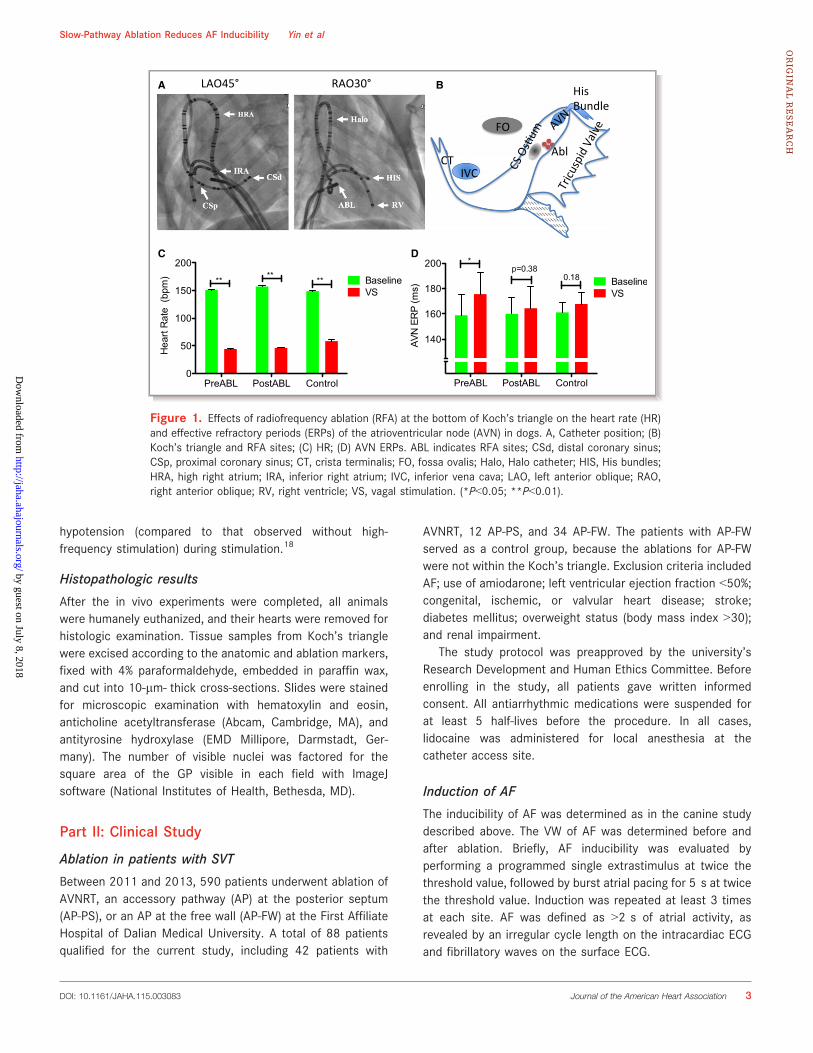
of Koch’s triangle for mapping and ablation. A quadrapolarcatheter was advanced into right ventricular apex for tempo-rary pacing. All catheters were manufactured by Cordis,Biosense Webster, Inc. (Diamond Bar, CA) and were posi-tioned with fluoroscopic guidance (Innova 2000; GE Health-care) (Figure 1A).
Electrophysiologic studies
An atrial pacing protocol involving a single extrastimulus wasperformed with a programmable multichannel stimulator(Model DF-5A; Dongfang Electric Co., Chengdu, China). Thepacing amplitude was set at twice the diastolic threshold. Theatrial ERP was defined as the longest coupling interval of theextrastimulus that failed to capture the local atria. The AVNERP was defined as the longest atrial coupling interval (A1–A2) observed on the His-bundle electrograms when A did notconduct to the His bundle. To determine the atrial ERP, weperformed an extrastimulus with coupling intervals of 200 msthat were progressively shortened by 10 ms with a basic drivecycle length of 250 ms at all sites. AF was defined as morethan 2 s of atrial activity appearing as an irregular cyclelength on the intracardiac ECG and fibrillatory waves on thesurface ECG.15 The vulnerability window (VW) for developmentof AF was defined as any coupling interval range of theextrastimulus at which fibrillation was induced.16,17 Thecontrol group underwent only the baseline electrophysiologystudy.
Radiofrequency ablation at the bottom of Koch’striangle in canines
Radiofrequency energy was delivered at the bottom of Koch’striangle with a 4-mm nonirrigated–tip ablation catheter(Cordis; Biosense Webster Inc.) connected to Stockert(Biosense Webster Inc.). Ablation was performed by movingthe tip in a point-by-point manner from the coronary sinusostium toward the His bundle until the His signal wasdetected.3 The tip was then moved back 2 mm for ablation(Figure 1B). For each ablation, energy was applied and thenwas stopped automatically when the abrupt impedance roseto 30 Ω above the baseline level. The highest energy givenwas 60 W, with a temperature threshold of 55°C and aduration of 60 s.
High-frequency stimulation
In dogs that underwent ablation at the Bottom of Koch’sTriangle, the function of the GPIVC-ICA within Koch’s trianglewas assessed by delivering high-frequency stimulation (20 Hz,10–150 V, 1–10-ms pulse width; S-88 stimulator; GrassInstruments Division, AstroNova, Inc., West Warwick, RI) toeach site for 5 s. The immediate response was defined as a>50% increase in R-R intervals and a >30% increase in
DOI: 10.1161/JAHA.115.003083 Journal of the American Heart Association 2
Slow-Pathway Ablation Reduces AF Inducibility Yin et alORIG
INALRESEARCH
by guest on July 8, 2018http://jaha.ahajournals.org/
Dow
nloaded from
hypotension (compared to that observed without high-frequency stimulation) during stimulation.18
Histopathologic results
After the in vivo experiments were completed, all animalswere humanely euthanized, and their hearts were removed forhistologic examination. Tissue samples from Koch’s trianglewere excised according to the anatomic and ablation markers,fixed with 4% paraformaldehyde, embedded in paraffin wax,and cut into 10-lm-‒thick cross-sections. Slides were stainedfor microscopic examination with hematoxylin and eosin,anticholine acetyltransferase (Abcam, Cambridge, MA), andantityrosine hydroxylase (EMD Millipore, Darmstadt, Ger-many). The number of visible nuclei was factored for thesquare area of the GP visible in each field with ImageJsoftware (National Institutes of Health, Bethesda, MD).
Part II: Clinical Study
Ablation in patients with SVT
Between 2011 and 2013, 590 patients underwent ablation ofAVNRT, an accessory pathway (AP) at the posterior septum(AP-PS), or an AP at the free wall (AP-FW) at the First AffiliateHospital of Dalian Medical University. A total of 88 patientsqualified for the current study, including 42 patients with
AVNRT, 12 AP-PS, and 34 AP-FW. The patients with AP-FWserved as a control group, because the ablations for AP-FWwere not within the Koch’s triangle. Exclusion criteria includedAF; use of amiodarone; left ventricular ejection fraction <50%;congenital, ischemic, or valvular heart disease; stroke;diabetes mellitus; overweight status (body mass index >30);and renal impairment.
The study protocol was preapproved by the university’sResearch Development and Human Ethics Committee. Beforeenrolling in the study, all patients gave written informedconsent. All antiarrhythmic medications were suspended forat least 5 half-lives before the procedure. In all cases,lidocaine was administered for local anesthesia at thecatheter access site.
Induction of AF
The inducibility of AF was determined as in the canine studydescribed above. The VW of AF was determined before andafter ablation. Briefly, AF inducibility was evaluated byperforming a programmed single extrastimulus at twice thethreshold value, followed by burst atrial pacing for 5 s at twicethe threshold value. Induction was repeated at least 3 timesat each site. AF was defined as >2 s of atrial activity, asrevealed by an irregular cycle length on the intracardiac ECGand fibrillatory waves on the surface ECG.
A B
C D
Figure 1. Effects of radiofrequency ablation (RFA) at the bottom of Koch’s triangle on the heart rate (HR)and effective refractory periods (ERPs) of the atrioventricular node (AVN) in dogs. A, Catheter position; (B)Koch’s triangle and RFA sites; (C) HR; (D) AVN ERPs. ABL indicates RFA sites; CSd, distal coronary sinus;CSp, proximal coronary sinus; CT, crista terminalis; FO, fossa ovalis; Halo, Halo catheter; HIS, His bundles;HRA, high right atrium; IRA, inferior right atrium; IVC, inferior vena cava; LAO, left anterior oblique; RAO,right anterior oblique; RV, right ventricle; VS, vagal stimulation. (*P<0.05; **P<0.01).
DOI: 10.1161/JAHA.115.003083 Journal of the American Heart Association 3
Slow-Pathway Ablation Reduces AF Inducibility Yin et alORIG
INALRESEARCH
by guest on July 8, 2018http://jaha.ahajournals.org/
Dow
nloaded from
Statistical AnalysisData were reported as the mean value�SD. A repeatedmeasurement of ANOVA and ANCOVA model followed by apostestimation was used to compare 3 groups of baseline, 4sites, and pre- and postablation on AERP and VW. Paired ttests were conducted between preablation and postablationvalues. A first-order exponential model was used to fit thecurves of A-H intervals versus the A-A pacing intervals fromindividual dogs. A P value of ≤0.05 was considered significant.All tests were performed with SPSS software, Version 16.0(IBM SPSS Statistics, Chicago, IL).
Results
Part I: Animal Study
RFA at the bottom of Koch’s triangle attenuated VS-induced prolongation of AVN ERPs
Figure 1A and 1B show the location of the catheters andablation sites, and Table 1 presents the parameters used forablation and VS. There was no significant difference betweenthe ablation and control groups in regard to the VS threshold.The ERPs of the sinoatrial and atrioventricular nodes, as wellas of 4 atrial sites, were tested with and without VS,preablation and postablation. The HRs and the ERPs of theAVN with VS were compared pre- and postablation at thebottom of Koch’s triangle. As shown in Figure 1C, RFA had nosignificant effect on VS-induced reduction of the HR(107�6.1 bpm preablation versus 111�6.2 bpm postabla-tion; P=0.21). The differences in HR responses to VS weresignificant preablation (152�6 versus 46�6 bpm; P<0.01)and postablation (158�5 versus 47�3 bpm; P<0.01), but VS-induced reductions in HR were comparable pre- and postab-lation (106.7�6.0 versus 110.9�6.2 bpm; P=0.08).
Vagal stimulation significantly prolonged the AVN ERPpreablation (from 159�16 to 176�17 ms; P<0.05), but itproduced no significant effects postablation (from 160�13 to164�17 ms; P=0.38) when tested at atrial pacing cyclelengths (A-A) of 250 ms with a half threshold voltage of VS.Therefore, RFA attenuated VS-induced prolongation of theAVN ERP (postablation: 4.44�4.74 ms versus preablation:16.67�5.53 ms; P<0.05) (Figure 1D). The AVN ERPs withoutVS were comparable to each other (P=0.85).
RFA at the bottom of Koch’s triangle attenuated VS-induced shifts of A-H conduction–time curves
The AVN conduction time, tested as the A-H interval, wasprolonged when atrial pacing cycle lengths A-A (or drive cyclelengths) were shortened from 300 to 150 ms in decrementsof 10 ms in the IRA. To determine the effects of VS on theAVN conduction time, A-H conduction-time curves werecompared to the A-A pacing interval pre- and postablation.As shown in Figure 2A, obtained from a representative dog,VS shifted the A-H curves toward the right, where the shiftcould be blocked by RFA.
During VS, the AVN conduction curves were discontinuous,or exhibited “jumping phenomena,” manifested as a suddenincrease in A-H and a decrement in A-A coupling intervals. Thediscontinuities were defined as the maximal prolongation ofA-H intervals (DA–Hmax) during reduction of the A-A interval in10-ms decrements. As shown in Figure 2B, VS increased theDA–Hmax significantly, from 13�2 to 33�4 ms (P<0.01).After RFA, however, the DA–Hmax response to VS wasattenuated from 14�2 to 13�2 ms (P=0.58).
In the first-order exponential model, the time-constants (s)of the curves were used to assess the A-H conductionintervals. As shown in Figure 2C, the s values with VS weredecreased from 88.46�7.36 to 52.03�6.99 ms (pairedP<0.01; n=7). Postablation, the s values were comparablewith VS (75.7�8.23 ms) and without VS (83.79�9.56 ms)(paired P=0.06). Moreover, the changes in s values betweenbaseline and VS were significantly decreased postablation(12.24�3.27 ms) (paired P <0.05) compared to preablation(36.43�9.01 ms) (Figure 2D).
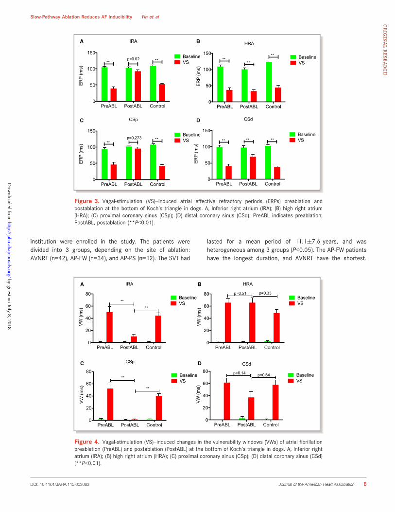
RFA at the bottom of Koch’s triangle partiallyattenuated VS-induced atrial ERP shortening
Before RFA, VS significantly shortened the atrial ERPs at allsites compared to the ERPs at baseline (Figure 3). Addition-ally, VS-induced shortening of atrial ERPs was comparableamong the 4 sites (ANOVA; P=0.11). However, after RFA, VS-shortened atrial ERPs were diverse (ANOVA; P<0.01). Short-ened atrial ERPs were comparable preablation and postabla-tion at sites in the HRA (DERP: 72�19 versus 67�22 ms;P=0.45) but were decreased at sites in the CSd (DERP: 59�7
Table 1. Canine Study: Parameters for Ablation and VS
Parameters Ablation (n=14) Control (n=7)
Ablation wattage, W 30.5�2.15 —
Ablation time, s 169.3�10.69 —
Ablation lesions (points) 5.2�0.5 —
Ablation temperature, °C 53.5�2.07 —
VS voltage, V 7.8�0.35 7.8�0.5
VS voltage at AVN ERP testing, V* 1.3�0.07 1.3�0.09
Procedure duration, hr 4.8�0.25 1.9�0.34
Animal weight, kg 12.7�0.42 13.2�0.63
AVN ERP indicates atrioventricular nodal effective refractory periods; VS, vagalstimulation.*11 dogs were tested for AVN ERP, because 3 dogs showed AV block with VS.
DOI: 10.1161/JAHA.115.003083 Journal of the American Heart Association 4
Slow-Pathway Ablation Reduces AF Inducibility Yin et alORIG
INALRESEARCH
by guest on July 8, 2018http://jaha.ahajournals.org/
Dow
nloaded from

versus 29�7 ms; P<0.05). However, VS-induced shorteningof the ERP was attenuated at sites of CSp (DERP: 48�7versus 6�4 ms; P<0.01) and IRA (DERP: 66�5 versus11�3 ms; P<0.01).
RFA at the bottom of Koch’s triangle reduced the VS-induced VWs of AF
Before and after ablation, AF was barely induced at all siteswithout VS, where the VWs trended to 0. Therefore, the VWsduring VS were compared only for preablation versuspostablation. The results showed that RFA significantlynarrowed the VWs at sites in the IRA and CSp (IRA: 49�36versus 1�3 ms; P<0.05; CSp: 45�34 versus 10�12 ms;P<0.05); however, there were no changes at sites in the HRA(63�25 versus 63�31 ms; P=0.99) and CSd (57�28 versus35�37 ms; P=0.07) (Figure 4). At all sites, the preablationand control VWs were comparable.
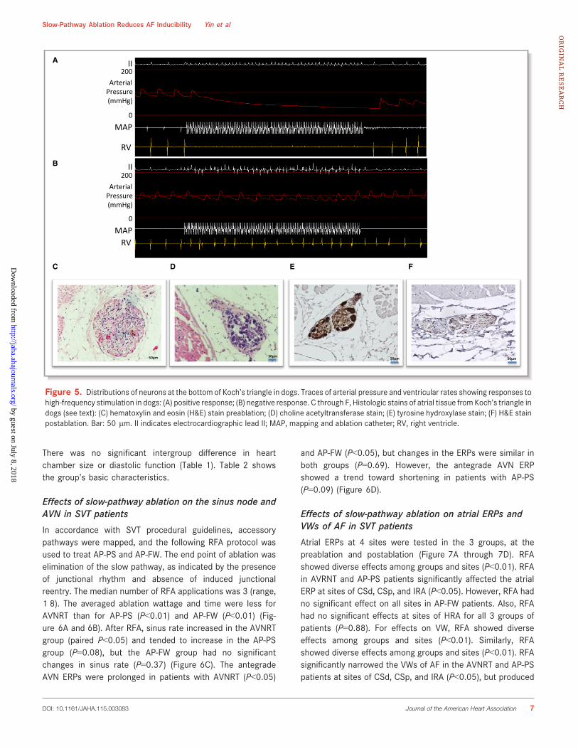
Neuronal mechanism of ablation at Koch’s triangle
The function of GPs was evaluated by high-frequencystimulation before and after ablation. At least 1 area ofresponse was identified within Koch’s triangle before ablation.There was no response after successful ablation. Typical
response and nonresponse pressure tracings are shown inFigure 5A and 5B, respectively.
At Koch’s triangle, the atrial tissues with ablation markerswere collected and sliced for histologic examination. Theneuronal ganglia and fibers were found on the epicardial side,located transmurally across the atrial wall from the ablationpoints (Figure 5). In samples from the control group, the GPwas clearly seen to connect with neuronal fibers and to besurrounded by adipose tissue (Figure 5C). Both cholinergicand adrenergic neurons were detected with antibodies forcholine acetyltransferase (Figure 5D) and antityrosine hydrox-ylase staining (Figure 5E), respectively. In addition, theneurons were severely impaired in the tissues from the RFAgroup (Figure 5F). The fraction of nuclear numbers in a cross-sectional area of the GPs was compared in the ablation versuscontrol groups. In the ablation group, the fraction of thenormal nuclear number in the cross-sectional area of the GPswas significantly reduced (33�8% versus 15�7%; P<0.01).
Part II: Clinical Study
Study population
Eight-eight patients (40 men and 48 women, aged47.9�14.3 years) who had undergone RFA for SVT at our
A B
C D
Figure 2. Effects of vagal stimulation (VS) on A-H conduction-time curves preablation (PreABL) andpostablation (PostABL) at the bottom of Koch’s triangle in dogs. A, Representative A–H curves from dog 3; (B)maximal vagal-induced A–H changes; (C) effects of VS on the s value of the A–H curve fitted by first-orderexponential model; (D) Changes in s between baseline (without VS) and with VS (*P<0.05; **P<0.01).
DOI: 10.1161/JAHA.115.003083 Journal of the American Heart Association 5
Slow-Pathway Ablation Reduces AF Inducibility Yin et alORIG
INALRESEARCH
by guest on July 8, 2018http://jaha.ahajournals.org/
Dow
nloaded from
institution were enrolled in the study. The patients weredivided into 3 groups, depending on the site of ablation:AVNRT (n=42), AP-FW (n=34), and AP-PS (n=12). The SVT had
lasted for a mean period of 11.1�7.6 years, and washeterogeneous among 3 groups (P<0.05). The AP-FW patientshave the longest duration, and AVNRT have the shortest.
A B
C D
Figure 3. Vagal-stimulation (VS)–induced atrial effective refractory periods (ERPs) preablation andpostablation at the bottom of Koch’s triangle in dogs. A, Inferior right atrium (IRA); (B) high right atrium(HRA); (C) proximal coronary sinus (CSp); (D) distal coronary sinus (CSd). PreABL indicates preablation;PostABL, postablation (**P<0.01).
A B
C D
Figure 4. Vagal-stimulation (VS)–induced changes in the vulnerability windows (VWs) of atrial fibrillationpreablation (PreABL) and postablation (PostABL) at the bottom of Koch’s triangle in dogs. A, Inferior rightatrium (IRA); (B) high right atrium (HRA); (C) proximal coronary sinus (CSp); (D) distal coronary sinus (CSd)(**P<0.01).
DOI: 10.1161/JAHA.115.003083 Journal of the American Heart Association 6
Slow-Pathway Ablation Reduces AF Inducibility Yin et alORIG
INALRESEARCH
by guest on July 8, 2018http://jaha.ahajournals.org/
Dow
nloaded from
There was no significant intergroup difference in heartchamber size or diastolic function (Table 1). Table 2 showsthe group’s basic characteristics.
Effects of slow-pathway ablation on the sinus node andAVN in SVT patients
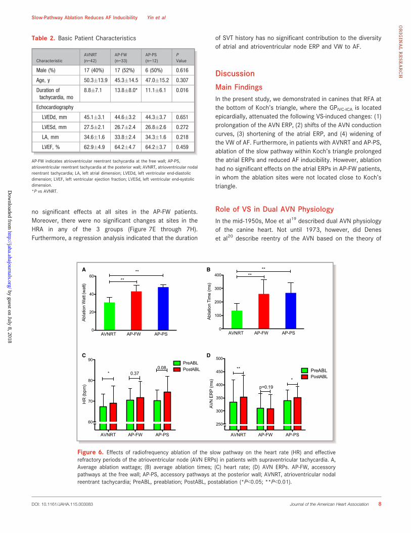
In accordance with SVT procedural guidelines, accessorypathways were mapped, and the following RFA protocol wasused to treat AP-PS and AP-FW. The end point of ablation waselimination of the slow pathway, as indicated by the presenceof junctional rhythm and absence of induced junctionalreentry. The median number of RFA applications was 3 (range,1‒8). The averaged ablation wattage and time were less forAVNRT than for AP-PS (P<0.01) and AP-FW (P<0.01) (Fig-ure 6A and 6B). After RFA, sinus rate increased in the AVNRTgroup (paired P<0.05) and tended to increase in the AP-PSgroup (P=0.08), but the AP-FW group had no significantchanges in sinus rate (P=0.37) (Figure 6C). The antegradeAVN ERPs were prolonged in patients with AVNRT (P<0.05)
and AP-FW (P<0.05), but changes in the ERPs were similar inboth groups (P=0.69). However, the antegrade AVN ERPshowed a trend toward shortening in patients with AP-PS(P=0.09) (Figure 6D).
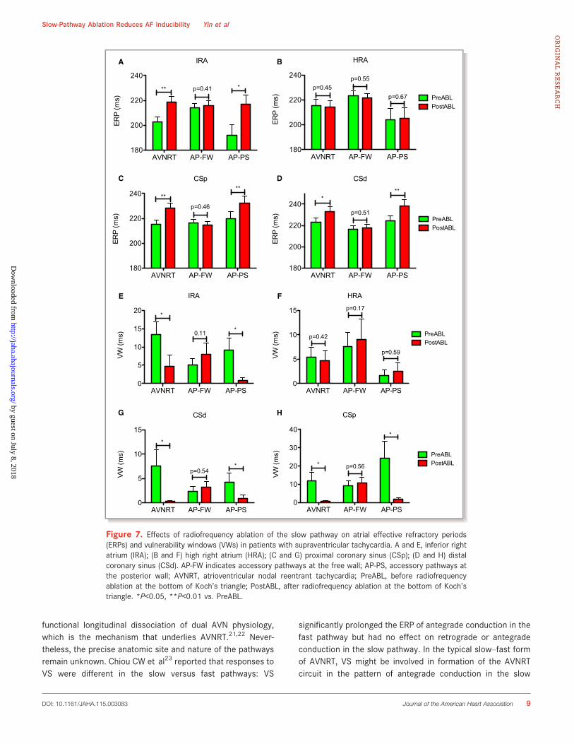
Effects of slow-pathway ablation on atrial ERPs andVWs of AF in SVT patients
Atrial ERPs at 4 sites were tested in the 3 groups, at thepreablation and postablation (Figure 7A through 7D). RFAshowed diverse effects among groups and sites (P<0.01). RFAin AVRNT and AP-PS patients significantly affected the atrialERP at sites of CSd, CSp, and IRA (P<0.05). However, RFA hadno significant effect on all sites in AP-FW patients. Also, RFAhad no significant effects at sites of HRA for all 3 groups ofpatients (P=0.88). For effects on VW, RFA showed diverseeffects among groups and sites (P<0.01). Similarly, RFAshowed diverse effects among groups and sites (P<0.01). RFAsignificantly narrowed the VWs of AF in the AVNRT and AP-PSpatients at sites of CSd, CSp, and IRA (P<0.05), but produced
A
B
C D E F
Figure 5. Distributions of neurons at the bottom of Koch’s triangle in dogs. Traces of arterial pressure and ventricular rates showing responses tohigh-frequency stimulation in dogs: (A) positive response; (B) negative response. C through F, Histologic stains of atrial tissue from Koch’s triangle indogs (see text): (C) hematoxylin and eosin (H&E) stain preablation; (D) choline acetyltransferase stain; (E) tyrosine hydroxylase stain; (F) H&E stainpostablation. Bar: 50 lm. II indicates electrocardiographic lead II; MAP, mapping and ablation catheter; RV, right ventricle.
DOI: 10.1161/JAHA.115.003083 Journal of the American Heart Association 7
Slow-Pathway Ablation Reduces AF Inducibility Yin et alORIG
INALRESEARCH
by guest on July 8, 2018http://jaha.ahajournals.org/
Dow
nloaded from
no significant effects at all sites in the AP-FW patients.Moreover, there were no significant changes at sites in theHRA in any of the 3 groups (Figure 7E through 7H).Furthermore, a regression analysis indicated that the duration
of SVT history has no significant contribution to the diversityof atrial and atrioventricular node ERP and VW to AF.
Discussion
Main FindingsIn the present study, we demonstrated in canines that RFA atthe bottom of Koch’s triangle, where the GPIVC-ICA is locatedepicardially, attenuated the following VS-induced changes: (1)prolongation of the AVN ERP, (2) shifts of the AVN conductioncurves, (3) shortening of the atrial ERP, and (4) widening ofthe VW of AF. Furthermore, in patients with AVNRT and AP-PS,ablation of the slow pathway within Koch’s triangle prolongedthe atrial ERPs and reduced AF inducibility. However, ablationhad no significant effects on the atrial ERPs in AP-FW patients,in whom the ablation sites were not located close to Koch’striangle.
Role of VS in Dual AVN PhysiologyIn the mid-1950s, Moe et al19 described dual AVN physiologyof the canine heart. Not until 1973, however, did Deneset al20 describe reentry of the AVN based on the theory of
A B
C D
Figure 6. Effects of radiofrequency ablation of the slow pathway on the heart rate (HR) and effectiverefractory periods of the atrioventricular node (AVN ERPs) in patients with supraventricular tachycardia. A,Average ablation wattage; (B) average ablation times; (C) heart rate; (D) AVN ERPs. AP-FW, accessorypathways at the free wall; AP-PS, accessory pathways at the posterior wall; AVNRT, atrioventricular nodalreentrant tachycardia; PreABL, preablation; PostABL, postablation (*P<0.05; **P<0.01).
Table 2. Basic Patient Characteristics
CharacteristicAVNRT(n=42)
AP-FW(n=33)
AP-PS(n=12)
PValue
Male (%) 17 (40%) 17 (52%) 6 (50%) 0.616
Age, y 50.3�13.9 45.3�14.5 47.0�15.2 0.307
Duration oftachycardia, mo
8.8�7.1 13.8�8.0* 11.1�6.1 0.016
Echocardiography
LVEDd, mm 45.1�3.1 44.6�3.2 44.3�3.7 0.651
LVESd, mm 27.5�2.1 26.7�2.4 26.8�2.6 0.272
LA, mm 34.6�1.6 33.8�2.4 34.3�1.6 0.218
LVEF, % 62.9�4.9 64.2�4.7 64.2�3.7 0.459
AP-FW indicates atrioventricular reentrant tachycardia at the free wall; AP-PS,atrioventricular reentrant tachycardia at the posterior wall; AVNRT, atrioventricular nodalreentrant tachycardia; LA, left atrial dimension; LVEDd, left ventricular end-diastolicdimension; LVEF, left ventricular ejection fraction; LVESd, left ventricular end-systolicdimension.*P vs AVNRT.
DOI: 10.1161/JAHA.115.003083 Journal of the American Heart Association 8
Slow-Pathway Ablation Reduces AF Inducibility Yin et alORIG
INALRESEARCH
by guest on July 8, 2018http://jaha.ahajournals.org/
Dow
nloaded from
functional longitudinal dissociation of dual AVN physiology,which is the mechanism that underlies AVNRT.21,22 Never-theless, the precise anatomic site and nature of the pathwaysremain unknown. Chiou CW et al23 reported that responses toVS were different in the slow versus fast pathways: VS
significantly prolonged the ERP of antegrade conduction in thefast pathway but had no effect on retrograde or antegradeconduction in the slow pathway. In the typical slow–fast formof AVNRT, VS might be involved in formation of the AVNRTcircuit in the pattern of antegrade conduction in the slow
A B
C D
E F
G H
Figure 7. Effects of radiofrequency ablation of the slow pathway on atrial effective refractory periods(ERPs) and vulnerability windows (VWs) in patients with supraventricular tachycardia. A and E, inferior rightatrium (IRA); (B and F) high right atrium (HRA); (C and G) proximal coronary sinus (CSp); (D and H) distalcoronary sinus (CSd). AP-FW indicates accessory pathways at the free wall; AP-PS, accessory pathways atthe posterior wall; AVNRT, atrioventricular nodal reentrant tachycardia; PreABL, before radiofrequencyablation at the bottom of Koch’s triangle; PostABL, after radiofrequency ablation at the bottom of Koch’striangle. *P<0.05, **P<0.01 vs. PreABL.
DOI: 10.1161/JAHA.115.003083 Journal of the American Heart Association 9
Slow-Pathway Ablation Reduces AF Inducibility Yin et alORIG
INALRESEARCH
by guest on July 8, 2018http://jaha.ahajournals.org/
Dow
nloaded from
pathway, as well as retrograde conduction in the fastpathway. Also, Mazgalev et al24–26 used VS to induce AVNRTin isolated rabbit hearts, thus providing evidence of VSmodulation in dual AVN physiology. In the present study, VSamplified the difference in conduction between the slow andfast pathways, thereby manifesting as discontinuous AVNconduction curves.
Hyperactive VS Triggers SVTAblation of the slow pathway has become an efficient, well-established treatment for AVNRT.27 However, evidence ofanatomic reentry or circulating pathways is lacking and themechanism of AVNRT remains controversial. In our study,after ablation of the slow pathway, discontinuous atrioven-tricular conduction was detectible in certain cases, but SVTcould not be induced. In contrast, some AVNRT patientsexhibited a smooth AVN conduction curve.28 This suggeststhat dual AVN physiology is not an exclusive mechanism ofAVNRT.28 Therefore, like others, we have demonstrated theimportant contributions of vagal activities in triggering SVT.Ablation at the bottom of Koch’s triangle attenuated the vagal-related heterogeneity in atrioventricular conduction, therebyreducing the potential for reentry, as confirmed by impairmentof the GP and blockade of vagal activity after ablation.
Ablation of the Slow Pathway May Prevent AFInductionLittle information exists about the specific distribution of thevagal pathways to the atrial myocardium. In this study, RFAwas delivered to the endocardial tissues at the bottom ofKoch’s triangle, which mirrored the epicardial fat pad at theGPIVC-LA.
29 Histologic results confirmed that RFA impaired thenerve fibers and neurons located in the epicardial fat pad.Ablation attenuated the ERP shortening response to VS nearthe region of ablation but not in any remote areas; thisindicated that ablation at the bottom of Koch’s triangleresulted in remarkable denervation of the atrial vagus nerves.Previous studies showed that innervation of the GPIVC-LA is theintegration center for extrinsic vagal pathways. Autonomicbranches from both the right and left vagus nerves passthrough the GPIVC-LA to the AVN.8,11 Ablation of the GPIVC-LAboth attenuated VS-induced VW widening and increased AFinducibility.30
Clinical ImplicationsAblation of the intrinsic cardiac autonomic nervous system,particularly targeting of the GP, has been shown to increasethe success of AF ablation. Indeed, it has been suggested thatRFA for AVNRT results in parasympathetic denervation with
inadvertent tachycardia. However, it is not clear whether suchdenervation affects the atria. In the present study, we showedthat RFA of the slow pathway at the bottom of Koch’s triangleimpaired the epicardial GPIVC-LA in canine atria and resulted invagal denervation in the atria and AVN. In patients withAVNRT, slow-pathway ablation has been well documented ashaving a high success rate, but the anatomic mechanism forthe reentrant or circulating pathway is unknown. In our dogs,VS induced discontinuities or similar “jumping phenomena” inAVN conduction curves, thereby indicating the role of vagalactivity in AVNRT formation. This role was supported by theobservation of increased vagal activity in patients withAVNRT.23 Moreover, in the present study, slow-pathwayablation in patients with AVNRT and AP-PS altered the atrialERP and AF inducibility.
LimitationsLack of information concerning the incidence of—andvulnerability to—AF in these patients limited our ability todetermine whether or not ablation contributed to AF. Follow-up studies of these patients are ongoing regarding theincidence of AF and other arrhythmias. Also, as a retrospec-tive study, the present study may not be representative of thegeneral population and may be prone to selection bias due tolacking of power analysis and sample size. However, thepresent study indicates that the area of Koch’s triangle mightneed more attention. Future prospective studies are war-ranted to optimize the strategies for AF.
The location of the GPIVC-LA was not identified with high-frequency stimulation before ablation. The endocardial abla-tion of the GPIVC-LA might have been confounded with tissueablation. Although the canine histologic findings indicatedthat GP structure was impaired after ablation, the location ofthe GPIVC-LA may have differed from subject to subject. OnlyERPs were tested and compared in this study, so it may notprecisely reflect the atrial effects of RFA, such as localconduction in the atria and AVN. Further study is warranted toinvestigate underlying mechanisms by using a mappingsystem to perform ex vivo mapping with direct ablation ofthe GPIVC-LA epicardially.
ConclusionsIn canines, RFA at the bottom of Koch’s triangle impaired theGPIVC-ICA and attenuated local autonomic innervation in theAVN and atrial tissue, which might contribute to VS-induceddiscontinuous AVN conduction and atrial ERP shortening.Moreover, in SVT patients, RFA of the slow pathway withinKoch’s triangle prolonged local atrial ERPs and decreased AFinducibility. These findings suggested that in certain types ofAF, such as vagal AF, the effect of slow-pathway ablation on
DOI: 10.1161/JAHA.115.003083 Journal of the American Heart Association 10
Slow-Pathway Ablation Reduces AF Inducibility Yin et alORIG
INALRESEARCH
by guest on July 8, 2018http://jaha.ahajournals.org/
Dow
nloaded from
AF might be related to the denervation of local autonomicnerves.
Sources of FundingThis work was supported by external grants to Yin fromLiaoning Province (201201009, L2011154, 2015020259,201501853) and from the Chinese Ministry of Health(w201003). It was also supported by internal grants fromDalian Medical University.
DisclosuresNone.
References1. Kocovic DZ, Harada T, Shea JB, Soroff D, Friedman PL. Alterations of heart rate
and of heart rate variability after radiofrequency catheter ablation ofsupraventricular tachycardia. Delineation of parasympathetic pathways inthe human heart. Circulation. 1993;88:1671–1681.
2. Havranek S, Souckova L, Simek J, Wichterle D. Slow pathway ablation fortypical atrioventricular nodal re-entrant tachycardia significantly alters theautonomic modulation of atrioventricular conduction. Clin Auton Res.2013;23:289–295.
3. Razavi M, Cheng J, Rasekh A, Yang D, Delapasse S, Ai T, Meade T, Donsky A,Goodman MJ, Massumi A. Slow pathway ablation decreases vulnerability topacing-induced atrial fibrillation: possible role of vagal denervation. Pacing ClinElectrophysiol. 2006;29:1234–1239.
4. Markowitz SM, Christini DJ, Stein KM, Mittal S, Iwai S, Slotwiner DJ, LermanBB. Time course and predictors of autonomic dysfunction after ablation of theslow atrioventricular nodal pathway. Pacing Clin Electrophysiol.2004;27:1638–1643.
5. Kowallik P, Escher S, Peters W, Braun C, Meesmann M. Preserved autonomicmodulation of the sinus and atrioventricular nodes following posteroseptalablation for treatment of atrioventricular nodal reentrant tachycardia. JCardiovasc Electrophysiol. 1998;9:567–573.
6. Kautzner J, Hartikainen J, Heald S, Malik M, Ward D, Rowland E. Is vagalinnervation to the atrioventricular node impaired after radiofrequency ablationof the slow atrioventricular nodal pathway? Pacing Clin Electrophysiol.1996;19:1993–1997.
7. Wilhelmy R, Pitschner H, Neuzner J, Dursch M, Konig S. Patients with AV nodalreentrant tachycardias show a reduced vagal impact on heart rate comparedto healthy subjects with further decrease of vagal tone after radiofrequencycatheter modification. Clin Sci (Lond). 1996;91(suppl):125.
8. Hou Y, Scherlag BJ, Lin J, Zhang Y, Lu Z, Truong K, Patterson E, Lazzara R,Jackman WM, Po SS. Ganglionated plexi modulate extrinsic cardiac autonomicnerve input: effects on sinus rate, atrioventricular conduction, refractoriness,and inducibility of atrial fibrillation. J Am Coll Cardiol. 2007;50:61–68.
9. Quan KJ, Lee JH, Van Hare GF, Biblo LA, Mackall JA, Carlson MD. Identificationand characterization of atrioventricular parasympathetic innervation inhumans. J Cardiovasc Electrophysiol. 2002;13:735–739.
10. Ardell JL, Randall WC. Selective vagal innervation of sinoatrial and atrioven-tricular nodes in canine heart. Am J Physiol. 1986;251:H764–H773.
11. Lin J, Scherlag BJ, Niu G, Lu Z, Patterson E, Liu S, Lazzara R, Jackman WM, PoSS. Autonomic elements within the ligament of Marshall and inferior left
ganglionated plexus mediate functions of the atrial neural network. JCardiovasc Electrophysiol. 2009;20:318–324.
12. Hayashi H, Usui M, Tani M, Nagasawa H, Fujiki A, Inoue H. Radiofrequencyablation at the coronary sinus ostium interrupts the vagal efferent input to theatrioventricular node in the canine heart. Jpn Circ J. 2001;65:667–672.
13. Soejima K, Akaishi M, Mitamura H, Ogawa S, Sakurada H, Okazaki H,Motomiya T, Hiraoka M. Increase in heart rate after radiofrequency catheterablation is mediated by parasympathetic nervous withdrawal and related tosite of ablation. J Electrocardiol. 1997;30:239–246.
14. Sauer WH, Alonso C, Zado E, Cooper JM, Lin D, Dixit S, Russo A, Verdino R, Ji S,Gerstenfeld EP, Callans DJ, Marchlinski FE. Atrioventricular nodal reentranttachycardia in patients referred for atrial fibrillation ablation: response toablation that incorporates slow-pathway modification. Circulation.2006;114:191–195.
15. Wyndham CR, Amat-y-Leon F, Wu D, Denes P, Dhingra R, Simpson R, RosenKM. Effects of cycle length on atrial vulnerability. Circulation. 1977;55:260–267.
16. Razavi M, Zhang S, Yang D, Sanders RA, Kar B, Delapasse S, Ai T, Moreira W,Olivier B, Khoury DS, Cheng J. Effects of pulmonary vein ablation on regionalatrial vagal innervation and vulnerability to atrial fibrillation in dogs. JCardiovasc Electrophysiol. 2005;16:879–884.
17. Morillo CA, Klein GJ, Jones DL, Guiraudon CM. Chronic rapid atrial pacing.Structural, functional, and electrophysiological characteristics of a new modelof sustained atrial fibrillation. Circulation. 1995;91:1588–1595.
18. Po SS, Nakagawa H, Jackman WM. Localization of left atrial ganglionated plexiin patients with atrial fibrillation. J Cardiovasc Electrophysiol. 2009;20:1186–1189.
19. Moe GK, Preston JB, Burlington H. Physiologic evidence for a dual A-Vtransmission system. Circ Res. 1956;4:357–375.
20. Denes P, Wu D, Dhingra RC, Chuquimia R, Rosen KM. Demonstration of dual A-V nodal pathways in patients with paroxysmal supraventricular tachycardia.Circulation. 1973;48:549–555.
21. Belhassen B, Fish R, Glikson M, Glick A, Eldar M, Laniado S, Viskin S.Noninvasive diagnosis of dual AV node physiology in patients with AV nodalreentrant tachycardia by administration of adenosine-50-triphosphate duringsinus rhythm. Circulation. 1998;98:47–53.
22. Wit AL, Weiss MB, Berkowitz WD, Rosen KM, Steiner C, Damato AN. Patternsof atrioventricular conduction in the human heart. Circ Res. 1970;27:345–359.
23. Chiou CW, Chen SA, Kung MH, Chang MS, Prystowsky EN. Effects ofcontinuous enhanced vagal tone on dual atrioventricular node and accessorypathways. Circulation. 2003;107:2583–2588.
24. Mazgalev T, Dreifus LS, Michelson EL, Pelleg A. Effect of postganglionic vagalstimulation on the organization of atrioventricular nodal conduction in isolatedrabbit heart tissue. Circulation. 1986;74:869–880.
25. Mazgalev T, Dreifus LS, Michelson EL, Pelleg A. Vagally induced hyperpolar-ization in atrioventricular node. Am J Physiol. 1986;251:H631–H643.
26. Mazgalev T, Dreifus LS, Michelson EL, Pelleg A, Price R. Phasic effects ofpostganglionic vagal stimulation on atrioventricular nodal conduction. Am JPhysiol. 1986;251:H619–H630.
27. Prystowsky EN. Atrioventricular node reentry: physiology and radiofrequencyablation. Pacing Clin Electrophysiol. 1997;20:552–571.
28. Tai CT, Chen SA, Chiang CE, Lee SH, Wen ZC, Chiou CW, Ueng KC, Chen YJ, YuWC, Huang JL, Chang MS. Complex electrophysiological characteristics inatrioventricular nodal reentrant tachycardia with continuous atrioventricularnode function curves. Circulation. 1997;95:2541–2547.
29. Chiou CW, Eble JN, Zipes DP. Efferent vagal innervation of the canine atria andsinus and atrioventricular nodes. The third fat pad. Circulation. 1997;95:2573–2584.
30. Zhou J, Scherlag BJ, Edwards J, Jackman WM, Lazzara R, Po SS. Gradients ofatrial refractoriness and inducibility of atrial fibrillation due to stimulation ofganglionated plexi. J Cardiovasc Electrophysiol. 2007;18:83–90.
DOI: 10.1161/JAHA.115.003083 Journal of the American Heart Association 11
Slow-Pathway Ablation Reduces AF Inducibility Yin et alORIG
INALRESEARCH
by guest on July 8, 2018http://jaha.ahajournals.org/
Dow
nloaded from
Chen, Jie Cheng and Yanzong YangXiaomeng Yin, Yutao Xi, Shulong Zhang, Yunlong Xia, Lianjun Gao, Jinqiu Liu, Nancy Cheng, Qi
Neuronal MechanismPathway Ablation Reduces Atrial Fibrillation Inducibility: A−Atrioventricular Node Slow
Online ISSN: 2047-9980 Dallas, TX 75231
is published by the American Heart Association, 7272 Greenville Avenue,Journal of the American Heart AssociationThe doi: 10.1161/JAHA.115.003083
2016;5:e003083; originally published June 10, 2016;J Am Heart Assoc.
http://jaha.ahajournals.org/content/5/6/e003083World Wide Web at:
The online version of this article, along with updated information and services, is located on the
for more information. http://jaha.ahajournals.orgAccess publication. Visit the Journal at
is an online only OpenJournal of the American Heart AssociationSubscriptions, Permissions, and Reprints: The
by guest on July 8, 2018http://jaha.ahajournals.org/
Dow
nloaded from