congenital and childhood atrioventricular blocks
TRANSCRIPT

REVIEW
Congenital and childhood atrioventricular blocks:pathophysiology and contemporary management
Alban-Elouen Baruteau1,2,3& Robert H. Pass4 & Jean-Benoit Thambo2 &
Albin Behaghel5 & Solène Le Pennec5 & Elodie Perdreau2& Nicolas Combes6 &
Leonardo Liberman7& Christopher J. McLeod8
Received: 28 January 2016 /Revised: 13 June 2016 /Accepted: 16 June 2016 /Published online: 28 June 2016# The Author(s) 2016. This article is published with open access at Springerlink.com
Abstract Atrioventricular block is classified as congeni-tal if diagnosed in utero, at birth, or within the firstmonth of life. The pathophysiological process is believedto be due to immune-mediated injury of the conductionsystem, which occurs as a result of transplacental pas-sage of maternal anti-SSA/Ro-SSB/La antibodies.Childhood atrioventricular block is therefore diagnosed
between the first month and the 18th year of life.Genetic variants in multiple genes have been describedto date in the pathogenesis of inherited progressive car-diac conduction disorders. Indications and techniques ofcardiac pacing have also evolved to allow safe perma-nent cardiac pacing in almost all patients, includingthose with structural heart abnormalities.
Communicated by Jaan Toelen
* Alban-Elouen [email protected]
Robert H. [email protected]
Jean-Benoit [email protected]
Albin [email protected]
Solène Le [email protected]
Elodie [email protected]
Nicolas [email protected]
Leonardo [email protected]
Christopher J. [email protected]
1 Cardiovascular and Cell Sciences Research Center, St George’sUniversity of London, London, UK
2 LIRYC Institute, CHU Bordeaux, Department of PediatricCardiology, Bordeaux-II University, Bordeaux, France
3 Service de Cardiologie Pédiatrique, Hôpital du Haut Lévèque,Institut Hospitalo-Universitaire LIRYC (Electrophysiology andHeart Modeling Institute), 5 avenue de Magellan,33600 Pessac, France
4 Division of Pediatric Electrophysiology, Albert Einstein College ofMedicine, Montefiore Children’s Hospital, Bronx, NY, USA
5 CHU Rennes, Department of Cardiology, LTSI, INSERM 1099,Rennes-1 University, Rennes, France
6 Department of Cardiology, Clinique Pasteur, Toulouse, France
7 Morgan Stanley Children’s Hospital, Division of PediatricCardiology, New York Presbyterian Hospital, Columbia UniversityMedical Center, New York, NY, USA
8 Mayo Clinic, Division of Cardiovascular Diseases, Mayo ClinicCollege of Medicine, Rochester, MN, USA
Eur J Pediatr (2016) 175:1235–1248DOI 10.1007/s00431-016-2748-0

Conclusion: Early diagnosis and appropriate managementare critical inmany cases in order to prevent sudden death, andthis review critically assesses our current understanding of thepathogenetic mechanisms, clinical course, and optimal man-agement of congenital and childhood AV block.
What is Known:• Prevalence of congenital heart block of 1 per 15,000 to 20,000live births. AV block is defined as congenital if diagnosed inutero, at birth, or within the first month of life, whereaschildhood AV block is diagnosed between the first month andthe 18th year of life. As a result of several different etiologies,congenital and childhood atrioventricular block may occur in anentirely structurally normal heart or in association withconcomitant congenital heart disease. Cardiac pacing isindicated in symptomatic patients and has several prophylacticindications in asymptomatic patients to prevent sudden death.
• Autoimmune, congenital AV block is associated with a high neonatalmortality rate and development of dilated cardiomyopathy in 5 to 30 %cases.
What is New:• Several genes including SCN5A have been implicated in autosomaldominant forms of familial progressive cardiac conduction disorders.
• Leadless pacemaker technology and gene therapy for biologicalpacing are promising research fields. In utero percutaneouspacing appears to be at high risk and needs further developmentbefore it can be adopted into routine clinical practice. Cardiacresynchronization therapy is of proven value in case of pacing-induced cardiomyopathy.
Keywords Heart block . Pacemaker . Pathophysiology .
Outcomes . Congenital heart disease
AbbreviationsACC American College of CardiologyAHA American Heart AssociationAV AtrioventricularCHB Congenital heart blockCHD Congenital heart diseaseDCM Dilated cardiomyopathyESC European Society of CardiologyHRS Heart Rhythm SocietyPCCD Progressive cardiac conduction diseaseSVC Superior vena cava
Background
Cardiac conduction disorders are rare syndromes in neonatesand children [15, 64]. As a result of several different etiolo-gies, it may occur in an entirely structurally normal heart or inassociation with concomitant congenital heart disease (CHD).In contrast to acquired atrioventricular (AV) conduction block,congenital heart block (CHB)—identified in utero in normalhearts—holds a significantly different prognosis with an
increased risk of late-onset cardiomyopathy. Fundamentally,the pathogenesis is also disparate, driven by different maternalclinical features and an increased risk of recurrence in futurepregnancies. For these reasons, AV block is classified as con-genital if diagnosed in utero, at birth, or within the first monthof life. Therefore, childhood AV block is diagnosed betweenthe first month and the 18th year of life [15]. The estimatedprevalence of congenital heart block is 1 per 15,000–20,000live births [64].
Atrioventricular conduction disorders in structurallynormal hearts
Immune-mediated AV block
Although some aspects remain to be clarified, pathophys-iology, therapeutic approach, and long-term prognosis ofimmune-mediated AV block have been extensively studied,as it is one of the leading causes of congenital heartblocks.
Pathophysiology Congenital AV block can be passively ac-quired via an autoimmune process affecting the developing heartdue to the transplacental passage ofmaternal anti-Ro/SSA and/oranti-La/SSB autoantibodies. Entering the fetal circulation, theycan directly bind L-type calcium channels on fetalcardiomyocytes and significantly, but reversibly, inhibit the relat-ed currents. However, in some cases, for unclear reasons, theprolonged exposure to anti-Ro/SSA antibodies may induce cal-cium channel internalization, in turn triggering a complex andonly in part mechanistically known perturbation of the cytoplas-mic calciummetabolismwhich ultimately leads to apoptosis andcell death, and then to local inflammation. If the process is notstopped in this phase, the inflammatory damage proceeds, thuseventually resulting in fibrosis and calcification of the cardiacconduction system. This mechanistic sequence, also known asBcalcium channel hypothesis,^ is currently recognized as themore attractive theory possible explaining the pathogenesis ofthe disease [1, 90]. Maternal autoantibodies can be detected inover 95 % of fetuses or newborns presenting with AV block,namely congenital AV block [19]. In contrast, maternal autoan-tibodies have been detected in only a minority of children, inwhom AV block was diagnosed beyond the neonatal period, adifferent, distinct clinical entity [15, 34, 95]. However, someisolated AV blocks diagnosed beyond the neonatal period arealso immune-mediated, even with late detection of maternal an-ti-Ro/SSA autoantibodies [12]. This condition, emerging inchildhood or even in the adult age, represents a late progressivecongenital form of immune AV block with the late developmentof a subclinical anti-Ro/SSA-induced congenital damage of theconduction system, related to a not fully understoodautoantibody-independent worsening with age [51]. Between 2
1236 Eur J Pediatr (2016) 175:1235–1248

and 5 % of fetuses and infants whose mothers are autoantibody-positive develop AV block, and the risk to subsequent pregnan-cies is substantial (ranging between 12 and 25 %) in motherswho have had a child with congenital AV block [16]. In up to athird of infants with congenital AV block, a characterized auto-immune disease, such as lupus, is present in the mother [16].However, in the large majority of the cases, AV block occursin fetuses of healthy, silently anti-Ro/SSA antibodies-carryingmothers. The diagnosis of mothers’ seropositivity is thus usuallymade only after congenital AV block detection. In a recent pro-spective study of 186 antibody-exposed fetuses and infants, it hasbeen demonstrated thatmothers of childrenwith cardiac involve-ment were less likely to have had a connective tissue disease thanmothers of children without cardiac involvement [36].
Diagnosis of fetal AV block Fetal echocardiography is thegold standard for the diagnosis of congenital AV block. AllM-mode and Doppler echocardiographic techniques rely onthe relationship between atrial and ventricular mechanical event[30]. Although clinical applications of both fetal electrocardi-ography and fetal magnetocardiography are more recent, thesetwo noninvasive tools are promising, being able to moreprecisely diagnose fetal arrhythmias and conduction disorders[23, 103]. Complete fetal AV block develops during gestationalweeks 16 to 24, although a later onset of the phenomenon up togestational week 34 has been described [2, 72].
Clinical course CHB derived from an autoimmune process isassociated with a high neonatal mortality rate [16, 37]. Theestimated overall mortality without pacing is estimated to bearound 8–16 % in infants and half as much in children andadults [37, 65]. Interestingly, cardiac dilatation and impairedventricular function can develop as long-term sequelae in thosewho forego pacing and those who are permanently paced.Without pacing support, it appears that the slow heart ratesand associated higher stroke volumes probably drive this pro-cess [65]. But with pacing, the current hypothesis centers on apacing-induced cardiomyopathy. Globally, the prevalence ofdilated cardiomyopathy (DCM) in CHB ranges from 5 to30 % [66, 91, 94]. Various pathophysiological processes, in-cluding transient myocarditis or immune-mediated myocardialinjury, have been proposed to explain the development of ven-tricular dilatation and dysfunction. Late-onset dilated cardio-myopathy in patients with complete heart block may be a se-quela of in utero autoimmunemyocarditis or due to its postnatalreactivation [95]. Right and left ventricular endocardialfibroelastosis and fibrosis have been observed at autopsy.These observations were not limited to the conduction systemand involved the working ventricular myocardium. The detri-mental effects of maternal antibodies directed against fetal car-diac tissue provided evidence in favor of the immunopatholog-ic role played by the maternal autoantibodies in congenital AVblock. In view of the myocardial dystrophic changes and
adverse remodeling caused by ventricular desynchronization,right ventricular pacing has been suggested as an importantcause of DCM [66, 91, 94]. In some patients, the discontinua-tion of right ventricular pacing or upgrade to cardiacresynchronization therapy alone normalized systolic function,which would not be expected to occur if ongoing myocarditisor other autoimmune factors were the only cause of DCM [13,39, 67].
Inherited AV block
Inherited progressive cardiac conduction disease (PCCD) isdiagnosed in patients less than 50 years of age with an unex-plained progressive conduction abnormality but with an other-wise structurally normal heart, especially if there is a familyhistory of PCCD. This excludes the skeletal myopathies andmuscular dystrophies, given the recognized impact of suchprogressive disorders on the cardiac muscle [70]. Since thepublication of Morquio’s first report of familial segregationof heart blocks in 1901, major advances have been made inour understanding of the clinical, genetic, and molecular char-acteristics of inherited PCCD. Familial clustering of PCCD ofunknown cause, including congenital AV block, has been re-ported. Published pedigrees have shown an autosomal domi-nant inheritance with incomplete penetrance and variable ex-pressivity [32, 56]. Inherited PCCD in structurally normalhearts presents as a primary electrical disease and has beenlinked to genetic variants in the ion channel genes SCN5A,SCN1B, SCN10A, TRPM4, and KCNK17 as well as in genescoding for cardiac connexin proteins [8, 58, 77]. Moreover,SCN5Amutation carriers tend to exhibit Bcardiac sodium chan-nelopathy overlap syndrome,^ with overlapping clinical man-ifestations of the distinct SCN5A-related syndromes such aslongQTsyndrome type 3 or Brugada syndrome, and an alteredcardiac conduction in many cases [44]. It is now clear thatcomplex pathophysiological processes involving many genesand gene networks may lead to the occurrence of atrioventric-ular and intraventricular block. It is likely that only a smallfraction of these genetic defects have been identified, and itis likely that genetic tests could help in the future to betterdetermine the risk of progression of a conduction defect andhence determine the best timing for pacemaker implantation.
Apparently Bidiopathic^ AV block
Rarely, AV block of unknown origin appears during childhood,in the absence of maternal antibodies, structural heart disease,or other overt causes. Scientific literature is scarce regarding theetiology and the clinical course of these patients with apparentlyidiopathic heart block. In the first large-scale study, looking forheritability of pediatric idiopathic heart block in a French na-tionwide cohort, Baruteau et al. observed a high degree of in-heritance and a strong genetic background in the pathogenesis
Eur J Pediatr (2016) 175:1235–1248 1237

of congenital and childhood nonimmune isolated AV block [9,10]. Thus, familial screening should be considered and mayprovide strong arguments for heritability, even in patients wherethe disorder appears to be sporadic and idiopathic.
Atrioventricular conduction disorders in associationwith congenital heart disease
Native
Recent genetic findings suggest that approximately 10 % ofsporadic CHD may have de novo mutations that significantlycontribute to the disease process [101]. Mutations in genesencoding for transcription factors critical for cardiac chamberformation, endocardial cushion remodeling, and conductionsystem development, like NKX2.5 and Tbx5, may lead toPCCD associated with CHD [61]. Numerous mutations inNKX2.5 have been reported with various CHD phenotypes,such as secundum atrial septal defect, tetralogy of Fallot,truncus arteriosus, double-outlet right ventricle, L-transposition of great arteries, interrupted aortic arch, ventricu-lar noncompaction, and hypoplastic left heart, with or withoutconduction disorders [62, 78]. Tbx5 mutations are responsiblefor Holt-Oram syndrome, an autosomal dominant inheriteddisease characterized by radial ray upper limb abnormalities,cardiac septation defects, and various degrees of cardiac con-duction disorders whichmay occur even in the absence of overtstructural heart disease [6].
Kearns-Sayre syndrome is amitochondrial disorder character-ized by onset before the age of 20, progressive externalophthalmoplegia, and pigmentary retinopathy, accompanied byeither cardiac conduction defects, elevated cerebrospinal fluidprotein, or cerebellar ataxia. Fifty percent of affected patientsdevelop cardiac complications, the most common of them beingconduction disease whichmay progress to complete AV block orbradycardia-related polymorphic ventricular tachycardia [42].
Heart block affects one third of fetuses with heterotaxysyndrome and left atrial isomerism, being a primary risk factorfor perinatal mortality [88]. The most common CHD associ-ated with conduction disorders is L-transposition of the greatarteries [97]. Abnormal development of the central fibrousbody with lack of union between AV node and AV bundleor formation of the conduction tissue from the anterior endo-cardium were suggested to be the possible causes of blockseen in L-transposition [3]. The lifelong risk for completeblock in these patients is roughly 1 % annually and roughly50 % to develop heart block spontaneously by age 50 [97].
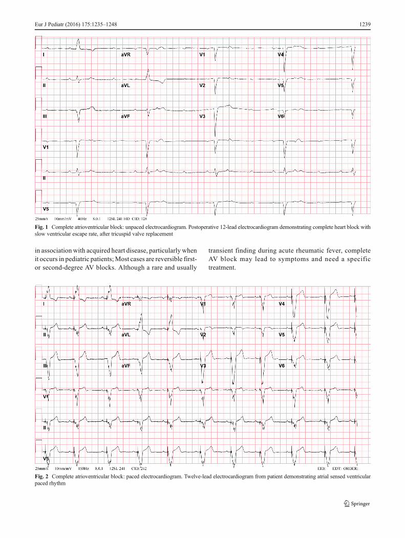
Postoperative
Following CHD surgery, any degree of AV block may be seen(Figs. 1 and 2). A retrospective multicenter study recently
evaluated incidence of postoperative complete heart block inchildren undergoing congenital heart surgery [53]. Among103,616 surgeries from 45 US tertiary care hospitals, the inci-dence of complete heart block requiring pacemaker placementwas low (1.2 %), mainly associated with mitral valve repair orreplacement (3.7 %), aortic valve repair or replacement(2.7 %), atrioventricular canal surgery (1.9 %), and ventricularseptal defect (VSD) surgery (1.8 %). However, these patientsincurred longer hospital stay and had higher mortality evenafter accounting for heart surgery complexity.
In roughly one third of the cases of postoperative completeheart block, AV conduction does not recover and those pa-tients should undergo pacemaker implantation. Permanentpacemaker implantation should be considered in all patientswho have postoperative high-grade AV block following CHDsurgery that exceeds 7–10 days, even in the setting of a narrowQRS escape rate [14, 26]. During this period, temporary pac-ing wires may be necessary to maintain adequate chronotropy.Postoperative heart block has also been rarely reported in pa-tients who had been previously discharged from the hospitalwith normal AV conduction after open-heart surgery. Closeand continued follow-up of postoperative CHD surgical cases,particularly VSD, is necessary due to the risk of possible pro-gression of block over time [54].
Atrioventricular conduction disorders in associationwith acquired heart disease
AV block in the young can also be derived from a wide vari-ety of causes such as surgical or catheterization-inducedtrauma, coronary artery disease, acute or chronic infectiousprocesses, myocarditis, hypersensitivity cardiomyopathy,metabolic abnormalities, hypothyroidism, infiltrative pro-cesses, or through a pathological neurocardiogenic mecha-nism [7]. Even if temporary pacing might be required in un-stable patients with Lyme carditis, complete heart block isusually reversible with appropriate antibiotics [28]. Chagasdisease is an endemic disease in most Latin American coun-tries, and around one third of affected patients develop car-diac conduction disorders requiring pacemaker implantation[4]. Incidence of catheterization-induced heart block wasrecently evaluated at 2.2 %, with a high rate of recoveryfollowing a similar course to that of postsurgical heart block[57]. Some interventional procedures, such as device closureof perimembranous VSD and catheter ablation of AV nodalreentrant tachycardia or parahissian accessory pathways,carry a risk of permanent heart block [49, 100, 102]. Theincidence of AV node dysfunction is apparently higher inpatients with Kawasaki disease, possibly caused bymyocar-ditis or an abnormal microcirculation in the AV node artery.Acute rheumatic carditis must also be kept in mind in thediagnostic work-up of patients with AV conduction disorder
1238 Eur J Pediatr (2016) 175:1235–1248
in association with acquired heart disease, particularly whenit occurs in pediatric patients;Most cases are reversible first-or second-degree AV blocks. Although a rare and usually
transient finding during acute rheumatic fever, completeAV block may lead to symptoms and need a specifictreatment.
Fig. 1 Complete atrioventricular block: unpaced electrocardiogram. Postoperative 12-lead electrocardiogram demonstrating complete heart block withslow ventricular escape rate, after tricuspid valve replacement
Fig. 2 Complete atrioventricular block: paced electrocardiogram. Twelve-lead electrocardiogram from patient demonstrating atrial sensed ventricularpaced rhythm
Eur J Pediatr (2016) 175:1235–1248 1239

Management
Treatment options during fetal life
Left untreated, congenital AV block is associated with a fetaland neonatal mortality ranging between 14 and 34 % [76].Fetal hydrops and ventricular escape rates <55 bpm have beenidentified as risk factors for mortality [54, 55]. Transplacentaltreatment options are not consensual. Dexamethasone usemay significantly lower fetal mortality [38], but its adminis-tration remains controversial because of its potential side ef-fects for both mother and fetus, especially potential fetal neu-rological development impairment [68]. Maternal administra-tion of terbutaline has also been reported, used alone or inaddition to dexamethasone [20]. Although largely controver-sial, prenatal treatment may also include intravenous immu-noglobulins and plasmapheresis, used alone or together incombination with steroids [31, 75].
Using the currently available techniques, in utero percutane-ous pacing appears to be at high risk, with fetal deaths occur-ring within a few hours of the procedure in a high proportion ofcases [5]. Further studies would be required to improve ourunderstanding of the natural history of congenital AV blockin order to identify more accurately the fetuses at highest risk.Development in the techniques and technologies available todeliver in utero pacing would also be required before this treat-ment can be adopted into routine clinical practice.
Postnatal medical therapy
Pharmacological therapy has a distinct role in the acute man-agement of severe bradycardia (whether sinus- or AV nodal-related) and should typically be carried out alongside parallelefforts to arrange for transcutaneous pacing and temporarycardiac pacing in order to prevent from sudden cardiac death[48]. Intravenous isoproterenol, atropine, epinephrine, and do-pamine are all recommended [48]. Beyond the acute manage-ment in this context, no medication is proven to improvechronic sinus- and AV nodal function. It is crucial, however,to recognize the potential for medications to compromise car-diac conduction, especially considering the atypical antihyper-tensive and antipsychotic agents, in addition to the AV nodalblocking agents they may contain. Monitoring of childrenwho do not require neonatal pacing is based on 24-h HolterECG and transthoracic echocardiography that should be per-formed frequently.
Permanent pacemaker implantation
The indications for permanent pacing in children or adultswith CHD are similar to those recommended in acquired heartdisease, yet there are important differences in the approachbased primarily on anatomy and somatic growth.
Indications In essence, every symptomatic, non-reversibleAV node disease requires permanent pacemaker implantation[14, 26]. Pacing must also be considered in asymptomatichigh degree AV blocks with specific risk conditions. Recentindications for cardiac pacing in children and CHD patientsare summarized in Table 1. Although historical series of iso-lated congenital AV block reported a high incidence of unpre-dictable Stokes-Adams attacks and a highmortality associatedwith the first attack [2, 18], latest studies performed in our eraof pediatric cardiac pacing showed that prophylactic pacing,as currently recommended [44, 45], is associated with strongreduction in the morbidity and mortality due to Stokes-Adamsattacks [32].
Endocardial versus epicardial deviceAlthough pacing indi-cations are clearly defined, whether an endocardial versus anepicardial system is preferred remains to be clarified [26, 48,96]. The large population of adult paced patients allows forsurgical practices to be studied more effectively and lead toevidence-based transitions in patient care. Meanwhile, thesmaller volume of paced pediatric patients does not provideenough information and clinical decisions based on faith,opinion, experience, and some retrospective data. Withinthose limitations, there is a general consensus that the smallestinfants are best served with epicardial pacing systems, with acutoff weight around 15–20 kg [26, 98]. Epicardial leads aremore likely to fracture and are prone to exit block, and im-plantation requires a major operation that is accompanied bythe inherent risks and need for perioperative support [89](Fig. 3). However, many groups standardly use epicardialpacing in infants and young children and argue that endocar-dial systems may carry a significant risk of venous thrombosisin infants, which can result in loss of venous access in thefuture, leading to a more complicated lead revision later inthe patient’s life [18, 29, 50]. Up to a 19 % transvenouslead-related failure rate has been reported by others whochoose the endocardial approach [80]. Extraction of aban-doned transvenous leads in the pediatric population is alsoproblematic, and optimal lead management still remains tobe defined [17, 60]. On the other hand, there is a global trendtowards using endocardial leads in younger patients and someinstitutions actively implant transvenous leads in childrenweighing less than 15 kg [43, 63, 73, 79, 83, 85]. It has beenshown that an 80-mm right atrial lead loop will allow 6 to12 years (mean, 8 years) of growth in infants and childrenwithout the need for reoperation to adjust lead length [33](Figs. 4, 5 and 6). Long-term follow-up demonstrates thatthe longevity of an endocardial system exceeds that of itsepicardial counterpart [89, 98], also an important consider-ation in patients who will be exposed to the cumulative burdenof repeated lead and device reimplantation. Despite growingexperience, endocardial implantation is not universally ac-cepted. Most publications reporting results of transvenous
1240 Eur J Pediatr (2016) 175:1235–1248

pacing in infants and young children are from small and/orolder studies [33, 40, 43, 55], so that long-term follow-up datafrom large patient populations should still be clarified.
Route of pacing Gaining access to the chamber requiringpacing is another central hurdle, frequently challenging in thispatient group, and thereby commonly dictating the route ofpacing but also highlighting why a detailed surgical history isvital. Modern-day pacemaker implantation is therefore suit-ably complemented by adjunctive CT or MR angiography,indicating whether an endocardial system is possible, and alsoproviding a map for coronary sinus lead placement. For com-plex CHD patients with prior operative intervention andwhose surgical reports are not available, venography at thetime of the procedure is recommended [14, 26]. Absolutecontraindications to conventional transvenous, endocardiallead placement include occlusion of the superior vena cava
(SVC) (bidirectional cavopulmonary anastomosis/Glenn),extra-cardiac Fontan procedures, baffle thrombosis, and se-vere baffle stenosis. Attempts to access the subpulmonic ven-tricle by crossing a mechanical atrioventricular valve shouldalso absolutely be avoided [27]. In this situation, coronarysinus lead placement can potentially be used for ventricularpacing.
In patients in whom a conventional transvenous approachvia the SVC is not possible, femoral and transhepatic leadimplantation can also be considered. Femoral techniques havebeen reported predominantly in children, suggesting this is aviable alternative with issues related primarily to the stabilityof the atrial lead and discomfort from the abdominally placedgenerator [59]. This approach does entail a higher incidence oflead failure given the additional mechanical stress associatedwith hip flexion. Transhepatic implants have also only beenreported in children and acutely present the additional risks of
Table 1 Pacing indications in children and patients with congenital heart disease
ESC guidelines ACCF/AHA/HRS guidelines
Congenital AV block
Symptomatic advanced second- or third-degree AV block Class I, level C Class I, level C
Asymptomatic high degree AV block with ventricular dysfunction Class I, level C Class I, level B
Asymptomatic high degree AV block with prolonged QTc interval Class I, level C –
Asymptomatic high degree AV block with complex ventricular ectopy Class I, level C Class I, level B
Asymptomatic high degree AV block with wide QRS escape rhythm Class I, level C Class I, level B
Asymptomatic high degree AV block with abrupt ventricular pauses>threefold the basic cycle length
Class I, level C Class IIa, level B
Asymptomatic third-degree AV block in the infant with a ventricular rate<55 bpm or with CHD and a ventricular rate <70 bpm
– Class I, level C
Third-degree AV block beyond the first year of life with an average heartrate <50 bpm
– Class IIa, level B
Asymptomatic high degree AV block with a ventricular rate <50 bpm Class I, level C –
Third-degree AV block beyond the first year of life with symptoms dueto chronotropic incompetence
– Class IIa, level B
High degree AV block in asymptomatic children/adolescents in absenceof the above risk conditions
Class IIb, level C Class IIb, level B
Asymptomatic type I second-degree AV block – Class III, level C
Postoperative AV block
Postoperative advanced second- or third-degree AV block that persists>7 days after cardiac surgery (10 days in ESC guidelines)
Class I, level B Class I, level B
Transient postoperative third-degree AV block that reverts to sinus rhythmwith residual bifascicular block
Class IIa, level C Class IIb, level C
Unexplained syncope in the patient with prior CHD surgery complicatedby transient complete heart block with residual fascicular block
– Class IIa, level B
Transient postoperative AV block with return of normal AV conductionin the otherwise asymptomatic patient
– Class III, level B
Asymptomatic postoperative bifascicular block with/without first-degreeAV block in the absence of prior transient complete AV block
– Class III, level C
Levels of evidence are classified in Blevel A^ if data are derived from multiple randomized clinical trials or meta-analyses, Blevel B^ if data are derivedfrom a single randomized clinical trial or large non-randomized studies, and Blevel C^ if there is a consensus of opinion of the experts and/or if data arederived from small studies, retrospective studies, or registries. Recommendations are listed according to the commonly used class I, IIa, IIb, and IIIclassification and the corresponding language: Bis recommended^ for a class I recommendation; Bcan be useful^ for a class IIa recommendation; Bmay beconsidered^ to signify a class IIb recommendation; and Bshould not^ or Bis not recommended^ for a class III recommendation. ESC guidelines: reference[44]; ACCF/AHA/HRS guidelines: reference [45]
AV atrioventricular, CHD congenital heart disease
Eur J Pediatr (2016) 175:1235–1248 1241

intraabdominal or intracapsular hemorrhage, and long-termoutcomes remain uncertain [25].
Placing leads in patients with prior atrial switch operations(Mustard/Senning) must be handled with utmost care, giventhe frequency of baffle leaks in this group [47]. Pre-emptivecovered stent implantation or even open repair should be con-sidered given the risk of thromboembolism across these veno-systemic shunts. A transcatheter approach is utilized for leakclosure but can also be applied for dilatation of a narrowedbaffle that would otherwise be occluded by transvenous leadimplantation [11, 24].
Thromboembolism Any patient with an intracardiac shunt isat higher risk for thrombus formation on transvenous pacingleads and subsequent systemic thromboembolic complications[21, 45]. Pre-emptive transesophageal echocardiography withbubble injections is therefore critical, and if any shunt is seen,the approach should be modified. If the shunt can be closed,either via an open approach or using a percutaneous approach,then this should be undertaken before endocardial leads areplaced or alternatively an epicardial device should be im-planted. It remains unclear how to exactly manage patients witha small shunt at an atrial level across a patent foramen ovale andfurther studies would be needed to develop recommendations[21]. It is also vital to recognize that the patients with a classicFontan operation frequently have very large atria and slow flowthrough their neo-chamber. Thus, it is not uncommon for largethrombi with a potential for pulmonary thromboembolism todevelop on transvenous leads. And although this does not pres-ent an absolute contraindication [69, 87], it does need to be
carefully considered and weighed against the risks of epicardiallead placement. Long-term oral anticoagulation does also needto be accordingly considered in this group.
Lead position The pacing site of the ventricular leads is a criticalissue, as it has a major impact on left ventricular mechanicalsynchrony, efficiency, and pump function in childrenwho requirelifelong pacing. The right ventricular apex has been the mostused pacing site, because it is easily accessible transvenouslyand provides a stable lead position with a low dislodgment rate.However, long-term right ventricular apical pacing induces aniatrogenic left bundle branch block and can lead to pacing-induced cardiomyopathy with left ventricular dilation and both
Fig. 3 Permanent pacemaker with epicardial leads (VVI pacing)
Fig. 4 Permanent pacemaker with transvenous leads. Growth and changein a loop of an endocardial lead. A 4-year-old boy with childhood isolatednonimmune atrioventricular block underwent pacemaker implantationusing a transvenous lead. Radiographs at 4 years of age (a, VVI pacing),6 years later (b, DDD pacing), and 9 years later (c, DDD pacing). Note thechange in the loop of the lead as the child grows
1242 Eur J Pediatr (2016) 175:1235–1248

systolic and diastolic left ventricular dysfunction, for both endo-cardial and epicardial leads [46, 67, 86, 91, 99]. There is earlyencouraging data that suggest improved overall hemodynamicsand ventricular function by cardiac resynchronization therapywith single-site left ventricular pacing via the coronary sinus inthis group [41]. Until further supportive evidence is accrued, it isimportant for the implanter and clinician to consider this ap-proach in any patient whose ejection fraction is suboptimal.
However, detrimental effects of the right ventricular apicalpacing led to the reassessment of traditional approaches and to
the research of alternative pacing sites, in order to get to morephysiological pattern of ventricular activation. Although be-ing theoretically the best technique, direct His-Bundle pacingand paraHisian cardiac pacing remain complex and associatedwith higher pacing thresholds are required, causing accelerat-ed battery depletion [93]. Right ventricular septal pacing is agood alternative, technically easy and maintaining a lowerpacing threshold [22]. By preserving septal to lateral left ven-tricular synchrony and systolic function, left ventricular apicaland midlateral wall pacingmay be the preferred pacing site forepicardial leads in the young [41, 81].
Mode of pacing A five-letter international code describespacemaker function, three letters being in common usage:the first letter is the chamber paced (A = atrium; V = ventricle;D = dual, namely both A and V; O = none), the second letter isthe chamber sensed and the third letter describes the algorithmused to integrate pacing and sensing functions. Most of chil-dren and teenagers will benefit from a dual-chamber pacing(DDD), allowing AV synchrony that is important to maintainventricular filling and stroke volume [89, 96]. In infants withcomplete AV block and normal sinus node function, a single-chamber ventricular pacing (VVI or VDD) should be selected,because it requires only a unique lead (uni- or bipolar) thusreducing the risk of venous occlusion [89, 96].
In patients with conduction system disease undergoingpacemaker implantation, coexistent reentrant atrial tachycar-dias are common. Therefore, the implantation of devices withatrial antitachycardia pacing capability is reasonable [84].This approach is more common as an adjunct to maze proce-dures and in conjunction with antiarrhythmic therapy [92].
Perspectives To overcome the potential short- and long-termcomplications related to transvenous leads and subcutaneouspulse generators, new technologies enabling leadless cardiac
Fig. 5 Cardiac resynchronization therapy with epicardial leads. A 5-year-old boy who underwent a neonatal Ross procedure had postoperativecomplete heart block and left ventricular dysfunction. He was implantedwith epicardial multisite pacing with right atrial, right ventricular (a and b,black star), and left ventricular (a and b, black arrow) leads. Biventricularpacing allow shortening of the paced QRS (c, 224 ms) compared with rightventricular pacing alone (d, 128 ms)
Fig. 6 Cardiac resynchronization therapy with transvenous leads. A 14-year-old boy with dilated cardiomyopathy and left ventricular dysfunction,second-degree AV block, and an episode of ventricular fibrillation hadimplantation of a biventricular implantable cardioverter-defibrillator witha transvenous right ventricular lead (a and b, black star) and a transvenousleft ventricular lead into the coronary sinus (a and b, black arrow)
Eur J Pediatr (2016) 175:1235–1248 1243
stimulation have been developed and proved to be applicableand safe in experimental models and in some small humanstudies [52, 71, 82]. These ultrasound or induction technolo-gies, based on leadless pacemakers, could be used to providecardiac stimulation from the endocardium at selected sites. Theabsence of a transvenous lead and subcutaneous pulse genera-tor could represent a paradigm shift in cardiac pacing. Futurestudies will need to address the safety/efficacy of alternate-siteRV and LV pacing and the long-term outcomes of these newapproaches. Although the continued development and successof electronic pacing are impressive, gene therapy for biologicalpacing is also a promising research field. This approach mightoffer an alternative treatment for pacemaker-related infectionsin the future, with right ventricular intramyocardial injection ofan adenoviral construct whose duration of effect is temporary,until such time as the infection is adequately treated and a newpermanent pacemaker can be inserted [35, 74].
Conclusion
Although rare, congenital and childhood AV block is importanttreatable cause of morbidity and mortality in the young. Thisreview crystallizes our contemporary understanding and man-agement strategies, highlighting the ongoing investigation in allaspects of care related to this complex group of disorders.Much work is needed, especially with regards to the antenataldetection and treatment of congenital AV block, and on a ge-netic level, and it is evident that as new insights are gleaned,management strategies continue to evolve. Despite the manydifferent etiologies, eachwith specific management and distinctoutcomes, these patients can do well with a careful, consideredapproach to the diagnostic evaluation and management plan.
Acknowledgments Dr. Baruteau is supported by the 2015 AcademicResearch Grant from the European Heart Rhythm Association and a re-search grant from the Lefoulon Delalande Foundation - Institut de France.
Author’s contribution Dr. Baruteau conceptualized and designed thereview article, drafted several parts of its initial version, and approved thefinal manuscript as submitted. Dr. Pass drafted the paragraph on postoper-ative heart blocks and the one on native heart blocks associated with con-genital heart disease. He approved the final manuscript as submitted. Dr.Thambo drafted the paragraph on immune-mediated AV block. He ap-proved the final manuscript as submitted. Dr. Behaghel drafted the partdescribing permanent pacemaker implantation. He approved the final man-uscript as submitted. Dr. Le Pennec drafted the paragraph on treatmentoptions during pregnancy and critically revised the initial version of thepaper. She approved the final manuscript as submitted. Dr. Perdreau draftedthe part on native heart blocks associated with congenital heart disease. Sheapproved the final manuscript as submitted. Drs. Liberman and Combescritically reviewed the article and added substantial contents to this paper.They approved the final manuscript as submitted. Dr. McLeod drafted themanagement section. He critically reviewed the whole paper and approvedthe final manuscript as submitted.
All authors approved the final manuscript as submitted and agreed to beaccountable for all aspects of the work.
Compliance with ethical standards
Financial disclosure None of the authors has any financial relationshipsrelevant to this article to disclose.
Conflict of interest The authors declare that they have no conflict ofinterest.
Funding None.
Ethical approval This article does not contain any studies with humanparticipants or animals performed by any of the authors.
Open Access This article is distributed under the terms of the CreativeCommons At t r ibut ion 4 .0 In te rna t ional License (h t tp : / /creativecommons.org/licenses/by/4.0/), which permits unrestricted use,distribution, and reproduction in any medium, provided you give appro-priate credit to the original author(s) and the source, provide a link to theCreative Commons license, and indicate if changes were made.
References
1. Ambrosi A, Wahren-Herlenius M (2012) Congenital heart block:evidence for a pathogenic role of maternal autoantibodies. ArthritisRes Ther 14:208
2. Andelfinger G, Fouron JC, Sonesson SE, Proulx F (2001) Referencevalues for time intervals between atrial and ventricular contractions ofthe fetal heart measured by two Doppler techniques. Am J Cardiol88:1433–6
3. Anderson RH, Becker AE, Arnold R, Wilkinson JL (1974) Theconducting tissues in congenitally corrected transposition. Circulation50:911–923
4. Arce M, Van Grieken J, Femenía F, Arrieta M, McIntyre WF,Baranchuk A (2012) Permanent pacing in patients with Chagas’disease. Pacing Clin Electrophysiol 35:1494–7
5. Assad RS, Zielinsky P, Kalil R, Lima G, Aramayo A, Santos A, CostaR, Marcial MB, Oliveira SA (2003) New lead for in utero pacing forfetal congenital heart block. J Thorac Cardiovasc Surg 126:300–2
6. Baban A, Pitto L, Pulignani S, Cresci M,Mariani L, Gambacciani C,Digilio MC, Pongiglione G, Albanese S (2014) Holt-Oram syn-drome with intermediate atrioventricular canal defect, and aortic co-arctation: functional characterization of a de novo TBX5 mutation.Am J Med Genet A 164A:1419–24
7. Barra SN, Providência R, Paiva L, Nascimento J, Marques AL(2012) A review on advanced atrioventricular block in young ormiddle-aged adults. Pacing Clin Electrophysiol 35:1395–405
8. BaruteauAE, Probst V,Abriel H (2015) Inherited progressive cardiacconduction disorders. Curr Opin Cardiol 30:33–39
9. Baruteau AE, Fouchard S, Behaghel A, Mabo P, Villain E, ThamboJB, Marçon F, Gournay V, Rouault F, Chantepie A, Guillaumont S,Godart F, Bonnet C, Fraisse A, Schleich JM, Lusson JR, Dulac Y,Leclercq C, Daubert JC, Schott JJ, Le Marec H, Probst V (2012)Characteristics and long-term outcome of nonimmune isolated atrio-ventricular block diagnosed in utero or early childhood: a multicentrestudy. Eur Heart J 33:622–629
10. Baruteau AE, Behaghel A, Fouchard S, Mabo P, Schott JJ, Dina C,Chatel S, Villain E, Thambo JB, Marçon F, Gournay V, Rouault F,Chantepie A, Guillaumont S, Godart F, Martins RP, Delasalle B,Bonnet C, Fraisse A, Schleich JM, Lusson JR, Dulac Y, Daubert
1244 Eur J Pediatr (2016) 175:1235–1248
JC, Le Marec H, Probst V (2012) Parental electrocardiographicscreening identifies a high degree of inheritance for congenital andchildhood non-immune isolated atrioventricular block. Circulation126:1469–1477
11. Bentham J, English K, Hares D, Gibbs J, Thomson J (2012) Effect oftranscatheter closure of baffle leaks following senning or mustardatrial redirection surgery on oxygen saturations and polycythaemia.Am J Cardiol 110:1046–1050
12. BergmanG, SkogA, Tingström J, OttossonV,HoxhaA,Ambrosi A,Swedish Congenital Heart Block Study Group, Salomonsson S,Wahren-HerleniusM (2014) Late development of complete atrioven-tricular block may be immune mediated and congenital in origin.Acta Paediatr 103:275–81
13. Bordachar P, Zachary W, Ploux S, Labrousse L, Haissaguerre M,Thambo JB (2013) Pathophysiology, clinical course, and managementof congenital complete atrioventricular block. Heart Rhythm 10:760–6
14. BrignoleM, Auricchio A, Baron-Esquivias G, Bordachar P, Boriani G,Breithardt OA, Cleland J, Deharo JC, Delgado V, Elliott PM, GorenekB, Israel CW, Leclercq C, Linde C, Mont L, Padeletti L, Sutton R,Vardas PE, ESC Committee for Practice Guidelines (CPG),Zamorano JL, Achenbach S, Baumgartner H, Bax JJ, Bueno H,Dean V, Deaton C, Erol C, Fagard R, Ferrari R, Hasdai D, Hoes AW,Kirchhof P, Knuuti J, Kolh P, Lancellotti P, Linhart A,Nihoyannopoulos P, Piepoli MF, Ponikowski P, Sirnes PA, TamargoJL, Tendera M, Torbicki A, Wijns W, Windecker S, DocumentReviewers, Kirchhof P, Blomstrom-Lundqvist C, Badano LP, AliyevF,BänschD,BaumgartnerH,BsataW,Buser P,CharronP,Daubert JC,Dobreanu D, Faerestrand S, Hasdai D, Hoes AW, Le Heuzey JY,Mavrakis H, McDonagh T, Merino JL, Nawar MM, Nielsen JC,Pieske B, Poposka L, Ruschitzka F, Tendera M, Van Gelder IC,WilsonCM (2013) 2013 ESC guidelines on cardiac pacing and cardiacresynchronization therapy: the task force on cardiac pacing andresynchronization therapy of the European Society of Cardiology(ESC). Developed in collaboration with the European Heart RhythmAssociation (EHRA). Eur Heart J 34:2281–2329
15. Brucato A, Jonzon A, Friedman D, Allan LD, Vignati G, GaspariniM, Stein JI, Montella S, Michaelsson M, Buyon J (2003) Proposalfor a new definition of congenital complete atrioventricular block.Lupus 12:427–35
16. Buyon JP,HiebertR,Copel J, Craft J, FriedmanD,KatholiM,LeeLA,Provost TT, Reichlin M, Rider L, Rupel A, Saleeb S, Weston WL,Skovron ML (1998) Autoimmune-associated congenital heart block:demographics, mortality, morbidity and recurrence rates obtained froma national neonatal lupus registry. J Am Coll Cardiol 31:1658–66
17. Cecchin F, Atallah J, Walsh EP, Triedman JK, Alexander ME, BerulCI (2010) Lead extraction in pediatric and congenital heart diseasepatients. Circ Arrhythm Electrophysiol 3:437–44
18. CohenMI, Bush DM,Vetter VL, Tanel RE,Wieand TS, Gaynor JW,Rhodes LA (2001) Permanent epicardial pacing in pediatric patients:seventeen years of experience and 1200 outpatient visits. Circulation103:2585–2590
19. Costedoat-Chalumeau N, Georgin-Lavialle S, Amoura Z, Piette JC(2005) Anti-SSA/Ro and anti-SSB/La antibody-mediated congenitalheart block. Lupus 14:660–4
20. Cuneo BF, Lee M, Roberson D, Niksch A, Ovadia M, Parilla BV,Benson DW (2010) A management strategy for fetal immune-mediated atrioventricular block. J Matern Fetal Neonatal Med 23:1400–5
21. DeSimone CV, Friedman PA, Noheria A, Patel NA, DeSimone DC,Bdeir S, Aakre CA, Vaidya VR, Slusser JP, Hodge DO, AckermanMJ, Rabinstein AA, Asirvatham SJ (2013) Stroke or transient ische-mic attack in patients with transvenous pacemaker or defibrillator andechocardiographically detected patent foramen ovale. Circulation128:1433–1441
22. Deshmukh P, Casavant DA, Romanyshyn M, Anderson K (2000)Permanent, direct His-bundle pacing: a novel approach to cardiac
pacing in patients with normal His-Purkinje activation. Circulation101:869–77
23. Donofrio MT, Moon-Grady AJ, Hornberger LK, Copel JA, SklanskyMS,AbuhamadA,CuneoBF,Huhta JC, JonasRA,KrishnanA,LaceyS, LeeW,Michelfelder EC Sr, Rempel GR, Silverman NH, Spray TL,Strasburger JF, Tworetzky W, Rychik J, American Heart AssociationAdults With Congenital Heart Disease Joint Committee of the Councilon Cardiovascular Disease in the Young and Council on ClinicalCardiology, Council on Cardiovascular Surgery and Anesthesia, andCouncil on Cardiovascular and Stroke Nursing (2014) Diagnosis andtreatment of fetal cardiac disease: a scientific statement from theAmerican Heart Association. Circulation 129:2183–242
24. Dragulescu A, Sidibe N, Aubert F, Fraisse A (2008) Successful useof covered stent to treat superior systemic baffle obstruction and leakafter atrial switch procedure. Pediatr Cardiol 29:954–956
25. EmmelM, SreeramN, Pillekamp F, BoehmW, Brockmeier K (2006)Transhepatic approach for catheter interventions in infants and chil-dren with congenital heart disease. Clin Res Cardiol 95:329–333
26. Epstein AE, DiMarco JP, Ellenbogen KA, Estes NA 3rd, FreedmanRA, Gettes LS, Gillinov AM, Gregoratos G, Hammill SC, Hayes DL,Hlatky MA, Newby LK, Page RL, Schoenfeld MH, Silka MJ,Stevenson LW, Sweeney MO, Tracy CM, Epstein AE, Darbar D,DiMarco JP, Dunbar SB, Estes NA 3rd, Ferguson TB Jr, HammillSC, Karasik PE, Link MS, Marine JE, Schoenfeld MH, Shanker AJ,Silka MJ, Stevenson LW, Stevenson WG, Varosy PD (2013) 2012ACCF/AHA/HRS focused update incorporated into the ACCF/AHA/HRS2008guidelines for device-based therapy of cardiac rhythmabnormalities: a report of the American College of CardiologyFoundation/American Heart Association Task Force on PracticeGuidelines and theHeart RhythmSociety. JAmCollCardiol 61:e6–75
27. Fadel BM, Di Salvo G (2013) Metal through metal: pacing leadacross a mechanical tricuspid valve. J Cardiovasc Med
28. Forrester JD,Mead P (2014) Third-degree heart block associated withLime carditis: review of published cases. Clin Infect Dis 59:996–1000
29. Fortescue EB, Berul CI, Cecchin F, Walsh EP, Triedman JK,Alexander ME (2004) Patient, procedural, and hardware factors as-sociated with pacemaker lead failures in pediatrics and congenitalheart disease. Heart Rhythm 1:150–159
30. Fouron JC, Proulx F,Miro J, Gosselin J (2000) Doppler andM-modeultrasonography to time fetal atrial and ventricular contractions.Obstet Gynecol 96:732–6
31. Friedman DM, Llanos C, Izmirly PM, Brock B, Byron J, Copel J,Cummiskey K, Dooley MA, Foley J, Graves C, Hendershott C,Kates R, Komissarova EV, Miller M, Paré E, Phoon CK, Prosen T,Reisner D, Ruderman E, Samuels P, Yu JK, Kim MY, Buyon JP(2010) Evaluation of fetuses in a study of intravenous immunoglob-ulin as preventive therapy for congenital heart block: results of amulticenter, prospective, open-label clinical trial. Arthritis Rheum62:1138–46
32. Gazes PC, Culler RM, Taber E, Kelly TE (1965) Congenital familialcardiac conduction defects. Circulation 32:32–4
33. Gheissari A, Hordof AJ, Spotnitz HM (1991) Transvenous pace-makers in children: relation of lead length to anticipated growth.Ann Thorac Surg 52:118–21
34. Hunscher O, Batista N, Rivero S, Marletta C, ArriagadaM, Boire G,MénardHA,AranaRM (1995) Clinical and serological identificationof 2 forms of complete heart block in children. J Rheumatol 22:1352–5.7
35. Hu Y-F, Dawkins JF, Cho HC, Marban E, Cingolani E (2014)Biological pacemaker created by minimally invasive somaticreprogramming in pigs with complete heart block. Sci Transl Med6:245–94
36. Jaeggi E1, Laskin C, Hamilton R, Kingdom J, Silverman E (2010)The importance of the level of maternal anti-Ro/SSA antibodies as aprognostic marker of the development of cardiac neonatal lupus
Eur J Pediatr (2016) 175:1235–1248 1245

erythematosus a prospective study of 186 antibody-exposed fetusesand infants. J Am Coll Cardiol 55:2778–84
37. Jaeggi ET, Hamilton RM, Silverman ED, Zamora SA, HornbergerLK (2002) Outcome of children with fetal, neonatal or childhooddiagnosis of isolated congenital atrio-ventricular block. J Am CollCardiol 39:130–7
38. Jaeggi ET, Fouron JC, Silverman ED, Ryan G, SmallhornJ, Hornberger LK (2004) Transplacental fetal treatment im-proves the outcome of prenatally diagnosed complete atrioven-tricular block without structural heart disease. Circulation 110:1542–8
39. Janousek J, Tomek V, Chaloupecky V, Gebauer RA (2004)Dilated cardiomyopathy associated with dual-chamber pacing ininfants: improvement through either left ventricular cardiacresynchronization or programming the pacemaker off allowingintrinsic normal conduction. J Cardiovasc Electrophysiol 15:470–4
40. Janousek J, Kubus P (2014) What’s new in cardiac pacing in chil-dren? Curr Opin Cardiol 29:76–82
41. Janousek J, van Geldorp IE, Krupickova S, Rosenthal E, NugentK, Tomaske M, Früh A, Elders J, Hiippala A, Kerst G, GebauerRA, Kubuš P, Frias P, Gabbarini F, Clur SA, Nagel B, Ganame J,Papagiannis J, Marek J, Tisma-Dupanovic S, Tsao S, NürnbergJH, Wren C, Friedberg M, de Guillebon M, Volaufova J, PrinzenFW, Delhaas T, Working Group for Cardiac Dysrhythmias andElectrophysiology of the Association for European PediatricCardiology (2013) Permanent cardiac pacing in children: choos-ing the optimal pacing site: a multicenter study. Circulation 127:613–623
42. Kabunga P, Lau AK, Phan K, Puranik R, Liang C, Davis RL, SueCM, SyRW (2015) Systematic review of cardiac electrical disease inKearns-Sayre syndrome and mitochondrial cytopathy. Int J Cardiol181:303–310
43. Kammeraad J, Rosenthal E, Bostock J, Rogers J, Sreeram N (2004)Endocardial pacemaker implantation in infants <10 kg weight.Pacing Clin Electrophysiol 27:1466–74
44. Kanter RJ, Pfeiffer R, Hu D, Barajas-Martinez H, Carboni MP,Antzelevitch C (2012) Brugada-like syndrome in infancy presentingwith rapid ventricular tachycardia and intraventricular conductiondelay. Circulation 125:14–22
45. Khairy P, Landzberg MJ, Gatzoulis MA, Mercier LA, FernandesSM, Côté JM, Lavoie JP, Fournier A, Guerra PG, Frogoudaki A,Walsh EP, Dore A, Epicardial Versus ENdocardial pacing andThromboembolic events Investigators, (2006) Transvenous pacingleads and systemic thromboemboli in patients with intracardiacshunts: a multicenter study. Circulation 113:2391–2397
46. Kim JJ, Friedman RA, EidemBW, Cannon BC, Arora G, Smith EO,Fenrich AL, Kertesz NJ (2007) Ventricular function and long-termpacing in children with congenital complete atrioventricular block. JCardiovasc Electrophysiol 18:373–377
47. Klein AJ, Kim MS, Salcedo E, Fagan T, Kay J (2009) The missingleak: a case report of a baffle-leak closure using real-time 3dtransoesophageal guidance. Eur J Echocardiogr 10:464–467
48. Kleinman ME, de Caen AR, Chameides L, Samson RA, HazinskiMF, Atkins DL, Berg MD, de Caen AR, Fink EL, Freid EB,Hickey RW, Marino BS, Nadkarni VM, Proctor LT, Qureshi FA,Sartorelli K, Topjian A, van der Jagt EW, Zaritsky AL, AmericanHeart Association. Pediatric Basic and Advanced Life SupportChapter Collaborators (2010) Pediatric basic and advanced lifesupport: 2010 International Consensus on CardiopulmonaryResuscitation and Emergency Cardiovascular Care Science withTreatment Recommendations. Pediatrics 126:e1261–318
49. Krause U, Backhoff D, Klehs S, Kriebel T, Paul T, Schneider HE(2015) Catheter ablation of pediatric AV nodal reentrant tachycardia:results in small children. Clin Res Cardiol 104:990–7
50. Kubus P, Materna O, Gebauer RA, Matejka T, Gebauer R, TláskalT, Janousek J (2012) Permanent epicardial pacing in children:long-term results and factors modifying outcome. Europace 14:509–14
51. Lazzerini PE, Capecchi PL, Laghi-Pasini F (2015) Isolated atrioven-tricular block of unknown origin in adults and anti-Ro/SSA antibodies:clinical evidence, putative mechanisms, and therapeutic implications.Heart Rhythm 12:449–54
52. Lee KL, Lau CP, Tse HF, Echt DS, Heaven D, Smith W, Hood M(2007) First human demonstration of cardiac stimulation with trans-cutaneous ultrasound energy delivery: implications for wireless pac-ing with implantable devices. J Am Coll Cardiol 50:877–83
53. Liberman L, Silver ES, Chai P, Anderson BR (2016) Incidence andcharacteristics of heart block after heart surgery in pediatric patients:a multicenter study. J Thorac Cardiovasc Surg. doi:10.1016/j.jtcvs.2016.03.081
54. Liberman L, Pass RH, Hordof AJ, Spotnitz HM (2008) Late onset ofheart block after open heart surgery for congenital heart disease.Pediatr Cardiol 29:56–9
55. Lotfy W, Hegazy R, Abdelaziz O, Sobhy R, Hasanein H, Shaltout F(2013) Permanent cardiac pacing in pediatric patients. Pediatr Cardiol34:273–280
56. Lynch HT, Mohiuddin S, Sketch MH, Krush AJ, Carter S, Runco V(1973) Hereditary progressive atrioventricular conduction defect. Anew syndrome? JAMA 225:1465–70
57. Mah DY, Porras D, Bergersen L, Marshall AC, Walsh EP, TriedmanJK (2014) Incidence of and risk factors for catheterization-inducedcomplete heart block in the pediatric cardiac catheterization labora-tory. Circ Arrhythm Electrophysiol 7:127–33
58. Makita N, Seki A, Sumitomo N, Chkourko H, Fukuhara S, WatanabeH, ShimizuW, Bezzina CR, Hasdemir C,MugishimaH,Makiyama T,Baruteau A, Baron E, Horie M, Hagiwara N, Wilde AA, Probst V, LeMarec H, Roden DM, Mochizuki N, Schott JJ, Delmar M (2012) Aconnexin40mutation associatedwith amalignant variant of progressivefamilial heart block type I. Circ Arrhythm Electrophysiol 5:163–72
59. MathurG, StablesRH,HeavenD, IngramA,SuttonR (2001)Permanentpacemaker implantation via the femoral vein: an alternative in cases withcontraindications to the pectoral approach. Europace 3:56–59
60. McCanta AC, Schaffer MS, Collins K (2011) Pediatric and AdultCongenital Endocardial Lead Extraction or Abandonment Decision(PACELEAD) survey of lead management. Pacing ClinElectrophysiol 34:1621–27
61. McCulley DJ, Black BL (2012) Transcription factor pathways andcongenital heart disease. Curr Top Dev Biol 100:253–277
62. McElhinney DB, Geiger E, Blinder J, Benson DW, Goldmuntz E(2003) NKX2.5 mutations in patients with congenital heart disease. JAm Coll Cardiol 42:1650–1655
63. McLeod KA (2010) Cardiac pacing in infants and children. Heart 96:1502–8
64. Michaelsson M, Riesenfeld T, Jonzon A (1997) Natural history ofcongenital complete atrioventricular block. Pacing ClinElectrophysiol 20:2098–2101
65. Michaëlsson M, Jonzon A, Riesenfeld T (1995) Isolated congenitalcomplete atrioventricular block in adult life. A prospective study.Circulation 92:442–9
66. Moak JP, Barron KS, Hougen TJ, Wiles HB, Balaji S, Sreeram N,Cohen MH, Nordenberg A, Van Hare GF, Friedman RA, Perez M,Cecchin F, Schneider DS, Nehgme RA, Buyon JP (2001) Congenitalheart block: development of late-onset cardiomyopathy, a previouslyunderappreciated sequela. J Am Coll Cardiol 37:238–42
67. Moak JP, Hasbani K, Ramwell C, Freedenberg V, Berger JT,DiRusso G, Callahan P (2006) Dilated cardiomyopathy followingright ventricular pacing for AV block in young patients: resolutionafter upgrading to biventricular pacing systems. J CardiovascElectrophysiol 17:1068–1071
1246 Eur J Pediatr (2016) 175:1235–1248

68. Modi N, Lewis H, Al-Naqeeb N, Ajayi-ObeM, Dore CJ, RutherfordM (2001) The effects of repeated antenatal glucocorticoid therapy onthe developing brain. Pediatr Res 50:581–5
69. Mosquera VX, Marini M, Portela F, Cao I (2008) Late complicationof classic Fontan operation: giant right atrial thrombus and massivepulmonary thromboembolism. J Card Surg 23:776–778
70. Priori SG, Wilde AA, Horie M, Cho Y, Behr ER, Berul C,Blom N, Brugada J, Chiang CE, Huikuri H, Kannankeril P,Krahn A, Leenhardt A, Moss A, Schwartz PJ, Shimizu W,Tomaselli G, Tracy C (2013) HRS/EHRA/APHRS expertconsensus statement on the diagnosis and management ofpatients with inherited primary arrhythmia syndromes.Heart Rhythm 10:1932–1963
71. Reddy VY, Knops RE, Sperzel J, Miller MA, Petru J, Simon J,Sediva L, de Groot JR, Tjong FV, Jacobson P, Ostrosff A,Dukkipati SR, Koruth JS, Wilde AA, Kautzner J, Neuzil P (2014)Permanent leadless cardiac pacing: results of the LEADLESS trial.Circulation 129:1466–71
72. Rein AJ, Mevorach D, Perles Z, Gavri S, Nadjari M, Nir A, ElchalalU (2009) Early diagnosis and treatment of atrioventricular block inthe fetus exposed to maternal anti-SSA/Ro-SSB/La antibodies: aprospective, observational, fetal kinetocardiogram-based study.Circulation 119:1867–72
73. Robledo-Nolasco R, Ortiz-Avalos M, Rodriguez-Diez G, Jimenez-Carrillo C, Ramírez-Machuca J, De Haro S, Castro-Villacorta H(2009) Transvenous pacing in children weighing less than 10 kilo-grams. Pacing Clin Electrophysiol 32:S177–81
74. RosenMR (2014)Gene therapy and biological pacing.NEngl JMed371:1158–9
75. Ruffatti A, Milanesi O, Chiandetti L, Cerutti A, Gervasi MT, DeSilvestro G, Pengo V, Punzi L (2012) A combination therapy totreat second-degree anti-Ro/La-related congenital heart block:a strategy to avoid stable third-degree heart block? Lupus 21:666–71
76. Schmidt KG, Ulmer HE, Silverman NH, Kleinman CS, Copel JA(1991) Perinatal outcome of fetal complete atrioventricular block: amulticenter experience. J Am Coll Cardiol 17:1360–6
77. Schott JJ, Alshinawi C, Kyndt F, Probst V, Hoorntje TM, HulsbeekM, Wilde AA, Escande D, Mannens MM, Le Marec H (1999)Cardiac conduction defects associate with mutations in SCN5A.Nat Genet 23:20–21
78. Schott JJ, Benson DW, Basson CT, Pease W, Silberbach GM, MoakJP, Maron BJ, Seidman CE, Seidman JG (1998) Congenital heartdisease caused by mutations in the transcription factor NKX2-5.Science 281:108–111
79. Silvetti MS, Drago F, Di Carlo D, Placidi S, Brancaccio G, Carotti A(2013) Cardiac pacing in paediatric patients with congenital heartdefects: transvenous or epicardial? Europace 15:1280–6
80. Silvetti MS, Drago F, Marcora S, Ravà L (2007) Outcome of single-chamber, ventricular pacemakerswith transvenous leads implanted inchildren. Europace 9:894–9
81. Silvetti MS, Di Carlo D, Ammirati A, Placidi S, DiMambro C, RavàL, Drago F (2015) Left ventricular pacing in neonates and infantswith isolated congenital complete or advanced atrioventricular block:short- and medium-term outcome. Europace 17:603–10
82. Sperzel J, Burri H, Gras D, Tjong FV, Knops RE, Hindricks G,Steinwender C, Defaye P (2015) State of the art of leadless pacing.Europace 17:1508–13
83. Spotnitz HM (1990) Transvenous pacing in infants and children withcongenital heart disease. Ann Thorac Surg 49:495–6
84. Stephenson EA, Casavant D, Tuzi J, Alexander ME, Law I,Serwer G, Strieper M, Walsh EP, Berul CI, ATTESTInvestigators (2003) Efficacy of atrial antitachycardia pacingusing the Medtronic AT500 pacemaker in patients with congenitalheart disease. Am J Cardiol 92:871–876
85. Stojanov PL, Savic DV, Zivkovic MB, Calovic ZR (2008)Permanent endovenous pediatric pacing: absence of lead fail-ure—20 years follow-up study. Pacing Clin Electrophysiol 31:1100–7
86. Sweeney MO, Hellkamp AS, Ellenbogen KA, Greenspon AJ,Freedman RA, Lee KL, Lamas GA, MOde Selection TrialInvestigators, (2003) Adverse effect of ventricular pacing on heartfailure and atrial fibrillation among patients with normal baselineQRS duration in a clinical trial of pacemaker therapy for sinus nodedysfunction. Circulation 107:2932–2937
87. Takahashi K, Cecchin F, Fortescue E, Berul CI, Alexander ME,Walsh EP, Fynn-Thompson F, Triedman JK (2009) Permanent atrialpacing lead implant route after Fontan operation. Pacing ClinElectrophysiol 32:779–785
88. Taketazu M, Lougheed J, Yoo SJ, Lim JS, Hornberger LK (2006)Spectrum of cardiovascular disease, accuracy of diagnosis, and out-come in fetal heterotaxy syndrome. Am J Cardiol 97:720–4
89. Takeuchi D, Tomizawa Y (2013) Pacing device therapy in infantsand children: a review. J Artif Organs 16:23–33
90. Taylor PV, Scott JS, Gerlis LM, Esscher E, Scott O (1986) Maternalantibodies against fetal cardiac antigens in congenital complete heartblock. N Engl J Med 315:667–72
91. Thambo JB, Bordachar P, Garrigue S, Lafitte S, Sanders P, ReuterS, Girardot R, Crepin D, Reant P, Roudaut R, Jaïs P, HaïssaguerreM, Clementy J, Jimenez M (2004) Detrimental ventricular remod-elling in patients with congenital complete heart block and chronicright ventricular apical pacing. Circulation 110:3766–3772
92. Tsao S, Deal BJ, Backer CL, Ward K, Franklin WH, Mavroudis C(2009) Device management of arrhythmias after Fontan conversion.J Thorac Cardiovasc Surg 138:937–940
93. Tung S, Lemaitre J (2015)His bundle pacing: in pursuit of the "sweetspot". Pacing Clin Electrophysiol 38:537–9
94. Udink ten Cate FE, Breur JM, Cohen MI, Boramanand N, KapustaL, Crosson JE, Brenner JI, Lubbers LJ, Friedman AH, Vetter VL,Meijboom EJ (2001) Dilated cardiomyopathy in isolated congenitalcomplete atrioventricular block: early and long-term risk in children.J Am Coll Cardiol 37:1129–1134
95. Villain E, Coastedoat-Chalumeau N, Marijon E, Boudjemline Y,Piette JC, Bonnet D (2006) Presentation and prognosis of completeatrioventricular block in childhood, according to maternal antibodystatus. J Am Coll Cardiol 48:1682–1687
96. Villain E (2008) Indications for pacing in patients with congenitalheart disease. Pacing Clin Electrophysiol 31:S17–20
97. Warnes C (2006) Transposition of the great arteries. Circulation114:2699–2709
98. WilhelmBJ, ThöneM, El-Scheich T, Livert D, Angelico R, OsswaldB (2015) Complications and risk assessment of 25 years in pediatricpacing. Ann Thorac Surg 100:147–53
99. Wilkoff BL, Cook JR, Epstein AE, Greene HL, Hallstrom AP, HsiaH, Kutalek SP, Sharma A, Dual Chamber and VVI ImplantableDefibrillator Trial Investigators (2002) Dual-chamber pacing or ven-tricular backup pacing in patients with an implantable defibrillator:the dual chamber and VVI implantable defibrillator (DAVID) trial.JAMA 288:3115–3123
100. Yildirim I, Karagöz T, Ertuğrul İ, Karagöz AH, Özer S (2013)Efficacy and safety of cryoablation of parahissian accessory path-ways in children: a single institution study. Pacing ClinElectrophysiol 36:1495–502
101. Zaidi S, ChoiM,Wakimoto H,Ma L, Jiang J, Overton JD, Romano-Adesman A, Bjornson RD, Breitbart RE, Brown KK, Carriero NJ,Cheung YH, Deanfield J, DePalma S, Fakhro KA, Glessner J,Hakonarson H, Italia MJ, Kaltman JR, Kaski J, Kim R, Kline JK,Lee T, Leipzig J, Lopez A, Mane SM, Mitchell LE, Newburger JW,Parfenov M, Pe’er I, Porter G, Roberts AE, Sachidanandam R,Sanders SJ, Seiden HS, State MW, Subramanian S, Tikhonova IR,Wang W, Warburton D, White PS, Williams IA, Zhao H, Seidman
Eur J Pediatr (2016) 175:1235–1248 1247

JG, Brueckner M, Chung WK, Gelb BD, Goldmuntz E, SeidmanCE, Lifton RP (2013)De novomutations in histone-modifying genesin congenital heart disease. Nature 498:220–3
102. Zartner P, Christians C, Stelter JC, Hraška V, Schneider MB (2014)Transvascular closure of single and multiple muscular ventricular
septal defects in neonates and infants <20 kg. Catheter CardiovascInterv 83:564–70
103. Zhao H, Cuneo BF, Strasburger JF, Huhta JC, Gotteiner NL, WakaiRT (2008) Electrophysiological characteristics of fetal atrioventricularblock. J Am Coll Cardiol 51:77–84
1248 Eur J Pediatr (2016) 175:1235–1248