atomic p & phosphorus molecules – the dna...
TRANSCRIPT
54
CHAPTER 3
Atomic P & Phosphorus molecules – The DNA
relevance
3.1 Introduction and motivation
It is well known that the biogenic elements (H, C, N, O, S, P) and organic
molecules [1] are some of the important constituents of the universe. The question of how
life began on Earth does not have an exact answer and inevitably there is scientific
speculation. There is evidence [2, 3] to support the view that a vast array of exotic but
important chemicals have been delivered to Earth through impacts from interstellar
objects.
Gulick [4] recognised a central role of phosphorus in the origin-of-life question
and pointed out a major problem. The principal source of prebiotic phosphorus present on
Earth crust was inorganic phosphate (containing the PO43- unit), which is insoluble in
water. Presumably, the solution phase of phosphates would be poorly available as a
biological nutrient on the early Earth. This question leads to consider extraterrestrial
sources of phosphorus and indeed, such sources exist. The phosphorus-containing
molecules like PH, PH2, PH3, PO, PO2, PN and PCradicals, have been observed in
interstellar gas clouds [5 - 7] and they are proposed to be present under appropriate
conditions. In a landmark paper of Cooper et al [8], the first account of the presence of
phosphonic acids in meteorites was published. Specifically, they observed water soluble
alkyl-phosphonic acids. Thus, Phosphorus is a bio-element important in the development
of life [9] and hence this chapter is aims to explore atomic phosphorus and its
compounds.
55
The present chapter addresses theelectron scattering cross sections, starting from
atomic P and several diatomic and large polyatomic phosphorus bearing molecules. After
atomic P, the next target of our study is Phosphorus monoxide (PO) which is an
important constituent of carbon rich stellar atmosphere [10]. Then, the dimer P2, is
principally an exotic gas phase species of astrophysical interest [11].Recently, Cummins
et al [12] have discussed direct inclusion of phosphorus atoms into organic substrates
that can have application in electronics.
It is interesting to study electron impact processes by Phosphorous hydrides
because of numerous applications. PH is major form of Phosphorus bearing molecule
near the photosphere as the spectrum of PH is observed in the Sun [13]. These radicals
are also detected in cool stellar atmosphere [14] and circumstellar envelopes. Phosphine
(PH3) has a wide range of industrial application, as for example it is used for the
production of P2 dimer. It is also an important doping agent in the semiconductor industry
and is widely used as source of phosphorus for InP and GaP thin film production [15]. It
is used to achieve devices for quantum computing. Also, presence of these molecules was
reported in the atmosphere of Jupiter, Saturn [16, 17] and Earth [18]. So, these
moleculesmust be playing important role in reaction mechanisms and atmospheric
chemistry of the interstellar and planetary systems [5, 19].
High energy ionizing radiation entering human body quickly attenuates via some
energy -loss process that liberates a large number of low energy (0–20 eV) secondary
electrons. These electrons then interact with bio-molecules such as water [20, 21], sugars
[22] and DNA bases [23 - 26], and have been shown to cause significant damage to DNA
through the process of dissociative attachment [27, 28]. This either directly leads to
single or double DNA strand breaks or it results into the formation of free radicals, which
can then chemically react with DNA to leading to strand break. Therefore, for complete
understanding and better physical description of all events leading to DNA damage we
must examine process induced by ionizing radiation. Comprehensive studies of Low
Energy Electron (LEE) interactions with deoxyribonucleic acid DNA, ribonucleic acid
RNA, their subunits, and molecular constituents of these biomolecules are necessary.
56
Especially, LEE interactions with components of DNA backbone, which consists of
repeated sugar-phosphate units, should be studied extensively.
Electron interactions with tetrahydrofuran (THF) – C4H8O have been studied
because it serves as a convenient model for the sugar ring in the DNA backbone. In
particular, the backbone of DNA may be viewed as a series of THF molecules held
together by phosphate bonds to which the bases are attached. Phosphate anion (PO4-)
forms the backbone of DNA. So in a way phosphoric acid – H3PO4 and trimethyl
phosphate (TMP) - OP(OCH3)3 is analogous to phosphate group present in DNA, and an
important constituent of DNA. Moreover, Phosphoric acid exists in atmosphere in form
of aerosols at lower altitudes (<30 km), whereas at higher altitudes it present in gaseous
form. Thus, Phosphates play an important role in terrestrial biochemistry, since they form
in combination with sugars; they form the backbone component of DNA and RNA.
Knowledge of ionization cross sections of Phosphorus bearing compounds can be an
important tool to study the damage of living cells; and study of Phosphorus chemistry for
‘Panspermia’ (hypotheses on the origin of life). With this background we were motivated
to investigate electron scattering with atomic P and related molecules.
3.2 Atomic Phosphorus
This atom is member of group 15 in the periodic table, wherein Nitrogen is the
lightest atom in this column. The stable molecular form of nitrogen is triply bonded
N2nitrogen molecule, whereas the stable molecular form of phosphorus is the tetrahedral
P4 molecule, which is an interesting dichotomy. Atomic P is less studied target in terms
of electron interactions. Here we have calculated the total ionization cross sections for
atomic P in its ground state as well as the metastable state. Our theoretical method of
calculations has been adequately discussed in chapters 2. In essence, we calculate by
SCOP and deduce by CSP- ic, vide equations (2.5.1) to (2.5.4).
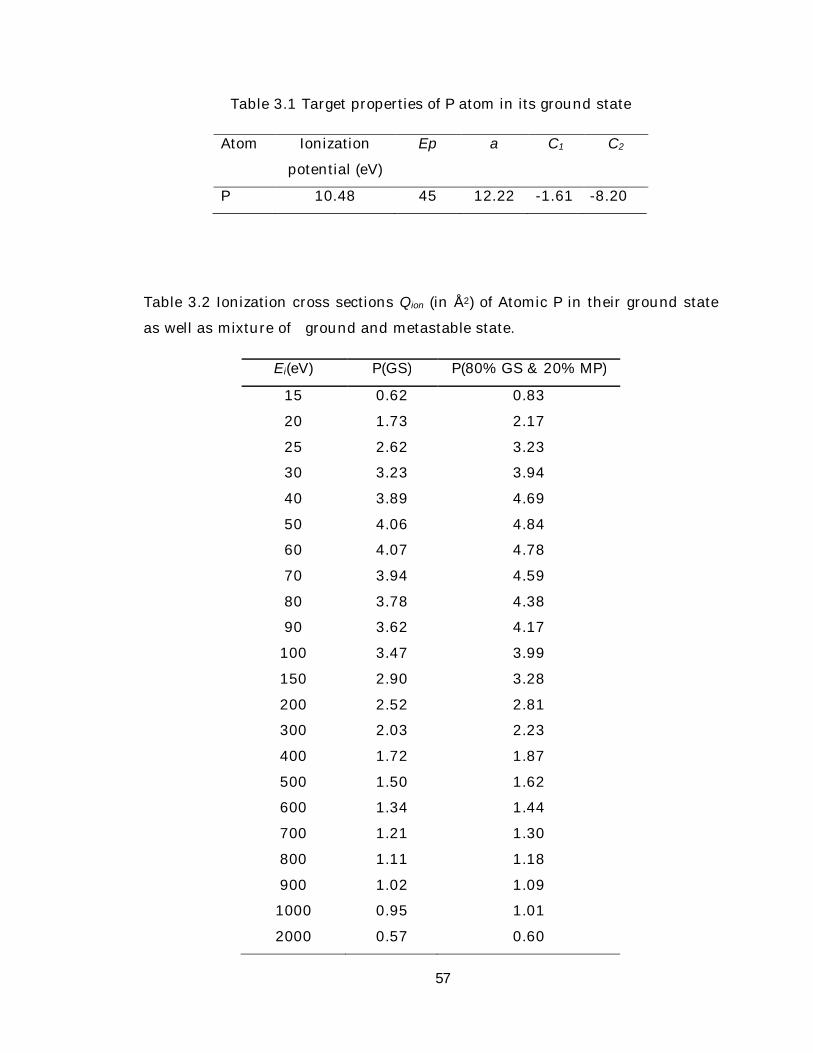
In table 3.1 we have shown target properties of atomic P along with the
parameters used for CSP- ic. In table 3.1 EP is the incident energy at which Qinel gets its
maximum and using this value of EP in CSP- ic method we derived a, C1 and C2
parameters which are also mentioned in the table.
57
Table 3.1 Target properties of P atom in its ground state
Atom Ionization
potential (eV)
Ep a C1 C2
P 10.48 45 12.22 -1.61 -8.20
Table 3.2 Ionization cross sections Qion (in Å2) of Atomic P in their ground state
as well as mixture of ground and metastable state.
Ei(eV) P(GS) P(80% GS & 20% MP)
15 0.62 0.83
20 1.73 2.17
25 2.62 3.23
30 3.23 3.94
40 3.89 4.69
50 4.06 4.84
60 4.07 4.78
70 3.94 4.59
80 3.78 4.38
90 3.62 4.17
100 3.47 3.99
150 2.90 3.28
200 2.52 2.81
300 2.03 2.23
400 1.72 1.87
500 1.50 1.62
600 1.34 1.44
700 1.21 1.30
800 1.11 1.18
900 1.02 1.09
1000 0.95 1.01
2000 0.57 0.60
58
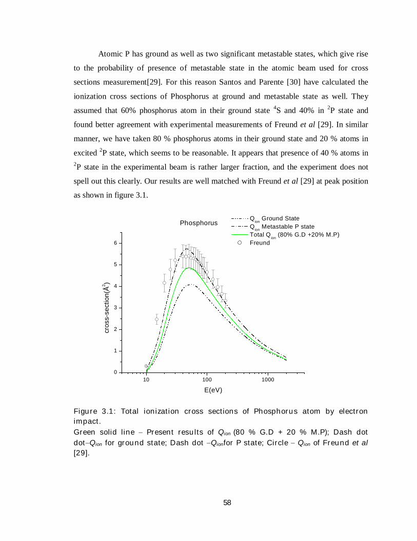
Atomic P has ground as well as two significant metastable states, which give rise
to the probability of presence of metastable state in the atomic beam used for cross
sections measurement[29]. For this reason Santos and Parente [30] have calculated the
ionization cross sections of Phosphorus at ground and metastable state as well. They
assumed that 60% phosphorus atom in their ground state 4S and 40% in 2P state and
found better agreement with experimental measurements of Freund et al [29]. In similar
manner, we have taken 80 % phosphorus atoms in their ground state and 20 % atoms in
excited 2P state, which seems to be reasonable. It appears that presence of 40 % atoms in 2P state in the experimental beam is rather larger fraction, and the experiment does not
spell out this clearly. Our results are well matched with Freund et al [29] at peak position
as shown in figure 3.1.
Figure 3.1: Total ionization cross sections of Phosphorus atom by electron impact. Green solid line Present results of Qion (80 % G.D + 20 % M.P); Dash dot dotQion for ground state; Dash dot Qionfor P state; Circle Qion of Freund et al [29].
10 100 10000
1
2
3
4
5
6
Phosphorus
cros
s-se
ctio
n(Å2 )
E(eV)
Qion
Ground State Qion Metastable P state Total Q
ion (80% G.D +20% M.P)
Freund

59
Here for atomic P we have also applied the same analogy. We have calculated the
cross sections of ground state phosphorus. We have also computed the cross sections for 2P metastable (MP) states as shown in figure 3.1. To reach the experimental scenario, we
have taken a mixture of 80 % G.D and 20 % M.P. As seen in the figure at lower and at
higher energies the experimental measurements are higher than the present results. Apart
from this our result basically highlights the cross sections for the ground state Phosphorus
atoms. In table 3.2 we have tabulated ionization cross sections of ground state as well as
mixture of ground and metastable state of P atom. In general experimental results on
atomic beams are more or less influenced by metastable contaminations [29].
3.3 Simple compounds of phosphorus
Let us now examine electron scattering with small molecules constituting
Phosphorus. We choose the molecules of astrophysical significance such as PO and P2.
The most stable form of Phosphorus element that is tetrahedral P4 molecule commonly
known as whitephosphorus is also included in the list.
In the previous chapter we have discussed the basic theoretical formalism to
calculate scattering cross sections. The method basically starts with simultaneous elastic
and inelastic electron scattering represented in the complex potential V(r, Ei) = VR(r, Ei) +
iVI(r, Ei), where r is the radial distance from the mass-centre of the target, VR(r, Ei) is the
real part and VI (r, Ei) is the imaginary part of the total potential. The real part consists of
static, exchange and polarization terms and the imaginary term is the absorption potential
Vabs as described in [31, 32]. The basic input required in constructing all these model
potentials is the atomic or molecular charge density. Since the targets are small
molecules, we have applied single centre expansion in order to obtain the charge density
of the molecules. Then we have computed the potentials to be used in Schrodinger
equation and solve it to obtain the cross sections QT,Qel andQinel.
Next, from our method CSP-ic the ionization cross sections and total excitation
cross sections are computed from Qinel. In the forthcoming sub-sections ionization cross
sections of the molecules PO, P2 and P4 are reported along with the comparison of the
60
results. Physical properties of the targets and parameters obtained from CSP-ic are
tabulated in Table 3.3.
Table 3.3 Molecular properties and parameters used for simple phosphorus
bearing molecules PO, P2 and P4
Target Ionization
potential (eV)
Bond
Length (Å)
Polarizability
(Å3)
Ep a C1
C2
PO 8.39 1.476 2.304 80 6.919 -0.955 -8.289
P2 10.53 1.893 5.155 100 6.192 -0.998 -7.204
P4 9.9 2.22 11.33 65 10.064 -1.635 -6.766
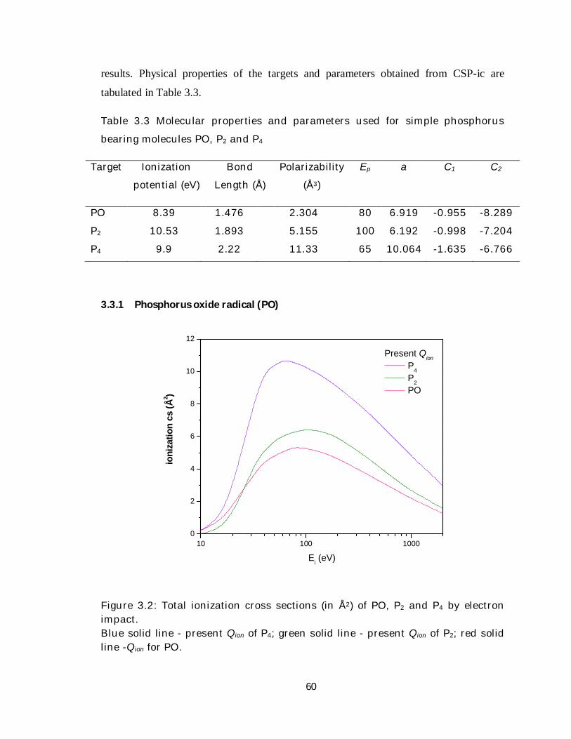
3.3.1 Phosphorus oxide radical (PO)
10 100 10000
2
4
6
8
10
12
ioni
zatio
n cs
(Å2 )
Ei (eV)
Present Qion P4 P2 PO
Figure 3.2: Total ionization cross sections (in Å2) of PO, P2 and P4 by electron impact. Blue solid line - present Qion of P4; green solid line - present Qion of P2; red solid line -Qion for PO.
61
Much work is done on spectroscopic data of the PO molecule but electron impact
processes are almost untouched in this case. To the best of our knowledge, there is no
data available for the target in the literature up to date. The cross sections of electron
impact on PO may supplement essential input formation of phosphonic acids in
meteorites. In figure 3.2 we have plotted our present ionization cross section for PO. We
have also compared the cross sections of PO with those of P2 and P4. It is clear from the
figure that cross sections Qionof PO are smallest while that of P4 are thelargest amongst
the three targets. This is because out of the three molecules PO is smallest and its
polarizability is also the smallest (table 3.3). We can also infer that the magnitude of
ionization cross sections depends on size and polarizability of molecules. The number of
electrons and ionization threshold of PO is smaller than P2 and P4 molecules. These facts
are reflected and explained by the peak positions and magnitudes of all the three targets
studied. Present ionization cross sections of PO, P2 and P4 are listed in Table 3.4.
3.3.2 P2 and P4
Not much literature is available on the ionization cross sections forP2 and P4
molecules. Molecular properties of these targets are listed in table 3.3. Monnon et al [33]
has measured direct ionization and dissociative ionization cross section of P2 and P4
molecules and these are the only measured data available till date. Bettaga et al [34]
have computed elastic cross section for electron scattering at low energy implementing
Schwinger Multichannel Method with Pseudopotentials. Scott [35] has calculated peak of
ionization cross sections of these molecules applying the binary-encounter-Bethe (BEB)
method and used effective core potential. Here we discuss our electron impact ionization
cross sections of these exotic species. The cross sections Qion of these molecular targets
are exhibited in table 3.4.
In figure 3.2 we have shown our present results on cross section calculation of P2
and P4. In the figure 3.3 we have also plotted our present ionization cross section of P2
and P4 as against our Qion of atomic phosphorus. The sequence shown in the figure 3.3
appears satisfactory and has broad peak. Adding the cross section of atomic P twice, we
obtain the cross section of P2 and is called Independent Atom Model (IAM). In similar
manner using IAM ionization cross section of P4 is obtained and plotted, as shown in the
62
figure. There is some discrepancy of in the data of ionization cross section for P4
published in earlier works. Peak of ionization cross section for P4 measured and reported
by Monnom[33] is 21 Å2, and calculated peak of ionization potential using BEB for
effective core potential is 15.1 as stated in Scott [35]. On the other hand, from the figure
we can see that the ionization peak for P4 using IAM is 15.65. So, this aspect needs
further detailed study, in order to verify and explain such variations.
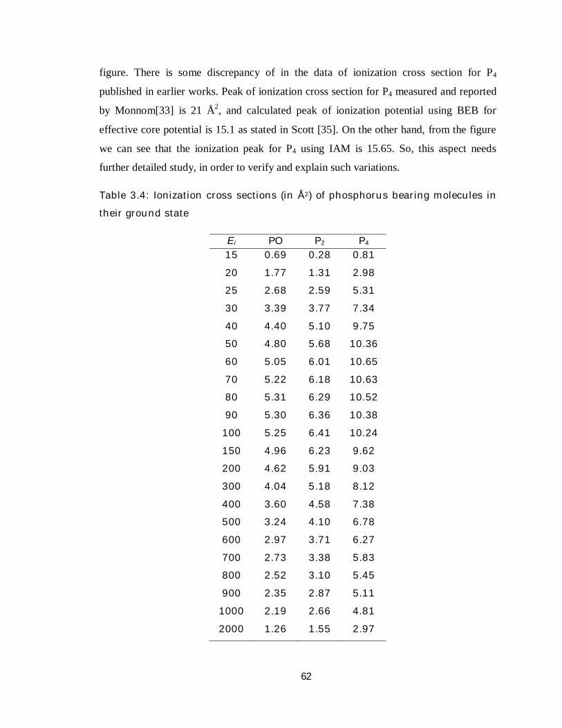
Table 3.4: Ionization cross sections (in Å2) of phosphorus bearing molecules in
their ground state
Ei PO P2 P4 15 0.69 0.28 0.81
20 1.77 1.31 2.98
25 2.68 2.59 5.31
30 3.39 3.77 7.34
40 4.40 5.10 9.75
50 4.80 5.68 10.36
60 5.05 6.01 10.65
70 5.22 6.18 10.63
80 5.31 6.29 10.52
90 5.30 6.36 10.38
100 5.25 6.41 10.24
150 4.96 6.23 9.62
200 4.62 5.91 9.03
300 4.04 5.18 8.12
400 3.60 4.58 7.38
500 3.24 4.10 6.78
600 2.97 3.71 6.27
700 2.73 3.38 5.83
800 2.52 3.10 5.45
900 2.35 2.87 5.11
1000 2.19 2.66 4.81
2000 1.26 1.55 2.97

63
10 100 10000
2
4
6
8
10
12
14
16
ioni
zatio
n cs
(Å2 )
Ei (eV)
QionP
4 present IAM (P4)
P2 present IAM (P2)
Atomic P
Figure 3.3: Present Qion for Atomic P, molecular P2 and P4, also ground state P-atom. Black dot- Atomic P; Orange line - P2Qion using single centre expansion; Pink line -Qion for P4 using single centre approach; Green dash - P2Qion using Independent Atom Model (IAM) ; Blue dash - P4Qion using IAM.
3.4 Hydrides of Phosphorus PHx(x=1–3)
For hydrides of Phosphorus, we intend to compute various total cross sections, viz.,
QT= Qel+ Qinel (3.4.1)
With Qinel = ΣQexc + Qion (3.4.2)
As we know Qinel(Ei) ≥ Qion(Ei) (3.4.3)
This is the basis for the CSP-ic method, already discussed in Chapter 2. Our
calculation of all these TCSs is based on a complex scattering potential, generated from
64
spherically averaged charge densities of the target molecules obtained by the single
centre. The molecular charge density ( )r . Here in table 3.5 and 3.6 we have shown
target properties andCSP-ic parameters (a, C1, C2) respectively for PH3 molecule.
As can be seen from table 3.5 these molecules are weakly dipolar and possess
very small dipole moment. A molecule with dipole moment D will exhibit long range
dipole potential at large r , and the form is given by [36]
2
ˆr
rDrV rD
(3.4.4)
The cross section for rotational excitation (J → J’) in a linear rigid rotator can be
calculated analytically [36] by applying the Born approximation to VD, and is given by
2
2
8( ) ( , ) ln3 2 1rot i JJ i
D J k kQ E Q D Ek J k k
(3.4.5)
Where, D is the dipole moment;k and k’ are the initial and final wave-vector
magnitudes of the scattering electron. Thus, Qrot calculated this way are added to the
QTto acquire the grand total cross sections QTOT of polar molecules. Thus we have,
( ) ( ) ( )TOT i T i rot iQ E Q E Q E (3.4.6)
Table 3.5 Molecular properties of phosphorus hydrides
Targets Ionization
potential (eV)
Bond
Length (Å)
Polarizability
(Å3)
Dipole
moment
(Debye)
PH 10.26 1.422 2.41 0.769
PH2 9.82 1.428 2.329 0.863
PH3 10.11 1.421 3.043 0.580

65
Table 3.6 Table of parameters for hydrides of phosphorus
Targets Ep a C1 C2
PH 55 8.707 -1.603 -6.056
PH2 70 8.796 -1.602 -6.113
PH3 55 5.873 -1.037 -6.629
3.4.1 PH3
Hardly any experimental work on phosphine (PH3) and other targets is carried out
due to its poisonous nature. Grand total cross sections of electron impact on PH3 are
reported by Aryasinghe et al [37] for electron energy range of 90 – 3500 eV. Absolute
cross section was measured by Szmytkowski et al [38] in energy range of 0.5 – 370 eV.
Mark et al [39] measured ionization cross sections PH3 upto 183 eV.
Many theoretical works on PH3 has been reported till date. Integral and
differential elastic CS for the hydrides of the V group (PH3, AsH3, SbH3) of the periodic
table is calculated by Bettega et al.[40, 41]. The differential, integral elastic and
momentum transfer cross sections for PH3 at 1–40 eV were calculated by Winstead et al.
[42] using Schwinger Multichannel (SMC) method. Bettega and Lima also estimated
integral elastic cross sections for low energy range 0.5 – 8 eV through SMC method. Jain
[43] used complex optical potential method to evaluate elastic and inelastic cross sections
for intermediate electron energy range. Varella et al [44] have reported cross sections for
rotational excitation by implementing SMC with pseudopotentails. Munjal and Baluja
[45] calculated the cross sections within 0.025–15 eV using R-matrix method. Recently,
Limbachiya et al [46] reported only the total elastic cross sections over a wide energy
range.
66
Table 3.7: Elastic and grand total cross sections (in Å2) of various phosphorus
hydrides PHx (x = 1, 3)
Ei PH3 PH2 PH
Qel QTOT Qel QTOT Qel QTOT
15 31.19 32.49 29.91 31.30 28.62 29.56
20 25.49 29.04 23.39 26.89 22.12 24.68
25 20.77 26.33 18.78 24.04 17.63 21.59
30 17.09 24.08 15.46 21.89 14.51 19.44
40 12.27 20.49 11.01 18.50 10.29 16.29
50 9.08 17.59 8.07 15.86 7.43 13.91
60 7.58 15.78 6.71 14.22 6.09 12.44
70 6.82 14.60 5.98 13.09 5.38 11.43
80 6.27 13.62 5.49 12.21 4.92 10.65
90 5.85 12.80 5.12 11.48 4.58 10.01
100 5.52 12.11 4.82 10.85 4.31 9.47
150 4.42 9.66 3.86 8.66 3.45 7.57
200 3.75 8.13 3.28 7.28 2.94 6.38
300 2.94 6.27 2.58 5.65 2.32 4.98
400 2.44 5.16 2.15 4.66 1.94 4.12
500 2.10 4.40 1.86 3.99 1.68 3.53
600 1.85 3.87 1.64 3.51 1.49 3.12
700 1.67 3.46 1.48 3.15 1.34 2.80
800 1.53 3.14 1.36 2.87 1.21 2.52
900 1.41 2.89 1.26 2.64 1.12 2.32
1000 1.32 2.69 1.18 2.46 1.07 2.20
2000 0.83 1.59 0.76 1.48 0.70 1.35
67
10 100 10000
10
20
30
40
50
60QTOT
present Aryasinghe Szmytkowski
Qel
present Jain Bettaga
Qion present Mark
TCS(
Å2 )
Ei (eV)
Figure 3.4: Present QTOT, Qel and Qion for e – PH3 scattering. QTOT → solid line present QTOT, circle – Aryasinghe et al [37], star – Szmytkowski et al [38]. Qel → Dash – present Qel, dash dot –Jain et al[45], dotted line- Bettaga [40], Qion → dash dot dot - present Qion; left triangle – Mark et al [39].
The numerical values of elastic and grand total cross sections of PH3 in the energy
range of 10 – 2000 eV are displayed in Table 3.7. Consider now figure 3.4 corresponding
to e – PH3 scattering. The grand total cross sections are below the measured values of
cross sections at lower energies, but at higher energies above 200 eV it is in good accord
with the reported measurements of Aryasinghe et al [37]. The ionization cross sections
are slightly greater than the only experimental measurement of Mark et al [39]. In this
figure we have also shown Qel which is again lesser than the other theoretical data given
by Jain et al [45].
68
From the figure 3.4 we observe a reasonable agreement between our present
calculations and the only experimental ionization cross section reported by Mark et
al[39]. At higher energies elastic cross sections and ionization cross sections have almost
an equal value. The values Qionfor PH3 are tabulated in table 3.8.
Table 3.8: Present Qion (in Å2) for various phosphorus hydrides PHx (x = 1, 3)
Ei PH3 PH2 PH
15 0.44 0.49 0.31
20 1.72 1.71 1.23
25 3.09 2.94 2.20
30 4.21 3.89 2.97
40 5.44 4.95 3.97
50 5.95 5.45 4.54
60 5.98 5.48 4.63
70 5.86 5.36 4.55
80 5.68 5.19 4.43
90 5.49 5.02 4.29
100 5.30 4.85 4.15
150 4.49 4.11 3.53
200 3.89 3.55 3.06
300 3.08 2.84 2.46
400 2.57 2.37 2.06
500 2.20 2.04 1.78
600 1.95 1.81 1.58
700 1.74 1.62 1.42
800 1.58 1.48 1.28
900 1.45 1.35 1.18
1000 1.35 1.26 1.11
2000 0.75 0.72 0.64
69
10 100 10000
1
2
3
4
5
6
ioni
zatio
n cs
(Å2 )
Ei (eV)
Qion PH3 PH2 PH
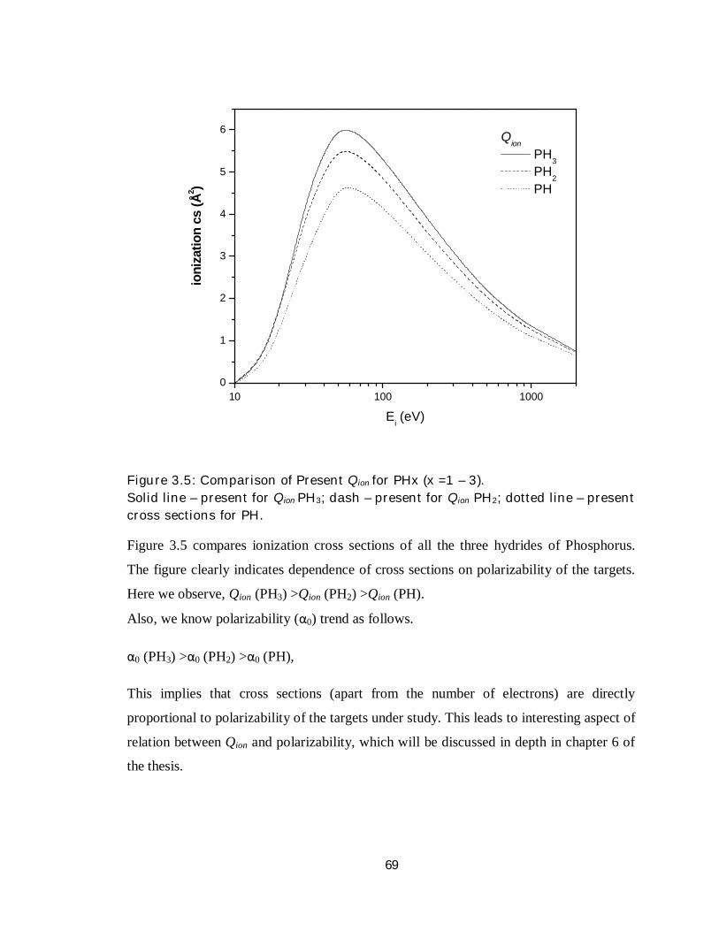
Figure 3.5: Comparison of Present Qion for PHx (x =1 – 3). Solid line – present for Qion PH3; dash – present for Qion PH2; dotted line – present cross sections for PH.
Figure 3.5 compares ionization cross sections of all the three hydrides of Phosphorus.
The figure clearly indicates dependence of cross sections on polarizability of the targets.
Here we observe, Qion (PH3) >Qion (PH2) >Qion (PH).
Also, we know polarizability (α0) trend as follows.
α0 (PH3) >α0 (PH2) >α0 (PH),
This implies that cross sections (apart from the number of electrons) are directly
proportional to polarizability of the targets under study. This leads to interesting aspect of
relation between Qion and polarizability, which will be discussed in depth in chapter 6 of
the thesis.
70
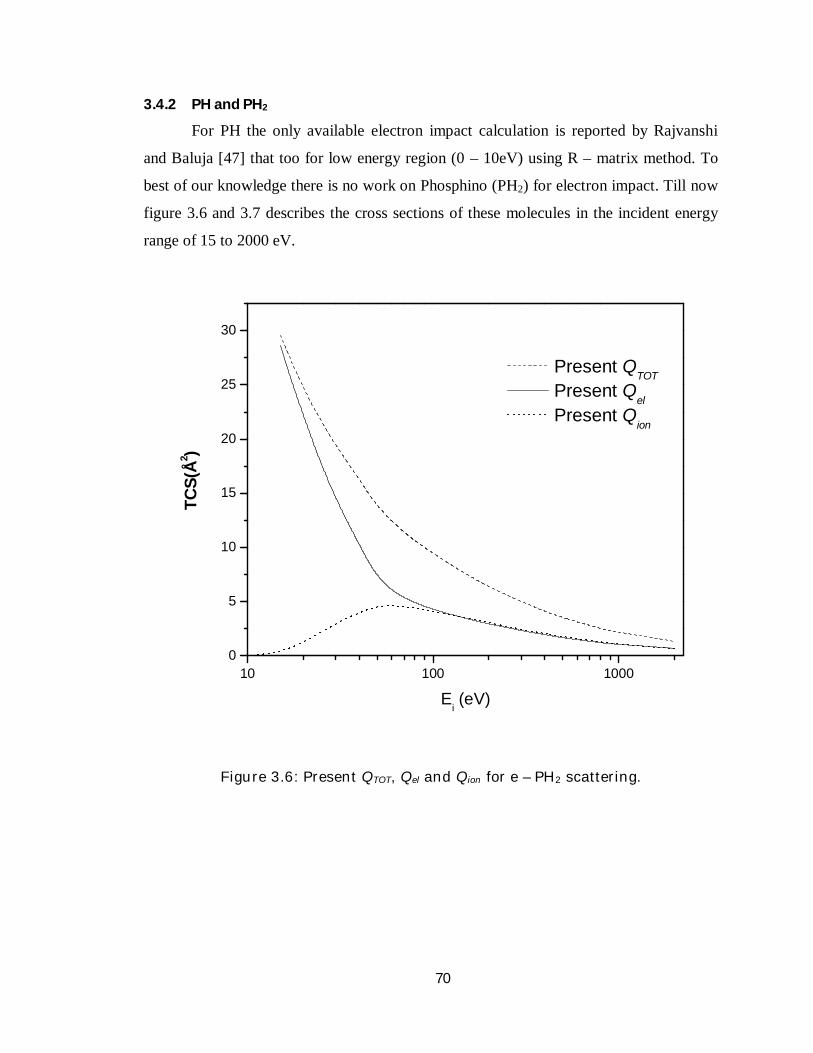
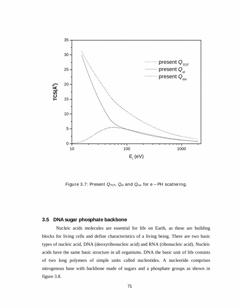
3.4.2 PH and PH2
For PH the only available electron impact calculation is reported by Rajvanshi
and Baluja [47] that too for low energy region (0 – 10eV) using R – matrix method. To
best of our knowledge there is no work on Phosphino (PH2) for electron impact. Till now
figure 3.6 and 3.7 describes the cross sections of these molecules in the incident energy
range of 15 to 2000 eV.
10 100 10000
5
10
15
20
25
30
TCS(
Å2 )
Ei (eV)
Present QTOT Present Q
el Present Q
ion
Figure 3.6: Present QTOT, Qel and Qion for e – PH2 scattering.
71
10 100 10000
5
10
15
20
25
30
35
TCS(
Å2 )
Ei (eV)
present QTOT
present Qel
present Qion
Figure 3.7: Present QTOT, Qel and Qion for e – PH scattering.
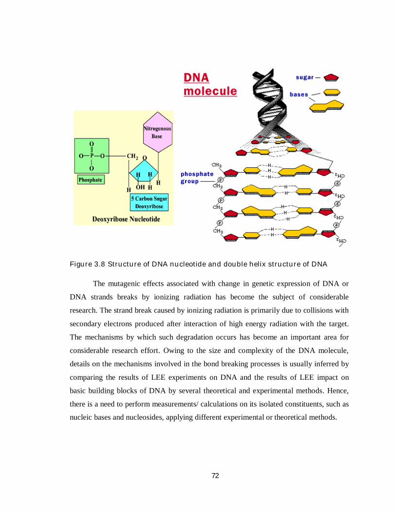
3.5 DNA sugar phosphate backbone
Nucleic acids molecules are essential for life on Earth, as these are building
blocks for living cells and define characteristics of a living being. There are two basic
types of nucleic acid, DNA (deoxyribonucleic acid) and RNA (ribonucleic acid). Nucleic
acids have the same basic structure in all organisms. DNA the basic unit of life consists
of two long polymers of simple units called nucleotides. A nucleotide comprises
nitrogenous base with backbone made of sugars and a phosphate groups as shown in
figure 3.8.
72
Figure 3.8 Structure of DNA nucleotide and double helix structure of DNA
The mutagenic effects associated with change in genetic expression of DNA or
DNA strands breaks by ionizing radiation has become the subject of considerable
research. The strand break caused by ionizing radiation is primarily due to collisions with
secondary electrons produced after interaction of high energy radiation with the target.
The mechanisms by which such degradation occurs has become an important area for
considerable research effort. Owing to the size and complexity of the DNA molecule,
details on the mechanisms involved in the bond breaking processes is usually inferred by
comparing the results of LEE experiments on DNA and the results of LEE impact on
basic building blocks of DNA by several theoretical and experimental methods. Hence,
there is a need to perform measurements/ calculations on its isolated constituents, such as
nucleic bases and nucleosides, applying different experimental or theoretical methods.
73
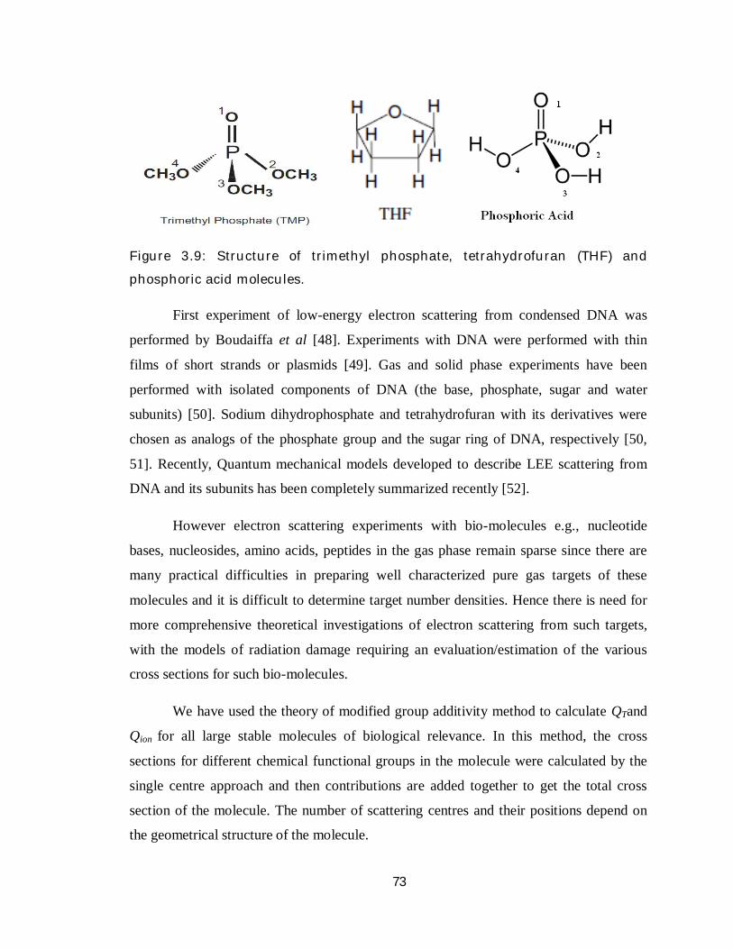
Figure 3.9: Structure of trimethyl phosphate, tetrahydrofuran (THF) and
phosphoric acid molecules.
First experiment of low-energy electron scattering from condensed DNA was
performed by Boudaiffa et al [48]. Experiments with DNA were performed with thin
films of short strands or plasmids [49]. Gas and solid phase experiments have been
performed with isolated components of DNA (the base, phosphate, sugar and water
subunits) [50]. Sodium dihydrophosphate and tetrahydrofuran with its derivatives were
chosen as analogs of the phosphate group and the sugar ring of DNA, respectively [50,
51]. Recently, Quantum mechanical models developed to describe LEE scattering from
DNA and its subunits has been completely summarized recently [52].
However electron scattering experiments with bio-molecules e.g., nucleotide
bases, nucleosides, amino acids, peptides in the gas phase remain sparse since there are
many practical difficulties in preparing well characterized pure gas targets of these
molecules and it is difficult to determine target number densities. Hence there is need for
more comprehensive theoretical investigations of electron scattering from such targets,
with the models of radiation damage requiring an evaluation/estimation of the various
cross sections for such bio-molecules.
We have used the theory of modified group additivity method to calculate QTand
Qion for all large stable molecules of biological relevance. In this method, the cross
sections for different chemical functional groups in the molecule were calculated by the
single centre approach and then contributions are added together to get the total cross
section of the molecule. The number of scattering centres and their positions depend on
the geometrical structure of the molecule.

74
Table 3.9: Molecular properties for THF, H3PO4 and TMP
Target Ionization
potential
(eV)
Bond Length (Å) Polarizability
(Å3)
Dipole
moment
(Debye)
Geometric
correction
factor Z
THF 9.55
CH 1.11
7.22 1.63 0.771 CO 1.42
CC 1.53
H3PO4 11.72
PO1 1.46
5.67 3.461 0.831 PO2,3,4 1.58
OH 0.96
TMP 9.99
PO1 1.47
10.86 3.3 0.683 PO2,3,4 1.61
OC 1.44
CH 1.08
For example the phosphoric acid (H3PO4) molecule will have four scattering
centres one at P and three at O atoms (refer figure 3.9). It has got three OH groups and
one PO group, and hence four scattering centres. The charge density for the OH group is
obtained by expanding H on O and is re-normalized to get the total number of electrons
in that group. Similarly the charge density for the other centre at P is also obtained.
Electron impact cross sections for each of these groups are obtained using the ionization
potential of the molecule. Then the cross section contributions from each of the centre are
added together to get the total cross section for the molecule. Simple addition of the cross
sections computed for the constituent groups overestimates the cross section values of the
molecules. This is because the bonding effects amongst the functional groups are not
taken into considerations that lead to overestimation. Here we are implementing Modified
Group Additivity Rule with Z correction, in order to reduce the overestimation. So we are
multiplying the added cross sections with Z factor and incorporate the geometric
75
correction as explained in chapter 2. The molecular properties of all the three targets are
listed in table 3.9. Also, in table 3.10 the parameters obtained from CSP-ic are displayed.
3.5.1 Tetra Hydrofuran (THF)- C4H8O
THF represents an analogue to the furan structure of the sugar in DNA backbone.
Due to this reason, a number of papers have been published on this target till date. Zecca
et al [53] and Mozejko et al [54] measured the absolute total cross section for electron
scattering by gas-phase THF. The elastic differential cross section (DCS) at 20 eV and
above was measured by Milosavljevi´c et al [55]. Absolute elastic cross-sections in the
energy range of 6.5–50 eV and angular range between 10◦ and 130◦ were measured by
Colyer et al [56], in the ranges 6–20 eV and 20◦–180◦ by Dampc et al [57]. Allan [58]
reported measurements of differential elastic, total elastic and vibrational excitation cross
sections over energy range from 0.1 eV to 20 eV. Homem et al [59] reported absolute
differential elastic cross section for THF at intermediate energy range (50 – 1000) eV.
Vibrational excitation cross sections have been measured very recently by Dampc et al
[60]. Several high-level scattering calculations on THF have been reported. Trevisan et al
[61] reported an ab initio calculation of the elastic differential and momentum-transfer
cross sections using the complex Kohn variational method. Winstead and McKoy [62]
calculated the elastic differential and momentum-transfer cross sections using the
Schwinger multichannel method. Bouchiha et al [63] used the R-matrix method with
Born correction to calculate the angle-integrated elastic cross section, inelastic cross
sections and the energies for a number of resonances. Tonzani and Greene [64] calculated
the integral elastic cross section and gained further insight into the resonant structure by
considering time delay. Pawel [65] reported elastic and ionization cross sections for THF
and H3PO4 within intermediate energy range. The only experimental measurement for
ionization cross section is recently reported by Dampc et al [66].
76
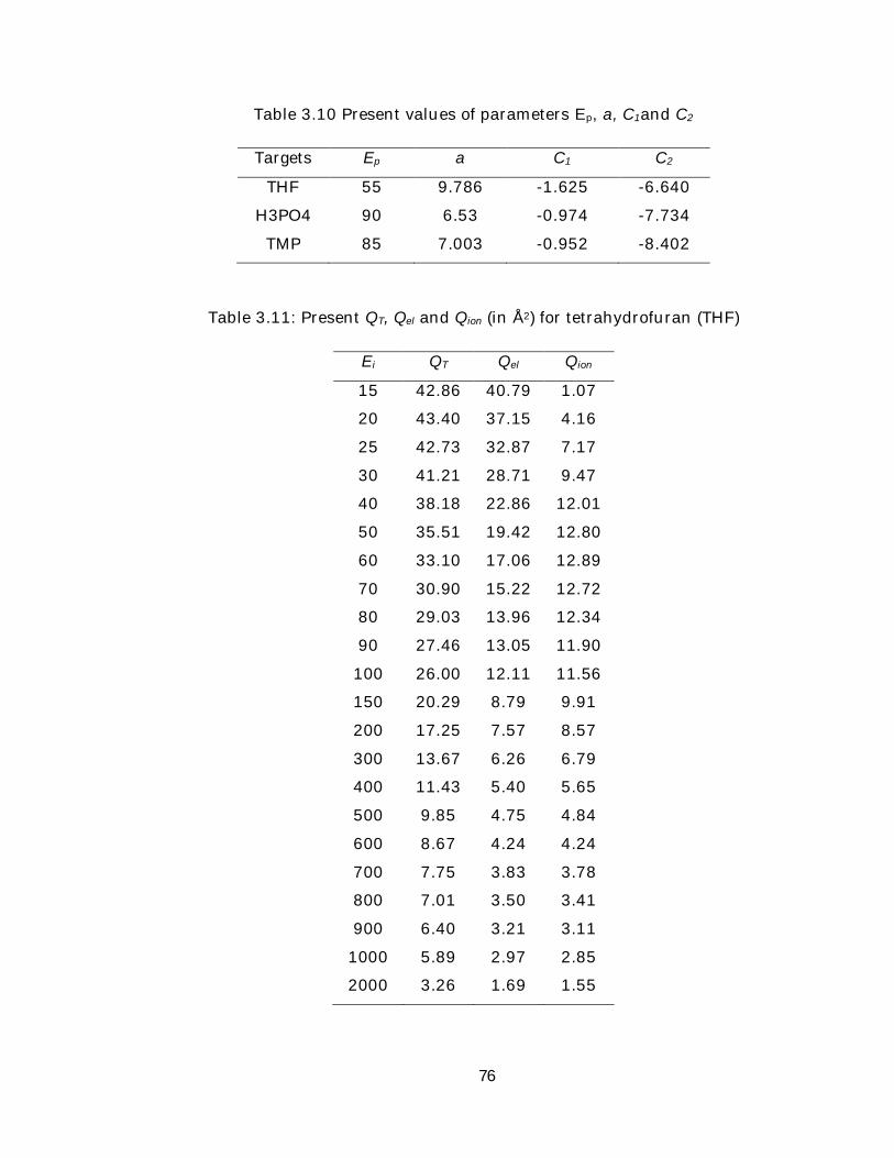
Table 3.10 Present values of parameters Ep, a, C1and C2
Targets Ep a C1 C2
THF 55 9.786 -1.625 -6.640
H3PO4 90 6.53 -0.974 -7.734
TMP 85 7.003 -0.952 -8.402
Table 3.11: Present QT, Qel and Qion (in Å2) for tetrahydrofuran (THF)
Ei QT Qel Qion
15 42.86 40.79 1.07
20 43.40 37.15 4.16
25 42.73 32.87 7.17
30 41.21 28.71 9.47
40 38.18 22.86 12.01
50 35.51 19.42 12.80
60 33.10 17.06 12.89
70 30.90 15.22 12.72
80 29.03 13.96 12.34
90 27.46 13.05 11.90
100 26.00 12.11 11.56
150 20.29 8.79 9.91
200 17.25 7.57 8.57
300 13.67 6.26 6.79
400 11.43 5.40 5.65
500 9.85 4.75 4.84
600 8.67 4.24 4.24
700 7.75 3.83 3.78
800 7.01 3.50 3.41
900 6.40 3.21 3.11
1000 5.89 2.97 2.85
2000 3.26 1.69 1.55
77
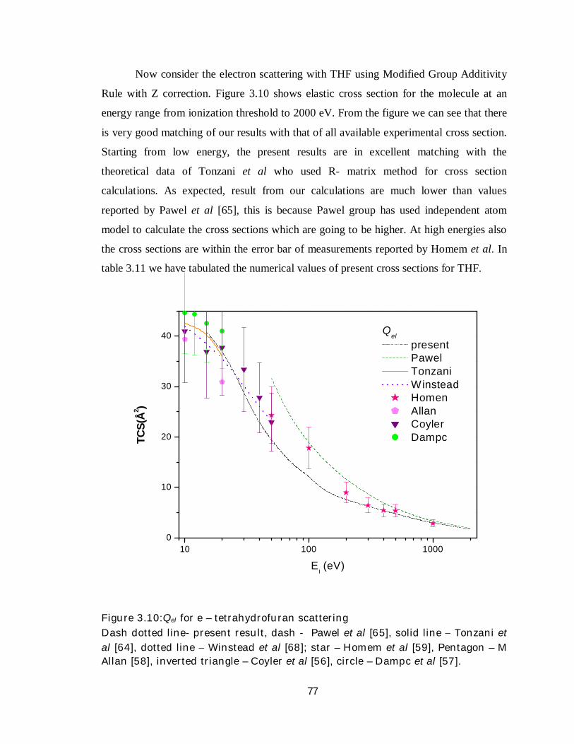
Now consider the electron scattering with THF using Modified Group Additivity
Rule with Z correction. Figure 3.10 shows elastic cross section for the molecule at an
energy range from ionization threshold to 2000 eV. From the figure we can see that there
is very good matching of our results with that of all available experimental cross section.
Starting from low energy, the present results are in excellent matching with the
theoretical data of Tonzani et al who used R- matrix method for cross section
calculations. As expected, result from our calculations are much lower than values
reported by Pawel et al [65], this is because Pawel group has used independent atom
model to calculate the cross sections which are going to be higher. At high energies also
the cross sections are within the error bar of measurements reported by Homem et al. In
table 3.11 we have tabulated the numerical values of present cross sections for THF.
10 100 10000
10
20
30
40
TCS(
Å2 )
Ei (eV)
Qel
present Pawel Tonzani Winstead Homen Allan Coyler Dampc
Figure 3.10:Qel for e – tetrahydrofuran scattering Dash dotted line- present result, dash - Pawel et al [65], solid line Tonzani et al [64], dotted line Winstead et al [68]; star – Homem et al [59], Pentagon – M Allan [58], inverted triangle – Coyler et al [56], circle – Dampc et al [57].
78
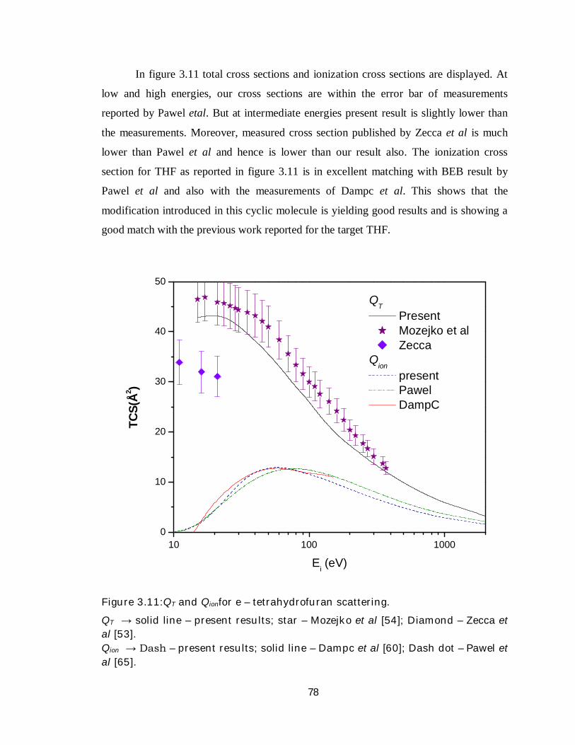
In figure 3.11 total cross sections and ionization cross sections are displayed. At
low and high energies, our cross sections are within the error bar of measurements
reported by Pawel etal. But at intermediate energies present result is slightly lower than
the measurements. Moreover, measured cross section published by Zecca et al is much
lower than Pawel et al and hence is lower than our result also. The ionization cross
section for THF as reported in figure 3.11 is in excellent matching with BEB result by
Pawel et al and also with the measurements of Dampc et al. This shows that the
modification introduced in this cyclic molecule is yielding good results and is showing a
good match with the previous work reported for the target THF.
10 100 10000
10
20
30
40
50
TCS(
Å2 )
Ei (eV)
QT
Present Mozejko et al Zecca
Qion
present Pawel DampC
Figure 3.11:QT and Qionfor e – tetrahydrofuran scattering.
QT → solid line – present results; star – Mozejko et al [54]; Diamond – Zecca et al [53]. Qion → Dash – present results; solid line – Dampc et al [60]; Dash dot – Pawel et al [65].
79
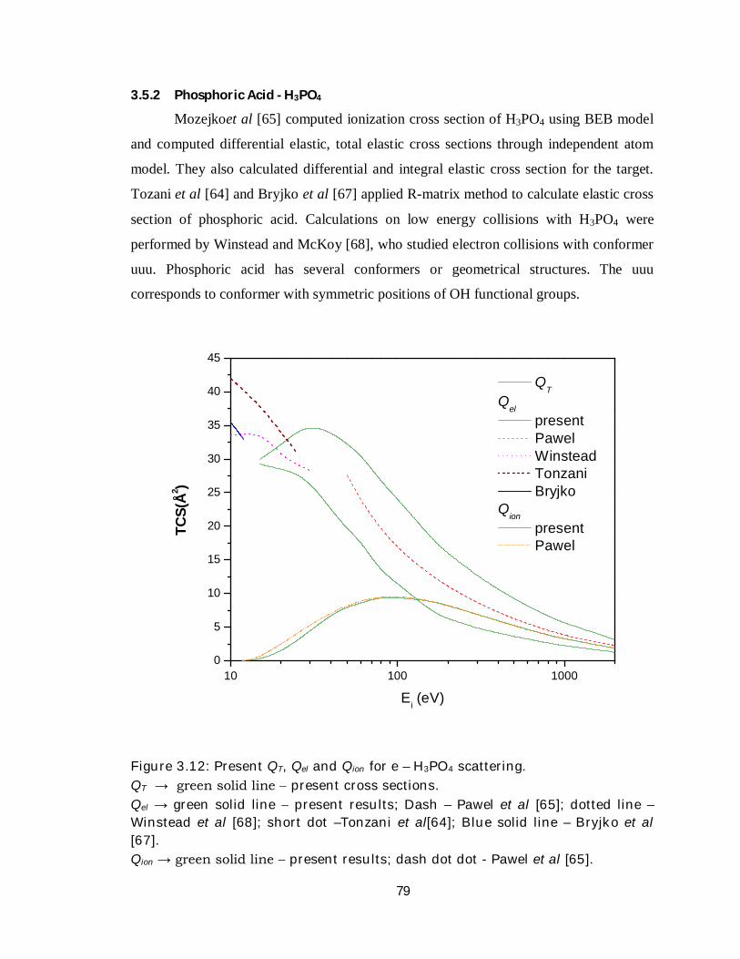
3.5.2 Phosphoric Acid - H3PO4
Mozejkoet al [65] computed ionization cross section of H3PO4 using BEB model
and computed differential elastic, total elastic cross sections through independent atom
model. They also calculated differential and integral elastic cross section for the target.
Tozani et al [64] and Bryjko et al [67] applied R-matrix method to calculate elastic cross
section of phosphoric acid. Calculations on low energy collisions with H3PO4 were
performed by Winstead and McKoy [68], who studied electron collisions with conformer
uuu. Phosphoric acid has several conformers or geometrical structures. The uuu
corresponds to conformer with symmetric positions of OH functional groups.
10 100 10000
5
10
15
20
25
30
35
40
45
TCS(
Å2 )
Ei (eV)
QT
Qel
present Pawel Winstead Tonzani Bryjko
Qion present Pawel
Figure 3.12: Present QT, Qel and Qion for e – H3PO4 scattering. QT → green solid line present cross sections. Qel → green solid line present results; Dash – Pawel et al [65]; dotted line – Winstead et al [68]; short dot –Tonzani et al[64]; Blue solid line – Bryjko et al [67]. Qion → green solid line present results; dash dot dot - Pawel et al [65].
80
As shown in figure 3.12 our present result of ionization cross section matches
well with the theoretically calculated cross sections based on BEB by Pawel et al [65].
H3PO4 is a polar molecule having permanent dipole moment of 3.461 debye, due to this
the total cross section shows a peak at lower energies; further the magnitude of QT is
lesser than Qel calculated by other theoretical groups.
Again, at low energy region, present Qel is much less than cross sections
calculated and reported by Tonzani et al [64]. Cross sections reported by Bryjko et al
[67] are slightly greater than the present Qel. As expected, the elastic cross sections by
Pawel et al are greater than the present cross sections, because they used IAM to evaluate
the cross sections. No experimental data of the target is available for comparison.
3.5.3 Trimethyl phosphate - OP(OCH3)3
Another anologue for the phoasphate group of DNA is trimethyl phosphate as in
figure 3.9. Recently Aflatooni et al. [69] have reported DA measurements for TMP,
while Burrow et al. [70] have reported electron transmission measurements for TMP.
Winstead et al [68] computed cross sections for low-energy elastic collisions of electrons
with trimethyl phosphate and phosphoric acid.
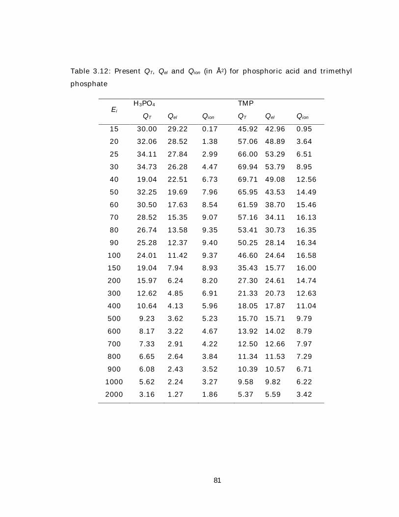
The numerical values of present cross sections for TMP and H3PO4 are given in
table number 3.12. The figure 3.13 portrays the cross sections of TMP within the energy
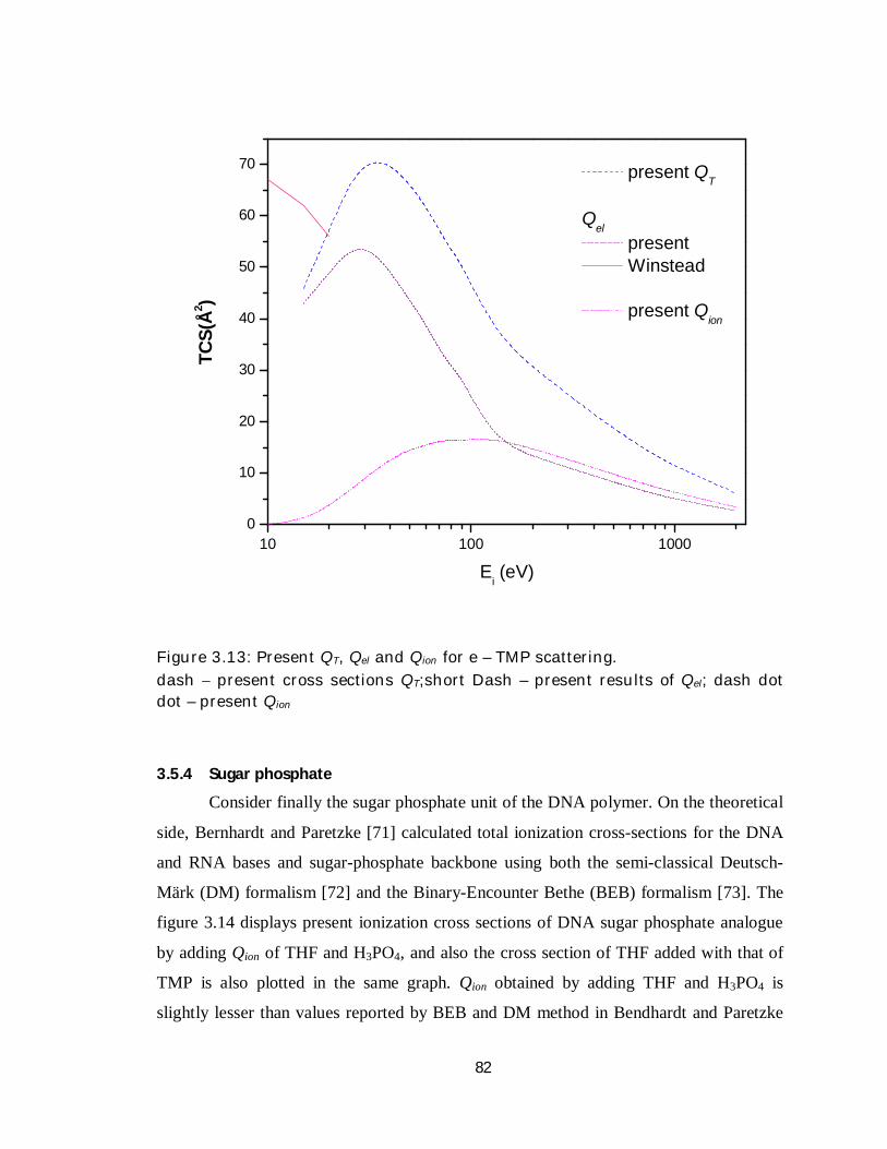
range from 10 eV to 2000 eV. Our present result for Qel at low energies below 20 eV is
much smaller than the values reported in Winstead et al [68]. This may be primarily due
to absence of rotational cross sections at lower energies and TMP being a polar molecule,
the addition of Qrot can improve cross section values at low energy regime. This is again
reflected in present total absolute cross sections of the target. There are no comparisons
of ionization cross sections for the present target.
81
Table 3.12: Present QT, Qel and Qion (in Å2) for phosphoric acid and trimethyl
phosphate
Ei H3PO4 TMP
QT Qel Qion QT Qel Qion
15 30.00 29.22 0.17 45.92 42.96 0.95
20 32.06 28.52 1.38 57.06 48.89 3.64
25 34.11 27.84 2.99 66.00 53.29 6.51
30 34.73 26.28 4.47 69.94 53.79 8.95
40 19.04 22.51 6.73 69.71 49.08 12.56
50 32.25 19.69 7.96 65.95 43.53 14.49
60 30.50 17.63 8.54 61.59 38.70 15.46
70 28.52 15.35 9.07 57.16 34.11 16.13
80 26.74 13.58 9.35 53.41 30.73 16.35
90 25.28 12.37 9.40 50.25 28.14 16.34
100 24.01 11.42 9.37 46.60 24.64 16.58
150 19.04 7.94 8.93 35.43 15.77 16.00
200 15.97 6.24 8.20 27.30 24.61 14.74
300 12.62 4.85 6.91 21.33 20.73 12.63
400 10.64 4.13 5.96 18.05 17.87 11.04
500 9.23 3.62 5.23 15.70 15.71 9.79
600 8.17 3.22 4.67 13.92 14.02 8.79
700 7.33 2.91 4.22 12.50 12.66 7.97
800 6.65 2.64 3.84 11.34 11.53 7.29
900 6.08 2.43 3.52 10.39 10.57 6.71
1000 5.62 2.24 3.27 9.58 9.82 6.22
2000 3.16 1.27 1.86 5.37 5.59 3.42
82
10 100 10000
10
20
30
40
50
60
70
TCS(
Å2 )
Ei (eV)
present QT
Qel
present Winstead
present Qion
Figure 3.13: Present QT, Qel and Qion for e – TMP scattering. dash present cross sections QT;short Dash – present results of Qel; dash dot dot – present Qion
3.5.4 Sugar phosphate
Consider finally the sugar phosphate unit of the DNA polymer. On the theoretical
side, Bernhardt and Paretzke [71] calculated total ionization cross-sections for the DNA
and RNA bases and sugar-phosphate backbone using both the semi-classical Deutsch-
Märk (DM) formalism [72] and the Binary-Encounter Bethe (BEB) formalism [73]. The
figure 3.14 displays present ionization cross sections of DNA sugar phosphate analogue
by adding Qion of THF and H3PO4, and also the cross section of THF added with that of
TMP is also plotted in the same graph. Qion obtained by adding THF and H3PO4 is
slightly lesser than values reported by BEB and DM method in Bendhardt and Paretzke
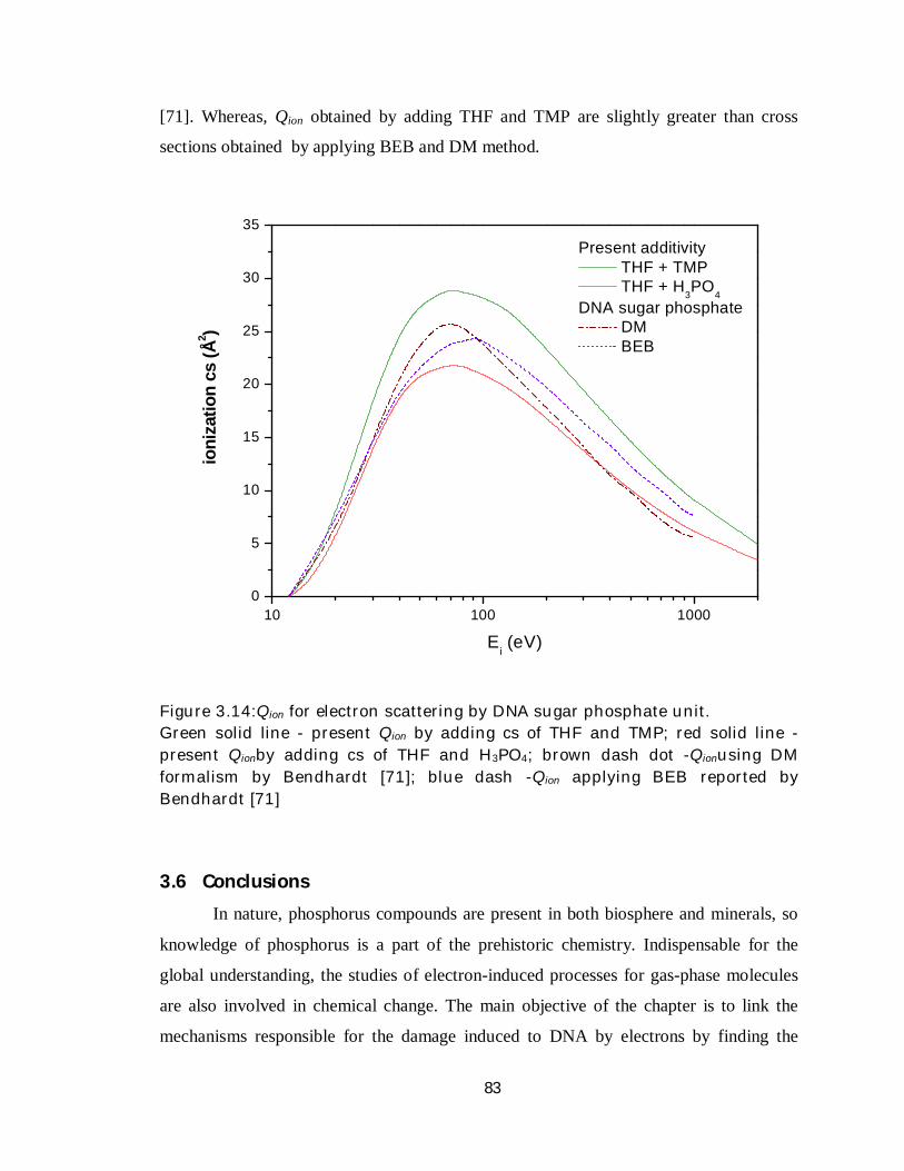
83
[71]. Whereas, Qion obtained by adding THF and TMP are slightly greater than cross
sections obtained by applying BEB and DM method.
10 100 10000
5
10
15
20
25
30
35
io
niza
tion
cs (Å
2 )
Ei (eV)
Present additivity THF + TMP THF + H
3PO
4DNA sugar phosphate
DM BEB
Figure 3.14:Qion for electron scattering by DNA sugar phosphate unit. Green solid line - present Qion by adding cs of THF and TMP; red solid line - present Qionby adding cs of THF and H3PO4; brown dash dot -Qionusing DM formalism by Bendhardt [71]; blue dash -Qion applying BEB reported by Bendhardt [71]
3.6 Conclusions
In nature, phosphorus compounds are present in both biosphere and minerals, so
knowledge of phosphorus is a part of the prehistoric chemistry. Indispensable for the
global understanding, the studies of electron-induced processes for gas-phase molecules
are also involved in chemical change. The main objective of the chapter is to link the
mechanisms responsible for the damage induced to DNA by electrons by finding the
84
contribution of these surrogates of its constituent building blocks and discuss potential
applications. To achieve this objective Modified Group Additivity Rule has been applied
successfully on large polyatomic molecules to calculate various cross sections. Initially
we started with sugar phosphate backbone, in this the idea of adding cross section of
sugar analog THF with phosphate analogues H3PO4 and TMP worked out quiet
satisfactorily. More work towards this direction can possibly produce some intuitive
proposition of DNA damage by electrons. Large magnitude of ionization cross section for
sugar phosphate unit supports the notion that phosphate backbone plays an important role
in electron induced damage of DNA. A number of analogs of DNA components are
awaited for the calculation, which can be a key to understand radiation damage
mechanism. The field however is infantile and the physics of life sciences is wide open
and challenging.
85
Bibliography
[1] E Macia, Chem. Soc. Rev., 34 (2005) 691.
[2] W Thomson, Presidential Address to the British Society for the Advancement
of Science, Edinburgh meeting, 1871.
[3] D A Allen, D T Wickramasinghe, Nature, 294 (1981) 239.
[4] A Gulick, Am. Sci.43 (1955) 479.
[5] B E Turner, T Tsuji, J Bally, M Guelin, J Cernicharo, Astrophys. J. 365 (1990)
569.
[6] D D S MacKay, S B Charnley, Mon. Not. R. Astron. Soc.,325 (2001) 545.
[7] S N Milam, D T Halfen, E D Tenenbaum, A J Apponi, N J Woolf, L M Ziurys,
Astrophys. J. 684 (2008) 618.
[8] G W Cooper, W M Onwo and J R Cronin, Geochim. Cosmochim. Acta 56
(1992) 4109.
[9] A W Schwartz, Phil. Trans. R. Soc. B.361 (2006) 1743.
[10] E D Tenenbaum, N J Woolf, L M Ziurys, Astrophys. J., 666 (2007) L29.
[11] A W Schwartz, Orig. Life Evol. Biosph. 27 (1997) 505.
[12] N A Piro, C C Cummins, J. Am. Chem. Soc. , 131 (2009) 8964
[13] E M De Gouveia, P D Singh, Sol. Phys.90 259 (1984)
[14] A J Sauval, Astron. Astrophys., Suppl. Ser.49 77 (1982)
[15] J Berkowitz , J. Chem. Phys. 89 (1988) 7065
[16] Th Encrenaz, B Bézard, J Crovisier, A Coustenis, E Lellouch, S Gulkis, S K
Atreya, Planet. Space Sci. 43 (1995) 1485
[17] R Courtin, D Gautier, A Marten, Astrophys. J. 287 (1984) 899.
[18] D Glindemann, M Edwards, P Kuschk, Atmos. Environ., 37 (2003) 2429.
[19] P Yin, Z L Wang, Z P Bai, Chem. Phys., 264 (2001) 1
[20] M Varella, M H F Bettega, M A P Lima, L G Ferreira, J. Chem. Phys. 111
(1999) 6396.
86
[21] R Greer, D Thompson, J. Phys. B 27 (1994) 3533.
[22] S Ptasinska, S Denifl, P Scheier, T D Mark, J. Chem. Phys. 120 (2004) 8505
[23] B Boudaiffa, P Cloutier, D Hunting, M A Huels and L Sanche, Radiat. Res. 157
(2002) 227
[24] M A Huels, I Hahndorf, E Illenberger, L Sanche, J. Chem. Phys. 108 (1998)
1309
[25] S Denifl, S Ptasinska, M Cingel, S Matejcik, P Scheier, T D Mark, Chem. Phys.
Lett. 377 (2003) 74
[26] H Abdoul-Carime, S Gohlke, E Illenberger, Phys. Rev. Lett. 92 (2004) 168103
[27] B Boudaiffa, P Cloutier, D Hunting, M A Huels and L Sanche, Science 287
(2000) 1658
[28] F Martin, P D Burrow, Z Cai, P Cloutier, D Hunting, L Sanche, Phys. Rev.
Lett. 93 (2004) 068101
[29] R S Freund, R C Wetzel, R J Shul, T R Hayes, Phys. Rev. A, 41 (1990) 3575.
[30] J P Santos, F Parente, Eur. J. Phys. D, 47 (2008) 339
[31] K N Joshipura, S Gangopadhyay, H N Kothari, F A Shelat, Phys. Lett. A,373
(2009) 2876.
[32] K N Joshipura, S Gangopadhyay, B G Vaishnav, J. Phys. B, 40 (2007)199.
[33] G Monnom, P Gaucherel, C Paparoditis, J. Phys. (Paris)45 (1984) 77
[34] M H F Bettega, M A P Lima, L G Ferreura, J. Phys. B, 31 (1998) 2091
[35] G E Scott, K K Irikura, J. Chem. Theory Comput.1 (2005) 1153.
[36] Y Itikawa, Phys. Rep. C46 (1978) 117
[37] W M Ariyasinghe, T Wijerathna, D Powers, Phys. Rev. A68 (2003) 032708.
[38] C Szmytkowski, L Klosowski, A Domaracka, M Piotrowicz, E Ptasinska-
Denga, J. Phys. B37 (2004) 1833.
[39] T D Märk, F Egger, J. Chem. Phys. 67(1977) 2629.
[40] M H F Bettega, M A P Lima, L G Ferreira, J. Chem. Phys., 105 (1996) 1029
[41] M H F Bettega, M A P Lima, J. Phys. B, 37 (2004) 3859.
[42] C Winstead, Q Sun, V McKoy, da Silva, J L Lino, M A P Lima, Z. Phys. D, 24
(1992) 141
[43] A Jain, K L Baluja, Phys. Rev. A45 (1992) 202.
87
[44] M Varella, M Bettega, A da Silva, M Lima, J. Chem. Phys.110(1999) 2452.
[45] M Gupta, K Baluja, Eur. Phys. J. D 41 (2007) 475.
[46] C Limbachiya, M Vinodkumar, N Mason, Phys. Rev. A83 (2011) 042708.
[47] J S Rajvanshi, K L Baluja, Phys. Rev. A. 81 (2010) 022709
[48] B Boudaiffa, P Cloutier, D Hunting, M A Huels, L Sanche, Rad. Res. 157
(2002) 227
[49] L Sanche, Radiation induced molecular phenomena in nucleic acid: A
comprehensive theoretical and experimental analysis, ed. by Manoj K. Shukla,
J. Leszczynski (Springer, Netherlands, 2008)
[50] L Sanche, Eur. Phys. J. D35 (2005) 367
[51] L Sanche, Radicals in Nucleic Acids, ed. by M. Greenberg (Wiley, 2009)
[52] L Caron, L Sanche, in Radical and radical ion reactivity in nucleic acid
chemistry, edited by P. Čársky, R. Čurik (Taylor & Francis, 2010)
[53] A Zecca, C Perazzolli, M Brunger, J. Phys. B 38 (2005) 2079
[54] P Mozejko, Ptasinska, A Domaracka, C Szmytkowski, Phys. Rev. A74 (2006)
012708
[55] A R Milosavljevic, A Giuliani, D Ševic, M J Hubin-Franskin, B P Marinkovic,
Eur. Phys. J. D 35 (2005) 411
[56] C J Colyer, V Vizcaino, J P Sullivan, M J Brunger, S J Buckman, New J. Phys.
9 (2007) 41
[57] M Dampc, A R Milosavljevic, I Linert, B P Marinkovic, M. Zubek, Phys. Rev.
A 75 (2007) 042710
[58] M Allan, J. Phys. B 40 (2008) 3531
[59] M G P Homem, R T Sugohara, I P Sanches, M T Lee, I Iga, Phys. Rev. A 80
(2009)032705
[60] M Dampc, I Linert, A R Milosavljevic, M Zubek, Chem. Phys. Lett. 443 (2007)
17
[61] C S Trevisan, A E Orel, T N Rescigno, J. Phys. B 39 (2006) L255
[62] C Winstead, V J McKoy. Chem. Phys. 125 (2006) 074302
[63] D. Bouchiha, J. D. Gorfinkiel, L. G. Caron, L. Sanche, J. Phys. B. 39 (2006)
975
88
[64] S. Tonzani, C. H. Greene, J. Chem. Phys. 125 (2006) 094504
[65] P Możejko, L Sanche, Rad. Phys. Chem.73 (2005) 77
[66] M Dampc, E Szymanska, B Mielewska, M Zubek J. Phys. B. 44 (2011)
055206
[67] L Bryjko, T Mourik, A Dora, J Tennyson, J. Phys. B. 43 (2010) 235203
[68] C Winstead, V. McKoy, Int. J. Mass. Spectrom. 277 (2008) 279
[69] K Aflatooni, A M Scheer, P D Burrow, J. Chem. Phys 125 (2006) 054301
[70] P D Burrow, G A Gallup, A Modelli, J. Phys. Chem. A112 (2008) 4106
[71] P H Bernhardt, H G Paretzke, Int. J. Mass. Spectrom.224 (2003) 599.
[72] H Deutsch, K Backer, S Matt, T D Mark, Int. J. Mas.s Spectrom.197 (2000) 37
[73] Y K Kim, M E Rudd, Phys. Rev. A.50 (1994) 3954