aspergillus niger and penicillium citrinum
TRANSCRIPT
1
FERMENTATION STUDIES AND GENETIC MODIFICATION
OF CELLOBIOHYDROLASE I GENE OF MUTATED
ASPERGILLUS NIGER AND PENICILLIUM CITRINUM
ISOLATED FROM SAWDUST (MITRAGYNA CILIATA)
A THESIS SUBMITTED TO THE SCHOOL OF POSTGRADUATE STUDIES
OF THE UNIVERSITY OF LAGOS, LAGOS NIGERIA, IN PARTIAL
FULFILMENT OF THE REQUIREMENTS FOR THE AWARD OF DOCTOR
OF PHILOSOPHY (Ph.D.) DEGREE IN BIOCHEMISTRY
BY
BABALOLA, OLADAYO MUSA
MATRICULATION NUMBER: 059093010
JULY, 2015
2
DECLARATION
The study titled ―Fermentation Studies and Genetic Modification of Cellobiohydrolase I Gene of
Mutated Aspergillus niger and Penicillium citrinum Isolated from Sawdust (Mitragyna ciliata)‖,
submitted to the School of Postgraduate Studies, University of Lagos, Lagos, Nigeria, for the
award of Doctor of Philosophy (Ph.D.) Degree in Biochemistry, is an original research carried
out by BABALOLA, Oladayo Musa, in the Department of Biochemistry, Faculty of Basic
Medical Sciences, College of Medicine of the University of Lagos under the supervision of Prof.
(Mrs.) O.A. Magbagbeola, Prof. O.A.T. Ebuehi and Prof. (Mrs.) V.I. Okochi. It is hereby
declared that this work has not been submitted previously (in whole or in part) to any institution
for the purpose of awarding of any academic degree.
Prof. (Mrs.) O.A. Magbagbeola (Supervisor)
Department of Biochemistry, Faculty of Basic Medical Sciences, College of Medicine of the
University of Lagos, Lagos, Nigeria
Prof. O.A.T. Ebuehi (Supervisor)
Department of Biochemistry, Faculty of Basic Medical Sciences, College of Medicine of the
University of Lagos, Lagos, Nigeria
Prof. (Mrs.) V.I. Okochi (Supervisor)
Department of Biochemistry, Faculty of Basic Medical Sciences, College of Medicine of the
University of Lagos, Lagos, Nigeria

3
BABALOLA, Oladayo Musa (Candidate)
CERTIFICATION
4
DEDICATION
This thesis is dedicated to Almighty Allah, The Omniscient, The All Knowing for His grace and
inspiration…Surely, my prayers, sacrifices, efforts and death are for the Lord of the
worlds… (Q6: 162).
To my late parents…
a journey we started at a time I was naive
I felt secured when I look back and see you
…but you left the race quite early enough without notice
leaving me behind to my fate
the journey became longer yet the road was rough
the Good Lord was there who saw me through
the task is accomplished but you are AWOL
notwithstanding, I present to you the report
May Allah increase His mercy on your souls
5
―Ameen‖
ACKNOWLEDGEMENTS
I am forever grateful to Almighty Allah for His grace over me. Al-Khaliq, The uncreated Creator,
who created the known and the unknown. Al-Aleem, the All-Knowing, ―Who has taught by the
pen…Who taught man that which he knew not… (Q96: 4-5).
In the wisdom of the last Apostle of Allah, [Prophet Muhammad (S.A.W)] ―Anyone not grateful
to his fellow men is not grateful to his Lord‖. On this premise, I shall express my appreciation to
some individuals who have contributed immensely towards the successful completion of this
programme in no particular order. I could not find words that perfectly explain my feelings.
Also, in a scientific treatise like this, I feel it is unprofessional to reminisce every remarkable and
memorable role everyone has played in my life and in the accomplishment of the objectives of
this research. As a personal tradition, I shall maintain brevity in my eulogy. However, I hope the
few words put together will be sufficient to pass across the message.
I am grateful to my supervisors and mentors. Interestingly, the trio have one feature in common;
motivation. They all have the capability to motivate the most helpless student to accomplish the
most onerous task. This is not an exaggeration, but a testimony in my life. The synergy of their
6
various supports has facilitated my accomplishments not just in research but in life. The more
reason I shall forever be grateful to them.
Prof. (Mrs.) Olubunmi Magbagbeola; a teacher per excellence, a role model and indeed a mother.
Your principles and stance on justice and fairplay is worth emulating. Mama I really appreciate
your contributions in my life.
I am also grateful to Prof. Osaretin Albert Taiwo Ebuehi, a teacher and motivator, I appreciate
you most especially the hope and confidence you gave me when I was timorous.
I must appreciate Prof. (Mrs.) Veronica Okochi, the one who inspired this study. Until your
retirement and even after, your support and motivations have been tremendous. The lessons
derived from you since the beginning of my postgraduate studies in the department remains
unforgettable.
I am grateful to my late parents, Alh. and Mrs. Salau Layiwola Babalola of blessed memory.
They both gave me life, love and upright upbringing, especially, my father for his principles and
commitment to qualitative education.
I must acknowledge the assistance I received from some distinguished individuals within and
outside the University in the course of undertaking this study. They are but not limited to; my
former boss, Prof. Alade Akintonwa and his managers; Alh. Sola Savage and Mr. Yemi Idowu of
the Toxicology Research Laboratories Limited. Their support and encouragements have been
quite motivational. Prof. Sunday Omilabu, a Czar of Virology and Molecular Biology. He
admitted me into his laboratory to carry out the beginning and later part of this study. I was
privileged to enjoy his tutelage and facilities at no financial cost. Thank you sir! May Allah
7
continue to strengthen you as you continue to give hope to the hopeless. While I was with him, I
enjoyed the routine assistance and the reception of Mrs. Mercy Orenolu, the one we fondly
called ―Aunty Remi‖, Mr. Banji Osho, Mr. Taye Yusuf and other members of staff of the Central
Research and the AIDS Prevention Initiative in Nigeria (APIN) Laboratories, College of
Medicine, University of Lagos. I appreciate the efforts of Prof. A.A. Adekunle and Mr. L.A.
Aderibigbe of the Departments of Botany and Microbiology respectively, UNILAG for their
guidance in the identification of fungi used in the study and other technical supports. Likewise,
Prof. Aweda, Department of Radiation Biology, Radiology, Radiography and Radio diagnosis
for his technical input in the mutation studies. At this point I must quickly acknowledge Prof.
Aldo J.P. Dillion, Institute of Biotechnology, University of Caxias do Sui, Brazil for the
technical materials provided for the mutation studies. Also, I must particularly express gratitude
to my friend and senior colleague, Dr. Uzoma Okafor, Albany State University, Georgia, USA
for his continuous technical input and laboratory materials provided. I say a big thank you! I
almost forgot to express my appreciation to Mrs. Eregbeni of the Laboratory Animal Centre of
the College of Medicine, UNILAG. Also, Mr. Jamiu Adeleke, who is always available to fix
―our‖ laptops and ancillary gadgets whenever the need arises. I will not hesitate to express my
gratitude to two individuals. Incidentally, both of them are of the Department of Pharmacology,
Therapeutics and Toxicology. One of them is my uncle and retired Chief Technologist while the
other is a senior lecturer in the department. The former actually was instrumental to my coming
to this college while the other was always encouraging me to continue with the struggle despite
all odds. They are Mr. Seun Okegbemi and Dr. Ibrahim Oreagba respectively.
I must express my truthful appreciation to all academic members of staff of the Department of
Biochemistry, College of Medicine, University of Lagos. Sincerely, I have derived at least one

8
and in some cases more benefits from you all at various times in this program me. You all have
peculiar features which are beneficial and worth emulating. I am constrained by space but I have
to quickly mention Dr. Niyi Osuntoki, Dr. (Mrs.) Ngozi Imaga and Dr. (Mrs.) Miriam Igwo-
Ezikpe. The trio, as well as others, has been quite resourceful to me.
Next are the technical members of staff of the department. You all have indeed been resourceful
to me. I have enjoyed your support and reception. At this point I must make particular
acknowledgement to Mr. Sunday Adenekan for the materials provided and technical inputs.
My colleagues within and outside the department have been quite supportive. At various points
in time or the other, we have cross exchanged scientific ideas and thoughts, enjoyed one
another‘s materials and facilities and even brought solace to one another in times of pain and
need... It has been a memorable experience. A special gratitude goes to a friend whose passion
for qualitative research, expertise and diligence has been a source of inspiration. Ayorinde
James, I have benefited immensely from you. Others include but not limited to Mrs. Ajisope
Fadunsi for her hands-on skills in Enzymology, Dr. Ridwan Lawal, Messrs Johnson Momoh,
Sola Ajibaye, Stella Nwoke, and Chinyere Eke. Others are my senior colleagues for their
encouragements and assistance whenever I beckon at them. Amongst them are Dr. Abiola
Ojokuku, Dr. H.S.A. Olasore and Dr. Olumide Adeyemi. Space will not allow me to go on.
This acknowledgement is incomplete if I fail to show gratitude to my siblings. They have been
supportive in terms of prayers, encouragement and financial relief in dire moments. Mr. Ahmed
Babalola (Tai Solarin University of Education) and Mr. Sulaimon Babalola (West African
Examinations Council) I say jazakumllah hairan! At this juncture, I must also express my
appreciation to my cousins; my good friends. From childhood up till this moment we have been
9
dependable allies. Also, kudos to my uncles and aunts for the love and affection given to me. I
acknowledge as well the motivations of my spiritual fathers, Sheiks Hassan Adebowale and Isiaq
Tijani.
My beloved and wonderful children; Haneef, Zumayah and Idrees, you gave me joy whenever
I‘m dull. I see my beloved parents reincarnated in you. Behind every successful man, they say,
lies a woman likewise vice versa. Titilayo Idiat Babalola, you have been dutiful, supportive,
responsive and faithful. You believed in my dreams and were patient enough to see it happen. No
doubt you have been a better half. Your complement with me yielded this accomplishment. I say
a big thank you!
TABLE OF CONTENTS
Title Page I
Declaration ii
Certification iii
Dedication iv
Acknowledgements v
Table of Contents ix
List of Figures xvi
List of Tables xx
Abstract xxi
CHAPTER ONE

10
1.0 INTRODUCTION 1
1.1 BACKGROUND OF STUDY 1
1.2 STATEMENT OF PROBLEM 4
1.3 AIM OF STUDY 6
1.4 OBJECTIVES OF STUDY 6
1.5 SIGNIFICANCE OF STUDY 7
1.6 DEFINITION OF TERMS 9
1.7 LIST OF ABBREVIATIONS 10
CHAPTER TWO
2.0 LITERATURE REVIEW 11
2.1 MICROFUNGI 11
2.1.1 CHARACTERISTICS AND IMPORTANCE OF MICROFUNGI 11
2.1.2 Aspergillus niger 12
2.1.2.1 Pathogenicity of Aspergillus niger 13
2.1.2.2 Industrial Uses 13
2.1.2.3 Other Uses 14
2.1.3 Penicillium citrinum 15
2.2 BIOMASS AND ITS POTENTIALS 15
2.3 STRUCTURE OF THE PLANT CELL WALL 16
2.4 CHEMICAL COMPOSITION OF LIGNOCELLULOSE 17
2.4.1 Cellulose 18
2.4.2 Hemicellulose 21

11
2.4.3 Lignin 24
2.4.4 Bonds in the Lignocellulosic Complex 26
2.4.5 Interactions Between the Lignocellulosic Components 28
2.5 ENZYMES INVOLVED IN THE BIODEGRADATION OF
LIGNOCELLULOSICS
28
2.5.1 Cellulases 30
2.5.1.1 Cellobiohydrolases 30
2.5.1.2 Endo-1,4-β-glucanases 32
2.5.1.3 β-glucosidases 33
2.5.2 Hemicellulases 34
2.5.3 Ligninases 37
2.5.4 Emerging Cell Wall Degrading Enzymes 37
2.5.4.1 Cellulase-Enhancing Proteins 38
2.5.4.2 Cellulose Induced Proteins 38
2.5.4.3 Expansin, Swollenin and Loosinin 39
2.5.4.4 Cellulosomes 39
2.6 IMPROVING ENZYME PRODUCTION 40
2.6.1 Mutagenesis 40
2.6.2 Co-culturing 42
2.7 HETEROLOGOUS ENZYME PRODUCTION 43
2.7.1 Change of AT Rich Sequence in Desired Gene 43
2.7.2 Use of Strong Promoters in Desired Gene 44
12
2.7.3 Construction and Use of Protease Deficient Fungal Strains 44
2.7.4 Optimization of Codon Usage of Desired Gene 45
2.7.5 Glycosylation of Produced Heterologous Protein 46
2.7.6 Use of Native or Artificial Intron Containing Genes in Fungal
Strains
46
2.7.7 Desired Fusion with well Expressed Gene 48
CHAPTER THREE
3.0 MATERIALS AND METHODS 49
3.1 MATERIALS 49
3.1.1 Source of Sawdust 49
3.1.2 Chemicals 49
3.2 PRETREATMENT OF SAWDUST 49
3.3 DETERMINATION OF LIGNOCELLULOSIC CONTENT 49
3.3.1 Determination of Cellulose Content 49
3.3.2 Determination of Hemicellulose Content 50
3.3.3 Determination of Lignin Content 50
3.4 PROXIMATE ANALYSIS 51
3.4.1 Determination of Moisture Content 51
3.4.2 Determination of Crude Protein Content 51
3.4.3 Determination of Ash Content 53
3.4.4 Determination of Carbohydrate Content 53

13
3.4.5 Determination of Crude Fat Content 54
3.4.6 Determination of Crude Fibre Content 54
3.5 ISOLATION OF FUNGI 55
3.5.1 Organism and Culture Conditions 55
3.5.2 Macroscopic and Microscopic Study 55
3.6 GENOTYPING OF SELECTED ISOLATES 55
3.6.1 Harvesting of Mycelia 55
3.6.2 DNA Extraction 56
3.6.2 DNA Quantification 57
3.6.4 PCR Amplification of ITS1, 5.8S and ITS 2 Fragments 57
3.6.5 Sequencing of PCR Amplified fungal ITS Fragments 58
3.6.6 Molecular Phylogenetic Analysis 58
3.7 CO-CULTURING OF FUNGI FOR BIODEGRADATION OF SAWDUST 58
3.8 DETERMINATION OF OPTIMUM FERMENTATION CONDITIONS 59
3.8. 1 Media Preparation and Enzyme Production 59
3.8.2 Reducing Sugar Assay 60
3.8.3 Protein Content Determination 60
3.9 MUTAGENESIS OF FUNGI FOR HYPER-PRODUCTION OF
CELLULASE
61
3.9.1 Ultraviolet Light Mutation 61
3.9.2 Selection of Hyper-Producing Mutant 61

14
3.10 IMPROVEMENT OF FUNGAL CELLULASE PRODUCTION BY
SEQUENTIAL UV MUTATION AND OPTIMIZATION OF SOLID-
STATE FERMENTATION
62
3.11 PARTIAL PURIFICATION AND CHARACTERIZATION OF
CELLULASE
62
3.11.1 Cellulase Production 63
3.11.2 Ammonium Sulfate Precipitation 63
3.11.3 Anion Exchange Chromatography 63
3.11.4 Determination of Protein 64
3.11.5 Effect of Substrate Concentration 64
3.12 GENETIC STUDIES 65
3.12.1 RNA Extraction 65
3.12.2 RNA Quantification 65
3.12.3 Reverse Transcription 65
3.12.4 Amplification of cDNA 66
3.12.5 Agarose Gel Electrophoresis 66
3.12.6 cDNA clean-up 66
3.12.7 cDNA Sequencing 67
3.12.8 Bioinformatics and Prediction of Cellulase Structure 67
3.13 STASTISTICAL ANALYSIS 68

15
CHAPTER FOUR
4.0 RESULTS 69
4.1 Analysis of Lignocellulosic Content of Sawdust 69
4.2 Proximate Analysis of Sawdust 69
4.3 Isolation of Fungi 72
4.4 Genotyping of Selected isolates 74
4.4.1 Integrity and Quantification of Extracted DNA 74
4.4.2 Amplification of DNA of Isolates 76
4.4.3 Molecular Phylogenetic Analysis 78
4.5 Co-culturing of Cellulolytic Fungi In the Biodegradation of Sawdust 78
4.6 Mutagenesis of Fungi for the Hyper-production of Cellulase 86
4.7 Improvement of A. niger Cellulase Production by Sequential UV Mutation
and Optimization of Solid-State Fermentation
87
4.8 Improvement of P. citrinum Cellulase Production by Sequential UV
Mutation and Optimization of Solid-State Fermentation
96
4.9 Partial Purification and Characterization of Cellulase 105
4.10 Effect of Substrate Concentration 112
4.11 Genetic studies 115
4.11.1 RNA Extraction 115
4.11.2 Amplification of cDNA 117
4.12 Bioinformatic Analysis and Prediction of the Structure of Cellulase 117

16
CHAPTER FIVE
5.0 DISCUSSION 125
6.0 SUMMARY OF FINDINGS 136
6.1 CONCLUSION 137
6.2 CONTRIBUTIONS TO KNOWLEDGE 138
REFERENCES 139
APPENDICES 165

17
LIST OF FIGURES
FIGURE PAGE
1 Plant Cell Wall Microfibril Cross Section 18
2 Structure of Single Cellulose Molecule 20
3 Demonstration of the Hydrogen Bonding that allows parallel arrangement of
the Cellulose Polymer Chain
21
4 Structure of Xylan; a Typical Hemicellulose 23
5a Dominant Building Blocks of Polymer Lignin 25
5b Structure of Lignin 27
6 Schematic Illustration of Cellulose Degradation by Cellulose Enzymes 34
7 Schematic Illustration of Hemicellulase Degradation by Hemicellulases 36
8 Isolated Fungi from Sawdust Waste Cultured on PDA and CDA 73
9 Agarose Gel Electrophoregram of DNA of Selected Fungi 74
10 Banding Pattern Produced by ITS1/ITS4 and NS7/LR3 primers 75
11 Phylogenetic Tree Showing Relationship Between Sample and Referenced
Aspergillus Species
79
12 Phylogenetic Tree Showing Relationship Between Sample And Referenced
Penicillium Species
80
13 Effect of Co-culturing on Cellulase Activity 81
14 Effect of Co-culturing on Release of Reducing Sugar 82
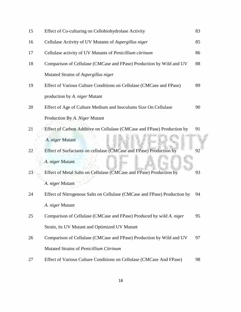
18
15 Effect of Co-culturing on Cellobiohydrolase Activity 83
16 Cellulase Activity of UV Mutants of Aspergillus niger 85
17 Cellulase activity of UV Mutants of Penicillium citrinum 86
18 Comparison of Cellulase (CMCase and FPase) Production by Wild and UV
Mutated Strains of Aspergillus niger
88
19 Effect of Various Culture Conditions on Cellulase (CMCase and FPase)
production by A. niger Mutant
89
20 Effect of Age of Culture Medium and Inoculums Size On Cellulase
Production By A. Niger Mutant
90
21 Effect of Carbon Additive on Cellulase (CMCase and FPase) Production by
A. niger Mutant
91
22 Effect of Surfactants on cellulase (CMCase and FPase) Production by
A. niger Mutant
92
23 Effect of Metal Salts on Cellulase (CMCase and FPase) Production by
A. niger Mutant
93
24 Effect of Nitrogenous Salts on Cellulase (CMCase and FPase) Production by
A. niger Mutant
94
25 Comparison of Cellulase (CMCase and FPase) Produced by wild A. niger
Strain, its UV Mutant and Optimized UV Mutant
95
26 Comparison of Cellulase (CMCase and FPase) Production by Wild and UV
Mutated Strains of Penicillium Citrinum
97
27 Effect of Various Culture Conditions on Cellulase (CMCase And FPase) 98
19
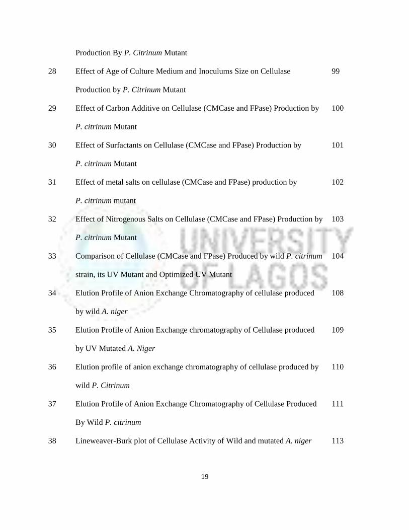
Production By P. Citrinum Mutant
28 Effect of Age of Culture Medium and Inoculums Size on Cellulase
Production by P. Citrinum Mutant
99
29 Effect of Carbon Additive on Cellulase (CMCase and FPase) Production by
P. citrinum Mutant
100
30 Effect of Surfactants on Cellulase (CMCase and FPase) Production by
P. citrinum Mutant
101
31 Effect of metal salts on cellulase (CMCase and FPase) production by
P. citrinum mutant
102
32 Effect of Nitrogenous Salts on Cellulase (CMCase and FPase) Production by
P. citrinum Mutant
103
33 Comparison of Cellulase (CMCase and FPase) Produced by wild P. citrinum
strain, its UV Mutant and Optimized UV Mutant
104
34 Elution Profile of Anion Exchange Chromatography of cellulase produced
by wild A. niger
108
35 Elution Profile of Anion Exchange chromatography of Cellulase produced
by UV Mutated A. Niger
109
36 Elution profile of anion exchange chromatography of cellulase produced by
wild P. Citrinum
110
37 Elution Profile of Anion Exchange Chromatography of Cellulase Produced
By Wild P. citrinum
111
38 Lineweaver-Burk plot of Cellulase Activity of Wild and mutated A. niger 113
20
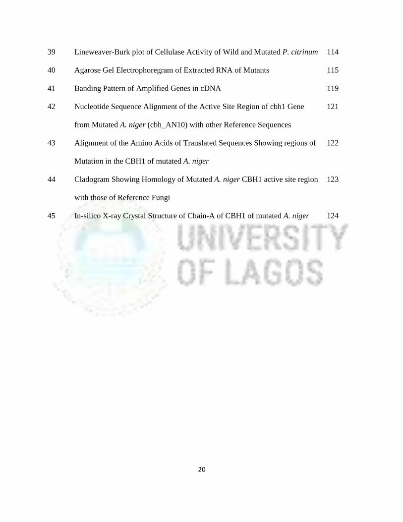
39 Lineweaver-Burk plot of Cellulase Activity of Wild and Mutated P. citrinum 114
40 Agarose Gel Electrophoregram of Extracted RNA of Mutants 115
41 Banding Pattern of Amplified Genes in cDNA 119
42 Nucleotide Sequence Alignment of the Active Site Region of cbh1 Gene
from Mutated A. niger (cbh_AN10) with other Reference Sequences
121
43 Alignment of the Amino Acids of Translated Sequences Showing regions of
Mutation in the CBH1 of mutated A. niger
122
44 Cladogram Showing Homology of Mutated A. niger CBH1 active site region
with those of Reference Fungi
123
45 In-silico X-ray Crystal Structure of Chain-A of CBH1 of mutated A. niger 124
21
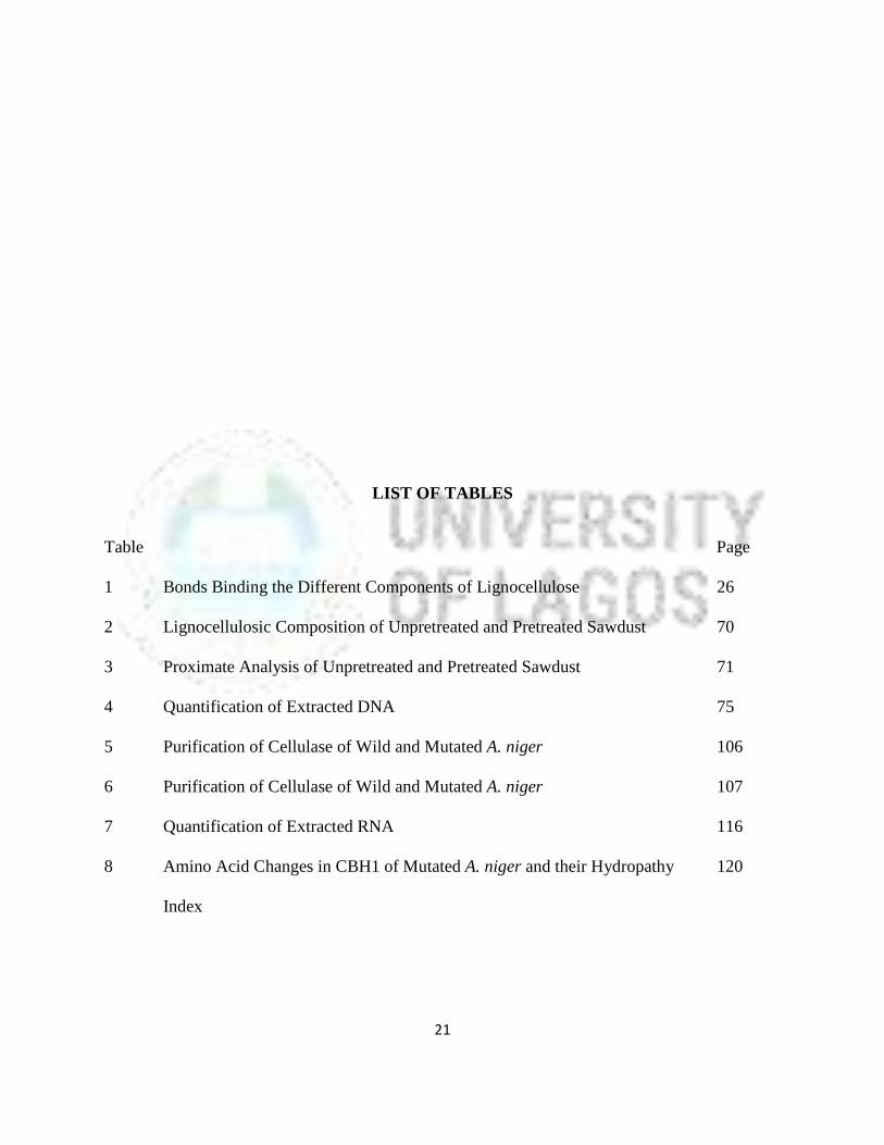
LIST OF TABLES
Table Page
1 Bonds Binding the Different Components of Lignocellulose 26
2 Lignocellulosic Composition of Unpretreated and Pretreated Sawdust 70
3 Proximate Analysis of Unpretreated and Pretreated Sawdust 71
4 Quantification of Extracted DNA 75
5 Purification of Cellulase of Wild and Mutated A. niger 106
6 Purification of Cellulase of Wild and Mutated A. niger 107
7 Quantification of Extracted RNA 116
8 Amino Acid Changes in CBH1 of Mutated A. niger and their Hydropathy
Index
120
22
ABSTRACT
The need for utilizing renewable resources to meet the future demand for fuel and other value
added products has increased the attention on lignocellulose, the most abundant and renewable
resource in the world. Lignocellulose is degraded by lignocellulolytic enzymes produced by
fungi and bacteria. Mutagenic agents can be used to achieve improvement of these strains.
However, there has been a challenge in improving and optimizing fermentation process which in
most cases has been carried out independently. The aim of this study was to hyper produce
cellulase from fungi in order to enhance lignocellulosic wastes biodegradation. Raw Abura
sawdust (Mitragyna ciliata) was collected from Okobaba sawmill, Ebute-meta, Lagos. It was
pretreated mechanically and chemically using ammonium hydroxide. The lignocellulosic and
proximate compositions of both the raw and pretreated sawdust types were determined. Fungi
were isolated from decomposing wood wastes. They were genotyped by amplifying the
internally transcribed spacer (ITS) regions on their DNA, sequencing the amplicons and
analyzing the sequences. Two fungal strains with best cellulolytic potential were selected and co-
cultured. They were genetically modified for enhanced cellulase production using ultraviolet
rays. The optimal conditions for the effective production of cellulase by both wild and mutant
strains of the fungi were investigated. The solid-state fermentation of these mutants was
optimized in order to further enhance the production of cellulase. The enzyme from both the wild
and mutant strains were partially purified and characterized. RNA of the fungal mutants was
extracted from their mycelia. The extracted RNA was reverse transcribed to complementary
DNA (cDNA). Selected genes coding for cellulase were amplified and the amplicons were
sequenced. The points of mutation were identified from the sequence and the sequences of the
23
mutants were aligned and compared with referenced sequences which included those of
Trichoderma reesei and Phanerochaete chrysosporium using MEGA5 and Jalview softwares.
The sequences were also translated to amino acid and in-silico X-ray crystallography structure of
the active site of the enzyme was constructed. The hemicellulose content of the sawdust reduced
significantly upon pretreatment (32.70 ± 2.20% to 19.80 ± 1.30%) while its cellulose content
increased (48.11 ± 1.60% to 64.94 ± 1.20%). Proximate analysis revealed that moisture (6.30 ±
0.60% to 3.60 ± 1.30%) and crude fibre contents (62.20 ± 3.40% to 52.80 ± 2.20%) reduced
significantly upon pretreatment. Aspergillus niger and Penicillium citrinum were selected out of
four fungi genotyped which included Trichosporon asahii and Penicillium corylophylum. Co-
culturing was more efficient in the biodegradation of sawdust. Aspergilus niger mutant strain had
a 2.1-fold and 2.4-fold increase in carboxymethylcellulase (CMCase) and filter paper cellulase
(FPase) production respectively more than the wild strain while Penicillium citrinum had a 1.8-
fold and 2.1-fold increase in CMCase and FPase production. However, optimized fermentation
of A. niger mutant produced a 7.4-fold and 7.6-fold higher increase in CMCase and FPase
production more than the wild strain while P. citrinum mutant produced a 5.3-fold and 5.8-fold
increase (CMCase and FPase). Purified cellulase from wild A. niger had a catalytic efficiency of
0.305M-1
s-1
while its mutant had a catalytic efficiency of 0.429M-1
s-1
. Purified cellulase from
wild P. citrinum had a catalytic efficiency of 0.858M-1
s-1
while its mutant had a catalytic
efficiency of 1.036M-1
s-1
. Bioinformatics analysis of the sequence of the cbh1 gene of the A.
niger mutant showed that it had strong similarity with compared industrially beneficial fungi;
Trichoderma reesei and Phanerochaete chrysosporium. Amino acid residues in its active site had
low hydropathy index. Predicted structure of cellulase from A. niger mutant revealed that
alteration occurred in the β-pleated sheets of the enzyme. Genetic modification of Aspergillus
niger and Penicillium citrinum and optimizing their solid-state fermentation resulted in enhanced
cellulase enzyme production.
CHAPTER ONE
1.0 INTRODUCTION
1.1 BACKGROUND OF STUDY
Technological advancement and industrial revolution have resulted in immense urbanization of
major cities in the world. This has led to attendant increase in commercial activities and
ultimately in generation of large volumes of solid wastes. These wastes usually accumulate in the
environment causing environmental menace. Lignocellulosic wastes are major renewable natural
resources of the world and represent a major source of renewable organic matter. Lignocellulosic
biomass can be grouped into four main categories: agricultural residues (corn stover and cobs,
sugar cane bagasse, banana peels and stalks etc), dedicated energy crops, wood residues (sawmill

24
and paper mill discards) and municipal paper waste. The plant biomass regarded as wastes are
biodegradable and can be converted into valuable products (Dashtban et al., 2009). The
bioconversion of lignocellulosic materials rather than its management is now a subject of
intensive research which will not only solve aesthetic problem they create, but generate wealth
by development of primary industries and creation of value added products. Lignocellulosic
wastes can be utilized not only for ethanol production but industrial chemicals. Howard et al.,
(2003) reported that many organic chemicals can be produced from ethylene, propylene,
benzene, toluene and xylene. Benzene, toluene and xylene can be obtained from lignin, being
aromatic while ethylene and propylene can be obtained from ethanol derived from fermentation
of glucose obtained from cellulose biodegradation.
Biodegradation of lignocellulosics can be achieved with the use of ligninocellulolytic enzymes
produced by bacteria and fungi (Kaur et al., 2007). Enzymes that act on cellulose are generally
referred to as cellulases, those that degrade hemicellulose and lignin are called hemicellulases
and ligninases respectively. Cellulases can be classified into three broad categories.
Endoglucanases (endo-1,4-β-D-glucanases); cellobiohydrolases or exoglucanases (exo-1,4-β-D-
glucanases); and β-glucosidases (1,4-β-D-glucosidases) (Gao et al., 2008). Cellulose hydrolysis
is a simultaneous and synergistic action of these three types of enzymes. The hydrolysis process
starts with endoglucanases that randomly hydrolyze internal β-1,4-glycosidic linkages in the
amorphous region of cellulose microfibrils, reducing significantly its degree of crystallinity and
opening new terminal ends. Simultaneously, the accessible reducing and non-reducing ends of
cellulose chains are attacked by types I and II cellobiohydrolases, respectively. The cellobiose
units released are then hydrolyzed into glucose units by β-glucosidases (Textor et al., 2013).
Cellulases can be classified into glycosyl hydrolase (GH) families based on their amino acid
25
sequence and folding similarities. Together with some endoglucanases, type I cellobiohydrolases
belong to the GH family 7 (Textor et al., 2013). The exoglucanase, Cel7A, is the most important
single enzyme component for cellulose depolymerization and conversion into cellobiose in fungi
(Lynd et al., 2002). It is considered to be the key enzyme in the hydrolysis process because it is
able to hydrolyze crystalline cellulose extensively, although at a slow rate (Textor et al., 2013).
The performance of microbes for hyper-production of lignocellulolytic enzymes can be
improved by different methods of mutation (Pradeep et al., 2012; De Nicolas Santiago et al.,
2006). Previously used physical mutagens include Ultraviolet light, microwaves and high energy
ionizing radiations (Li et al., 2010; Xu et al., 2011). Chemical mutagenesis had been carried out
by treatment with Nitrosoguanidine, Ethyl methane sulphonate, Diethyl sulphonate and
colchicines (Bhargavi and Singara, 2010). On the other hand, enhanced enzyme production can
be achieved by supplementations with salts (Junior et al., 2009), metals (Nikolic et al., 2009) and
optimization of fermentation conditions (Acharya et al., 2008).
Modification of fungi for enhanced performance by mutation and optimization of their
fermentation conditions are the major approaches for enhanced ligninocellulolytic enzymes
production. In view of the utilization of sawdust and agro-wastes in general, the present research
was carried out with the aim of hyper producing cellulase from fungi by strain improvement in
order to enhance lignocellulosic wastes biodegradation
.
26
1.2 STATEMENT OF PROBLEM
The recognition that environmental pollution is a worldwide threat to public health has given rise
to new massive industries for environmental restoration (Milala et al., 2009). The activities of
these industries have ultimately led to generation of large volumes of wastes. Waste generation is
an inevitable aspect of living which, at least can only be managed. The problems posed by these
wastes are many: they degrade the urban environment, reduce its aesthetic value, produce
offensive odours during the rains and pollute the air with smoke when the wastes are burnt
uncontrollably. They also constitute health hazards in themselves if they are not timely disposed
(Ogunbode et al., 2013). Agricultural wastes represent the largest class of cellulosic wastes
which are grossly underutilized leaving a large proportion to constitute source of environmental

27
pollution (Rahman et al., 2000). In Nigeria, agricultural wastes constitute over 60% of cellulosic
wastes which are currently underutilized (Abu et al., 2002).
A huge volume of wood residue is generated annually from timber processing activities around
sawmills, plank markets, and furniture-making factories in cities within Nigeria. For instance, about
294,798 tons of wood waste is generated yearly in the city of Lagos (Dosunmu and Ajayi, 2002),
while about 2288 m3 is generated daily in Abeokuta, while estimated 31,324.3 tons annually in Ilorin
and a total of 104,000 m3 of wood waste is generated daily in Nigeria (Aina, 2006).
Lignocellulosic wastes alone account for over 80% urban refuse in Lagos, Nigeria (LAWMA
report, 2004). Nwankwo (2004) observed that wood wastes constitute the largest class of
industrial wastes which have been reported to have caused enormous environmental pollution. It
was estimated that 5,666.19 tons of wastes are generated in one week by the saw mills in Lagos
in 2011. This produces about 6.6 tons of Sulfure (IV) oxide and 3331.7 tons of ash per annum if
it gets burned (Ogunbode et al., 2013).
Enzyme cost is estimated to represent approximately 50% of the total hydrolysis process cost.
The cost of enzyme on the economics of lignocellulosic waste bioconversion has been a subject
of controversy. There have been arguments that the cost of enzyme production itself is more
expensive than the product of the bioconversion (Klein-Marcuschammer et al., 2012). At 20%
solids loading during saccharification, a typical enzyme loading is 10 FPU/g cellulose,
equivalent to approximately 20mg enzyme/g cellulose (Gusakov, 2011). The typical yield for the
saccharification of cellulose at this enzyme loading is 70% after 5 days (Roche et al., 2009). It
must be noted that the optimal value of enzyme loading varies depending on feedstock, solids

28
loading, and pretreatment technology, among other variables (Kazi et al., 2010; Kristensen et al.,
2009).
A thorough understanding of the mechanisms guiding the expression of the genes coding for
cellulase is imperative for the development of the genetic improvement programme for cellulase
producing organisms. There is paucity of data on studies on tropical fungi and their mutants.
1.3 AIM OF STUDY
The aim of this study is to hyper-produce cellulase from fungi in order to enhance lignocellulosic
wastes biodegradation.
1.4 OBJECTIVES OF STUDY
The specific objectives of this study are to:
1. determine the lignocellulosic composition of raw and pretreated sawdust.
2. isolate and genotype cellulolytic fungi.

29
3. mutate isolates and optimize their solid-state fermentation.
4. sequence cDNA amplicons obtained from wild and mutant fungi strains.
5. analyze the sequence using bioinformatics tools and predict the structure of cellulase
from mutant fungi.
1.5 SIGNIFICANCE OF STUDY
Generally, sawdust as well as other lignocellulosic wastes are disposed by burning, thus
constitutes a source of greenhouse gases and other forms of environmental pollution. This study
will enhance the transformation of sawdust and other lignocellulosic wastes from pollutant to
raw materials. By implication, the massive turnover of wood wastes which is estimated to be
about hundreds of tons per day will lead to the creation of primary and secondary industries
producing valuable products thus creating jobs for the teeming unemployed youths of the nation.
30
This will inevitably contribute to Nigeria‘s Gross Domestic earnings. Simultaneously, the
environment will be salvaged from further deterioration.
This study will also provide alternative for energy crops, which itself is uneconomical for
hydrolysis to bioproducts.
The lignocellulosic composition of a number of biomass wastes has been determined. These
include sugarcane chaffs, paper, corn-cobs, and banana peels, to name a few. There is however,
no data on sawdust. This study will provide data on the composition of sawdust and the
implication of chemical pretreatment its composition.
The traditional method of identifying a microorganism is from its distinct feature on plates and
under the microscope. These observations are comfirmed using biochemical tests. However, it
has been revealed that some organisms share similar features macroscopically and
microscopically which makes it difficult to distinguish organisms belonging to the same phyla.
DNA barcoding is now in use in the past two decades to identify microorganisms most especially
fungi, thus solving the problems of misidentification of microorganisms. This study seeks to add
to the already existing bank of fungal gene sequences in public databases. Thus further
enhancing the identification of fungi isolated from our environment.
The kinetic parameters of the cellulase obtained shall be studied. This will elucidate the catalytic
mechanism of hydrolysis by cellulase from both wild and mutant strains of different fungi.
Strain improvement has been the target of researchers in the field of lignocellulosic
biotechnology in this decade. Several attempts have been made which proved auspicious albeit
delimited by cost of the procedure, safety and generation of harmful mutants. This study set out
to create strains which are efficient yet cheap to produce. The efficiency of these mutants shall
be complemented by improving their solid-state fermentation. The improved solid-state

31
fermentation procedure shall be premised on independent experiments which this study set to
carry-out.
Based on the emerging trend in solving problems in lignocellulosic biotechnology using
recombinant DNA technology and protein engineering, this study seeks to propose a sequence
which can be cloned and expressed in suitable vectors for hyper production of cellulase. The
efficiency of an enzyme largely depends on its structure. This study will provide a model
structure of cellulase enzyme from mutant fungi which will be an addition to the database needed
in studying the structure and function relationship in enzyme biotechnology.
1.6 OPERATIONAL DEFINITION OF TERMS
Lignocellulosic Wastes (LCW): are plant biomass wastes that are composed of cellulose,
hemicellulose, and lignin. They are majorly agricultural residues and municipal solid wastes.
Pretreatment: Preparation of lignocellulosic materials for enzymatic degradation. It involves the
alteration of structural and chemical composition of lignocellulosics to facilitate rapid and
efficient hydrolysis of carbohydrates to fermentable sugars.
32
Cellulases: are enzymes responsible for the hydrolysis of cellulose with specificities to
hydrolyze glycosidic bonds.
Endoglucanase (CMCase): are type of cellulases which initiate attack randomly at multiple
internal sites for subsequent attack by cellobiohydrolase. The are also known as
carboxymethylcellulase.
Solid State Fermentation (SSF): Growth of microorganisms in the absence or near absence of
free water with inert natural substrates as solid support.
Gene Mutation: Permanent change in DNA sequence that makes up a gene.
Mutagenesis: Conscious introduction of mutation in the genome of an organism which is
achieved with physical or chemical agents called mutagens.
Polymerase Chain Reaction (PCR): Primer specific in-vitro enzymatic amplification of a target
segment of DNA from a complex mixture of starting material usually termed the template.
Complementary DNA (cDNA): DNA molecule generated from RNA by reverse transcription.
DNA Barcoding: Molecular identification and phylogenetic classification of organisms from
bioinformatic analysis of sequence of specific segment of their DNA.
1.7 LIST OF ABBREVIATIONS
ace1 – transcriptional activating gene class1
aep1 - aldose epimerase gene class1
ANM - Aspergillus niger (mutant strain)
ANW- Aspergillus niger (wild strain)
BLAST – Basic Local Alignment Search Tool
33
cbh - cellobiohydrolase gene class 1
CBH1- cellobiohydrolase enzyme class 1
CBM – cellulose binding module
cDNA – complimentary deoxyribonucleic acid
DNA- deoxyribonucleic acid
FPase – filter paper cellulase
k(off) – dissociation rate
Lacc1 – laccase gene class 1
LCW – lignocellulosic wastes
PCM - Penicillium citrinum (mutant strain)
PCW – Penicillium citrinum (wild strain)
RNA – ribonucleic acid
UV- ultraviolet
X – times
CHAPTER TWO
2.0 LITERATURE REVIEW
2.1 MICROFUNGI
Microfungi are diverse group of fungi consisting of yeasts and moulds. Microfungi belong to
three major phyla; Ascomycota, Deuteromycota and Zygomycota. The fungal body consists of
microscopic threads called hyphae, extending through the substrate through which they grow

34
(Carlile et al., 2001). Typically only the ―fruiting body‖ of the fungus is visible, producing
thousands of tiny spores that are carried by the air, spreading the fungus to new locations.
Spores are produced in a variety of ways and occur in a bewildering array of shapes and sizes. In
spite of this diversity, spores are quite constant in their shapes, sizes (about 2–20 μm), colour and
form and as such these characteristics are very useful for identification of microfungi. The basic
difference between spores lies in their method of initiation, which can be either sexual or asexual
(Carlile et al., 2001).
2.1.1 CHARACTERISTICS AND IMPORTANCE
Microfungi are well adapted to extreme environmental conditions. They tolerate a wide range of
temperature, pH, dryness, oxygen concentrations and ultraviolet radiation better than the wood-
rotting basidiomycetes called white or brown rot fungi. In addition they are found in all climatic
zones ranging from the poles to the tropics (Blanchette, 2000). Generally, fungi prefer an acidic
environment although microfungal activities occur within a broad pH range of between 3.7 and
8.6 (Daniel and Nilsson, 1998). Microfungi can protect themselves by relatively quick growth in
natural niches and by the production of antibiotics and toxic substances (mycotoxins).
Microfungi play important role in carbon cycling, and are also involved in many
biotechnological processes. These processes include: brewing, wine making, baking, cheese-
making and the preparation of other fermented food (e.g. soy sauce) together with edible
mushroom production are the most important microfungal applications. Production of enzymes
(amylase, cellulase, invertase, lipase, pectinase, proteinase, rennin and xylanase), organic acids
(citric, itaconic and lactic acids), antibiotics and other pharmaceuticals (penicillin, mevinolin,
cephalosporin, griseofulvin and cyclosporine) by fungi are common processes that have been
reviewed (Demain et al., 2004).

35
2.1.2 Aspergillus niger
Aspergillus niger or A. niger is a ubiquitous fungus and one of the most common species of the
genus Aspergillus. It causes a disease called black mould on certain fruits and vegetables. It is
characteristic with black colonies which can be confused with those of Stachybotrys (Samson et
al., 2001). Studies have proved that some true A. niger strains do produce ochratoxin A. It also
produces the isoflavone orobol (Samson et al., 2001; Schuster et al., 2002). The black aspergilli
are among the most common fungi causing food spoilage and deterioration of other materials.
They have also been used for diverse biotechnological purposes, not limited to production of
organic acids and enzymes (Schuster et al. 2002). The taxonomy of Aspergillus section Nigri has
been studied by many taxonomists and was recently reviewed by Abarca et al., (2004). Al-
Musallam (1980) did a comprehensive revision of the taxonomy of the A. niger group based on
morphological features. Seven species (A. japonicus, A. carbonarius, A. ellipticus, A. helicothrix,
A. heteromorphus, A. foetidus, and A. niger) were recognized. A. niger was described as an
aggregate consisting of seven varieties and two formae. Samson et al., (2004) reported that
Kozakiewicz (1989) distinguished A. japonicus, A. helicothrix, A. atroviolaceus, A.
heteromorphus, A. ellipticus and A. carbonarius as species exhibiting echinulate conidial
ornamentations distinct from the remaining black Aspergillus taxa, which produce verrucose
conidia. Within the verrucose category, A. acidus, A. fonsecaeus, A. niger var. ficuum, A. niger
var. phoenicis, A. niger var. niger, A. niger var. awamori, A. niger var. pulverulentus, A.
niger var. tubingensis, A. citricus (A. foetidus) and A. citricus var. pallidus were recognized.
Aspergillus niger is the most frequently reported species in this section and has often been
included in biotechnological processes that are Generally Regarded as Safe (GRAS). However,
36
species concepts are uncertain in this complex and occasionally the name A. niger has been
used for any member of the section. Taxonomic studies using molecular methods have divided
the A. niger complex into two species, A. niger and A. tubingensis (Abarca et al., 2004).
2.1.2.1 Pathogenicity
A. niger is less likely to cause human disease than some other Aspergillus species. In extremely
rare instances, humans may become ill, but this is due to a serious lung disease, aspergillosis,
that can occur. Aspergillosis is, in particular, frequent among horticultural workers that inhale
peat dust, which can be rich in Aspergillus spores. A. niger is one of the most common causes
of otomycosis (fungal ear infections), which can cause pain, temporary hearing loss and in severe
cases, damage to the ear canal and tympanic membrane. In plants, A. niger causes a common
postharvest disease of onions, in which the black conidia can be observed between the scales of
the bulb. The fungus also causes disease in peanuts, grapes and other fruits (Samson et al. 2001).
2.1.2.2 Industrial Uses
A. niger is cultured for the industrial production of many substances. Various strains of A. niger
are used in the industrial preparation of gluconic acid (E574) and citric acid (E330) and have
been assessed as acceptable for daily intake by the World Health Organisation (WHO). A. niger
fermentation is ―generally recognized as safe‖ (GRAS) by the United States Food and Drug
Administration under the Federal Food, Drug, and Cosmetic Act (FDA, 2008). Enzymes of
economic importance are produced using industrial fermentation of A. niger . These include
alpha-galactosidase used in the food industry, pectinases used in cider and wine clarification,
glucoamylase used in the production of high fructose corn syrup. Another use for A. niger

37
within the biotechnology industry is in the production of magnetic isotope-containing variants of
biological macromolecules for nuclear magnetic reasonance (NMR) studies. The enzyme
protease is derived from Aspergillus niger and used to produce the supplement Clarity-Ferm.
This product is being used in the brewing industry to reduce gluten content of wheat and barley
based beers. A Clarity-Ferm treated beer made from barley or wheat usually tests below 20 ppm
of gluten, the current international standard for gluten free (Mitea et al., 2008).
2.1.2.3 Other Uses
A microbial-derived enzyme, prolyl endoprotease, which cleaves gluten has been found to be
producible by A. niger . This enzyme has strong implications in the treatment of coeliac disease
or other metabolic gluten sensitivity disease processes (Mitea, 2008). A placebo controlled,
double blind study was initiated in December 2008 to determine the efficacy of this enzyme in
treating humans with coeliac disease (Mulder, 2008). A. niger is also cultured for the extraction
of the enzymes glucose oxidase (GO) and alpha-galactosidase (AGS). Glucose oxidase is used in
the design of glucose biosensors, due to its high affinity for β-D-glucose (Staiano et al., 2005). α-
galactosidase can be produced by A. niger fermentation and is used to hydrolyze α-1-6 bonds
found in melibiose, raffinose, and stachyose.
2.1.3 Penicillium citrinum
Penicillium citrinum is an anamorph, mesophilic fungus species of the genus of Penicillium
which produces tanzawaic acid, Mevastatin, Quinocitrinine A, Quinocitrinine B, and nephrotoxic
citrinin (John et al., 2009; Mossini and Kemmelmeier, 2008). Penicillium citrinum is often found
on moldy citrus fruits and occasionally occurs in tropical spices and cereals. This Penicillium
38
species is also mortally to the mosquito Culex quinquefasciatus (Maketon, 2014). In view of its
mesophilic character, Penicillium citrinum occurs worldwide (John et al., 2009). The first statin
(Mevastatin) was isolated from this species in 1970.
2.2 BIOMASS AND ITS POTENTIALS
Any mass of biological material is termed Biomass. These include whole or plant parts, plant
constituents and byproducts, animal byproducts, municipal and industrial wastes (Howard et al.,
2003). Bioproducts can be generated from these materials following a thorough knowledge of
techniques to be used in manipulating its constituents to obtain the desired product.
Biomass of plant origin is composed mainly of cellulose, hemicelluloses and lignin, hence, called
lignocellulosic biomass. In addition, small amounts of other components can be found in them
depending on source (Sanchez, 2008). These components may include pectin, protein, and ash.
Agricultural resources of lignocellulosic waste are quite abundant as estimated by the Food and
Agriculture Organization (FAOSTAT, 2006). Around 2.9x10³ million tons from cereal crops and
1.6x10² millions tons from pulse crops, 1.4x10 million tons from oil seed crops and 5.4x10
2
million tons from plantation crops are produced annually worldwide (Kumar et al., 2008). The
various types of lignocellulosic raw materials include wheat straw, rice straw, palm, corncobs,
corn stems and husk etc., have varying amounts of cellulosic components. It has been estimated
that the yearly biomass production of cellulose is 1.5 trillion tons, making it an essentially
inexhaustible source of raw material for environmentally friendly and biocompatible products
(Kim and Yun, 2006). Therefore, the bioconversion of large amounts of lignocellulosic biomass
into fermentable sugars has potential application in the area of bioenergy generation. Although
extensive studies have been carried out to meet the future challenges of bioenegy generation,
39
there is no self-suficient process or technology available to convert the lignocellulosic biomass
for bioenegy generation (Kim and Yun, 2006).
2.3 STRUCTURE OF THE PLANT CELL WALL
The plant cell wall consists of three types of layers, namely middle lamella, primary wall, and
secondary wall. In the primary wall, the main structure is a skeleton of cellulose cross-linked
with glycans, and there are two types according to the cross-link types. Type I walls are found in
dicotyledonous plants and consist of equal amounts of glucan and xyloglucan embedded in a
matrix of pectin. Type II walls are present in cereals and other grasses having
glucuronoarabinoxylans as their cross-linking glucans, but lacking of pectin and structural
proteins (Mohnen et al., 2008).
The secondary wall usually has three sub-layers, which are named S1, S2 and S3. S1 is the outer
layer, S2 is the middle layer while S3 is the inner layer (Chundawat et al., 2011a).
The cellulose microfibrils of secondary wall are embedded in lignin, (Sticklen, 2008). Cellulose,
hemicellulose, and lignin have different distribution in these layers. In wood fibers, it has been
found that cellulose concentration is increased from middle lamella to the secondary wall. S2 and
S3 lamellaes have the highest cellulose concentration. Most of the hemicellulose distributes in
the secondary wall (Zhao et al., 2012). Lignin is found to be the dominant composition in the
outer portion of the compound middle lamellae. The percentage of lignin in the lignocellulosic
matrix decreases with increasing distance into the middle lamella. The percentages of lignin in
the primary wall and in the S1 layer of the secondary wall are much higher than those in the S2
and S3 sections (Zhao et al., 2012). However, the plant cell wall is indeed a complex
nanocomposite material at the molecular and nanoscales (Ding et al., 2008). Although much is

40
known of the structure of the plant cell wall, more information should be gained to understand
the microstructure and even anostructure of plant cell wall and how these structures build the
recalcitrance of biomass. Many modern analytic and simulation methods have been employed to
deeply understand the molecular mechanism of biomass recalcitrance (Zhao et al., 2012) and the
molecular dynamics of lignin (Petridis and Smith, 2009).
2.4 CHEMICAL COMPOSITION OF LIGNOCELLULOSE
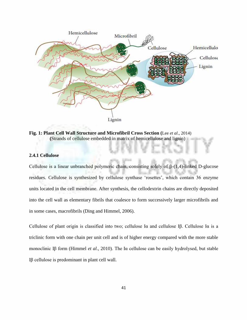
Plant cell wall consists mainly of monosaccharides which could be structural or storage. The
structural form could be celluloses, hemicelluloses, or pectins (Himmel et al., 2010). Their
predominance is as enumerated. The most abundant weight fraction cell wall type in plant tissue
is the secondary cell wall, produced after the cell has stopped growing. Secondary cell walls
contain structural polysaccharides, strengthened further with lignin covalently cross-linked to
hemicellulose. Cellulose is the most abundant. It exists as microfibrils and is embedded in matrix
of hemicellulose and lignin as shown in Fig. 1 below.
41
Fig. 1: Plant Cell Wall Structure and Microfibril Cross Section (Lee et al., 2014)
(Strands of cellulose embedded in matrix of hemicellulose and lignin)
2.4.1 Cellulose
Cellulose is a linear unbranched polymeric chain, consisting solely of β-(1,4)-linked D-glucose
residues. Cellulose is synthesized by cellulose synthase ‗rosettes‘, which contain 36 enzyme
units located in the cell membrane. After synthesis, the cellodextrin chains are directly deposited
into the cell wall as elementary fibrils that coalesce to form successively larger microfibrils and
in some cases, macrofibrils (Ding and Himmel, 2006).
Cellulose of plant origin is classified into two; cellulose Iα and cellulose Iβ. Cellulose Iα is a
triclinic form with one chain per unit cell and is of higher energy compared with the more stable
monoclinic Iβ form (Himmel et al., 2010). The Iα cellulose can be easily hydrolysed, but stable
Iβ cellulose is predominant in plant cell wall.
42
In cellulose, all of the glucosyl hydroxyl groups are in the equatorial position, whereas all of the
axial positions are occupied by nonpolar (and nonhydrogen-bonding) aliphatic protons. This
means that the ‗sides‘ of the elementary microfibrils are polar and hydrogen bonding and the
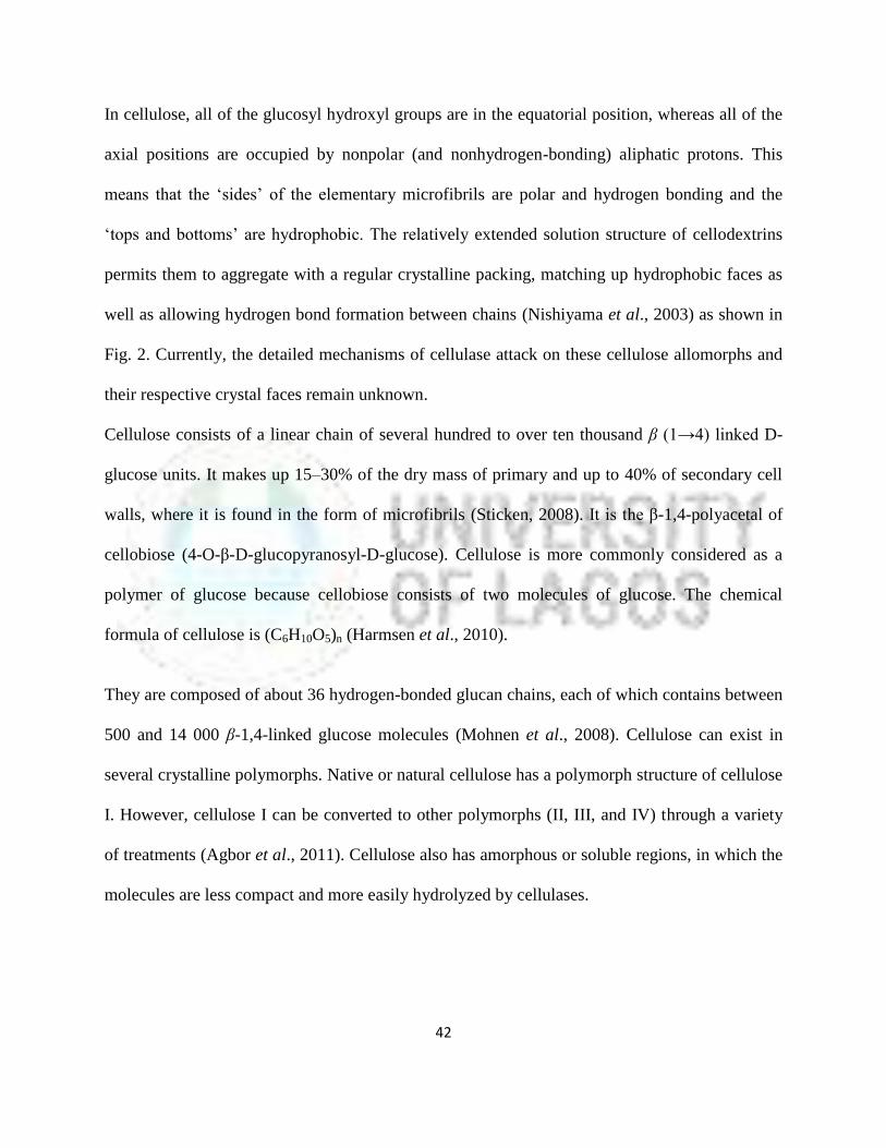
‗tops and bottoms‘ are hydrophobic. The relatively extended solution structure of cellodextrins
permits them to aggregate with a regular crystalline packing, matching up hydrophobic faces as
well as allowing hydrogen bond formation between chains (Nishiyama et al., 2003) as shown in
Fig. 2. Currently, the detailed mechanisms of cellulase attack on these cellulose allomorphs and
their respective crystal faces remain unknown.
Cellulose consists of a linear chain of several hundred to over ten thousand β (1→4) linked D-
glucose units. It makes up 15–30% of the dry mass of primary and up to 40% of secondary cell
walls, where it is found in the form of microfibrils (Sticken, 2008). It is the β-1,4-polyacetal of
cellobiose (4-O-β-D-glucopyranosyl-D-glucose). Cellulose is more commonly considered as a
polymer of glucose because cellobiose consists of two molecules of glucose. The chemical
formula of cellulose is (C6H10O5)n (Harmsen et al., 2010).
They are composed of about 36 hydrogen-bonded glucan chains, each of which contains between
500 and 14 000 β-1,4-linked glucose molecules (Mohnen et al., 2008). Cellulose can exist in
several crystalline polymorphs. Native or natural cellulose has a polymorph structure of cellulose
I. However, cellulose I can be converted to other polymorphs (II, III, and IV) through a variety
of treatments (Agbor et al., 2011). Cellulose also has amorphous or soluble regions, in which the
molecules are less compact and more easily hydrolyzed by cellulases.
43
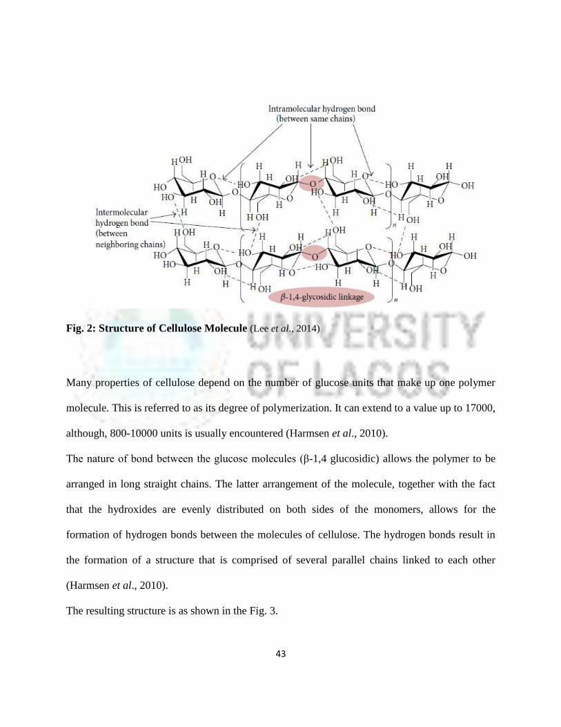
Fig. 2: Structure of Cellulose Molecule (Lee et al., 2014)
Many properties of cellulose depend on the number of glucose units that make up one polymer
molecule. This is referred to as its degree of polymerization. It can extend to a value up to 17000,
although, 800-10000 units is usually encountered (Harmsen et al., 2010).
The nature of bond between the glucose molecules (β-1,4 glucosidic) allows the polymer to be
arranged in long straight chains. The latter arrangement of the molecule, together with the fact
that the hydroxides are evenly distributed on both sides of the monomers, allows for the
formation of hydrogen bonds between the molecules of cellulose. The hydrogen bonds result in
the formation of a structure that is comprised of several parallel chains linked to each other
(Harmsen et al., 2010).
The resulting structure is as shown in the Fig. 3.

44
Fig. 3: Demonstration of Hydrogen Bonding that Allows the Parallel Arrangement of the
Cellulose Polymer Chains (Harmsen et al., 2010)
2.4.2 Hemicellulose
In addition to cellulose, the plant cell wall matrix contains two additional major types of cell wall
polysaccharides; the hemicelluloses and the pectins. Unlike cellulose, both are synthesized in the
Golgi apparatus, delivered to the cell membrane via small vesicles and secreted into the cell wall.
Hemicelluloses are generally complex, branched carbohydrate polymers that are formed from
different monomeric sugars attached through different linkages (Chundawat et al., 2011a).
Carbohydrate substituents and noncarbohydrate components occur in hemicelluloses on either
the main chain or on the carbohydrate branches. The complex structures of the hemicelluloses
are thought to confer a wide range of biophysical and biomechanical properties on the plant
tissues in which they occur, as well as on products made from these tissues. The principal
pentose sugar in the major plant cell wall hemicellulose is β-D-xylopyranose, which has only the
position 2- and 3-carbons available for O-linked substitution by substituent sugars when β-(1,4)
linked as in xylan. Hemicelluloses also include xyloglucan, arabinoxylan and glucomannans,
which contain other sugars including the pentose arabinose and the aldohexoses glucose,
45
mannose and galactose. The hemicelluloses can also be esterified by acetylation and/or cross-
linked to lignins via p-coumaroyl and feruloyl groups (Chundawat et al., 2011a).
Hemicellulose consists of polysaccharides other than cellulose. Its structure reveals that either
type of bonds is the main one that forms its molecule. The main difference with cellulose is that
the hydrogen bonds are absent and that there is significant amount of carboxyl groups. The
carboxyl groups can be present as carboxyl or as esters or even as salts in the molecule (Harmsen
et al., 2010). Hemicellulose is a diverse group of short-chain branched, substituted polymer of
sugars with a degree of polymerization ~70 to 200, and is usually characterized as the
heterogeneous polysaccharides being soluble in strong alkali (.Scheller and Ulvskov, 2010). It
has a backbone composed of 1, 4-linked β-D-hexosyl residues and may contain pentoses,
hexoses, and/or uronic acids. Other sugars, such as α-L-rhamnose and α-L-fucose, may also be
present in small amounts and the hydroxyl groups of sugars can be partially substituted with
acetyl groups (Zhao et al., 2009). Hemicelluloses comprises a family of polysaccharides such as
arabinoxylans, glucomannans, galactans, and others that are found in the plant cell wall and have
different composition and structure depending on their source. The most common type of
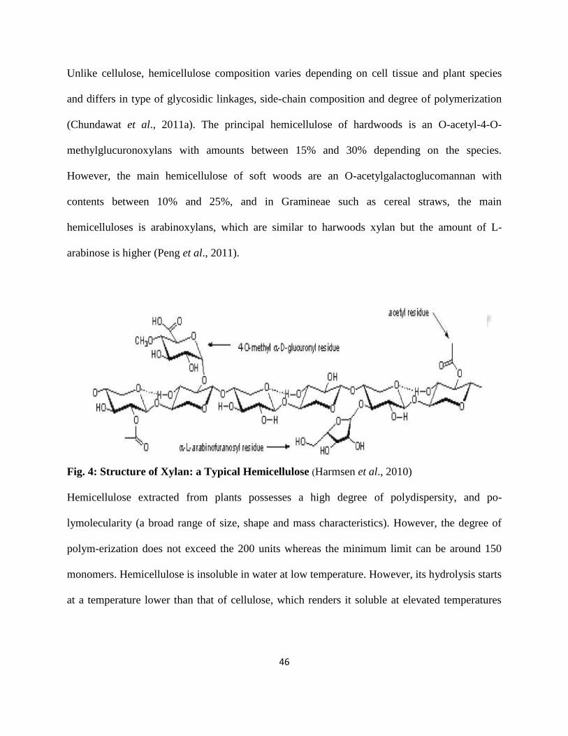
polymers that belongs to the hemicellulose family of polysaccharides is xylan. As shown in Fig.
3, the molecule of a xylan involves 1->4 linkages of xylopyranosyl units with α-(4-O)-methyl-D-
glucuronopyranosyl units attached to anhydroxylose units (Fig. 4). The result is a branched
polymer chain that is mainly composed of five carbon sugar monomers, xylose, and to a lesser
extent six carbon sugar monomers such as glucose. Important aspects of the structure and
composition of hemicellulose are the lack of crystalline structure, mainly due to the highly
branched structure and the presence of acetyl groups connected to the polymer chain (Harmsen et
al., 2010).
46
Unlike cellulose, hemicellulose composition varies depending on cell tissue and plant species
and differs in type of glycosidic linkages, side-chain composition and degree of polymerization
(Chundawat et al., 2011a). The principal hemicellulose of hardwoods is an O-acetyl-4-O-
methylglucuronoxylans with amounts between 15% and 30% depending on the species.
However, the main hemicellulose of soft woods are an O-acetylgalactoglucomannan with
contents between 10% and 25%, and in Gramineae such as cereal straws, the main
hemicelluloses is arabinoxylans, which are similar to harwoods xylan but the amount of L-
arabinose is higher (Peng et al., 2011).
Fig. 4: Structure of Xylan: a Typical Hemicellulose (Harmsen et al., 2010)
Hemicellulose extracted from plants possesses a high degree of polydispersity, and po-
lymolecularity (a broad range of size, shape and mass characteristics). However, the degree of
polym-erization does not exceed the 200 units whereas the minimum limit can be around 150
monomers. Hemicellulose is insoluble in water at low temperature. However, its hydrolysis starts
at a temperature lower than that of cellulose, which renders it soluble at elevated temperatures
47
(Thermowoodhandbook, 2003). The presence of acid highly improves the solubility of
hemicellulose in water.
2.4.3 Lignin
As a complex phenolic polymer, lignins exist widely in cell walls of plants and some algae.
There are three types of functional groups in lignins, including p-hydroxyphenyl, guaiacyl and
syringyl, from which the monolignols, 4-hydroxycinnamyl, coniferyl and sinapyl alcohols,
respectively are comprised. The complex and highly variable chemical heterogeneity of lignin is
due to the diversity of substitution patterns and intermolecular linkages utilized during
polymerization. Although lignins enable critical functions for the plant, including mechanical
support, water transport and defense, lignin is also an undesirable component in the biomass
conversion process, due to its ability to shield polysaccharides from enzymatic hydrolysis and
generally impede diffusion into plant tissue by chemicals and enzymes. In native plant cell walls,
lignins are covalently linked to hemicellulose, which forms a matrixing layer around the
cellulose comprising the microfibril core that further hinders cellulolytic and hemicellulolytic
enzymes. Moreover, many of the lignin degradation products are either inhibitory or generally
detrimental to the plant cell polysaccharide-degrading enzymes. It is therefore necessary to take
this into account when designing an enzymatic process for degradation of lignocellulosic
biomass (Harmsen et al., 2010).
Lignin is the most complex natural polymer (Calvo-Flores and Dobado, 2010). It is an
amorphous three-dimensional polymer with phenylpropane units as the predominant building
blocks. These building blocks are mainly (1) sinapyl alcohol, (2) p-coumaryl alcohol and (3)
coniferyl alcohol as shown in Fig. 5a. As an organic substance binding the cells, fibers, and
vessels, lignin has an important role in protecting the plants against invasion by pathogens and
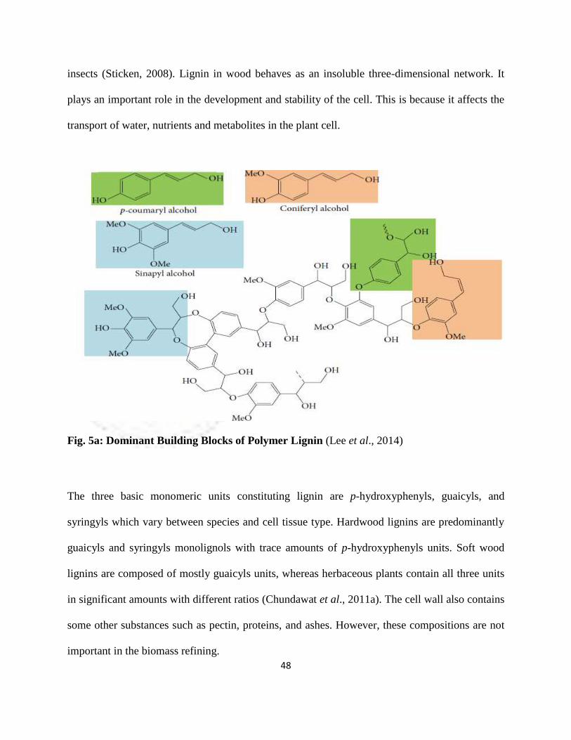
48
insects (Sticken, 2008). Lignin in wood behaves as an insoluble three-dimensional network. It
plays an important role in the development and stability of the cell. This is because it affects the
transport of water, nutrients and metabolites in the plant cell.
Fig. 5a: Dominant Building Blocks of Polymer Lignin (Lee et al., 2014)
The three basic monomeric units constituting lignin are p-hydroxyphenyls, guaicyls, and
syringyls which vary between species and cell tissue type. Hardwood lignins are predominantly
guaicyls and syringyls monolignols with trace amounts of p-hydroxyphenyls units. Soft wood
lignins are composed of mostly guaicyls units, whereas herbaceous plants contain all three units
in significant amounts with different ratios (Chundawat et al., 2011a). The cell wall also contains
some other substances such as pectin, proteins, and ashes. However, these compositions are not
important in the biomass refining.

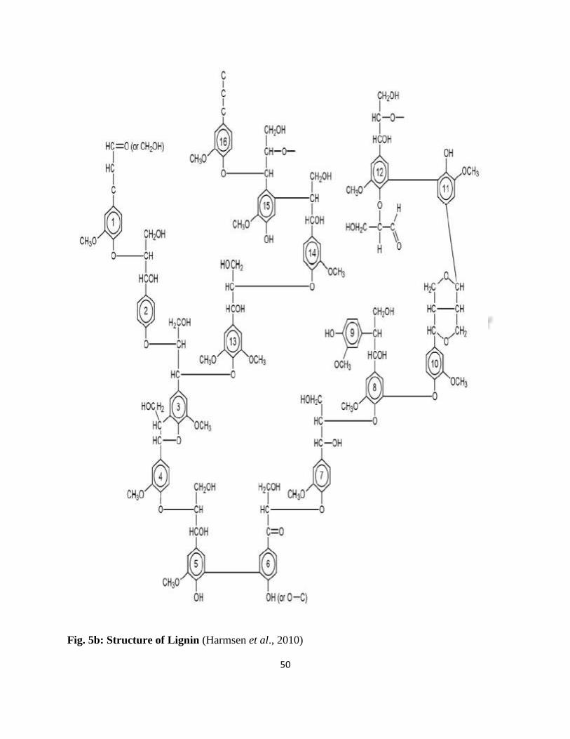
49
Lignin from softwood is made up of more than 90% of coniferyl alcohol with the remaining
being mainly p-coumaryl alcohol units. However, lignin found in hardwood is made up of
varying ratios of coniferyl and sinapyl alcohol type of units. Low molecular alcohols
significantly dissolve lignin, likewise di-oxane, acetone, pyridine, and dimethyl sulfoxide. Also,
it has been found out that at elevated temperatures, lignin softens, and this allows
depolythmerization reactions of acidic or alkaline nature to accelerate (O‘Connor et al., 2007).
The property of polydispersity, just as with hemicellulose, characterizes lignin as well. Different
branching and bonding in otherwise similar molecules are encountered (Harmsen et al., 2010).
Fig. 5b shows a model structure of lignin from spruce pine.
2.4.4 Bonds in the Lignocellulosic Complex
There are four main types of bonds identified in the lignocellulose complex. These four bonds
are the main types of bonds that provide linkages within the individual components of
lignocellulose (intrapolymer link-ages) and connect the different components to form the
complex (interpolymer linkages). The position and bonding function of the latter linkages is
summarized in the Table 1 below.
Table 1: Bonds Linking the Different Components of Lignocellulose (Harmsen et al., 2010)
Bonds within different components (intrapolymer linkages)
Ether bond Lignin, (hemi)cellulose
Carbon to carbon Lignin
Hydrogen bond Cellulose
Ester bond Hemicellulose
Bonds connecting different components (interpolymer linkages)
Ether bond Cellulose-Lignin Hemicellulose lignin
Ester bond Hemicellulose-lignin
Hydrogen bond Cellulose-hemicellulose Hemicellulose-Lignin Cellulose-Lignin
50
Fig. 5b: Structure of Lignin (Harmsen et al., 2010)

51
2.4.5 Interactions between the Lignocellulosic Components
The cellulose macromolecule is formed on the basis of two main linkages:
1. The glucosidic linkage is the one that forms the initial polymer chain. More specifically,
it is a 1-4 β D-glucosidic bond that binds the glucose units together. The glucosidic bond can
also be considered as an ether bond, since it is in fact the connection of two carbon atoms
with an elementary oxygen interfering (Harmsen et al., 2010).
2. The hydrogen bond is considered to be responsible for the crystalline fibrous structure of
cellulose. The arrangement of the polymer in long straight parallel chains together with the
fact that the hydroxyl groups are evenly distributed in both sides of the glucose monomer,
allow the formation of hydrogen bond between two hydroxyl groups of different polymer
chains (Harmsen et al., 2010).
2.5 ENZYMES INVOLVED IN THE BIODEGRADATION OF LIGNOCELLULOSICS
Biological degradation of lignocellulosics is achieved with enzymes usually termed
lignocellulolytic enzymes. Theses enzymes are produced by species of fungi from the phylum
Basidiomycetes, Ascomycetes and some members of the Orpinomycetes (Dashtban et al., 2009)
and some of bacteria. Consortiums of enzymes that degrade cellulose are called cellulases, while
those that degrade hemicelluloses are called hemicellulases and those that degrade lignin are
called ligninases. Celluases and majority of hemicellulases belong to a group of enzymes called
Glycoside hydrolases (GH) (Dashtban et al., 2009). It has been estimated that 2500 glycoside
hydrolases exists and they have been classified into 115 families (Cantarel et al., 2009).

52
However, fungal cellulases are found within glycoside GH 5-9, 12, 44, 45, 48, 61 and 74
(Dashtban et al., 2009).
According to the International Union of Biochemistry and Molecular Biology‘s Enzyme
Nomenclature and Classification (http://www.chem.qmul.ac.uk/iubmb/enzyme/ Sweeney and Xu,
2012), enzymes involved in the bioconversion of lignocellulosics may be classified according to their
structural, evolutionary relationship and specificity, although other criteria for classification are
available. Based on these, they have been classified to belong to glycosidases (EC 3.2.1), lyases (EC
4.2.2), esterases (EC 3.1.1), peroxidises (EC 1.11.1), carbohydrate oxidases (EC 1.1.3), phenol
oxidase (EC 1.10.3) and other EC classes, according to their main reactions.
Based on Carbohydrate-Active EnZYmes (http://www.cazy.org/) and Fungal Oxidative Lignin
Enzymes (FOLy) (http://foly.esil.univ-mrs.fr/ Sweeney and Xu, 2012) databases, enzymes involved
in biodegradation of lignocellulosics belong to Glycoside Hydrolases (GH), Carbohydrate Esterases
(CE), Polysaccharide Lyases (PL), Lignin Oxidases (LO), and Lignin Degrading Auxiliary enzymes
(LDA) families according to their sequence and structural homology. Each family has shared three-
dimensional structure and catalytic mechanism. This feature may facilitate bioinformatic analyses of
genomic data. It has been found out that enzymes from families which do not share structural
homology and evolutionary relationship may however, catalyze the same reaction.
Enzymes degrading lignocelluloses possesses some structural features. Some of these features may
not be involved in their catalytic activities. They possess a catalytic core for catalysis and
carbohydrate binding modules which Guillen et al., (2010) claimed anchors host enzymes to targeted
carbohydrate substrates. Other modules include dockerins, fibronectin 3-like modules,
immunoglobulin-like domains, or functionally unknown ―X‖ domains (Sweeney and Xu, 2012).
53
These enzymes also disrupt cellulose microfibrils to enhance cellulase enzymes in hydrolysis (Moser
et al., 2008). Through affinity to cohesion, dockerin attaches host enzymes onto scaffoldin to
assemble a cellulosome consisting a clustering of different though synergistic enzymes. Modularity
equips lignocellulololytic enzymes with enormous flexibility (Sweeney and Xu, 2012).
In most cases, lignocellulosic enzymes degrade celluloses and hemicelluloses by hydrolytic reactions
while they degrade lignin by oxido-reduction reactions. Virtually all cellulases and hemicellulases
fall into carbohydrate hydrolases family. They utilise two mechanisms in their hydrolytic reactions.
First, is the ―retaining‖ mechanism which leads to product of the same anomeric configuration after
cleaving a glycosidic bond with a ―double-displacement‖ hydrolysis. Also an ―inverting‖ mechanism,
which leads to a product of the opposite anomeric configuration after cleaving a glycosidic bond with
a ―single nucleophilic-displacement‖ hydrolysis. In both cases two acidic amino acid residues (Glu or
Asp) as a proton donor and as a nucleophile are involved (Vocaldo and Davies, 2008)
2.5.1 Cellulases
Hydrolytic cleavage of the β(1→4) glucosidic bond in cellulose, leading to the release of glucose
(Glc) and short cellodextrins, is carried out mainly by cellulases, a group of enzymes comprising
cellobiohydrolase (CBH), endo-1,4-β-D-glucanase (EG), and β-glucosidase (BG). Although cellulose
is relatively simple in terms of composition and morphology, there is a vast natural diversity of
cellulases with catalytic modules belonging to about fourteen GH families to accommodate four
major reactions modes and different synergisms (Sweeney and Xu, 2012).
2.5.1.1. Cellobiohydrolase
Cellobiohydrolases hydrolyze β-1,4-glycosidic bonds from chain ends, producing cellobiose as
the main product. CBHs create a substrate-binding tunnel with their extended loops which
54
surround the cellulose (Dashtban et al., 2009). Cellobiohydrolases are monomers with no or low
glycosylation with pH optima between 4.0 and 5.0, but the temperature optima are wider, from
37 to 60 °C. Studies have shown that some CBHs can act from the non-reducing ends and others
from the reducing ends of the cellulosic chains, which increases the synergy between opposite-
acting enzymes (Dashtban et al., 2009).
Degradation of crystalline cellulose is carried out mainly by Cellobiohydrolases (CBHs), thus the
enzymes are essential for industrial enzymatic lignocellulose degradation. Archetypical CBHs are
found in GH6 and 7, as well as 48, families. GH7 CBH is found in all known cellulolytic fungi. GH6
CBH is also found in many cellulolytic fungi. Chundawat et al., (2011b) observed that about 70%
secreted proteins and enzymes of cellulolytic fungi may be CBHs. Also known as CBH-I, GH7 CBH
has specificity towards the reducing end of a cellulose chain. In contrast, GH6 CBH, also known as
CBH-II (EC 3.2.1.91), can be specific towards the non-reducing end of a cellulose chain. Such
―opposing‖ specificities render GH7 and 6 CBHs highly synergistic and cooperative in degrading
their common substrate (Sweeney and Xu, 2012).
The CBH catalytic core features tunnel-like active sites, a topology that equips CBH with the ability
to hydrolyze cellulose: it threads into the end of a cellulose chain through its active site, cleaves off a
cellobiosyl unit, glides down the chain, and starts the next hydrolysis step (Liu et al., 2011). A CBM
may assist the catalytic core with processivity (Beckham et al., 2010). Processive CBH movement
can be obstructed by kinks or other impediments on the cellulose surface and as such it has been
suggested that k(off) values may be a major factor in CBH efficiency (Praestgaard et al, 2011). GH7
CBH-I may have approximately ten anhydro-Glc-binding subsites within its active tunnel, in which a
cellulose segment or cellodextrin is bound and activated via H-bonding and π-stacking with key
55
amino acid residues. In addition to the catalytic core, many CBHs also have CBMs, which is believed
key in CBH‘s action on crystalline cellulose (Sweeney and Xu, 2012).
2.5.1.2 Endo-1,4-β-Glucanase
Endoglucanases (EG) are also referred to as carboxymethylcellulases (CMCase). Initiate
cellulose degradation by attacking the amorphous regions of the cellulose. This makes cellulose
more easily reached for cellobiohydrolases by providing new free chain ends. Fungal
Endoglucanases are generally monomers with no or low glycosylation and have an open binding
cleft. They mostly have pH optima between 4.0 and 5.0 and temperature optima from 50-70 °C
(Dashtban et al., 2009). There is a significant synergism between CBH and EG, this synergism is
inevitable for efficient enzymatic systems of industrial biomass-conversion.
Different EGs have a catalytic core belonging to over ten GH families, of which GH5, 7, 9, 12, 45,
and 48 are representative. Typical cellulolytic fungi secrete EGs at approximately 20% wt level in
their secretomes (Sipos et al., 2010). Also known as EG-I, II, III, and V, respectively, GH7, 5, 12,
and 45 EG are most common in natural fungal cellulase mixes. Most cellulolytic fungi and bacteria
produce numerous EGs. Although they all act on the same cellulose substrate, they do so through
differing mechanisms (―inverting‖ for GH6, 9, 45, and 48 EGs; ―retaining‖ for GH5, 7, 12 EGs).
Such EG ―plurality‖ may relate to different EGs‘ side-activities on hemicellulose in degrading
complex lignocelluloses (Vlasenko et al., 2010), or synergism between processive and conventional
EGs (Wilson, 2008).
56
The active sites of most EGs are cleft- or groove-shaped, inside which a cellodextrin or a cellulose
segment may be bound and acted on by EG. In addition to the catalytic core, EGs may possess CBMs
or other domains. CBMs may direct host EG, but is not a pre-requisite, for EG‘s action.
2.5.1.3 β-Glucosidases
β-glucosidases hydrolyze soluble cellobiose and cellodextrins to glucose. They are competitively
inhibited by glucose. β-glucosidases have been placed in families 1 and 3 of glycoside
hydrolases based on their amino acid sequences (Dashtban et al., 2009). β-glucosidases from
fungi, bacteria, and plants are classified as family 3, while β-glucosidases of bacterial, plant and
mammalian origins which have galactosidase activity in addition to β-glucosidase activity are
classified as family 1.
Degradation of cellobiose, as well as other cellodextrins, is carried out by β-Glucosidases (BG) or
cellobiose hydrolase (EC 3.2.1.21). Unlike CBH and EG, BGs in general are not modular (lacking
distinct CBMs), and have pocket-shaped active sites to act on the non-reducing sugar unit from
cellobiose or cellodextrin (Langston et al., 2006). BGs belong to the GH1, 3, and 9 families, with
GH1 and 3 BGs being archetypical (Eyzaguirre et al., 2005). Unlike the majority of biomass
degrading enzymes, the activity of BG, which acts upon soluble rather than insoluble substrate, can
be studied using traditional kinetic models (Jeoh et al.,2005).
Many cellulolytic fungi produce one or more BGs at levels of about 1% of total secreted proteins,
significantly lower than that of CBH and EG (Sipos et al., 2010; Chundawat et al., 2011b). However,
BG plays a key role in the efficiency of an enzymatic lignocellulose-degrading system, because its

57
action on cellobiose mitigates product inhibition on CBH and EG. For industrial biomass conversion
targeting high feedstock loads, supplementing BG to common microbial cellulolytic enzyme
preparations can be imperative, because of high cellobiose level during the enzymatic conversion
(Kristensen et al., 2009).
GH1 BGs tend to be more resilient to product inhibition, as well as more active on different di- or
oligosaccharides, than GH3 BGs do. Thus having GH1 BG might enable a cellulolytic enzyme
system to be more potent in degrading complex lignocellulose.
Fig. 6: Schematic Illustration of Cellulose Degradation by Cellulase Enzymes (Van den Brink and
deVries, 2011)
2.5.2 Hemicellulases
In plant cell walls, cellulose is entangled with and shielded by hemicellulose, a group of complex
polysaccharides made by different glyco-units and glycosidic bonds. Degradation of hemicellulose,
which not only ―liberates‖ cellulose for cellulases but also converts hemicellulose into valuable
saccharides, is carried out mainly by an array of interdependent and synergistic hemicellulases
(Scheller and Ulvskov, 2010).
58
Common hemicelluloses include β-glucan, xylan, xyloglucan, arabinoxylan, mannan,
galactomannan, arabinan, galactan, polygalacturonan, etc., which are targets of β-glucanase,
xylanase, xyloglucanase, mannanase, arabinase, galactanase, polygalacturonase, glucuronidase,
acetyl xylan esterase, and other enzymes (Van den Brink and de Vries, 2011; Scheller and Ulvskov,
2010).
Among hemicellulases, glycoside hydrolases hydrolyze glycosidic bonds, carbohydrate esterases
hydrolyze ester bonds, polysaccharide lyases cleave glycosidic bonds, endo-Hemicellulases cleave
backbone glycosidic bonds, whereas other glycosidases remove mainly the chain‘s substituents or
side chains. Cellulolytic microbes produce many hemicellulases along with cellulases for effective
lignocellulose degradation (Sipos et al., 2010).
Different plants have different hemicelluloses: acetylated (galacto)glucomannan (as well as
arabinoglucuronoxylan), glucuronoxylan, and arabinoxylan are major hemicellulose in softwood,
hardwood, and grass, respectively (Scheller and Ulvskov, 2010). Hence different hemicellulase
combinations are needed for different biomass feedstocks in industrial biomass conversion.
Synergism of hemicellulases is found both amongst hemicellulases themselves and between
hemicellulases and cellulases (Banerjee et al., 2010; Gottschalk et al., 2010; Couturier et al., 2011).
Fig. 7 below depicts degradation of typical hemicelluloses.
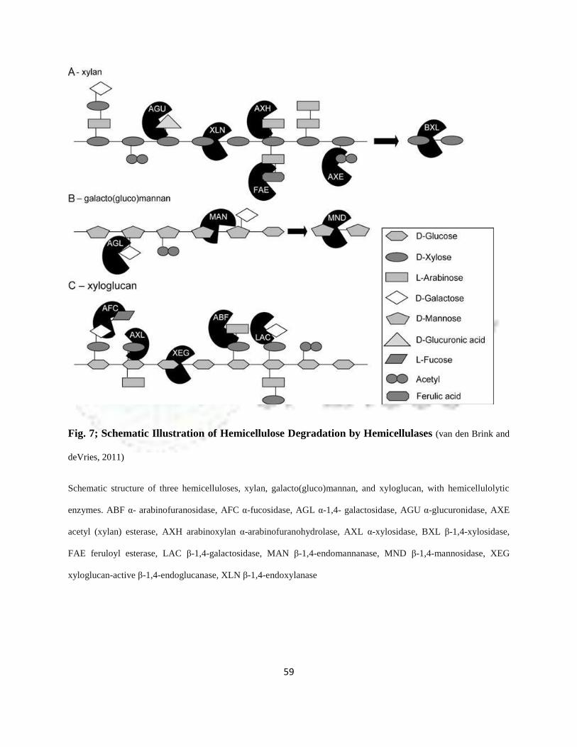
59
Fig. 7; Schematic Illustration of Hemicellulose Degradation by Hemicellulases (van den Brink and
deVries, 2011)
Schematic structure of three hemicelluloses, xylan, galacto(gluco)mannan, and xyloglucan, with hemicellulolytic
enzymes. ABF α- arabinofuranosidase, AFC α-fucosidase, AGL α-1,4- galactosidase, AGU α-glucuronidase, AXE
acetyl (xylan) esterase, AXH arabinoxylan α-arabinofuranohydrolase, AXL α-xylosidase, BXL β-1,4-xylosidase,
FAE feruloyl esterase, LAC β-1,4-galactosidase, MAN β-1,4-endomannanase, MND β-1,4-mannosidase, XEG
xyloglucan-active β-1,4-endoglucanase, XLN β-1,4-endoxylanase
60
2.5.3. Ligninase
Lignin degradation is inevitable for industrial enzymatic biomass-conversion because it not only
increases hemicelluloses and cellulose accessibility for their respective enzymes but also
diminishes hemicellulase and cellulase inactivation caused by lignin adsorption (Sweeney and
Xu, 2012). Fungi degrade lignin by secreting enzymes collectively termed ―ligninases‖. These
include two ligninolytic families; (i) phenol oxidase (laccase) and (ii) peroxidases [lignin
peroxidase (LiP) and manganese peroxidase (MnP)] (Martinez et al., 2005). Interestingly, LiP is
able to oxidize the non-phenolic part of lignin, but it was not detected in many lignin degrading
fungi. In addition, it has been widely accepted that the oxidative ligninolytic enzymes are not
able to penetrate the cell walls due to their size. Thus, it has been suggested that prior to the
enzymatic attack, low-molecular weight diffusible reactive oxidative compounds have to initiate
changes to the lignin structure (Dashtban et al., 2009).
Lignin peroxidase (EC 1.11.1.14), Manganese peroxidase (EC 1.11.1.13), and versatile peroxidase
(EC 1.11.1.16) are extracellular fungal heme peroxidases (belonging to LO2 family) with high
potency to oxidatively degrade lignin. Upon interaction with H2O2, these enzymes form highly
reactive Fe(V) or Fe(IV)-oxo species, which abstract electrons from lignin (to cause oxidation or
radicalization) either directly or via Mn(III) species. Laccase (EC 1.10.3.2) is a multi-copper oxidase
(belonging to LO1 family) secreted by numerous lignocellulolytic fungi. This enzyme can directly
oxidize phenolic parts of lignin, or indirectly oxidize non-phenolic lignin parts with the aid of
suitable redox-active mediator (Sweeney and Xu, 2012).
2.5.4 Emerging Cell Wall Degrading Enzymes
61
Besides the major lignocellulolytic enzymes classifications, the microbes producing lignocellulolytic
enzymes secrete some proteins, which co-induced and secreted with cellulases and hemicellulases,
with more mysterious functions. Some of these molecules, when added to cellulases, enhance their
performance, while others has little or no stimulatory effects on cellullase activity (Sweeney and Xu,
2012). A proper understanding to how these enzymes function will lead to improvements in
industrial lignocellulose degradation,
2.5.4.1 Cellulase-Enhancing Proteins
Analysis of the polysaccharide cleavage products formed by GH61 and CBM33 molecules reveals
that these enzymes release oxidized cellooligosaccharides, though significant quantities of non-
oxidized oligosaccharides are also detected in some studies (Forsberg et al., 2011; Quinlan et al.,
2011; Westereng et al., 2011). Unlike other glycoside hydrolases, which are more active on
cellooligosaccharides than crystalline cellulose, GH61 and CBM33 enzymes appear inactive upon
cellooligosaccahrides (Forsberg et al., 2011; Quinlan et al., 2011). The position of the oxidation on
the oligosaccharide products has been reported on the reducing end (Langston et al., 2011; Phillips et
al., 2011), non-reducing end (Langston et al., 2011; Phillips et al., 2011), or both (Quinlan et al.,
2011), which could suggest differences amongst these enzymes. GH61 proteins also require a metal
for this cellulose cleaving activity, specifically copper which binds tightly to the protein in a type-2
copper site geometry (Vaaje-Kolstad et al., 2010). Interestingly, despite the large potential of this
enzyme class to promote lignocellulose breakdown, the precise cleavage mechanism of GH61 and
CBM33 enzymes is unclear and is the subject of ongoing study by multiple groups.
2.5.4.2 Cellulose Induced Proteins
62
CIP1 and CIP2 (cellulose induced protein-1 and -2,) were first found in a transcriptional analysis of
T. reesei. Both contain a carbohydrate binding module and are co-regulated with known cellulases
(Foreman et al., 2003). The function of CIP1 is unknown, though it is claimed that CIP1 from T.
reesei has weak activity on p-nitrophenyl β-D-cellobioside (Foreman et al., 2011) and some
synergistic activity with both GH61 and swollenin (Scott et al., 2011). CIP2, found in both T. reesei
and Schizophyllum commune, has recently been shown to be an esterase that cleaves the methyl ester
of 4-O-methyl-D-glucuronic acid (Li et al., 2007). This enzyme, now classified as the first member of
CE15 family, likely acts in the cleavage of hemicellulose-lignin crosslinks. Further investigation of
both the functions and the potential of these enzymes in industrial applications are needed.
2.5.4.3. Expansin, Swollenin, and Loosinin
Expansins are of plant proteins which interact with cell walls and/or cell wall components by an
unknown activity. This interaction causes expansion or lengthening of cell wall structures. These
proteins have two domains. A domain is homologous to the GH45 endoglucanase catalytic core and
the other domain is homologous to Group II grass pollen allergens besides both domains have no
known catalytic function and display no detectable hydrolytic activity on lignocellulosic or model
substrates (Sweeney and Xu, 2012). Swollenin is also a protein causes a disruptive effect on
cellulosic substrates. It has sequence homology to plant expansions. It has been expressed in T. reesei
(Sweeney and Xu, 2012). Loosinin, like swollenin and expasin, increases cellulose activity (Quiroz-
Casterneda et al., 2011). They are produced by the basidiomycete fungus Bjerkandera adusta.
Bacterial species, including Bacillus subtilis (Kerff et al., 2008) and Hahella chejuensis (Lee et al.,
2010), also produces expansin-like molecules. Similar to fungal swollenins, bacterial expansins
modifies cellulose fiber structure and promotes the hydrolysis of cellulose without showing
detectable direct hydrolase activity (Kim et al., 2009)
63
2.5.4.4 Cellulosomes
Cellulosomes multiple cellulase and hemicellulase proteins, assembled by specific connections
between dockerin domains on the enzyme and cohesins bound to structural scaffoldins on the
microbial surface (Bayer et al., 2008; Fontes and Gilbert, 2010). These proteins are usually
produced by anaerobic organisms. This spatial clustering of multiple lignocellulose degrading
enzymes results in an increased synergy between lytic activities (Morais et al., 2011). It has been
shown that recombinant cellulosomes can be transplanted to other industrially useful organisms,
such as S. cerevisiae (Tsai et al., 2010) and B. subtilis (Anderson et al., 2011).
2.6 IMPROVING ENZYME PRODUCTION
There has been increasing interest in hyper-producing enzymes that have the potential to
hydrolyze lignocellulose. This is influenced by many factors which include the large potential
market for these enzymes, especially cellulase in the production of many products including
bioethanol, glucose syrups, animal feed, extractives etc. These have provided the platform and
incentives for the development of genetic improvement programmes which are based on
mutagenesis and selection (Chand et al., 2005) which produce new strains (Dillion et al., 2006)
and genetic transformants (Meittinen-Oinonem and Suominen, 2002).
There are a number of ways of improving enzyme production. These vary from co-culturing,
mutagenesis, genetics and recombinant gene technology to name a few. The techniques to be
used depend on the desired change in the organism.
2.6.1 Mutagenesis
64
Mutagenesis is a process by which the genetic information of an organism is changed in a stable
manner, resulting in a mutation. It may occur spontaneously in nature or as a result of exposure
to mutagens. It can also be achieved experimentally using laboratory procedures. Major
producers of fermentation products usually make use of mutation and selection. The production
of cellulases by the microbial cell is regulated by controls that include induction and catabolite
repression, or end product inhibition (Kumar et al., 2008). These controls are operative under
cellulase production conditions, thus resulting in limited yields of the enzymatic constituents.
There have been several attempts to improve enzyme yield using different mutagenic agents
combined or alone. Different techniques including UV-light and chemicals were used to induce
point mutations in fungi to enable them produce large amounts of degradative enzymes
(Dashtban et al., 2009).
Strain improvement has been achieved using chemical mutagens (Parekh et al., 2000), selection
and genetic recombination (De Nicolas-Santiago et al., 2006). Chand et al. (2005) used
simultaneous treatment of fungi with N-methyl-N‘-nitro-N-nitrosoguanidine (NTG), ethidium
bromide and UV light to enhance cellulase production more than wild type fungi. Abo-State et
al., (2010) isolated twenty-nine fungal strains and carried out solid-state fermentation (SSF) on
agricultural wastes and compared with Trichoderma reesei. They were able to enhance CMCase,
FPase, and β-glucosidase production by mutating with 0.5KGy dose of Gamma radiation.
Pradeep et al., (2012) subjected A. niger to mutations involving treatment with ethidium
bromide for the production of cellulases. Mutant strains were found to show increase in activities
of cellulase.
Mutagenic treatments of Trichoderma reesei Qm 6a, led to the development of mutants with
higher cellulolytic activity (Kumar et al., 2008). Vu et al. (2011) reported that a hypercellulolytic
65
mutant from Fusarium oxysporum was developed by ultraviolet treatment followed by chemical
mutagenesis using NTG (100 µg ml-1
). The resultant mutant strain had substantially higher
(80%) cellulolytic activity than its parent strain. NTG treatment of Cellulomonas Xavigena also
produced four mutants with improved xylanolytic activities. A mutant creAd30 with the end
product inhibition resistance showed improved levels of D-glucose metabolism and was
constructed from Aspergillus nidulans. However, this effort did not result in robust strains that
consistently produce ethanol at high yields under a broad range of conditions and in the hands of
different investigators (Kumar et al., 2008).
Ultraviolet (UV) light is one of the most utilised physical mutagenic agents. This may not be
unconnected to its high success rate. It has strong genotoxic effects to produce DNA damage and
induce mutations. UV produces specific DNA damage such as cyclobutane pyrimidine dimers
(CPDs) and pyrimidine-pyrimidone photoproducts (64PPs) at dipyrimidine sites, where two
pyrimidine (Py) bases are juxtaposed in tandem in the nucleotide sequence of DNA. These UV
lesions are formed through a photochemical reaction, whose efficiency depends on the
wavelength, following direct UV energy absorption by DNA bases. The yields of CPD and 64PP
are highest at around 260 nm, and the action spectra of their formation are in parallel to the
absorption spectrum of DNA. It is also known that 64PP can change into an isomeric secondary
product, Dewar valence isomer, after subsequent absorption of ultraviolet light band A (UVA)
with wavelengths around 325 nm (Matsunaga et al., 1991). These photolesions, CPD, 64PP and
Dewar, are assumed to cause UV-specific mutations (Ikehata and Ono, 2011).
2.6.2 Co-Culturing

66
Conversion of both cellulolytic and hemicellulosic hydrolytic products can be achieved by co-
culturing of two or more hydrolytic microorganisms. Co-culturing enhances hydrolysis of
lignocellulosic residues, and also enhances product utilization which diminishes the need for use
of additional enzymes in the bioconversion process. In the case of cellulose degradation, all three
enzymatic components have to be present in large amounts. Unfortunately, none of the fungal
strains, not even the best mutants, are able to produce high levels of the enzymes at the same
time (Dashtban et al., 2009). Conversion of both cellulosic and hemicellulosic hydrolytic
products in a single process can be achieved by co-culturing two or more compatible
microorganisms with the ability to utilize the materials. In fact, in nature, lignocellulosic residues
are degraded by multiple co-existing microorganisms. Co-culturing of two or more fungal strains
in fermentation is widely used in many biological processes including the production of
antibiotics, enzymes and fermented food (Ahamed and Vermette, 2008). Mixed fungal cultures
have many advantages compared to their monocultures, including improving productivity,
adaptability and substrate utilization.
Terijan and Xu (2011) was able demonstrate that co-culturing of various fungi could be
important in the complete biodegradation of lignocellulosic biomass since cellulase hydrolytic
reactions are prone to inhibition by some oligomeric phenolics contained in lignin molecules.
The main drawback of co-culturing however is the complexity of growing multiple
microorganisms in the same culture (Lynd et al., 2002). Alternative, to co-culture,
microorganisms can be metabolically engineered which and this enable one microorganism to
complete an entire task from beginning to end. This can be done by altering metabolic flux by
blocking undesirable pathway(s) and/or enhancement of desirable pathway(s).
2.7 HETEROLOGOUS CELLULASE EXPRESSION

67
2.7.1 Change of AT-Rich Sequences in Desired Gene
Partial processing of pre-mRNA can lead to limitation of heterologous genes expression at the
transcriptional level. It has been suggested that AT-rich sequences in the coding regions of
heterologous genes act as internal polyadenylation sequences and produce short and incomplete
transcripts. It has been reported that premature termination of transcription can be overcome by
replacing an AT-rich sequence in gene with a more GC-rich sequence or changing codon usage
(Te‘o et al., 2000). Also, the partial processing of transcripts has been overcome by increasing
the GC-content in the AT-rich area without changing the amino acid sequences of protein.
2.7.2 The Use of Strong Promoters in Desired Gene
Strong promoters have a high affinity for RNA polymerase. The strength and specificity of
promoter are characteristics of regulated endogenous gene expression and are inevitable for
genetic engineering of heterologous gene expression. Limited numbers of promoters allow high
level of transgenic expression, usually by ubiquitous and constitutive expression in all tissue
types. A promoter for transgenic expression in fungal strains should have certain characteristics
to be selected as a suitable promoter for a high-level expression of the interested gene. It must be
strong (30% or more of the total gene products), exhibit a minimal level of basal transcriptional
activity, be easily transferable to other vectors genomes and to be induced via simple, natural or
cost-effective manner (Koushki et al., 2011). In transformation constructs, promoters to be used
should be induced in a simple and natural condition. Gene expression of cell wall-degrading
enzymes (CWDEs) in Trichoderma spp. frequently has been reported to be induced by fungal
cell wall components and repressed by carbon catabolite repressors, such as glucose (Donzelli et

68
al., 2001). In some cases, starvation conditions alone could trigger CWDEs production, while in
others, cell walls or cell wall components were needed (Ramot et al., 2000).
2.7.3 The Construction and Use of Protease-Deficient Fungal Strains
Proteases are accountable for the degradation of many heterologous proteins (Van den
Hombergh et al., 1997). Fungi can secrete extracellular proteases. Extracellular proteases of
Aspergillus are responsible for the degradation of many heterologous proteins (Broekhuijsen et
al., 1993). Intracellular or cell wall localized proteases have been implicated for low yields of
secreted heterologous proteins as in the case of hIL-6 in A. awamori (Gouka et al., 1996a &b).
So far, most researchers focused their efforts on selecting and using protease deficient strains as
hosts for recombinant DNA to overcome protease degradation problems. However, such strains
are usually incapacitated and unsteady for large-scale bioreactor and biocontrol applications.
Therefore, it is often vital to match the protease loss of function strain to the desired protein and
identify genes involved in regulating extracellular protease secretion, since knocking out all
native proteases would likely be dangerous (Katz et al., 2000). Fungal strains deficient in
extracellular proteases have been constructed by random mutagenesis (Mattern et al., 1992; van
den Hombergh et al., 1997) or molecular genetic approaches (Berka et al., 1990; van den
Hombergh et al., 1997). The use of these protease deficient strains has resulted in the
enhancement of production level of heterologous proteins (Berka, 1991; Roberts et al., 1992;
Broekhuijsen et al., 1993). The use of strains deficient in vacuolar proteases has led to increased
level of heterologous proteins by Saccharomyces cerevisiae (Wingfield and Dickinson, 1993).
2.7.4 Optimization of Codon Usage of Desired Gene

69
The use of synonymous codons varies between different genes and organisms, the codon usage
of the desired gene can be optimized to perk up expression in each used transformants. Genes
can then be manipulated with rare codons replaced by their optimal synonyms and then
reconstructed by overlap extension of synthetic oligonucleotides. When the native family 11
xylanase gene (xynB) from Dictyoglomus thermophilum was introduced into T. Reesei, enzyme
was not produced (Hazell et al., 2000; Te‘o et al., 2002). It was explained that excessive
differences in the codon usage between the heterologous gene and the expression host could
prevent expression at the transcriptional level. They overcame this problem by constructing a
synthetic xynB gene carrying changes in 20 codons which was generated by advanced primer
extension PCR using the codon preference of highly-expressed T. reesei genes (Te'o et al.,
2000).
2.7.5 Glycosylation of Produced Heterologous Proteins
Wrong glycosylation of glycoproteins has been suggested as a problem for heterologous protein
processing. Engineering the protein glycosylation sites for enhanced usage in vivo has made huge
improvements in yield of several glycoproteins (van den Brink et al., 2006). N-glycosylation has
been shown to improve the amount of secreted chymosin in A. awamori, although, the specific
activity of the chymosin was reduced (Ward, 1989). It has been reported that the overexpression
of mannosylphospho dolichol synthase encoding gene from S. cerevisiae in T. reesei, which is
needed for O-glycan precursor synthesis, improved the production of CBHI (Kruszewska et al.,
1999). Overexpression of a yeast glycosylating enzyme has improved protein yields in an A.
nidulans mutant impaired in glycosylation (Perlinska-Lenart et al., 2005). These observations
could indicate that the problems encountered in glycoprotein production could be reduced by
manipulating glycosylation. Earlier studies with endogenous Trichoderma cellulases have
70
indicated that N-glycosylation of cellulases of T. reesei was not required for enzyme activity and
secretion (Kubicek et al., 1987), but had an effect on their thermostability and resistance to
proteolysis (Merivuori et al., 1985; Wang et al., 1996).
2.7.6 Use of Native or Artificial Intron-Containing Genes in Fungal Strains
In several eukaryotic genes, introns are necessary for efficient mRNA cytoplasmic accumulation
(Nesic et al., 1993). It has been suggested that introns regulate gene expression at transcription
and post-transcription (Morello et al., 2002; Sivak et al., 1999) levels. Introns might also protect
pre- mRNA from undergoing degradation in the nucleus, facilitate polyadenylation or transport
mRNA to the cytoplasm (Liu and Mertz, 1995). Jonsson et al., (1992) also concluded that certain
genes cannot be expressed without their introns. Two possible mechanisms for intron dependent
mRNA accumulation are commonly accepted. One is that introns protect the pre-mRNA from
degradation, either by accepting a stable secondary structure or by providing binding sites for
factors protecting the pre-mRNA, such as oligo-U binding proteins (Kurachi et al., 1995;
Gniadkowski et al., 1996) or heterogeneous nuclear ribonucleoproteins (Krecic and Swanson,
1999). Other mechanism is that spliceosome assembly onto the introns in pre-mRNA facilitates
an association with enzymes involved in other aspects of RNA maturation (such as
polyadenylation) and transport of the mRNA to the cytoplasm. Koziel et al., (1996) proposed the
phenomenon of intron-dependent mRNA accumulation in mammalian and plant cells. It also
proposed that the small introns in the genes of filamentous fungi may not only act as intervening
elements, but may also play vital roles in gene expression by increasing the stability of the
mRNA or by assisting the export of mRNA (Xu and Gong, 2003). In a trial, after introducing the
coding genes of ABH1 (Agaricus bisporus), SC3, SC6 (S. commune) and GFP (Aequorea
victoria), without introns, no or very low expression level were seen contrasting with the
71
expression of intron containing genes (Scholtmeijer et al., 2001). Xu and Gong (2003) showed
that introns are required for AFP gene (a gene encoding antifungal protein in A. giganteus)
expression in T. atroviride transformants, as demonstrated by the level of mRNA and confirmed
by analysis of AFP synthesis. However, in S. cerevisiae only 2 to 5% of the genes contain introns
and no obligation for introns has been observed for vertebrate and plant genes expression
(Hiraiwa et al.,1997; Lin et al., 1997).
2.7.7 Desired Gene Fusion with Well-Expressed Genes
A low mRNA stability can affect heterologous gene expression at the transcriptional level. Many
of mRNAs in eucaryotic cells are unstable, because they contain specific sequences (AU-rich) in
the 3' untranslated region (UTR) that induce their degradation (Koushki et al., 2011). mRNA
degradation is facilitated by AU-rich sequence. This is done by stimulating the removal of the
poly-A tail found at the 3' end of almost all eucaryotic mRNAs. Other unstable mRNAs contain
recognition sites in their 3' UTR for specific endonucleases that cleave the mRNA. Low mRNA
stability can be partly overcome by fusing the desired gene to the 3' end of a homologous gene.
Fusion of the gene to be expressed behind a highly expressed homologous gene does not only
increase stability of the mRNAs, but can also resolve limitations at early stages in the secretion
pathway (ER) of protein metabolites. The production of antibody fragments in T. reesei as a
CBHI fusion resulted in more than 150-fold increase in the yield (Koushki et al., 2011).
72
CHAPTER THREE
3.0 MATERIALS AND METHODS
3.1 MATERIALS
3.1.1 Source of Sawdust
Abura (Mitragyna ciliata) sawdust was collected from Okobaba Saw Mill, Ebute-metta (N06o
28. 714‘ E003o 23. 426‘). Lagos. The sawdust was pulverised and sieved by Jayant Test Sieves
(B. S. S. Mesh No. 60 and 0.250mm) to make uniform particle size and to obtain the respective
fine powder which was used for the study.
3.1.2 Chemicals
Media used were obtained from Biomark, India; yeast exract from Oxoid, England and other
chemicals were obtained from Fluka, Germany.
3.2 Pretreatment of Sawdust
Mechanical pretreatment was done by pulverizing the sawdust. The cellulosic substrate was
milled in a communitor and stored in polythene bags (De Sousa et al., 2004). Modified chemical
pretreatment methods using NH4OH as described by Abu et al., (2002) were used. Ammonia
solution (2.9M) was prepared to steep Abura sawdust. The substrates were left to steep for five
73
days after which they were properly washed and sun-dried. The pretreated substrates were
maintained to constant weight at 80oC in an oven.
3.3 Determination of Lignocellulosic Content of Sawdust
3.3.1 Determination of Cellulose Content of Sawdust
To 1g of pretreated and unpretreated dried sawdust samples, 15 mL of 80 % acetic was added
followed by 1.5 mL of concentrated nitric acid. It was refluxed for 20 min. It was filtered, the
residue was washed with ethanol, dried in the oven at 100-105 oC and weighed. Then it was
incinerated at 540 oC. Cellulose content was determined in accordance with the method of
Abdullah et al., (2006) using the formula below.
% cellulose = dried material – incinerated material
weight of sample
3.3.2 Determination of Hemicellulose Content of Sawdust
Pretreated and unpretreated dried sawdust samples (1g) were added to 15ml of 80% acetic acid
and 1.5mL of concentrated nitric acid and refluxed for 20 mins. It was filtered, the residue was
washed with ethanol, dried in the oven at 100-105oC and weighed. Then it was incinerated at
540oC. Cellulose content was determined in accordance with the method of Abdullah et al.,
(2006) using the formula below:
% hemicellulose = dried material – incinerated material
weight of sample
3.3.3 Determination of Lignin Content of Sawdust
Pretreated and unpretreated samples (1g) were dispensed into 2 different conical flasks. A 70ml
portion of 1.25 % H2SO4 (sulphuric acid) was added and the mixture was refluxed for 120min,

74
filtered and washed with water. Thirty mL of 72% H2SO4 was added to this material and allowed
to stand for 4hr with occasional stirring. It was dried at 105oC after previously filtered and dried,
then incinerated at 540oC.
Lignin content for both pretreated and unpretreated samples was calculated in accordance with
the method of Abdullah et al., (2006) using the formula below:
% lignin = dried material – incinerated material
weight of sample
3.4 Proximate Analysis
The methods of the Association of Official Analytical Chemists (A.O.A.C., 2005) was used to
determine the proximate composition of the unpretreated and pretreated sawdust.
3.4.1 Determination of Moisture Content
Dishes with lids were oven dried to remove traces of moisture and weighed. A 2 g portion of
each of the samples was weighed into dishes. The samples were dried in an oven at 100oC for
5hr without the lid. The samples were removed from the oven after drying and the lid replaced.
The dishes were placed in a dessicator containing silica, cooled to room temperature and
weighed. The processes of drying, cooling and weighing were continued until a constant weight
for each sample dish was obtained.
Calculation: % moisture = t-u/s×100
Where s = weight of sample for the analysis
t= weight of sample + dish before drying
u= weight of sample + dish after drying
t - u = moisture content or weight loss
75
3.4.2 Determination of Crude Protein Content
Finely ground sample (1g) was introduced into the Kjeldahl digestion flask. Concentrated H2SO4
(20ml) and 8 g of the catalyst (10 g K2SO4 and 0.5g HgO) were added to the flask and mixed to
speed up the digestion. Flask was placed on digestion unit and the sample digested in a fume
cupboard until a clear pale green solution was obtained and the time noted. The solution was
allowed to cool and 150mL of distilled water added, mixed and the content transferred to
distillation flask. Saturated 0.1M NaOH (80mL) was added to the distillation flask. The solution
was distilled with 50 ml of 2% Boric acid using methyl red indicator. The ammonium borate
formed was titrated with standard HCl and titre value noted.
Calculation:
0.01M HCl 1.40 mg of Nitrogen
Therefore the nitrogen content = titre value × 1.40
%N =1.40×XY
105
Where X is the titre value when the digest is distilled with Boric acid, Y is the weight of the
sample used. Crude protein (%) = 1.40XY/105×6.25 (a factor derived from the fact that protein
contains 16% Nitrogen).
Equation of the reaction:
Nitrogen in sample conc.
H
2SO
4 catalyst
NH3
The ammonia is produced in the form of ammonium tetraoxosulphate (vi), i.e.,
2NH3 + H2SO4→ NH4 (SO4)2

76
When the ammonium tetraoxosulphate (VI) is reacted with an alkali, such as NaOH, NH3 is
evolved and then trapped into boric acid.
(NH4)2SO4 + NaOH → NH2SO4 +2H2O +2NH3
H3BO3 +NH3→ NH4+H2BO3
-
When the complex is steam-distilled, the NH3 comes out as NH4OH which was titrated with
HCl. Thus,
NH4OH +HCl → NH4Cl + H2O
3.4.3 Determination of Ash Content
Four dry porcelain dishes were weighed using an analytical balance. A 2g portion of each the
samples was weighed separately into the dish and dried in an oven for 3 hr. The dishes were
heated on a bunsen burner until the content turned black. The dishes and contents were placed
into a muffle furnace and heated for 7hr until greyish white residue was obtained. The dishes
were removed and some drops of water added to expose unashed carbon still present. The dishes
were dried again in oven for 3hr and re-ashed in muffle furnace for 45min. The dishes were
removed and cooled. The cooled dishes were placed in a desiccator to cool to remove traces of
moisture. The dishes with the contents were weighed to determine the level of ash using the
calculation below.
Calculation
Ash (%) = 100(X-Y)
Z
Where X= weight (g) of dish and content after drying
Y= weight (g) of empty dish
Z= weight of sample (g) used for the analysis

77
3.4.4 Determination of carbohydrate content
Samples (2 g) were weighed on filter paper and transferred to a measuring cylinder containing 10
ml of distilled water. Fifty two percent cold perchloric acid (10 mL) was added to the dissolved
sample and stirred for 30 min. The solution was further diluted to 100mL with distilled water.
The solution was filtered into a 250 mL graduated flask and filled to mark. A 45 mL portion of
the extract was diluted to 450 mL with distilled water. 1mL of the diluted extract was pipetted
into test tube using 1mL of distilled water as blank and glucose as standard (0.15 mg/mL). A
5 mL portion of anthrone reagent was added to the tubes and incubate in a water bath for 12 min;
cooled and absorbance was read at 630 nm with a spectrophotometer.
Calculation
% glucose = 25 A1/ A2 × 100
Where A1 = Absorbance of diluted sample
A2 = Absorbance of diluted standard
3.4.5 Determination of Crude Fat
Samples (2 g) were weighed on Whatman 542 filter paper and transferred to extraction thimble.
The thimble was placed in a beaker and put in an oven for 7 hr to expel traces of moisture. The
beakers were removed from the oven and placed into the soxhlet apparatus and the solvent
mixture (chloroform : methanol 2:1) was added. The lipid content was extracted at condensation
rate of 240 drops/min for 7hr. The extract was transferred to an already weighed evaporating
dish. The solvent was allowed to evaporate in a fume chamber and dried in an oven at 100⁰C for
25min. The dish was removed and taken to a dessicator to cool and weighed.
Calculation
Crude fat (%) = (X-Y)
78
Z × 100
Where X = weight (g) of dish and content after drying
Y = weight (g) of empty evaporating dish
Z = weight of sample taken for analysis
3.4.6 Crude Fiber Content
This was determined by subtracting the proximate components from 100% as shown below as
shown below:
%Crude fiber = 100 - (%ash +%protein+%moisture+%CHO+%crude fat)
3.5 Isolation of Fungi
3.5.1 Organisms and Culture Conditions
Fungi were isolated from decomposing wood wastes obtained from Okobaba Sawmills, Ebute-
metta, south-west Nigeria, using different selective and differential media (Guarro et al., 1999).
They were maintained on potato dextrose agar, PDA (Biomark Laboratories, Pune, India) and
modifed Czapek Dox agar (CDA) under ambient laboratory conditions (28 ± 2 oC, 12hr light and
12hr darkness). Morphological studies of their mycelia were done by inoculation of sample from
pure culture into PDA broth and Czapek Dox broth into 100ml conical flasks and were
maintained at 28 ± 2 oC for 5 days. The fungi were identified from their cultural and
morphological characteristics by Prof. Adejare Adekunle at the Department of Botany,
University of Lagos, Nigeria.
3.5.2 Macroscopic and Microscopic Study
Macroscopic study was done by studying the growth rate, texture, pigmentation of both top and
reverse plates, and topography of colony using PDA, yeast extract agar and Czapek Dox agar.
79
Microscopic study was done by preparing slide mount with lactophenol cotton blue stain and
observed under light microscope.
3.6 Genotyping of Selected Isolates
3.6.1 Harvesting of Mycelia
Mycelia of isolates were harvested from Czapek Dox media grown for 36 hr in 100 mL flask
containing 30 mL of modified Czapek Dox media which contained: 3 g NaNO3, 0.5 g KCl, 0.5 g
MgSO4, 1 g KH2PO4, 0.01 g FeSO4, 20 g glucose, 20 g agar per litre (Nwodo-Chinedu et al.,
2007). The mycelia were collected by filtration/decantation through muslin cloth, washed with
cold sterile water and cold acetone on a Buchner funnel. The shrunken mycelia material was then
washed for 3hr under cold acetone. The dehydrated mycelia so obtained were desiccated under
vacuum (to remove vapour) for 20 min. The dried mycelia were stored at 4 oC in the refrigerator
for use.
3.6.2 DNA Extraction
The extraction was achieved using the modified method of Keb-Llanes et al., (2002). Mycelia of
isolates were harvested from Czapek Dox media grown for 36 hr in 100 mL flask containing 30
mL of Czapek Dox media (per litre). The mycelia were collected by filtration through muslin
cloth, washed with cold sterile water and cold acetone on a Buchner funnel. The shrunken
mycelia material was washed for 3 hr under cold acetone. The dehydrated mycelia so obtained
were dessicated under vacuum for 20 min. The dried mycelia obtained from each (500mg)
isolate was washed twice with sterile Ca2+
/Mg2+
free phosphate buffered saline (PBS) at pH 7.4.
200µL of N-Cetyl-N,N,N- trimethylammonium bromide (CTAB) buffer (2X) containing 55
mmol/L CTAB, 1.4 mol/L NaCL, 20 mmol/L EDTA and 0.1 M Tris (pH 7.0) was added to the
80
washed pellets. Pellets were crushed with a sterile dounce homogenizer and incubated at 65 oC
for 2h r with occasional vigorous shaking. A 120 µL portion of chloroform/isoamylalcohol
(24:1) was added and the mixture was placed on an orbital shaker at room temperature for
20min. Mixture phases were resolved by centrifugation at 4000 rpm for 30 min. The aqueous
phase was carefully pipetted into a fresh microcentrifuge tube and 300 µL of pre-chilled
isopropanol was added and incubated at -80oC for 1hr. DNA pellets was collected by
centrifugation at 3000 rpm for 5min, the supernatant was discarded and the pellet was washed
with 70 % ethanol twice and dried. Pellets were dissolved in 100µL TE buffer (10 mM Tris: 1
mM EDTA, pH 8) after extraction by heating to 56°C for 10min.
3.6.3 DNA Quantification
The integrity of the isolated DNA was confirmed by running agarose gel electrophoresis (0.8%
gel strength) alongside a negative control. Staining was done by mixing 5µL of DNA template
with 2µL (each) of loading dye and SyberGreen. Electrophoresis was carried out at 120V, 50Hz,
300mA for about 30min. Bands were viewed using BioRadTM
Gene Viewer. DNA quantification
was done using nanodrop spectrophotometric measurement of UV absorption of 260nm and
280nm (Tiwari et al., 2011). DNA concentration was calculated using the formula:
DNA concentration (µg/mL) = OD at 260nm X dilution times X standard value (1nm =50µg).
3.6.4 PCR Amplification of ITS1, 5.8S, and ITS2 fragments
PCR amplification of the internal transcribed spacer regions ITS1, ITS2, and 5.8S of the rRNA
gene cluster (Bellemain et al., 2010) was performed in a 25µL reaction containing 5µL total
genomic DNA template, 1X buffer, 0.2mM dNTP mix, 0.25 µM of each primer pair ITS1(5‘-
TCC GTA GGT GAA CCT GCG G-3‘), ITS4 (5‘- TCC TCC GCT TAT TGA TAT GC-3‘) and
81
0.04 U/µL High Yield taq polymerase (Jena Biosciences, Germany). The PCR conditions were
initial denaturation at 94°C for 5 min, followed by 35 cycles for 30sec at 94°C, 30sec at 52°C,
and 30 sec at 72°C. A final extension step was conducted at 72°C for 10 min. The PCR products
were analyzed by electrophoresis on 1.2% agarose gel containing ethidium bromide in tris-
acetate EDTA buffer at 120V for 25min. Gel Images were captured and analyzed using Gel
analyzer version 2010a.
3.6.5 Sequencing of PCR Amplified Fungal ITS fragments
PCR amplicon bands of approximately 648 bp on the 1.2 % agarose gel were excised and
purified using the NucleoSpin® Gel and PCR clean-up kit (Macherey-Nagel, Germany) as
specified by the manufacturer. Purified PCR amplicons were sequenced in one direction using
the forward primer (ITS1 primer) on an ABI377 DNA sequencer (Jena Bioscience, Germany).
3.6.6 Molecular Phylogenetic Analysis
The 18S rDNA and 5.8S rDNA sequences were compared to sequences received from GenBank
database by using BLASTN algorithm version 2.2.28+ (http://www.ncbi.nlm.nih.gov/BLAST/).
Other known sequences of similar fungal species were downloaded and aligned using CLUSTAL
W of the MEGA-5 Software (Tamura et al., 2011). Aligned sequences were pruned and edited
with Jalview software 2.8 (Waterhouse et al., 2009). Phylogenetic analyses were conducted in
MEGA-5 (Tamura et al., 2011); evolutionary history was inferred using the Maximum
82
Likelihood method based on the Tamura-Nei model with 1000 replicates of bootstrap cycles
(Tamura and Nei, 1993).
3.7 Co-culturing of Fungi for Biodegradation of Sawdust
Cellulolytic activity of the isolates was determined and two fungi with lead activities were
selected. The effect of biodegradation using single cellulolytic organism or synergistic
combination of organisms was assessed. The effect of pretreatment was also determined using
these two organisms on the substrate. Ten discs (8mm size) of fungal spores which were freshly
sub-cultured on PDA were used to inoculate 5g of the pretreated and the unpretreated sawdust
submerged in mineral salt media earlier described. Control was set up using pretreated and
unpretreated sawdust submerged in flasks containing the media without the organisms. The pH
of the medium, released reducing sugar, enzyme activity, and protein were monitored using
modified methods of Nwodo-Chinedu et al., 2007.
3.8 Determination of Optimum Fermentation Conditions
The modified method of Acharya et al., (2008) was adopted for the optimization studies.
Optimum substrate concentration was determined by carrying out fermentation with different
amounts of pretreated sawdust (2.4-12 % w/v) as substrate in the fermentation media.
Determination of optimum pH (4.0-6.0) was carried out. The different nitrogen sources
optimized were peptone, NaNO3, (NH4)2SO4 and urea. Peptone was used in range from 0.05-
0.15% w/v. (NH4)2SO4 was used in range from 0.1-0.18 % w/v while sodium nitrate was used in
the range 0.05-0.15% respectively. However, urea was used in range from 0.01-0.05 % w/v. All
the flasks were inoculated with 10 discs of 8 mm size of A. niger and P. citrinum and incubated
83
at 28 ± 2°C in a 250ml Erlenmeyer flask in an orbital shaker incubator at 120 rpm. At regular
intervals, enzyme assays were performed.
3.8.1 Media Preparation and Enzyme Production
Medium composition described by Mandels and Weber as reported by Acharya et al., (2008) was
used for fermentation. The media contained 0.3 g Urea, 1.4 g (NH4)2SO4, 2.0 g KH2PO4, 0.3 g
CaCl2, 0.3 g MgSO4.7H2O, 1.0 g protease peptone, 5.0 mg FeSO4.7H2O, MnSO4.7H2O 1.6 mg,
ZnSO4.7H2O 1.4 mg, CoCl2 2.0 mg per litre. The pH of media was adjusted to 5.0 ± 0.2.
3.8.2 Reducing Sugar Assay
Extract from the fermentation crude was centrifuged and the supernatant (1mL) was collected
into test tubes. 1mL of prepared DNS reagent was added. It was boiled for 5min and allowed to
cool before the absorbance was read at 540nm against blank (Singh et al., 2009).
Enzyme assay was carried out according to the methods described by Singh et al. (2009).
Carboxyl-methyl cellulase (CMCase) activity was determined at 40oC by using carboxy-methyl
cellulose as substrate. For total cellulase/filter paper (FPase) activity, Whatman No. 1 filter paper
strip of dimension 1.0 x 6cm (50mg) was placed into each assay tube. The filter paper strip was
saturated with 1.0ml of Na-Citrate buffer (0.05M, pH 4.8) and was heated for 10min at 50OC.
Half milliliter of an appropriate diluted enzyme (in Na-citrate buffer, 0.05M; pH 4.8) was added
to the tube and incubated at 50OC for 60min. Cellulase assay was determined using the method
of Singh et al., (2009).
84
3.8.3 Protein Content Determination
Five microlitre each of bovine serum albumin (BSA) standard and tests were pipetted into the
appropriate microplate wells (Thermo Scientific PierceTM
96-Well Plates). 250µL of Coomassie
blue reagent was added to each well and was mixed with plate shaker for 30min. It was removed
from plate shaker and incubated at room temperature for 10min in order to obtain a consistent
result. The absorbance was measured at 595nm with a plate reader. The average measurement for
the blank replicates was subtracted from all other measurements (standard and unknown
samples). Standard curve was plotted from which protein concentration of the tests were
determined.
3.9 Mutagenesis of Fungi for Hyper-Production of Cellulase
3.9.1 UV Mutation
The mutagenesis step involved finding the exposure time at which 50% of the fungal spores were
inactivated by ultraviolet radiation (LD50) at different distances. Four days old spores of fungi
were harvested, counted in a Neubauer chamber and their concentration adjusted to
1x10⁶spores/mL. Fifteen mL of the spore suspension were poured into sterile petri dish and
agitated during the procedure. The ultraviolet radiation source was placed at different distances
above the surface of the spores suspension (10-50cm), radiation intensity was delivered at
300µW/cm2 (modified method of De Nicholas-Santiago et al., 2006) at different times ranging
from 10-60 mins. Samples were taken at intervals of 5 min, kept in the dark and concentration
adjusted to 1x10⁴spores/mL and inoculated in petri dishes containing mineral salt agar at 30
oC
for 72 hr.
85
3.9.2 Selection of Hyper Producing Mutants
The selection of mutants was based on the modified method of De Nicholas-Santiago et al.,
(2006). The parent and mutated strains were inoculated (0.5mL) separately in flasks containing
mineral salt medium (MSM) which contained 10g glucose, 0.3g Urea, 1.4g (NH4)2SO4, 2.0g
KH2PO4, 0.3g CaCl2, 0.3g MgSO4.7H2O, 1.0g protease peptone, 5.0mg FeSO4.7H2O, 1.6mg
MnSO4.7H2O, 1.4mg ZnSO4.7H2O, 2.0mg CoCl2 and 1.0g 2-deoxy-D-glucose. The pH of media
was adjusted to 5.0 ± 0.2, and incubated at 30oC for 72hr. The contents of the flasks were
harvested by centrifugation at 4000 x g for 10min. The clear extract was used as enzyme source.
The crude enzyme was subjected to screening using the cup-plate method. Two grams of
ammonium oxalate, 2g of agar and 1g of cellulose were dissolved in 100ml of water in a 150ml
conical flask. The mixture was autoclaved at 121oC for 15min. It was then poured into sterile
petri dishes and allowed to solidify. Holes were made into the agar which were filled with 1ml of
the crude enzyme and sealed with cellophane paper. The plates were incubated at room
temperature for 24hr. The zone of hydrolysis was detected with potassium iodide-iodine solution
which contained 1.0g Iodine, 5.0g potassium iodide and 330ml H20/Litre. Potency index was
calculated as the ratio of diameter of clearance (DC) to diameter of cup (DC0).
3.10 Improvement of Fungal Cellulase Production by Sequential UV Mutation and
Optimizing Solid State Fermentation
The effect of various physicochemical parameters required for maximal production of cellulase
by the A. niger and P. citrinum mutants were investigated using solid state fermentation (SSF).
The parameters optimized were moisture (10-60 % v/w), incubation temperature (25-500C), pH
(3-6), fermentation time (1-8 days), size of inoculum (%, w/w), and age of culture medium
86
(days). Studies were also done to investigate the effect of various supplementations into the
culture on cellulase production. The supplementations include carbon source (glucose, lactose,
sucrose, maltose, corn starch), surfactants (sodium dodecyl sulfate SDS, ethylene-diamine-tetra
acetic acid EDTA, Tween 20, Tween 80), metal salts (ZnSO4, FeSO4, CuSO4, CaCl2, MgCl2,
CoCl2, KCl), and nitrogen supplements (malt extract, yeast extract, ammonium nitrate,
ammonium chloride, urea).
3.11 Partial Purification and Characterization of Cellulase
The modified method of Kolawole et al., (2006) was followed in the partial purification and
characterization of the cellulase enzyme. The crude enzyme preparation was obtained from 72h
solid-state fermentation.
3.11.1 Cellulase Production
The medium composition described by Mandels and Weber as reported by Acharya et al., (2008)
was used for fermentation. The media contained 20g glucose, 0.3g Urea, 1.4g (NH4)2SO4, 2.0g
KH2PO4, 0.3g CaCl2, 0.3g MgSO4.7H2O, 1.0g protease peptone, 5.0mg FeSO4.7H2O,
MnSO4.7H2O 1.6 mg, ZnSO4.7H2O 1.4 mg, CoCl2 2.0 mg per litre. The pH of media was
adjusted to 5.0 ± 0.2. After sterilization at 121°C for 15min, flasks were cooled, inoculated
(1·105 spores/mL) and incubated at 30°C for a period of 72hr. The content of the flask was
harvested by adding 100mL of 50mM Na-Citrate buffer pH 5 and centrifuged at 5000 x g for 10
min, the clear filtrate was used as crude enzyme. The clear filtrates were pooled and freeze dried
at 4oC, to about 120mL.
3.11.2 Ammonium Sulphate Precipitation
87
The concentrated fractions of the crude enzyme (50mL) were precipitated using 10-80%
(NH4)2SO4 precipitation with gentle agitation until final dissolution of the salt. The mixture was
centrifuged at 1000 x g for 20 min. The precipitate was reconstituted in 50 mM Na-Citrate buffer
pH 5 and dialyzed over-night at 4oC against the same buffer.
3.11.3 Anion Exchange Chromatography
Gel chromatography was carried out using anion exchanger; Sephadex A25-120 and Whatman
DE-52. 10 g of Sephadex A25-120 was suspended in Tris-HCl buffer pH 8.3, to equilibrate and
allowed to swell for 10hr. After swelling it was packed in a vertical glass tube chromatography
column of 1.5 × 70cm. The column was eluted several times with 0.05M acetate buffer (pH 5.0).
Whatman DE-52 suspension was prepared using the same buffer but allowed to swell for 5hr. The
void volume was determined with blue dextran. Ten millilitres of the ammonium sulphate-
dialysate enzyme concentrate was applied to the column eluted with buffer and fractions of 5mL at
a flow rate of 20mL h-1
were collected. The bound proteins were eluted with 0-1.0M NaCl in the
same eluting buffer. The pooled bound cellulase was dialyzed for 24hr with four changes of the
acetate buffer (0.05M, pH 5.0) and stored at -5oC. All purifications were carried out in the cold
laboratory.
3.11.4 Determination of Protein
Protein was determined spectrophotometrically at 280 nm using the Microplate method (Thermo
Scientific) and cellulase activity was assayed using the method of Singh et al., (2009).
Five microlitre each of Bovine Serum Albumin (BSA) standard and tests were pipetted into the
appropriate microplate wells (Thermo Scientific PierceTM
96-Well Plates). Two hundred and fifty
microliter of Coomassie blue reagent was added to each well and was mixed with plate shaker for
88
30min. It was removed from plate shaker and incubated at room temperature for 10min in order to
obtain a consistent result. The absorbance was measured at or near 595nm with a plate reader. The
average measurement for the blank replicates was subtracted from all other measurements
(standard and unknown samples). Standard curve was plotted from which protein concentration of
the tests were determined.
3.11.5 Effect of Substrate Concentration
The effect of various concentrations of carboxymethylcellulose CMC (2mg/mL-20mg/mL) on the
enzyme activity was studied at optimum temperature and pH. The maximum velocity (Vmax) and
Michaelis Menten constant (Km) of the enzyme were obtained using the Line-weaver Burk-plot.
3.12 GENETIC STUDIES
3.12.1 RNA extraction
Spores from the wild and mutated fungi were grown on PDA for four days. The spores were
inoculated in mineral salt media (MSM) and allowed to grow for another four days. Fungal
mycelia were harvested and 50µg was transferred aseptically into sterile eppendorf tubes
containing the lysis buffer.
The Direct-zol RNA MiniPrep kit (Zymo Research, USA) was used according to manufacturer‘s
instruction for the extraction.
3.12.2 RNA Quantification
89
This was done using the Nanodrop spectrophotometric technique as applied by Aranda et al.,
(2012). RNA sample (0.1µL) was introduced into the probe pedestal of the spectrophotometer
and the absorbance read at 230nm, 260nm and 280nm. The quality of the extracted RNA was
confirmed by running the samples in 1% agarose gel electrophoresis using 1X TAE buffer.
3.12.3 Reverse Transcription
This polymerase chain reaction based technique was accomplished using SCRIPT reverse
Transcriptase (Jena Bioscience, Germany) kit. The following components were mixed gently in a
nuclease-free microtube on ice: 5µg RNA template, 50-100ng primer, 1X SCRIPT RT complete
buffer, 500nM dNTP mix, 5mM DTT stock solution, 40 units RNase inhibitor, 100-200 units
SCRIPT reverse transcriptase, the content of the tube was made up to 20µl. The mixture was
incubated at 65-67oC for 5min after which it was placed at room temperature. Thermal
conditions applied were 42oC for 10 min, 50
oC for 10 min, 70
oC for 10 minutes and finally left at
4oC for an extended period.
3.12.4 Amplification of cDNA
This was carried out using the SCRIPT DNeasy kit (Jena Biosciences, Germany) according to
the procedure described by Bellemain et al., (2010).
PCR amplification of the cbh1 gene was performed in a 25µL reaction containing 2µL cDNA
template, 1X buffer, 0.2mM dNTP mix, 0.25 µM of each primer pair forward (5‘-GTC ATT
GAC GCC AAC TGG CGC TGG-3‘), reverse (5‘- ACG CTC CCA GCC CTC AAC GTT GG-
3‘) and 0.04 U/µL High Yield taq polymerase (Jena Biosciences, Germany). The PCR conditions
were initial denaturation at 94°C for 5 min, followed by 35 cycles for 30sec at 94°C, 30sec at
52°C, and 30sec at 72°C. A final extension step was conducted at 72°C for 10 min. The PCR
90
products were analyzed by electrophoresis on 1.2% agarose gel containing ethidium bromide in
tris-acetate EDTA buffer at 120V for 25min. Gel images were captured and analyzed using Gel
analyzer version 2010a.
3.12.5 Agarose Gel Electrophoresis
Amplifcation was confirmed by electrophoresing amplicons in 1% agarose. Staining was done
by mixing 5µL of cDNA template with 2µL of each of loading dye and SyberGreen.
Electrophoresis was carried out at 120V, 50Hz, 300mA for about 30min. On completion of
electrophoresis, bands were viewed using PrepOneTM
Sapphire Gene Viewer (Embi Tec).
3.12.6 cDNA Clean-Up
Gel slice containing the cDNA fragment of interest was excised using sterile sharp scalpel. The
Gel cleanup kit (MCLAB, USA) was used. The protocol for spin column chromatography as
described by manufacturer was followed. A 200mg portion of gel was transferred into sterile 2ml
centrifuge tube. 1ml of QG buffer (provided by manufacturer) was added onto it. It was
incubated at 37oC for 10min until the gel was completely dissolved. The column was placed in a
collection tube and 0.7ml of dissolved gel mixture was loaded into the column and centrifuged
for 60sec at 8000rpm. The flow-through was discarded. This step was repeated for the rest of the
mixture. The column was washed twice with PE buffer by centrifuging for 30sec. The flow
through was discarded. The column was centrifuged at full speed for 5 min to remove residual
ethanol. The column was placed in a new 1.5ml centrifuge tube and 30µl of Elution Buffer added
to the centre of the membrane. The column was centrifuged at maximum speed for 2min after
allowing it to stand for 10min. Eluted cDNA was stored at -20oC for further use.
91
3.12.7 cDNA Sequencing
Purified PCR amplicons were sequenced in one direction using the forward primers (lacc1, ace1,
aep1 and cbh1 primers) using Sanger dideoxy method on an ABI377 DNA sequencer (MCLAB,
USA).
3.12.8 Bioinformatics and Prediction of Cellulase Structure
The sequences of the mutant fungi amplified using primers designed for lacc1, ace1, aep1 and
cbh1 genes were compared to those in the NCBI GenBank database by using BLASTN
algorithm version 2.2.28+ (http://www.ncbi.nlm.nih.gov/BLAST/). Sequences of wild species of
the same organism were aligned using CLUSTAL W of the MEGA-5 Software (Tamura et al.,
2011). Aligned sequences were pruned and edited with Jalview software 2.8 (Waterhouse et al.,
2009). Regions where mutation has occurred were identified.
3.13 Statistical Analysis
All experiments were carried out in triplicates, unless otherwise stated. The results were
expressed as mean ± standard error of mean. The treatment means were tested for significant
difference and compared with the control using the Dunnet interval test. The Student‘s t-test was
used to compare between two groups. One-way analysis of variance (ANOVA) with Turkey‘s
test was used to compare the level of significant difference between the samples more than two;
p<0.05 was considered to be significant. These were done using the GraphPad Prism 5 statistical
package.
92
CHAPTER FOUR
4.0 RESULTS
4.1 Analysis of Lignocellulosic Content of Sawdust
The lignin, cellulose and hemicellulose content of both unpretreated and pretreated sawdust are
shown in Table 2. After the pretreatment process, the lignin content and the hemicellulose
content reduced showing the removal of part of hemicelluloses and lignin components as a result
of the pretreatment process. The cellulose and hemicellulose content of both types of sawdust are
however, significantly different from each other (p<0.05). This consequently makes cellulose
more accessible for microbial hydrolysis.

93
4.2 Proximate Composition of Sawdust
The sawdust was found to be high in crude fibre but low in carbohydrate, lipid, ash, protein and
moisture content (Table 3). However, when it was chemically pretreated, the moisture content
and crude fibre reduced significantly.
Table 2: Lignocellulosic Composition of Unpretreated and Pretreated Sawdust
Sawdust Type Lignin Content
(%)
Cellulose
Content (%)
Hemicellulose
Content (%)
Ash Content
(%)
Unpretreated 19.10±0.30 48.11±1.60* *32.70±2.20 0.09±0.10
Pretreated 15.20±0.20 64.94±1.20* *19.80±1.30 0.06±0.10
*The values were significantly different from each other (p < 0.05). Values are expressed as
Mean±SEM; N=3

94
Table 3: Proximate Composition of Unpretreated and Pretreated Sawdust
Sawdust type Moisture
(%)
Protein
(%)
Ash (%) Lipid
(%)
Crude
(%)Fibre
CHO (%)
Unpretreated 6.30±0.60* 2.50±0.20 1.70±0.10 6.40±0.40 62.20±3.40* 24.40±1.80
Pretreated 3.60±1.30* 4.00±0.20 2.00±0.10 5.70±0.20 52.80±2.20)* 29.80±1.60
*Values were statistically different from each other (p < 0.05). Values are expressed as
Mean±SEM; N=3

95
4.3 Isolation of fungi
Four foremost cellulase producers were retained for further studies. Selected fungi were
Penicillium corylophilum, Penicillium citrinum, Aspergillus niger, and Trichosporon asahii.
Colonies of Penicillium corylophilum were green in colour on PDA plate. The reverse side of
plate was yellow. Microscopic feature include septate hyphae (Fig.s 8A, 8E, and 8I). Colonies of
Penicillium citrinum were powdery and purplish-green in colour on PDA plates. The reverse side
of the colony was pale yellow in colour, similar characteristics were observed on Czapek Dox

96
media. Hyphae were septate and rough (Fig.s 8B, 8F, and 8J). Aspergillus niger was black in
colour on PDA plate. The reverse view of the plate was slightly yellow. Hyphae were septate and
rough with very dense spores (Fig.s 8C, 8G and 8K). Colonies of Trichosporon asahii on PDA
were white, creamy with a wide margin. The reverse side of the plate was also creamy in colour.
Hyphae were arial and smooth (Fig.s 8D, 8H, and 8L).
Fig. 8: Isolated Fungi from Sawdust Waste Cultured On PDA and Czapek Dox Agar
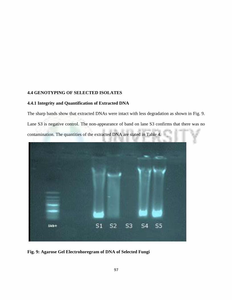
97
4.4 GENOTYPING OF SELECTED ISOLATES
4.4.1 Integrity and Quantification of Extracted DNA
The sharp bands show that extracted DNAs were intact with less degradation as shown in Fig. 9.
Lane S3 is negative control. The non-appearance of band on lane S3 confirms that there was no
contamination. The quantities of the extracted DNA are stated in Table 4.
Fig. 9: Agarose Gel Electrohoregram of DNA of Selected Fungi
98
S1, S2, S4 and S5 represent the DNA of Penicillium citrinum, Penicilium corylophylum,
Meyerozyme quillamondii, Aspergillus niger, and Trichosporon asahii respectively. To the
extreme left is the DNA ladder (1Kb+). S3 is a negative control, hence, no band shown on the
lane.
Table 4 below depicts the spectrophotometric measurements of the DNA samples. A260/280nm
measurements above 1.00 shows integrity of extracted DNA while the A260/A230nm
measurements below 0.50 shows no contamination in the extraction.
Table 4: Quantification of Extracted DNA
DNA Sample Quantity (g/mL) A260/280 nm A260/230 nm
S1 1.93 1.45 0.25
S2 1.75 1.87 0.47
S4 2.31 2.08 0.65
S5 2.53 2.41 0.23

99
4.4.2 Amplification of DNA of Isolates
ITS1/ITS4 primers amplified the selected genes with all the samples with band size of about
648bp. NS7/LR3 primers also amplified the selected genes with band size of 1642bp (Fig. 10).
Amplification of the rDNA regions with primers ITS1 and ITS4 yielded products of
approximately 700bp as estimated by agarose gel electrophoresis (Fig. 10). The sequences used
for the final phylogenetic analysis were 482 to 533 bp after manual counting trimming and
corresponded to the ITS 1 and 2 complete regions; the 5´ portion of the 18S gene, 5.8S
completes sequence and the 3´ end of the 28S gene. In addition to ITS 1, 5.8S rDNA and ITS 2,
such sequences contained the last base of the 28S rDNA and the first bases of the 18S rDNA.
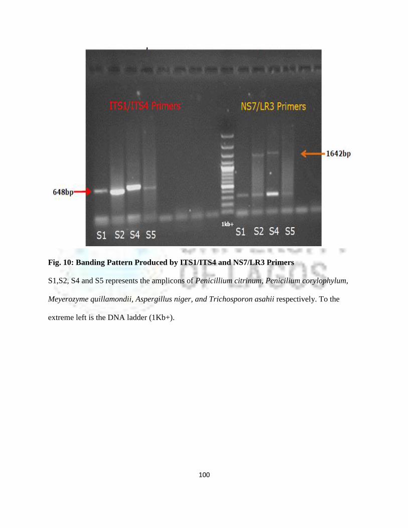
100
Fig. 10: Banding Pattern Produced by ITS1/ITS4 and NS7/LR3 Primers
S1,S2, S4 and S5 represents the amplicons of Penicillium citrinum, Penicilium corylophylum,
Meyerozyme quillamondii, Aspergillus niger, and Trichosporon asahii respectively. To the
extreme left is the DNA ladder (1Kb+).

101
4.4.3 Molecular Phylogenetic Analysis
Analysis revealed that fungi isolates were Aspergillus niger (Fig. 11), Penicilium citrinum,
Penicillium corylophilum (Fig. 12) and Trichosporon asahii. They all had boostrap values which
confirm their evolutionary and phylogenetic relationship with compared sequences.
4.5 Co-culturing of Cellulolytic Fungi in the Biodegradation Of Sawdust
An enzyme activity of 0.92 U/mL was attained in 96 hr when both fungi were co-cultured (Fig.
13). Pretreated sawdust fermented with co-cultured A. niger and P. citrinum released more
reducing sugar (2.72 mg/mL) from the sawdust at 72 hr (Fig. 14). Likewise, pretreated sawdust
fermented with co-cultured Aspergillus niger and Penicilium citrinum released more
cellobiohydrolase (0.58 U/mL) from the sawdust at 100 hr (Fig. 15).

102
Fig. 11: Phylogenetic Tree Showing Relationship Between Sample and Reference
Aspergillus species
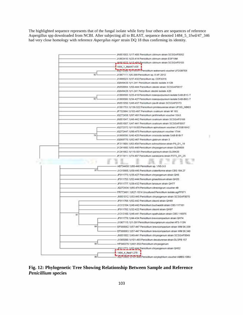
103
The highlighted sequence represents that of the fungal isolate while forty four others are sequences of reference
Aspergillus spp downloaded from NCBI. After subjecting all to BLAST, sequence denoted 1484_5_1fwd/47_346
had very close homology with reference Aspergilus niger strain DQ 18 thus confirming its identity.
Fig. 12: Phylogenetic Tree Showing Relationship Between Sample and Reference
Penicillium species
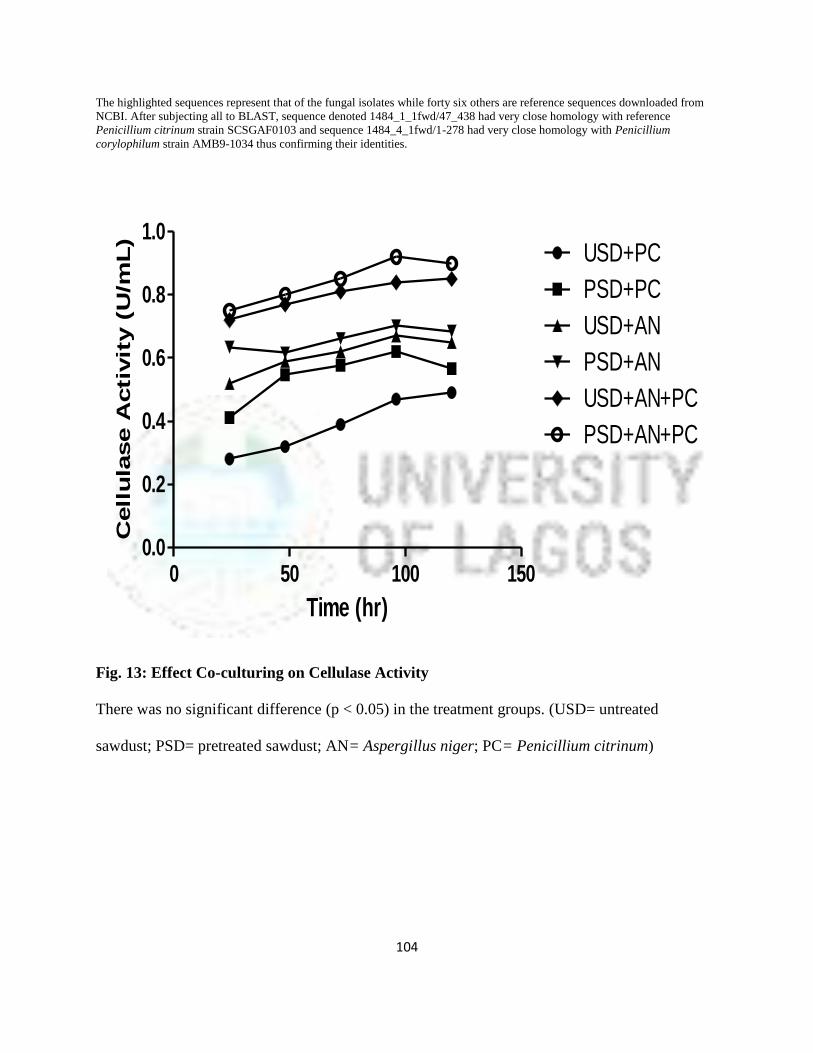
104
The highlighted sequences represent that of the fungal isolates while forty six others are reference sequences downloaded from
NCBI. After subjecting all to BLAST, sequence denoted 1484_1_1fwd/47_438 had very close homology with reference
Penicillium citrinum strain SCSGAF0103 and sequence 1484_4_1fwd/1-278 had very close homology with Penicillium
corylophilum strain AMB9-1034 thus confirming their identities.
0 50 100 1500.0
0.2
0.4
0.6
0.8
1.0USD+PC
PSD+PC
USD+AN
PSD+AN
USD+AN+PC
PSD+AN+PC
Time (hr)
Cell
ula
se A
ctiv
ity (
U/m
L)
Fig. 13: Effect Co-culturing on Cellulase Activity
There was no significant difference (p < 0.05) in the treatment groups. (USD= untreated
sawdust; PSD= pretreated sawdust; AN= Aspergillus niger; PC= Penicillium citrinum)
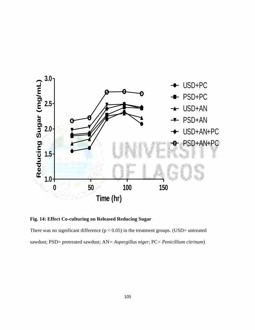
105
0 50 100 1501.0
1.5
2.0
2.5
3.0USD+PC
PSD+PC
USD+AN
PSD+AN
PSD+AN+PC
USD+AN+PC
Time (hr)
Red
ucin
g S
ug
ar (
mg
/mL
)
Fig. 14: Effect Co-culturing on Released Reducing Sugar
There was no significant difference (p < 0.05) in the treatment groups. (USD= untreated
sawdust; PSD= pretreated sawdust; AN= Aspergillus niger; PC= Penicillium citrinum)
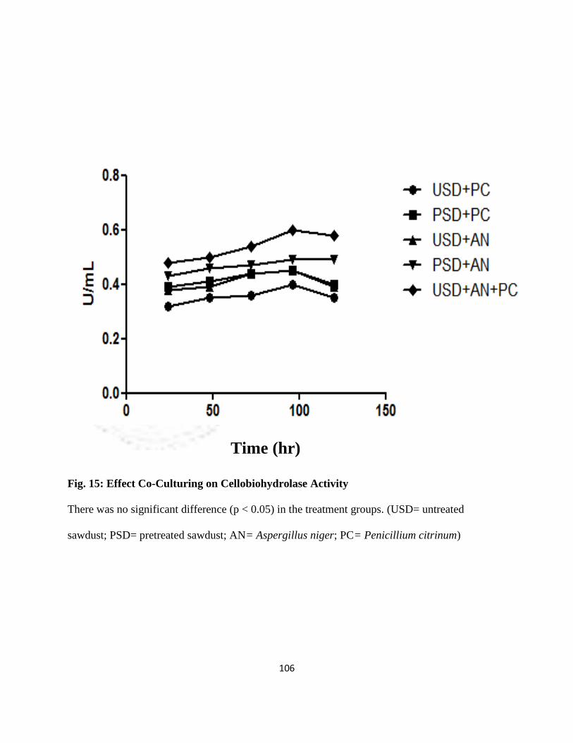
106
Time (hr)
Fig. 15: Effect Co-Culturing on Cellobiohydrolase Activity
There was no significant difference (p < 0.05) in the treatment groups. (USD= untreated
sawdust; PSD= pretreated sawdust; AN= Aspergillus niger; PC= Penicillium citrinum)
107
4.6 Mutagenesis of Fungi for the hyperproduction of cellulase
Eleven mutants of A. niger were selected after the irradiation. Fig. 16 below shows the
comparison of their cellulase activities with the wild strain. All had cellulase activities higher
than the wild strain. Mutant ANM202 was retained for further studies. Likewise, eleven mutants
of P. citrinum were selected after the irradiation (Fig. 17). All mutants had cellulase activity
more than the wild except PCM403, PCM502 and PCM504. Mutant PCM505 was retained for
further studies.
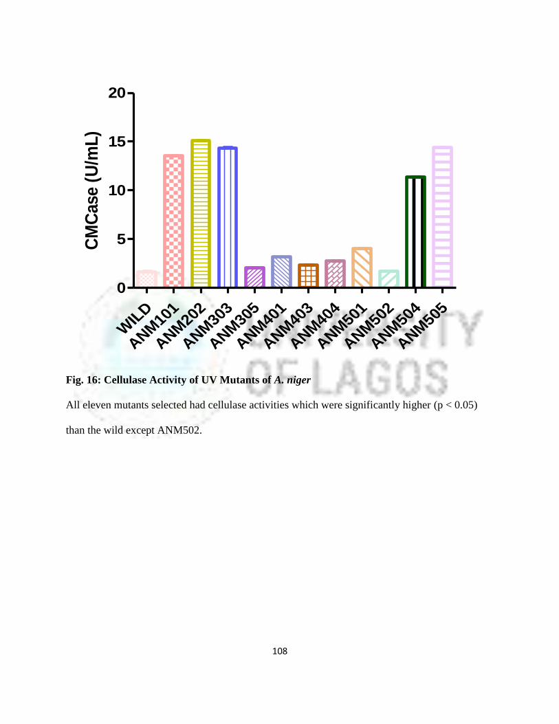
108
WIL
D
ANM
101
ANM
202
ANM
303
ANM
305
ANM
401
ANM
403
ANM
404
ANM
501
ANM
502
ANM
504
ANM
505
0
5
10
15
20C
MC
ase (
U/m
L)
Fig. 16: Cellulase Activity of UV Mutants of A. niger
All eleven mutants selected had cellulase activities which were significantly higher (p < 0.05)
than the wild except ANM502.
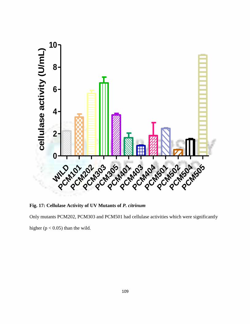
109
WIL
D
PCM10
1
PCM20
2
PCM30
3
PCM30
5
PCM40
1
PCM40
3
PCM40
4
PCM50
1
PCM50
2
PCM50
4
PCM50
5
0
2
4
6
8
10 c
ell
ula
se a
cti
vit
y (
U/m
L)
Fig. 17: Cellulase Activity of UV Mutants of P. citrinum
Only mutants PCM202, PCM303 and PCM501 had cellulase activities which were significantly
higher (p < 0.05) than the wild.
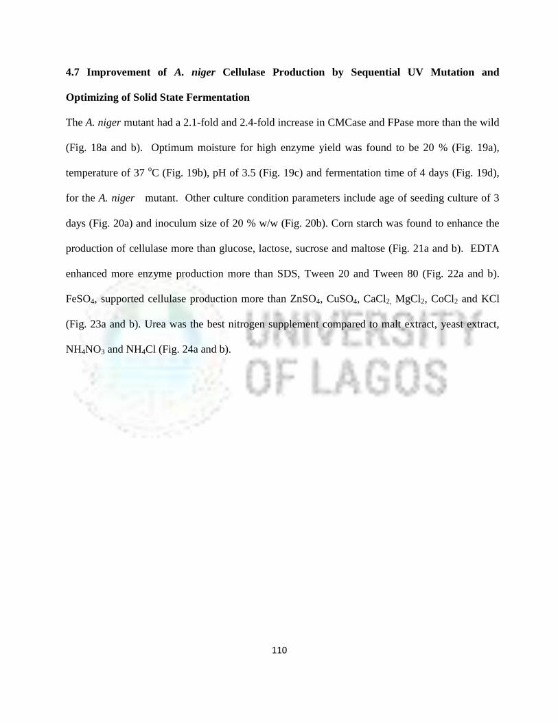
110
4.7 Improvement of A. niger Cellulase Production by Sequential UV Mutation and
Optimizing of Solid State Fermentation
The A. niger mutant had a 2.1-fold and 2.4-fold increase in CMCase and FPase more than the wild
(Fig. 18a and b). Optimum moisture for high enzyme yield was found to be 20 % (Fig. 19a),
temperature of 37 oC (Fig. 19b), pH of 3.5 (Fig. 19c) and fermentation time of 4 days (Fig. 19d),
for the A. niger mutant. Other culture condition parameters include age of seeding culture of 3
days (Fig. 20a) and inoculum size of 20 % w/w (Fig. 20b). Corn starch was found to enhance the
production of cellulase more than glucose, lactose, sucrose and maltose (Fig. 21a and b). EDTA
enhanced more enzyme production more than SDS, Tween 20 and Tween 80 (Fig. 22a and b).
FeSO4, supported cellulase production more than ZnSO4, CuSO4, CaCl2, MgCl2, CoCl2 and KCl
(Fig. 23a and b). Urea was the best nitrogen supplement compared to malt extract, yeast extract,
NH4NO3 and NH4Cl (Fig. 24a and b).
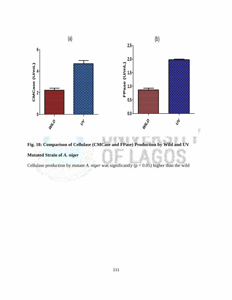
111
WILD
UV
0
2
4
6
(a)C
MC
ase (U
/m
L)
WILD
UV
0.0
0.5
1.0
1.5
2.0
2.5
(b)
FP
ase (U
/m
L)
Fig. 18: Comparison of Cellulase (CMCase and FPase) Production by Wild and UV
Mutated Strain of A. niger
Cellulase production by mutant A. niger was significantly (p < 0.05) higher than the wild
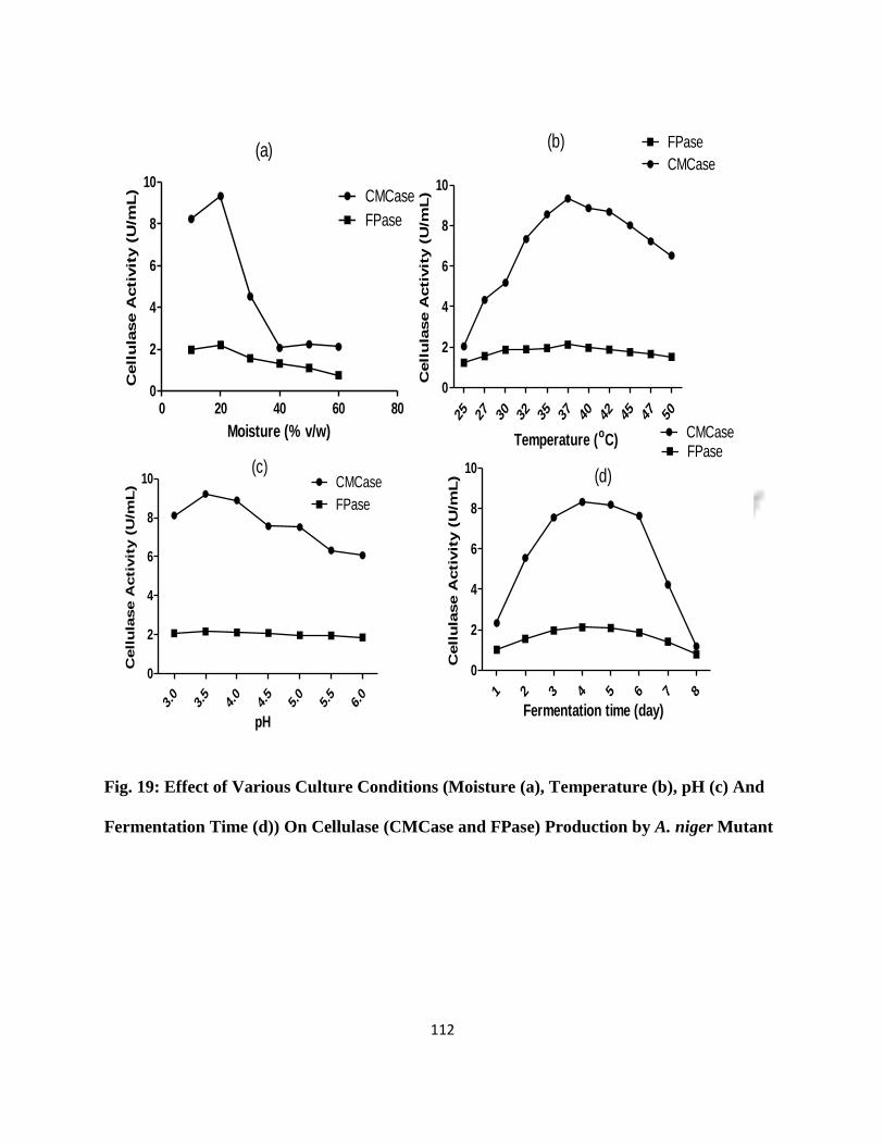
112
0 20 40 60 800
2
4
6
8
10CMCase
FPase
(a)
Moisture (% v/w)
Cell
ula
se A
cti
vit
y (
U/m
L)
25 27 30 32 35 37 40 42 45 47 50
0
2
4
6
8
10
CMCase
FPase(b)
Temperature (oC)
Cell
ula
se A
cti
vit
y (
U/m
L)
3.0
3.5
4.0
4.5
5.0
5.5
6.0
0
2
4
6
8
10 CMCase
FPase
(c)
pH
Cell
ula
se A
cti
vit
y (
U/m
L)
1 2 3 4 5 6 7 8
0
2
4
6
8
10
CMCaseFPase
(d)
Fermentation time (day)
Cell
ula
se A
cti
vit
y (
U/m
L)
Fig. 19: Effect of Various Culture Conditions (Moisture (a), Temperature (b), pH (c) And
Fermentation Time (d)) On Cellulase (CMCase and FPase) Production by A. niger Mutant
113
1 2 3 4 5 6
0
2
4
6
8CMCase
FPase
Age of culture (day)
cellu
lase activity (U
/m
L)
(a)
510
15
20
25
30
35
40
45
50
0
2
4
6
8
10CMCase
FPase
Inoculum size (%, w/w)cellu
lase activity (U
/m
L)
(b)
Fig. 20: Effect of Age of Culture Medium and Inoculum Size on Cellulase (CMCase and
FPase) Production by A. niger Mutant
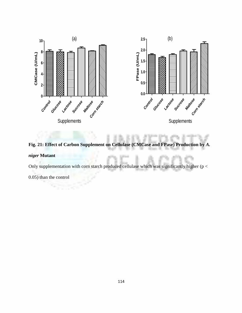
114
Control
Gluco
se
Lac
tose
Sucr
ose
Malto
se
Corn
starc
h
0
2
4
6
8
10
Supplements
(a)C
MC
ase (
U/m
L)
Control
Gluco
se
Lac
tose
Sucr
ose
Malto
se
Corn
starc
h
0.0
0.5
1.0
1.5
2.0
2.5
Supplements
(b)
FP
ase (
U/m
L)
Fig. 21: Effect of Carbon Supplement on Cellulase (CMCase and FPase) Production by A.
niger Mutant
Only supplementation with corn starch produced cellulase which was significantly higher (p <
0.05) than the control

115
Control
SDS
EDTA
Twee
n 20
Twee
n 80
0
5
10
15
Surfactants
CM
Case (
U/m
L)
(a)
Control
SDS
EDTA
Twee
n 20
Twee
n 80
0.0
0.5
1.0
1.5
2.0
2.5
SurfactantsF
Pase (
U/m
L)
(b)
Fig. 22: Effect of surfactants on cellulase (CMCase and FPase) production by A. niger
Mutant
Only supplementation with EDTA produced cellulase which was significantly higher (p < 0.05)
than the control
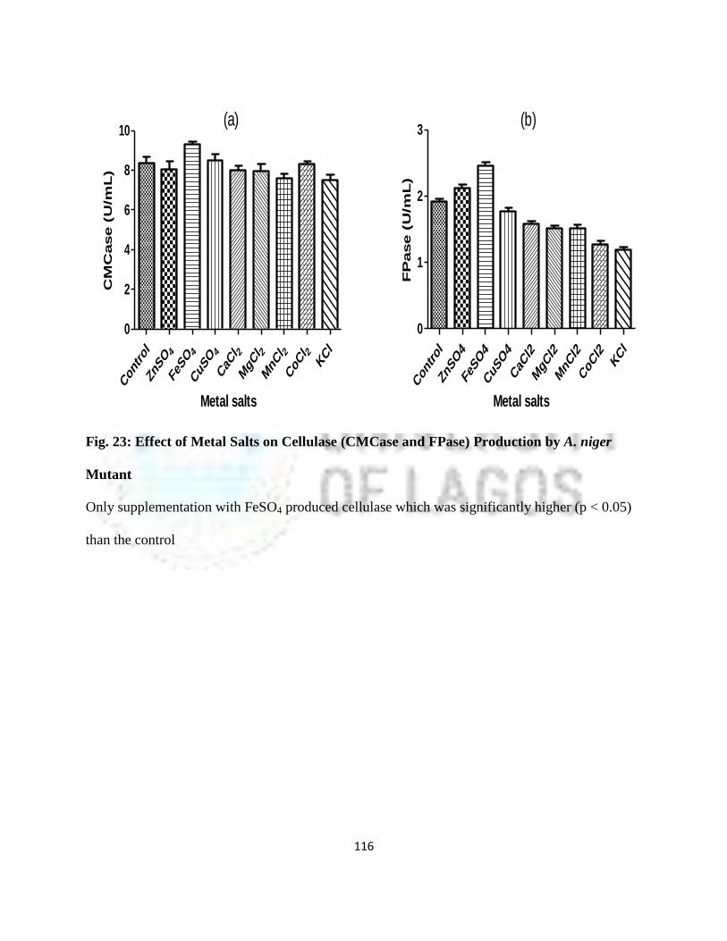
116
Control 4
ZnSO 4
FeS
O 4
CuSO
2
CaC
l 2
MgCl 2
MnCl 2
CoCl
KCl
0
2
4
6
8
10
Metal salts
CM
Case (
U/m
L)
(a)
Control
ZnSO4
FeS
O4
CuSO4
CaC
l2
MgCl2
MnCl2
CoCl2
KCl
0
1
2
3
Metal saltsF
Pase (
U/m
L)
(b)
Fig. 23: Effect of Metal Salts on Cellulase (CMCase and FPase) Production by A. niger
Mutant
Only supplementation with FeSO4 produced cellulase which was significantly higher (p < 0.05)
than the control

117
Control
Malt Extract
Yea
st Extract 3
NO
4
NH
Cl
4
NH U
rea
0
5
10
15
Nitrogen supplements
CM
Case (
U/m
L)
(a)
Control
Malt Extract
Yea
st Extract
NH4N
O3
NH4C
l
Urea
0
1
2
3
Nitrogen supplementsF
Pase (
U/m
L)
(b)
Fig. 24: Effect of Nitrogenous Salts on Cellulase (CMCase and FPase) Production by A.
niger Mutant
Only supplementation with Urea produced cellulase which was significantly higher (p < 0.05)
than the control.
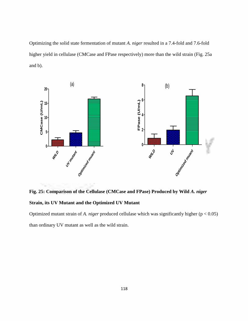
118
Optimizing the solid state fermentation of mutant A. niger resulted in a 7.4-fold and 7.6-fold
higher yield in cellulase (CMCase and FPase respectively) more than the wild strain (Fig. 25a
and b).
WILD
UV m
utant
Optimized m
uan
t
0
5
10
15
20(a)
CM
Case (
U/m
L)
WILD
UV
Optimized
muan
t
0
2
4
6
8 (b)
FP
ase (
U/m
L)
Fig. 25: Comparison of the Cellulase (CMCase and FPase) Produced by Wild A. niger
Strain, its UV Mutant and the Optimized UV Mutant
Optimized mutant strain of A. niger produced cellulase which was significantly higher (p < 0.05)
than ordinary UV mutant as well as the wild strain.
119
4.8 Improvement of P. citrinum Cellulase Production by Sequential UV Mutation and
Optimization of Solid State Fermentation
Fig. 26 a and b shows that mutant P. citrinum had a 1.8-fold and 2.1-fold increase in CMCase and
FPase more than the wild. Optimum moisture for high enzyme yield was found to be 20 % (Fig. 27a),
temperature of 37 OC (Fig. 27b), pH of 5.5 (Fig. 27c) and fermentation time of 7 days (Fig. 27d) for the P.
citrinum mutant. Other parameters include age of seeding culture of 5days (Fig. 28a) and inoculum size of
30% w/w and (Fig. 28b). Maltose was found to enhance the production of cellulase more than corn-starch,
glucose, lactose, sucrose (Fig. 29a and b). SDS enhanced more enzyme production than EDTA, Tween 20
and Tween 80 (Fig. 30a and b). FeSO4 supported cellulase production more than ZnSO4, CuSO4, CaCl2,
MgCl2, CoCl2 and KCl were used (Fig. 31a and b). Urea was the best nitrogen supplement compared to
malt extract, yeast extract, NH4NO3 and NH4Cl (Fig. 32a and b).
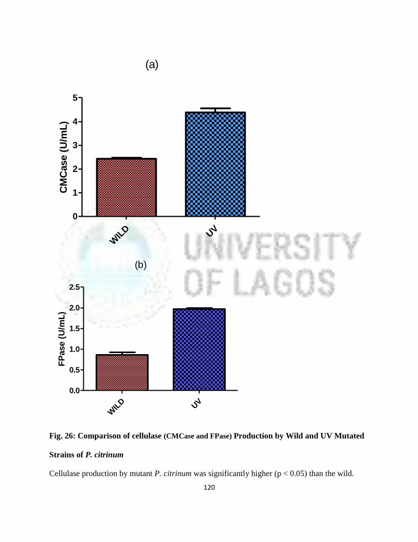
120
WIL
DUV
0
1
2
3
4
5
(a)C
MC
ase (
U/m
L)
WIL
DUV
0.0
0.5
1.0
1.5
2.0
2.5
(b)
FP
ase (
U/m
L)
Fig. 26: Comparison of cellulase (CMCase and FPase) Production by Wild and UV Mutated
Strains of P. citrinum
Cellulase production by mutant P. citrinum was significantly higher (p < 0.05) than the wild.

121
0 20 40 60 800
2
4
6
8
10CMCase
FPase
Moisture (% v/w)
cell
ula
se a
cti
vit
y (
U/m
L)
(a)
25 27 30 32 35 37 40 42 45 47 50
0
5
10
15CMCase
FPase
Temperature (oC)
cell
ula
se a
cti
vit
y (
U/m
L)
(b)
3.0
3.5
4.0
4.5
5.0
5.5
6.0
0
2
4
6
8
10
CMCase
FPase
pH
cell
uls
e a
cti
vit
y (
U/m
L)
(c)
1 2 3 4 5 6 7 8
0
2
4
6
8
10CMCase
FPase
Fermentation time (days)
cell
ula
se a
cti
vit
y (
U/m
L)
(d)
Fig. 27: Effect of Various Culture Conditions (Moisture (a), Temperature (b), pH (c) and
Fermentation time (d)) on Cellulase (CMCase and FPase) Production by P. citrinum Mutant
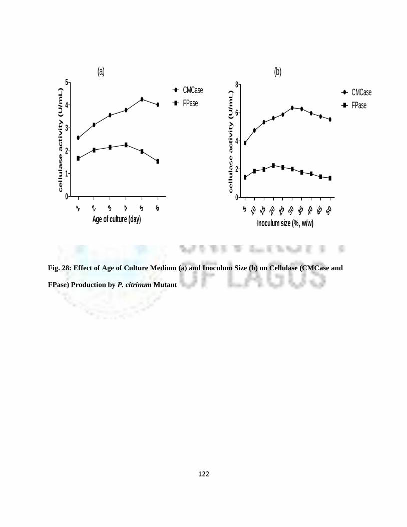
122
1 2 3 4 5 6
0
1
2
3
4
5CMCase
FPase
Age of culture (day)
cellu
lase activity (U
/m
L)
(a)
510
15
20
25
30
35
40
45
50
0
2
4
6
8CMCase
FPase
Inoculum size (%, w/w)cellu
lase activity (U
/m
L)
(b)
Fig. 28: Effect of Age of Culture Medium (a) and Inoculum Size (b) on Cellulase (CMCase and
FPase) Production by P. citrinum Mutant
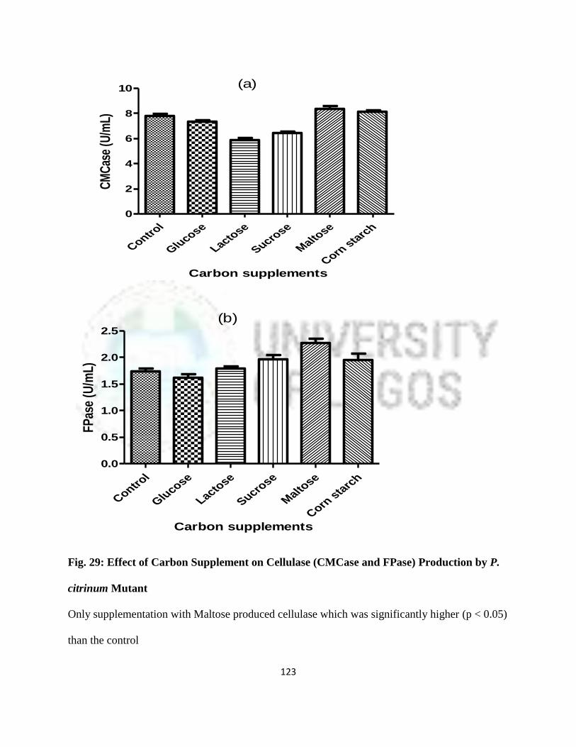
123
Control
Glucose
Lactose
Sucrose
Malto
se
Corn s
tarc
h
0
2
4
6
8
10
Carbon supplements
CM
Cas
e (U
/mL)
(a)
Control
Glu
cose
Lactose
Sucrose
Malto
se
Corn s
tarc
h
0.0
0.5
1.0
1.5
2.0
2.5
Carbon supplements
FPas
e (U
/mL)
(b)
Fig. 29: Effect of Carbon Supplement on Cellulase (CMCase and FPase) Production by P.
citrinum Mutant
Only supplementation with Maltose produced cellulase which was significantly higher (p < 0.05)
than the control
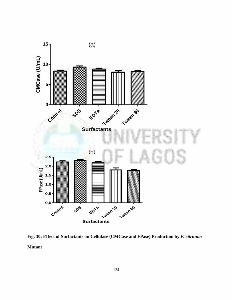
124
Fig. 30: Effect of Surfactants on Cellulase (CMCase and FPase) Production by P. citrinum
Mutant
Contr
ol
SDS
EDTA
Twee
n 20
Twee
n 80
0
5
10
15
Surfactants
CM
Case (
U/m
L)
(a)
Control
SDS
EDTA
Tween 2
0
Tween 8
0
0.0
0.5
1.0
1.5
2.0
2.5
Surfactants
FPas
e (U
/mL)
(b)
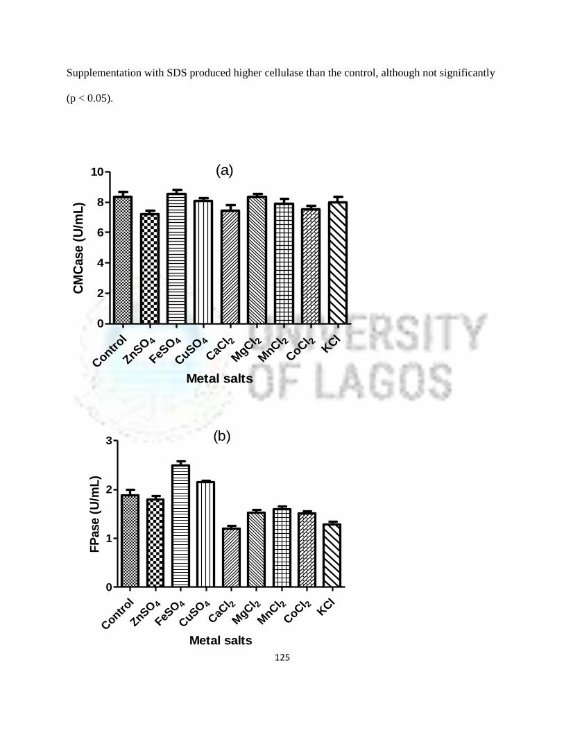
125
Supplementation with SDS produced higher cellulase than the control, although not significantly
(p < 0.05).
Contr
ol 4
ZnSO 4
FeSO 4
CuSO 2
CaC
l 2
MgC
l 2
MnC
l 2
CoC
lKCl
0
2
4
6
8
10
Metal salts
CM
Case (
U/m
L)
(a)
Contr
ol 4
ZnSO 4
FeSO 4
CuSO 2
CaC
l 2
MgC
l 2
MnC
l 2
CoC
lKCl
0
1
2
3
Metal salts
FP
ase (
U/m
L)
(b)
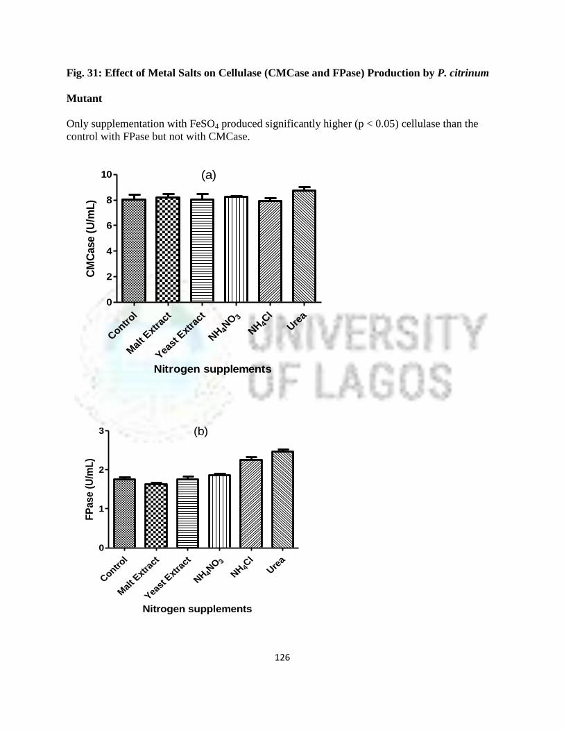
126
Fig. 31: Effect of Metal Salts on Cellulase (CMCase and FPase) Production by P. citrinum
Mutant
Only supplementation with FeSO4 produced significantly higher (p < 0.05) cellulase than the
control with FPase but not with CMCase.
Contr
ol
Mal
t Ext
ract
Yeast
Ext
ract 3
NO
4NH
Cl
4NH U
rea
0
2
4
6
8
10
Nitrogen supplements
CM
Case (
U/m
L)
(a)
Contr
ol
Mal
t Ext
ract
Yeast
Ext
ract 3
NO
4NH
Cl
4NH U
rea
0
1
2
3 (b)
Nitrogen supplements
FP
as
e (
U/m
L)

127
Fig. 32: Effect of Nitrogen Supplement on Cellulase (CMCase and FPase) Production by P. citrinum
Mutant
There was no significant difference (p < 0.05) in the cellulase production in media supplemented
with the different nitrogen supplements with CMCase. However, yeast extract and NH4Cl
produced FPase which was significantly higher (p < 0.05) than the control
Optimizing the solid state fermentation of mutant P. citrinum resulted in a 5.3-fold and 5.8-fold
higher yield in cellulase (CMCase and FPase respectively) more than the wild strain (Fig. 33a
and b).
WIL
D
UV m
utant
Opt m
uant
0
5
10
15
20(a)
CM
Case (
U/m
L)
WIL
DUV
Opt m
uant
0
2
4
6
8(b)
FP
ase (
U/m
L)
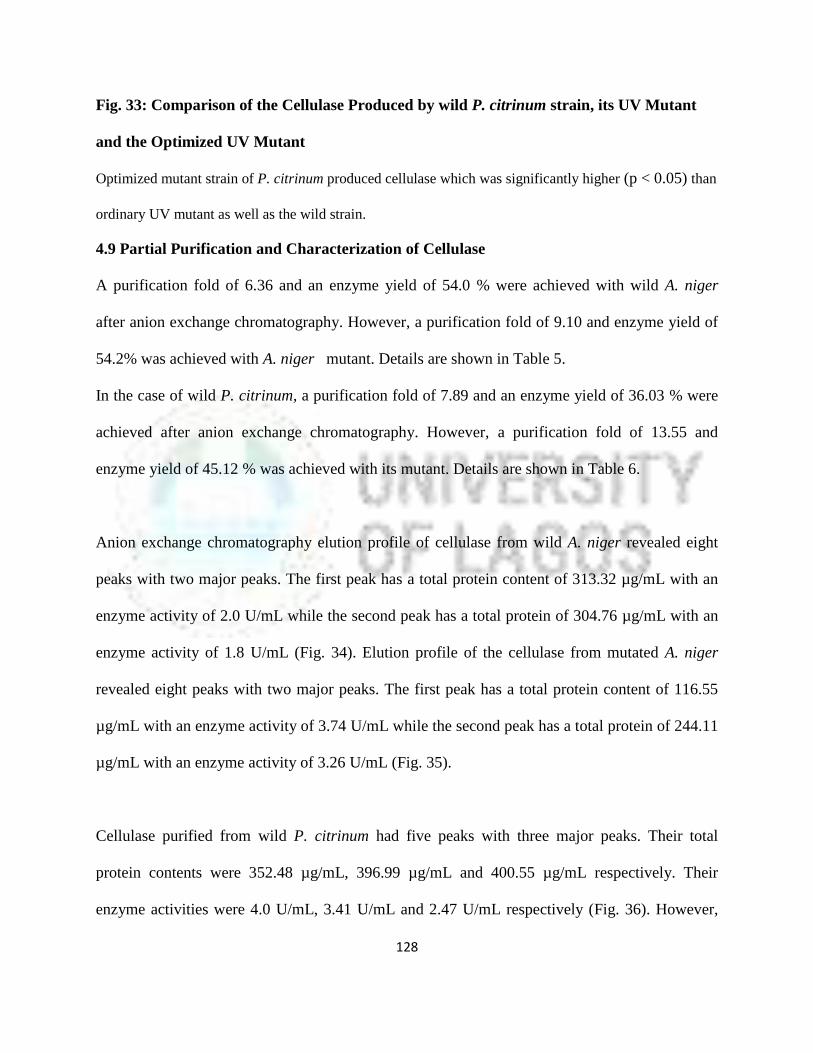
128
Fig. 33: Comparison of the Cellulase Produced by wild P. citrinum strain, its UV Mutant
and the Optimized UV Mutant
Optimized mutant strain of P. citrinum produced cellulase which was significantly higher (p < 0.05) than
ordinary UV mutant as well as the wild strain.
4.9 Partial Purification and Characterization of Cellulase
A purification fold of 6.36 and an enzyme yield of 54.0 % were achieved with wild A. niger
after anion exchange chromatography. However, a purification fold of 9.10 and enzyme yield of
54.2% was achieved with A. niger mutant. Details are shown in Table 5.
In the case of wild P. citrinum, a purification fold of 7.89 and an enzyme yield of 36.03 % were
achieved after anion exchange chromatography. However, a purification fold of 13.55 and
enzyme yield of 45.12 % was achieved with its mutant. Details are shown in Table 6.
Anion exchange chromatography elution profile of cellulase from wild A. niger revealed eight
peaks with two major peaks. The first peak has a total protein content of 313.32 µg/mL with an
enzyme activity of 2.0 U/mL while the second peak has a total protein of 304.76 µg/mL with an
enzyme activity of 1.8 U/mL (Fig. 34). Elution profile of the cellulase from mutated A. niger
revealed eight peaks with two major peaks. The first peak has a total protein content of 116.55
µg/mL with an enzyme activity of 3.74 U/mL while the second peak has a total protein of 244.11
µg/mL with an enzyme activity of 3.26 U/mL (Fig. 35).
Cellulase purified from wild P. citrinum had five peaks with three major peaks. Their total
protein contents were 352.48 µg/mL, 396.99 µg/mL and 400.55 µg/mL respectively. Their
enzyme activities were 4.0 U/mL, 3.41 U/mL and 2.47 U/mL respectively (Fig. 36). However,

129
mutated P. citrinum had nine peaks with one major peak. Its total protein content was 167.34
µg/mL with an enzyme activity of 4.50 U/mL (Fig. 37).
Table 5: Purification of Cellulase of Wild and Mutated A.niger
Organism
Purification Step Total Activity
(U/mL)
Total Protein
(mg)
Specific Activity
(U/mg protein)
Purification
Fold
Enzyme Yield
(%)
Wild A.
niger
Crude enzyme 72.8±1.3 129.4±2.5 0.56±1.1 1.0 1.0
(NH4)2SO4 (70-80 %) 46.4±0.3 56.3±0.2 0.82±0.2 1.46 64.0
Whatman DE-52 38.5±1.4 10.8±3.2 3.56±2.4 6.36 53.0
Mutant A.
niger
Crude enzyme 82.5±1.2 138.4±0.3 0.60±1.2 1.00 100.0
(NH4)2SO4 (70-80 %) 56.4±0.1 64.4±0.2 0.87±0.2 1.45 68.4
Whatman DE-52 44.7±1.3 8.2±2.1 5.45±2.4 9.10 54.2

130
Table 6: Purification of Cellulase of Wild and Mutant P. citrinum
Organism Purification Step Total Activity
(U/mL)
Total Protein
(mg)
Specific Activity
(U/mg protein)
Purification
Fold
Enzyme Yield
(%)
Wild P.
citrinum
Crude enzyme 60.5±5.1 93.2±2.2 0.65±2.6 1.0 100
(NH4)2SO4 (70-80 %) 33.4±0.2 38.5±1.3 0.87±0.6 1.34 55.21
Whatman DE-52 21.8±1.1 4.2±0.8 5.19±1.0 7.98 36.03
Mutant P.
citrinum
Crude enzyme 72.3±2.1 104.1±1.1 0.69±1.0 1.00 100.0
(NH4)2SO4 (70-80 %) 45.4±0.3 44.7±1.1 1.02±0.5 1.48 63.00
Whatman DE-52 27.3±2.1 3.1±0.2 8.81±1.5 13.55 45.12
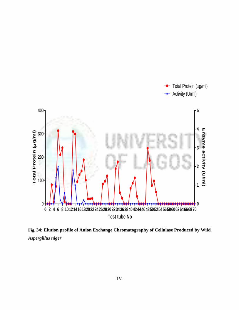
131
0 2 4 6 8 101214161820222426283032343638404244464850525456586062646668700
100
200
300
400
0
1
2
3
4
5
Total Protein (g/ml)
Activity (U/ml)
Test tube No
To
tal
Pro
tein
(
g/m
l)E
nzym
e a
ctiv
ity (U
/ml)
Fig. 34: Elution profile of Anion Exchange Chromatography of Cellulase Produced by Wild
Aspergillus niger

132
0 2 4 6 8 10 12 14 16 18 20 22 24 26 28 30 32 34 36 38 40 42 44 46 48 50 52 54 56 58 60 62 64 66 68 700
50
100
150
200
250
0
1
2
3
4
5
Total Protein (g/ml)
Activity (U/ml)
Test tube No
To
tal
Pro
tein
(
g/m
l)A
ctiv
ty (
U/m
L)
Fig. 35: Elution Profile of Anion Exchange Chromatography of cellulase Produced by UV
Mutated Aspergillus niger
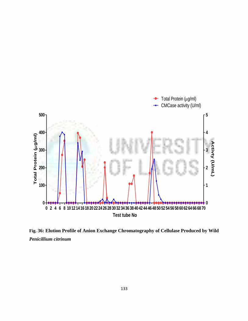
133
0 2 4 6 8 10 12 14 16 18 20 22 24 26 28 30 32 34 36 38 40 42 44 46 48 50 52 54 56 58 60 62 64 66 68 700
100
200
300
400
500
0
1
2
3
4
5
Total Protein (g/ml)
CMCase activity (U/ml)
Test tube No
To
tal
Pro
tein
(
g/m
l)A
ctiv
ty (U
/mL
)
Fig. 36: Elution Profile of Anion Exchange Chromatography of Cellulase Produced by Wild
Penicillium citrinum
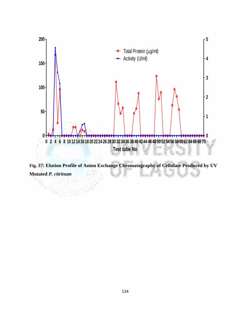
134
0 2 4 6 8 10 12 14 16 18 20 22 24 26 28 30 32 34 36 38 40 42 44 46 48 50 52 54 56 58 60 62 64 66 68 700
50
100
150
200
0
1
2
3
4
5
Total Protein (g/ml)
Activity (U/ml)
Test tube No
Fig. 37: Elution Profile of Anion Exchange Chromatography of Cellulase Produced by UV
Mutated P. citrinum

135
4.10 Effect of Substrate Concentration
The Michaelis-Menten constant (Km) and Maximum velocity (Vmax) for wild A. niger were 15.31 g/L
and 4.67 g/L respectively while for the mutant A. niger, they were 7.9g/L and 3.39g/L respectively (Fig.
38).
The Km and Vmax values for wild P. citrinum were 7.94 g/L and 6.81 g/L respectively while the mutant
P. citrinum had Km and Vmax values of 6.60 g/L and 6.84 g/L respectively as shown in Fig. 39.

136
A. niger
-0.4 -0.2 0.2 0.4 0.6
-0.4
-0.2
0.2
0.4
0.6
0.8
1.0
1.2
1.4
1.6
1.8
2.0 1/[V] A. niger (Wild)
1/[V] A. niger (Mutant)1/[V]
1/[S]
Fig. 38: Lineweaver-Burk plot of Cellulase Activity of Wild and Mutated A. niger

137
P citrinum
-0.4 -0.2 0.2 0.4 0.6
-0.4
-0.2
-0.0
0.2
0.4
0.6
0.8
1.0 1/[V] P. citrinum (Wild)
1/[V] P. citrinum (Mutant)1/[V]
1/[S]
Fig. 39: Lineweaver-Burk plot of Cellulase Activity of Wild and Mutated P. citrinum

138
4.11 Genetic Studies
4.11.1 RNA Extraction
Bands of extracted RNA of wild and mutated strains of Aspergillus niger and Penicillium
citrinum are shown in Fig. 40 below. Non-smearing of bands confirms quality of extraction.
Quantities of extracted RNA are shown in Table 7. Measurement of the ratio of their absorbance
at A260/A280 further confirmed the integrity of the extract while A260/A230 values show very
minimal contaminations from extraction.
Fig. 40: Agarose Gel Electrophoregram of Extracted RNA Mutants
Lane1= ANW, Lane 2= PCW, Lane 3= ANM, Lane 4= PCM
2 1 3 4
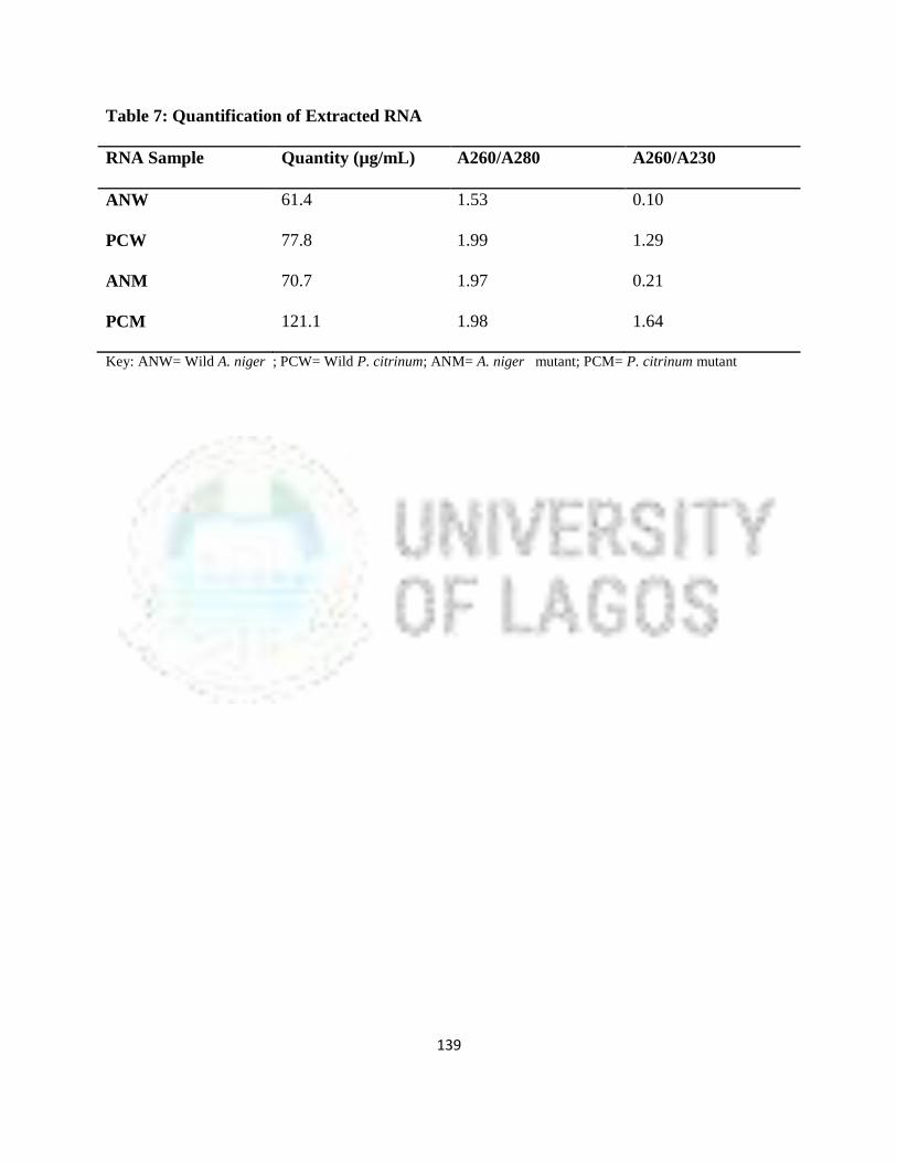
139
Table 7: Quantification of Extracted RNA
RNA Sample Quantity (µg/mL) A260/A280 A260/A230
ANW 61.4 1.53 0.10
PCW 77.8 1.99 1.29
ANM 70.7 1.97 0.21
PCM 121.1 1.98 1.64
Key: ANW= Wild A. niger ; PCW= Wild P. citrinum; ANM= A. niger mutant; PCM= P. citrinum mutant

140
4.11.2 Amplification of cDNA
Bands of the amplified cDNA are shown in Fig. 41. Selected genes amplified in both wild and
mutant strains of the two fungi are arranged in the order; Wild A. niger, Wild P. citrinum, A.
niger Mutant, P. citrinum mutant. Lane 1-4 shows Lacc1 gene; Lane 5-8 is ace1 gene; Lane 9 is
empty; Lane 10-13 shows aep1 gene; Lane 14-17 shows cbh1 gene; while Lane 18 shows the
ladder. There were poor amplifications in the Lacc1 gene of the four fungi strains and the ace1 of
Wild Apergillus niger (Lane 5). There was amplification of ace1 gene in wild P.citrinum and
mutant A. niger (Lane 6 and 7). There were poor amplification of aep1 gene in all the fungi
strains (Lane 10-13), but there was amplification of cbh1 gene in all the four fungi (Lane 14-17).
It was found that the cbh1 gene which is about 730bp was amplified successfully.
4.12 Bioinformatic Analysis and Prediction of the Structure of Cellulase
The percentage identity between the active site region of uncultured fungus (JF347834), Wild A.
niger strain nl-1 (HM769954.1), Phanarechaete chrysosporium (M22220.1), Trichoderma reesei
(E00389) and A. niger mutant was very high (Fig. 42). This was also confirmed in their
consensus region shaded in black (Fig. 42). Amino acid translation of their sequences using
JPred suite of Jalview software showed high consensus as well (Fig. 43). Cladogram showed that
A. niger mutant (cbh_AN10_5b_cbh_forward) had the closest value (6.81 and 7.17) with
uncultured fungus (JF347834.1). However, values are not too different from other sequences
analyzed (Fig. 44). X-ray crystal structure of the enzyme from the A. niger mutant revealed that
the regions of the enzyme that have been altered as a result of the mutation were mainly in the β-
pleated sheets of the enzyme structure which is represented in blue and white (Fig. 45). The translation
141
of the cbh1 gene (excluding introns) into the CBH1 protein was performed using the translate
tool (http://web.expasy.org/translate/). The analysis revealed amino acid sequence similar to
cellobiohydrolase of T.reesei, P. chrysosporium and other reference sequences downloaded from
public databases (Fig. 43). There were changes in amino acids residues of the enzyme as a result
of the mutation. Table 8 shows the amino acids that arose from the mutational change, their
location in the amino acid chain and their hydropathy index.
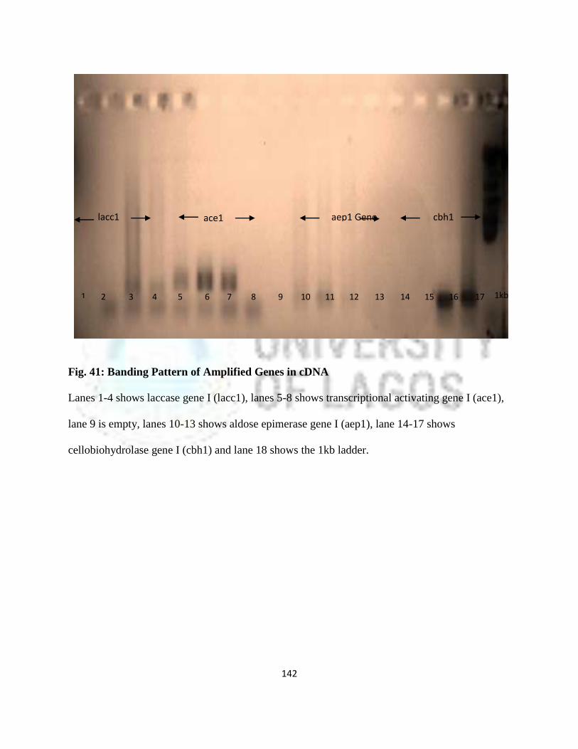
142
Fig. 41: Banding Pattern of Amplified Genes in cDNA
Lanes 1-4 shows laccase gene I (lacc1), lanes 5-8 shows transcriptional activating gene I (ace1),
lane 9 is empty, lanes 10-13 shows aldose epimerase gene I (aep1), lane 14-17 shows
cellobiohydrolase gene I (cbh1) and lane 18 shows the 1kb ladder.
1 2 3 4 5 6 7 8 9 10 11 12 13 14 15 16 17 1kb
+
lacc1
Gene
ace1
Gene
aep1 Gene cbh1
Gene

143
Table 8: Amino Acid Changes in CBH1 of Mutated A. niger and their Hydropathy Index
Amino acid Symbol Position in the Protein Structure Hydropathy Index
Lysine K 8th -3.9
Serine S 18th
-0.8
Asparagine N 27th -3.5
Aspartate D 28th -3.5
Threonine T 36th -0.7
Glutamate E 52nd -3.5
Asparagine N 53rd -3.5
Lysine K 56th -3.5
Glutamine Q 75th -3.5
Asparagine N 89th -3.5
Serine S 92th -0.8
The amino acid on the 8th
position on the chain has changed from Asparagine (N) to Lysine (K)
while on the 18th
position, there was a change from alanine (A) to serine (S). Similarly, there
were changes 27th
, 28th
, 36th
, up till the 92nd
position where there was a change from alanine (A)
to serine (S) (Fig. 43).
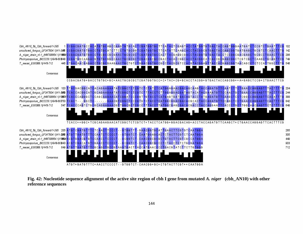
144
Fig. 42: Nucleotide sequence alignment of the active site region of cbh I gene from mutated A. niger (cbh_AN10) with other
reference sequences
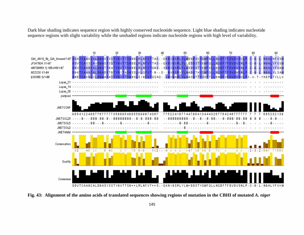
145
Dark blue shading indicates sequence region with highly conserved nucleotide sequence. Light blue shading indicates nucleotide
sequence regions with slight variability while the unshaded regions indicate nucleotide regions with high level of variability.
Fig. 43: Alignment of the amino acids of translated sequences showing regions of mutation in the CBHI of mutated A. niger
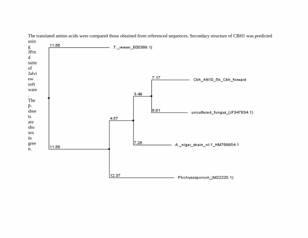
146
The translated amino acids were compared those obtained from referenced sequences. Secondary structure of CBH1 was predicted
usin
g
JPre
d
suite
of
Jalvi
ew
soft
ware
.
The
β-
shee
ts
are
sho
wn
in
gree
n.
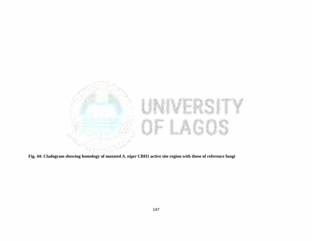
147
Fig. 44: Cladogram showing homology of mutated A. niger CBH1 active site region with those of reference fungi
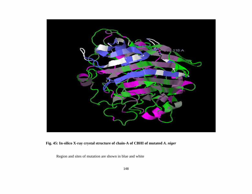
148
Fig. 45: In-silico X-ray crystal structure of chain-A of CBHI of mutated A. niger
Region and sites of mutation are shown in blue and white
cxlix
CHAPTER FIVE
5.0 DISCUSSION
Microbes, most especially fungi posses the capability of producing a wide range of
industrially important enzymes which find use in almost all industries including food, feed,
textile, cosmetics and pharmaceuticals. These enzymes may however, not be available in
commercial quantities. Hence, the need to modify these organisms for enhanced enzymes
production.
The lignocellulose content (cellulose, hemicellulose and lignin) of Abura wood sawdust were
comparable with the lignocellulose content of common agricultural residues and wastes
compiled by Lee et al., (2014). Hardwood stem was said to be composed of 40-55%
cellulose, 24-40% hemicelluloses, and 18-25% lignin while softwood contained 45-50%
cellulose, 25-30% hemicelluloses and 25-35% lignin. This range for lignocelluloses
composition is also similar to compilation of Pedersen and Meyer (2010). However, when the
sawdust was pretreated, the cellulose content was 64.94%, hemicellulose was 19.80% and
lignin was 15.20%. The sharp reduction was as a result of the removal of hemicelluloses and
lignin due to the pretreatment process.
There was reduction in moisture and crude fibre content. This is unconnected to the fact that
due to chemical pretreatment, lignin and hemicelluloses are removed. Pretreatment is
considered to be the disruption of resistant carbohydrate-lignin shield that inhibits enzymes
access to target substrates, which are cellulose and hemicelluloses (Yang and Wyman, 2008).
Different methods have been used to achieve pretreatment. This varies from physical,
mechanical, chemical, physico-chemical and even biological methods (Abu et al., 2000; Abu
et al., 2002; Yang and Wyman, 2008; Harmsen et al., 2010). Cost plays a major role in

cl
deciding the choice of pretreatment. In this study, ammonia steeping, otherwise called
soaking in aqueous ammonia (SAA) was employed. It leads to about 60% reduction in lignin
and hemicelluloses content. This was in agreement with observations of Abu et al., (2002).
Kim et al., 2008 achieved 60% lignin solubilization and observed saccharification yields of
83% for glucan when destarched barley husk was treated with 15% aqueous ammonia at 75oC
during 48h. In this study, pretreatment was carried out at room temperature and comparable
success was achieved.
The fungal isolates selected (P. corylophilum, P. citrinum, A. niger and T. asahii) had
similar morphological characteristics with the species described by Tiwari et al., (2011).
Molecular identification of fungi by DNA barcoding has been described to be the most useful
method of identification (Bellemain et al., 2010). The internally transcribed spacer regions
(ITS1 &2) were targetted for the amplification of the DNA fragments. It is located between
the short sequence unit (SSU) and the long sequence unit (LSU) regions separated by the
5.8S gene in the rDNA repeat unit. It is specific for fungi and other organisms of plant origin.
They had very close homology with reference sequence. The results of the genotyping were
consistent with the findngs of Jang et al., (2012) where isolates which included Aspergillus
and Penicilium species where identified by amplification of the ITS region.
Alkaline pretreated sawdust fermented with co-cultured A. niger and P. citrinum released
more cellulase enzyme more than unpretreated sawdust co-cultured with both organisms.
However, when both sawdust types were fermented with either fungi, alkaline pretreated
sawdust fermented with A. niger produced more enzyme more than unpretreated sawdust
fermented with A. niger. The sawdust types fermented with P. citrinum did not release
cellulase as much as those fermented with A. niger. This shows the efficiency of A. niger
cli
over P. citrinum in the biodegradation of sawdust. However, synergism between both
organisms was demonstrated when co-culturing of both organism in unpretreated sawdust
could release more cellulase more than the use of either fungi on alkaline pretreated sawdust.
Similar trend was observed with the release of reducing sugars and cellobiase (β-glucosidase)
activity. This confirms the synergistic effect of both fungi.
The decrease in conidiospores of A. niger and P. citrinum mutant growing on mineral salt
agar containing 2-deoxy glucose was due to the cytotoxity of the ultraviolet radiation as well
as the presence of 2-deoxy glucose in the culture media. The glucose derivative is an anti-
metabolite which could cause catabolite repression to non-mutant strains (Anwar et al., 1996;
Farkas et al., 1981). Only mutated strains of fungi can survive this repression (Dillon et al.,
2006).
The increase in cellulase activity was a confirmation of mutation in the genome of the
organism. UV irradiation could lead to unspecific mutation which may be beneficial or not.
In this case it has enhanced the production of cellulase in an appreciable manner. In most
studies, mutation is achieved after exposure of organisms to repeated rounds of mutagenic
agents and / or combination of different mutagenic agents. Vu et al., (2009) succeeded in
improving carboxymethylcellulase, filter paper cellulase and β-glucosidase yield of A. niger
by 2.03, 3.20, and 1.80 fold respectively by sequential treatments with two repeated rounds of
µ-radaition of Co60
, ultraviolet treatment and four repeated rounds of treatment with a
chemical mutagen, N-methyl-N‘-nitro-N-nitrosoguanidine. In this study, a competent mutant
was selected from different mutants created from fungi exposed to different doses of UV
irradiation. A. niger mutant with CMCase of 4.69U/mL and FPase of 1.97U/mL and P.
citrinum mutant with CMCase of 4.83U/mL and an FPase of 1.97U/mL.

clii
Moisture is very essential for production of cellulase. It is pertinent that moisture level should
be at a specific quantity required for optimum enzyme production. Too much moisture in a
solid-state fermentation medium could affect porosity of substrate (Vu et al., 2010). The
optimum moisture content obtained is comparable to the findings obtained by Vu et al.,
(2010) where moisture content higher than 50 % v/w (76.6 U/g) was said to be optimum for
mutated A. niger.
The optimum temperature of 37 oC was found suitable for both CMCase and FPase of A.
niger and P. citrinum mutants. The optimum temperature obtained quite agrees with the
findings obtained in earlier works of Narasimha et al., (2006). A pH of 3.5 was found to be
optimum for enzyme production of the A. niger mutant while a pH of 4.5 was optimum for P.
citrinum mutant. There was a slight variation to findings of Acharya et al., 2008. They
identified an optimum pH of 4.0 and 4.5 for wild A. niger species. The slight change could be
due to the slight change in the make-up of the organism due to its mutation by UV. Vu et al.,
(2011) obtained 28.5 U/g for an optimum pH of 3.5. The rate of enzyme catalyzed reactions
increase with temperature up to a certain limit. Above a certain temperature enzyme activity
decreases with increase in temperature because of enzyme denaturation. The optimum pH for
the growth of the organism leads to reduction in unwanted extra-cellular proteins other than
the required product (Rajesh et al., 2012). Other parameters include fermentation time of 4
days was obtained for A. niger mutant while it was 7 days for P. citrinum mutant. Age of
culture medium was 3 days for A. niger mutant while it was 7 days for P.citrinum mutant.
Inoculum size was 20% w/w for A. niger mutant while it was 30% (w/w) for P. citrinum. In
the studies conducted by Vu et al., (2011) fermentation time of 3 days was obtained, age of
culture medium of 2 days and inoculum size of 25 % for A. niger mutant.
cliii
Gilna and Khaleel, (2011) identified a pH of 6.5 to be optimum for enzyme production by A.
niger, temperature of 32 oC, yeast extract as the best nitrogen source, and cellulose as a good
inducer. The A. niger mutant used in this study had an optimum pH of 3.5, temperature of 37
oC and EDTA as an inducer. Changes in pH may also alter the structural architecture of the
enzyme. This change in pH of the medium activates the active site of the enzyme and
facilitates the reaction rate. Pradeep and Narasimha, (2011) mutated A. niger with ethyl
methane sulfonate (EMS) and were able to obtain a CMCase of 18.09 U/mL and FPase of
19.73 U/mL after fermenting the mutant using pea seed husk as substrate. Pradeep et al.,
(2012) was however, able to obtain CMCase of 3.15 U/mL and FPase of 3.15 U/mL after
chemical mutagenesis of A. niger using ethidium bromide. Vu et al., (2011) treated A. niger
spores with Co60
rays, UV and NTG and was able to obtain a mutant. The solid state
fermentation parameters of the mutant were optimized and a CMCase activity of 82.5 U/g
was obtained using sawdust as substrate. Their findings represented an 8.5-fold increase in
enzyme yield compared to the wild.
However, in this study, optimized fermentation of UV mutant of A. niger produced a
CMCase of 17.2 U/mL and FPase of 6.5U/mL. This represented a 7.4-fold increase in
enzyme yield compared to the wild. EDTA best enhanced cellulase production and this
agrees with the findings of Acharya et al., (2008). Corn-starch enhanced cellulase production
which disagrees with the findings of Zhu et al., (2011). The preference for natural carbon
sources in enzyme production may be due to the presence of growth promoters in enough
amounts covering the requirements of fungal growth and enzyme production (Rajesh et al.,
2012). It could also be explained that the metabolic pattern of the organism may have been
altered as a result of the mutation, thus making it to easily metabolize glucose biopolymers
without hindrance. Increase in cellulase activity after UV mutation may be due to possible
changes in the promoter regions of the genes (De Nicolas-Santiago et al., 2006). During SSF,
cliv
the components of the cell wall are degraded and lignocellulolytic enzymes (LCEs) are
produced and act optimally (Vu et al, 2011).
Solid-state fermentation of polysaccharide using Paecilomyces cicadae was carried out by
Ren et al., (2014). The Box-Behnken design was used to optimize the final levels of culture
conditions. Relative humidity of 56.07%, inoculums of 13.51mL/100g and temperature of
27.09oC were found to be optimal parameters for polysaccharide production.
Solid State Fermentation (SSF) has been demonstrated to offer better hydrolytic enzyme yield
more than submerged. It is fermentation in the absence or near absence of free water with
inert natural substrates as solid support (Pandey et al., 1999). Additional carbon source is
needed in the fermentation tank to enhance growth. It is expected that the supplemented
carbon source should be easily metabolized (Zhu et al., 2011). In this study, cornstarch
enhanced production of endoglucanase (CMCase) and total cellulase (FPase) activities more
than glucose for A. niger mutant while maltose enhanced both enzymes in the fermentation
by P. citrinum mutant. This was contrary to findings of Zhu et al., (2011) where
supplementation of glucose and increased moisture content enhanced endoglucanase
production with Trametes versicolor during solid-state fermentation of corn stover. It could
be explained that the metabolic pattern of the organism may have been altered as a result of
the mutation, thus enabling it to metabolize glucose biopolymers easily.
Surfactants have been used to enhance lignocellulolytic hydrolysis and when used, they
reduce enzyme loadings. A number of mechanisms have been proposed for their action.
Surfactants help the enzyme to adjust its structure to allow the substrate to fit into the active
site of the enzyme. Yang and Wyman (2005) explained that Tween 80 bring cellulase in
close proximity with cellulose. EDTA and SDS enhanced enzyme activity of A. niger and P.

clv
citrinum mutants. The enhancement could be due to the ability of the surfactants to reduce
unnecessary attachment of cellulase to substrates other than cellulose in the medium, thus
enhancing its performance. The variation may however, be due to the physiological
differences in the organisms. Yang and Wyman (2005) explained that bovine serum albumin,
a biosurfactant, could attach to lignin, thus allowing non-specific attachment of cellulase to
lignin when it is used in the hydrolysis of lignocellulosics.
Metal salts play significant roles in microbial metabolism. They act as cofactors or as co-
enzymes. Some fermentation will not yield product without the inclusion of metal salts.
FeSO4 best enhanced the production of endoglucanase and total cellulase in both mutants.
However, MgSO4 was able to enhance endoglucanase production more than other salts when
Vu et al., (2011) carried out solid-state fermentation after mutating A. niger.
Urea supplementation enhanced enzyme activity more than other nitrogen supplements. This
corroborates the findings of Acharya et al., (2008). Optimization of nitrogen source for
cellulase production was carried out on sawdust using wild A. niger. Urea was found to
enhance optimum CMCase production more than peptone and sodium nitrite.
Optimization of the solid-state fermentation of mutant A. niger resulted in 7.4-fold and 7.6-
fold higher yield (CMCase and FPase respectively) more than the wild strain while with P.
citrinum mutant, it led to 5.3-fold and 5.8-fold higher yield. Vu et al., 2011 obtained 8.5-fold
increase over wild strain after optimizing solid-state fermentation of mutant.
Cellulase enzyme from the mutated A. niger was purified to 9.10 purification fold. The
enzyme yield was 54.2% with a specific activity of 5.42 ± 2.4 U/mg protein. Its activity was
quite higher than cellulase from the wild which was 3.56 ± 2.4 U/mg protein (Table 5). The
increase was due to the effect of the mutation. Ultraviolet mutation has been proven to impact
clvi
on cellulase activity (De Nicholas-Santiago et al., 2006; Vu et al., 2009; Vu et al., 2011). The
Michaelis constant (KM) of cellulase from A. niger (15.31g/L) reduced after mutation
(7.9g/L). A low KM indicates a high affinity between the enzyme and the substrate (Aehle et
al., 2007). This probably explains the enhanced activity of the enzyme from the mutant.
Substrates that are tightly bound to the enzyme has low KM. This affinity for the substrate will
enhance its activity which was reflected in the elevated specific activity of the enzyme. The
catalytic efficiency of cellulase from A. niger mutant also increased. Catalytic efficiency is
the ability of an enzyme to release a product after its conversion almost instantaneously
(Murray et al., 2012). It is a ratio of the turnover number (Kcat) of the enzyme to its
Michaelis constant (KM). Turnover number represents the µmoles of substrate converted per
µmole of enzyme per second. Alternatively, the number of molecules of enzymes converted
by one molecule of enzyme per second (Bugg, 2004). Since cellulase has one active site, at
maximum substrate concentration, its turnover number is equivalent to its Vmax which was
7.9s-1
. The catalytic efficiency (Kcat / KM) of cellulase from A. niger mutant was calculated
to be 0.429M-1
s-1
as compared to the wild which was 0.305M-1
s-1
.
Cellulase enzyme from the mutated P. citrinum was purified to 13.55 purification fold. The
enzyme yield was 45.12% with a specific activity of 8.81 ± 1.5 U/mg protein. Its activity was
quite higher than cellulase from wild P. citrinum which was 5.19 ± 1.0 U/mg protein. The
increase is also due to the effect of the mutation. The Michaelis constant (KM) of cellulase
from P. citrinum (7.94g/L) reduced after mutation (6.60g/L). Like it‘s A. niger counterpart,
the catalytic efficiency of cellulase from P. citrinum mutant also increased.
Likewise, at maximum substrate concentration, its turnover number is equivalent to its Vmax
which was 6.84s-1
. The catalytic efficiency (Kcat / KM) of cellulase from P. citrinum mutant
was calculated to be 1.036M-1
s-1
as compared to the wild which was 0.858M-1
s-1
. In the
studies of Wang et al., (2012), two cellulases purified from Trichoderma reesei were found to
clvii
have a Km of 22.68 ± 0.28 g/Land Vmax of 0.269 ± 0.066 mg/min at 5.0mmol/L AlCl3. It
was however, inhibited at 10 mmol/L MgCl2 when the Km increased to 50.0 g/L and Vmax
of 0.434mg/min.
Studies on fungal cellulolytic system are only limited to a few key species such as
Trichoderma reesei and Phanerochaete chrysosporium (Chukaetirote et al., 2012). This data
provides additional information on the presence of cbh1 gene in A. niger and P. citrinum. The
cbh1 gene of A. niger showed in this study was distinct, although, it showed high homology
to the cbh1 of T. reesei and P. chrysosporium as shown Figs. 42 and 45. This finding is in
agreement with previous work of Chukaetirote et al., (2012) in which several forms of the
cbh1 gene has been reported. Cellobiohydrolases are classified as glycosyl hydrolases.
Typical basic structures of fungal CBH are composed of three domains or regions: a cellulose
binding domain which contains conserved cysteine residues, a catalytic domain which
contains conserved glutamic and aspartic acid residues, and a linker region which is rich in
serine, glycine and threonine residues (Chukaetirote et al., 2012). A. niger CBH has been
shown to consist solely a catalytic domain (Li et al., 2009). The mutated A. niger in this study
is rich in aspartic acid but not glutamic acid. In addition to the homologous amino acids it has
with the compared sequence, it is rich in alanine. The nature of amino acid present in the
active site of the enzyme determines the efficacy of the enzyme in performing its function.
Hydrolysis of cellulose requires amino acids with low hydropathy index at the active site of
the enzyme. The hydropathy index is a scale combining the hydrophobicity and
hydrophilicity of amino acids functional groups. It is used to measure the tendency of an
amino acid to seek an aqueous environment (- values) or a hydrophobic environment (+
values) (Nelson and Cox, 2008). Amino acids peculiar to the active site of the mutant A.

clviii
niger alone were found to have hydropathy index that favours aqueous environment which
enhances cellulose hydrolysis. Lysine (K), which has a hydropathy index of -3.9 was found in
the 8th
position. Likewise, asparagine (N), glutamate (E), and glutamine (Q) and other amino
acids with favourable hydropathy index are found in the enzyme amino acid chain. Almost all
amino acids peculiar to the mutant have low hydropathy index which suggests the reason for
their elevated activity.
The in-silico X-ray crystal structural elucidation of the active site of the enzyme revealed that
the α-helices are not affected by the irradiation. However, mutation occurred in the β-strands
(Fig. 44). Cladogram constructed based on relationship between the mutated A. niger and
industrially important referenced fungi showed homology between the mutant and
Trichoderma reesei and Phanerochaete chrysosporium with a bootstrap values of 11.56 and
12.37 respectively as compared with 7.17 of the mutated A. niger.
Homology in the sequence of the mutant A. niger and Trichoderma reesei, a model hyper
producer of cellulase, signifies its potential in the industry for hyper production of cellulase.
The analysis will allow the use of the exquisite tools of fungal genetics to further interrogate
protein function and interactions (Bennett et al., 2001).

clix
6.0 SUMMARY OF FINDINGS
OBJECTIVES FINDINGS
1. Determination of composition of raw
and pretreated sawdust.
Cellulose was 48.11%, Hemicellulose was 32.70% and lignin was
19.10%.
2. Isolation and genotyping of cellulolytic
fungi.
Four cellulolytic fungi; Penicillium citrinum, Aspergillus niger,
Trichosporon asahii and Penicillium corylophylum were genotyped.
3. Mutation of isolates and optimizing
their Solid-State Fermentation.
A. niger mutant required optimum moisture of 20%,
temperature of 37 oC, pH of 3.5, fermentation time of 4 days,
inoculum size of 25 % w/w, age of seeding culture of 3 days,
corn starch as carbon supplement, EDTA as surfactant, FeSO4
and urea as metal salt and nitrogen supplements respectively.
However, P. citrinum mutant required an optimum moisture of
20 %, temperature of 37 oC, pH of 5.5, fermentation time of 7
days, inoculum size of 30 % w/w, age of seeding culture of 5
days, maltose as carbon supplement, SDS as surfactant, FeSO4
and urea as metal salt and nitrogen supplements respectively.
.A. niger mutant resulted in a 7.4-fold and 7.6-fold higher yield

clx
in CMCase and FPase respectively more than the wild strain.
Likewise, P. citrinum mutant resulted in a 5.3-fold and 5.8-fold
higher yield in CMCase and FPase respectively more than the wild
strain.
4. Sequencing of cDNA amplicons
obtained from wild and mutant fungi
strains.
Lacc1, ace1, aep1 and cbh1 genes were amplified but only cbh1
gene sequencing was successful.
5. Analysis of sequence using
Bioinformatics tools and prediction of
the structure of cellulase from
mutant.
Mutated A. niger had strong similarity with compared inࠀustrially
beneficial cellulolytic fungi. Amino acid residues in its active site
had low hydropathy index.
Predicted structure of cellulase from A. niger mutant revealed that
alteration in structure occurred in the β-pleated sheets of enzyme.
6.1 CONCLUSION
Filamentous fungi are considered to be the most important and main group of
microorganisms for the production of plant cell wall degrading enzymes in solid state
fermentation. Mutagenesis using ultraviolet light and enrichment are cost effective yet
efficient way of enhancing their performance. In this study, optimization of the solid state
fermentation of mutated A. niger resulted in a 7.4-fold and 7.6-fold higher yield in CMCase
and FPase respectively more than the wild strain while mutated P. citrinum resulted in 5.3-
fold and 5.8-fold increase (CMCase and FPase). The cellulase produced by these modified
fungi has high turnover rates. The amino acids in their active site has low hydropathy index
which explains their superlative performance. This signifies their potential for use in the
overproduction of enzymes for cellulosic wastes bioconversion. The huge tonnage of
agricultural wastes produced everyday, which is an environmental threat, are raw materials
for generation of valuable products and ultimately medium of wealth generation.
clxi
6.3 CONTRIBUTIONS TO KNOWLEDGE
1. The study sequenced cellobiohydrolase 1 gene from mutated Aspergilus niger and
deposited its sequence in the National Center for Biotechnology Information, USA.
2. The study identified the region of alteration in the β-strands in chain-A of
cellobiohydrolase I enzyme obtained from mutated Aspergillus niger.
3. DNA barcoding of four wild fungi; Aspergillus niger ANR-352L (KM192155.1),
Penicillium citrinum PCM-254L (KJ425589), Trichosporon asahii TAS-150L
(KM192153.1) and Meyerozyma guilamondii MQL-105L (KM192154.1) was achieved in
the study. Their sequences were deposited in the the National Center for Biotechnology
Information, USA.
4. The study determined the lignocellulosic content of Abura (Mitragyna ciliata) sawdust.

clxii
REFERENCES
Abarca, M.L., Accensi, F., Cano, J. and Cabanes, F.J. (2004). Taxonomy and significance of
black aspergilli. Antonie van Leeuwenhoek 86:33–49.
Abdullahi, N., Ejaz, N., Abdullah, M., Nisa, A. and Firdous, S. (2006). Lignocellulosic
degradation in Solid-State fermentation of sugar cane bagasse by Termytomyces sp.
Micologia Aplicada International, 18(2): 15-19.
Abo-State, M.A.M., Hammed, A.I., Swelim, M. and Gannam, R.B. (2010). Enhanced
production of Cellulase(s) by Aspergillus spp Isolated from Agricultural Wastes by
Solid-State Fermentation. American-Eurosian J. Agric. And Environ. Sci. 8(4): 402-410
Abu, E.A., Ameh, D.A., Onyenekwe, P.C., Agbaji, A.S., Ado, S.A. and Nwaeze, A.R.
(2002). Comparative Studies of the Effect of Pretreatments of three Agrowastes on
Cellulase Production by Aspergillus niger SL1. Scientia Africana. 15(1): 57-63.
Abu, E.A., Oyenekwe, P.C., Ameh, D.A., Agbaji, A.S. and Ado, S.A. (2000). Cellulase (EC
3.2.1.3) Production from sorghum bran by Aspergilus niger SL1: An assessment of
pretreatment methods. Proceedings from the international conference on
Biotechnology; Commercializtion and Food Security. Abuja, Nigeria. Pp 153-157.
clxiii
Acharya, P.B., Acharya, D.K. and Modi, H.A. (2008). Optimiztion or cellulase production by
Aspergillus niger using saw dust as substrate. Afr. J. Biotechnol. 7(22): 4147-4152.
Aehle, W., Antrim, R.L, Becker, T., Bott, R. et al., (2007). Catalytic Activity of Enzymes. In:
Enzymes in Industry: Production and Applications. W. Aehle (Ed.). 3rd
edn. Wiley
VCH Verlag GmbH & Co.KGaA, Weinhem, Germany.pp: 13-21.
Agbor, V.B., Cicek, N., Sparling, R., Berlin, A. and Levin, D.B. (2011). Biomass
pretreatment: Fundamentals toward application. Biotechnol Adv. 29:675–685.
Ahamed, A. and Vermette P. (2008). Culture-based strategies to enhance cellulase enzyme
production from Trichoderma reesei RUT-C30 in bioreactor culture conditions.
Biochem.Eng. J., 40: 399-407.
Aina, O. M. (2006). Wood waste utilization for energy generation, Proceedings of the
International Conference on Renewable Energy for Developing Countries,
http://cere.udc.edu/Aina.pdf.[Aaccessed: 1st August, 2009].
Al-Musallam, A. (1980). Revision of the black Aspergillus species. Ph.D. thesis.
Rijksuniversiteit Utrecht, Utrecht. Unpublished data.
Anderson, T.D., Robson, S.A., Jiang, X.W., Malmirchegini, G.R., Fierobe, H.P., Lazazzera,
B.A. and Clubb, R.T. (2011). Assembly of minicellulosomes on the surface of Bacillus
subtilis. Appl. Environ. Microbiol., 77:4849–4858.
Anwar, M.N., Suto, M., and Tomida, F. (1996). Isolation of mutants of Penicillium
purpurogenum resistant to catabolite repression. Appl Microbiol Biotechnol. 45:684–
687
A.O.A.C (2005) Association of Official Analytical Chemist. Official Methods of Analysis.
18th
edition. Washington, DC. USA.
clxiv
Aranda, P. S., LaJoie, D. M. and Jorcyk, C. L. (2012). Bleach Gel: A Simple Gel for Analyzing
RNA Quality. Electrophoresis. 33(2):366-369.
Banerjee, G., Car, S., Scott-Craig, J.S., Borrusch, M.S. and Walton, J.D. (2010). Rapid
optimization of enzyme mixtures for deconstruction of diverse pretreatment/biomass
feedstock combinations. Biotechnol. Biofuels. 3: 22.
Bayer, E.A., Lamed, R. and Himmel, M.E. (2008). The potential of cellulases and
cellulosomes for cellulosic waste management. Curr. Opin. Biotechnol., 18:237–245.
Beckham, G.T., Matthews, J.F., Bomble, Y.J., Bu, L., Adney, W.S., Himmel, M.E., Nimlos,
M.R. and Crowley, M.F. (2010). Identification of amino acids responsible for
processivity in a Family 1 carbohydrate-binding module from a fungal cellulase. J.
Phys. Chem. B. 114:1447–1453.
Bellemain, E., Calsen, T., Brochmann, C., Coissac, E., Taberlet, P. and Kauserud, H. (2010).
ITS as an environmental DNA barcode for fungi: an in silico approach reveals
potential PCR biases. BMC Microbiology, 10: 189-197.
Bennett, C.B., Lewis, L.K, Karthikeyan, G., Lobachev, K.S., Jin, Y.H., Sterling, J.F., Snipe
J.R. and Resnick, M.A. (2001). Genes required for ionizing radiation resistance in
yeast. Nat. Genet. 29: 426–434.
Berka, R.M., Kodama, K.H., Rey, M.W., Wilson, L.J. and Ward, M. (1991). The
development of A. niger var. awamori as a host for the expression and secretion of
heterologous gene products. Biochem. Soc. Trans.19: 681−685.
Berka, R.M., Ward, M., Wilson, L.J., Hayenga, K.J., Kodama K.H., Carlomagno, L.P. and
Thompson, S.A. (1990). Molecular cloning and deletion of the gene encoding
aspergillopepsin A from A. awamori. Gene 86: 153−162.
clxv
Bhargavi, M.C. and Singara (2010). Influence of physical and chemical mutagens on dye
decolourising Mucormucedo. Afr. J. Microbiol. Res. 4(17):1808-1813.
Blanchette, R.A., (2000). A review of microbial deterioration found in archaeological wood
from different environments. International Biodeterioration and Biodegradation 46:
189-204.
Broekhuijsen, M.P., Mattern, I.E., Contreras, R., Kinghorn, J.R. and Van den Hondel, C.A.
(1993). Secretion of heterologous proteins by A.niger. Production of active human
interleukin-6 in a protease deficient mutant by KEX-2 processing of a glucoamylase-
hIL-6 fusion protein. J. Biotechnol. 31: 135-145.
Bugg, T.D.H. (2004). Methods for Studying Enzymatic Reactions. In: Introduction to
Enzyme and Co-enzyme Chemistry. 2nd
edn. Blackwell Publishing. Great Britain.
pp: 51-64.
Calvo-Flores, F.C. and Dobado, J.A. (2010). Lignin as renewable raw material.
ChemSusChem 3:1227–1235.
Cantarel, B.L., Coutinho, P.M., Rancurel, C., Bernard, T., Lombard, V. and Henrissat, B.
(2009). The Carbohydrate-Active enZymes database (CAZy): an expert resource for
Glycogenomics. Nucleic Acids Res. 37: D233-238.
Carlile M.J., Gooday G.W, and Watkinson S.C.(2001). The fungi. 2nd
edn. Academic Press
Ltd, San Diego.
Chand P., Aruna, A., Maqsood, A.M. and Rao, L.V. (2005). Novel mutation method for
increased cellulase production. J Appl Microbiol. 98:318–323.
Chukaetirote, E., Maharchchikumbura, S., Wongkham, S., Sysouphanthong, P., Phookamsak,
R. and Hyde., K. (2012) Cloning and Sequence Analysis of the cellobihydrolase I
genes from some Basidiomycetes. Mycobiology 40(2): 107-110.
clxvi
Chundawat, S.P.S., Beckham, G.T., Himmel, M.E. and Dale. B.E. (2011a). Deconstruction of
lignocellulosic biomass to fuels and chemicals. Annu Rev Chem Biomol Eng 2:6.1–
6.25.
Chundawat, S.P.S., Lipton, M.S., Purvine, S.O., Uppugundla, N., Gao, D., Balan, V. and
Dale, B.E. (2011b) Proteomics-based compositional analysis of complex cellulase-
hemicellulase mixtures. J. Proteome Res. 10:4365–4372.
Couturier, M., Haon, M., Coutinho, P.M. and Henrissat, J.G. (2011). Hemicellulases
potentiate the Trichoderma reesei secretome for saccharification of lignocellulosic
biomass. Appl. Environ. Microbiol. 77:237–246.
Damisa, D., Ameh, J. and Umoh, V.J. (2008). Effect of chemical pretreatment on some
lignocellulosic wastes on the recovery of cellulose from Aspergillus niger AH3 mutant.
Afr. J. Biotechnol. 7(14): 2444 – 2450.
Daniel, G.F., and Nilsson, T. (1998). Developments in the study of soft rot and bacterial
decay. In: Bruce, A. and Palfreyman, J. W. (Eds.) Forest Products Biotechnology.
Taylor and Francis, London. pp326.
Dashtban, M., Schraft, H., and Qin, W. (2009). Fungal bioconversion of lignocellulosic
residues; opportunities & perspectives. Int. J. Biol. Sci. 5:578–595.
Demain, A.L., Valesco, J. and Aldrio, J.L. (2004). Industrial Mycology: past, present and
future. In: Handbook of Industrial Mycology (An, Z. ed.) Marcell Dekker
Publishers.NYpp1-25.
De Nicolas-Santiago, S., Regalado-Gonzalez, C., Garcia-Almendarez, B., Fernandez, F.J.,
Tellez-Jurado, A. and Huerta-Ochoa, S.A. (2006). Physiological, morphological, and
mannanase production studies on Aspergillus niger uam-gs 1 mutants. Electron.
J.Biotechnol. 9(1): 50-60.
clxvii
De Sousa, M.V., Monteiro, S.N. and d‘Almeida, J.R.M. (2004). Evaluation of pretreatment,
size and molding pressure on flexural mechanical behavior of chopped bagasse–
polyester composites. Poly. Testing 23(3): 253-258.
Dillon A.J.P., Zorgi, C., Camassola, M. and Henriques J.A.P. (2006). Use of 2-deoxyglucose
in liquid media for the selection of mutant strains of Penicillium echinulatum
producing increased cellulase and β-glucosidase activities. Appl Microbiol. Biotechnol.
70:740-746.
Ding, S.Y., Himmel, M. and Xie, S.X. (2008). Identify molecular structural features of biomass
recalcitrance using nondestructive microscopy and spectroscopy. Microsc Microanal
14:1494–1495.
Ding, S.Y., Xu, Q., Crowley, M., Zeng, Y., Nimlos, M., Lamed, R., Bayer, E.A. and Himmel,
M.E. (2008). A biophysical perspective on the cellulosome: new opportunities for
biomass conversion. Curr. Opin. Biotechnol. 19:218–227.
Ding, SY, and Himmel ME. (2006). The maize primary cell wall microfibril: a new model
derived from direct visualization. J. Agric. Food Chem. 54:597-606
Donzelli, B.G.G., Lorito, M., Scala, F. and Harman, G.E. (2001). Cloning, sequence and
structure of a gene encoding an antifungal glucan 1, 3-glucosidase from T. atroviride
(T._harzianum). Gene. 277: 199-208.
Dosunmu, O.O. and Ajayi, A.B. (2002). Problems and management of sawmill waste in
Lagos. Proceedings of International Symposium on Environmental Pollution
Control and Waste Management. Tunis (EPCOWM 2002), pp 271-278.
Eyzaguirre, J., Hidalgo, M., and Leschot, A. (2005). Beta-glucosidases from filamentous
fungi: Properties, structure, and applications. In Handbook of Carbohydrate
Engineering; Yarema, K.J., Ed.; CRC Press: Boca Raton, FL, USA. pp. 645–685.
clxviii
FAOSTAT (2006). FAO statistical databases. http://faostat.fao.org/
Farkas, V., Labudova, I., Bauers, S., and Ferenczy, L. (1981). Preparation of mutants of
Trichoderma viride with increased production of cellulase. Folia Microbiol 26:105–
110
FDA (2008). ―Inventory of GRAS Notices: Summary of all GRAS Notices‖. US
FDA/CFSAN. 2008-10-22. Archived from the original on 11 October 2008. Retrieved
2008-10-31.
Fontes, C.M. and Gilbert, H.J. (2010). Cellulosomes: highly efficient nanomachines designed
to deconstruct plant cell wall complex carbohydrates. Ann. Rev. Biochem. 79:655–681.
Foreman, P., van Solingen, P., Goedegebuur, F. and Ward, M. (2011). CIP1 polypeptides and
their uses. U.S. Patent 7,923,235.
Foreman, P.K., Brown, D., Dankmeyer, L., Dean, R., Diener, S., Dunn-Coleman, N.S.,
Goedegeburr, F., Houfek., T.D., England, G.J., Kelly, A.S., Meerman, H.J., Mitchell,
T., Mitchinson, C., Olivares, H.A., Teunissen, P.J., Yao, J. and Ward, M. (2003).
Transcriptional regulation of biomass-degrading enzymes in the filamentous fungus
Trichoderma reesei. J. Biol. Chem., 278, 31988–31997.
Forsberg, Z., Vaaje-Kolstad, G., Westereng, B., Bunæs, A.C. et al. (2011). Cleavage of
cellulose by a CBM33 protein. Protein Sci. 20:1479–1483.
Gao J.M., Weng, H.B., Zhu, D.H., Yuan, M.X., Guan, F.X. and Xi, Y. (2008). Production
and characterization of cellulolytic enzymes from the thermoacidophilic fungal
Aspergillus terreus M11 under solid-state cultivation of corn stover. Bioresour
Technol 99, 7623–7629.
clxix
Gilna, V.V. and Khaleel, K.M. (2011). Cellulase enzyme activity of Aspergillus fumigatus
from Mangrove Soil on Lignocelulosic substrate. Recent Research in Science and
Technology. 3(1): 132-134.
Gniadkowski, M., Hemmings-Mieszczak, M., Klahre, U., Liu, H.X. and Filipowicz, W.
(1996). Characterization of intronic uridine-rich sequence elements acting as possible
targets for nuclear proteins during pre-mRNA splicing in Nicotiana plumbaginifolia.
Nucleic Acids Res. 24: 619-627.
Gottschalk, L.M.F., Oliveira, R.A. and Bon, E.P.D.S. (2010). Cellulases, xylanases, beta-
glucosidase and ferulic acid esterase produced by Trichoderma and Aspergillus act
synergistically in the hydrolysis of sugarcane bagasse. Biochem. Eng. J. 51:72–78.
Gouka, R.J., Hessing, J.G., Punt, P.J., Stam, H., Musters, W. and Van den Hondel, C.A.
(1996a). An expression system based on the promoter region of the A. awamori 1,4-
beta-endoxylanase A gene. Appl. Microbiol.Biotechn. 46: 28−35.
Gouka, R.J., Punt, P.J., Hessing, J.G., and van den Hondel, C.A. (1996b). Analysis of
heterologous protein production in defined recombinant A.awamori strains. Appl.
Environ. Microbiol. 62: 1951−1957.
Guarro, J. Gene J. and Stchigel A.M., (1999). Development of Fungal Taxonomy. Clinical
Microbiol. Reviews 12(3): 454-500.
Guillen, D., Sanchez, S. and Rodriguez-Sanoja, R. (2010). Carbohydrate-binding domains:
Multiplicity of biological roles. Appl. Microbiol. Biotechnol. 85:1241–1249.
Gusakov, A.V. (2011). Alternatives to Trichoderma reesei in biofuel production. Trends
Biotechnol. 29:419–425.
clxx
Harmsen, P.F.H., Huigen, W.J.J., Bermidez, L.L.M and Bakker, R.R.C. (2010). Literature
Review of Physical and Chemical Pretreatment process for lignocellulosic Biomass.
Food and Biobased Research. 113: 1-49
Hazell, B.W., Te‘o, V.S.J., Bradner, J.R., Bergquist, P.L. and Nevalainen, K.M.H. (2000).
Rapid transformation of high secreting mutant strains of T. reesei by microprojectile
bombardment. Lett. Appl. Microbiol. 30:282–286.
Himmel, M.E, Xu, Q., Luo, Y., Ding, S., Lamed, R. and Bayer, E.A. (2010). Microbial
enzyme systems for biomass conversion: emerging paradigms. Biofuels. 1(2): 323-341
Hiraiwa, N., Nishimura, M. and Hara-Nishimura, I. (1997). Expression and activation of the
vacuolar processing enzyme in Saccharomycescerevisiae. Plant J. 12: 819-829.
Howard, R.L., Abotsi, E., Jansen Van Rensburg, E.L. and Howard S. (2003). Lignocellulosic
Biotechnology: issues of bioconveion and enzyme production. Afr J. Biotechnol. 2(12):
602-619.
Ikehata, H. and Ono, T. (2011). The mechanisms of UV mutagenesis. J. Radiat. Res. 52: 115-
125
Jang, J.H., Jang, H.L., Chang-Seok, K. and Lee, N.Y. (2012). Identification of clinical mold
isolates by sequence analysis of the internally transcribed spacer region, Ribosomal
Large subunit D1/D2, and β-tubulin. Annals of Laboratory Medicine. 32: 126-132.
Jeoh, T., Baker, J.O., Ali, M.K., Himmel, M.E. and Adney, W.S. (2005). Beta-D-glucosidase
reaction kinetics from isothermal titration microcalorimetry. Anal. Biochem. 347:244–
253.
John, I. P., and Hocking, A.D. (2009). Fungi and Food Spoilage. Springer Science &
Business Media. ISBN 0387922075.
clxxi
Jonsson, J.J., Foresman, M.D., Wilson, N., and McIvor, R.S. (1992). Intron requirement for
expression of the human purine nucleoside phosphorylase gene. Nucleic Acids Res.
20: 3191– 3198.
Junior, M.M., Batistote, M., Cilli, E.M. and Ernandes, J.R. (2009). Sucrose fermentation by
Brazillian ethanol production yeast in media containing structural complex nitrogen
sources. J. Inst. Brewing. 115: 191-197.
Katz, M.E., Masoumi, A., Burrows, S.R., Shirtliff, C.G. and Cheetham, B.F. (2000). The A.
nidulans xprF gene encodes a hexokinase-like protein involved in the regulation of
extracellular proteases. Genetics 156 (4): 1559–1571.
Kaur, J, Chandha, B.S. and Kumar, B.A. (2007). Purification and Characterization of B-
glucosidase from Melanocarpus Sp. MTCC3922. Elect. J. Biotechnol., 10: 260-270
Kazi, F.K., Fortman, J.A., Anex, R.P., Hsu, D.D., Aden, A., Dutta, A. and Kothandaraman,
G. (2010). Techno-economic comparison of process technologies for biochemical
ethanol production from corn stover. Fuel. 89:S20–S28.
Keb-Llanes, M., Gonzalez, G., Chi-Manzanero and B., Infante, D. (2002). A rapid and simple
method for small scale DNA extraction in Agavaceae and other tropical plants. Plant
Molecular Biology Reporter.20(3): 299a-299e.
Kerff, F., Amoroso, A., Herman, R., Sauvage, E., Petrella, S., Filée, P., Charlier, P., Joris, B.,
Tabuchi, A., Nikolaidis, N. and Cosgrove, D.J. (2008). Crystal structure and activity of
Bacillus subtilis YoaJ (EXLX1), a bacterial expansin that promotes root colonization.
Proc. Natl. Acad. Sci. USA. (105):16876–16881.
Kim J and Yun S (2006). Discovery of cellulose as a smart material. Macromolecules
39:4202–4206
clxxii
Kim, E.S., Lee, H.J., Bang, W.G., Choi, I.G. and Kim, K.H. (2009) Functional
characterization of a bacterial expansin from Bacillus subtilis for enhanced enzymatic
hydrolysis of cellulose. Biotechnol. Bioeng., 102:1342–1353.
Klein- Marcuschamer, D., Oleskowicz-Popiel, P., Simmons, B.A. and Blanch, H.W. (2012).
The Challenge of Enzyme Cost in the Production of Lignocellulosic Biofuels.
Biotechnology and Bioengineering. 30(30):1-5
Kolawole A.O., Sanni D.M. and Ajele J.O. (2006). Catalytic and thermodynamic properties
of Digitalis exilis β-amylase. J. Plant Sciences 1(4): 273-285.
Koushki, M.M., Rouhani, H. and Farsi, M. (2011) Genetic manipulation of fungal strains for
the improvement of heterologous gene expression (a mini-review). Afr. J. Biotechnol.
Vol. 10(41): 7939-7948
Kozakiewicz, Z. (1989). Aspergillus species on stored products. Mycological Papers. 161: 1–
188.
Koziel, M.G., Carozzi, N.B., and Desai, N. (1996). Optimizing expression of transgenes with
an emphasis on post-transcriptional events. Plant Mol. Biol. 32:393-405.
Krecic, A.M. and Swanson, M.S. (1999). hnRNP complexes. composition, structure and
function. Curr. Opin. Cell Biol. 11: 363-371.
Kristensen, J.B., Felby, C., and Jørgensen, H. (2009). Yield-determining factors in high-
solids enzymatic hydrolysis of lignocellulose. Biotechnol. Biofuels. 2: 11.
Kruszewska, J., Butterweck, A., Kurzatkowski, W., Midgalski, A., Kubicek, C. and
Palamarczyk, G. (1999). Overexpression of the Saccharomyces cerevisiae
mannosylphosphodolichol synthase-encoding gene in T. reesei results in an increased
level of protein secretion and abnormal ultrastructure. Appl. Environ. Microbiol. 65:
2382.2387.
clxxiii
Kubicek, C., Panda, T., Schreferl-Kunar, G., Gruber, F. and Messner, R. (1987). O-linked .
but not N-linked . glycosylation is necessary for endolucanase I and II secretion by T.
reesei. Can. J. Microbiol. 33:698-703.
Kumar, R., Singh, S. and Singh, O.V (2008). Bioconversion of lignocellulosic biomass:
biochemical and molecular perspectives. J. Ind. Microbiol. Biotechnol. 35: 377-391
Kurachi, S., Hitomi, Y., Furukawa, M. and Kurachi, K. (1995). Role of intron I in expression
of the human factor IX gene. J. Biol. Chem. 270: 5276-5281.
Lagos State Wastes Management Authority (LAWMA) (2004). Tonnage of wastes deposited
at landfill sites from 19978-2003. Lagos State Waste Management Authority
(LAWMA): landfill gate records.
Langston, J., Sheehy, N., and Xu, F. (2006). Substrate specificity of Aspergillus oryzae
family 3 β-glucosidase. Biochim. Biophys. Acta, 1764, 972–978.
Langston, J.A., Shaghasi, T., Abbate, E., Xu, F., Vlasenko, E. and Sweeny, M.D. (2011).
Oxidoreductive cellulose depolymerization by the enzymes cellobiose dehydrogenase
and glycoside hydrolase 61. Appl. Environ. Microbiol. 77:7007–7015.
Lee, H.J., Lee, S., Ko, H.J., Kim, K.H., Choi, I.G. (2010). An expansin-like protein from
Hahella chejuensis binds cellulose and enhances cellulase activity. Mol. Cells.
29:379–385.
Lee, H.V., Hamid, S.B.A., and Zain, S.K. (2014). Conversion of lignocelluloses Biomass to
Nanocellulose structure and Chemical Process. The Scientific World Journal. http://
dx.doi.org/10.1105/2014/631013. 20 pages.
Li, X.H., Yang, J., Roy, B., Park, E.Y., and Jiang, L.J.(2010). Enhanced cellulose production
of the Trichoderma viride mutated by microwave and ultraviolet. Microbiol. Res.
165(3):190-8.
clxxiv
Li, X.L., Špániková, S., de Vries R.P. and Biely, P. (2007). Identification of genes encoding
microbial glucuronoyl esterases. FEBS Lett., 581:4029–4035.
Li, Y.L., Li, H., Li, A.N. and Li, D.C. (2009). Cloning of a gene encoding thermostable
cellobiohydrolase from the thermophilic fungus Chaetomium thermophilum and its
expression in Pichia pastoris. Journal of Applied Microbiology.106: 1867-1875.
Lin, G., Cai, X., and Johnstone, R.M. (1997). Expression cloning of a mammalian amino acid
transporter or modifier by complementation of a yeast transport mutant. J. Cell
Physiol. 173: 351-360.
Liu, X. and Mertz, J.E. (1995). HnRNP L binds a cis acting RNA sequence element that
enables intron dependent gene expression. Genes Dev. 9:1766–1780.
Liu, Y.S., Baker, J.O., Zeng, Y., Himmel, M.E., Haas, T. and Ding, S.Y. (2011).
Cellobiohydrolase hydrolyzes crystalline cellulose on hydrophobic faces. J. Biol. Chem.
286: 11195–11201.
Lynd, L. R., Weimer, P. J., van Zyl, W. H. and Pretorius, I. S. (2002). Microbial Cellulose
Utilization: Fundamentals and Biotechnology. Microbiol Mol BioloReviews, 66, 506–
577.
Lynd, L.R., Laser, M.S., Bransby, D. and Dale, B.E. (2008). How biotech can transform
biofuels. Nat. Biotechnol. 26:169–172.
Lynd, L.R., Weimer, P.J., van Zyl, W.H. and Pretorius, I.S. (2002). Microbial cellulose
utilization: fundamentals and biotechnology. Microbiol. Mol. Biol. Rev. 66:739–739.
Maketon, M., Amnuaykanjanasin, A., Kaysorngup, A. (2014). ―A rapid knockdown effect of
Penicillium citrinum for control of the mosquito Culex quinquefasciatus in Thailand‖.
World Journal of Microbiology and Biotechnology 30 (2): 727–36.

clxxv
Martínez, A.T., Speranza, M., Ruiz-Dueñas, F.J., Ferreira, P., Camarero, S., Guillén, F.,
Martínez, M.J., Gutiérrez, A., and del Río, J.C. (2005). Biodegradation of
lignocellulosics: microbial, chemical, and enzymatic aspects of the fungal attack of
lignin. Int. Microbiol. 8: 195–204.
Matsunaga, T., Kotaro, H. and Nikaido, O. (1991). Wavelength dependent formation of
thymine dimers and (6-4)photoproducts in DNA by monochromatic ultraviolet light
ranging from 150 to 365 nm. Photochem Photobiol 54: 403–410.
Mattern, I.E., van Noort, J.M., van den Berg, P., Archer, D.B., Roberts, I.N. and van den
Hondel, C.A.M.J.J. (1992). Isolation and characterization of mutants of A. niger
deficient in extracellular proteases. Mol. Gen. Genet. 234:332-336.
Merivuori, H., Sands, J. and Montenecourt, B. (1985). Effects of tunicamycin on secretion
and enzymatic activities of cellulase from T. reesei. Appl. Microbiol. Biotechnol. 23:
60-66.
Miettinen-Oinonen, A., and Suominen, P. (2002). Enhanced production of Trichoderma
reesei endoglucanases and use of the new cellulase preparations in producing the
stonewashed effect on denim fabric. Appl Environ Microbiol 68:3956–3964.
Milala, M.A., Shehu, B.B., Zanna, H., and Osmosioda, V.O. (2009). Degradation of
Agrowaste by cellulase from Aspergillus condidus. Asian J. Biotechnol., 1: 51-56.
Mitea, C., Havenaar, R., Drijfhout, J.W., Edens, L., Dekking, L. and Koning, F. (2008).
―Efficient degradation of gluten by a prolyl endoprotease in a gastrointestinal model:
implications for coeliac disease‖. Gut 57 (1): 25–32.
Mohnen, D., Bar-Peled, M. and Somerville, C. (2008). Cell wall polysaccharide synthesis, in
Biomass Recalcitrance: Deconstructing the Plant Cell Wall for Bioenerg. Himmel,
M.E.(ed) Wiley-Blackwell, Oxford, UK, pp. 94–159.
clxxvi
Montiel-Gonzalez, A.M., Viniegra-Gonzalez, G, Fernandez, F.J., and Leora, O. (2004).
Effect of water activity on invertase production in solid state fermentation by improved
diploid strains of Aspergillus niger. Process Biochemistry. 39(12): 2085-2090.
Moraïs, S., Barak, Y., Hadar, Y., Wilson, D.B., Shoham, Y., Lamed, R. And Bayer, E.A.
(2011). Assembly of xylanases into designer cellulosomes promotes efficient
hydrolysis of the xylan component of a natural recalcitrant cellulosic substrate. MBio,
2, e00233-11.
Morello, L., Bardini, M., Sala, F. and Breviario, D. (2002). A long leader intron of the
Ostub16 rice beta tubulin gene is required for high level gene expression and can
autonomously promote transcription both in vivo and in vitro. Plant J. 29: 33–44.
Moser, F., Irwin, D., Chen, S.L. and Wilson, D.B. (2008). Regulation and characterization of
Thermobifida fusca carbohydrate-binding module proteins E7 and E8. Biotechnol.
Bioeng. 100: 1066–1077.
Mossini, S. A. G. and Kemmelmeier, C. (2008). ―Inhibition of Citrinin Production in
Penicillium citrinum Cultures by Neem [Azadirachta indica A. Juss (Meliaceae)]".
International Journal of Molecular Sciences 9 (9): 1676. doi:10.3390/ijms9091676.
Mulder, C. J. (2008). ―Effect of Aspergillus niger Prolyl Endoprotease (AN-PEP) Enzyme on
the Effects of Gluten Ingestion in Patients with Coeliac Disease‖. Archived from the
original on 4 September 2009. Retrieved 2009-10-07.
Murray, R.K., Bender, D.A., Bothman, K.M., Kenelly, P.J., Rodwell, V.W. and Weil, P.A.
(2012). Enzymes: Kinetics. In: Harper’s Illustrated Biochemistry. Weitz, M and
Kearns B (Eds). International Edition. McGraw Hill Medical. New York. Pp: 70-83.
clxxvii
Narasimha, G., Sridevi, A., Viswanath, B., Chandra, M. S. and Reddy, R. B.(2006). Nutrient
effects on production of cellulolytic enzymes by Aspergillus niger. Afr. J. of
Biotechnol. 5(5): 472-476
Nelson D.L. and Cox M.M. (2008). Amino Acids, Peptides and Proteins. In: Lehninger‘s
Principle of Biochemistry, K. Ahr (Ed.). 5th
edn .W.H. Freeman and Company. Ney
York. pp: 73-78.
Nesic, D., Cheng, J. and Maquat, L.E. (1993). Sequences within the last intron function in
RNA 3'_end formation in cultured cells. Mol. Cell Biol. 13:3359–3369.
Nikolic, S., Mojovic, L., Pejin, D., Rakin, M. and Vucurovic, V. (2009). Improvement of
ethanol fermentation of corn Semolina hydrolyzates with immobilized yeast by medium
supplementation. Food Technol. Biotechnol., 47: 83-89.
Nishiyama, Y.S.J., Chanzy, H. and Langan, P. (2003). Crystal structure and hydrogen
bonding system in cellulose Iα from synchrotron X-ray and neutron fiber diffraction.
J. Am. Chem. Soc. 124:14300–14306.
Nwankwo D.I. (2004). The microalgae: Our indispensable allies in aquatic monitoring. An
inaugural lecture of the University of Lagos. University of Lagos Press. Lagos.
Nwodo-Chinedu, S., Okochi, V.I., Smith, H.A., Okafor, U.A., Onyegeme-Okerenta, B.M.
and Omidiji, O. (2007). Effect of carbon sources on cellulase (EC 3.2.1.4) production
by Penicillium chrysogenum PL501. Afr. J. Biochemical Res. 1(1): 006-010.
O'Connor, R.P., Woodley, R., Kolstad, J.J., Kean, R., Glassner, D.A., Mastel, J.M.,
Ritzenthaler, H.J., Warwick, J., Hettenhaus, J.R. and Brooks, R.K. (2007). Process for
fractionating lignocellulosic biomass into liquid and solid products. assignee U. S. A.
Nature-works LLC, patent number. WO 20071-20210.
clxxviii
Ogunbode, E.B.1, Fabunmi, F.O., Ibrahim, S.M., Jimoh, I.O. and Idowu, O.O. (2013).
Management of Sawmill Wastes in Nigeria: Case Study of Minna, Niger State.
Greener J. Sci. Eng. and Technol. Res. 3(2), pp. 034-041.
Pandey, A., Selvakumar, P., Soccol, C.R., and Nigam, P. (1999). Solid state fermentation for
the production of industrial enzymes. Curr. Sci. 77: 149-162
Parekh, S., Vinci, V.A., and Strobel, R.J. (2000). Improvement of microbial strains and
fermentation processes. Appl Microbiol. Biotechnol. Vol 54(3): 287-301
Pedersen, M. and Meyer , A.S. (2010). ―Lignocellulose pretreatment severity—relating pH
to biomatrix opening,‖ New Biotechnology.27(6):739–750.
Peng, F., Ren, J.L., Xu, F. and Sun, R.C. (2011). Chemicals from hemicelluloses: A review,
in Sustainable Production of Fuels, Chemicals, and Fibers from Forest Biomass. Zhu,
J.Y., Zhang, X. and Pan, X. (eds.) ACS Symposium Series, Washington DC, USA,
pp. 219–259.
Perlinska-Lenart, U., Kurzatkowski, W., Janas, P., Kopinska, A., Palamarczyk, G., and
Kruszewska, J.S. (2005). Protein production and secretion in an A. nidulans mutant
impaired in glycosylation. Acta. Biochimica. Polonica. 52(1):195–205.
Petridis, L. and Smith, J.C. (2009). A molecular mechanics force field for lignin. J Comput
Chem 30:457–467.
Phillips, C., Beeson, W., Cate, J. and Marletta, M. (2011). Cellobiose dehydrogenase and a
copper dependent polysaccharide monooxygenase potentiate fungal cellulose. ACS
Chem. Biol., 6:1399–1406.
Pitt, J.I. and Hocking, A.D. (2009). Fungi and Food Spoilage. Springer Science & Business
Media. ISBN 0387922075.
clxxix
Pradeep, M.R., and Narasimha, G. (2011). Utilization of Pea seed husk as substrate for
cellulase Production by Mutant Aspergillus niger. Insight Biotechnology 1(2):17-22.
Pradeep, M.R., Janardhan, A., Kumar, A.P. and Narasimha, G. (2012). Induction of chemical
mutations in Aspergillus niger to enhance cellulase production. Int’l J. Environ. Biol.
2(3): 129-132.
Praestgaard, E., Elmerdahl, J., Murphy, L., Nymand, S., McFarland, K.C., Borch, K. and
Westh, P. A (2011). kinetic model for the burst phase of processive cellulases. FEBS J.
278:1547–1560.
Quinlan, R.J., Sweeney, M.D., Lo Leggio, L., Otten, H., Poulsen, J.C., Johansen, K.S. et al.
(2011). Insights into the oxidative degradation of cellulose by a copper metalloenzyme
that exploits biomass components. Proc. Natl. Acad. Sci. USA, 108, 15079–15084.
Quiroz-Castañeda, R.E., Martínez-Anaya, C., Cuervo-Soto, L.I., Segovia, L., Folch-Mallol,
J.L. (2011). Loosenin, a novel protein with cellulose-disrupting activity from
Bjerkandera adusta. Microb. Cell Factories, 10, 8.
Rahman A.M.T.F., Kyesumu, P.M. and Tom, T.U (2000). Biodegradation of wheat and corn
offal for poultry feed production. In: Proc. Int. Conf. on Biotechnol.
Commercialization and food security, Abuja, Nigeria. Pp63-71..
Rajesh, M.J., Rajesh, L. and Wondimu Abachire, L.W. (2012). Optimization of Solid State
Fermentation Conditions for the Production of Cellulase by Using Trichoderma
Reesei. Euro. J. Appl. Eng. Sci. Res. 1(4):196-200
Ramot, O., Cohen-Kupiek, R. and Chet, I. (2000). Regulation of -1, 3-glucanase by carbon
starvation in the mycoparasite T. harzianum. Mycol. Res. 104: 415-420.
clxxx
Ren, X, He, L, Cheng, J and Chang, J (2014). Optimization of the Solid-state Fermentation
and Properties of a Polysaccharide from Paecilomyces cicadae (Miquel) Samson and
Its Antioxidant Activities In Vitro. PLoS ONE 9(2): e87578. 1-12.
Roberts, I.N., Jeenes, D.J., MacKenzie, D.A., Wilkinson, A.P., Summer, I.G. and Archer,
D.B. (1992). Heterologous gene expression in A. niger . a glucoamylase - porcine
pancreatic prophospholipase A2 fusion protein is secreted and processed to yield
mature enzyme. Gene.122: 155−61.
Roche, C.M., Dibble, C.J., Knutsen, J.S., Stickel, J.J. and Liberatore, M.W. (2009). Particle
concentration and yield stress of biomass slurries during enzymatic hydrolysis at
high-solids loadings. Biotechnol Bioeng. 104:290–300.
Samson, R.A., Houbraken, J., Summerbell. R.C., Flannigan, B. and Miller, J.D. (2001).
―Common and important species of fungi and actinomycetes in indoor environments‖.
Microogranisms in Home and Indoor Work Environments. CRC. pp. 287–292.
Samson, R.A., Houbraken, J.A.M.P., Kuijpers, A.F.A., Frank, J.M. and Frisvad, J.C. (2004).
New ochratoxin A or sclerotium producing species in Aspergillus section Nigri.
Studies in Mycology. 50:45-61.
Sanchez, O. J. and Cardona, C.A. (2008). "Trends in biotechnological production of fuel
ethanol from different feedstocks." Bioresource Technology 99(13): 5270-5295.
Scheller, H.V. and Ulvskov, P. (2010). Hemicelluloses. Ann. Rev. Plant Biol. 61:263–289.
Scholtmeijer, K., Ws ten, H.A.B., Springer, J. and Wessels, J.G.H. (2001). Effect of introns
and AT-rich sequences on expression of the bacterial hygromycin B resistance gene in
the basidiomycete Schizophyllumcommune. Appl. Environ. Microb. 67: 481–483.
clxxxi
Schuster, E., Dunn-Coleman, N., Frisvad, J.C. and Van Dijck, P.W. (2002). ―On the safety of
Aspergillus niger- a review‖. Applied Microbiology and Biotechnology. 59 (4–5):
426–35.
Scorer, C.A., Buckholz, R.G., Clare, J.J., and Romanos, M.A. (1993). The intracellular
production and secretion of HIV-1 envelope protein in the methylotrophic yeast Pichia
pastoris. Gene, 136: 111-119.
Scott, B.R., Hill, C., Tomashek, J. and Liu, C. (2011). Enzymatic hydrolysis of
lignocellulosic feedstocks using accessory enzymes. U.S. Patent 8,017,361 B2, 13
September 2011.
Singh, A., Singh, N. and Bishnoi, N.R. (2009). Production of Cellulases by Aspergillus
heteromorphous from wheat straw under submerged Fermentation. International
Journal of Environmental Science and Engineering 1(1):23-26.
Sipos, B., Benko, Z., Dienes, D., Reczey, K., Viikari, L. and Siika-aho, M. (2010).
Characterisation of specific activities and hydrolytic properties of cell-wall-degrading
enzymes produced by Trichoderma reesei Rut C30 on different carbon sources. Appl.
Biochem. Biotechnol. 161:347–364.
Sivak, L.E., Pont-Kingdon, G., Le, K., Mayr, G., Tai, K.F., Stevens, B.T. and Carroll, W.L.
(1999). A novel intron element operates posttranscriptionally to regulate human
N_myc expression. Mol Cell Biol 19: 155–163.
Staiano, M., Bazzicalupo, P., Rossi, M., and d'Auria, S. (2005). ―Glucose biosensors as
models for the development of advanced protein-based biosensors‖. Molecular
bioSystems 1 (5–6): 354–362.
Sticklen, M.B. (2008). Plant genetic engineering for biofuel production: Towards affordable
cellulosic ethanol. Nat Rev Genet 9:433–443.
clxxxii
Sun, Y., and Cheng, J. (2002). Hydrolysis of lignocellulosic material from ethanol
production: A review. Biores. Technol. 83: 1-11.
Sweeney, M.D. and Xu, F. (2012). Biomass Converting Enzymes as Industrial Biocatalysts
for Fuels and Chemicals: Recent Developments. Catalysts. 2: 244-263.
Tamura, K., and Nei, M. (1993). Estimation of the Number of Nucleotide Subtituion in the
control region of mitochondrial DNA in humans and chimpanzees. Mol. Biol. Evol.
10(3): 512-526.
Tamura, K., Peterson, D., Peterson, N., Stecher, G., Nei, S. and Kumar, M. (2011). MEGA5:
Molecular Evolutionary Genetics Analysis using Maximum Likelihood, Evolutionary
Distance, and Maximum Parsimony Methods. Molecular Biology and Evolution 28:
2731-2739.
Te.‘o V.S.J., Bergquist, P.L. and Nevalainen, K.M.H. (2002). Biolistic transformation of T.
reesei using the Bio-Rad seven barrels Hepta Adaptor system. J. Microbiol. Methods
51: 393–399.
Te.‘o, V.S.J., Cziferszky, A.E., Bergquist, P.L., Nevalainen ,K.M.H. (2000). Codon
optimization of xylanase gene xynB from the thermophilic bacterium Dictyoglomus
thermophilum for expression in the filamentous fungus T. reesei. FEMSmMicrobiol.
Lett. 190: 13-19
Terijan, A., and Xu, F. (2011). Inhibition of enzymatic cellulolysis of phenolic compounds.
Enzyme and Microbial. Technol. 48: 239-247.
Textor, L.C., Colussi, F., Silveira, R.L., Serpa, V., de Mello, B.L., Muniz, J.R., Squina, F.M.,
Pereira, N., Jr., Skaf, M.S. and Polikarpov, I. (2013). Joint X-ray crystallographic
and molecular dynamics study of cellobiohydrolase I from Trichoderma harzianum:
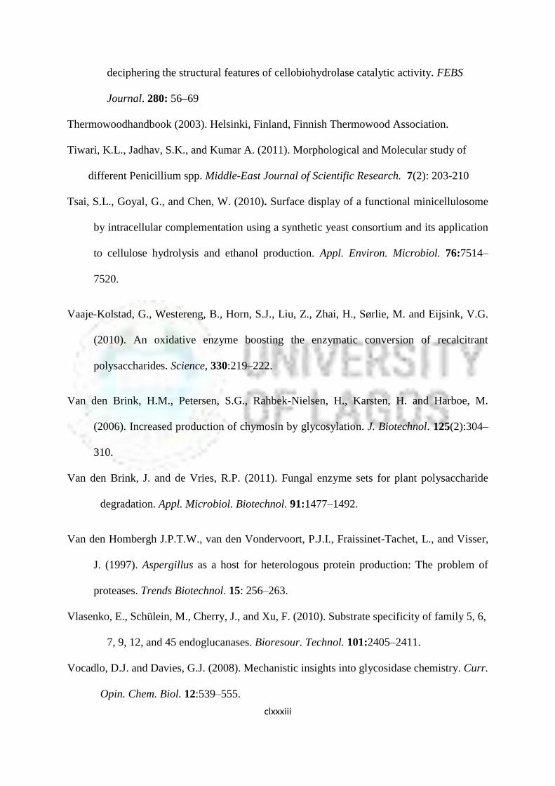
clxxxiii
deciphering the structural features of cellobiohydrolase catalytic activity. FEBS
Journal. 280: 56–69
Thermowoodhandbook (2003). Helsinki, Finland, Finnish Thermowood Association.
Tiwari, K.L., Jadhav, S.K., and Kumar A. (2011). Morphological and Molecular study of
different Penicillium spp. Middle-East Journal of Scientific Research. 7(2): 203-210
Tsai, S.L., Goyal, G., and Chen, W. (2010). Surface display of a functional minicellulosome
by intracellular complementation using a synthetic yeast consortium and its application
to cellulose hydrolysis and ethanol production. Appl. Environ. Microbiol. 76:7514–
7520.
Vaaje-Kolstad, G., Westereng, B., Horn, S.J., Liu, Z., Zhai, H., Sørlie, M. and Eijsink, V.G.
(2010). An oxidative enzyme boosting the enzymatic conversion of recalcitrant
polysaccharides. Science, 330:219–222.
Van den Brink, H.M., Petersen, S.G., Rahbek-Nielsen, H., Karsten, H. and Harboe, M.
(2006). Increased production of chymosin by glycosylation. J. Biotechnol. 125(2):304–
310.
Van den Brink, J. and de Vries, R.P. (2011). Fungal enzyme sets for plant polysaccharide
degradation. Appl. Microbiol. Biotechnol. 91:1477–1492.
Van den Hombergh J.P.T.W., van den Vondervoort, P.J.I., Fraissinet-Tachet, L., and Visser,
J. (1997). Aspergillus as a host for heterologous protein production: The problem of
proteases. Trends Biotechnol. 15: 256–263.
Vlasenko, E., Schülein, M., Cherry, J., and Xu, F. (2010). Substrate specificity of family 5, 6,
7, 9, 12, and 45 endoglucanases. Bioresour. Technol. 101:2405–2411.
Vocadlo, D.J. and Davies, G.J. (2008). Mechanistic insights into glycosidase chemistry. Curr.
Opin. Chem. Biol. 12:539–555.
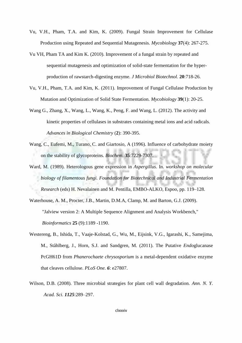
clxxxiv
Vu, V.H., Pham, T.A. and Kim, K. (2009). Fungal Strain Improvement for Cellulase
Production using Repeated and Sequential Mutagenesis. Mycobiology 37(4): 267-275.
Vu VH, Pham TA and Kim K. (2010). Improvement of a fungal strain by repeated and
sequential mutagenesis and optimization of solid-state fermentation for the hyper-
production of rawstarch-digesting enzyme. J Microbiol Biotechnol. 20:718-26.
Vu, V.H., Pham, T.A. and Kim, K. (2011). Improvement of Fungal Cellulase Production by
Mutation and Optimization of Solid State Fermentation. Mycobiology 39(1): 20-25.
Wang G., Zhang, X., Wang, L., Wang, K., Peng, F. and Wang, L. (2012). The activity and
kinetic properties of cellulases in substrates containing metal ions and acid radicals.
Advances in Biological Chemistry (2): 390-395.
Wang, C., Eufemi, M., Turano, C. and Giartosio, A (1996). Influence of carbohydrate moiety
on the stability of glycoproteins. Biochem. 35:7229-7307.
Ward, M. (1989). Heterologous gene expression in Aspergillus. In. workshop on molecular
biology of filamentous fungi. Foundation for Biotechnical and Industrial Fermentation
Research (eds) H. Nevalainen and M. Pentilla, EMBO-ALKO, Espoo, pp. 119–128.
Waterhouse, A. M., Procter, J.B., Martin, D.M.A, Clamp, M. and Barton, G.J. (2009).
"Jalview version 2: A Multiple Sequence Alignment and Analysis Workbench,"
Bioinformatics 25 (9):1189 -1190.
Westereng, B., Ishida, T., Vaaje-Kolstad, G., Wu, M., Eijsink, V.G., Igarashi, K., Samejima,
M., Ståhlberg, J., Horn, S.J. and Sandgren, M. (2011). The Putative Endoglucanase
PcGH61D from Phanerochaete chrysosporium is a metal-dependent oxidative enzyme
that cleaves cellulose. PLoS One. 6: e27807.
Wilson, D.B. (2008). Three microbial strategies for plant cell wall degradation. Ann. N. Y.
Acad. Sci. 1125:289–297.

clxxxv
Wingfield, J.M. and Dickinson, J.R. (1993). Increased activity of a model heterologous
protein in Saccharomyces cerevisiae strains with reduced vacuolar proteinases. Appl.
Microbiol. Biotechnol. 39: 211-215.
Xu, F., Wang, J., Chen, S., Qin, W., and Yu, Z. (2011). Strain improvement for enhanced
production of cellulase in Trichoderma viride. Prikl. Biokhim. Mikrobiol. 47(1):61-5.
Xu, J. and Gong, Z.Z. (2003) Intron requirement for AFP gene expression in T. viride.
Microbiol. 149: 3093-3097.
Yang B and Wyman CE (2006). BSA Treatment to Enhance Enzymatic Hydrolysis of
Cellulose in Lignin Containing substrates. Biotechnology and Bioengineering. 94(4):
611-617
Yang, B. and Wyman, C.E. (2008). Pretreatment: Key to unlocking low-cost cellulosic
ethanol. Biofuels, Bioprod. Bioref. 2: 26-40.
Zhao, X., Peng, F., Cheng, K. and Liu, D. (2009). Enhancement of the enzymatic digestibility
of sugarcane bagasse by alkali-peracetic acid pretreatment. Enzyme Microb Technol
44:17–23.
Zhao, X., Zhang, L. and Liu, D. (2012). Biomass recalcitrance. Part I: The chemical
compositions and physical structures affecting the enzymatic hydrolysis of
lignocelluloses. Biofuels Bioprod. Bioref. DOI.1002/bbb.1331.p1-18
Zhu, H., Zhang, H., Zhang, Y. and Huang, F. (2011). Lignocellulose degradation, enzyme
production and protein enrichment by Trametes versicolor during solid-state
fermentation of corn stover. Afr. J. Biotechnol.10(45): 9182-9192.
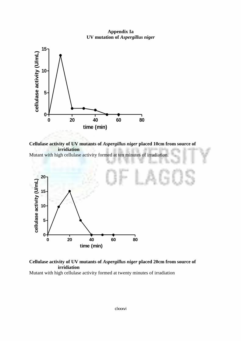
clxxxvi
Appendix Ia
UV mutation of Aspergillus niger
0 20 40 60 800
5
10
15
time (min)
cell
ula
se a
cti
vit
y (
U/m
L)
Cellulase activity of UV mutants of Aspergillus niger placed 10cm from source of
irridiation
Mutant with high cellulase activity formed at ten minutes of irradiation.
0 20 40 60 800
5
10
15
20
time (min)
cell
ula
se a
cti
vit
y (
U/m
L)
Cellulase activity of UV mutants of Aspergillus niger placed 20cm from source of
irridiation
Mutant with high cellulase activity formed at twenty minutes of irradiation
clxxxvii
0 20 40 60 800
5
10
15
20
time (min)
cell
ula
se a
cti
vit
y (
U/m
L)
Cellulase activity of UV mutants of Aspergillus niger placed 30cm from source of
irridiation
Mutant with high cellulase activity formed at thirty minutes of irradiation
0 20 40 60 800
1
2
3
4
time (min)
cell
ula
se a
cti
vit
y (
U/m
L)
Cellulase activity of UV mutants of Aspergillus niger placed 40cm from source of
irridiation
Mutants with high cellulase activity formed at ten minutes and thirty minutes of irradiation
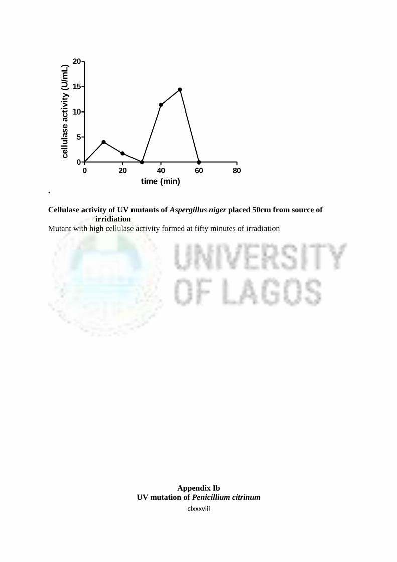
clxxxviii
.
0 20 40 60 800
5
10
15
20
time (min)
cell
ula
se a
cti
vit
y (
U/m
L)
Cellulase activity of UV mutants of Aspergillus niger placed 50cm from source of
irridiation
Mutant with high cellulase activity formed at fifty minutes of irradiation
Appendix Ib
UV mutation of Penicillium citrinum
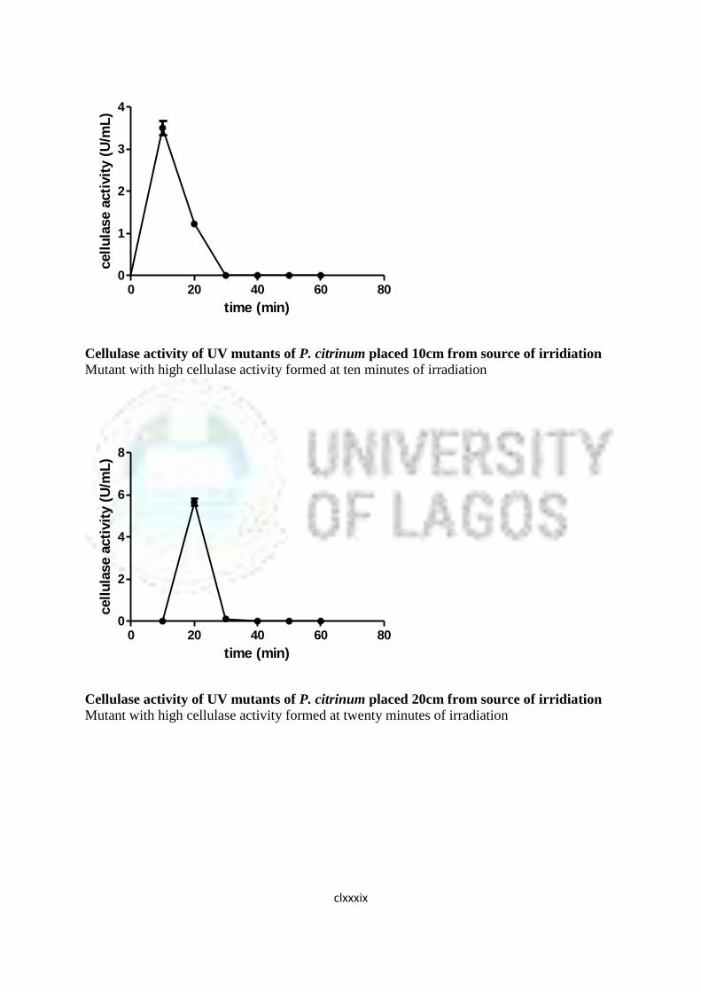
clxxxix
0 20 40 60 800
1
2
3
4
time (min)
cell
ula
se a
cti
vit
y (
U/m
L)
Cellulase activity of UV mutants of P. citrinum placed 10cm from source of irridiation
Mutant with high cellulase activity formed at ten minutes of irradiation
0 20 40 60 800
2
4
6
8
time (min)
cell
ula
se a
cti
vit
y (
U/m
L)
Cellulase activity of UV mutants of P. citrinum placed 20cm from source of irridiation
Mutant with high cellulase activity formed at twenty minutes of irradiation

cxc
0 20 40 60 800
2
4
6
8
time (min)
cell
ula
se a
cti
vit
y (
U/m
L)
Cellulase activity of UV mutants of P. citrinum placed 30cm from source of irridiation
Mutants with high cellulase activity formed at thirty minutes and fifty minutes of irradiation
0 20 40 60 800
1
2
3
time (min)
Cellulase activity of UV mutants of P. citrinum placed 40cm from source of irridiation
Mutants with high cellulase activity formed at ten minutes and forty minutes of irradiation
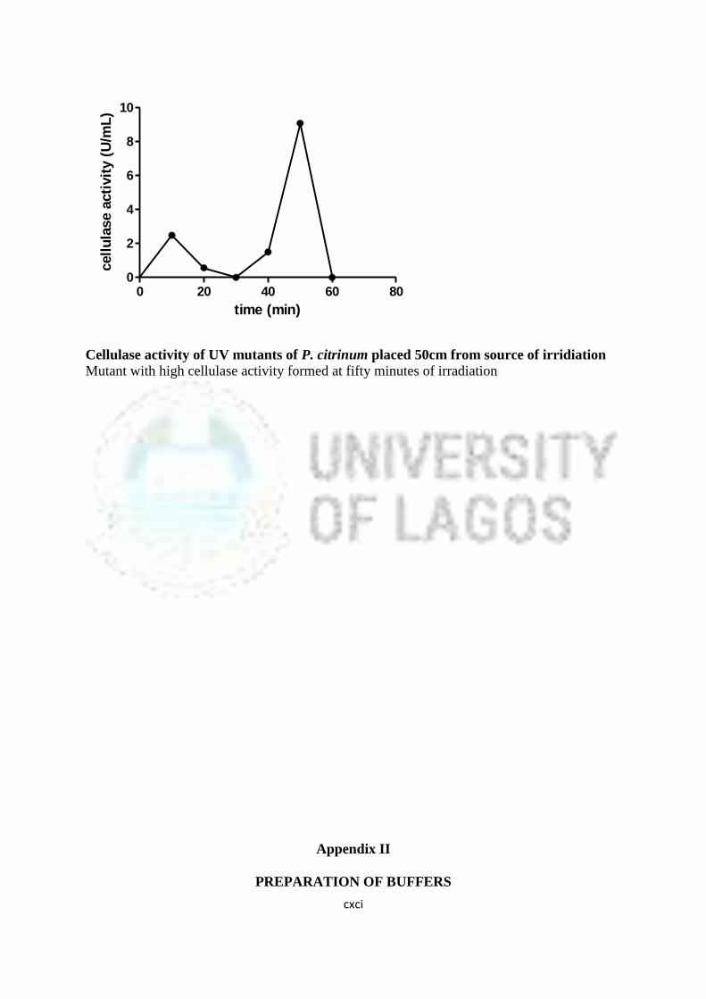
cxci
0 20 40 60 800
2
4
6
8
10
time (min)
cell
ula
se a
cti
vit
y (
U/m
L)
Cellulase activity of UV mutants of P. citrinum placed 50cm from source of irridiation
Mutant with high cellulase activity formed at fifty minutes of irradiation
Appendix II
PREPARATION OF BUFFERS
cxcii
ACETIC ACID - SODIUM ACETATE BUFFER:
Acetic Acid 0.2M: 1.5 ml of glacial acetic acid is made up to 100ml with distilled water.
Sodium Acetate Solution: 0.64g of sodium acetate or 2.72g of sodium acetate trihydrate was
dissolved in 100mL distilled water.
PROCEDURE:
Exactly 36.2ml of sodium acetate solution was drawn into 100ml of standard flask and
14.8ml of glacial acetic acid was added. The volume was made up to 100ml using distilled
water. This gave 0.2 M of acetic acid and sodium acetate buffer. The pH was measured with
pH meter. Adjustments were made using 5N NaOH and 1N HCl.
CITRATE BUFFER (0.1M pH 2.5)
REAGENT S REQUIRED:
Citric acid: 2.101g of citric acid was dissolved in 100ml distilled water.
Sodium citrate solution 0.1 M: 2.941g of sodium citrate was dissolved in 100mL distilled
water.
PROCEDURE:
Citric acid (46.5mL) was added to sodium citrate solution then made up to 100mL with
distilled water. Adjustments were made using 5N NaOH and 1N HCl.
PHOSPHATE BUFFER pH 6.8
REAGENTS REQUIRED:
Monobasic: 2.78g of sodium dihydrogen phosphate was dissolved in 100mL of distilled
water.
Dibasic sodium phosphate (0.2M): 5.3g of disodium hydrogen phosphate or 7.17g sodium
hydrogen phosphate was dissolved in 100mL distilled water.
cxciii
PROCEDURE:
Thirty nine mL of dihydrogen sodium phosphate was mixed with 61mL of disodium
hydrogen phosphate. The solution was made up to 200ml with distilled water. This gave
phosphate buffer of 0.2M. Adjustments were made using 5N NaOH and 1N HCl.
POTASSIUM PHOSPHATE BUFFER
Dipotassium hydrogen phosphate
Potassium dihydrogen phosphate
PROCEDURE:
Dipotassium hydrogen phosphate (174.18 g/mol) and Potassium dihydrogen phosphate
(136.09 g/mol) were taken and made up to 200ml using distilled water. This gave the
potassium buffer.
Standardised pH meter with standard buffer. Washed electrode with distilled water was
introduced into potassium buffer prepared. The pH of the solution was 6.5.
Dipotassium hydrogen phosphate (K2HPO4) and potassium dihydrogen phosphate (KH2PO4)
solution were prepared and the pH was measured to be 9.87 and 4.23 respectively, the
solution were made using 1N HCl and 5N NaOH respectively and the pH was found to be
6.5.
CTAB Buffer (100mL) preparation
1.1.1Materials: CTAB buffer, microfuge tubes, mortar and pestle, liquid nitrogen,
microcentriguge, absolute ethanol (ice cold), 70% ethanol (ice cold), 7.5M Ammonium
acetate, 55oC water bath, chloroform : isoamyl alcohol (24:1), sterile water, agarose, 6x
loading buffer, 1x TBE, agarose gel electrophoresis system, ethidium bromide solution.

cxciv
CTAB buffer system 100ml
2.0g CTAB (Hexadecyl trimethyl-ammonium bromide)
10.0mL 1M Tris pH 8.0
4.0mL 0.5M EDTA pH 8.0 (EthylenediaminetetraAcetic acid Di-sodium salt)
28.0mL 5M NaCl
40.0mL H2O
Adjust all pH 5.0 with HCl and make up to 100mL with distilled water
1M Tris pH 8.0
To 800mL of distilled water, 121.1g of Tris base was added. The pH was adjusted to 8.0 by
adding 42mL of concentrated HCl. The solution was allowed to cool to room temperature
before making the final adjustments to the pH. The volume was adjusted to 1L with distilled
water and it was sterilized using an autoclave.
5X TBE Buffer
54g Tris base
27.5g boric acid
20mL of 0.5M EDTA (pH 8.0)
It was made up to 1L with distilled water
To make a 0.5X working solution, a 1:10 dilution of the concentrated stock was done.
1% Agarose gel: one gram agarose was dissolved in 100mL TBE
cxcv
Appendix III
DNA quality confirmation
One percent solution of agarose was prepared by melting 1g of agarose in 100mL of 0.5X
TBE buffer in a microwave for approximately 2min. It was allowed to cool for a couple of
minutes before adding 2.5µL of ethidium bromide. It was was the stirred to mix.
The gel was casted using the supplied tray and comb. The gel was allowed to set for a
minimum of 20min at room temperature on a flat surface.
The following was loaded into separate wells
10 µL 1Kb ladder
5 µL sample + 5 µL water +2 µL 6X loading buffer
The gel was run for 30min at 110V. Then it was exposed to UV light and photographed.
The DNA quality was confirmed. Presence of highly resolved high molecular weight band
indicates good quality DNA, presence of smeared band indicates DNA degradation.

cxcvi
Appendix IV
Primers used in the amplification of selected genes in cDNA
cbh1
Forward 5‘ GTCATTGACGCCAACTGGCGCTGG 3‘
Reverse 5‘ ACGCTCCCAGCCCTCAACGTTGG 3‘
ace1
Forward 5‘ AGCGCGATCCATGCGGTCCATGGCCCGCCG 3‘
Reverse 5‘ AGCCGGAATTCGTAGCTGGGCGTGGAGGAAG 3‘
aep 3
Forward 5‘ CTGGCTGTCTTTGCTCTG 3‘
Reverse 5‘ TCCCACTCTGCTCAAACC 3‘
lacc1
Forward 5‘ GGTACATCCTAGCACCCAATG 3‘
Reverse 5‘ GACGAGATCAGTTTCCAAGAGG 3‘
lacc5
Forward 5‘ CGCATTTGCCGCTTTCTT 3‘
Reverse 5‘ GGTGACTAGGACTGAGTATCTC 3‘
`