asnr 2015 use of a novel mri-compatible head-positioning device for the three dimensional kinematic...
TRANSCRIPT

ASNR 2015
Use of a Novel MRI-Compatible Head-Positioning Device for the Three Dimensional Kinematic Analysis of the Cervical
Spine in Axial Rotation
1. Student, Illinois Mathematics and Science Academy, Aurora, IL, USA
2. PhD Candidate, Interdepartmental Neuroscience Program, Northwestern University, Chicago, IL, USA
3. Professor and Director, Spine Biomechanics Laboratory, Department of Orthopedic Surgery, Rush University Medical Center, Chicago, IL, USA
4. Assistant Director, Spine Biomechanics Laboratory, Department of Orthopedic Surgery, Rush University Medical Center, Chicago, IL, USA
5. Professor, Department of Radiology, Northwestern University, Chicago, IL, USA
Poster #: EP-153
Authors and Affiliations
Jyotsna Bitra1, Jessica Phung1, Kenneth A. Weber II2, Nozomu Inoue3, Alejandro A. Espinoza Orías4, and Todd B. Parrish5

ASNR 2015
The authors have no disclosures.
Disclosures

ASNR 2015
Cervical Spine Anatomy
● Upper Cervical Spine○ Occiput = C0○ Atlas = C1○ Axis = C2○ Most of the axial rotation occurs
between C1 and C2● Lower Cervical Spine
○ C3-C7● Intervertebral Joints
○ Intervertebral discs between adjacent vertebrae
○ Fibrocartilaginous joint○ Shock absorbers
● Facet Jointso Synovial planar jointso Between adjacent superior and
inferior articulating processes
Austin et al., 2014

ASNR 2015
Segmental Motion
Ishii, T., et al. (2004)
● 3D cervical spine segmental motions (6 degrees of freedom)○ +/- Rotations around X,Y, and Z axes○ +/- Translations along X, Y, and Z axes
● Characteristic coupled segmental motions of the cervical spine

ASNR 2015
Previous Studies
● Ishii T et al. 2004○ Imaged healthy patients in 15 degree intervals of axial head rotation○ 1.0 T MRI Scanner○ Calculated 3D segmental motions using a voxel-based registration
technique● Upper Cervical Spine
○ Coupled lateral bending with axial rotation in the direction opposite of axial rotation at C0-C1 and C1-C2
○ Coupled extension with axial rotation at C0-C1 and C1-C2● Subaxial Cervical Spine
○ Coupled lateral bending with axial rotation in the same direction of axial rotation at all levels
○ Coupled extension with axial rotation occurred at C2-C3, C3-C4, and C4-C5
○ Coupled flexion with axial rotation at C5-C6, C6-C7, and C7-T1● No head-positioning device was used to control position of the head
relative to the thorax

ASNR 2015
Purpose
1. MRI allows a non-invasive method of analyzing cervical spine segmental motion
2. Previous studies failed to utilize a head-positioning device to control the amount of angular displacement of the head relative to thorax
3. Design and test an MRI-compatible head-positioning device to image the cervical spine with controlled axial rotation of head
Windsor, R.E, (2014).

ASNR 2015
Head-Positioning Device● MRI-compatible device made from ABS (acrylonitrile
butadiene styrene) plastic and football helmet (Riddell, Rosemont, IL, USA)
● Wheel and axle design● Allows for 180 degrees head rotation

ASNR 2015
Imaging
● Siemens TIM Trio 3.0 T Scanner
● Two 4-channel flex coils were placed anterior and posterior to the head and neck while scanning
● Imaged using navigated multi-echo (4) 3D MPRAGE acquisition with IPAT
○ TE 2 ms, TR 2540 ms, flip angle 7°,○ 1 average, 1 mm x 1 mm x 2 mm voxels○ Acquisition time 5.5 minutes per image
● 4 males, 2 females (mean age 33.5+- 8.2 years) with no complaints of neck pain
● Subjects were scanned in three different positions of axial head rotation○ Neutral, 40 degrees of right head rotation,
and 40 degrees of left head rotation

ASNR 2015
MRI Images Using the Head Positioning Device
40° Right Rotation 40° Left RotationNeutral
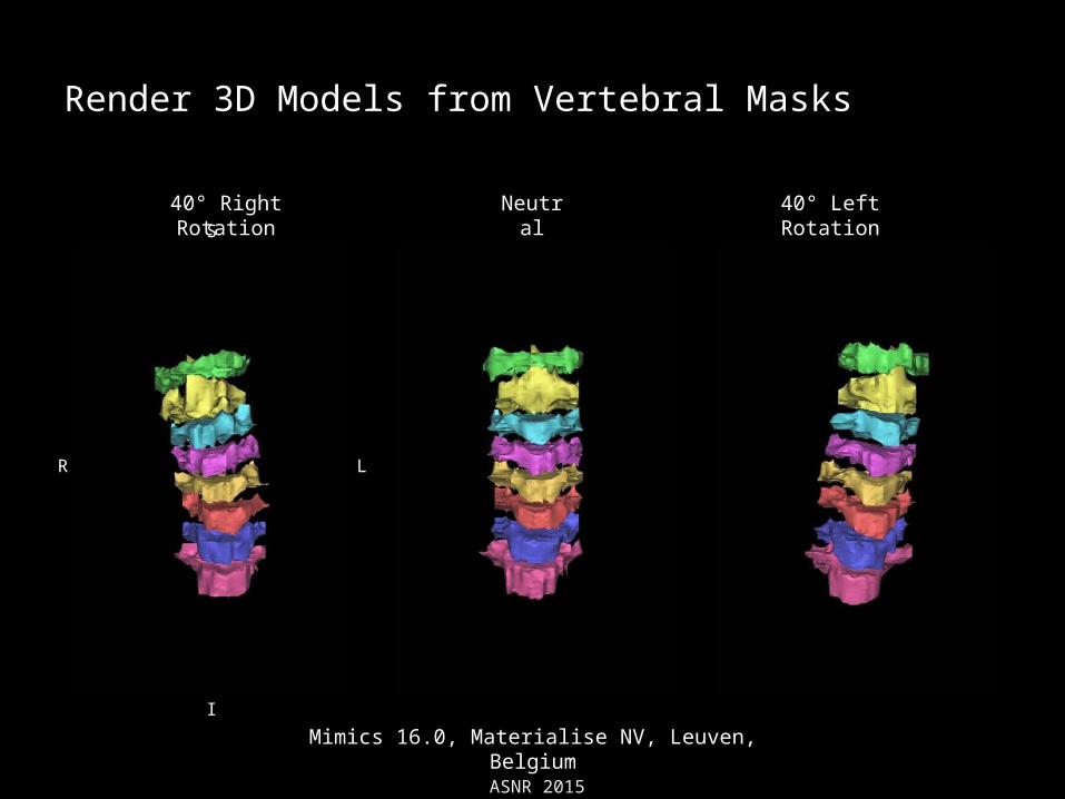
Mimics 16.0, Materialise NV, Leuven, Belgium
R L
S
I

ASNR 2015
Manual Segmentation of Cervical Vertebrae
40° Right Rotation 40° Left RotationNeutral
Mimics 16.0, Materialise NV, Leuven, Belgium
R L
S
I
ASNR 2015
Render 3D Models from Vertebral Masks
40° Right Rotation 40° Left RotationNeutral
Mimics 16.0, Materialise NV, Leuven, Belgium
R L
S
I

ASNR 2015
Calculation of 3D Segmental Motions using the Volume Merge Method
Render 3D Models Point Cloud Format Transformation of neutral vertebra to transformed
vertebra
Ochia, R.S. et al., (2006)
ASNR 2015
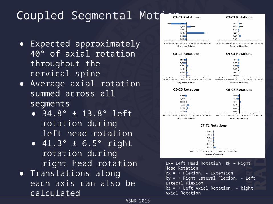
Coupled Segmental Motion
● Expected approximately 40° of axial rotation throughout the cervical spine
● Average axial rotation summed across all segments ● 34.8° ± 13.8° left rotation
during left head rotation● 41.3° ± 6.5° right rotation
during right head rotation● Translations along each axis
can also be calculated
LR= Left Head Rotation, RR = Right Head RotationRx = + Flexion, - ExtensionRy = + Right Lateral Flexion, - Left Lateral FlexionRz = + Left Axial Rotation, - Right Axial Rotation

ASNR 2015
Conclusions
● The device allowed for the precise control of axial head rotation while in the MRI scanner
● All subjects were able to tolerate the device and head positioning throughout the scans
● Image quality was adequate to manually segment the data
● Our results were similar to previously published studies● In the future, the feasibility of using this device in a neck
pain population will be assessed

ASNR 2015
References
● Austin N, Krishnamoorthy V, Dagal A. Airway management in cervical spine injury. International Journal of Critical Illness and Injury Science. 2014;4(1):50-56.
● Ishii T, Mukai Y, Hosono N, Sakaura H, Nakajima Y, Sato Y, Sugamoto K, Yoshikawa H. Kinematics of the upper cervical spine in rotation: in vivo three-dimensional analysis. Spine. 2004 Apr 1;29(7):E139-44.
● Ishii T, Mukai Y, Hosono N, Sakaura H, Fujii R, Nakajima Y, Tamura S, Sugamoto K, Yoshikawa H. Kinematics of the subaxial cervical spine in rotation in vivo three-dimensional analysis. Spine. 2004 Dec 15;29(24):2826-31.
● Ochia RS, Inoue N, Renner SM, Lorenz EP, Lim TH, Andersson GB, An HS. Three-dimensional in vivo measurement of lumbar spine segmental motion. Spine (Phila Pa 1976). 2006 Aug 15;31(18):2073-8.

ASNR 2015
Acknowledgments
Authors and Collaborators:
Parrish Neuroimaging Laboratory
Department of Radiology
Northwestern University
Center for Translational Imaging
•Todd B. Parrish, PhD
•Kenneth A. Weber II, DC
•Jennie Chen, PhD
•Xue Wang, PhD
•Brian Williams, BSN, RN
Rush University Medical Center
Spine Biomechanics Laboratory
•Nozomu Inoue, MD, PhD
•Alejandro A. Espinoza Orías, PhD
Funding Support
•NCMIC Foundation, Inc.
•NCCIH F32AT007800
•NICHD T32HD057845