asd validity - springer · asd,klin etal.(2015) positedthatthe developmentalfailure of the...
TRANSCRIPT

REVIEW PAPER
ASD Validity
Lynn Waterhouse1 & Eric London2& Christopher Gillberg3
Received: 15 February 2016 /Accepted: 19 July 2016 /Published online: 10 August 2016# Springer Science+Business Media New York 2016
Abstract ASD research is at an important crossroads. TheASD diagnosis is important for assigning a child to early be-havioral intervention and explaining a child’s condition. ButASD research has not provided a diagnosis-specific medicaltreatment, or a consistent early predictor, or a unified lifecourse. If the ASD diagnosis also lacks biological and con-struct validity, a shift away from studying ASD-defined sam-ples would be warranted. Consequently, this paper reviewsrecent findings for the neurobiological validity of ASD, theconstruct validity of ASD diagnostic criteria, and the constructvalidity of ASD spectrum features. The findings reviewedindicate that the ASD diagnosis lacks biological and constructvalidity. The paper concludes with proposals for research go-ing forward.
Keywords DSM-5 . ASD . Autism . Diagnosis . Validity .
Comorbidity . Heterogeneity
The goal of the DSM-3 nosology (American PsychiatricAssociation 1980) was to create reliable and standard categor-ical psychiatric diagnoses (Robins and Guze 1970). However,in the past 30 years, clinical, genetics, and neuroscience find-ings have revealed that the DSM diagnoses are not biological-ly valid. The National Institutes of Mental Health (NIMH)
responded by proposing the Research Domain Criteria(RDoC) framework for a brain-based transdiagnostic psychi-atric symptom nosology (Cuthbert and Insel 2013; Insel et al.2010; Lilienfeld and Treadway 2016). Peterson (2015) andWeinberger et al. (2015) argued that the RDoC could notreplace the DSM-5 psychiatric nosology (AmericanPsychiatric Association 2013) or the parallel InternationalClassification of Diseases (ICD) psychiatric nosology(World Health Organization 2012). But BFor the foreseeablefuture, RDoC is not envisioned as a system of psychiatricclassification in its own right. Instead, in the near term,RDoC and DSM-ICD are expected to coexist. Nevertheless,RDoC is intended to provide scaffolding for a large-scale re-search program that will ultimately yield an alternative toDSM-ICD^ (Lilienfeld and Treadway 2016, p. 445).
RDoC advocates accept that DSM-5/ICD psychiatric cate-gories remain necessary in clinical practice, but argue thatresearchers should shift to RDoC study designs immediately.They assert that studying psychiatric categories lacking bio-logical validity blocks the discovery of brain bases for psy-chopathology and thus cannot lead to effective medical treat-ments for specific psychiatric symptoms (Cuthbert and Insel2013; Insel et al. 2010; Lilienfeld and Treadway 2016; Yeeet al. 2015). Against this RDoC imperative for biological va-lidity, Weinberger et al. (2015) countered that current DSM-5psychiatric behavioral diagnoses were valid when theyyielded effective medical treatment, clear prognosis, and a lifecourse specific to a diagnosis.
Autism spectrum disorder (ASD) research has been produc-tive (Dawson 2016; de la Torre-Ubieta et al. 2016; Szatmariet al. 2016), but no ASD research findings have met the validitycriteria of Weinberger et al. (2015). DSM-5 ASD research hasfound no specific effective pharmacotherapy or other medicaltreatment (Na Young and Findling 2015). The only broadlysuccessful ASD treatment has been early behavioral intervention
* Lynn [email protected]
1 The College of New Jersey, Ewing, NJ, USA2 Autism Treatment Research Laboratory, New York State Institute for
Basic Research in Developmental Disabilities, Staten Island, NY,USA
3 University of Gothenburg, Gothenburg, Sweden
Rev J Autism Dev Disord (2016) 3:302–329DOI 10.1007/s40489-016-0085-x

programs (Kasari 2015; Schreibman et al. 2015; Smith andIadarola 2015; Volkmar et al. 2014), but these treatments arenot unique to ASD (Losinski et al. 2014), and the long-termeffectiveness of these programs is not yet known (Fernell et al.2011, 2014). Researchers who studied infant siblings of childrenwith ASD concluded no single early behavior could predictASD diagnoses (Zwaigenbaum et al. 2015), and ASD has beenfound with widely varied trajectories of development (Fountainet al. 2012; Lord et al. 2015) and varied life outcomes (Fein et al.2013; Helles et al. 2015; Steinhausen et al. 2016).
ASD research is at a crucial decision point. The ASD di-agnosis remains necessary in the clinic to assign a child toearly behavioral intervention and to explain a child’s condi-tion. But researchers must decide whether to continue study-ing ASD-defined samples or not. Given that ASD lacks anydiagnosis-specific medical treatment, any consistent early pre-dictor, or any specific life course, if the ASD diagnosis alsolacks biological and construct validity, a shift away fromstudying ASD-defined samples would be warranted.
Consequently, this paper reviews recent findings for ASDbiological and construct validity. This paper is not a meta-analysis; instead it brings together competing and unresolvedfindings. The first section examines evidence for the neurobiolo-gical validity of ASD. The second section outlines evidence forthe construct validity of ASD diagnostic criteria. The third sectionexplores evidence for the construct validity of ASD spectrumfeatures beyond ASD diagnostic symptoms. The paper concludesthat the evidence reviewed does not provide support for theneurobiological or construct validity of the ASD diagnosis, andtherefore the ASD diagnosis should be disbanded in research.
Does ASD Have Neurobiological Validity?
TheASD diagnosis would be biologically valid if all or nearly allindividuals diagnosed with ASD shared one or a few relatedbrain impairments. Researchers have tried different means to finda unitary ASD brain impairment. Researchers have measured thebrain structures of idiopathic ASD, defined as having no knowngenetic or environmental cause. Researchers have proposed andtested models of unitary ASD brain impairment. Researchershave sought brain impairments unique to each core ASD diag-nostic symptom. Researchers have looked for an independentASD brain impairment in syndromic ASD, defined as occurringwith known genetic and environmental syndromes. Fifth, re-searchers have grouped ASD genetic risk genes theorized toproduce a narrowed set of disrupted brain circuits.
Brain Validity Research Approach 1: Is There a SpecificASD Brain Impairment?
Findings for ASD global and regional brain sizes have beenvaried. Riddle et al. (2016) found that 443 individuals with
ASD had a 2.17 % increase in gray matter compared to 390typically developing (TD) individuals, but found no group dif-ference in white matter. However, when Riddle et al. (2016)compared matched subsamples of 300 ASD and TD children,no differences between TD and ASD white matter or graymatter were found. Vasa et al. (2012) reported that only 8 of73 individuals diagnosed with ASD had any atypical brainfeatures. Lenroot and Yeung (2013) reported that the majorityof individuals diagnosed with ASD had typical brain and headsize. However, in other studies, approximately 10 to 24 % ofindividuals diagnosed with ASD had persisting macrocephaly(Gillberg and de Souza 2002; Lainhart 2015; Nebel et al. 2015;Sacco et al. 2015; Tammimies et al. 2015). In addition, a num-ber of studies have reported that 3 to 15 % of individuals withASD had persistingmicrocephaly (Gillberg and de Souza 2002;Nebel et al. 2015; Roullet et al. 2013; Stevens et al. 2013).
Although Riddle et al. (2016) found no regional brain dif-ferences in 443 individuals with ASD compared to 390 TDindividuals, Lefebvre et al. (2015) noted that many studies hadfound significantly smaller corpus callosum in ASD than TDcontrols. By contrast, Lefebvre et al. (2015) found no differ-ence in corpus callosum size in their sample of 694 individualswith ASD compared to TD controls. Studies of the amygdalain ASD have reported significantly Bincreased, decreased andpreserved volumes^ of the amygdala (Bellani et al. 2013,p. 3). Similarly, Stigler et al. (2011) noted that studies havereported increased, decreased, and typical (preserved) vol-umes for the fusiform gyrus in ASD.
D’Mello et al. (2015) reported that ASD was characterizedby reduced gray matter in the cerebellum in lobule VII.However, a consensus paper on the cerebellum in ASD(Fatemi et al. 2012) concluded that only a subgroup of ASDhad atypical cerebellar anatomy, and this was a smaller cere-bellar vermis volume and fewer Purkinje cells. Studies of thebrainstem in ASD have reported both typical and atypicallysmaller volumes of brainstem gray matter (Jou et al. 2013).Despite evidence for abnormal auditory brainstem response(ABR) in ASD (Rosenhall et al. 2003; Lukose et al. 2015),Jou et al. (2013) concluded that ASD brainstem studies wereconflicting and inconclusive.
SummaryASD global and regional brain structures are variedand do not provide brain structure biological validity for ASD.
To date, no unitary atypical brain size or volume has beenfound for ASD, and no unitary atypical ASD regional brainstructure has been found. Equally important, varied ASD elec-trophysiology findings (Billeci et al. 2013) and varied ASDmolecular neurochemistry findings (Zürcher et al. 2015) standagainst the idea that there is any shared single ASD brainimpairment in electrophysiology or molecular neurochemistry.
Of course, research has not uncovered the full complexityof human brain development, functions, and networks (Spornsand Betzel 2016). When much more is known about the
Rev J Autism Dev Disord (2016) 3:302–329 303

emergence of regional functions and connectivity of the brain,it may be possible in the future to establish a unitary model ofASD brain impairment.
Brain Validity Research Approach 2: Is Therea Replicated ASD Brain Impairment Model?
Hundreds of unitary models of ASD brain impairment havebeen theorized and studied. For example, Baron-Cohen et al.(2015) proposed that ASD stemmed from fetal steroidogenicabnormalities. Fishman et al. (2014) proposed that ASD re-sulted from atypical overconnectivity of the brain’s mirrorneuron regions and theory of mind regions. By contrast,Khan et al. (2015) proposed that ASD resulted fromunderconnectivity in local brain regions when activated by aBfunctionally relevant task^ (p. 1407). Mullins et al. (2016)posited that mutations in the FMR1 gene and TSC genes dis-rupt long-term depression (LTD) and long-term potentiation(LTP), and therefore, these mutations in ASD cause atypicalLTP and LTD processes that impair the excitation-inhibitionbalance in brain development and function which determinesASD (Mullins et al. 2016).
Robertson et al. (2015) claimed that the Bprime suspect^single cause for ASDwas disrupted GABAergic signaling thatimpaired neurodevelopment and cortical computations.However, Estes and McAllister (2015) theorized that ASDwas caused by atypical immune system factors that convergedon the MEF2 and mTOR signaling hubs and thus disruptedthe brain’s developmental synaptic function and plasticity.Irimia et al. (2014) theorized a basis for ASD brain impair-ment in atypically greater misregulation of nSR100-dependent microexons. Focusing on early visual attention inASD, Klin et al. (2015) posited that the developmental failureof the reward-based interactional eye fixation to co-opt thebrain’s innate reflexive eye fixation caused ASD.
Because these eight theories explain different aspects of ASDbrain function, they are not necessarily mutually exclusive.However, there has been no attempt to synthesize any subsetof these eight theories, or synthesize any subset of prior unitaryASD brain theories (Waterhouse 2008). Most importantly, nounitary brain impairment theory to date has been replicated tobecome a standard explanation of ASD brain disruption.
The theory with the most active support argues that ASDresults from impaired brain connectivity that is likely to havebeen preceded by early brain overgrowth (Solso et al. 2015;Stoner et al. 2014; Venkataraman et al. 2015). Although theunderconnectivity theory has been studied formore than 15 years(Just et al. 2004) and has many supporters (Anderson 2013;Dawson 2016; Ecker et al. 2015; Green et al. 2015; Shen et al.2013), nonetheless, this theory has not become standard becausethere is as much evidence against the theory as there is for it.
Supporting the existence of early ASD brain overgrowth,Ecker et al. (2015) asserted that, on average, toddlers with
ASD have Ba larger brain volume than typically developingchildren^ (p. 1), and Shen et al. (2013) and Anderson (2013)reported that ASD was characterized by atypical early brainovergrowth. Sussman et al. (2015) reported that early brainovergrowth was limited to males with ASD, and Chaste et al.(2013) found that the subgroup of ASD with an atypicallylarger head circumference expressed greater symptom severityand lower IQ.
Counter to the early brain overgrowth model, though,Chaste et al. (2013), Raznahan et al. (2013), and Cederlundet al. (2014) all concluded that very few children with ASDhad early brain overgrowth, and Raznahan et al. (2013) report-ed that larger ASD head circumference was not found whenlocal TD controls were used. Lainhart (2015) reviewed re-search and noted that only Ba very small subgroup of ASDchildren^ (p. 79) had larger brain volumes. In a meta-analysis,Sacco et al. (2015) found only 5.7 % of individuals with ASDhad macrocephaly and just 9.1 % had brain overgrowth.Tammimies et al. (2015) also found that only 63 of 258 chil-dren with ASD had macrocephaly. Also countering the ASDearly brain overgrowth model, 3 to 15 % of individuals diag-nosed with ASD have been born with microcephalic brains(Nebel et al. 2015; Roullet et al. 2013; Stevens et al. 2013).Most importantly, the generalizability of the ASD early brainovergrowth model is limited by the evidence that the majorityof individuals diagnosed with ASD have typically developinghead and brain growth (Lenroot and Yeung 2013; Nebel et al.2015; Riddle et al. 2016; Vasa et al. 2012).
Supporting the malconnectivity component of the currentlydominant model, Dawson (2016) asserted that Blong-rangeconnections between different brain regions are weaker inpeople with ASD^ (para. 4). Venkataraman et al. (2015) de-termined that ASD brain malconnectivity occurred in two net-works: a language network including temporal lobe areas anda Bsocial-person^ network including temporal and parietalareas. Venkataraman et al. (2015), however, found no impair-ment in ASD frontal lobe connectivity. Conversely,Kitzbichler et al. (2015) reported that ASD malconnectivitywas centered in frontal lobe-linked connections.
Counter to both Kitzbichler et al. (2015) and Venkataramanet al. (2015), though, Redcay et al. (2013) reported finding noevidence for atypical connectivity in ASD. Tyszka et al.(2014) also stated that their ASD sample showed, Bno evi-dence at all for altered connectivity at the whole-brain level^(pp. 7–8). Kirkovski et al. (2015) found no differences be-tween high functioning individuals with ASD and typical con-trols in white matter in major tract bundles determined by anymethod: fractional anisotropy (FA), mean diffusivity (MD),radial diffusivity (RD), or axial diffusivity (AD). Koldewynet al. (2014) reported finding no general impaired white matterin ASD. Lefebvre et al. (2015) found no differences between694 individuals diagnosed with ASD and typical controls inthe largest white matter tract in the brain—the corpus
304 Rev J Autism Dev Disord (2016) 3:302–329
callosum. The researchers questioned whether any regionalASD malconnectivity model could be meaningful becausemost of the brain is involved in connectivity (Lefebvre et al.2015).
Another problem for standardizing the overgrowth-malconnectivity model is that there are so many different ver-sions of this model (Fishman et al. 2014; Kennedy et al. 2015;Khan et al. 2015; Kitzbichler et al. 2015; Venkataraman et al.2015). As noted by Kennedy et al. (2015), Bthe connectivityhypothesis has been vague ever since its inception… slowlymorphing from a theory about underconnectivity in ASD, toone about distal underconnectivity paired with localoverconnectivity, to one about atypical connectivity in eitherdirection (or both)^ (p. 81).
Summary No ASD brain impairment model has beenadequately replicated to become standard, and thus, noexisting model provides neurobiological validity forASD.
Despite intense research efforts, the malconnectivity modelhas not been successfully replicated (Ecker et al. 2015;Kennedy et al. 2015; Khan et al. 2015; Lefebvre et al.2015), and neither has any of the myriad other unitary ASDbrain impairment models. Moreover, none of these models hasaccounted for the variation in ASD global and regional brainstructures, or the variation in ASD electrophysiology findings(Billeci et al. 2013), or the variation in neurochemistry find-ings (Zürcher et al. 2015).
Brain Validity Research Approach 3: Is Each ASDDiagnostic Symptom Caused by a Distinct BrainImpairment?
Many studies have looked for the biological validity of indi-vidual ASD diagnostic and associated nondiagnostic symp-toms (Anderson et al. 2014; Boucher 2011; Brunsdon andHappé 2014; Chen et al. 2015; Harrop et al. 2013;Hormozdiari et al. 2015; Jason et al. 2015; Pina-Camacho et al.2012; Shuster et al. 2014).
Is ASD Social Impairment Linked to a Unique BrainImpairment?
DSM-5 defines ASD diagnostic social impairment as persis-tent impaired social reciprocity, with impaired social nonver-bal communication behaviors, along with an inability to de-velop and maintain relationships. Pina-Camacho et al. (2012)analyzed multiple studies of the association between ASDsymptoms and brain impairments and reported that ASD di-agnostic social impairment was linked to four distinct regionalbrain dysfunctions. The face-fusiform area (FFA) was foundas the basis for ASD social impairment, and thefrontotemporal cortical networks including mirror neuron
system circuits were found as the basis for ASD social impair-ment (Pina-Camacho et al. 2012). Still other findings impli-cated dysfunction of the anterior cingulate cortex (ACC) inASD social impairment, and finally, some research suggestedthat ASD social behaviors were impaired by disruptions in thesubcortical amygdala-hippocampal network (Pina-Camachoet al. 2012).
In addition to the brain regions reported by Pina-Camachoet al. (2012), Ecker et al. (2012) claimed that ASD diagnosticsocial impairment was specifically linked to Ba significantdecrease in gray matter volume in a large cluster located inthe occipital and medial parietal regions^ (p. 202). However,Ecker et al. (2015) later asserted that impaired ASD socialcommunication resulted from dysfunctions in Broca’s andWernicke’s areas, while impaired ASD social/emotional com-prehension resulted from dysfunctions in frontotemporal re-gions and the amygdala.
Zilbovicius et al. (2013) reported that ASD diagnostic so-cial impairment was linked to abnormalities in the superiortemporal sulcus (STS), including decreased gray matter andhypoperfusion at rest. Maillard et al. (2015) found that diag-nostic ASD social impairment and chromosomal number var-iant (CNV) 16p11.2 carrier social impairment were bothlinked to brain dysfunctions in left and right putamen,insula, posterior cingulate, thalamus, and superior temporalgyrus. By contrast, Byrge et al. (2015) reported that five (of17) individuals diagnosed with ASD all had profound diag-nostic social impairment but none of the five shared any spe-cific atypical brain dysfunction. Instead, these five individualsexpressed five different idiosyncratic brain responses to ob-served social interaction (Byrge et al. 2015).
Ameis and Catani (2015) reported that three differentmethods yielded different brain bases for ASD social impair-ment. Ameis and Catani (2015) noted that while neuropathol-ogy studies found frontolimbic pathways linked to ASD diag-nostic social impairment, MRI imaging studies did not.Instead, Ameis and Catani (2015) reported that MRI imagingstudies found early atypical brain growth and atypical volumeof frontal white matter linked to ASD social impairment.Finally, fMRI findings differed from both neuropathologyand MRI findings, linking ASD diagnostic social impairmentto decreased temporal lobe activity and decreased frontal lobeactivity. Ameis and Catani (2015), in turn, theorized that thebrain basis for ASD diagnostic social impairment wasdisrupted uncinate fasciculus and thalamic projections to pre-frontal and temporal lobes.
Are ASD Diagnostic Repetitive Behaviors, RestrictedInterests, and Resistance to Change, Together Identifiedas the RRBs, Linked to a Unitary Brain Impairment?
The DSM-5 ASD diagnosis now requires the presence of twoof four behaviors of any of the RRBs, and/or atypical sensory
Rev J Autism Dev Disord (2016) 3:302–329 305

responsiveness. Pina-Camacho et al. (2012) reviewed multi-ple studies of the brain basis of the RRBs and reported that sixdistinct brain regions had been linked to ASD RRBs. Onebrain region was the frontocerebellar network, and anotherregion was the frontostriatal system (Pina-Camacho et al.2012). Fourmore regions linked to the RRBswere the anteriorand posterior cingulate, some posterior parietal regions, theposterior regions of corpus callosum, and the cerebellar ver-mis and peduncles (Pina-Camacho et al. 2012).
The RRBs have also been linked to specific frontal lobestructural abnormalities (Ecker et al. 2012), and the RRBshave been linked to atypical caudate overgrowth (Langenet al. 2014). By contrast, Doyle-Thomas et al. (2014) reportedthat RRBs in general were associated with atypically lowcholine/creatine in the thalamus, whereas ASD social impair-ment was not. In addition, Gabriels et al. (2013) reported thatthe general severity of RRBs was inversely correlated withdaytime cortisol levels.
Are ASD Nondiagnostic Symptoms Linked to Specific BrainImpairments?
Although ASD diagnostic criteria specifically exclude manyneurodevelopmental symptoms such as intellectual disability(ID), language delay, language impairment, attention deficits,and seizures, more than 96 % of those diagnosed with ASDactually express one or more of these symptoms and/or othernon-ASD symptoms (Lundström et al. 2015b). The preva-lence of non-ASD neurodevelopmental symptoms varies.ADHD symptoms were found in 17 to 83 % of individualswith ASD (Matson et al. 2013; Nebel-Schwalm and Worley2014). ID has been reported in 50–70 % of individuals withASD (Matson and Williams 2014). Nearly 100 % of individ-uals diagnosed with ASD have been found to exhibit mutism,or language delay, or language impairment (Boucher 2012).Finally, 40 % with ASD have experienced seizures (Amietet al. 2013), and 60 % with ASD have exhibited epileptiformbrain activity (Mulligan and Trauner 2014).
Variation in the pathophysiology of non-ASD epilepsy isvery similar to the variation in the pathophysiology of epilep-sy in ASD (Amiet et al. 2013; Blackmon 2015; Stafstrom andCarmant 2015). Some epilepsy is the result of malformationsof cortical development (MCDs), including focal cortical dys-plasia and heterotopias, and these malformations have beenfound in ASD (Blackmon 2015).
Attention problems are inherent in some ASD criteria, suchas the preoccupation with unusual objects, and up to 83 % ofthose with ASD have expressed ADHD symptoms (Nebel-Schwalm and Worley 2014). However, before DSM-5, ASDand ADHD could not be diagnosed together in one individual.Johnson et al. (2015) reported that frontal lobe impairmentsand hypo- and hyperconnectivity have been reported for bothASD and ADHD. Johnson et al. (2015) also noted that some
studies found slower early brain size increase in both ADHDand ASD.
Up to 70 % of individuals with ASD express ID, and someof the comorbidity of ASD and ID is known to stem fromshared gene risk factors. For example, 17 % of all ID riskgenes with de novo loss of function (LoF) mutations are alsoreported for ASD (Vissers et al. 2016). Both ASD and ID havebeen found with gene variants that cause impairment in manyaspects of brain development and function, includingneurogenesis, neuronal migration, synaptic function, and reg-ulation of transcription and translation (Vissers et al. 2016).
Mayes et al. (2015) found that language impairment inchildren without ASD was associated with atypical structureand function in traditional language regions including theinferior frontal gyrus, posterior superior temporal gyrus, andcaudate nucleus. As noted earlier, Venkataraman et al. (2015)reported that ASD included impaired brain connectivity inlanguage regions including right temporal pole, left posteriorcingulate cortex, left supramarginal gyrus, and left middletemporal gyrus. ASD has also been found with both decreasedand increased rightward functional activation of language cor-tex (Joseph et al. 2014).
Summary Each of the two ASD core diagnostic symptomshas been found with multiple varied brain impairments; thus,each core ASD diagnostic symptom lacks neurobiologicalvalidity.
Neither core ASD diagnostic symptom has been linked to asingle consistent brain impairment. Moreover, nondiagnosticsymptoms commonly found with ASD, such as ID, ADHD,language impairments, and seizures, each occur with variedbrain impairments.
That ASD social impairment is found with so many variedbrain impairments may reflect the many different andinterconnecting brain systems that shape typical human socialbehaviors (Barrett and Satpute 2013; Doré et al. 2014). Thevaried brain impairments, in turn, may, in part, reflect themyriad genes that regulate the structure and function of socialbrain systems (Weitekamp and Hofmann 2014; Westberg andHasse Walum 2015).
The many varied brain impairments found with the RRBsmay be consonant with the variation in the types of RRBs. TheRRBs include stereotyped movements, repetitive manipula-tion of objects, repetitive self-injurious behavior, specific ob-ject attachments, compulsions, rituals and routines, an insis-tence on sameness in the environment, repetitive use of lan-guage, as well as narrow and circumscribed interests (Bishopet al. 2013; Leekam et al. 2011). Given this wide range ofRRB behaviors, it is possible that, in part, different forms ofRRBs may be caused by different brain dysfunctions.
Finally, nondiagnostic symptoms found with ASD suchas ID and language dysfunction have many different brainbases reflecting the wide variation in ASD etiology.
306 Rev J Autism Dev Disord (2016) 3:302–329

Brain Validity Research Approach 4: Are Syndromic ASDDiagnostic Symptoms Caused by an Independent UniqueASD Brain Impairment?
Syndromic ASD is found with a known genetic or environ-mental syndrome, such as fragile X syndrome (FXS) orvalproate syndrome. Syndromic ASD occurs withMendelian single gene syndromes, CNV syndromes, and en-vironmental syndromes. Although syndromic ASD has beenestimated to account for only 5 to 25 % of all cases of ASD(Adviento et al. 2014; de la Torre-Ubieta et al. 2016; Jeste andGeschwind 2014), more forms of syndromic ASD continue tobe identified (Adviento et al. 2014; Richards et al. 2015;Roullet et al. 2013; Smile et al. 2013; Yu and Berry-Kravis2014; Yuen et al. 2015). In addition, syndromic and idiopathicASD brain impairments have been reported that aresimilar (Adviento et al. 2014; Blackmon 2015; D’Angelo et al.2015; Guilmatre et al. 2014; Klusek et al. 2015; Sala et al.2015).
The key question is whether syndromic ASD diagnosticsymptoms are caused by an independent brain impairmentunique to ASD. For example, when ASD occurs with cerebralpalsy (Smile et al. 2013), are ASD diagnostic symptomscaused by an ASD-specific brain connectivity problem? Orare ASD diagnostic symptoms caused by characteristic cere-bral palsy gray matter injuries, brain malformations, and focalvascular insults? If syndromic ASD symptoms do not resultfrom an independent ASD-specific brain impairment, thenASD diagnostic symptoms in syndromic ASD must resultfrom widely varied brain impairments.
Peters et al. (2013) and Tye and Bolton (2013) argued for anindependent unique ASD brain impairment in syndromic ASD.Peters et al. (2013) asserted that in syndromic ASD with tube-rous sclerosis complex (TSC), the TSC brain tubers and TSCmalorganization of the brain caused TSC symptoms, but thatbrain underconnectivity was independently and uniquely causalfor ASD alone. The researchers found that underconnectivityoccurred in syndromic ASD with TSC, and in idiopathic ASD,but not in TSC alone. Similarly, Lainhart (2015) claimed thatsyndromic ASD with TSC and idiopathic ASD both expressedthe ASD-specific reduced long-range connectivity with in-creased short-range connectivity.
However, Jeste and Geschwind (2014), Hall et al. (2010),and Adviento et al. (2014) proposed that syndromic ASDarose from the syndrome’s brain impairments. Specifically,for the case of syndromic ASD with TSC, Jeste andGeschwind (2014) argued that the TSC brain tubers andTSC malorganization of the brain that caused TSCsymptoms also simultaneously caused ASD symptoms. Jesteand Geschwind (2014) argued that both ASD and TSCsymptoms resulted from TSC tubers that occurred in thetemporal, frontal, and occipital cortex and in the cerebellum,because these tubers cause many brain impairments, including
disorganized axonal tracts, increased axonal growth, abnormalmyelination, aberrant synapse formation, and aberrant whitematter organization. Similarly, for syndromic ASD with FXS,Hall et al. (2010) argued that an individual with FXS whoexpressed ASD symptoms did not have two disorders, buthad one genetic disorder, FXS, that caused one brain impair-ment, the lack of fragile X mental retardation protein (FMRP)that, in turn, caused both FXS symptoms and ASD symptoms.Gabis and Pomeroy (2014) also supported the single brain im-pairment model of syndromic ASD with evidence suggestingthat ASD symptoms expressed in syndromic ASD likely result-ed from a unitary Bbiological or neural pathway^ (p. 297).
Genetic Syndromic ASD Has Been Found with Varied BrainImpairments
There are many forms of genetic syndromic ASD (Cederlöfet al. 2014; Kern et al. 2015; Plasschaert et al. 2015; Poot2015; Richards et al. 2015). The fact that many different ge-netic syndromes do yield ASD diagnostic symptoms is theresult of locus heterogeneity, wherein different gene variantsproduce the same phenotype, or the same phenotypic trait orsymptom. Syndromic ASD has been found with genetic syn-dromes called RASopathies that are caused by mutations inRas/mitogen-activated protein kinase (Ras/MAPK) expres-sion genes. Adviento et al. (2014) reported that ASD occurredin four RASopathies: neurofibromatosis type 1 (NF1),Noonan syndrome (NS), Costello syndrome (CS), andcardio-facio-cutaneous syndrome (CFC). Syndromic ASDhas also been found with the Shankopathies, which are geneticsyndromes caused largely by mutations in SHANK2, andSHANK3 genes (Guilmatre et al. 2014; Leblond et al. 2014;Sala et al. 2015). Many genetic variants, like the mutations inSHANK2 and SHANK3, have pleiotropic effects, that is, asingle mutation results in a variety of phenotypes. A mutationin SHANK3 has the pleiotropic effect of generating four sep-arate diagnostic phenotypes: ASD, schizophrenia (SCZ), se-vere ID, and epilepsy (Guilmatre et al. 2014; Leblond et al.2014; Sala et al. 2015). Hommer and Swedo (2015) noted thatSHANK3 mutations, and the 22q11.2 deletion syndrome, andduplications at the Williams syndrome locus (7q11.23) allpleiotropically generated both the ASD and SCZ phenotypes.However, the ACD and SCZ phenotypes had disrupted deeplayer cortical projection neurons in different brain locations; inASD, the disruption occurred in the prefrontal and primarymotor-somatosensory regions, but in SCZ, the disruption oc-curred in the dorsolateral and ventrolateral prefrontal corticalregions (Hommer and Swedo 2015).
ASD has frequently been reported with the Phelan-McDermid syndrome caused by the CNV chromosome22q13 deletion that disrupts the gene SHANK3. TheSHANK protein regulates synaptic cell adhesion moleculesand cell scaffolding in brain development via dendrites and
Rev J Autism Dev Disord (2016) 3:302–329 307

glutamatergic synapses. SHANK2 and SHANK3 gene muta-tions reduce the total number of dendritic spines and synapseson neurons, and the lack of mature glutamatergic synapsesappears to cause the impaired brain function that results inID and ASD symptoms.
In a meta-analysis of 168 papers, Richards et al. (2015)reported varied rates of syndromic ASD across 11 geneticsyndromes. Richards et al. (2015) reported that of those diag-nosed with Rett’s syndrome (RTT), 61 % were found to haveASD diagnoses, and of those with Cohen’s syndrome, 54 %had ASD, while for Cornelia de Lange syndrome, 43 % hadASD. Richards et al. (2015) also noted that of those with TSC,36 % were diagnosed with ASD; for those with Angelman’ssyndrome, 34% had ASD; for CHARGE syndrome, 30% hadASD; and also 30 % of males with FXS were found to bediagnosed with ASD. Richards et al. (2015) further reportedthat 18 % of those diagnosed with NF1 had ASD diagnoses,and the rate of ASD for Down’s syndrome was 16 %, for NSwas 15 %, and for Williams’ syndrome was 12 %. Finally, theresearchers reported that 11% of those found with the 22q11.2deletion syndrome met the criteria for an ASD diagnosis.
Genetic syndromes found with ASD diagnostic symptomsalso occur without ASD diagnostic symptoms. For example,the Cornelia de Lange syndrome, which is caused by muta-tions in NIP-BL, SMC3, and X-linked SMC1A and HDAC8genes, occurs with and without ASD diagnostic symptoms(Moss et al. 2012). However, with or without ASD diagnosticsymptoms, Cornelia de Lange syndrome involves ID, socialanxiety, and mutism. Similarly, the RASopathy syndromesNS, CS, and CFC have all been reported with and withoutASD diagnostic symptoms. With or without ASD diagnosticsymptoms, NS, CS, and CFC are consistently characterized byatypical head and face development, brain lesions, and im-paired cognition.
Research has demonstrated that syndromic ASD and idio-pathic ASD may cause similar brain impairments. As notedabove, ASD occurs with four Ras/MAPK syndromes: NF1,NS, CS, and CFC. However, ASD has also occurred withCNVs and single nucleotide polymorphisms (SNPs) that dis-rupt the Ras/MAPK pathway (Adviento et al. 2014). TheseCNVs and SNPs do not technically cause a RASopathy syn-drome such as Noonan syndrome; nonetheless, these CNVsand SNPs result in the same brain impairments as those foundwith the RASopathy syndromes. Similarly, while syndromicASD occurs with SHANK genemutations, idiopathic ASD hasbeen found with mutations in neuroligin genes, includingNLGN2 and NLGN3, and mutations in neurexin (NRXN)genes, whose deleterious effects on synaptic cell adhesionmolecules and cell scaffolding are very much like the delete-rious effects of SHANK gene mutations and the Phelan-McDermid 22q13 deletion.
Another example linking syndromic and idiopathic ASD isthat some individuals with idiopathic ASD have expressed
hyperarousal, dampened parasympathetic tone, and atypicalreactivity, and these same symptoms have been found forsyndromic ASD with FXS (Klusek et al. 2015). In syndromicASD with FXS, these arousal and reactivity deficits resultfrom brain dysfunction caused by the genetic mutation ofthe FMR1 gene, and therefore, Klusek et al. (2015) arguedthat these symptoms in idiopathic ASD must result from brainimpairments that are similar to those caused by FMR1 genemutations.
Genetic syndrome risk factors and individual nonsyndromicgenetic mutations may also together contribute to an individualcase of autism, and multiple nonsyndromic genetic mutationsmay also operate together (De Rubeis and Buxbaum2015; Jiang et al. 2013; Lim et al. 2013; Murdoch and State2013; Sanders et al. 2015), such that Beach individual geneaccounts for a small fraction of ASD^ (Lim et al. 2013,p. 240). Combinations of CNVs also work together to causeASD. For example, D’Angelo et al. (2015) reported that ASDwas linked to both the CNV 16p11.2 BP4-BP5 duplication andthe CNV 16p11.2 BP4-BP5 deletion. However, D’Angelo et al.(2015) found that those individuals who had the duplication of16p11.2 BP4-BP5 also had many additional deleterious CNVs,but those with the 16p11.2 BP4-BP5 deletion did not haveadditional deleterious CNVs. This indicated that the duplicationform had less damaging brain effects, because it required thepresence of additional deleterious CNVs to produce a brainimpairment that could yield ASD diagnostic symptoms.
Similarly, syndromic ASD brain impairment may be bur-dened by the additional deleterious effects of other gene var-iants. Wassink et al. (2014) studied syndromic ASD with FXSin relation to a low activity allele of the MAOA gene that islinked to impaired arousal regulation, aggression, impairedsocial communication skills, and cortical enlargement.Wassink et al. (2014) found that when individuals withsyndromic ASD with FXS, idiopathic ASD, and FXS withoutASD carried the low activity MAOA allele, all three groupshad identical atypical increases in gray matter and white mat-ter. Thus, theMAOA allele functioned identically in three dif-ferent diagnoses—idiopathic ASD, ASD with FXS, and FXSalone.
Environmental Syndromic ASD Has Been Found with VariedBrain Impairments
There are fewer defined environmental ASD syndromes thangenetic ASD syndromes, but there are numerous ASD envi-ronmental risk factors (Boukhris et al. 2015; Croen et al. 2011;Grabrucker 2013; Hviid et al. 2013; Lyall et al. 2014;Maramara et al. 2014; Ornoy et al. 2015; Rossignol et al.2014; Schieve et al. 2014). Ornoy et al. (2015) reviewedASD environmental risk factors and reported 17 significantprenatal, perinatal, and postnatal risk factors: maternal inflam-mation and immune activation, rubella, cytomegalovirus
308 Rev J Autism Dev Disord (2016) 3:302–329

(CMV), influenza, fever, diabetes, folic acid deficiency, toxo-plasmosis; exposure to ethanol, cocaine, valproic acid, miso-prostol, thalidomide, antidepressant serotonin reuptake inhib-itors (SSRIs); and exposure to pesticides, insecticides, and airpollution. Also, Maramara et al. (2014) found 15 significantASD prenatal, perinatal, and postnatal risk factors in a singlestudy sample: father’s age, mother’s age, maternal drug andalcohol use, maternal hypertension during pregnancy, gesta-tional diabetes, vaginal bleeding, cesarean section, prematuri-ty, induced labor, prolonged labor, lack of infant oxygen dur-ing delivery, low birth weight, newborn jaundice, and new-born infection.
Though not every ASD environmental risk factor is iden-tified as a syndrome, many environmental syndromes havebeen identified. For example, Roullet et al. (2013) reportedthat ASD was seven times more frequent in infants born tomothers who took valproic acid during their first trimester tocontrol epilepsy, or as a mood stabilizer for bipolar disorder.Children with fetal valproate syndrome (FVS) who expressASD symptoms are likely to have ID, microcephaly, and var-ied other physical disabilities. In addition, children with fetalalcohol syndrome (FAS) have also expressed ASD symptoms(Stevens et al. 2013). FAS is found with an atypically smallerbrain, and an impaired, or atypically small, or missing corpuscallosum.
Kuzniewicz et al. (2014) reported that ASD is associatedwith prematurity, and the risk of ASD increases with decreas-ing gestational age at birth: the rate of ASD was three timeshigher in infants born at less than 27 weeks gestational age.Kuzniewicz et al. (2014) also reported that intracranial hem-orrhage (ICH) in premature infants was also associated with ahigher rate of ASD.
Even pollution has been linked to ASD. Kalkbrenneret al. (2014) theorized that five chemicals contributed toASD by disrupting endocrine system development:polychlorinated biphenyls (PCBs), flame retardants, non-stick chemicals, bisphenol A (BPA), and phthalates.However, pollution risk findings have been variable.Boggess et al. (2016) pooled heavy metals and organicpollutants into a single variable of xenobiotic exposureand reported that xenobiotic exposure level correlatedwith severity of ASD communication impairment(p = .02) and ASD social impairment severity (p = .05).Conversely, Guxens et al. (2015) found no link betweenprenatal air pollution and ASD in a study of 8000children.
Volk et al. (2014), however, demonstrated that if individ-uals homozygous for the C allele of the MET gene were ex-posed to air pollution, they had an increased ASD risk rate of2.9 (1.0–10.6). The C allele of the MET gene reduces genetranscription by 50 %, disrupting the brain’s structural net-works, resting state connectivity, and social-emotional infor-mation processing. The process by which the C allele of the
MET gene reduces gene transcription is likely to be furtherdisrupted by the xenobiotics in air pollution. This and othergene variant-environment interaction findings suggest that xe-nobiotics may add to existing genetic vulnerability.
Summary Evidence indicates that many varied brain impair-ments cause syndromic ASD diagnostic symptoms, thus pro-viding no neurobiological validity for ASD.
The ASD diagnosis has been found with more than 100genetic and environmental syndromes, and these syndromesare found with varied brain impairments. Although similarbrain impairments have been reported for some types ofsyndromic and idiopathic ASD, the wide variation insyndromic ASD brain impairments argues that there is nounitary brain basis for ASD diagnostic symptoms.Moreover, the etiology of brain impairments in bothsyndromic and idiopathic ASD is complex. Syndromic ASDmay occur with additional deleterious syndromic or nonsyn-dromic genetic mutations, and syndromic and idiopathic ASDbrain impairments may stem from the interaction of multiplegenetic risk factors, or from the additive effect or interaction ofmultiple genetic and environmental risk factors.
Brain Validity Research Approach 5: Do Multiple ASDGenes Disrupt Few Brain Circuits?
Some researchers have theorized that a network or networksof multiple ASD risk genes disrupt just a few impaired braincircuits or pathways in ASD (Chen et al. 2015; de la Torre-Ubieta et al. 2016; Ecker et al. 2015; Geschwind and State2015; Parikshak et al. 2013; Ruggeri et al. 2014; Willsey et al.2013). For example, de la Torre-Ubieta et al. (2016) statedBevidence from known mutations does suggest significantconvergence in the pathways in which the mutations arefound^ (p. 349). Willsey et al. (2013) predicted that ASD riskgenes together would yield, Ba much smaller set of underlyingpathophysiological mechanisms^ (p. 1004), and Geschwindand State (2015) also proposed that ASD genetic risk factorswould converge on only a few Bspecific molecular pathwaysor brain circuits^ (p. 9).
There is evidence that ASD risk gene variants do convergeon specific brain tissue in development (Parikshak et al. 2013;Willsey et al. 2013). Willsey et al. (2013) reported that nineASD risk genes were expressed in one brain developmentlayer at a shared time of mouse fetal brain growth. Parikshaket al. (2013) reported that ASD risk genes were linked toBlaminae containing postmitotic neurons during early fetaldevelopment…(and) upper-layer glutamatergic neurons inadult cortex^ (p. 1118). Ziats and Rennert (2016) lauded thefindings ofWillsey et al. (2013) and Parikshak et al. (2013) forproviding evidence Bthat a few common mechanisms mayultimately relate the heterogeneous set of ASD candidategenes to one another^ (p. 4).
Rev J Autism Dev Disord (2016) 3:302–329 309

However, there are limits to the explanatory power of theBmultiple genes-few brain circuits^ model because Bthe func-tion of most known genes is not fully understood; the group-ing of affected genes is often arbitrary; and the concepts ofpathways and networks are based on biochemistry, whichmaynot be appropriately capturing the complex scenarios of thetrue biological system^ (Vissers et al. 2016, p. 5). Most of thenearly 800 identified ASD risk genes (Butler et al. 2015) arenot yet well understood, and the full extent of polygenicity forASD is not yet known. It could be that ASD polygenicity is asgreat as that found for schizophrenia (SCZ). Loh et al. (2015)reported SCZ was so polygenic that most of the human ge-nome contained SCZ-linked loci, raising the concern that afuture more powerful analysis could link SCZ to the entirehuman genome, making genetic analysis effectivelyuninformative.
Yet another difficulty for the ASDmultiple genes-few braincircuits model is that there are many known functional genepathway groups. For example, Wen et al. (2016) found ASDrisk genes in pathways for cell signaling, metabolism, neuro-active ligand-receptor interaction, and nervous systemdevelopment. Wen et al. (2016) noted yet another ASD genet-ics inference problem. The researchers reported that theMAPK signaling pathway and the calcium signaling pathwaythat were central to the ASD pathway network were integratedby BASD genes that encode proteins functioning in multiplesteps^ (Wen et al. 2016, p. 16). Because these proteins affectmultiple body systems and thus can cause problemsthroughout the body, Wen et al. (2016) concluded that ASDsymptoms were likely to be caused Bby underlying more per-vasive processes that are not specific toASD brain or behaviorfeatures^ (p. 13).
Another concern is that some Bmultiple genes yielding fewdisrupted brain circuits^ models have not addressed knownvariation in ASD brain impairments. For example, Parikshaket al. (2013) stated that gene variants disrupted laminae in fetaldevelopment that resulted in upper-layer glutamatergic neuronmalconnectivity that was the malconnectivity that definedASD. However, Parikshak et al. (2013) did not discuss theevidence that many with ASD have no brain malconnectivity(Redcay et al. 2013; Tyszka et al. 2014). Thus, the Parikshaket al. (2013) model cannot apply to ASD in general. Parikshaket al. (2013) also proposed that the chromatin remodelingASD risk gene ARID1B caused the corpus callosum abnor-malities that characterized ASD. Here again, Parikshak et al.(2013) did not discuss evidence that many with ASD have nocorpus callosum abnormalities (Lefebvre et al. 2015).Consequently, the role of the ARID1B risk gene in ASD can-not be generalized.
Finally, models of multiple genes disrupting a few braincircuits have ignored ASD environmental risk factors. Forexample, de la Torre-Ubieta et al. (2016) stated that ASDgenetic risk factors caused Bdeficits in neurogenesis, cell fate,
neuronal migration and morphogenesis during fetal develop-ment and dysregulated synaptic function^ (p. 349). This in-ventory does not include intracranial hemorrhage effectsfound in ASD diagnosed with extreme prematurity(Kuzniewicz et al. 2014), or the brain cysts and apoptosisof neurons infected with cytomegalovirus (CMV) found forASD occurring with congenital CMV infection (Engman et al.2015).
While evidence suggests that gene variants are the domi-nant cause of the pathophysiology of ASD symptoms, just asgene variants have been found to be the dominant cause formost human traits and disorders (Polderman et al. 2015), thereare significant findings for many ASD environmental riskfactors (Boukhris et al. 2015; Grabrucker 2013; Lyall et al.2014; Maramara et al. 2014; Ornoy et al. 2015; Rossignolet al. 2014; Schieve et al. 2014). Moreover, heritability studieshave variably calculated ASD environmental risk factor influ-ence at 7 to 35 % (Tick et al. 2016), 5 to 44 % (Colvert et al.2015), 50 % (Sandin et al. 2014), and 55 % (Hallmayer et al.2011). Huguet et al. (2016) reviewed ASD findings and pro-posed a specific distribution of ASD risk factors: 49.8 % com-mon inherited variants, 2.6 % rare inherited CNVs and SNVs,6.6 % de novo SNVs, 2.9 % de novo CNVs, and 38.1 %environmental risk factors. Huguet et al. (2016) consideredthe de novo SNVs and CNVs to be environmental and thus52.4 % of ASD had genetic causes, and 47.6 % had environ-mental causes. Most importantly, gene-environment interac-tions have been identified for ASD (LaSalle 2013; Jeste andGeschwind 2014; Volk et al. 2014).
Summary BMultiple gene-few brain circuits^models are pre-mature and lack explanatory coverage; thus, these modelscannot provide neurobiological validity for ASD.
The Bmultiple gene-few brain circuits^ models are prema-ture and insufficiently explanatory because ASD risk genefunctions in networks are complex and not yet fully understood.There are hundreds of ASD risk genes, and there is evidence forgene-gene and gene-environment interaction in ASD etiology.It is also possible that ASD polygenicity is so extreme as tohinder specific causal inferences, and possible that ASD symp-toms result from gene variants causing systemic processes thatare Bnot specific to ASD^ (Wen et al. 2016, p. 13).
Do ASD Diagnostic Criteria Have ConstructValidity?
The two core ASD diagnostic criteria of social impairmentand ASD RRBs and/or atypical sensory responsiveness wouldhave construct validity if these diagnostic symptoms wereinvariantly expressed together or were expressed togetherwithout additional nondiagnostic symptoms. Research to de-termine the links between the ASD diagnostic criteria has
310 Rev J Autism Dev Disord (2016) 3:302–329
taken different approaches. One approach documented theseparate expression of the two core ASD diagnostic symptomsin affected individuals. Another research approach looked forwhat causes ASD diagnostic social impairment and ASDRRBs to uniquely occur together. A third research approachinvestigated whether ASD diagnostic criteria occur in TDchildren. A fourth approach looked for the comorbidity ofASD diagnostic symptoms with symptoms of other psychiat-ric and neurodevelopmental disorders.
Criteria Validity Research Approach 1: Do ASDDiagnostic Symptoms Occur Independently in AffectedIndividuals?
Kanner (1943, 1951) asserted that autism social withdrawalalways occurred with the obsessive desire for the preservationof sameness. Although Kanner’s claim of an invariant bondbetween these core symptoms provided construct validity forthe infantile autism diagnosis, subsequent research has notsupported Kanner’s claim. No invariant link between ASDsocial impairment and ASD RRBs has been discovered(Frazier et al. 2014; Harrop et al. 2013; Pina-Camacho et al.2012; Shuster et al. 2014). Many who express ASD diagnosticsocial impairment do not express the need for the preservationof sameness or any of the RRBs (Brennan et al. 2015; Kimet al. 2014; Kulage et al. 2014; Mandy et al. 2011; McPartlandet al. 2012; Ventola et al. 2006). For example, in comparingtoddler diagnostic instruments, Ventola et al. (2006) found thatwhen a diagnostic instrument required RRBs, a majority of thechildren who had clinical autism diagnoses did not meetDSM-IV-TR autistic disorder (AD) diagnostic criteria(American Psychiatric Association 2000). However, all clini-cally diagnosed children were correctly diagnosed with ADwhen assessed by instruments that did not require the expres-sion of RRBs (Ventola et al. 2006).
In a study of 256 children with DSM-IV-TR pervasivedevelopmental disorder-not otherwise specified (PDD-NOS),Mandy et al. (2011) found that 97 % of those with PDD-NOSexpressed AD diagnostic social impairment, but only 3 %expressed AD RRBs. Comparing DSM-IV-TR and DSM-5diagnostic categories, McPartland et al. (2012) reported thatmost individuals previously diagnosed with Asperger’s syn-drome or PDD-NOS did not meet DSM-5 ASD diagnosticcriteria, but would meet DSM-5 criteria if the requirementfor RRBs was eliminated. Similarly, Kim et al. (2014) report-ed that nearly all individuals with a prior diagnosis of AD,Asperger’s disorder, or PDD-NOS diagnosis who did not meetDSM-5 ASD criteria did express ASD social impairment, butdid not express two of the four RRBs and/or atypicalresponsiveness.
Individuals who express ASD diagnostic social impairmentbut no RRBs or one RRB are in diagnostic limbo (Happé et al.2006; Hus et al. 2007; Lam et al. 2008; Mandy and Skuse
2008; Szatmari et al. 2006; Watt et al. 2008). These individ-uals might meet criteria for the new DSM-5 social communi-cation disorder (SCD) diagnosis (Smith et al. 2015). However,Bishop (2014) claimed that the DSM-5 category of languagedisorder was flawed, and Norbury (2014) warned that theSCD diagnosis would confusingly overlap with the ASD di-agnosis because many diagnosed with SCD express full ASDsocial impairment and also express one of the two requiredRRBs and/or atypical sensory responsiveness.
ASD Social Impairment and RRBs Form Independent Factors
ASD criteria have been shown to form independent factors(Frazier et al. 2014). Frazier et al. (2014) reported a two-factor model with one factor for each core ASD diagnosticcriterion. Similarly, Harstad et al. (2015) reported findingone ASD social impairment factor and one RRBs factor.Shuster et al. (2014) reviewed 36 factor analytic studies ofASD symptoms and concluded that ASD social interactionimpairment formed one factor, while RRBs formed a separateindependent factor.
Separate factors for the ASD core criteria have been repli-cated despite mixed evidence for the intercorrelations of ASDsymptoms. Although some researchers have reported positivecorrelations between social impairment and RRBs (Frazieret al. 2014; Lam et al. 2008; Szatmari et al. 2006; Watt et al.2008), other researchers reported finding no or limited asso-ciations between social impairment and the RRBs (Harropet al. 2013; Hus et al. 2007; Mandy and Skuse 2008; Veatchet al. 2014).
Distinct Subgroups Have Been Identified Within ASD SocialImpairment
Wing and Gould (1979) reported finding three distinct typesof ASD social impairment—aloof, active-but-odd, and pas-sive. More recently, Scheeren et al. (2012) identified two so-cial impairment subgroups in higher functioning ASD, indi-viduals with active-but-odd interaction, and individuals withaloof social interaction. Corbett et al. (2014) reported twoASD social-cortisol level groups: the ASD low social motiva-tion group engaged in less social play and expressed higherlevels of cortisol in interaction, and the ASD moderate socialmotivation group engaged in relatively more social play andexpressed lower levels of cortisol in interaction.
Pierce et al. (2015) reported finding two ASD social atten-tion subgroups: 80 % of toddlers with ASD preferred to lookat dynamic social images, but 20 % of toddlers with ASDstrongly preferred to look at dynamic geometric images.Pierce et al. (2015) noted that none of the children in compar-ison groups, including Btoddlers with typical development,language delay, and global developmental delay as well asunaffected siblings of toddlers with ASD^ (p. 6), preferred
Rev J Autism Dev Disord (2016) 3:302–329 311
to look at dynamic geometric images. Bishop et al. (2016)reported finding two clusters of ASD social impairments.One cluster included basic social communication deficits ineye contact, facial expression, shared enjoyment, and gesturethat Bishop et al. (2016) identified as characterizing true coreASD. These social skills were relatively more Bintact in …children with severe intellectual disability, early trauma/ne-glect, prenatal teratogenic exposure, (and) extreme prematuri-ty^ (Bishop et al. 2016, p. 5). The other cluster of socialimpairments included impaired quality of social reciprocityand impaired social rapport that Bishop et al. (2016) statedwas more prevalent than the first cluster of social impairmentsin non-ASD neurodevelopmental disorders.
Distinct Subgroups Have Been Identified Within the ASDRRBs
Some factor analyses of the RRBs have reported multiplesubgroups (Esbensen et al. 2009; Mirenda et al. 2010;Frazier et al. 2014). Leekam et al. (2011) noted an age strati-fication in the RRBs where, Blower level RRBs are moreapparent in younger and more developmentally delayed cases,and preoccupations, special interests, and obsessions moreoften found in older and more able cases^ (p. 564). This de-velopmental split in the RRBs has appeared as two clusters: ayounger motor cluster with stereotyped movements and repet-itive manipulation of objects; and an older cognitive clusterwith compulsions, rituals, insistence on sameness, andcircumscribed interests (Bishop et al. 2013; Georgiades et al.2010; Leekam et al. 2011).
Summary The independence of ASD symptoms in clinicalautism does not support the construct validity of an invariantlink between core ASD diagnostic symptoms.
Many who express ASD diagnostic social impairment donot express two of the RRBs and/or atypical sensory respon-siveness, and therefore, ASD does not have construct validitybased on an invariant link between the two core diagnosticsymptoms. Equally important, there is no adequate clinicaldiagnosis for individuals who nearly meet all criteria for ASD.
Criteria Validity Research Approach 2: Do the Two CoreASD Diagnostic Criteria Occur Together in the Absenceof Other Symptoms?
As reviewed in the section above, ASD social impairment andASD RRBs do not have an invariant link, and many individ-uals exhibit ASD diagnostic social impairment but expressnone or only one of the RRBs. Consequently, Happé et al.(2006), Boucher (2011), Shuster et al. (2014), and Brunsdonand Happé (2014) all raised the concern that no brain impair-ment had been found that caused only the ASD socialimpairment and ASD RRBs to occur together, and no theory
had explained why the core ASD diagnostic symptoms didoccur together. Boucher (2011) and Shuster et al. (2014) ar-gued for studying core symptoms separately, and Waterhouse(2013) recommended studying ASD social impairment alone.
Brunsdon and Happé (2014) argued that there was no uni-tary basis for ASD diagnostic symptoms because each ASDdiagnostic symptom was linked to Bdifferent genes, neuralpatterns and cognitive components that influence distinct be-havioral symptoms^ (p. 27). The claim of different neuralpatterns for each symptom is supported by the evidencediscussed above that each ASD diagnostic symptom has beenfound with a different set of varied brain impairments (Harropet al. 2013; Hormozdiari et al. 2015; Jason et al. 2015; Pina-Camacho et al. 2012; Shuster et al. 2014; Zilbovicius et al.2013). However, existing evidence argues that ASD socialimpairment and the RRBs are very unlikely to be generatedby separate genes in one individual. Although studies of TDtwins found social impairment and RRBs to be independentlyheritable (Robinson et al. 2011; Ronald et al. 2011), this is nottrue for syndromic ASD with FXS, RTT, TSC, theRASopathies, the Shankopathies, FAS, or ASD with extremeprematurity. In these and other cases of syndromic ASD, aspecific gene mutation or prenatal event appears to cause bothASD social impairment and RRBs as well as other symptoms.In addition, individual SNPs, such as those that disrupt theRas/MAPK pathway (Adviento et al. 2014), also appear tocause both core diagnostic symptoms in idiopathic ASD.Finally, where multiple risk genes combine to cause ASD, ithas not yet been determined that one gene or set of genesyields social impairment while another gene or set of genesyields the RRBs and/or atypical sensory responsivity (Jianget al. 2013; Lim et al. 2013; Murdoch and State 2013; Sanderset al. 2015).
ASD diagnostic symptoms are expressed together becauseASD genetic and nongenetic risk factors cause brain impair-ments that yield both ASD diagnostic symptoms (Chen et al.2015; Ornoy et al. 2015) and at rates above chance (Happéet al. 2006). However, ASD risk factors do not cause onlyASD social impairment and RRBs to be expressed togetherat rates above chance. In fact, fewer than 5 % of individualswith ASD have been found to express ASD diagnostic socialimpairment and RRBs together without any non-ASD symp-toms (Lundström et al. 2015b), and/or minor physical anom-alies (MPAs) (Tammimies et al. 2015), and/or MCAs(Timonen-Soivio et al. 2015). More than 95 % of individualswith ASD express ASD diagnostic symptoms along withADHD, ID, epilepsy, language impairment, motor dysfunc-tions, attention deficits, anxiety, varied medical conditions,and many other symptoms (Coleman and Gillberg 2012;Lundström et al. 2015b; Pine et al. 2008; Richards et al.2015; Waterhouse 2013). Because the vast majority of thosediagnosed with ASD also express one or more non-ASDsymptoms, ASD lacks the construct validity that would be
312 Rev J Autism Dev Disord (2016) 3:302–329
provided by diagnostic symptom co-expression in the absenceof other symptoms.
Counter to this, however, Rutter (2014) argued that Bthereis a problem in defining autism on the basis of particularfeatures without considering a broader pattern^ (p. 55) ofnon-ASD symptoms found with ASD. Rutter (2014) arguedthat ADHD, ID, epilepsy, language impairment, and othernondiagnostic symptoms provided ASDwith a validating pat-tern of ASD-specific nondiagnostic symptoms. However, thisvalidating pattern of non-ASD symptoms is not one consistentpattern, but instead, non-ASD symptoms vary widely fromone diagnosed individual to another. Moreover, these associ-ated symptoms stand against the construct validity of a uniqueco-expression of ASD symptoms. Most importantly, ADHD,ID, epilepsy, and language impairment occurring with ASDsymptoms are linked to varied brain impairments that standagainst the biological validity of ASD (Gabis and Pomeroy2014; Jeste and Geschwind 2014; Klusek et al. 2015; Peterset al. 2013).
Summary Because nearly 100 % of those with ASD alsoexpress non-ASD symptoms, there are too few instances ofthe unique co-expression of just the two ASD core diagnosticsymptoms to provide construct validity for the ASDdiagnosis.
ASD core diagnostic symptoms occur alone together with-out other nondiagnostic symptoms in vanishingly few individ-uals. Consequently, there is insufficient coverage for ASDconstruct validity based on unique ASD symptom co-expres-sion. Some researchers have proposed to make ASD morehomogeneous by refining ASD diagnostic criteria (Bishopet al. 2016; Sonuga-Barke 2016) and by developing moresensitive ASD diagnostic screening instruments (Bone et al.2016). However, it is unlikely that the many nondiagnosticsymptoms such as ADHD, ID, epilepsy, and language impair-ment that occur with ASD that stem from varied ASD brainimpairments caused by varied ASD risk factors (Kida andKato 2015) will be eliminated by refinement of the ASDcriteria or refinement of ASD screening measures.
Criteria Validity Research Approach 3: Do ASDDiagnostic Criteria Occur Independently in TypicalChildren?
Many very young TD children express RRBs, including re-stricted interests, repetitive motor behaviors, change resis-tance, and/or atypical sensory responsiveness (Camarata2014; Harrop et al. 2013; Van Hulle et al. 2012). In fact,Harrop et al. (2013) noted that young TD children and youngchildren diagnosed with ASD express similar rates of RRBs.However, RRBs disappear in typically developing childrenafter they Bserve the purpose of developmental mastery^(Harrop et al. 2013, p. 3). Thus, RRBs in older children with
ASDmay be evidence for developmental delay or evidence ofatypical limits to development (Camarata 2014; Harrop et al.2013). However, only extremely shy but otherwise typicallydeveloping children show severe social withdrawal in thatthey Brarely initiate social contacts with available playmates,tend to withdraw from social interactions^ (Coplan et al. 2013,p. 862).
Happé et al. (2006) found modest or weak correlationsbetween ASD social impairment symptoms and RRBs in typ-ically developing 7- and 8-year-old twins. Posserud et al.(2013) reported separate factors for social impairment andRRBs in a sample of 10,220 typically developingadolescents. Conversely, Constantino and Charman (2015)concluded that ASD Bcharacteristic traits and symptoms…are as highly inter-related in the general population as theyare in ASD syndromes^ (p. 7), and asserted that ASD traitswere continuous; therefore, any boundary between TD andASD was arbitrary.
Summary The independence of the two core ASD symptomsin typical children stands against the construct validity of ageneral invariant link of ASD diagnostic symptoms in TD.
RRBs are commonly expressed in young typically devel-oping children (Harrop et al. 2013), but ASD social withdraw-al is extremely rare in typical development (Coplan et al.2013). These findings and TD twin study findings(Robinson et al. 2011; Ronald et al. 2011) argue against ageneral invariant coupling of ASD social impairment andASD RRBs in TD.
Criteria Validity Research Approach 4: Is ASD Comorbidwith Other Psychiatric Disorders?
The DSM-III nosology (American Psychiatric Association1980) triggered an increase in the comorbidity of diagnosesbecause DSM-III divided complex psychiatric phenotypes in-to fixed diagnostic categories with specific required symp-toms. As a result, often more than one diagnosis was neededto cover the full range of an individual’s symptoms (Maj2005).
Skokauskas and Frodl (2015) found moderately high levelsof comorbidity of ASD and bipolar disorder (BPD). Levyet al. (2010) found that 39 % of those with ASD expressedanxiety and mood disorder symptoms, compared with only4 % of controls. Pine et al. (2008) reported that 57 % ofchildren with BPD, 38 % of children with major depressivedisorder (MDD), and 25 % of children with anxiety disorderexpressed ASD symptoms. Croen et al. (2015) reported that54 % of adults with ASD were diagnosed with an additionalpsychiatric disorder, including anxiety (29 %), BPD (11 %),depression (26 %), SCZ (8 %), and OCD (8 %).
OCD and ASD symptoms have been reported to be comor-bid in frequencies ranging from 1.5 to 81 % and OCD
Rev J Autism Dev Disord (2016) 3:302–329 313
compulsions have parallels in ASD insistence on samenessand ASD repetitive behaviors (Meier et al. 2015; Stone andChen 2015). Surprisingly, although OCD is often found withincreased gray matter volumes in the caudate nuclei (Meieret al. 2015), the OCD-like insistence on sameness in ASDwithout OCD has been linked to every subcortical regionexcept the caudate (Eisenberg et al. 2015).
Half of those diagnosed with SCZ have expressed ASDdiagnostic social impairment (Matsuo et al. 2015), and socialcognitive impairment was found to be the same in Asperger’sdisorder and SCZ (Lugnegård et al. 2013), as well as the samein ASD and SCZ (Eack et al. 2013). Moreover, SCZ wasdiagnosed in 2.4 % of those with ASD (Kohane et al. 2012).Evidence also has indicated that if one parent was diagnosedwith SCZ, there was an increased risk for having a child withASD (Larsson et al. 2005).
Multiple complex developmental disorder (MCDD) is apsychosis prodrome disorder leading to overt psychosis orSCZ that is often found with ASD or PDD symptoms, as wellas with panic, explosive emotional behaviors, magical think-ing, easy confusability, and paranoid preoccupations (Ad-Dab’bagh and Greenfield 2001; de Bruin et al. 2007;Kyriakopoulos et al. 2015; Sprong et al. 2008; Oranje et al.2013; Ziermans et al. 2009). MCDD is not a DSM-5diagnosis, and the brain basis of MCDD remains unknown.Oranje et al. (2013) reported no deficits in P50 wave suppres-sion and prepulse inhibition (PPI) of the startle reflex inMCDD with ASD suggesting that there were no SCZ-likesensory gating problems. Ziermans et al. (2009) also foundno abnormalities in brain gray matter or white matter inMCDD with PDD.
Summary Comorbidity of psychiatric symptoms in ASDstands against ASD construct validity.
The comorbidity of SCZ, BPD, MDD, OCD, MCDD, andanxiety symptoms in ASD (along with symptoms of ID, epi-lepsy, language impairment, motor dysfunctions, and others)stands against the construct validity of the ASD diagnosis bydemonstrating that most complete ASD phenotypes are inad-equately covered by the two core ASD diagnostic symptoms.One likely contributor to such high ASD comorbidity is thatASD shares risk genes with other disorders (Brandler andSebat 2015).
Do Shared Features Provide Construct Validityfor an ASD Spectrum?
An ASD spectrum of related disorders would have constructvalidity if the spectrum had features common to all diagnosedwith ASD beyond simply the ASD diagnostic symptoms.Researchers have looked for five types of unifying ASD fea-tures. One line of research has looked for a consistent early
behavioral or biological predictor shared by all with ASD. Asecond line of research has looked for a consistent develop-mental course or life outcome for ASD. A third line of re-search has looked for a single predictive recurrence risk forpossible future siblings of those with ASD. A fourth line ofresearch has looked for a consistent broader ASD phenotype(BAP) in siblings and parents. Finally, a fifth line of researchhas looked for valid subgroups within the ASD spectrum.
Feature Validity Research Approach 1: Is Therea Consistent Early ASD Predictor?
Yirmiya and Charman (2010) observed that little is knownabout Bthe prodrome of ASDs^ (p. 450), and Reeb-Sutherland and Fox (2015) noted that studies attempting topredict which infant siblings would go on to develop ASDBhad little success reliably identifying behavioral markers dur-ing infancy that predict the later manifestation of ASD^ (p.390). Volkmar and Reichow (2014) suggested that one diffi-culty for early ASD diagnosis was Bthe broad range of severityand associated communicative and cognitive disability^ (p.11). Barbaro and Dissanayake (2013) reported that at12months, deficits in pointing, waving, imitation, eye contact,and name response were significant markers for an ASD di-agnosis, but that by 24 months, only deficits in eye contactremained a reliable index of ASD.
Elsabbagh and Johnson (2016) reported that social impair-ment was not a key early characteristic of ASD, which insteadincluded five features: (1) head lag when an infant is pulled upto sitting, (2) atypically high sensitivity to sensory experience,(3) trouble with consonants in earliest language, (4) atypicallyslower ability to shift away attention, and (5) general atypical-ly lower level of activity. Similarly, in a study of young sib-lings of individuals with ASD, Sutera et al. (2007) reportedthat ASD diagnostic social and communicative skills were notpredictive but that motor skills were predictive of later diag-nostic outcome.
A consensus panel of ASD researchers reported there wasno Bsingle behavioral sign or a single developmental trajectorythat is predictive of all diagnoses of ASD^ (Zwaigenbaumet al. 2015, p. S37), and the panel asserted that ASD hetero-geneity made it unlikely that any defining early ASD behav-ioral marker will ever be found.
As discussed in the first section of this paper, a dominanttheory has argued that ASD results from early brain over-growth with later impaired brain connectivity (Solso et al.2015). Consequently, early brain overgrowth has been pro-posed as the primary biomarker for ASD (Anderson 2013;Ecker et al. 2015; Shen et al. 2013). However, as previouslyoutlined, early brain overgrowth is rare in ASD. The meta-analysis of Sacco et al. (2015) found macrocephaly in lessthan 6 % of ASD, and also, as reported earlier, Chaste et al.(2013), Raznahan et al. (2013), and Cederlund et al. (2014)
314 Rev J Autism Dev Disord (2016) 3:302–329

found no evidence that ASD was characterized by early brainovergrowth.
Jones and Klin (2013) argued that early decline in eyefixation would be the best single biomarker of ASD. Theresearchers claimed this biomarker reflected that a disruptedreward-based interactional eye fixation regulatory system wasthe core ASD brain impairment (Klin et al. 2015). Reeb-Sutherland and Fox (2015), however, suggested that atypicaleyeblink conditioning was a possible single ASD biomarker,and Jeste et al. (2015) proposed that EEG patterns might be apossible biomarker for ASD.
Small and Pelphrey (2015) proposed, BInnate olfactory be-haviors may provide a link between early emerging sensorymotor behaviors and the social deficits that characterize ASD^(p. R675). Rozenkrantz et al. (2015) reported that longer timesniffing an unpleasant odor was 81 % accurate in differentiat-ing children with ASD from typically developing children,and Rozenkrantz et al. (2015) and Small and Pelphrey(2015) suggested that a sniff test could serve as an effectivesingle biomarker for ASD.
However, Varcin and Nelson (2016) argued that BThe het-erogeneity inherent to ASD necessitates… sets of markers,rather than a single marker^ (p. 127). Glatt et al. (2012) iden-tified a blood-based gene expression profile of 48 genes thatreliably identified young children with ASD. Taylor et al.(2014) and Ruggeri et al. (2014) proposed large panels ofbiomarkers to include markers such as head circumferenceabove the 97th percenti le, long-range functionalhypoconnectivity and short-range hyperconnectivity, ERP-measured speed of response to human faces, elevated bloodserotonin (5-HT) levels, and autoantibodies against a range ofbrain antigens localized in GABAergic neurons. However,Varcin and Nelson (2016) concluded that BBiomarkers withsufficient sensitivity and specificity for clinical application areyet to be identified in ASD^ (p. 124).
Summary There is no consistent early behavioral or brainpredictor for ASD; thus, these findings do not support con-struct validity for an ASD spectrum.
Ideally, clinicians use early symptoms to determine prog-nosis and assign individuals to treatment. However, ASD re-search has discovered many varied early behaviors and variedbrain markers, and no consistent predictor pattern has beenfound. Panels of multiple biomarkers have been proposed tohelp net ASD heterogeneity.
Feature Validity Research Approach 2: Is There One ASDDevelopmental Course?
Understanding the developmental course of ASD is cruciallyimportant for managing life care and planning treatment.However, ASD occurs with many varied developmental paths(Fountain et al. 2012; Lord et al. 2015), and there is no one
consistent life outcome (Fein et al. 2013; Helles et al. 2015;Steinhausen et al. 2016). Variation in ASD developmentalcourse has ranged from typical development in infancy thatbecomes atypical in early childhood, to marked infant impair-ment in social and cognitive skills that changes to becomeoptimal adaptive functioning in adulthood (Anderson et al.2014; Fein et al. 2013; Fountain et al. 2012; Helles et al.2015; Howlin et al. 2013; Levy and Perry 2011; Lord et al.2015; Yirmiya and Charman 2010).
Predictors of ASD Outcome Are Varied
Although Howlin et al. (2013) reported that ASD childhoodIQ was not a predictor of adult outcome in their sample, none-theless, many researchers have found that IQ was a good pre-dictor, or even the best predictor of outcome in ASD (Hedvallet al. 2014; Jones et al. 2014; Magiati et al. 2014). Billstedtet al. (2007) reported that childhood IQ and socialcommunication before age 5 were the strongest predictors ofadult outcome in ASD. Anderson et al. (2014) reported that25 % of higher IQ children with ASD experienced notablyimproved functioning at age 19, but that lower functioningchildren with ASD did not have comparable improvement.
Rates of Recovery from ASD Are Varied
Howlin et al. (2013) determined that long-term follow-upstudies indicated that a majority of adults with ASD hadnot recovered. Steinhausen et al. (2016) conducted ameta-analysis of ASD adolescent and adult outcome stud-ies and determined that 19.7 % had a good outcome,31.1 % had a fair outcome, but 47.7 % of those withASD had a poor outcome. Fein et al. (2013) reported thatgood or optimal outcomes for ASD ranged widely, fromabout 1 % to nearly 50 %.
Blumberg et al. (2015) reported that 13 % of 1607 individ-uals diagnosed with ASD in childhood no longer met criteriafor ASD: 9 % had been initially misdiagnosed and 4 % Blost^their ASD due to treatment or maturation. Helles et al. (2015)found that 24 % of individuals diagnosed initially withAsperger’s disorder no longer met criteria for any develop-mental disorder. However, developmental changes are com-plex. For example, Olsson et al. (2015) found that a majorityof preschool children who were clinically judged to be recov-ered from an early ASD diagnosis were rediagnosed withASD or other neurodevelopmental disorders just 4 years later.Fein et al. (2013) recruited 34 adults who had been diagnosedwith ASD as children, but whose behavior was now compa-rable to that of typical adults. Magiati et al. (2014) reviewed18 ASD adult outcome studies and reported that 50 % ofadults with ASD were able to live independently.
Rev J Autism Dev Disord (2016) 3:302–329 315

Summary Existing evidence for varied developmentalcourses and life outcomes does not provide construct validityfor an ASD spectrum.
The findings for outcome and developmental trajectoriessuggest that the ASD diagnosis does not identify a consistentspecifiable developmental course or life outcome.
Feature Validity Research Approach 3: Is There a UnitaryASD Recurrence Risk?
The assumption that ASD was a unitary entity led to efforts toestablish a single recurrence risk rate for ASD. Recurrencerisk is the likelihood that a second child with ASD would beborn in a family where a child has already been diagnosedwith ASD. No consistent recurrence risk for ASD has beenestablished, and rates have varied from 6 to 19 % (Grønborget al. 2013; Ozonoff et al. 2011; Ronemus et al. 2014; Rostiet al. 2014; Werling and Geschwind 2013).
However, Matsunami et al. (2014) asserted that no sin-gle inheritance model for ASD could be correct, becausemany varied forms of genetic transmission occur withASD, and Ronemus et al. (2014) noted that ASD genetictransmission recurrence risk must vary for families withone child with ASD (simplex) and families with morethan one child with ASD (multiplex). An example of var-ied genetic transmission was reported by Jiang et al.(2013), who found multiple varied ASD inheritance pat-terns in just 16 families: 12 rare X-linked deleterious var-iants, 7 rare deleterious autosomal-dominant mutations,13 deleterious missense mutations, and 15 de novo dele-terious mutations. Another problem for recurrence ratedetermination was discovered by Grønborg et al. (2013),who found increased ASD risk in maternal half-siblings,indicating that environmental factors unique to themother’s pregnancy history may contribute to ASD recur-rence risk.
Summary Recurrence risk findings do not provide constructvalidity for an ASD spectrum.
Researchers have identified many varied ASD genetic riskfactors with varied transmission patterns, and because thereare also many varied ASD environmental risk factors, no sin-gle recurrence risk can ever be determined for the ASDdiagnosis.
Feature Validity Research Approach 4: Is Therea Consistent Broader Autism Phenotype (BAP)?
The assumption that ASD was a unitary disorder led re-searchers to look for a unitary BAP in families of individualsdiagnosed with ASD. The BAP is theorized to be the expres-sion of attenuated ASD symptoms in family members.However, BAP symptoms have been found to be as
heterogeneous as ASD symptoms and have included cogni-tive, language, and social skill impairments, as well as atten-uated and nonattenuated psychiatric symptoms (Sucksmithet al. 2011).
Do Siblings of Individuals with ASD Express Varied BAPSymptoms?
Ozonoff et al. (2014) reported that 28 % of siblings ofindividuals with ASD expressed a BAP consisting of mildsocial impairment. However, Sucksmith et al. (2011) re-ported that BAP symptoms in siblings were quite variedand included impaired social responsiveness, impairedmental state recognition, impaired basic emotionrecognition, impaired face processing, language delay,pragmatic language impairment, atypical eye gazepatterns, and impaired joint attention. Szatmari et al.(2016) concluded that the siblings of children with ASDexpress a significant Bvariability in terms of both type andseverity^ (p. 183) of BAP symptoms.
Taylor et al. (2015) reported that in families with two ormore children with ASD, the undiagnosed siblings had signif-icantly greater social impairment and pragmatic language im-pairment than did undiagnosed siblings in families where onlyone child was diagnosed with ASD.
Do Parents of Individuals with ASD Express Varied BAPSymptoms?
Cruz et al. (2013) reviewed studies of BAP in parents ofindividuals with ASD and found widely varied BAPsymptoms in parents, including pragmatic languageimpairment, lack of friends, difficulties in planning,impaired working memory, and impaired detection ofemotions in face expressions. Cruz et al. (2013) also not-ed a great variation in BAP symptom prevalence, rangingfrom no parent deficits in skills such as reading and spell-ing to more than 40 % of parents expressing some form ofpragmatic language deficit.
Losh et al. (2008) examined specific features of BAPin parents, including overconscientiousness, rigidity,aloofness, anxiety, hypersensitivity, tactlessness, pragmat-ic language deficits, speech deficits, and lack of friend-ships, and discovered four general deficit trait factors:language, rigidity, anxiety, and sociability. Losh et al.(2008) reported a high prevalence of BAP symptoms intheir samples. In the sample of parents who had more thanone child diagnosed with ASD, 92 % of parents expressedat least one BAP symptom, and in the sample of parentswith a single child diagnosed with ASD, 66 % of parentsexpressed at least one BAP symptom.
Losh et al. (2008) reported that in families with more thanone child diagnosed with ASD, parents were four times as
316 Rev J Autism Dev Disord (2016) 3:302–329

likely to express two or more of the deficit traits than parentswith a single child diagnosed with ASD. Similarly, Bernieret al. (2012) reported that in families where more than onefamily member was diagnosed with ASD, more undiagnosedfamily members expressed BAP symptoms. Woodbury-Smithet al. (2015) looked for a genetic link between parents’ BAPand presence of specific CNVs in their children diagnosedwith ASD, but could only find, Ba small number of CNVstransmitted from BAP parents to ASD offspring^ (p. 196).
Summary Varied BAP findings provide no construct validityfor an ASD spectrum.
Pisula and Ziegart-Sadowska (2015) reviewed sibling stud-ies and concluded that evidence was, Binsufficient to formu-late final conclusions regarding BAP characteristics in sib-lings of people with ASD^ (p. 13250). Cruz et al. (2013)similarly concluded that findings for BAP in parents were sovaried that Bfurther studies should be conducted before there isa definition of which traits are undoubtedly part of the groupof BAP characteristics^ (p. 261). It is likely that widely variedbrain dysfunctions in family members with BAP result invaried constellations of social, language, cognitive, and psy-chiatric symptoms.
Feature Validity Research Approach 5: Are There ValidASD Spectrum Subgroups?
Researchers have proposed that ASD includes biologicallyvalid subgroups (Aldinger et al. 2015; Brandler and Sebat2015; Ellegood et al. 2015; Georgiades et al. 2013; Williamset al. 2014; Whitehouse and Stanley 2013; Williams andBowler 2014). Williams et al. (2014) predicted that ASDwould eventually become a set of biologically valid subgroupsdefined Bby genetic and/or neurological…findings^ (p. 339),and Brandler and Sebat (2015) predicted that ASD wouldnaturally disband into valid Bquanta of separate geneticdisorders^ (p. 502).
Researchers have subtyped many aspects of ASD. Therehas been ASD gender subgrouping (Halladay et al. 2015;Ypma et al. 2016), ASD brain feature subgrouping (Piraset al. 2014; Hahamy et al. 2015), ASD behavior subgrouping(Chaste et al. 2015; Kim et al. 2016; Veatch et al. 2014), ASDdevelopmental course subgrouping (Fountain et al. 2012;Lord et al. 2015), ASD behavior and brain featuresubgrouping (Lombardo et al. 2015), ASD MPAssubgrouping (Tammimies et al. 2015), ASD MCAssubgrouping (Timonen-Soivio et al. 2015), and ASD risk genevariant subgrouping (Hormozdiari et al. 2015; Krumm et al.2014; Noh et al. 2013).
Males and females are valid biological subgroups. Maleswith ASD show more aggression and repetitive behaviors,while females with ASD express more mood disorders, and
that females with ASD have greater cognitive impairmentthan males with ASD (Jeste and Geschwind 2014). There isa sex ratio of four or five males for every female with ASD(Halladay et al. 2015; Jeste and Geschwind 2014; Romanoet al. 2016; Ypma et al. 2016). Romano et al. (2016)asserted that male genetic variants make them more vulner-able to ASD risk factors than are females. Jeste andGeschwind (2014) proposed that a possible reduced femalevulnerability, termed the female protective effect (FPE),arises from genes in females that are found on the sexchromosomes , and the effec ts of sex hormonesthemselves. Halladay et al. (2015) noted support for theFPE in that females with ASD carry a higher number ofdnCNVs and de novo loss of function point mutations thando males with ASD.
However, the sex ratio of males and females in ASD hasnot been adequately explained by either increased male vul-nerability or a special female protective factor (Halladay et al.2015; Ypma et al. 2016). For example, at the lowest level ofID, the male-female ASD sex ratio is 1:1. In addition, dif-ferent etiologies have different sex ratios. Syndromic ASDwith RTT includes only females, and syndromic ASD withFXS includes only males. Taking ASD apart by etiologieswill likely reveal that the current overall ASD sex ratio iscomposed of many different separate sex ratios that vary byetiology, sex chromosome gene effects, and sex hormoneeffects.
Tammimies et al. (2015) divided children diagnosed withASD into three groups: those with many MPAs, those withfew MPAs, and those with some MPAs. Children with fewMPAs had fewer molecular diagnoses than the children withmany MPAs (Tammimies et al. 2015). However, there wasgenetic heterogeneity in each of the three ASD MPA sub-groups, including varied de novo mutations and other variedmolecular diagnoses (Tammimies et al. 2015).
Similarly, Chaste et al. (2015) found that subgrouping ASDby distinct phenotypes did not result in finding sets of geneticvariants that were unique to distinct clinical phenotypes. Infact, fewer and greater numbers of overlapping genetic vari-ants were reported across the ASD phenotypic groups. Chasteet al. (2015) nonetheless argued that clinical phenotypes musthave some Bpower for discovering genetic associations^ (p.781).
Summary ASD subgroups have been insufficiently homoge-neous to provide construct validity for an ASD spectrum.
Despite many efforts to form valid subgroups of ASD, noASD subgroups have become standard, and noASD subgrouphas yet achieved adequate internal homogeneity (Chen et al.2015). Thus, to date, ASD has not been subdivided into validsubgroups that together would provide construct validity forthe ASD spectrum. Males and females are valid biological
Rev J Autism Dev Disord (2016) 3:302–329 317
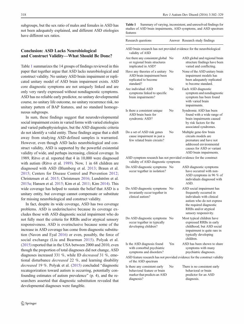
subgroups, but the sex ratio of males and females in ASD hasnot been adequately explained, and different ASD etiologieshave different sex ratios.
Conclusion: ASD Lacks Neurobiologicaland Construct Validity—What Should Be Done?
Table 1 summarizes the 14 groups of findings reviewed in thispaper that together argue that ASD lacks neurobiological andconstruct validity. No unitary ASD brain impairment or repli-cated unitary model of ASD brain impairment exists. ASDcore diagnostic symptoms are not uniquely linked and areonly very rarely expressed without nondiagnostic symptoms.ASD has no reliable early predictor, no unitary developmentalcourse, no unitary life outcome, no unitary recurrence risk, nounitary pattern of BAP features, and no standard homoge-neous subgroups.
In sum, these findings suggest that neurodevelopmentalsocial impairment exists in varied forms with varied etiologiesand varied pathophysiologies, but the ASD diagnostic criteriado not identify a valid entity. These findings argue that a shiftaway from studying ASD-defined samples is warranted.However, even though ASD lacks neurobiological and con-struct validity, ASD is supported by the powerful existentialvalidity of wide, and perhaps increasing, clinical coverage. In1989, Ritvo et al. reported that 4 in 10,000 were diagnosedwith autism (Ritvo et al. 1989). Now, 1 in 68 children arediagnosed with ASD (Blumberg et al. 2013; Braun et al.2015; Centers for Disease Control and Prevention 2012;Christensen et al. 2015; Christensen 2016; Lundström et al.2015a; Hansen et al. 2015; Kim et al. 2011; Kim 2014). Thiswide coverage has helped to sustain the belief that ASD is aunitary entity, but coverage cannot compensate or substitutefor missing neurobiological and construct validity.
In fact, despite its wide coverage, ASD has two coverageproblems. ASD is underinclusive because its coverage ex-cludes those with ASD diagnostic social impairment who donot fully meet the criteria for RRBs and/or atypical sensoryresponsiveness. ASD is overinclusive because some of theincrease in ASD coverage has come from diagnostic substitu-tion (Navon and Eyal 2016) or even, possibly, the force ofsocial exchange (Liu and Bearman 2015). Polyak et al.(2015) reported that in the USA between 2000 and 2010, eventhough the proportion of total diagnoses did not change, ASDdiagnoses increased 331 %, while ID decreased 31 %, emo-tional disturbance decreased 22 %, and learning disabilitydecreased 19 %. Polyak et al. (2015) concluded Bdiagnosticrecategorization toward autism is occurring, potentially con-founding estimates of autism prevalence^ (p. 4), and the re-searchers asserted that diagnostic substitution revealed thatdevelopmental diagnoses were fungible.
Table 1 Summary of varying, inconsistent, and unresolved findings forstudies of ASD brain impairments, ASD symptoms, and ASD spectrumfeatures
Research questions Answer Research study findings
ASD brain research has not provided evidence for the neurobiologicalvalidity of ASD
Are there any consistent globalor regional brain structuresimplicated in ASD?
No ASD global and regional brainstructure findings have beenvaried and conflicting.
Have any theories of a unitaryASD brain impairment beenreplicated to becomestandard?
No None of the ASD unitary brainimpairment models hasbeen adequately replicatedto become standard.
Are individual ASDsymptoms linked to specificbrain impairments?
No Each ASD diagnosticsymptom and nondiagnosticsymptom has been foundwith varied brainimpairments.
Is there a consistent uniqueASD brain basis forsyndromic ASD?
No Syndromic ASD has beenfound with a wide range ofbrain impairments causedby risk factors for theassociated syndromes.
Do a set of ASD risk genescause impairment in just afew related brain circuits?
No Multiple gene-few braincircuits models arepremature and have notaddressed environmentalcauses for ASD or variantASD brain impairments.
ASD symptom research has not provided evidence for the constructvalidity of ASD diagnostic symptoms
Do ASD diagnostic symptomsoccur together in isolation?
No ASD diagnostic symptomshave occurred with non-ASD symptoms in 96 % ofindividuals diagnosed withASD.
Do ASD diagnostic symptomsinvariantly occur together inclinical autism?
No ASD social impairment hasfrequently occurred inindividuals with clinicalautism who do not expressthe required diagnosticRRBs and/or atypicalsensory responsivity.
Do ASD diagnostic symptomsoccur together in typicallydeveloping children?
No Most typical children haveexpressed RRBs in earlychildhood, but ASD socialimpairment is quite rare intypically developingchildren.
Is the ASD diagnosis foundwith comorbid psychiatricsymptoms and disorders?
Yes ASD has been shown to sharesymptoms with manypsychiatric diagnoses.
ASD feature research has not provided evidence for the construct validityof the ASD spectrum
Is there any consistent earlybehavioral feature or brainmarker that predicts an ASDdiagnosis?
No There is no consistent earlybehavioral or brainpredictor for an ASDdiagnosis.
318 Rev J Autism Dev Disord (2016) 3:302–329

Proposals for Future Research: Disband the ASDDiagnosis, Study Individual Variation, and ReviseInstruments and Health Records
Disband the ASD Diagnosis in Research
ASD should be disbanded in research because it lacks validity,and finding effective medical treatments requires the analysisof individual variation in valid pathophysiological mecha-nisms linked to specific risk factors in complete neuro-developmental phenotypes (Kennedy et al. 2015; London2014; Volkmar and McPartland 2015; Waterhouse andGillberg 2014). Because the ASD diagnosis is defined by justtwo symptoms from an individual’s complete set of symp-toms, an ASD-defined sample is widely over-inclusive, cap-turingmany different Blifelong conditions that can arise from acomplex combination of multiple genetic and environmentalfactors^ (Dawson 2016, para. 1), in which Bthe genetic archi-tecture of ASD seems to be different from one individual toanother^ (Huguet et al. 2016, p. 118), and there are manyindividually varied Bconnections between brain and behavior^(Kennedy et al. 2015, p. 81). In sum, the heterogeneity of riskfactors, brain impairments, and non-diagnostic symptoms inan ASD-defined sample blocks valid scientific inference.
Study Individual Variation in Brain Impairmentsin Neurodevelopmental Disorders
There is no catalog of brain impairments for neuro-developmental disorders because much past research wasbased on the belief that each neurodevelopmental disorderhad its own specific brain impairment (Bourgeron 2015a).Jeste and Geschwind (2014) highlighted the need for a catalog
of brain impairments in genetic research by inserting a ques-tion mark where (ASD) brain impairments should have beenlisted (Jeste and Geschwind 2014, Fig. 1, p. 75). Individualvariation in brain impairments reflects variation in risk factors,and as argued for RDoC, pathophysiology variation is notaligned with diagnostic categories but is a focus for specificmedical treatment.
Revise Instruments and Health Recordsfor Neurodevelopmental Disorders
Disbanding the ASD diagnosis will require new screening, eval-uations, and health records for all neurodevelopmental disordersto include social impairment and RRBs and sensory atypicalresponsivity independent of the ASD diagnosis. Many formsof electronic health records (EHRs) have been outlined(Castillo et al. 2015; McCoy et al. 2015; Simon et al. 2014).
Gillberg (2010) proposed BEarly Symptomatic SyndromesEliciting Neurodevelopmental Clinical Examinations^ orESSENCE, to include early appearing difficulties in generaldevelopment, communication and language, social interrelated-ness, motor coordination, attention, activity, behavior, mood,and/or sleep. ESSENCE disorders include ASD, ADHD, ID,BPD, oppositional defiant disorder (ODD), specific languageimpairment (SLI), learning disabilities (LD), nonverbal learn-ing disabilities (NVLD), tic disorders, behavioral phenotypesyndromes, rare epilepsy syndromes, and reactive attachmentdisorder. Gillberg (2010) found that comorbidity resulted inmost children having two or more diagnoses. Empirical supportfor ESSENCE (Bourgeron 2015b; Hatakenaka et al. 2016) hasdemonstrated that it effectively documents the variation andcomplexity of neurodevelopmental disorder symptoms.
London (2014) similarly recommended that researchshould study DSM-5 neurodevelopmental disorders as awhole, but proposed a multiaxial model that would allow forthe identification of specific symptoms such as sensoryoverresponsiveness and ID, allow for possible etiologicfactors such as genetic findings or infection duringpregnancy, allow for functional assessment, and also allowfor treatment outcome history. Most importantly, London(2014) noted that this reformulated nosology would directresearchers and clinicians to attend to an individual’s specificsymptoms and not force researchers to assign a categoricaldiagnosis. Instead an inventory of individual symptoms andrisk factors would provide a template suitable for clinical usein lieu of a diagnostic category.
Drawbacks and Benefits of Disbanding ASDin Research
Disbanding ASD in research is likely to be reductive anduncomfortable. Most painfully, many existing ASD group
Table 1 (continued)
Research questions Answer Research study findings
Is there a consistentdevelopmental course or lifeoutcome for ASD?
No ASD is found with varieddevelopmental courses andvaried life outcomes.
Is there a single recurrence riskfor ASD?
No ASD does not and cannot havea single recurrence risk ratebecause there are so manydifferent forms of genetictransmission and because somany environmental riskfactors cause ASD.
Is there a consistent ASDbroader autism phenotype(BAP)?
No BAP symptoms in siblings andparents of children withASD have been found tovary widely.
Is there any set of valid ASDsubgroups?
No Existing ASD subgroups haveshown a lack internalhomogeneity and have notbeen adequately replicated.
Rev J Autism Dev Disord (2016) 3:302–329 319

findings and ASD theories will be irrelevant for futuretransdiagnostic studies, and future studies will likely be basedon varied grouping factors, making study findings less compa-rable. There will also likely be conflicting interests as clinicianscontinue to use DSM-5 ASD, while researchers develop newtypes of studies that aggregate multiple neurodevelopmentaldiagnoses. However, Zhao and Castellanos (2016) argued thatmany large sample studies of psychiatric diagnoses have beenunproductive exactly because they studied traditional DSMdiagnoses. Zhao and Castellanos (2016) argued thatBNeuropsychiatric disorders are frequently transdiagnostic withdistinct subtypes likely among all disorders^ (p. 432); therefore,large-scale imaging and genetics studies should benefit fromexploring multiple neurodevelopmental diagnoses together.
Most importantly, Lilienfeld and Treadway (2016) notedthat although DSM-5 diagnoses have captured much of indi-viduals’ impairments, RDoC research is likely to yieldBstratified medicine, in which interventions are tailored to in-dividuals within well-defined subgroups^ (p. 447), somethingthat should be the central focus for all future research onneurodevelopmental disorders.
Compliance with Ethical Standards All procedures performed inreviewed studies conducted by the authors involving human participantswere in accordance with the ethical standards of the institutional and/ornational research committee and with the 1964 Helsinki declaration andits later amendments or comparable ethical standards. This article doesnot contain any studies with animals performed by any of the authors.
Conflict of Interest The authors declare that they have no conflict ofinterest.
Informed Consent Research reports reviewed herein have includedstatements that informed consent was obtained from all individual partic-ipants included in the study.
References
Ad-Dab’bagh, Y., & Greenfield, B. (2001). Multiple complex develop-mental disorder: the Bmultiple and complex^ evolution of theBchildhood borderline syndrome^ construct. Journal of theAmerican Academy of Child & Adolescent Psychiatry, 40(8), 954–964.
Adviento, B., Corbin, I. L., Widjaja, F., Desachy, G., Enrique, N., Rosser,T., Risi, S., Marco, E. J., Hendren, R. L., Bearden, C. E., Rauen, K.A., & Weiss, L. A. (2014). Autism traits in the RASopathies.Journal of Medical Genetics, 51(1), 10–20. doi:10.1136/jmedgenet-2013-101951.
Aldinger, K. A., Lane, C. J., Veenstra-VanderWeele, J., & Levitt, P.(2015). Patterns of risk for multiple co-occurring medical conditionsreplicate across distinct cohorts of children with autism spectrumdisorder. Autism Research. doi:10.1002/aur.1492.
Ameis, S. H., & Catani, M. (2015). Altered white matter connectivity as aneural substrate for social impairment in autism spectrum disorder.Cortex, 62, 158–181. doi:10.1016/j.cortex.2014.10.014.
American Psychiatric Association. (1980). Diagnostic and statisticalmanual of mental disorders (3rd ed.). Washington: AmericanPsychiatric Association.
American Psychiatric Association. (2000). Diagnostic and statisticalmanual of mental disorders (4th ed.). Washington: AmericanPsychiatric Association.
American Psychiatric Association. (2013). Diagnostic and statisticalmanual of mental disorders (5th ed.). Washington: AmericanPsychiatric Association.
Amiet, C., Gourfinkel-An, I., Laurent, C., Bodeau, N., Génin, B.,Leguern, E., Tordjiman, S., & Cohen, D. (2013). Does epilepsy inmultiplex autism pedigrees define a different subgroup in terms ofclinical characteristics and genetic risk?Molecular Autism, 4(1), 47.http://www.molecularautism.com/content/4/1/47.
Anderson, J. S. (2013). Functional connectivity MRI in autism. In M. F.Casanova, A. S. El-Baz, & J. S. Suri (eds.), Imaging the brain inautism (pp. 325–47). Springer.
Anderson, D. K., Liang, J. W., & Lord, C. (2014). Predicting young adultoutcome among more and less cognitively able individuals withautism spectrum disorders. Journal of Child Psychology andPsychiatry, 55(5), 485–494. doi:10.1111/jcpp.12178.
Barbaro, J., & Dissanayake, C. (2013). Early markers of autism spectrumdisorders in infants and toddlers prospectively identified in theSocial Attention and Communication Study. Autism, 17(1), 64–86.doi:10.1177/1362361312442597.
Baron-Cohen, S., Auyeung, B., Nørgaard-Pedersen, B., Hougaard, D.M.,Abdallah, M.W., Melgaard, L., Cohen, A. S., Chakrabarti, B., Ruta,L., & Lombardo, M. V. (2015). Elevated fetal steroidogenic activityin autism. Molecular Psychiatry, 20(3), 369–376. doi:10.1038/mp.2014.48.
Barrett, L. F., & Satpute, A. B. (2013). Large-scale brain networks inaffective and social neuroscience: towards an integrative functionalarchitecture of the brain. Current Opinion in Neurobiology, 23(3),361–372. doi:10.1016/j.conb.2012.12.012.
Bellani, M., Calderoni, S., Muratori, F., & Brambilla, P. (2013). Brainanatomy of autism spectrum disorders II. Focus on amygdala.Epidemiology and Psychiatric Sciences, 1–4. doi:10.1017/S2045796013000346.
Bernier, R., Gerdts, J., Munson, J., Dawson, G., & Estes, A. (2012).Evidence for broader autism phenotype characteristics in parentsfrom multiple-incidence autism families. Autism Research, 5(1),13–20. doi:10.1002/aur.226.
Billeci, L., Sicca, F., Maharatna, K., Apicella, F., Narzisi, A., Campatelli,G., Calderoni, S., Pioggia, G., & Muratori, F. (2013). On the appli-cation of quantitative EEG for characterizing autistic brain: a sys-tematic review. Frontiers in Human Neuroscience, 7, 442.
Billstedt, E., Gillberg, C. I., & Gillberg, C. (2007). Autism in adults:symptom patterns and early childhood predictors. Use of theDISCO in a community sample followed from childhood. Journalof Child Psychology and Psychiatry, 48(11), 1102–1110.
Bishop, D. V. M. (2014). Ten questions about terminology for childrenwith unexplained language problems. International Journal ofLanguage & Communication Disorders, 49(4), 381–415.
Bishop, S. L., Hus, V., Duncan, A., Huerta, M., Gotham, K., Pickles, A.,Kreiger, A., Buja, A., Lund, S., & Lord, C. (2013). Subcategories ofrestricted and repetitive behaviors in children with autism spectrumdisorders. Journal of Autism and Developmental Disorders, 43(6),1287–1297. doi:10.1007/s10803-012-1671-0.
Bishop, S. L., Havdahl, K. A., Huerta, M., & Lord, C. (2016).Subdimensions of social-communication impairment in autismspectrum disorder. Journal of Child Psychology and Psychiatry.doi:10.1111/jcpp.12510.
Blackmon, K. (2015). Structural MRI biomarkers of shared pathogenesisin autism spectrum disorder and epilepsy. Epilepsy & Behavior.doi:10.1016/j.yebeh.2015.02.017.
320 Rev J Autism Dev Disord (2016) 3:302–329
Blumberg, S. J., Bramlett, M. D., Kogan,M., Schieve, L., Jones, J., & Lu,M. (2013). Changes in prevalence of parent-reported autism spec-trum disorder in school-aged U. S. children: 2007 to 2011–2012.National Health Statistics Reports, 65, 1–12. Hyattsville, MD:National Center for Health Statistics. http://www.cdc.gov/nchs/data/nhsr/nhsr065.pdf.
Blumberg, S. J., Zablotsky, B., Avila, R. M., Colpe, L. J., Pringle, B. A.,Kogan, M. D. (2015). Diagnosis lost: differences between childrenwho had and who currently have an autism spectrum disorder diag-n o s i s . Au t i sm , 1 3 623 61315607724 . d o i : 1 0 . 1177/1362361315607724.
Boggess, A., Faber, S., Kern, J., & Kingston, H. S. (2016). Mean serum-level of common organic pollutants is predictive of behavioral se-verity in children with autism spectrum disorders. Scientific Reports,6, 26185.
Bone, D., Bishop, S., Black, M. P., Goodwin, M. S., Lord, C., &Narayanan, S. S. (2016). Use of machine learning to improve autismscreening and diagnostic instruments: effectiveness, efficiency, andmulti-instrument fusion. Journal of Child Psychology andPsychiatry. doi:10.1111/jcpp.12559.
Boucher, J. (2011). Redefining the concept of autism as a unitary disor-der: multiple causal deficits of a single kind? In D. Fein (Ed.), Theneuropsychology of autism (pp. 469–82). New York: OxfordUniversity Press.
Boucher, J. (2012). Research review: structural language in autistic spec-trum disorder—characteristics and causes. Journal of ChildPsychology and Psychiatry, 53(3), 219–233. doi:10.1111/j.1469-7610.2011.02508.x.
Boukhris, T., Sheehy, O., Mottron L., Bérard, A. (2015). Antidepressantuse during pregnancy and the risk of autism spectrum disorder inc h i l d r e n . J AMA P e d i a t r i c s , 1 – 8 . d o i : 1 0 . 1 0 0 1/jamapediatrics.2015.3356.
Bourgeron, T. (2015a). From the genetic architecture to synaptic plasticityin autism spectrum disorder. Nature Reviews Neuroscience, 16(9),551–563. doi:10.1038/nrn3992.
Bourgeron, T. (2015b). The genetics and neurobiology of ESSENCE: thethird Birgit Olsson lecture. Nordic Journal of Psychiatry, 1–9.doi:10.3109/08039488.2015.1042519.
Brandler, W. M., & Sebat, J. (2015). From de novo mutations to person-alized therapeutic interventions in autism. Annual Review ofMedicine, 66, 487–507. doi:10.1146/annurev-med-091113-024550.
Braun, K. V. N., Christensen, D., Doernberg, N., Schieve, L., Rice, C.,Wiggins, L., …, Yeargin-Allsopp, M. (2015). Trends in the preva-lence of autism spectrum disorder, cerebral palsy, hearing loss, in-tellectual disability, and vision impairment, Metropolitan Atlanta,1991–2010. PloS One, 10(4). doi:10.1371/journal.pone.0124120.
Brennan, L., Barton, M., Chen, C. M., Green, J., & Fein, D. (2015).Detecting subgroups in children diagnosed with PervasiveDevelopmental Disorder-Not Otherwise Specified. Journal ofAutism and Developmental Disorders, 45(5), 1329–1344.
Brunsdon, V. E., & Happé, F. (2014). Exploring the ‘fractionation’ ofautism at the cognitive level. Autism, 18(1), 17–30. doi:10.1177/1362361313499456.
Butler, M. G., Rafi, S. K., & Manzardo, A. M. (2015). High-resolutionchromosome ideogram representation of currently recognized genesfor autism spectrum disorders. International Journal of MolecularSciences, 16(3), 6464–6495. doi:10.3390/ijms16036464.
Byrge, L., Dubois, J., Tyszka, J. M., Adolphs, R., & Kennedy, D. P.(2015). Idiosyncratic brain activation patterns are associated withpoor social comprehension in autism. The Journal ofNeuroscience, 35(14), 5837–5850. doi:10.1523/jneurosci.5182-14.201.
Camarata, S. (2014). Early identification and early intervention in autismspectrum disorders: accurate and effective? International Journal ofSpeech-Language Pathology, 16(1), 1–10. doi:10.3109/17549507.2013.864708.
Castillo, E. G., Olfson, M., Pincus, H. A., Vawdrey, D., & Stroup, T. S.(2015). Electronic health records in mental health research: a frame-work for developing valid research methods. Psychiatric Services.doi:10.1176/appi.ps.201400200.
Cederlöf, M., Gotby, A. O., Larsson, H., Serlachius, E., Boman, M.,Långström, N., …, Lichtenstein, P. (2014). Klinefelter syndromeand risk of psychosis, autism and ADHD. Journal of PsychiatricResearch, 48(1), 128–130. doi:10.1016/j.jpsychires.2013.10.001.
Cederlund, M., Miniscalco, C., & Gillberg, C. (2014). Pre-schoolchildrenwith autism spectrum disorders are rarely macrocephalic: a popula-tion study. Research in Developmental Disabilities, 35(5), 992–998.
Centers for Disease Control and Prevention. (2012). Prevalence of autismspectrum disorders—Autism and Developmental DisabilitiesMonitoring Network, 14 Sites, United States, 2008. Morbidity andMortality Weekly Report, 61(3), 1–19. http://www.cdc.gov/mmwr/pdf/ss/ss6103.pdf.
Chaste, P., Klei, L., Sanders, S. J., Murtha,M. T., Hus, V., Lowe, J. K.,…,Fombonne, E. (2013). Adjusting head circumference for covariatesin autism: clinical correlates of a highly heritable continuous trait.Biological Psychiatry, 74(8) , 576–84. doi :10.1016/j .biopsych.2013.04.018.
Chaste, P., Klei, L., Sanders, S. J., Hus, V., Murtha, M. T., Lowe, J. K.,…& Geschwind, D. (2015). A genomewide association study of au-tism using the Simons Simplex Collection: Does reducing pheno-typic heterogeneity in autism increase genetic homogeneity?.Biological Psychiatry, 77(9), 775–784.
Chen, J. A., Peñagarikano, O., Belgard, T. G., Swarup, V., & Geschwind,D. H. (2015). The emerging picture of autism spectrum disorder:genetics and pathology. Annual Review of Pathology: Mechanismsof Disease, 10, 111–144.
Christensen, D. L. (2016). Prevalence and characteristics of autism spec-trum disorder among children aged 8 years—Autism andDevelopmental Disabilities Monitoring Network, 11 Sites, UnitedStates, 2012. MMWR. Surveillance Summaries, 65.
Christensen, D., Bilder, D., Zahorodny, W., Pettygrove, S., Durkin, M.,Fitzgerald, R.,…, Yeargin-Allsopp,M. (2015). Prevalence and char-acteristics of autism spectrum disorder among 4-year-old children inthe Autism and Developmental Disabilities Monitoring Network.Journal of Developmental and Behavioral Pediatrics, 37(1), 1–8.
Coleman, M., & Gillberg, C. (2012). The autisms. Oxford UniversityPress.
Colvert, E., Tick, B., McEwen, F., Stewart, C., Curran, S. R.,Woodhouse,E.,…, Bolton, P. (2015). Heritability of autism spectrum disorder ina UK population-based twin sample. JAMA Psychiatry, 72(5), 415–423. doi:10.1001/jamapsychiatry.2014.3028.
Constantino, J. N., & Charman, T. (2015). Series: diagnosis of autismspectrum disorder: reconciling the syndrome, its diverse origins, andvariation in expression. The Lancet Neurology. doi:10.1016/S1474-4422(15)00151-9.
Coplan, R. J., Rose-Krasnor, L., Weeks, M., Kingsbury, A., Kingsbury,M., & Bullock, A. (2013). Alone is a crowd: social motivations,social withdrawal, and socioemotional functioning in later child-hood. Developmental Psychology, 49(5), 861. http://psycnet.apa.org/doi/10.1037/a0028861.
Corbett, B. A., Swain, D. M., Newsom, C., Wang, L., Song, Y., &Edgerton, D. (2014). Biobehavioral profiles of arousal and socialmotivation in autism spectrum disorders. Journal of ChildPsychology and Psychiatry, 55(8), 924–934.
Croen, L. A., Grether, J. K., Yoshida, C. K., Odouli, R., & Hendrick, V.(2011). Antidepressant use during pregnancy and childhood autismspectrum disorders. Archives of General Psychiatry, 68(11), 1104–12. doi:10.1001/archgenpsychiatry.2011.73.
Croen, L. A., Zerbo, O., Qian, Y., Massolo, M. L., Rich, S., Sidney, S., &Kripke, C. (2015). The health status of adults on the autism spec-trum. Autism, 19(7), 814–823.
Rev J Autism Dev Disord (2016) 3:302–329 321

Cruz, L. P., Camargos-Junior, W., & Rocha, F. L. (2013). The broadautism phenotype in parents of individuals with autism: a systematicreview of the literature. Trends in Psychiatry and Psychotherapy,35(4), 252–263.
Cuthbert, B. N., & Insel, T. R. (2013). Toward the future of psychiatricdiagnosis: the seven pillars of RDoC. BMC Medicine, 11(1), 126.doi:10.1186/1741-7015-11-126.
D’Angelo, D., Lebon, S., Chen, Q., Martin-Brevet, S., Snyder, L. G.,Hippolyte, L., …, Pain, A. (2015). Defining the effect of the16p11.2 duplication on cognition, behavior, and medical comorbid-i t i e s . J A M A P s y c h i a t r y , 1 – 1 1 . d o i : 1 0 . 1 0 0 1/jamapsychiatry.2015.2123.
D’Mello, A. M., Crocetti, D., Mostofsky, S. H., & Stoodley, C. J. (2015).Cerebellar gray matter and lobular volumes correlate with core au-tism symptoms. NeuroImage: Clinical, 7, 631–639. doi:10.1016/j.nicl.2015.02.007.
Dawson, G. (2016). On the brink of breakthroughs in diagnosing andtreating autism. Retrieved from http://blogs.scientificamerican.com/mind-guest-blog/on-the-brink-of-breakthroughs-in-diagnosing-and-treating-autism/.
de Bruin, E. I., de Nijs, P. F., Verheij, F., Hartman, C. A., & Ferdinand, R.F. (2007). Multiple complex developmental disorder delineatedfrom PDD-NOS. Journal of Autism and Developmental Disorders,37(6), 1181–1191.
de la Torre-Ubieta, L., Won, H., Stein, J. L., & Geschwind, D. H. (2016).Advancing the understanding of autism disease mechanismsthrough genetics. Nature Medicine, 22(4), 345–361.
De Rubeis, S., & Buxbaum, J. D. (2015). Genetics and genomics ofautism spectrum disorder: embracing complexity. HumanMolecular Genetics, 24(R1), R24–31.
Doré, B. P., Zerubavel, N., Ochsner, K. N. (2014). Social cognitive neu-roscience: a review of core systems. APA Handbook of Personalityand Social Psychology, 693–720.
Doyle-Thomas, K. A., Card, D., Soorya, L. V., Ting Wang, A., Fan, J., &Anagnostou, E. (2014). Metabolic mapping of deep brain structuresand associations with symptomatology in autism spectrum disor-ders. Research in Autism Spectrum Disorders, 8(1), 44–51.doi:10.1016/j.rasd.2013.10.003.
Eack, S. M., Bahorik, A. L., McKnight, S. A., Hogarty, S. S., Greenwald,D. P., Newhill, C. E.,…, Minshew, N. J. (2013). Commonalities insocial and non-social cognitive impairments in adults with autismspectrum disorder and schizophrenia. Schizophrenia Research,148(1), 24–28. doi:10.1016/j.schres.2013.05.013.
Ecker, C., Suckling, J., Deoni, S. C., Lombardo, M. V., Bullmore, E. T.,Baron-Cohen, S.,…, Williams, S. C. (2012). Brain anatomy and itsrelationship to behavior in adults with autism spectrum disorder: amulticenter magnetic resonance imaging study. Archives of GeneralPsychiatry, 69(2), 195–209.
Ecker, C., Bookheimer, S. Y., & Murphy, D. G. (2015). Neuroimaging inautism spectrum disorder: brain structure and function across thelifespan. The Lancet Neurology. doi:10.1016/S1474-4422(15)00050-2.
Eisenberg, I. W., Wallace, G. L., Kenworthy, L., Gotts, S. J., &Martin, A.(2015). Insistence on sameness relates to increased covariance ofgray matter structure in autism spectrum disorder. MolecularAutism, 6(1), 1.
Ellegood, J., Anagnostou, E., Babineau, B. A., Crawley, J. N., Lin, L.,Genestine, M., …, Lerch, J. P. (2015). Clustering autism: usingneuroanatomical differences in 26mouse models to gain insight intothe heterogeneity. Molecular Psychiatry, 20(1), 118–125.doi:10.1038/mp.2014.98.
Elsabbagh,M., & Johnson,M. H. (2016). Autism and the social brain: thefirst-year puzzle. Biological Psychiatry, 80(2), 94–9.
Engman, M. L., Sundin, M., Miniscalco, C., Westerlund, J., Lewensohn-Fuchs, I., Gillberg, C., & Fernell, E. (2015). Prenatal acquired
cytomegalovirus infection should be considered in children withautism. Acta Paediatrica, 104(8), 792–795.
Esbensen, A. J., Seltzer, M. M., Lam, K. S., & Bodfish, J. W. (2009).Age-related differences in restricted repetitive behaviors in autismspectrum disorders. Journal of Autism and DevelopmentalDisorders, 39(1), 57–66. doi:10.1007/s10803-008-0599-x.
Estes, M. L., & McAllister, A. K. (2015). Immune mediators in the brainand peripheral tissues in autism spectrum disorder. Nature Reviews.Neuroscience, 16(8), 469–486. doi:10.1038/nrn3978.
Fatemi, S. H., Aldinger, K. A., Ashwood, P., Bauman, M. L., Blaha, C.D., Blatt, G. J.,…, Welsh, J. P. (2012). Consensus paper: patholog-ical role of the cerebellum in autism. The Cerebellum, 11(3), 777–807. doi: 10.1007/s12311-012-0355-9.
Fein, D., Barton, M., Eigsti, I. M., Kelley, E., Naigles, L., Schultz, R. T.,…, Troyb, E. (2013). Optimal outcome in individuals with a historyof autism. Journal of Child Psychology and Psychiatry, 54(2), 195–205. doi:10.1111/jcpp.12037.
Fernell, E., Hedvall, Å., Westerlund, J., Carlsson, L. H., Eriksson, M.,Olsson, M. B., …, Gillberg, C. (2011). Early intervention in 208Swedish preschoolers with autism spectrum disorder. A prospectivenaturalistic study. Research in Developmental Disabilities, 32(6),2092–2101.
Fernell, E., Wilson, P., Hadjikhani, N., Bourgeron, T., Neville, B., Taylor,D.,…, Gillberg, C. (2014). Screening, intervention and outcome inautism and other developmental disorders: the role of randomizedcontrolled trials. Journal of Autism and Developmental Disorders,44(8), 2074–2076.
Fishman, I., Keown, C. L., Lincoln, A. J., Pineda, J. A., & Müller, R. A.(2014). Atypical cross talk between mentalizing and mirror neuronnetworks in autism spectrum disorder. JAMA Psychiatry, 71(7),751–760. doi:10.1001/jamapsychiatry.2014.83.
Fountain, C., Winter, A. S., & Bearman, P. S. (2012). Six developmentaltrajectories characterize children with autism. Pediatrics, 129(5),e1112–e1120.
Frazier, T. W., Ratliff, K. R., Gruber, C., Zhang, Y., Law, P. A., &Constantino, J. N. (2014). Confirmatory factor analytic structureand measurement invariance of quantitative autistic traits measuredby the Social Responsiveness Scale-2. Autism, 18(1), 31–44.doi:10.1177/1362361313500382.
Gabis, L. V., & Pomeroy, J. (2014). An etiologic classification of autismspectrum disorders. The Israel Medical Association Journal: IMAJ,16(5), 295–298.
Gabriels, R. L., Agnew, J. A., Pan, Z., Holt, K. D., Reynolds, A., &Laudenslager, M. L. (2013). Elevated repetitive behaviors are asso-ciated with lower diurnal salivary cortisol levels in autism spectrumdisorder. Biological Psychology, 93(2), 262–268. doi:10.1016/j.biopsycho.2013.02.017.
Georgiades, S., Papageorgiou, V., & Anagnostou, E. (2010). Brief report:repetitive behaviours in Greek individuals with autism spectrumdisorder. Journal of Autism and Developmental Disorders, 40(7),903–906.
Georgiades, S., Szatmari, P., Boyle, M., Hanna, S., Duku, E.,Zwaigenbaum, L., …, Thompson, A. (2013). Investigating pheno-typic heterogeneity in children with autism spectrum disorder: afactor mixture modeling approach. Journal of Child Psychologyand Psychiatry, 54(2) , 206–215. doi:10.1111/ j .1469-7610.2012.02588.x.
Geschwind, D. H., & State, M. W. (2015). Gene hunting in autism spec-trum disorder: on the path to precision medicine. Lancet Neurology,14(11), 1109–1120.
Gillberg, C. (2010). The ESSENCE in child psychiatry: EarlySymptomatic Syndromes Eliciting Neurodevelopmental ClinicalExaminations. Research in Developmental Disabilities, 31, 1543–1551. doi:10.1016/j.ridd.2010.06.002.
322 Rev J Autism Dev Disord (2016) 3:302–329
Gillberg, C., & De Souza, L. (2002). Head circumference in autism,Asperger syndrome, and ADHD: a comparative study.Developmental Medicine & Child Neurology, 44(05), 296–300.
Glatt, S. J., Tsuang,M. T., Winn,M., Chandler, S. D., Collins, M., Lopez,L.,…, Courchesne, E. (2012). Blood-based gene expression signa-tures of infants and toddlers with autism. Journal of the AmericanAcademy of Child & Adolescent Psychiatry, 51(9), 934–944.
Grabrucker, A. (2013). Environmental factors in autism. Frontiers inPsychiatry, 18(3), 118. doi:10.3389/fpsyt.2012.00118.
Green, C., Dissanayake, C., & Loesch, D. (2015). A review of physicalgrowth in children and adolescents with autism spectrum disorder.Developmental Review, 36, 156–178.
Grønborg, T. K., Schendel, D. E., & Parner, E. T. (2013). Recurrence ofautism spectrum disorders in full-and half-siblings and trends overtime: a population-based cohort study. JAMA Pediatrics, 167(10),947–953. doi:10.1001/jamapediatrics.2013.2259.
Guilmatre, A., Huguet, G., Delorme, R., & Bourgeron, T. (2014). Theemerging role of SHANK genes in neuropsychiatric disorders.Developmental Neurobiology, 74(2), 113–122.
Guxens, M., García Esteban, R., De Nazelle, A., Forns i Guzman, J.,Nieuwenhuijsen, M. J., & Sunyer Deu, J. (2015). Air pollution ex-posure during pregnancy and childhood autistic traits in fourEuropean population-based cohort studies: the ESCAPE Project.
Hahamy, A., Behrmann, M., & Malach, R. (2015). The idiosyncraticbrain: distortion of spontaneous connectivity patterns in autismspectrum disorder. Nature Neuroscience, 18(2), 302–309.doi:10.1038/nn.3919.
Hall, S. S., Lightbody, A. A., Hirt,M., Rezvani, A., &Reiss, A. L. (2010).Autism in fragile X syndrome: a category mistake? Journal of theAmerican Academy of Child & Adolescent Psychiatry, 49(9), 921–933. doi:10.1016/j.jaac.2010.07.001.
Halladay, A. K., Bishop, S., Constantino, J. N., Daniels, A. M., Koenig,K., Palmer, K.,…, Taylor, J. L. (2015). Sex and gender differencesin autism spectrum disorder: summarizing evidence gaps and iden-tifying emerging areas of priority. Molecular Autism, 6(1), 1.
Hallmayer, J., Cleveland, S., Torres, A., Phillips, J., Cohen, B., Torigoe,T., …, Risch, N. (2011). Genetic heritability and shared environ-mental factors among twin pairs with autism. Archives of GeneralPsychiatry, 68(11), 1095–1102.
Hansen, S. N., Schendel, D. E., & Parner, E. T. (2015). Explaining theincrease in the prevalence of autism spectrum disorders: the propor-tion attributable to changes in reporting practices. JAMA Pediatrics,169(1), 56–62. doi:10.1001/jamapediatrics.2014.1893.
Happé, F., Ronald, A., & Plomin, R. (2006). Time to give up on a singleexplanation for autism. Nature Neuroscience, 9(10), 1218–1220.doi:10.1038/nn1770.
Harrop, C., McConachie, H., Emsley, R., Leadbitter, K., Green, J. (2013.)Restricted and repetitive behaviors in autism spectrum disorders andtypical development: cross-sectional and longitudinal comparisons.Journal of Autism and Developmental Disorders, 1–13. doi:10.1007/s10803-013-1986-5.
Harstad, E. B., Fogler, J., Sideridis, G., Weas, S., Mauras, C., &Barbaresi, W. J. (2015). Comparing diagnostic outcomes of autismspectrum disorder using DSM-IV-TR and DSM-5 criteria. Journalof Autism and Developmental Disorders, 45(5), 1437–1450.doi:10.1007/s10803-014-2306-4.
Hatakenaka, Y., Kotani, H., Yasumitsu-Lovell, K., Suzuki, K., Fernell, E.,& Gillberg, C. (2016). Infant motor delay and ESSENCE in Japan.Pediatric Neurology, 54, 55–63.
Hedvall, Å., Westerlund, J., Fernell, E., Holm, A., Gillberg, C., &Billstedt, E. (2014). Autism and developmental profiles in pre-schoolers: stability and change over time. Acta Paediatrica,103(2), 174–181.
Helles, A., Gillberg, C. I., Gillberg, C., & Billstedt, E. (2015). Aspergersyndrome in males over two decades: stability and predictors of
diagnosis. Journal of Child Psychology and Psychiatry, 56(6),711–718.
Hommer, R. E., & Swedo, S. E. (2015). Schizophrenia and autism-relateddisorders. Schizophrenia Bulletin, 41(2), 313–314.
Hormozdiari, F., Penn, O., Borenstein, E., & Eichler, E. E. (2015). Thediscovery of integrated gene networks for autism and related disor-ders. Genome Research, 25(1), 142–154. doi:10.1101/gr.178855.114.
Howlin, P., Moss, P., Savage, S., & Rutter, M. (2013). Social outcomes inmid to later adulthood among individuals diagnosedwith autism andaverage nonverbal IQ as children. Journal of the American Academyof Child & Adolescent Psychiatry, 52(6), 572–581. doi:10.1016/j.jaac.2013.02.017.
Huguet, G., Benabou, M., Bourgeron, T. (2016). The genetics of autismspectrum disorders. In A time for metabolism and hormones (pp.101–129). Springer International Publishing.
Hus, V., Pickles, A., Cook, E. H., Jr., Risi, S., & Lord, C. (2007). Usingthe autism diagnostic interview—revised to increase phenotypic ho-mogeneity in genetic studies of autism. Biological Psychiatry, 61,438–448. doi:10.1016/j.biopsych.2006.08.044.
Hviid, A., Melbye,M., & Pasternak, B. (2013). Use of selective serotoninreuptake inhibitors during pregnancy and risk of autism. NewEngland Journal of Medicine, 369(25), 2406–2415. doi:10.1056/NEJMoa1301449.
Insel, T., Cuthbert, B., Garvey, M., Heinssen, R., Pine, D. S., Quinn, K.,…, Wang, P. (2010). Research domain criteria (RDoC): toward anew classification framework for research on mental disorders.American Journal of Psychiatry, 167(7), 748–751.
Irimia, M., Weatheritt, R. J., Ellis, J. D., Parikshak, N. N., Gonatopoulos-Pournatzis, T., Babor, M., …, Blencowe, B. J. (2014). A highlyconserved program of neuronal microexons is misregulated in autis-tic brains. Cell, 159(7), 1511–1523. doi:10.1016/j.cell.2014.11.035.
Jason, J. Y., Berrios, J., Newbern, J. M., Snider, W. D., Philpot, B. D.,Hahn, K. M., & Zylka, M. J. (2015). An autism-linked mutationdisables phosphorylation control of UBE3A. Cell, 162(4), 795–807.
Jeste, S. S., & Geschwind, D. H. (2014). Disentangling the heterogeneityof autism spectrum disorder through genetic findings. NatureReviews Neurology, 10(2), 74–81. doi:10.1038/nrneurol.2013.278.
Jeste, S. S., Frohlich, J., & Loo, S. K. (2015). Electrophysiological bio-markers of diagnosis and outcome in neurodevelopmental disorders.Current Opinion in Neurology, 28(2), 110–116. doi:10.1097/WCO.0000000000000181.
Jiang, Y. H., Yuen, R. K., Jin, X.,Wang,M., Chen, N.,Wu,X., Ju, J., Mei,J., Shi, Y., He, M., et al. (2013). Detection of clinically relevantgenetic variants in autism spectrum disorder by whole-genome se-quencing. The American Journal of Human Genetics, 93(2), 249–263. doi:10.1016/j.ajhg.2013.06.012.
Johnson, M. H., Gliga, T., Jones, E., & Charman, T. (2015). AnnualResearch Review: infant development, autism, and ADHD–earlypathways to emerging disorders. Journal of Child Psychology andPsychiatry, 56(3), 228–247.
Jones,W., & Klin, A. (2013). Attention to eyes is present but in decline in2–6-month-old infants later diagnosed with autism. Nature,504(7480), 427–431.
Jones, E. J., Gliga, T., Bedford, R., Charman, T., & Johnson, M. H.(2014). Developmental pathways to autism: a review of prospectivestudies of infants at risk. Neuroscience & Biobehavioral Reviews,39, 1–33.
Joseph, R.M., Fricker, Z., Fenoglio, A., Lindgren, K. A., Knaus, T. A., &Tager-Flusberg, H. (2014). Structural asymmetries of language-related gray and white matter and their relationship to languagefunction in young children with ASD. Brain Imaging andBehavior, 8(1), 60–72. doi:10.1007/s11682-013-9245-0.
Jou, R. J., Frazier, T. W., Keshavan, M. S., Minshew, N. J., & Hardan, A.Y. (2013). A two-year longitudinal pilot MRI study of the brainstemin autism. Behavioural Brain Research, 251, 163–167.
Rev J Autism Dev Disord (2016) 3:302–329 323

Just, M. A., Cherkassky, V. L., Keller, T. A., & Minshew, N. J. (2004).Cortical activation and synchronization during sentence comprehen-sion in high-functioning autism: evidence of underconnectivity.Brain, 127(8), 1811–1821.
Kalkbrenner, A. E., Schmidt, R. J., & Penlesky, A. C. (2014).Environmental chemical exposures and autism spectrum disorders:a review of the epidemiological evidence. Current Problems inPediatric and Adolescent Health Care, 44(10), 277–318.doi:10.1016/j.cppeds.2014.06.001.
Kanner, L. (1943). Autistic disturbances of affective contact. The NervousChild, 2, 217–250.
Kanner, L. (1951). The conception of wholes and parts in early infantileautism. American Journal of Psychiatry, 108(1), 23–26.
Kasari, C. (2015). Update on behavioral interventions for autism anddevelopmental disabilities. Current Opinion in Neurology, 28(2),124–129. doi:10.1097/WCO.0000000000000185.
Kennedy, D. P., Paul, L. K., & Adolphs, R. (2015). Brain connectivity inautism: the significance of null findings. Biological Psychiatry,78(2), 81–82. doi:10.1016/j.biopsych.2015.05.002.
Kern, J. K., Geier, D. A., King, P. G., Sykes, L. K., Mehta, J. A., & Geier,M. R. (2015). Shared brain connectivity issues, symptoms, and co-morbidities in autism spectrum disorder, attention deficit/hyperactivity disorder, and Tourette syndrome. Brain Connectivity.doi:10.1089/brain.2014.0324.
Khan, S., Michmizos, K., Tommerdahl, M., Ganesan, S., Kitzbichler, M.G., Zetino, M., …, Kenet, T. (2015). Somatosensory cortex func-tional connectivity abnormalities in autism show opposite trends,depending on direction and spatial scale. Brain. doi:10.1093/brain/awv043.
Kida, S., & Kato, T. (2015). Microendophenotypes of psychiatric disor-ders: phenotypes of psychiatric disorders at the level of moleculardynamics, synapses, neurons, and neural circuits. CurrentMolecular Medicine, 15(2), 111–118.
Kim, Y. S. (2014). Recent challenges to the psychiatric diagnostic nosol-ogy: a focus on the genetics and genomics of neurodevelopmentaldisorders. International Journal of Epidemiology, 43(2), 465–475.
Kim, Y. S., Leventhal, B. L., Koh, Y. J., Fombonne, E., Laska, E., Lim, E.C., Cheon, K.-A., Kim, S.-J., Kim, Y.-K., Lee, H., et al. (2011).Prevalence of autism spectrum disorders in a total population sam-ple. American Journal of Psychiatry, 168(9), 904–912. doi:10.1176/appi.ajp.2011.10101532.
Kim, Y. S., Fombonne, E., Koh, Y. J., Kim, S. J., Cheon, K. A., &Leventhal, B. L. (2014). A comparison of DSM-IV PervasiveDevelopmental Disorder and DSM-5 Autism Spectrum Disorderprevalence in an epidemiologic sample. Journal of the AmericanAcademy of Child & Adolescent Psychiatry, 53(5), 500–508.doi:10.1176/10.1016/j.jaac.2013.12.021.
Kim, S. H., Macari, S., Koller, J., & Chawarska, K. (2016). Examiningthe phenotypic heterogeneity of early autism spectrum disorder:subtypes and short-term outcomes. Journal of Child Psychologyand Psychiatry, 57(1), 93–102.
Kirkovski, M., Enticott, P. G., Maller, J. J., Rossell, S. L., & Fitzgerald, P.B. (2015). Diffusion tensor imaging reveals no white matter impair-ments among adults with autism spectrum disorder. PsychiatryResearch: Neuroimaging. doi:10.1016/j.pscychresns.2015.05.003.
Kitzbichler, M. G., Khan, S., Ganesan, S., Vangel, M. G., Herbert, M. R.,Hämäläinen, M. S., & Kenet, T. (2015). Altered development andmultifaceted band-specific abnormalities of resting state networks inautism. Biological Psychiatry, 77(9), 794–804.
Klin, A., Shultz, S., & Jones, W. (2015). Social visual engagement ininfants and toddlers with autism: early developmental transitionsand a model of pathogenesis. Neuroscience & BiobehavioralReviews, 50, 189–203.
Klusek, J., Roberts, J. E., & Losh, M. (2015). Cardiac autonomic regula-tion in autism and fragile X syndrome: a review. PsychologicalBulletin, 141(1), 141.
Kohane, I. S., McMurry, A., Weber, G., MacFadden, D., Rappaport, L.,Kunkel, L., … & Churchill, S. (2012). The co-morbidity burden ofchildren and young adults with autism spectrum disorders. PloSOne, 7(4), e33224.
Koldewyn, K., Yendiki, A., Weigelt, S., Gweon, H., Julian, J.,Richardson, H.,…&Kanwisher, N. (2014). Differences in the rightinferior longitudinal fasciculus but no general disruption of whitematter tracts in children with autism spectrum disorder. Proceedingsof the National Academy of Sciences, 111(5), 1981–1986.
Krumm, N., O’Roak, B. J., Shendure, J., & Eichler, E. E. (2014). A denovo convergence of autism genetics and molecular neuroscience.Trends in Neurosciences, 37(2), 95–105. doi:10.1016/j.tins.2013.11.005.
Kulage, K. M., Smaldone, A. M., Cohn, E. G. (2014). How will DSM-5affect autism diagnosis? A systematic literature review and meta-analysis. Journal of Autism and Developmental Disorders 1–15.doi:10.1007/s10803-014-2065-2.
Kuzniewicz, M.W., Wi, S., Qian, Y., Walsh, E. M., Armstrong, M. A., &Croen, L. A. (2014). Prevalence and neonatal factors associatedwithautism spectrum disorders in preterm infants. Journal of Pediatrics,164, 20–25. doi:10.1016/j.jpeds.2013.09.021.
Kyriakopoulos, M., Stringaris, A., Manolesou, S., Radobuljac, M. D.,Jacobs, B., Reichenberg, A., … , Frangou, S. (2015).Determination of psychosis-related clinical profiles in children withautism spectrum disorders using latent class analysis. EuropeanChild & Adolescent Psychiatry, 24(3), 301–307.
Lainhart, J. E. (2015). Brain imaging research in autism spectrum disor-ders: in search of neuropathology and health across the lifespan.Current Opinion in Psychiatry, 28(2), 76–82. doi:10.1097/YCO.0000000000000130.
Lam, K. S. L., Bodfish, J. W., & Piven, J. (2008). Evidence for threesubtypes of repetitive behavior in autism that differ in familiarityand association with other symptoms. Journal of ChildPsychology and Psychiatry, 49, 1193–1200. doi:10.1111/j.1469-7610.2008.01944.x.
Langen, M., Bos, D., Noordermeer, S. D., Nederveen, H., van Engeland,H., & Durston, S. (2014). Changes in the development of striatumare involved in repetitive behavior in autism. Biological Psychiatry,76(5), 405–411.
Larsson, H. J., Eaton, W.W., Madsen, K. M., Vestergaard, M., Olesen,A. V., Agerbo, E., …, Mortensen, P. B. (2005). Risk factors forautism: perinatal factors, parental psychiatric history, and socio-economic status. American Journal of Epidemiology, 161(10),916–925.
LaSalle, J. M. (2013). Epigenomic strategies at the interface of geneticand environmental risk factors for autism. Journal of HumanGenetics, 58(7), 396–401. doi:10.1038/jhg.2013.49.
Leblond, C. S., Nava, C., Polge, A., Gauthier, J., Huguet, G., Lumbroso,S., …, Pinto, D. (2014). Meta-analysis of SHANK mutations inautism spectrum disorders: a gradient of severity in cognitive im-pairments. PLoS Genetics, e1004580. doi: 10.1371/journal.pgen.1004580.
Leekam, S. R., Prior, M. R., & Uljarevic, M. (2011). Restricted andrepetitive behaviors in autism spectrum disorders: a review of re-search in the last decade. Psychological Bulletin, 137(4), 562–593.doi:10.1037/a0023341.
Lefebvre, A., Beggiato, A., Bourgeron, T., & Toro, R. (2015).Neuroanatomical diversity of corpus callosum and brain volume inautism: meta-analysis, analysis of the Autism Brain Imaging DataExchange project, and simulation. Biological Psychiatry.doi:10.1016/j.biopsych.2015.02.010.
Lenroot, R. K., Yeung, P. K. (2013). Heterogeneity within autism spec-trum disorders: what have we learned from neuroimaging studies?Fron t i e r s in Human Neurosc i ence , 7 . do i : 10 .3389/fnhum.2013.00733.
324 Rev J Autism Dev Disord (2016) 3:302–329

Levy, A., & Perry, A. (2011). Outcomes in adolescents and adults withautism: a review of the literature. Research in Autism SpectrumDisorders, 5(4), 1271–1282. doi:10.1016/j.rasd.2011.01.023.
Levy, S. E., Giarelli, E., Lee, L. C., Schieve, L. A., Kirby, R. S., Cunniff,C., …, Rice, C. E. (2010). Autism spectrum disorder and co-occurring developmental, psychiatric, and medical conditionsamong children in multiple populations of the United States.Journal of Developmental & Behavioral Pediatrics, 31(4), 267–275.
Lilienfeld, S. O., & Treadway, M. T. (2016). Clashing diagnostic ap-proaches: DSM-ICD versus RDoC. Annual Review of ClinicalPsychology, 12(1).
Lim, E. T., Raychaudhuri, S., Sanders, S. J., Stevens, C., Sabo, A.,MacArthur, D. G., Neale, B. M., Kirby, A., Ruderfer, D. M., et al.(2013). Rare complete knockouts in humans: population distributionand significant role in autism spectrum disorders. Neuron, 77(2),235–242. doi:10.1016/j.neuron.2012.12.029.
Liu, K., & Bearman, P. S. (2015). Focal points, endogenous processes,and exogenous shocks in the autism epidemic. SociologicalMethods & Research , 44 (2 ) , 272–305. do i :10 .1177/0049124112460369.
Loh, P. R., Bhatia, G., Gusev, A., Finucane, H. K., Bulik-Sullivan, B. K.,Pollack, S. J., …, O’Donovan, M. C. (2015). Contrasting geneticarchitectures of schizophrenia and other complex diseases using fastvariance-components analysis. Nature genetics, 47(12), 1385–92.
Lombardo,M. V., Pierce, K., Eyler, L. T., Barnes, C. C., Ahrens-Barbeau,C., Solso, S.,…, Courchesne, E. (2015). Different functional neuralsubstrates for good and poor language outcome in autism. Neuron,86(2), 567–577. doi: 10.1016/j.neuron.2015.03.02.
London, E. B. (2014). Categorical diagnosis: a fatal flaw for autism re-search? Trends in Neurosciences, 37(12), 683–686. doi:10.1016/j.tins.2014.10.003.
Lord, C., Bishop, S., & Anderson, D. (2015). Developmental trajectoriesas autism phenotypes. American Journal of Medical Genetics PartC: Seminars in Medical Genetics. doi:10.1002/ajmg.c.31440.
Losh, M., Childress, D., Lam, K., & Piven, J. (2008). Defining keyfeatures of the broad autism phenotype: a comparison across parentsof multiple- and single-incidence autism families. American Journalof Medical Genetics Part B: Neuropsychiatric Genetics, 147(4),424–433.
Losinski, M., Maag, J. W., Katsiyannis, A., & Ennis, R. P. (2014).Examining the effects and quality of interventions based on theassessment of contextual variables: A meta-analysis. ExceptionalChildren, 80(4), 407–422.
Lugnegård, T., Hallerbäck, M. U., Hjärthag, F., & Gillberg, C. (2013).Social cognition impairments in Asperger syndrome and schizo-phrenia. Schizophrenia Research, 143(2), 277–284.
Lukose, R., Beebe, K., & Kulesza, R. J. (2015). Organization of thehuman superior olivary complex in 15q duplication syndromesand autism spectrum disorders. Neuroscience, 286, 216–230.doi:10.1016/j.neuroscience.2014.11.0330306-4522.
Lundström, S., Reichenberg, A., Anckarsäter, H., Lichtenstein, P.,Gillberg, C. (2015a). Autism phenotype versus registered diagnosisin Swedish children: prevalence trends over 10 years in generalpopulation samples. British Medical Journal, 350, h1961.
Lundström, S., Reichenberg, A., Melke, J., Råstam, M., Kerekes, N.,Lichtenstein, P.,…, Anckarsäter, H. (2015b). Autism spectrum dis-orders and coexisting disorders in a nationwide Swedish twin study.Journal of Child Psychology and Psychiatry, 56(6), 702–710.doi:10.1111/jcpp.12329
Lyall, K., Schmidt, R. J., & Hertz-Picciotto, I. (2014). Maternal lifestyleand environmental risk factors for autism spectrum disorders.International Journal of Epidemiology, 43(2), 443–464.doi:10.1093/ije/dyt282.
Magiati, I., Tay, X. W., & Howlin, P. (2014). Cognitive, language, socialand behavioural outcomes in adults with autism spectrum disorders:
a systematic review of longitudinal follow-up studies in adulthood.Clinical Psychology Review, 34(1), 73–86.
Maillard, A. M., Ruef, A., Pizzagalli, F., Migliavacca, E., Hippolyte, L.,Adaszewski, S., … & Zazhytska, M. (2015). The 16p11. 2 locusmodulates brain structures common to autism, schizophrenia andobesity. Molecular Psychiatry, 20(1), 140–147.
Maj, M. (2005). ‘Psychiatric comorbidity’: an artefact of current diagnos-tic systems? The British Journal of Psychiatry, 186(3), 182–184.
Mandy, W. P. L., & Skuse, D. H. (2008). Research review: What is theassociation between the social-communication element of autismand repetitive interests, behaviours and activities? Journal of ChildPsychology and Psychiatry, 49(8), 795–808. doi:10.1111/j.1469-7610.2008.01911.x.
Mandy, W. P., Charman, T., & Skuse, D. H. (2011). Testing the constructvalidity of proposed criteria for DSM-5 autism spectrum disorder.Journal of the American Academy of Child & AdolescentPsychiatry, 51(1), 41–50. doi:10.1016/j.jaac.2011.10.013.
Maramara, L. A., He, W., & Ming, X. (2014). Pre-and perinatal riskfactors for autism spectrum disorder in a New Jersey cohort.Journal of Child Neurology, 29(12), 1645–1651. doi:10.1177/0883073813512899.
Matson, J. L., & Williams, L. W. (2014). The making of a field: thedevelopment of comorbid psychopathology research for personswith intel lectual disabil i t ies and autism. Research inDevelopmental Disabilities, 35(1), 234–238. doi:10.1016/j.ridd.2013.09.043.
Matson, J. L., Rieske, R. D., & Williams, L. W. (2013). The relationshipbetween autism spectrum disorders and attention-deficit/hyperactiv-ity disorder: an overview. Research in Developmental Disabilities,34(9), 2475–2484. doi:10.1016/j.ridd.2013.05.021.
Matsunami, N., Hensel, C. H., Baird, L., Stevens, J., Otterud, B., Leppert,T.,…, Kim, C. (2014). Identification of rare DNA sequence variantsin high-risk autism families and their prevalence in a large case/control population. Molecular Autism, 5(1), 5. doi:10.1186/2040-2392-5-5.
Matsuo, J., Kamio, Y., Takahashi, H., Ota, M., Teraishi, T., Hori, H., …,Kunugi, H. (2015). Autistic-like traits in adult patients with mooddisorders and schizophrenia. PloS One, 10(4), e0122711.doi:10.1371/journal.pone.0122711.
Mayes, A. K., Reilly, S., & Morgan, A. T. (2015). Neural correlates ofchildhood language disorder: a systematic review. DevelopmentalMedicine & Child Neurology. doi:10.1111/dmcn.12714.
McCoy, T. H., Castro, V. M., Rosenfield, H. R., Cagan, A., Kohane, I. S.,Perlis, R. H. (2015). A clinical perspective on the relevance ofResearch Domain Criteria in electronic health records. AmericanJournal of Psychiatry. http://dx.doi.org.ucsf.idm.oclc.org/10.1176/appi.ajp.2014.14091177.
McPartland, J. C., Reichow, B., & Volkmar, F. R. (2012). Sensitivity andspecificity of proposed DSM-5 diagnostic criteria for autism spec-trum disorder. Journal of the American Academy of Child &Adolescent Psychiatry, 51(4), 368–383. doi:10.1016/j.jaac.2012.01.007.
Meier, S.M., Petersen, L., Schendel, D. E., Mattheisen,M.,Mortensen, P.B., & Mors, O. (2015). Obsessive-compulsive disorder and autismspectrum disorders: longitudinal and offspring risk. PloS One,10(11), e0141703.
Mirenda, P., Smith, I. M., Vaillancourt, T., Georgiades, S., Duku, E.,Szatmari, P., …, Zwaigenbaum, L. (2010). Validating theRepetitive Behavior Scale-revised in young children with autismspectrum disorder. Journal of Autism and DevelopmentalDisorders, 40(12), 1521–1530. doi:10.1007/s10803-010-1012-0.
Moss, J., Howlin, P., Magiati, I., & Oliver, C. (2012). Characteristics ofautism spectrum disorder in Cornelia de Lange syndrome. Journalof Child Psychology and Psychiatry, 53(8), 883–891.
Mulligan, C. K., & Trauner, D. A. (2014). Incidence and behavioralcorrelates of epileptiform abnormalities in autism spectrum
Rev J Autism Dev Disord (2016) 3:302–329 325
disorders. Journal of Autism and Developmental Disorders, 44(2),452–458. doi:10.1007/s10803-013-1888-6.
Mullins, C., Fishell, G., & Tsien, R. W. (2016). Unifying views of autismspectrum disorders: a consideration of autoregulatory feedbackloops. Neuron, 89(6), 1131–1156.
Murdoch, J. D., & State, M. W. (2013). Recent developments in thegenetics of autism spectrum disorders. Current Opinion inGenetics & Development, 23(3), 310–315. doi:10.1016/j.gde.2013.02.003.
Na Young, J., & Findling, R. L. (2015). An update on pharmacotherapyfor autism spectrum disorder in children and adolescents. CurrentOpinion in Psychiatry, 28(2), 91–101.
Navon, D., & Eyal, G. (2016). Looping genomes: diagnostic change andthe genetic makeup of the autism population 1. American Journal ofSociology, 121(5), 1416–1471.
Nebel, R. A., Kirschen, J., Cai, J., Woo, Y. J., Cherian, K., & Abrahams,B. S. (2015). Reciprocal relationship between head size, an autismendophenotype, and gene dosage at 19p13.12 points to AKAP8 andAKAP8L. PloS One, 10(6), e0129270.
Nebel-Schwalm, M., & Worley, J. (2014). Other disorders frequentlycomorbid with autism. In Handbook of autism and anxiety (pp.47–60). Springer International Publishing.
Noh, H. J., Ponting, C. P., Boulding, H. C., Meader, S., Betancur, C.,Buxbaum, J. D., …, Webber, C. (2013). Network topologies andconvergent aetiologies arising from deletions and duplications ob-served in individuals with autism. PLOS Genetics, 9(6), e1003523.doi:10.1007/s10803-011-1348-0.
Norbury, C. F. (2014). Practitioner review: Social (pragmatic) communi-cation disorder conceptualization, evidence and clinical implica-tions. Journal of Child Psychology and Psychiatry, 55(3), 204–216.
Olsson, M. B., Westerlund, J., Lundström, S., Giacobini, M., Fernell, E.,& Gillberg, C. (2015). BRecovery^ from the diagnosis of autism—and then? Neuropsychiatric Disease and Treatment, 11, 999.
Oranje, B., Lahuis, B., van Engeland, H., van der Gaag, R. J., & Kemner,C. (2013). Sensory and sensorimotor gating in children with multi-ple complex developmental disorders (MCDD) and autism.Psychiatry Research, 206(2), 287–292.
Ornoy, A., Weinstein-Fudim, L., & Ergaz, Z. (2015). Prenatal factorsassociated with autism spectrum disorder (ASD). ReproductiveToxicology. doi:10.1016/j.reprotox.2015.05.007.
Ozonoff, S., Young, G. S., Carter, A., Messinger, D., Yirmiya, N.,Zwaigenbaum, L., …, Hutman, T. (2011). Recurrence risk for au-tism spectrum disorders: a Baby Siblings Research Consortiumstudy. Pediatrics, 128(3), e488–e495. http://pediatrics.aappublications.org/content/early/2011/08/11/peds.2010-2825.
Ozonoff, S., Young, G. S., Belding, A., Hill, M., Hill, A., Hutman, T.,…,Steinfeld, M. (2014). The broader autism phenotype in infancy:when does it emerge? Journal of the American Academy of Child& Adolescent Psychiatry, 53(4), 398–407. doi:10.1016/j.jaac.2013.12.020.
Parikshak, N. N., Luo, R., Zhang, A.,Won, H., Lowe, J. K., Chandran, V.,…, Geschwind, D. H. (2013). Integrative functional genomic anal-yses implicate specific molecular pathways and circuits in autism.Cell, 155(5), 1008–1021. doi::10.1016/j.cell.2013.10.031.
Peters, J. M., Taquet, M., Vega, C., Jeste, S. S., Sanchez Fernandez, I.,Tan, J., …, Warfield, S. K. (2013). Brain functional networks insyndromic and non-syndromic autism: a graph theoretical study ofEEG connectivity. BMC Medicine, 11(1), 54. doi:10.1186/1741-7015-11-54.
Peterson, B. S. (2015). Editorial: Research Domain Criteria (RDoC): anew psychiatric nosology whose time has not yet come. Journal ofChild Psychology and Psychiatry, 56(7), 719–722.
Pierce, K., Marinero, S., Hazin, R., McKenna, B., Barnes, C. C., &Malige, A. (2015). Eye tracking reveals abnormal visual preferencefor geometric images as an early biomarker of an autism spectrum
disorder subtype associated with increased symptom severity.Biological Psychiatry. doi:10.1016/j.biopsych.2015.03.032.
Pina-Camacho, L., Villero, S., Fraguas, D., Boada, L., Janssen, J., Navas-Sánchez, F. J., …, Parellada, M. (2012). Autism spectrum disorder,does neuroimaging support the DSM-5 proposal for a symptomdyad? A systematic review of functional magnetic resonance imag-ing and diffusion tensor imaging studies. Journal of Autism andDevelopmental Disorders, 42(7), 1326–1341. doi:10.1007/s10803-011-1360-4.
Pine, D. S., Guyer, A. E., Goldwin, M., Towbin, K. A., & Leibenluft, E.(2008). Autism spectrum disorder scale scores in pediatric mood andanxiety disorders. Journal of the American Academy of Child &Adolescent Psychiatry, 47(6), 652–661. doi:10.1097/CHI.0b013e31816bffa5.
Piras, I. S., Haapanen, L., Napolioni, V., Sacco, R., Van de Water, J., &Persico, A. M. (2014). Anti-brain antibodies are associated withmore severe cognitive and behavioral profiles in Italian childrenwith autism spectrum disorder. Brain, Behavior, and Immunity, 38,91–99. doi:10.1016/j.bbi.2013.12.020.
Pisula, E., & Ziegart-Sadowska, K. (2015). Broader autism phenotype insiblings of children with ASD—a review. International Journal ofMolecular Sciences, 16(6), 13217–13258. doi:10.3390/ijms160613217.
Plasschaert, E., Descheemaeker, M. J., Van Eylen, L., Noens, I., Steyaert,J., & Legius, E. (2015). Prevalence of autism spectrum disordersymptoms in children with neurofibromatosis type 1. AmericanJournal of Medical Genetics Part B: Neuropsychiatric Genetics,168(1), 72–80. doi:10.1002/ajmg.b.32280.
Polderman, T. J., Benyamin, B., De Leeuw, C. A., Sullivan, P. F., VanBochoven, A., Visscher, P. M., & Posthuma, D. (2015). Meta-analysis of the heritability of human traits based on fifty years oftwin studies. Nature Genetics, 47(7), 702–9.
Polyak, A., Kubina, R. M., & Girirajan, S. (2015). Comorbidity of intel-lectual disability confounds ascertainment of autism: Implicationsfor genetic diagnosis. American Journal of Medical Genetics PartB: Neuropsychiatric Genetics. doi:10.1002/ajmg.b.32338.
Poot, M. (2015). SHANK mutations may disorder brain development.Molecular Syndromology, 6(1), 1–3. doi:10.1159/000368949.
Posserud, M. B., Breivik, K., Gillberg, C., & Lundervold, A. J. (2013).ASSERT—The Autism Symptom SElf-ReporT for adolescents andadults: bifactor analysis and validation in a large adolescent popula-tion. Research in Developmental Disabilities, 34(12), 4495–4503.
Raznahan, A., Wallace, G. L., Antezana, L., Greenstein, D., Lenroot, R.,Thurm, A.,…, Giedd, J. N. (2013). Compared to what? Early brainovergrowth in autism and the perils of population norms. BiologicalPsychiatry, 74(8), 563–575. doi:10.1016/j.biopsych.2013.03.022.
Redcay, E., Moran, J. M., Mavros, P. L., Tager-Flusberg, H., Gabrieli, J.D., & Whitfield-Gabrieli, S. (2013). Intrinsic functional networkorganization in high-functioning adolescents with autism spectrumdisorder. Frontiers in Human Neuroscience, 7, 573. doi:10.3389/fnhum.2013.00573.
Reeb-Sutherland, B. C., & Fox, N. A. (2015). Eyeblink conditioning: anon-invasive biomarker for neurodevelopmental disorders. Journalof Autism and Developmental Disorders, 45(2), 376–394.doi:10.1007/s10803-013-1905-9.
Richards, C., Jones, C., Groves, L., Moss, J., & Oliver, C. (2015).Prevalence of autism spectrum disorder phenomenology in geneticdisorders: a systematic review and meta-analysis. The LancetPsychiatry. doi:10.1016/S2215-0366(15)00376-4.
Riddle, K., Cascio, C. J., Woodward, N. D. (2016). Brain structure inautism: a voxel-based morphometry analysis of the Autism BrainImaging Database Exchange (ABIDE). Brain Imaging andBehavior, 1–11.
Ritvo, A., Freeman, B. J., Pingree, C., Mason-Brothers, A., Jorde, L.,Jensen, W. R., …, Ritvo, A. (1989). The UCLA-University ofUtah epidemiologic survey of autism: prevalence. American
326 Rev J Autism Dev Disord (2016) 3:302–329

Journal of Psychiatry, 146(2), 194–199. doi:10.1016/S2215-0366(15)00376-4.
Robertson, C. E., Ratai, E.-M., & Kanwisher, N. (2015). ReducedGABAergic action in the autistic brain. Current Biology, 26, 1–6.doi:10.1016/j.cub.2015.11.019.
Robins, E., & Guze, S. B. (1970). Establishment of diagnostic validity inpsychiatric illness: its application to schizophrenia. AmericanJournal of Psychiatry, 126(7), 983–987.
Robinson, E. B., Koenen, K. C., McCormick, M. C., Munir, K., Hallett,V., Happé, F., Plomin, R., & Ronald, A. (2011). Evidence that au-tistic traits show the same etiology in the general population and atthe quantitative extremes (5%, 2.5%, and 1%). Archives of GeneralPsychiatry, 68(11), 1113.
Romano, E., Cosentino, L., Laviola, G., & De Filippis, B. (2016). Genesand sex hormones interaction in neurodevelopmental disorders.Neuroscience & Biobehavioral Reviews, 67, 9–24.
Ronald, A., Larsson, H., Anckarsater, H., & Lichtenstein, P. (2011). Atwin study of autism symptoms in Sweden. Molecular Psychiatry,16(10), 1039–1047. doi:10.1038/mp.2010.82.
Ronemus, M., Iossifov, I., Levy, D., & Wigler, M. (2014). The role of denovo mutations in the genetics of autism spectrum disorders. NatureReviews Genetics, 15(2), 133–1341. doi:10.1038/nrg3585.
Rosenhall, U., Nordin, V., Brantberg, K., & Gillberg, C. (2003). Autismand auditory brain stem responses. Ear and Hearing, 24(3), 206–214.
Rossignol, D. A., Genuis, S. J., & Frye, R. E. (2014). Environmentaltoxicants and autism spectrum disorders: a systematic review.Translational Psychiatry, 4(2), e360. doi:10.1038/tp.2014.4.
Rosti, R. O., Sadek, A. A., Vaux, K. K., & Gleeson, J. G. (2014). Thegenetic landscape of autism spectrum disorders. DevelopmentalMedicine & Child Neurology, 56(1), 12–18. doi:10.1111/dmcn.12278.
Roullet, F. I., Lai, J. K., & Foster, J. A. (2013). In utero exposure tovalproic acid and autism—a current review of clinical and animalstudies. Neurotoxicology and Teratology, 36, 47–56. doi:10.1016/j.ntt.2013.01.004.
Rozenkrantz, L., Zachor, D., Heller, I., Plotkin, A., Weissbrod, A., Snitz,K., Secundo, L., & Sobel, N. (2015). A mechanistic link betweenolfaction and autism spectrum disorder. Current Biology, 25(14),1904–1910. doi:10.1016/j.cub.2015.05.048.
Ruggeri, B., Sarkans, U., Schumann, G., & Persico, A. M. (2014).Biomarkers in autism spectrum disorder: the old and the new.Psychopharmacology, 231(6), 1201–1216.
Rutter, M. (2014). Addressing the issue of fractionation in autism spec-trum disorder: a commentary on Brunsdon and Happé, Frazier et al.,Hobson and Mandy et al. Autism, 18(1), 55–57. doi:10.1177/1362361313513522.
Sacco, R., Gabriele, S., & Persico, A. M. (2015). Head circumference andbrain size in autism spectrum disorder: a systematic review andmeta-analysis. Psychiatry Research, 234(2), 239–251. doi:10.1016/j.pscychresns.2015.08.016.
Sala, C., Vicidomini, C., Bigi, I., Mossa, A., & Verpelli, C. (2015). Shanksynaptic scaffold proteins: keys to understanding the pathogenesisof autism and other synaptic disorders. Journal of Neurochemistry,135(5), 849–858.
Sanders, S. J., He, X., Willsey, A. J., Ercan-Sencicek, A. G., Samocha, K.E., Cicek, A. E., …, Autism Sequencing Consortium. (2015).Insights into autism spectrum disorder genomic architecture andbiology from 71 risk loci. Neuron, 87(6), 1215–1233.
Sandin, S., Lichtenstein, P., Kuja-Halkola, R., Larsson, H., Hultman, C.M., & Reichenberg, A. (2014). The familial risk of autism. JAMA,311(17), 1770–1777.
Scheeren, A. M., Koot, H. M., & Begeer, S. (2012). Social interactionstyle of children and adolescents with high-functioning autism spec-trum disorder. Journal of Autism and Developmental Disorders,42(10), 2046–2055.
Schieve, L. A., Tian, L. H., Baio, J., Rankin, K., Rosenberg, D., Wiggins,L., …, King, L. (2014). Population attributable fractions for threeperinatal risk factors for autism spectrum disorders, 2002 and 2008Autism and Developmental Disabilities Monitoring Network.Annals of Epidemiology, 24(4), 260–266. doi:10.1016/j.annepidem.2013.12.014.
Schreibman, L., Dawson, G., Stahmer, A. C., Landa, R., Rogers, S. J.,McGee, G. G., …, Halladay, A. (2015). Naturalistic developmentalbehavioral interventions: empirically validated treatments for autismspectrum disorder. Journal of Autism and Developmental Disorders,1–18. doi:10.1007/s10803-015-2407-8.
Shen, M. D., Nordahl, C. W., Young, G. S., Wootton-Gorges, S. L., Lee,A., Liston, S. E.,…, Amaral, D. G. (2013). Early brain enlargementand elevated extra-axial fluid in infants who develop autism spec-trum disorder. Brain, awt166. doi:10.1093/brain/awt166.
Shuster, J., Perry, A., Bebko, J., & Toplak, M. E. (2014). Review of factoranalytic studies examining symptoms of autism spectrum disorders.Journal of Autism and Developmental Disorders, 44(1), 90–110.http://link.springer.com/article/10.1007%2Fs10803-013-1854-3.
Simon, T. D., Cawthon, M. L., Stanford, S., Popalisky, J., Lyons, D.,Woodcox, P., …, Mangione-Smith, R. (2014). Pediatric medicalcomplexity algorithm: a new method to stratify children by medicalcomplexity. Pediatrics, 133(6), e1647–e1654.
Skokauskas, N., & Frodl, T. (2015). Overlap between autism spectrumdisorder and bipolar affective disorder. Psychopathology, 48(4),209–216. doi:10.1159/00043578.
Small, D. M., & Pelphrey, K. A. (2015). Autism spectrum disorder:sniffing out a new biomarker. Current Biology, 25(15), R674–R676. doi:10.1016/j.cub.2015.06.050.
Smile, S., Dupuis, A., MacArthur, C., Roberts,W., & Fehlings, D. (2013).Autism spectrum disorder phenotype in children with ambulatorycerebral palsy: a descriptive cross-sectional study. Research inAutism Spectrum Disorders, 7(2), 391–397. doi:10.1016/j.rasd.2012.10.008.
Smith, T., & Iadarola, S. (2015). Evidence base update for autism spec-trum disorder. Journal of Clinical Child & Adolescent Psychology,44(6), 897–922.
Smith, I. C., Reichow, B., Volkmar, F. R. (2015). The effects of DSM-5criteria on number of individuals diagnosed with autism spectrumdisorder: a systematic review. Journal of Autism and DevelopmentalDisorders, 1–12. doi: 10.1007/s10803-015-2423-8.
Solso, S., Xu, R., Proudfoot, J., Hagler, D. J., Campbell, K.,Venkataraman, V., …, Courchesne, E. (2015). DTI provides evi-dence of possible axonal over-connectivity in frontal lobes in ASDt o d d l e r s . B i o l o g i c a l P s y c h i a t r y . d o i : 1 0 . 1 0 1 6 / j .biopsych.2015.06.029.
Sonuga-Barke, E. J. (2016). Editorial: Distinguishing between the chal-lenges posed by surface and deep forms of heterogeneity to diag-nostic systems: do we need a new approach to subtyping of childand adolescent psychiatric disorders. Journal of Child Psychologyand Psychiatry, 57(1), 1–3.
Sporns, O., & Betzel, R. F. (2016). Modular brain networks. AnnualReview of Psychology, 67, 613–640.
Sprong, M., Becker, H. E., Schothorst, P. F., Swaab, H., Ziermans, T. B.,Dingemans, P. M., …, Van Engeland, H. (2008). Pathways to psy-chosis: a comparison of the pervasive developmental disorder sub-type multiple complex developmental disorder and the Bat risk men-tal state^. Schizophrenia Research, 99(1), 38–47.
Stafstrom, C. E., & Carmant, L. (2015). Seizures and epilepsy: an over-view for neuroscientists. Cold Spring Harbor Perspectives inMedicine, 5(6), a022426.
Steinhausen, H. C., Mohr Jensen, C., & Lauritsen, M. B. (2016). Asystematic review and meta-analysis of the long-term overall out-come of autism spectrum disorders in adolescence and adulthood.Acta Psychiatrica Scandinavica, 133(6), 445–452.
Rev J Autism Dev Disord (2016) 3:302–329 327
Stevens, S. A., Nash, K., Koren, G., & Rovet, J. (2013). Autism charac-teristics in children with fetal alcohol spectrum disorders. ChildN e u ro p s y c h o l o g y , 1 9 ( 6 ) , 5 7 9 – 5 8 7 . d o i : 1 0 . 1 0 8 0/09297049.2012.727791.
Stigler, K. A., McDonald, B. C., Anand, A., Saykin, A. J., & McDougle,C. J. (2011). Structural and functional magnetic resonance imagingof autism spectrum disorders. Brain Research, 1380, 146–161.doi:10.1016/j.brainres.2010.11.076.
Stone, W. S., & Chen, G. (2015). Comorbidity of autism spectrum andobsessive-compulsive disorders. North American Journal ofMedicine and Science, 8(3), 105. doi:10.7156/najms.2015.0803105.
Stoner, R., Chow,M. L., Boyle, M. P., Sunkin, S. M.,Mouton, P. R., Roy,S., … & Courchesne, E. (2014). Patches of disorganization in theneocortex of children with autism. New England Journal ofMedicine, 370(13), 1209–1219.
Sucksmith, E., Roth, I., & Hoekstra, R. A. (2011). Autistic traits belowthe clinical threshold: re-examining the broader autism phenotype inthe 21st century. Neuropsychology Review, 21(4), 360–389.doi:10.1007/s11065-011-9183-9.
Sussman, D., Leung, R. C., Vogan, V. M., Lee, W., Trelle, S., Lin, S.,…,Taylor, M. J. (2015). The autism puzzle: diffuse but not pervasiveneuroanatomical abnormalities in children with ASD. NeuroImage:Clinical, 8, 170–179. doi: 10.1016/j.nicl.2015.04.008.
Sutera, S., Pandey, J., Esser, E. L., Rosenthal, M. A., Wilson, L. B.,Barton, M., …, Fein, D. (2007). Predictors of optimal outcome intoddlers diagnosed with autism spectrum disorders. Journal ofAutism and Developmental Disorders, 37(1), 98–107. doi:10.1007/s10803-006-0340-6.
Szatmari, P., Georgiades, S., Bryson, S., Zwaigenbaum, L., Roberts, W.,& Mahoney, W. (2006). Investigating the structure of the restricted,repetitive behaviors and interests domain of autism. Journal of ChildPsychology and Psychiatry, 47, 582–90. doi:10.1111/j.1469-7610.2005.01537.x.
Szatmari, P., Chawarska, K., Dawson, G., Georgiades, S., Landa, R.,Lord, C., …, Halladay, A. (2016). Prospective longitudinal studiesof infant siblings of children with autism: lessons learned and futuredirections. Journal of the American Academy of Child & AdolescentPsychiatry, 55(3), 179–187.
Tammimies , K. , Marsha l l , C. R. , Walker, S . , Kaur, G. ,Thiruvahindrapuram, B., Lionel, A. C., … & Woodbury-Smith,M. (2015). Molecular diagnostic yield of chromosomal microarrayanalysis and whole-exome sequencing in children with autism spec-trum disorder. JAMA, 314(9), 895–903.
Taylor, L. J., Maybery, M. T., & Whitehouse, A. J. (2014). Movingbeyond behaviour-only assessment: incorporating biomarkers to im-prove the early detection and diagnosis of autism spectrum disor-ders. International Journal of Speech-Language Pathology, 16(1),19–22. doi:10.3109/17549507.2013.855262.
Taylor, L. J., Maybery, M. T., Wray, J., Ravine, D., Hunt, A., &Whitehouse, A. J. (2015). Are there differences in the behaviouralphenotypes of autism spectrum disorder probands from simplex andmultiplex families? Research in Autism SpectrumDisorders, 11, 56–62.
Tick, B., Bolton, P., Happé, F., Rutter, M., & Rijsdijk, F. (2016).Heritability of autism spectrum disorders: a meta-analysis of twinstudies. Journal of Child Psychology and Psychiatry, 57(5), 585–595.
Timonen-Soivio, L., Vanhala, R., Malm, H., Leivonen, S., Jokiranta, E.,Hinkka-Yli-Salomäki, S.,…, Sourander, A. (2015). The associationbetween congenital anomalies and autism spectrum disorders in aFinnish national birth cohort. Developmental Medicine & ChildNeurology, 57(1), 75–80.
Tye, C., & Bolton, P. (2013). Neural connectivity abnormalities in autism:insights from the tuberous sclerosis model. BMC Medicine, 11, 55.doi:10.1186/1741-7015-11-55.
Tyszka, J. M., Kennedy, D. P., Paul, L. K., & Adolphs, R. (2014). Largelytypical patterns of resting-state functional connectivity in high-functioning adults with autism. Cerebral Cortex, 24(7), 1894–1905. doi:10.1093/cercor/bht040.
Van Hulle, C. A., Schmidt, N. L., & Goldsmith, H. H. (2012). Is sensoryover-responsivity distinguishable from childhood behavior prob-lems? A phenotypic and genetic analysis. Journal of ChildPsychology and Psychiatry, 53(1), 64–72. doi:10.1111/j.1469-7610.2011.02432.x.
Varcin, K. J., & Nelson, C. A., III. (2016). A developmental neuroscienceapproach to the search for biomarkers in autism spectrum disorder.Current Opinion in Neurology, 29(2), 123–129.
Vasa, R. A., Ranta, M., Huisman, T. A., Pinto, P. S., Tillman, R. M., &Mostofsky, S. H. (2012). Normal rates of neuroradiological findingsin children with high functioning autism. Journal of Autism andDevelopmental Disorders, 42(8), 1662–70. doi:10.1007/s10803-001-1407-6.
Veatch, O. J., Veenstra-VanderWeele, J., Potter, M., Pericak-Vance, M.A., & Haines, J. L. (2014). Genetically meaningful phenotypic sub-groups in autism spectrum disorders. Genes, Brain and Behavior,13(3), 276–285. doi:10.1111/gbb.12117.
Venkataraman, A., Duncan, J. S., Yang, D. Y. J., & Pelphrey, K. A.(2015). An unbiased Bayesian approach to functional connectomicsimplicates social-communication networks in autism. NeuroImage:Clinical. doi:10.1016/j.nicl.2015.04.021.
Ventola, P. E., Kleinman, J., Pandey, J., Barton, M., Allen, S., Green, J.,…., Fein, D. (2006). Agreement among four diagnostic instrumentsfor autism spectrum disorders in toddlers. Journal of Autism andDevelopmental Disorders, 36(7), 839–847. doi:10.1007/s10803-006-0128-8.
Vissers, L. E., Gilissen, C., & Veltman, J. A. (2016). Genetic studies inintellectual disability and related disorders. Nature ReviewsGenetics, 17, 9–18. doi:10.1038/nrg3999.
Volk, H. E., Kerin, T., Lurmann, F., Hertz-Picciotto, I., McConnell, R., &Campbell, D. B. (2014). Autism spectrum disorder: interaction of airpollution with theMET receptor tyrosine kinase gene. Epidemiology(Cambridge, MA), 25(1), 44–47.
Volkmar, F. R., &McPartland, J. C. (2015). Moving beyond a categoricaldiagnosis of autism. The Lancet Neurology. doi:10.1016/S1474-4422(15)00299-9.
Volkmar, F. R., & Reichow, B. (2014). Infants and toddlers with autism:the promise and the challenges. International Journal of Speech-Languag e Pa t h o l o g y, 1 6 ( 1 ) , 11–14 . d o i : 1 0 . 3 1 09/17549507.2013.862859.
Volkmar, F., Siegel, M., Woodbury-Smith, M., King, B., McCracken, J.,& State, M. (2014). Practice parameter for the assessment and treat-ment of children and adolescents with autism spectrum disorder.Journal of the American Academy of Child & AdolescentPsychiatry, 53(2), 237–257. doi:10.1016/j.jaac.2013.10.013.
Wassink, T. H., Hazlett, H. C., Davis, L. K., Reiss, A. L., & Piven, J.(2014). Testing for association of the monoamine oxidase A pro-moter polymorphism with brain structure volumes in both autismand the fragile X syndrome. Journal of NeurodevelopmentalDisorders, 6(1), 6. http://www.jneurodevdisorders.com/content/6/1/6.
Waterhouse, L. (2008). Autism overflows: increasing prevalence andproliferating theories. Neuropsychology Review, 18(4), 273–286.
Waterhouse, L. (2013). Rethinking autism: variation and complexity.Waltham: Academic.
Waterhouse, L., & Gillberg, C. (2014). Why autism must be taken apart.Journal of Autism and Developmental Disorders, 44(7), 1788–1792.
Watt, N., Wetherby, A., Barber, A., & Morgan, L. (2008). Repetitive andstereotyped behaviors in children with autism spectrum disorders inthe second year of life. Journal of Autism and DevelopmentalDisorders, 38, 1518–1533. doi:10.1007/s10803-007-0532-8.
328 Rev J Autism Dev Disord (2016) 3:302–329
Weinberger, D. R., Glick, I. D., & Klein, D. F. (2015). Whither ResearchDomain Criteria (RDoC)?: the good, the bad, and the ugly. JAMAPsychiatry, 72(12), 1161–1162.
Weitekamp, C. A., & Hofmann, H. A. (2014). Evolutionary themes in theneurobiology of social cognition. Current Opinion in Neurobiology,28, 22–27.
Wen, Y., Alshikho, M. J., & Herbert, M. R. (2016). Pathway networkanalyses for autism reveal multisystem involvement, major overlapswith other diseases and convergence upon MAPK and calcium sig-naling. PloS One, 11(4), e0153329.
Werling, D. M., & Geschwind, D. H. (2013). Understanding sex bias inautism spectrum disorder. Proceedings of the National Academy ofSciences, 110(13), 4868–4869. doi:10.1073/pnas.1301602110.
Westberg, L., &HasseWalum, H. (2015). Oxytocin and vasopressin genevariation and the neural basis of social behaviors. The OxfordHandbook of Molecular Psychology, 145.
Whitehouse, A. J. O., & Stanley, F. J. (2013). Is autism one or multipledisorders? Medical Journal of Australia, 198(6), 302–303.doi:10.5694/mja12.11667.
Williams, D. M., & Bowler, D. M. (2014). Autism spectrum disorder:fractionable or coherent? Autism, 18(1), 2–5. doi:10.1177/1362361313513523.
Williams, K., Woolfenden, S., Roberts, J., Rodger, S., Bartak, L., & Prior,M. (2014). Autism in context 1: classification, counting and causes.Journal of Paediatrics and Child Health, 50(5), 335–340.doi:10.1111/jpc.12451.
Willsey, A. J., Sanders, S. J., Li, M., Dong, S., Tebbenkamp, A. T.,Muhle, R. A., …, State, M. W. (2013). Coexpression networks im-plicate human midfetal deep cortical projection neurons in the path-ogenesis of autism. Cell, 155(5), 997–1007. doi: 10.1016/j.cell.2013.10.020.
Wing, L., & Gould, J. (1979). Severe impairments of social interactionand associated abnormalities in children: epidemiology and classifi-cation. Journal of Autism and Developmental Disorders, 9(1), 11–29.
Woodbury-Smith, M., Paterson, A. D., Thiruvahindrapduram, B., Lionel,A. C., Marshall, C. R., Merico, D.,…, Scherer, S. W. (2015). Usingextended pedigrees to identify novel autism spectrum disorder(ASD) candidate genes. Human Genetics, 134(2), 191–201.doi:10.1007/s00439-014-1513-6.
World Health Organization. (2012). International classification of dis-eases (ICD).
Yee, C. M., Javitt, D. C., & Miller, G. A. (2015). Replacing DSM cate-gorical analyses with dimensional analyses in psychiatry research:
the Research Domain Criteria Initiative. JAMA Psychiatry, 72(12),1159–1160.
Yirmiya, N., & Charman, T. (2010). The prodrome of autism: early be-havioral and biological signs, regression, peri- and post-natal devel-opment and genetics. Journal of Child Psychology and Psychiatry,51(4), 432–458. doi:10.1111/j.1469-7610.2010.02214.x.
Ypma, R. J., Moseley, R. L., Holt, R. J., Rughooputh, N., Floris, D. L.,Chura, L. R., … , Rubinov, M. (2016). Default modehypoconnectivity underlies a sex-related autism spectrum.Biological Psychiatry: Cognitive Neuroscience and Neuroimaging,1(4), 364–371.
Yu, T. W., & Berry-Kravis, E. (2014). Autism and fragile X syndrome.Seminars in Neurology, 34(3), 258–265. doi:10.1055/s-0034-1386764.
Yuen, R. K., Thiruvahindrapuram, B., Merico, D., Walker, S.,Tammimies, K., Hoang, N., …, Scherer, S. W. (2015). Whole-genome sequencing of quartet families with autism spectrum disor-der. Nature Medicine. doi:10.1038/nm.3792.
Zhao, Y., & Castellanos, F. X. (2016). Annual research review: Discoveryscience strategies in studies of the pathophysiology of child andadolescent psychiatric disorders: promises and limitations. Journalof Child Psychology and Psychiatry, 57(3), 421–39.
Ziats, M. N., & Rennert, O. M. (2016). The evolving diagnostic andgenetic landscapes of autism spectrum disorder. Frontiers inGenetics, 7, 65.
Ziermans, T. B., Durston, S., Sprong, M., Nederveen, H., van Haren, N.E., Schnack, H. G., …, van Engeland, H. (2009). No evidence forstructural brain changes in young adolescents at ultra high risk forpsychosis. Schizophrenia Research, 112(1), 1–6.
Zilbovicius, M., Saitovitch, A., Popa, T., Rechtman, E., Diamandis, L.,Chabane, N., Brunelle, F., Samson, Y., & Boddaert, N. (2013).Autism, social cognition and superior temporal sulcus. OpenJournal of Psychiatry, 3, 46–55. doi:10.4236/ojpsych.2013.32A008.
Zürcher, N. R., Bhanot, A., McDougle, C. J., & Hooker, J. M. (2015). Asystematic review ofmolecular imaging (PETand SPECT) in autismspectrum disorder: current state and future research opportunities.Neuroscience & Biobehavioral Reviews, 52, 56–73.
Zwaigenbaum, L., Bauman, M. L., Stone, W. L., Yirmiya, N., Estes, A.,Hansen, R. L., McPartland, J. C., Natowicz, M. R., Choueiri, R.,Fein, D., Kasari, C., Pierce, K., Buie, T., Carter, A., Davis, P. A.,Granpeesheh, D., Mailloux, Z., Newschaffer, C., Robins, D., Roley,S. S., Wagner, S., & Wetherby, A. (2015). Early identification ofautism spectrum disorder: recommendations for practice and re-search. Pediatrics, 136(Supplement 1), S10–S40.
Rev J Autism Dev Disord (2016) 3:302–329 329