article selective enhacement of mesocortical …digital.csic.es/bitstream/10261/32978/1/selective...
TRANSCRIPT

Selective enhacement of mesocorticaldopaminergic transmission by noradrenergicdrugs: therapeutic opportunitiesin schizophrenia
Merce Masana1,2, Analıa Bortolozzi1,2 and Francesc Artigas1,2
1 Department of Neurochemistry and Neuropharmacology, IIBB – CSIC (IDIBAPS), Spain2 CIBERSAM, Barcelona, Spain
Abstract
The superior efficacy of atypical vs. classical antipsychotic drugs to treat negative symptoms and cognitive
deficits in schizophrenia appears related to their ability to enhance mesocortical dopamine (DA) function.
Given that noradrenergic (NE) transmission contributes to cortical DA output, we assessed the ability of
NE-targeting drugs to modulate DA release in medial prefrontal cortex (mPFC) and nucleus accumbens
(NAc), with the aim of selectively increasingmesocortical DA. Extracellular DAwasmeasured using brain
microdialysis in rat mPFC and NAc after local/systemic drug administration, electrical stimulation and
selective brain lesions. Local GBR12909 [a selective DA transporter (DAT) inhibitor] administration
increased DA output more in NAc than in mPFC whereas reboxetine [a selective NE transporter (NET)
inhibitor] had an opposite regional profile. DA levels increased comparably in both regions of control rats
after local nomifensine (DAT+NET inhibitor) infusion, but this effect was much lower in PFC of NE-
lesioned rats (DSP-4) and in NAc of 6-OHDA-lesioned rats. Electrical stimulation of the locus coeruleus
preferentially enhanced DA output in mPFC. Consistently, the administration of reboxetine+RX821002
(an a2-adrenoceptor antagonist) dramatically enhanced DA output in mPFC (but not NAc). This effect
also occurred when reboxetine+RX821002 were co-administered with haloperidol or clozapine. The
preferential contribution of the NE system to PFC DA allows selective enhancement of DA transmission
by simultaneously blocking NET and a2-adrenoceptors, thus preventing the autoreceptor-mediated
negative feedback on NE activity. Our results highlight the importance of NET and a2-adrenoceptors as
targets for treating negative/cognitive symptoms in schizophrenia and related psychiatric disorders.
Received 19 April 2010 ; Reviewed 1 June 2010 ; Revised 1 July 2010 ; Accepted 6 July 2010 ;
First published online 12 August 2010
Key words : Antipsychotics, dopamine, noradrenaline, nucleus accumbens, prefrontal cortex.
Introduction
Mesocortical and mesolimbic dopamine (DA) systems
play a crucial role in many psychiatric disorders in-
cluding schizophrenia (Carlsson, 1978). A general en-
hancement of brain dopaminergic neurotransmission
in schizophrenia was suggested by pharmacological
evidence (Creese et al. 1976 ; Seeman & Lee, 1975).
However, current views indicate a hyperactivity of
subcortical DA transmission together with a hypo-
active mesocortical system (Abi-Dargham et al. 2000 ;
Akil et al. 1999; Laruelle et al. 1996; Lewis &
Lieberman, 2000 ; Weinberger et al. 1988).
The overall efficacy of classical (DA D2 receptor
antagonists) and atypical antipsychotics (APDs, pre-
ferential 5-HT2A/2C vs. DA D2 receptor antagonists) to
treat positive symptoms is similar (Lieberman et al.
2005). In contrast, some APDs, and particularly cloza-
pine, are superior to classical APDs for the treatment
of negative symptoms and cognitive impairment
(Kane et al. 1988 ; Keefe et al. 2006 ; Leucht et al. 2009 ;
Meltzer & McGurk, 1999). This clinical feature has
been related to the ability of atypical (but not classical)
Address for correspondence : Professor F. Artigas, Department of
Neurochemistry and Neuropharmacology, IIBB-CSIC (IDIBAPS),
C/Rossello, 161, 6th floor, 08036 Barcelona, Spain.
Tel. : +3493-3638315 Fax : +3493-363 8301
Email : [email protected]
International Journal of Neuropsychopharmacology (2011), 14, 53–68. f CINP 2010doi:10.1017/S1461145710000908
ARTICLE
THEMATIC SECTIONNew Developments inSchizophrenia Research

APDs to increase DA release in the mesocortical
pathway (Diaz-Mataix et al. 2005 ; Ichikawa et al. 2001;
Kuroki et al. 1999 ; Rollema et al. 1997; Westerink et al.
2001). Indeed, an optimal prefrontal DA function is
crucial for working memory and executive functions
(Castner et al. 2000 ; Floresco & Magyar, 2006;
Goldman-Rakic et al. 2000 ; Robbins & Arnsten, 2009;
Vijayraghavan et al. 2007 ; Williams & Goldman-Rakic,
1995).
A key step determining the intensity and duration
of synaptic DA signalling is the reuptake of the
released transmitter into nerve terminals through
high-affinity plasma membrane transporters. Previous
studies indicate a lower density of DA transporter
(DAT) in PFC compared to striatum (Letchworth
et al. 2000; Marshall et al. 1990 ; Sesack et al. 1998).
Conversely, the PFC contains a higher density of
noradrenaline (NE) transporter (NET) (Miner et al.
2003 ; Schroeter et al. 2000) compared to NAc. In fact,
NE axons may contribute to the removal of DA from
the extracellular brain space, since NET shows a
similar affinity for NE and DA (Raiteri et al. 1977). NET
inhibitors seem to preferentially increase the extra-
cellular DA concentration in the medial prefrontal
cortex (mPFC) compared to caudate or nucleus ac-
cumbens (NAc) (Carboni et al. 1990, 2006 ; Mazei et al.
2002 ; Pozzi et al. 1994). Furthermore, NE axons from
locus coeruleus (LC) neurons may contribute to regu-
late extracellular DA concentration in PFC by either
taking up or co-releasing DA (Devoto et al. 2001, 2005;
Devoto & Flore, 2006 ; Kawahara et al. 2001). However,
a systematic comparison of these factors in the meso-
cortical and mesolimbic pathways is lacking.
Here we evaluated simultaneously the contribution
of noradrenergic transmission to DA reuptake and
release in both mesocortical and mesolimbic pathways
As a result, we report on a marked and selective
enhancement of mesocortical DA transmission by
combining the NET inhibitor reboxetine and the
a2-adrenoceptor antagonist RX821002.
Materials and methods
Animals
Male Wistar rats (250–320 g, Iffa-Credo, France) were
maintained on a 12-h light/dark cycle (lights on 07:00
hours), room temperature 22¡2 xC, with food and
water available ad libitum. Animal care followed
European Union regulations (O. J. of E. C. L358/1 18/
12/1986) and was approved by the Institutional
Animal Care and Use Committee of the School of
Medicine, University of Barcelona.
Drugs and reagents
Desipramine hydrochloride, GBR12909 dihydrochlo-
ride, haloperidol, nomifensine maleate, N-(2-chloro-
ethyl)-N-ethyl-2-bromobenzylamine hydrochloride
(DSP-4), 6-hydroxydopamine hydrochloride (6-OHDA)
and RX821002 hydrochloride were purchased from
Sigma (Spain). Clozapine, fluoxetine hydrochloride
and reboxetine mesylate were obtained from Tocris
(UK). Drugs were dissolved in artificial cerebrospinal
fluid [aCSF (mm): NaCl, 125; KCl, 2.5 ; CaCl2, 2.52 ;
MgCl2, 1.18] and distilled water for local and systemic
administration (pH adjusted to 6–7), respectively.
Clozapine was dissolved in few drops of glacial
acetic acid and diluted with saline. DSP-4, 6-OHDA
and GBR12909 solutions were prepared prior to use.
6-OHDA was dissolved in water containing 0.1% as-
corbic acid. All reagents used were of analytical grade
and obtained from Merck (Germany).
Microdialysis procedures
Microdialysis experiments were conducted as pre-
viously described (Bortolozzi et al. 2005 ; Diaz-Mataix
et al. 2005). Briefly, concentric dialysis probes were
implanted under pentobarbital anaesthesia (60 mg/kg
i.p.) at the following brain coordinates (in mm):
mPFC, AP +3.2, L x0.8, DV x6.0, 4-mm membrane
length ; or NAc, AP +1.6, L x1.1, DV x8.0, 1.5-mm
membrane length (Paxinos & Watson, 1998). The
probe in NAc samples included core and shell sub-
divisions (Fig. 1b). Groups of rats were implanted with
two probes ipsilaterally in mPFC (as above) and NAc
(AP +1.6, L x3.9, DV x7.5, with a lateral 20x angle).
Experiments were performed in freely moving rats
y20 h after surgery except those involving the elec-
trical stimulation of LC (see below). Probes were per-
fused with aCSF pumped at 1.5 ml/min. After an initial
30-min stabilization period, four baseline samples
were collected (20 min/fraction) before local or sys-
temic drug administration. Control groups were per-
fused with aCSF or injected with vehicle.
To examine the effects of electrical stimulation of LC
on DA release, a stimulating electrode was implanted
in LC at AP x2.0 (from lambda; nose down 15x from
horizontal plane), L x1.2, DV –7.2 (Mateo et al. 1998).
The bipolar stimulating electrode consisted of two
stainless-steel enamel-coated wires (California Fine
Wire, USA) with a diameter of 150 mm and in-vitro
impedances of 10–30 kV. Additionally, two dialysis
probes were implanted in mPFC and NAc (as above).
On the following day, rats were anaesthetized with
chloral hydrate (400 mg/kg i.p. followed by sup-
plementary doses of 50–70 mg/kg.h i.p.). Ten-min
54 M. Masana et al.
dialysate fractions were collected (flow rate 3.0 ml/
min) and body temperature was maintained at 37 xC
with a heating pad. LC was phasically stimulated for
20 min after four basal fractions : 0.1-ms pulses deliv-
ered at 20 Hz for 250 ms every 1 s (average frequency
5 Hz) at 0.7 mA for 20 min using a Grass stimulation
unit S-48 (Devoto et al. 2005 ; Florin-Lechner et al. 1996).
Brain dialysate fractions were collected on micro-
vials containing 5 ml of 10 mM HClO4 and rapidly
injected into the HPLC equipment as described pre-
viously (Diaz-Mataix et al. 2005). DA was detected
amperometrically (+0.7 V) (Hewlett-Packard 1049,
USA) with a limit of detection of 2–3 fmol/sample. At
the end of the experiments, animals were killed by an
anaesthetic overdose. Brains were quickly removed
and frozen on dry ice before sectioning (40 mm) with
a cryostat (HM500-Om Microm, Germany). Coronal
brain sections were stained with Neutral Red to verify
the correct placement of probes and electrodes (Fig. 1).
Brain lesions
To examine the relative contribution of noradrenergic
and dopaminergic systems to the release of DA
in mPFC and NAc, we performed specific lesions :
(a) treatment with the NE neurotoxin DSP-4, and
(b) lesion of ventral tegmental area (VTA) DA system
with 6-OHDA. To lesion NE neurons, DSP-4 (40 mg/
kg i.p.) was administered 60 min after the injection of
fluoxetine (10 mg/kg i.p.) and GBR12909 (20 mg/kg
s.c.), to protect serotonin (5-HT) and DA neurons,
respectively (Bortolozzi & Artigas, 2003 ; Dailly et al.
2006 ; Fritschy & Grzanna, 1989). Microdialysis ex-
periments were performed 5 d after DSP-4 adminis-
tration in awake rats implanted with a single probe in
mPFC or NAc.
For 6-OHDA lesions, rats were pre-treated 60 min
before with fluoxetine (10 mg/kg i.p.) and desipramine
(25 mg/kg s.c) to protect 5-HT and NE neurons,
respectively (Robinson & Whishaw, 1988; Tseng et al.
2005). Rats were unilaterally injected with 6-OHDA
(total dose 8 mg/1 ml) in two locations within the VTA
[AP x5.2, L x2.2 (10x), DV-7.8 ; AP-5.8, L x1.9 (10x),
DV-7.7]. The injection rate was 1 ml/min, followed by a
3-min pause before slowly withdrawing the infusion
cannula. Microdialysis experiments were performed
8 d later in rats implanted with two microdialysis
probes, one in mPFC (or NAc) ipsilateral to the
lesioned VTA and the other one in the contralateral
mPFC (or NAc), used as a control [mPFC: AP +3.2,
L ¡1.5 (10x), DV x5.7 ; NAc: AP +1.6, L ¡3.9 (20x),
DV x7.5].
The efficacy of DSP-4 and 6-OHDA to lesion NE and
DA systems, respectively, was assessed by analysis of
NE and DA in brain tissue by HPLC-ED (Adell et al.
1989 ; Bortolozzi & Artigas, 2003). The effect of lesions
was examined in brain areas containing a substantial
innervation of NE (PFC, for DSP-4 lesion) and DA
(NAc, for 6-OHDA lesion). At the end of the micro-
dialysis experiments, rats were killed and their brains
were quickly removed and placed over a cold plate.
In these animals, probe location was assessed by vis-
ual inspection with a low power magnification micro-
scope. Brains were sectioned at 1-mm-wide coronal
sections and PFC (for DSP-4 lesion) and NAc (for
6-OHDA lesion) were carefully dissected out. Only
rats with more than 90% depletions in NE (DSP-4) or
DA (6-OHDA) were included.
Statistical analysis
Microdialysis results are expressed as fmol/30-ml
fraction and shown as percentages of baseline.
Area under the curve (AUC) of selected time-
periods was also used. Statistical analysis was per-
formed using one- or two-way ANOVA of AUC or
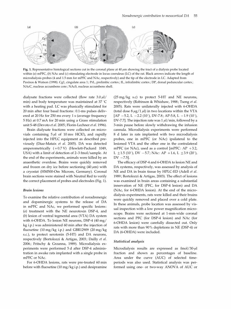
(a) (b) (c)
Cg1
PrL
ILDP
NAcC
NAcS
LC
Fig. 1. Representative histological sections cut in the coronal plane at 40 mm showing the tract of a dialysis probe located
within (a) mPFC, (b) NAc and (c) stimulating electrode in locus coeruleus (LC) of the rat. Black arrows indicate the length of
microdialysis probes (4 and 1.5 mm for mPFC and NAc, respectively) and the tip of the electrode in LC. Adapted from
Paxinos & Watson (1998). Cg1, cingulate area 1 ; PrL, prelimbic cortex ; IL, infralimbic cortex ; DP, dorsal peduncular cortex ;
NAcC, nucleus accumbens core ; NAcS, nucleus accumbens shell.
Noradrenergic contribution to mesocortical DA 55

DA values (repeated measures) followed by
Newman–Keuls post-hoc test. Basal DA dialysate levels
and tissue monoamine contents were compared using
Student’s t test. Data are expressed as means¡S.E.M.
The significance level was set at p<0.05.
Results
Local effect of DAT and NET inhibitors on DA
output in mPFC and NAc
Perfusion of aCSF did not significantly alter DA
output in mPFC and NAc of awake rats. The mean
baseline concentrations of DA dialysate samples from
mPFC and NAc (single probe experiments) were
19¡1 fmol/fraction (n=33) and 19¡3 fmol/fraction
(n=27), respectively.
Local infusion of the DAT+NET inhibitor nomi-
fensine (1, 10 and 30 mM) increased extracellular DA
in both areas in a concentration-dependent manner
(Fig. 2a). The maximal DA increase (AUC6x16) was
660¡52% in mPFC and 527¡130% in NAc at 30 mM
(n=5 each). Two-way ANOVA of AUCs revealed a
significant effect of concentration (F3,32=27.13, p<0.00001) and non-significant effects of region and con-
centrationrregion interaction. Post-hoc test (Newman–
Keuls) revealed significant differences among all tested
concentrations.
Unlike nomifensine, significant regional differences
were found for the selective DAT (GBR12909) and
NET (reboxetine) inhibitors after local administration.
Local GBR12909 infusion markedly increased DA
output in NAc and evoked a minor increase in mPFC
(Fig. 2b). Mean DA elevations at 30, 100 and 300 mM,
expressed as percentage of baseline, were respect-
ively : (a) mPFC (n=6) : 146¡11, 150¡24 and 122¡28
and (b) NAc (n=4) : 382¡75, 390¡91 and 287¡80.
Two-way ANOVA of AUCs showed a significant
effect of the drug concentration (F3,32=6.39, p<0.01),
region (F1,32=27.92, p<0.00001) and concentrationrregion interaction (F3,32=3.19, p<0.05). Post-hoc test
(Newman–Keuls) revealed significant differences be-
tween NAc and mPFC at concentrations of 30
and 100 mM and a marginal difference (p=0.052) at
300 mM.
Local reboxetine application elevated extracellular
DA in mPFC but not in NAc (Fig. 2c). Reboxetine
effects at 1, 10 and 30 mM, expressed as percentage of
baseline, were respectively : (a) mPFC (n=6) : 267¡19,
278¡21, and 282¡33 and (b) NAc (n=4) : 129¡21,
117¡28 and 90¡19. Two-way ANOVA showed a
significant effect of concentration (F3,32=8.50, p<0.001),
region (F1,32=60.07, p<0.0001) and concentrationr
800
700
600
500
400
300
200
100
0
Dia
lysa
te D
A(%
of b
asal
val
ues)
Dia
lysa
te D
A(%
of b
asal
val
ues)
Dia
lysa
te D
A(%
of b
asal
val
ues)
0 1 10 30
Nomifensine (µM)
(a)
800
700
600
500
400
300
200
100
00 30 100 300
GBR12909 (µM)
(b)
800
700
600
500
400
300
200
100
00 1 10 30
Reboxetine (µM)
(c)
* *
* *
**
+ + †
* *
*
+ + +
* * *
Fig. 2. Local effect of catecholamine transporter inhibitors on
DA release in mPFC (%) and NAc (&) of freely moving rats.
The application of artificial CSF did not alter DA levels in any
area. (a) Nomifensine (DAT+NET inhibitor), perfused at 1,
10 and 30 mM (12 fractions each), showed a marked dose effect
without regional differences at any concentration.
(b) GBR12909 (a selective DAT inhibitor) was perfused at
increasing concentrations (30, 100 and 300 mM, five fractions
each) and produced a significant increase of DA levels in
NAc, but not in mPFC. (c) Reboxetine (a selective NET
inhibitor) was perfused at increasing concentrations
(1, 10 and 30 mM, four fractions each) and increased DA
output in mPFC, but not in NAc. Bars show the AUC
calculated by averaging DA percentage of baseline values
in fractions 6–16 for nomifensine, and three DA values for
each concentration for GBR12909 and reboxetine. Data are
expressed as mean¡S.E.M., n=4–7 rats/group, except for
1 mM nomifensine in NAc (n=3). * p<0.05 vs. respective
control (0 mM), + p<0.01 for mPFC vs. NAc and # p=0.05.
See text for detailed statistical analysis.
56 M. Masana et al.

region interaction (F3,32=7.19, p<0.001). Post-hoc test
(Newman–Keuls) revealed significant differences be-
tween NAc and mPFC at all concentrations.
Effects of selective brain lesions on the modulation
of DA output in mPFC and NAc induced by
nomifensine
We further explored the effect of nomifensine in the
mPFC and NAc of rats underlying specific lesions
with (a) the NE neurotoxin DSP-4 and (b) unilateral
lesion of VTA DA system with 6-OHDA, respectively.
Rats pretreated with DSP-4 showed a 90% depletion
of tissue NE level (85¡8 vs. 894¡92 pmol/g of wet
tissue in PFC, p<0.00001) without a significant change
in tissue DA in this area. DSP-4-lesioned rats had
higher basal values (fmol/fraction) of dialysate DA in
mPFC (37¡8, n=6) and NAc (31¡6, n=6), compared
to their respective controls in mPFC (15¡2, n=5) and
NAc (24¡7, n=5), yet only the DA increase in mPFC
reached statistical significance (p<0.05) (Fig. 3c).
Local infusion of nomifensine (30 mM) by reverse
dialysis increased mPFC DA output in control rats to
660¡52% of baseline, but to a much lower extent
(180¡11% of baseline) in mPFC of DSP-4-pretreated
rats (Fig. 3a). However, DSP-4 pretreatment did not
alter the ability of nomifensine to increase extracellular
DA in NAc (DA increase of 528¡130% and 682¡
176% in control and DSP-4-pretreated rats, respect-
ively). Two-way ANOVA revealed a significant effect
of lesionrregion interaction (F1,18=7.48, p<0.05).
Post-hoc t test revealed that the effect of nomifensine in
mPFC of DSP-4-lesioned rats was significantly differ-
ent to the other groups.
The unilateral dopaminergic VTA lesion by 6-OHDA
was assessed in the same rats comparing lesion
side (ipsilateral to 6-OHDA application) with control
side (contralateral to 6-OHDA application). Tissue DA
and NE levels were significantly decreased to 93%
and 71%, respectively in NAc (492¡174 vs. 7226¡
2272 pmol/g of DA in wet tissue and 298¡53 vs.
1019¡1173 pmol/g of NE in wet tissue, p<0.01). Basal
DA values (fmol/fraction) in the control and lesioned
sides were: (a) control mPFC: 12¡4, (b) lesioned
mPFC: 9¡3, (c) control NAc: 11¡4 and, (d) lesioned
NAc: 9¡2 (n=5–6). Non-significant differences of DA
output were found between control and lesioned sides.
Simultaneous local nomifensine (30 mM) infusion by
reverse dialysis in both control and lesioned mPFC
increased DA output to 660¡72% (control mPFC) and
405¡47% (lesioned mPFC) of baseline (Fig. 3b). On
the other hand, nomifensine (30 mM) perfusion en-
hanced DA output to 798¡194% of baseline in control
NAc and to 284¡69% in lesioned NAc. Two-way
ANOVA revealed a significant effect of lesion (F1,19=11.03, p<0.01) and non-significant effects of region
and lesionrregion interaction. Post-hocNewman–Keuls
1200
(a)
1000
800
600
400
200
0
Dia
lysa
te D
A(%
of b
asal
val
ues)
Control DSP-4
Nomifensine (30 µM)
1200
(b)
1000
800
600
400
200
0
Dia
lysa
te D
A(%
of b
asal
val
ues)
Control side 6-OHDA side
Nomifensine (30 µM)
*
+
*
60
(c)
50
40
30
20
10
0
Bas
al D
A v
alue
s(f
mol
/frac
tion)
Control Controlside
6-OHDAside
DSP-4
*
Fig. 3. Local effect of nomifensine 30 mM in mPFC (%)
and NAc (&) of rats underlying specific lesions.
(a) Noradrenergic lesion with DSP-4 (40 mg/kg i.p., 5 d
before experiments) changed DA response to nomifensine
in mPFC but not NAc, compared to control groups. (b) VTA
was unilaterally lesioned with 6-OHDA (8 mg/1 ml, 8 d before
experiments) and the contralateral mPFC and NAc were used
as control. 6-OHDA lesion produced a significant change to
nomifensine in NAc-lesioned (6-OHDA ipsilateral side)
compared to control side. Bars show AUC values, averaged
DA values in fractions 6–16, expressed as percentage of
baseline. (c) Basal DA values (fmol/fraction) in the mPFC
and NAc of control and lesioned rats (DSP-4 and 6-OHDA).
Data are expressed as mean¡S.E.M., n=5–6 per group.
* p <0.05 vs. respective control groups and + p<0.01 for
mPFC vs. NAc. See text for statistical analysis.
Noradrenergic contribution to mesocortical DA 57

test indicated that only DA values in lesioned NAc
were statistically different from control NAc.
Electrical stimulation of LC differentially increases
DA output in mPFC and NAc
We then assessed the effect of electrical stimulation of
LC on DA output in mPFC and NAc of the same rats.
Mean DA basal values (fmol/10-min fraction) were
15¡3 (n=18) and 17¡3 (n=17) in mPFC and NAc,
respectively.
Burst LC stimulation (n=5) significantly elevated
extracellular DA in mPFC and NAc compared to
animals with misplaced electrodes, including peri-
coeruleus area (n=9–10) and sham control rats (n=3,
no current was passed through the LC electrode)
(Fig. 4). The DA output in mPFC showed a sharp rise
during the stimulation period which declined rapidly.
Maximal DA increase was 244¡44% of baseline.
Two-way ANOVA revealed significant effects of the
treatment (F2,15=6.17, p<0.05), time (F9,135=8.94, p<0.0001) and treatmentrtime interaction (F18,135=3.78,
p<0.0001). Burst LC stimulation induced a modest
and slow increase of DA release in NAc (153¡20% of
baseline). Two-way ANOVA indicated a significant
effect of time (F9,126=2.13, p<0.05) and treatmentrtime interaction (F18,126=1.89, p<0.05) and non-
significant effects of stimulation and time. Post-hoc
Newman–Keuls test indicated that DA output in
mPFC during LC stimulation was significantly greater
than in control rats.
Finally, two-way ANOVA of DA output of the sti-
mulated groups revealed significant effects of time
(F9,72=11.67, p<0.00001) and timerregion interaction
(F9,72=2.39, p<0.05). Post-hoc Newman–Keuls test in-
dicated that the DA output in the mPFC during LC
stimulation was significantly greater than in NAc.
Selective enhancement of cortical DA output by
noradrenergic drugs in APD pre-treated rats
Overall, the above results suggest that (1) the extra-
cellular DA concentration is distinctly regulated in
mPFC and NAc, and (2) NE terminals markedly con-
tribute to the control of mPFC DA (but not of NAc DA)
either by co-releasing DA and/or taking up DA via
NET.
We next studied the feasibility of selectively in-
creasing mesocortical DA transmission through NE-
acting drugs. We conducted two sets of experiments in
rats implanted with two microdialysis probes (mPFC
and NAc). Baseline DA concentrations in mPFC and
NAc (double-probe experiments) were 9¡1 fmol/
fraction (n=69) and 9¡2 fmol/fraction (n=62), re-
spectively.
In the first set of experiments, the selective NET
inhibitor reboxetine (30 mM) was locally applied by re-
verse dialysis in mPFC and NAc followed by the sys-
temic administration of the selective a2-adrenoceptor
antagonist RX821002 (1 mg/kg s.c.) 2 h later, to dis-
inhibit the autoreceptor-mediated negative feedback
on NE neuron activity. Figure 5 shows the increase in
350
300
250
200
150
100
50
00 1 2 3 4 5 6 7 8 9 10
Dia
lysa
te D
A (%
of b
asal
val
ues)
5 Hz
5 Hz
250 ms 1 s
+*+
*
Fraction number (10 min each)
Control LC, mPFC
Control LC, NAc
Stimulated outside-LC, mPFC
Stimulated outside-LC, NAc
Stimulated LC, mPFC
Stimulated LC, NAc
Fig. 4. Effect of electrical stimulation of LC on DA levels in mPFC and NAc of the same anaesthetized rats. LC stimulation
increased DA release in mPFC to a larger extent than in NAc. Data are expressed as mean¡S.E.M., n=3 for control ; n=5 for
stimulated. Inset : LC was stimulated after four basal fractions in phasic mode with 0.1-ms pulses delivered at 20 Hz for
250 ms every 1 s (average frequency of 5 Hz) and 0.7 mA for 20 min. See text for statistical analysis. * p<0.05 vs. control
(non-stimulated) and + p<0.05 for mPFC vs. NAC. Dotted lines show the effect on extracellular DA of rats with stimulating
electrodes implanted outside the LC.
58 M. Masana et al.

extracellular DA (AUCs) obtained in mPFC and NAc
during local reboxetine application plus its combi-
nation with RX821002. Two-way ANOVA revealed a
significant effect of treatment (F2,18=79.18, p<0.0001)
and region (F1,18=50.40, p<0.0001) as well as a sig-
nificant treatmentrregion interaction (F2,18=20.74,
p<0.0001).
In the second set of experiments, both drugs
were given systemically (reboxetine, 3 mg/kg i.p. ;
RX821002, 1 mg/kg s.c.). The effect of the reboxetine+RX821002 combination was examined alone and in
rats pretreated with APD drugs: haloperidol (classical,
0.1 mg/kg s.c.) and clozapine (APD, 3 mg/kg s.c.).
The whole set of data are shown in Fig. 6 and were
analysed using two-way ANOVA with treatment
and time as main factors (main effects are shown in
Table 1). The AUC data of relevant fractions (12–16)
for each combination treatment were also calculated
and analysed by two-way ANOVA with treatment
and region as main factors (Fig. 7).
Vehicle+reboxetine+RX821002
Vehicle injections did not alter DA output in mPFC
or NAc. The administration of either reboxetine or
RX821002 alone moderately enhanced DA output, yet
their combined administration dramatically increased
DA levels in mPFC (869¡139% of baseline, n=5)
but not in NAc (188¡33%, n=4) (Fig. 6a, b ; see
Table 1 for detailed statistical analysis). Post-hoc t tests
(Newman–Keuls) revealed a significant difference of
the reboxetine+RX821002 treatment vs. the rest of ex-
perimental groups in mPFC. No significant differences
were found for DA values in NAc.
Two-way ANOVA of AUC data (Fig. 7a) revealed a
significant effect of treatment (F3,30=20.6, p<0.00001),
region (F1,30=21.0, p<0.0001) and regionrtreatment
interaction (F3,30=14.9, p<0.00001).
Clozapine+reboxetine+RX821002
The systemic administration of clozapine elevated DA
output to 241¡27% of baseline in mPFC (n=4, data
are AUC6x16). In NAc, systemic clozapine adminis-
tration did not significantly alter DA output (99¡13%,
n=4).
The combination of clozapine+reboxetine+RX821002 markedly increased DA output to 781¡
123% of baseline in mPFC (n=6). This effect was sig-
nificantly greater than that of clozapine alone and that
of clozapine+reboxetine (Newman–Keuls test post-
ANOVA) (Fig. 6b, Table 1). In NAc, the administration
of reboxetine+RX821002 increased the effect of cloza-
pine to 335¡127% of baseline (n=5) (Fig. 6d, Table 1).
No significant differences between treatments were
found in NAc.
When comparing AUC data within regions (Fig. 6b),
two-way ANOVA revealed a significant effect of
treatment (F3,30=11.9, p<0.0001), region (F1,30=10.5,
p<0.005) and a marginal significance of regionrtreatment interaction (F3,30=2.5, p=0.078).
Haloperidol+reboxetine+RX821002
The systemic administration of haloperidol, given
alone, did not alter the DA output in mPFC (116¡
12%, n=9, AUC6x16), while in NAc it significantly in-
creased DA levels to 162¡20% of baseline (n=7).
The combination of haloperidol+reboxetine+RX821002 induced a large elevation of DA levels in
mPFC to 1375¡275% of baseline (n=6). This effect
was significantly greater than that of haloperidol alone
or that of the combined administration of reboxetine+RX821002 (post-hoc Newman–Keuls ; Fig. 6e, Table 1).
In NAc, the co-administration of haloperidol+reboxetine+RX821002 increased DA levels to 238¡
43% of baseline (n=5) (Fig. 6 f, Table 1). No significant
differences between treatments were found for DA
values in this region.
Two-way ANOVA of AUC data (Fig. 7c) revealed a
significant effect of treatment (F3,39=18.2, p<0.00001),
region (F1,39=19.5, p<0.0001) and regionrtreatment
interaction (F3,39=14.2, p<0.00001).
1800
1600
1400
1200
1000
800
600
400
200
0
Dia
lysa
te D
A (%
of b
asal
val
ues)
Veh Reb 30 µM Reb 30 µM
+ RX1
mPFCNAc
+
+
*
§*
§*
Fig. 5. Effect of the combination treatment with NE-targeting
drugs [local reboxetine (Reb), 30 mM and RX821002 (RX),
1 mg/kg s.c.] on DA release in mPFC and NAc. Reboxetine
was locally applied by reverse dialysis at 30 mM for the whole
experiment (fractions 4–16) and RX was systemically
administered 2 h after the beginning of reboxetine perfusion.
Bars are AUC of four DA values for Reb and for Reb+RX.
See text for statistical analysis. * p<0.01 vs. control, · p<0.01
vs. 30 mM Reb and + p<0.01 for mPFC vs. NAc.
Noradrenergic contribution to mesocortical DA 59

Discussion
The results of the present study confirm and extend
previous observations on a differential regulation of
DA release in mPFC and NAc. We systematically
compared the effect of agents modulating extracellular
DA in both pathways. The data support a significant
contribution of noradrenergic neurotransmission to
DA release in mPFC compared to NAc. The different
mechanisms involved in the control of the active
(extracellular) DA fraction in both areas offer new
therapeutic opportunities to treat non-psychotic symp-
toms in schizophrenia, associated with a reduced
cortical dopaminergic function. Hence, we demon-
strate that noradrenergic drugs dramatically and selec-
tively enhance mesocortical DA, using a strategy
previously shown to potentiate the effects of reuptake
blockers on serotoninergic (Artigas et al. 1996) and
noradrenergic systems (Mateo et al. 1998).
Relative contribution of NE neurotransmission to
DA output in mPFC and NAc
The effects of DAT and/or NET inhibitors agree with
previous observations indicating that NET inhibitors
increase extracellular NE and DA in PFC, but not in
0200400600800
10001200140016001800
0 4 8 12 16
Dia
lysa
te D
A(%
of b
asal
val
ues)
Fraction number (20 min each)
(a)mPFC
Veh
Veh/Reb/RX
*
0200400600800
10001200140016001800
0 4 8 12 16
Dia
lysa
te D
A(%
of b
asal
val
ues)
Fraction number (20 min each)
(b)NAc
Veh
Veh/Reb/RX
0200400600800
10001200140016001800
0 4 8 12 16
Dia
lysa
te D
A(%
of b
asal
val
ues)
Fraction number (20 min each)
(c)
Clz
Veh/Reb/RX
*
0200400600800
10001200140016001800
0 4 8 12 16
Dia
lysa
te D
A(%
of b
asal
val
ues)
Fraction number (20 min each)
(d)
Clz
Veh/Reb/RX
0200400600800
10001200140016001800
0 4 8 12 16
Dia
lysa
te D
A(%
of b
asal
val
ues)
Fraction number (20 min each)
(e)
Hal
Veh/Reb/RX
*
*0
200400600800
10001200140016001800
0 4 8 12 16
Dia
lysa
te D
A(%
of b
asal
val
ues)
Fraction number (20 min each)
(f )
Hal
Veh/Reb/RX
Reb3 + RX1 Reb3 + Veh Veh + RX1 Veh + Veh
Fig. 6. Effect of the combination treatment with NE-targeting drugs [reboxetine (Reb), 3 mg/kg i.p. ; and RX821002 (RX),
1 mg/kg s.c.] on DA release in rats pretreated with (a, b) vehicle (Veh), (c, d) clozapine (Clz 3 mg/kg s.c.) and (e, f)
haloperidol (Hal 0.1 mg/kg s.c.). Control groups received vehicle injections (n=4–6 ; except for Veh/Veh+Veh, where n=3).
Left (a, c, e) and right (b, d, f) panels correspond to the effects in mPFC and NAc, respectively. Data are expressed as mean¡s.e.m.
Statistical analyses shown in Table 2.
60 M. Masana et al.

NAc or dorsal striatum (Bymaster et al. 2002 ; Carboni
et al. 1990, 2006; Di Chiara et al. 1992 ; Mazei et al. 2002 ;
Pozzi et al. 1994; Tanda et al. 1994). The local appli-
cation of nomifensine, with similar affinity for DAT
and NET (PDSP database: http://pdsp.med.unc.edu/
pdsp.php; Bymaster et al. 2002), increased comparably
dialysate DA in mPFC and NAc. However, GBR12909
preferentially increased DA in NAc, suggesting a poor
contribution of DAT-containing fibres to extracellular
DA in mPFC, a view consistent with the low density of
DAT in PFC (Sesack et al. 1998) compared to NAc or
dorsal striatum (Letchworth et al. 2000 ; Marshall et al.
1990). In contrast, mPFC contains a higher density of
NET (Miner et al. 2003 ; Schroeter et al. 2000) than the
NAc. Hence, the NAc core contains scarce NE fibres
(Seguela et al. 1990) and those in NAc shell mainly
arise from the nucleus tractus solitarius (Berridge et al.
1997 ; Delfs et al. 1998). This, together with the similar
affinity of NE and DA for NET (Gu et al. 1994; Raiteri
et al. 1977) may account for the effect of reboxetine,
which increased DA output only in mPFC. Despite NE
axons being infrequently apposed to DA axons in PFC
(Miner et al. 2003), newly released DA may diffuse
trans-synaptically to reach NET sites, as observed for
DA itself (Sesack et al. 1998) (see Fig. 8a).
Furthermore, DSP-4 (Fig. 3a) almost abolished the
effect of nomifensine on DA output in mPFC, but not
in NAc, further supporting a preferential contribution
of NE fibres to mPFC DA release. We used standard
lesion procedures to extensively damage DA and NE
neurons/fibres. Thus, similarly to previous studies
(Bortolozzi & Artigas, 2003 ; Fritschy & Grzanna, 1989 ;
Robinson & Whishaw, 1988; Tseng et al. 2005) DSP-4
and 6-OHDA depleted tissue NE and DA concen-
trations by o90%, respectively. The clear-cut mPFC–
NAc difference of DSP-4 on nomifensine’s effect may
reflect the aforementioned differences on NE axon
densities innervating both brain structures and also
their ability to release DA. Moreover, a preferential LC
sensitivity to the DSP-4 lesion cannot be discounted
(Dailly et al. 2006 ; Grzanna et al. 1989; Jonsson et al.
1981). A limitation of the present study is the lack of
immunohistochemical or autoradiographic analysis of
lesions, which, as in other studies (Grzanna et al. 1989;
Table 1. Statistical analyses of the combination treatment of NE-targeting drugs, reboxetine and RX821002, on dopamine
release in vehicle, clozapine and haloperidol pre-treated rats
Treatment
mPFC NAc
Effect F p Effect F p
Vehicle
Veh+Veh 93¡19 (6)T3,16=19.0
t15,240=33.6
Trt45,240=19.0
<0.0001
<0.00001
<0.00001
121¡9 (5)T3,14=3.1
t15,210=6.3
Trt45,210=2.3
n.s.
<0.00001
<0.0001
Reb+Veh 195¡38 (5) 93¡12 (5)
Veh+RX 234¡42 (4) 216¡61 (4)
Reb+RX 869¡139* (6) 188¡33 (5)
Clozapine
Veh+Veh 197¡30 (4)T3,15=5.0
t15,225=24.2
Trt45,225=10.3
<0.05
<0.00001
<0.00001
110¡15 (4)T3,15=2.2
t15,225=4.3
Trt45,225=2.2
n.s.
<0.00001
<0.001
Reb+Veh 180¡46 (4) 108¡21 (4)
Veh+RX 348¡67 (5) 203¡23 (6)
Reb+RX 781¡123+ (4) 335¡127 (4)
Haloperidol
Veh+Veh 120¡16 (6)T3,22=18.8
t15,330=29.2
Trt45,330=16.0
<0.0001
<0.00001
<0.00001
174¡23 (5)T3,17=2.7
t15,225=5.9
Trt45,225=1.7
n.s.
<0.00001
<0.01
Reb+Veh 323¡40 (5) 95¡7 (3)
Veh+RX 197¡18 (6) 167¡22 (6)
Reb+RX 1375¡275* (9) 238¡43 (7)
Reb, Reboxetine ; RX, RX821002 ; Veh, vehicle ; n.s., not significant.
Data (effect) are given as percentage of baseline in each experimental group (AUC12x16). Number of animals in each group is
given in parentheses.
* Significantly different to all other treatments.+ Significantly different to all treatments except for Veh+RX.
Data have been analysed using two-way ANOVA with treatment (T) and time (t) as main factors.
Noradrenergic contribution to mesocortical DA 61

Vos et al. 1999; Szot et al. 2010) would have permitted a
detailed assessment of lesion effects on NE and DA
axons.
Conversely, local nomifensine increased DA out-
put less in NAc than in mPFC of the unilaterally
6-OHDA-lesioned rats (Fig. 3b), compared to the
contralateral (unlesioned) side, respectively, suggest-
ing that most DA output in NAc arises from VTA DA
fibres. Even with the prior protection of NE fibres by
desipramine prior to 6-OHDA application, a marked
reduction of tissue NE was found in the ipsilateral
NAc (71% for NE vs. 93% for DA), possibly due to
toxin diffusion to the neighbouring median forebrain
bundle. Previous studies have shown the inability of
desipramine to fully protect NE axons from 6-OHDA
lesions (Harden et al. 1998 ; King & Finlay, 1995).
Although DSP-4 and 6-OHDA markedly affected
the nomifensine-induced rise in extracellular DA,
baseline levels were almost unaltered, with the ex-
ception in mPFC of DSP-4 pretreated rats. This is
consistent with previous literature indicating com-
pensatory changes to maintain extracellular mono-
amine concentrations despite marked differences in
tissue concentrations (Abercrombie & Zigmond, 1989;
Robinson et al. 1994; Romero et al. 1998; Zigmond et al.
1990 ; see however Devoto et al. 2008).
The enhancement of DA output in mPFC (244% of
baseline) after electrical stimulation of LC agrees with
previous observations on a potential co-release of DA
from LC NE fibres in PFC (Devoto et al. 2001, 2005;
Devoto & Flore, 2006). Since rats in the present study
were implanted with two probes (in mPFC and NAc),
we were able to compare the effect of LC stimulation
on DA output in mPFC and NAc of the same animals.
LC stimulation also moderately increased (153% of
baseline) the DA output in NAc although with a
blunted time-course, an effect that may arise from
a1-adrenergic stimulation of VTA DA neurons fol-
lowing LC stimulation (Grenhoff & Svensson, 1993).
Further, a1-adrenoceptor blockade abolishes the be-
havioural effect produced by the stimulation of VTA
DA neurons (Auclair et al. 2002, 2004 ; Darracq et al.
1998). Although an a1-adrenergic stimulation of meso-
cortical DA neurons cannot be excluded, the larger DA
increase in mPFC, its temporal association with LC
stimulation and the lack of effect when the stimulating
electrodes were placed outside the LC supports a
noradrenergic origin of the DA release in mPFC.
Thus, the present results suggest that the extra-
cellular DA concentration in NAc mainly arise from
VTA DA fibres whereas that in mPFC has a dual con-
tribution, from the VTA and from LC NE fibres. Since
DA is an intermediate metabolite in the synthesis of
NE in noradrenergic neurons, the co-release of DA
and NE may reflect a deficient activity of dopamine-
b-hydroxylase in cortical noradrenergic axons, a
possibility that deserves further investigation. This
region-specific noradrenergic contribution to DA
0200400600800
10001200140016001800
Dia
lysa
te D
A(%
of b
asal
val
ues)
(a)
Veh +
Veh
Reb3 +
Veh
Veh +
RX1
Reb3 +
RX1
0200400600800
10001200140016001800
Dia
lysa
te D
A(%
of b
asal
val
ues)
(b)
Veh +
Veh
Reb3 +
Veh
Veh +
RX1
Reb3 +
RX1
0200400600800
10001200140016001800
Dia
lysa
te D
A(%
of b
asal
val
ues)
(c)
Veh +
Veh
Reb3 +
Veh
Veh +
RX1
Reb3 +
RX1
Vehicle
Clozapine 3 mg/kg
Haloperidol 0.1 mg/kg
+
*
+
*
+
*
Fig. 7. Bars show the average effect of the combination
treatment of reboxetine (Reb, 3 mg/kg i.p.) plus RX821002
(RX, 1 mg/kg s.c.) on DA release in mPFC (%) and NAc (&)
of rats pre-treated with (a) vehicle, (b) clozapine (3 mg/kg
s.c.) or (c) haloperidol (0.1 mg/kg s.c.), respectively. Data are
AUC of fractions 12–16 (Fig. 5), expressed as percentage of
baseline ; n=4–6, except for Veh/Veh+Veh, where n=3.
See text for statistical analysis. * p<0.001 vs. control
treatments and + p<0.01 for mPFC vs. NAc.
62 M. Masana et al.

release allows the selective modulation of mesocortical
DA function.
Selective enhancement of DA release in mPFC:
association with APD treatments
Many studies implicate prefrontal catecholamine
function in cognition (Arnsten & Li, 2005; Goldman-
Rakic et al. 2000 ; Robbins & Roberts, 2007 ; Sara, 2009).
The present and previous observations show striking
similarities between the factors governing DA and NE
release in mPFC. Current views indicate a hypoactive
mesocortical DA pathway in schizophrenia that may
underlie negative and/or cognitive symptoms and
APDs increase mPFC DA output (see Introduction).
This has been considered a useful pharmacological
feature accounting for the clinical superiority of some
APDs in non-psychotic symptoms (Kane et al. 1988 ;
Keefe et al. 2006 ; Leucht et al. 2009 ; Meltzer &McGurk,
1999). Actually, blockade of DA D2 receptors is effec-
tive in treating positive symptoms, possibly related to
a subcortical DA hyperactivity (Laruelle et al. 1996),
but not negative/cognitive symptoms. Conversely,
DA D2 receptor blockade induces negative symptoms
in healthy individuals (Artaloytia et al. 2006).
Previous reports indicate that reuptake blockade in
serotonergic and noradrenergic neurons induces a
very marked increase of the respective neuro-
transmitter in the vicinity of cell bodies in the raphe
nuclei (Adell & Artigas, 1991 ; Bel & Artigas, 1992) and
LC (Grandoso et al. 2004 ; Mateo et al. 1998). The excess
neurotransmitter in this area activates their respective
autoreceptors (5-HT1A and a2-adrenoceptors), which
leads to a reduced neuronal activity and terminal
monoamine release. Autoreceptor blockade enables
the recovery of cell firing and terminal release, thus
permitting the full pharmacological effect of reuptake
blockade (Artigas et al. 1996 ; Grandoso et al. 2004 ;
Invernizzi & Garattini, 2004 ; Mateo et al. 1998; Romero
& Artigas, 1997). Given the marked involvement of
noradrenergic fibres in the uptake/co-release of DA in
mPFC, we used this strategy to selectively enhance
cortical DA function (Fig. 8).
a2-adrenergic antagonists and NE reuptake inhibi-
tors, acting mainly on NE neurons, increase moderately
(a) (b) (c)
NE
NE DA NET DAT α2R D2R
DA
(–) (–)
(–)(–)(–)
Fig. 8. Schematic representation of the contribution of NET inhibition (reboxetine) and a2-adrenoreceptor antagonism
(RX821002) in NE terminals on the control of extracellular DA levels in mPFC. (a) In a physiological situation, DA can be
co-released with NE by noradrenergic terminals and/or taken up by NET, given the similar affinity of the membrane
transporters for both monoamines. (b) The moderate increase in extracellular DA levels in mPFC evoked by reboxetine probably
results from two opposing factors, (i) an elevation resulting from NET blockade itself, and (ii) a reduction resulting from the
activation of a2-adrenoceptors. Activation of somatodendritic a2-adrenoceptors in the LC (not shown in the figure) also
contributes to attenuate catecholamine release through a reduction of the firing rate of noradrenergic neurons after systemic
reboxetine administration. (c) The co-treatment with RX821002 removes the a2-adrenoceptor-mediated negative feedback on
noradrenergic release of catecholamines, and markedly potentiates the increase in extracellular DA evoked by reboxetine.
Noradrenergic contribution to mesocortical DA 63

but selectively prefrontal DA output (Devoto et al.
2004 ; Gresch et al. 1995 ; Hertel et al. 1999a, b ; Linner
et al. 2001; Millan et al. 2000 ; Swanson et al. 2006;
Valentini et al. 2004 ; Wadenberg et al. 2007). However,
our results show for the first time that a2-adrenoceptor
blockade markedly potentiates the effect of NET in-
hibitors on DA output in mPFC (as also observed for
cortical NE output ; Swanson et al. 2006) but not in
NAc (Figs 5–8).
The increase in cortical DA output also occurred
when reboxetine+RX-821002 were administered in
combination with classical (haloperidol – lacking ap-
preciable affinity for a2-adrenoreceptor) and APDs
(clozapine – with antagonist properties at a2-adreno-
receptors). These drug combinations did not elevate
DA output in NAc or produce even a small effect
compared to the marked elevation seen in mPFC. The
doses used (3 mg/kg for clozapine, 0.1 mg/kg for
haloperidol) are close to their ED50 values for occu-
pation of their primary receptor targets (5-HT2A/2C
and D2, respectively) (Schotte et al. 1993). Further, the
dose of clozapine is in the lower range of those shown
to enhance cortical DA release (Diaz-Mataix et al. 2005;
Ichikawa et al. 2001; Rollema et al. 1997).
Clozapine increased DA output in mPFC, an
effect mediated by cortical 5-HT1A receptor activation
(Bortolozzi et al. 2010 ; Diaz-Mataix et al. 2005)
although a2-adrenoceptor blockade has also been
suggested (Ashby & Wang, 1996 ; Svensson, 2003).
However, since the elevation of DA output produced
by reboxetine+RX821002 was much larger than that
of reboxetine+clozapine, it seems reasonable to as-
sume that clozapine antagonizes a2-adrenoceptors
much less than RX821002.
Consistent with previous reports, haloperidol pro-
duced a moderate DA increase in NAc but not in
mPFC (Kuroki et al. 1999 ; Li et al. 1998). However,
the effect of haloperidol+reboxetine+RX821002 was
more marked than that of the latter two drugs, poss-
ibly due to blockade of DA D2 receptors by haloper-
idol, which would remove the D2-mediated negative
feedback on DA release following the large increase
produced by reboxetine+RX821002. The lower occu-
pancy of DA D2 receptors produced by clozapine
probably accounts for the lower enhancement of
mPFC DA output when combined with reboxetine+RX821002.
Therapeutic implications
Overall, the above results indicate that (1) extracellular
DA concentration is distinctly regulated in mPFC and
NAc, (2) NE terminals largely contribute to the control
of DA output in mPFC (but not NAc) by co-releasing
DA and taking up DA via NET, and (3) a marked and
selective enhancement of DA function in mPFC is
feasible through the combined administration of NET
blockers and a2-adrenoceptor antagonists. The latter
effect occurs when these dugs are administered alone
or in combination with haloperidol or clozapine.
This opens the way to perform clinical trials in
which reboxetine or other NET blockers, used as anti-
depressants, can be combined with a2-adrenoceptor
antagonists in order to test their clinical efficacy on
negative symptoms and/or cognitive dysfunction in
schizophrenia and other psychiatric disorders.
Acknowledgements
This work was supported by grant SAF 2007-62378
(MICIN, Spain). Support from SENY Fundacio is also
acknowledged. M.M. is a recipient of a predoctoral
fellowship from CSIC (I3P programme). A.B. is sup-
ported by the research stabilization programme of
the Health Department of Generalitat de Catalunya.
We thank Leticia Campa for skilful maintenance
and supervision of HPLC equipment and analysis of
dialysate samples. We also thankMrs Veronica Paz for
excellent technical support.
Statement of Interest
None.
References
Abercrombie ED, Zigmond MJ (1989). Partial injury to
central noradrenergic neurons : reduction of tissue
norepinephrine content is greater than reduction of
extracellular norepinephrine measured by microdialysis.
Journal of Neuroscience 9, 4062–4067.
Abi-Dargham A, Rodenhiser J, Printz D, Zea-Ponce Y, et al.
(2000). Increased baseline occupancy of d2 receptors by
dopamine in schizophrenia. Proceedings of the National
Academy of Sciences USA 97, 8104–8109.
Adell A, Artigas F (1991). Differential effects of clomipramine
given locally or systemically on extracellular
5-hydroxytryptamine in raphe nuclei and frontal cortex.
an in vivo brain microdialysis study.Naunyn-Schmiedeberg’s
Archives of Pharmacology 343, 237–244.
Adell A, Sarna GS, Hutson PH, Curzon G (1989). An in vivo
dialysis and behavioural study of the release of 5-HT by
p-chloroamphetamine in reserpine-treated rats. British
Journal of Pharmacology 97, 206–212.
Akil M, Pierri JN, Whitehead RE, Edgar CL, et al. (1999).
Lamina-specific alterations in the dopamine innervation
of the prefrontal cortex in schizophrenic subjects.
American Journal of Psychiatry 156, 1580–1589.
64 M. Masana et al.

Arnsten AF, Li BM (2005). Neurobiology of executive
functions : catecholamine influences on prefrontal cortical
functions. Biological Psychiatry 57, 1377–1384.
Artaloytia JF, Arango C, Lahti A, Sanz J, et al. (2006).
Negative signs and symptoms secondary to
antipsychotics : a double-blind, randomized trial of a single
dose of placebo, haloperidol, and risperidone in healthy
volunteers. American Journal of Psychiatry 163, 488–493.
Artigas F, Romero L, de Montigny C, Blier P (1996).
Acceleration of the effect of selected antidepressant drugs
in major depression by 5-HT1A antagonists. Trends in
Neuroscience 19, 378–383.
Ashby CR, Wang RY (1996). Pharmacological actions of the
atypical antipsychotic drug clozapine : a review. Synapse
24, 349–394.
Auclair A, Cotecchia S, Glowinski J, Tassin JP (2002).
D-amphetamine fails to increase extracellular dopamine
levels in mice lacking alpha 1b-adrenergic receptors :
relationship between functional and nonfunctional
dopamine release. Journal of Neuroscience 22, 9150–9154.
Auclair A, Drouin C, Cotecchia S, Glowinski J, et al. (2004).
5-HT2A and alpha1b-adrenergic receptors entirely
mediate dopamine release, locomotor response and
behavioural sensitization to opiates and psychostimulants.
European Journal of Neuroscience 20, 3073–3084.
Bel N, Artigas F (1992). Fluvoxamine preferentially increases
extracellular 5-hydroxytryptamine in the raphe nuclei : an
in vivo microdialysis study. European Journal of
Pharmacology 229, 101–103.
Berridge CW, Stratford TL, Foote SL, Kelley AE (1997).
Distribution of dopamine beta-hydroxylase-like
immunoreactive fibers within the shell subregion of the
nucleus accumbens. Synapse 27, 230–241.
Bortolozzi A, Artigas F (2003). Control of
5-hydroxytryptamine release in the dorsal raphe nucleus
by the noradrenergic system in rat brain. Role of alpha-
adrenoceptors. Neuropsychopharmacology 28, 421–434.
Bortolozzi A, Diaz-Mataix L, Scorza MC, Celada P, et al.
(2005). The activation of 5-HT receptors in prefrontal cortex
enhances dopaminergic activity. Journal of Neurochemistry
95, 1597–1607.
Bortolozzi A, Masana M, Dıaz-Mataix L, Cortes R, et al.
(2010). Dopamine release induced by atypical
antipsychotics in prefrontal cortex requires 5-HT1A
receptors but not 5-HT2A receptors. International Journal of
Neuropsychopharmacology. Published online : 17 February
2010. doi :10.1017/S146114571000009X.
Bymaster FP, Katner JS, Nelson DL, Hemrick-Luecke SK,
et al. (2002). Atomoxetine increases extracellular levels of
norepinephrine and dopamine in prefrontal cortex of rat :
a potential mechanism for efficacy in attention deficit/
hyperactivity disorder. Neuropsychopharmacology 27,
699–711.
Carboni E, Silvagni A, Vacca C, Di Chiara G (2006).
Cumulative effect of norepinephrine and dopamine carrier
blockade on extracellular dopamine increase in the nucleus
accumbens shell, bed nucleus of stria terminalis and
prefrontal cortex. Journal of Neurochemistry 96, 473–481.
Carboni E, Tanda GL, Frau R, Di Chiara G (1990).
Blockade of the noradrenaline carrier increases
extracellular dopamine concentrations in the prefrontal
cortex : evidence that dopamine is taken up in vivo by
noradrenergic terminals. Journal of Neurochemistry 55,
1067–1070.
Carlsson A (1978). Antipsychotic drugs, neurotransmitters,
and schizophrenia. American Journal of Psychiatry 135,
165–173.
Castner SA, Williams GV, Goldman-Rakic PS (2000).
Reversal of antipsychotic-induced working memory
deficits by short-term dopamine D1 receptor stimulation.
Science 287, 2020–2022.
Creese I, Burt DR, Snyder SH (1976). Dopamine receptor
binding predicts clinical and pharmacological potencies
of antischizophrenic drugs. Science 192, 481–483.
Dailly E, Chenu F, Petit-Demouliere B, Bourin M (2006).
Specificity and efficacy of noradrenaline, serotonin
depletion in discrete brain areas of Swiss mice by
neurotoxins. Journal of Neuroscience Methods 150, 111–115.
Darracq L, Blanc G, Glowinski J, Tassin JP (1998).
Importance of the noradrenaline-dopamine coupling in
the locomotor activating effects of D-amphetamine.
Journal of Neuroscience 18, 2729–2739.
Delfs JM, Zhu Y, Druhan JP, Aston-Jones GS (1998).
Origin of noradrenergic afferents to the shell
subregion of the nucleus accumbens: anterograde and
retrograde tract-tracing studies in the rat. Brain Research
806, 127–140.
Devoto P, Flore G, Pani L, Gessa GL (2001). Evidence for
co-release of noradrenaline and dopamine from
noradrenergic neurons in the cerebral cortex. Molecular
Psychiatry 6, 657–664.
Devoto P, Flore G, Pira L, Longu G, et al. (2004).
Alpha2-adrenoceptor mediated co-release of
dopamine and noradrenaline from noradrenergic
neurons in the cerebral cortex. Journal of Neurochemistry
88, 1003–1009.
Devoto P, Flore G, Saba P, Fa M, et al. (2005). Stimulation
of the locus coeruleus elicits noradrenaline and
dopamine release in the medial prefrontal and parietal
cortex. Journal of Neurochemistry 92, 368–374.
Devoto P, Flore G (2006). On the origin of cortical dopamine :
is it a co-transmitter in noradrenergic neurons? Current
Neuropharmacology 4, 115–125.
Devoto P, Flore G, Saba P, Castelli MP, et al. (2008).
6-Hydroxydopamine lesion in the ventral tegmental area
fails to reduce extracellular dopamine in the cerebral
cortex. Journal of Neuroscience Research 86, 1647–1658.
Di Chiara G, Tanda GL, Frau R, Carboni E (1992).
Heterologous monoamine reuptake : lack of transmitter
specificity of neuron-specific carriers. Neurochemistry
International 20, 231S–235S.
Diaz-Mataix L, Scorza MC, Bortolozzi A, Toth M, et al.
(2005). Involvement of 5-HT1A receptors in prefrontal
cortex in the modulation of dopaminergic activity : role
in atypical antipsychotic action. Journal of Neuroscience 25,
10831–10843.
Noradrenergic contribution to mesocortical DA 65

Floresco SB, Magyar O (2006). Mesocortical dopamine
modulation of executive functions : beyond working
memory. Psychopharmacology (Berlin) 188, 567–585.
Florin-Lechner SM,Druhan JP, Aston-JonesG,ValentinoRJ
(1996). Enhanced norepinephrine release in prefrontal
cortex with burst stimulation of the locus coeruleus.
Brain Research 742, 89–97.
Fritschy JM, Grzanna R (1989). Immunohistochemical
analysis of the neurotoxic effects of DSP-4 identifies two
populations of noradrenergic axon terminals. Neuroscience
30, 181–197.
Goldman-Rakic PS, Muly III EC, Williams GV (2000). D(1)
receptors in prefrontal cells and circuits. Brain Research
Reviews 31, 295–301.
Grandoso L, Pineda J, Ugedo L (2004). Comparative study
of the effects of desipramine and reboxetine on locus
coeruleus neurons in rat brain slices.Neuropharmacology 46,
815–823.
Grenhoff J, Svensson TH (1993). Prazosin modulates the
firing pattern of dopamine neurons in rat ventral
tegmental area. European Journal of Pharmacology 233, 79–84.
Gresch PJ, Sved AF, Zigmond MJ, Finlay JM (1995). Local
influence of endogenous norepinephrine on extracellular
dopamine in rat medial prefrontal cortex. Journal of
Neurochemistry 65, 111–116.
Grzanna R, Berger U, Fritschy JM, Geffard M (1989).
Acute action of DSP-4 on central norepinephrine axons :
biochemical and immunohistochemical evidence for
differential effects. Journal of Histochemistry and
Cytochemistry 37, 1435–1442.
Gu H, Wall SC, Rudnick G (1994). Stable expression of
biogenic amine transporters reveals differences in inhibitor
sensitivity, kinetics, and ion dependence. Journal of
Biological Chemistry 269, 7124–7130.
Harden DG, King D, Finlay JM, Grace AA (1998). Depletion
of dopamine in the prefrontal cortex decreases the basal
electrophysiological activity of mesolimbic dopamine
neurons. Brain Research 794, 96–102.
Hertel P, Fagerquist MV, Svensson TH (1999b). Enhanced
cortical dopamine output and antipsychotic-like effects of
raclopride by alpha2 adrenoceptor blockade. Science 286,
105–107.
Hertel P, Nomikos GG, Svensson TH (1999a). Idazoxan
preferentially increases dopamine output in the rat medial
prefrontal cortex at the nerve terminal level. European
Journal of Pharmacology 371, 153–158.
Ichikawa J, Ishii H, Bonaccorso S, Fowler WL, et al. (2001).
5-HT(2A) and D(2) receptor blockade increases cortical DA
release via 5-HT(1A) receptor activation : a possible
mechanism of atypical antipsychotic-induced cortical
dopamine release. Journal of Neurochemistry 76, 1521–1531.
Invernizzi RW, Garattini S (2004). Role of presynaptic
alpha2-adrenoceptors in antidepressant action : recent
findings from microdialysis studies. Progress in
Neuro-Psychopharmacology and Biological Psychiatry 28,
819–827.
Jonsson G, Hallman H, Ponzio F, Ross S (1981). DSP4
(N-(2-chloroethyl)-N-ethyl-2-bromobenzylamine) – a useful
denervation tool for central and peripheral noradrenaline
neurons. European Journal of Pharmacology 72, 173–188.
Kane J, Honigfeld G, Singer J, Meltzer H (1988). Clozapine
for the treatment-resistant schizophrenic. A double-blind
comparison with chlorpromazine. Archives of General
Psychiatry 45, 789–796.
Kawahara H, Kawahara Y, Westerink BH (2001). The
noradrenaline-dopamine interaction in the rat medial
prefrontal cortex studied by multi-probe microdialysis.
European Journal of Pharmacology 418, 177–186.
Keefe RS, Young CA, Rock SL, Purdon SE, et al. (2006).
One-year double-blind study of the neurocognitive
efficacy of olanzapine, risperidone, and haloperidol in
schizophrenia. Schizophrenia Research 81, 1–15.
King D, Finlay JM (1995). Effects of selective dopamine
depletion in medial prefrontal cortex on basal and evoked
extracellular dopamine in neostriatum. Brain Research 685,
117–128.
Kuroki T, Meltzer HY, Ichikawa J (1999). Effects of
antipsychotic drugs on extracellular dopamine levels in rat
medial prefrontal cortex and nucleus accumbens. Journal of
Pharmacology and Experimental Therapeutics 288, 774–781.
Laruelle M, Abi-Dargham A, van Dyck CH, Gil R, et al.
(1996). Single photon emission computerized tomography
imaging of amphetamine-induced dopamine release in
drug-free schizophrenic subjects. Proceedings of the National
Academy of Sciences USA 93, 9235–9240.
Letchworth SR, Smith HR, Porrino LJ, Bennett BA, et al.
(2000). Characterization of a tropane radioligand,
[(3)H]2beta-propanoyl-3beta-(4-tolyl) tropane ([(3)H]PTT),
for dopamine transport sites in rat brain. Journal of
Pharmacology and Experimental Therapeutics 293, 686–696.
Leucht S, Corves C, Arbter D, Engel RR, et al. (2009).
Second-generation vs. first-generation antipsychotic drugs
for schizophrenia : a meta-analysis. Lancet 373, 31–41.
Lewis DA, Lieberman JA (2000). Catching up on
schizophrenia : natural history and neurobiology.
Neuron 28, 325–334.
Li XM, Perry KW, Wong DT, Bymaster FP (1998).
Olanzapine increases in vivo dopamine and
norepinephrine release in rat prefrontal cortex, nucleus
accumbens and striatum. Psychopharmacology (Berlin) 136,
153–161.
Lieberman JA, Stroup TS, McEvoy JP, Swartz MS, et al.
(2005). Effectiveness of antipsychotic drugs in patients
with chronic schizophrenia. New England Journal of
Medicine 353, 1209–1223.
Linner L, Endersz H, Ohman D, Bengtsson F, et al. (2001).
Reboxetine modulates the firing pattern of dopamine cells
in the ventral tegmental area and selectively increases
dopamine availability in the prefrontal cortex. Journal of
Pharmacology and Experimental Therapeutics 297, 540–546.
Marshall JF, O’Dell SJ, Navarrete R, Rosenstein AJ (1990).
Dopamine high-affinity transport site topography in rat
brain : major differences between dorsal and ventral
striatum. Neuroscience 37, 11–21.
Mateo Y, Pineda J, Meana JJ (1998). Somatodendritic
alpha2-adrenoceptors in the locus coeruleus are involved
66 M. Masana et al.
in the in vivo modulation of cortical noradrenaline release
by the antidepressant desipramine. Journal of
Neurochemistry 71, 790–798.
Mazei MS, Pluto CP, Kirkbride B, Pehek EA (2002). Effects
of catecholamine uptake blockers in the caudate-putamen
and subregions of the medial prefrontal cortex of the rat.
Brain Research 936, 58–67.
Meltzer HY, McGurk SR (1999). The effects of clozapine,
risperidone, and olanzapine on cognitive function in
schizophrenia. Schizophrenia Bulletin 25, 233–255.
MillanMJ, Gobert A, Rivet JM, Adhumeau-Auclair A, et al.
(2000). Mirtazapine enhances frontocortical dopaminergic
and corticolimbic adrenergic, but not serotonergic,
transmission by blockade of alpha2-adrenergic and
serotonin2C receptors : a comparison with citalopram.
European Journal of Neuroscience 12, 1079–1095.
Miner LH, Schroeter S, Blakely RD, Sesack SR (2003).
Ultrastructural localization of the norepinephrine
transporter in superficial and deep layers of the rat
prelimbic prefrontal cortex and its spatial relationship to
probable dopamine terminals. Journal of Comparative
Neurology 466, 478–494.
Paxinos G, Watson C (1998). The Rat Brain in Stereotaxic
Coordinates. Sydney : Academic.
Pozzi L, Invernizzi R, Cervo L, Vallebuona F, et al. (1994).
Evidence that extracellular concentrations of dopamine are
regulated by noradrenergic neurons in the frontal cortex of
rats. Journal of Neurochemistry 63, 195–200.
Raiteri M, del Carmine R, Bertollini A, Levi G (1977).
Effect of sympathomimetic amines on the
synaptosomal transport of noradrenaline, dopamine and
5-hydroxytryptamine. European Journal of Pharmacology 41,
133–143.
Robbins TW, Roberts AC (2007). Differential regulation of
fronto-executive function by the monoamines and
acetylcholine. Cerebral Cortex 17, i151–i160.
Robbins TW, Arnsten AF (2009). The
neuropsychopharmacology of fronto-executive function :
monoaminergic modulation. Annual Review of Neuroscience
32, 267–287.
Robinson TE, Mocsary Z, Camp DM, Whishaw IQ (1994).
Time course of recovery of extracellular dopamine
following partial damage to the nigrostriatal dopamine
system. Journal of Neuroscience 14, 2687–2696.
Robinson TE, Whishaw IQ (1988). Normalization of
extracellular dopamine in striatum following recovery
from a partial unilateral 6-OHDA lesion of the substantia
nigra : a microdialysis study in freely moving rats.
Brain Research 450, 209–224.
Rollema H, Lu Y, Schmidt AW, Zorn SH (1997). Clozapine
increases dopamine release in prefrontal cortex by 5-HT1A
receptor activation. European Journal of Pharmacology 338,
R3–R5.
Romero L, Artigas F (1997). Preferential potentiation of the
effects of serotonin uptake inhibitors by 5-HT1A receptor
antagonists in the dorsal raphe pathway : role of
somatodendritic autoreceptors. Journal of Neurochemistry
68, 2593–2603.
Romero L, Jernej B, Bel N, Cicin-Sain L, et al. (1998). Basal
and stimulated extracellular serotonin concentration in the
brain of rats with altered serotonin uptake. Synapse 28,
313–321.
Sara SJ (2009). The locus coeruleus and noradrenergic
modulation of cognition. Nature Reviews Neuroscience 10,
211–223.
Schroeter S, Apparsundaram S, Wiley RG, Miner LH, et al.
(2000). Immunolocalization of the cocaine- and
antidepressant-sensitive l-norepinephrine transporter.
Journal of Comparative Neurology 420, 211–232.
Seeman P, Lee T (1975). Antipsychotic drugs : direct
correlation between clinical potency and presynaptic
action on dopamine neurons. Science 188, 1217–1219.
Seguela P, Watkins KC, Geffard M, Descarries L (1990).
Noradrenaline axon terminals in adult rat neocortex : an
immunocytochemical analysis in serial thin sections.
Neuroscience 35, 249–264.
Sesack SR, Hawrylak VA,Matus C, GuidoMA, et al. (1998).
Dopamine axon varicosities in the prelimbic division of the
rat prefrontal cortex exhibit sparse immunoreactivity for
the dopamine transporter. Journal of Neuroscience 18,
2697–2708.
Schotte A, Janssen PF, Megens AA, Leysen JE (1993).
Occupancy of central neurotransmitter receptors by
risperidone, clozapine and haloperidol, measured ex vivo
by quantitative autoradiography. Brain Research 631,
191–202.
Svensson TH (2003). Alpha-adrenoceptor modulation
hypothesis of antipsychotic atypicality. Progress in
Neuro-Psychopharmacology & Biological Psychiatry 27,
1145–1158.
Swanson CJ, Perry KW, Koch-Krueger S, Katner J, et al.
(2006). Effect of the attention deficit/hyperactivity disorder
drug atomoxetine on extracellular concentrations of
norepinephrine and dopamine in several brain regions
of the rat. Neuropharmacology 50, 755–760.
Szot P, Miguelez C, White SS, Franklin A, et al. (2010).
A comprehensive analysis of the effect of DSP4 on the
locus coeruleus noradrenergic system in the rat.
Neuroscience 166, 279–291.
Tanda G, Carboni E, Frau R, Di Chiara G (1994). Increase of
extracellular dopamine in the prefrontal cortex : a trait of
drugs with antidepressant potential? Psychopharmacology
(Berlin) 115, 285–288.
Tseng KY, Kargieman L, Gacio S, Riquelme LA, et al. (2005).
Consequences of partial and severe dopaminergic lesion
on basal ganglia oscillatory activity and akinesia.
European Journal of Neuroscience 22, 2579–2586.
Valentini V, Frau R, Di Chiara G (2004). Noradrenaline
transporter blockers raise extracellular dopamine in
medial prefrontal but not parietal and occipital cortex :
differences with mianserin and clozapine. Journal of
Neurochemistry 88, 917–927.
Vijayraghavan S, Wang M, Birnbaum SG, Williams GV,
et al. (2007). Inverted-U dopamine D1 receptor actions
on prefrontal neurons engaged in working memory.
Nature Neuroscience 10, 376–384.
Noradrenergic contribution to mesocortical DA 67

Vos PE, Steinbusch HW, Ronken E, van Ree JM (1999).
Short and long term plasticity after lesioning of the cell
body or terminal field area of the dopaminergic
mesocorticolimbic system in the rat. Brain Research 831,
237–247.
Wadenberg ML, Wiker C, Svensson TH (2007). Enhanced
efficacy of both typical and atypical antipsychotic drugs by
adjunctive alpha2 adrenoceptor blockade : experimental
evidence. International Journal of Neuropsychopharmacology
10, 191–202.
Weinberger DR, Berman KF, Chase TN (1988). Mesocortical
dopaminergic function and human cognition. Annals of the
New York Academy of Sciences 537, 330–338.
Westerink BH, Kawahara Y, De Boer P, Geels C, et al.
(2001). Antipsychotic drugs classified by their effects on the
release of dopamine and noradrenaline in the prefrontal
cortex and striatum. European Journal of Pharmacology 412,
127–138.
Williams GV, Goldman-Rakic PS (1995). Modulation of
memory fields by dopamine D1 receptors in prefrontal
cortex. Nature 376, 572–575.
Zigmond MJ, Abercrombie ED, Berger TW,
Grace AA, et al. (1990). Compensations after
lesions of central dopaminergic neurons : some
clinical and basic implications. Trends in Neuroscience
13, 290–296.
68 M. Masana et al.