archaebacteria: lipids, membrane structures, and ... · archaebacteria: lipids, membrane...
TRANSCRIPT

Archaebacteria: Lipids, Membrane Structures, and Adaptation to Environmental Stresses
M. DE ROSA1 ,2, A. TRINCONEt, B. NICOLAUSt, and A. GAMBACORTA1
1 Introduction
In the past few years a revolution has occurred in the taxonomy of living organisms. In fact, on the basis of genetic studies and on the aoquisition of other general biochemical features, organisms are no longer merely gathered into two groups of eubacteria and eukaryotes, but may be considered to belong to a third line, the archaebacteria (Woese 1987).
The archaebacteria have been recognized as a phylogenetically coherent separate group of microorganisms, and differ from eubacteria and eukaryotes in a series of genetic and molecular aspects (Woese 1987). They are a collection of disparate phenotypes and thrive in environments that would normally kill many other known organisms. They are segregated into a few peculiar ecological niches, such as saturated brine for halophiles, strict anaerobiotic environment for methanogens, and thermal habitats for extreme thermophiles (Woese 1987; Konig 1988).
From the point of view of the early evolution oflife, archaebacteria have to be considered quite interesting organisms, since they were probably the dominant forms of life in the primeval biosphere. Many metabolic features of these microorganisms seem to be suitable for the ecological parameters which have characterized the early history of life on earth. The importance of the archaebacteria lies in the improvement of our knowledge of the early events in the evolution of cells, contributing to a better understanding of the universal ancestor. Although archaebacterial taxonomy is complex and controversial, the new recent isolates all conform essentially to one of the three original types: halophiles, methanogens, and thermophiles (De Rosa and Gambacorta 1988).
Recent studies based on the comparison of complete 16S ribosomal RNA sequences confirm, refine, and extend earlier archae bacterial phylogeny. In particular, these studies show that archaebacteria could now be divided into the following two major branches: (1) the sulfur-dependent thermophilic organisms, and (2) the methanogens, which comprise the extreme halophiles and some
1 Istituto per la Chimica di Molecole di Interesse Biologico CNR, Via Toiano 6, Arco Felice, Napoli, Italy. 2Istituto di Biochimica delle Macromolecole, Universita' di Napoli I, Facolta' di Medicina, Via Costantinopoli 16, Napoli, Italy.
Guido di Prisco (Ed) Life Under Extreme Conditions © Springer-Verlag Berlin Heidelberg 1991

62 M. De Rosa et al.
thermophilic species belonging to the orders of Thermoplasmatales, Thermococcales and Archaeglobales (Fig. 1; Woese 1987).
In view of the key role of the membrane ability of archaebacteria to survive in the face of drastic environmental stresses, extensive studies have thus been undertaken to characterize the structural identity and the biogenetic origin of their membrane lipids (Langworthy 1985; De Rosa and Gambacorta 1988; Kamekura and Kates 1988).
Upon comparing molecular components of archae bacteria with prokaryotes and eukaryotes, one is struck by the major chemical differences observed in the lipids, especially in comparison with the other components whose essential chemical features appear to be preserved. In fact, the structure of membrane lipids remains one of the major distinctions between all archaebacteria and other organisms.
All the membrane lipids of the archae bacteria so far identified are characterized by unusual structural features, which can be considered to be specific taxonomic markers of this group of microorganisms (Langworthy 1985; De Rosa and Gambacorta 1988; Kamekura and Kates 1988). Archaebacteria do not contain fatty acyl chains in their lipids; instead, they are characterized by the presence of phytanyl chains derived from saturated C20 ' C25 , or C40 alcohols joined to glycerol or other polyols by ether linkage, resulting in ether lipids.
Moreover, it is worth noting that all glycerol ethers in archae bacteria contain a 2,3-sn-glycerol, which is unusual, since the glycerol, in naturally occurring glycerophosphatides or diacylglycerols, is known to have 1 ,2-sn stereochemistry.
Although isoprenoids with specific functions occur in the lipid membrane of most cells, the ether lipids dealt with here are the only such compounds that provide the major structural components of the membrane in which they occur.
HllCaliphllit halophile,
Neutral halophile,
Methanospirillum
rhermopla,ma
MethaDothermus MethanobDtterlum
Hrtheoglobus
Thermotottales
Fig. 1. Phylogenetic tree of archae bacteria group
Sulfolobales
.-..::::....---- Staphylothermus
DesulfurotottuS

Archaebacteria 63
We may wonder how archaebacteriallipids with a chemical composition so different from conventional ester lipids can manage to perform similar functions, and why that chemical variability is required for living organisms. In this respect, one of the most important aspects is the organization of ether lipids in the plasma membrane of archaebacteria.
This presentation is meant to be a survey of the lipid structure occurring in archaebacteria so far examined, and of the most important features of archaebacteriallipid biosynthesis. Moreover, some data about phenotypic adaptation of archae bacteria to environmental stresses are discussed.
2 Lipid Structure of Archaebacteria
2.1 Core Lipids
The complex lipids of archae bacteria are mainly based on two classes of isopranoid ether core lipids: diethers and tetraethers. These compounds are easily obtained by acid or alkaline hydrolysis of complex lipids (De Rosa and Gambacorta 1988).
2.2 Diether Archaebacterial Lipids
Different types ofisopranoid diethers, basic structural elements ofthe membrane lipids occuring in different groups of archae bacteria, are reported in Fig. 2. These molecules could be formally considered the archaebacterial counterparts of the conventional diglycerides of eukaryotic or eubacterial origin.
A general feature of all archae bacterial glycerol lipids is the unusual 2,3-sn glycerol stereochemistry, opposite to the one found in common diglycerides, which is probably related to the biochemical mechanism of ether linkage assembly.
The diethers found in archaebaeteria are reported in Fig. 2. Figure 2a shows the 2,3-di-O-phytanyl-sn-glycerol formed by condensation via ether linkage between two C20 isopranoid alcohols and a glycerol molecule. The chiral centers of the isopranoid chains have the 3R, 7R, llR configuration. The 2,3-di-Ophytanyl-sn-glycerol, a (Fig. 2), could be considered a universal core lipid in such types of microorganisms. It may be present as the major component depending on the type of archaebacteria. This glycerol diether represents 100% of ether core lipids in the majority of neutral halophiles and in some coccoid forms belonging to the methanogens and thermophiles (Langworthy and Pond 1986; De Rosa and Gambacorta 1988).
Structural types b-c (Fig. 2) occur in the extremely alkaliphilic halophiles of the genera Natronococcus and Natronobacterium, living at pH 10, and in a few strains of neutral halophiles of the genera Halobacterium and Halococcus. The 2-0-sesterterpanyl-3-0-phytanyl-sn-glycerol, b (Fig. 1), represents up to 80% of

64 M. De Rosa et al.
a H-·E
b H"[
c H-E
d H"[ CH20H
I CHOH
e I CH·O
I CH20
HE: Fig. 2a-f. Isopranoid diether lipids in archaebacteria (see text)

Archaebacteria 65
total ether core lipids in the haloalkaliphiles (De Rosa and Gambacorta 1988; Kamekura and Kates 1988).
Macrocyclic glycerol diether, d (Fig. 2), is characterized by the presence ofa 36-membered ring, which formally originates from the condensation ofa glycerol moiety with a C40W,W1 isopranyl diol. From a biosynthetic point of view, the macrocyclic structure probably derives from the unprecedented head-tohead condensation of two isopranoid residues of a 2,3-di-O-geranylgeranyl-snglycerol precursor (Comita et al. 1984).
Diphytanyl tetritol diether, e (Fig. 1), isolated from Methanosarcina barkeri and Methanosarcina mazei, represents up until now the only example, among archaebacteria, of a core lipid in which glycerol is absent (De Rosa et al. 1986b; De Rosa and Gambacorta 1988).
Recently, Ferrante et al. (l988a) isolated from the aceticlastic methanogen Methanothrix concilii GP6 the diether core lipid, f, (Fig. 2). This new core lipid, accounting for 30% of total polar lipids of the microorganisms, is obtained only when mild hydrolytic conditions are used. It possesses, in fact, a hydroxyl group on C-3 of the phytanyl chain at the sn-3 position of the glycerol.
2.3 Tetraether Archaebacterial Lipids
Figure 3 shows two families of macrocyclic tetraethers which are the basic components of the membrane lipids of many methanogens and, with few exceptions, of all thermophilic archaebacteria (Langworthy and Pond 1986; De Rosa and Gambacorta 1988).
The tetraether a (Fig. 3) has a 72-member ring with 18 stereocenters, formed by two 2,3-sn-glycerol moieties, bridged through ether linkages by two isopranoid C40 diols formally derived from head to head linkage oftwo O-phytanyl residues. The series oftetraethers b-i (Fig. 3) can be considered to derive from the first one bya process ofcyclization of the aliphatic component (3R, 7R, llR, ISS, 18S,22R, 26R, 30R)-3,7 ,26,30-octamethyldotriacontane, between methyls in 3,7,26,30 and methylenes 6,10,23,27, respectively. All these structures are simply named glycerol dialkyl glycerol tetraethers (GDGTs).
The second series a' -i' (Fig. 3) has a structural organization similar to that of GDGTs, but a more complex polyol, with nine carbon atoms replacing one ofthe glycerols. By analogy, these compounds are named glycerol dialkyl nonitol tetraethers (GDNTs; De Rosa and Gambacorta 1988).
A high degree of structural specificity characterizes these tetra ethers, originating from two polyols and five different C40 isopranoids with up to four cyclopentane rings per chain. In all the structures identified so far in Sulfolobus solfataricus, the two polyols are always antiparallel and the two C40 chains are either identical or differ byone cycle. Moreover, in tetraethers with an asymmetric end-to-end C40 chain (mono- and tricyclic C40 isopranoids), the more cyclized end is always linked to the primary carbinol of the polyols (De Rosa et al. 1983).
The tetraether a (Fig. 3), together with the 2,3-di-O-phytanyl-sn-glycerol, a (Fig. 2), in different proportions, are the basic components of complex lipids of

66 M. De Rosa et al.
CH20H
"b ~~~~~"'-/"': l···· H
a;a'
"E" "E" "E" "E"
CHOH I R
~~~~:l""H b ;b'
d;d'
CHOH I R
~'"""'~1" CHOH I R
:1" CHOH I R
Fig. 3. Isopranoid tetraether lipids in archae bacteria. a-i Glycerol-dialkyl-glycerol tetraethers (GDGTs); a'-i':glycerol-dialkyl-nonitol tetraethers (GDNTs) (see text)
methanogenic archaebacteria belonging to the orders Methanobacteriales and Methanomicrobiales. Only in Methanothermus sociabilis and in Methanosarcina barkeri tetraethers with cyclopentane rings in the isopranoid chains have been detected (Langworthy and Pond 1986; De Rosa and Gambacorta 1988).
G DNT lipids are found only in microorganisms belonging to the Sulfolobales and seem to be a specific taxonomic marker for this order (Langworthy and Pond 1986; De Rosa and Gambacorta 1988).
Tetraethers are the main components of lipids of thermophilic archaebacteria, with the exception of Pyrococcus woesei, Thermococcus celer, and ANI isolate, whose lipids are based on diether a (Fig. 2; De Rosa et al. 1987; Lanzotti et al. 1989b).

Archaebacteria
a-i R=H a'-i' R=
~lH CHOH I R
.--l ~ /18 9, OHI oHi OH OH OH OH
2.4 Minor Archaebacterial Lipids
In Fig. 4, three minor core lipids are reported which occur in archaebacteria.
67
The structure a (Fig. 4) has been isolated from lipids of Methanosarcina barkeri and, in trace amounts, also in S. solfataricus (De Rosa et al. 1986b; De Rosa et al. 1989).
Compound b (Fig. 4) is a glycerol trialkyl glycerol tetraether based upon two glycerol linked by four ether bonds with two C20 and one C40 isopranoid chain. The absence in S. solfataricus of tetraethers with this type of molecular architecture, but with cyclopentane rings in the isopranoid chains, as in GDGT and in GDNT (Fig. 3), could indicate that the enzyme system devoted to cyclopentane ring formation operates only if the 72-membered macrocycle is closed (De Rosa et al. 1989).
Recently, Thurl and Schafer (1988) reported the occurrence, as a minor component of tetraethers in Thermoproteus tenax, of a new GDGT having in

68 M. De Rosa et al.
a
"F O~ 01 ~O··r··H
b CH20H
c
Fig.4a-c. Minor ether lipid in archae bacteria
the macrocycle an acyclic and a bicyclic C40 isopranoid chain, c, (Fig. 4); the relative orientation of the two glycerol moieties in this tetraether remains unknown.
3 Polar Lipids
3.1 Halophiles
Although polar complex lipids based on 2,3-di-O-phytanyl-sn-glycerol are found in all archaebacterial phenotypes, they differ in the nature of the attached polar heads.
In the halophilic archaebacteria four basic complex lipid structures are found: (1) the isoprenoid ether analogues of phosphatidylglycerol (PG); (2) phosphatidylglycerophosphate (PGP); (3) phosphatidylglycerosulfate (PGS;

Archaebacteria 69
Fig. 5); and (4) a family of glycolipids and their sulfate derivatives, differing among themselves in the number and type of sugars (Fig. 5; De Rosa et al. 1989).
The amino lipids are absent in halophilic archaebacteria, which is unusual in that most of eubacterial counterparts usually contain phosphatidylethanolamine or lipoamino acids (Kamekura and Kates 1988).
Recently, Tsujimoto et al. (1989) and Fredrickson et al. (1989) revised the structure of PGP in Halobacterium halobium and H. cutirubrum, respectively, by using the F AB technique. The authors describe that in both organisms the major component of polar lipids is a methyl ester ofPGP (PGP-Me, Fig. 5) instead of the corresponding free acid previously reported.
The glycolipids of halophilic archaebacteria appear to be derived from the basic structure diglycosyl diether, mannosyl-glucosyl-diphytanylglycerol (DGD), bysubstitutionofa sugar or sulfate group atthe 6 positionofthe mannose residue, giving rise to triglycosyl diethers and sulfate diglycosyl (S-DGD-I) diethers, respectively (Fig. 5; Kamekura and Kates 1988).
Triglycosyl glycolipids differ among themselves in the more external sugar residue, which can be galactose (TGD-I) or glucose (TGD-2). TGD-I in turn originates a sulfate triglycosyl derivative when the sulfate group is linked to C-3 of the third sugar residue (S-TGD-I, Fig. 5). Minor branched tetraglycosyl glycolipid (TeGD) and its sulfated derivative (S-TeGD) show essentially the structure of the sulfate triglycosylglycolipid with galactose residue (TGD-I), with the addition of a-galactose to the 3 position of the mannose residue (Fig. 5; Kamekura and Kates 1988).
Another sulfated diglycosyl diether, designated as S-DGD-2, has been detected in the extreme halophile strain isolated in Japan, but its structure is yet unknown (Kamekura and Kates 1988).
The membrane lipid composition ofnonalkaliphilic halobacteria has proved particularly useful in the taxonomy of such microorganisms. PGS, for example, is not as widely distributed in all strains as PG P and PG. Variations also occur in the type and amounts of the glycolipids; the presence or absence of these compounds and other minor unidentified lipids can be used to rapidly assign an isolate to a particular group. Most Halobacterium species are characterized by the presence of PGS, S-TG D-l, and S-TeGD. Members of Haloarcula contain PGS as well as TGD-2 as major glycolipids, together with a minor unidentified glycolipid component, and do not possess sulfated glycolipid. The species of the Haloferax genus, on the contrary, do not contain PGS but are characterized by the presence of S-DGD-l. The main lipids of the Halococcus genus are PG, PGP, TGD-2, and a sulfated diglycosyllipid, probably S-DGD-I (Fig. 5; Torreblanca et al. 1986; Kamekura and Kates 1988; Grant and Larsen 1989).
The polar lipid extract from a neutrophilic halophilic archaebacterium isolated from a salt mine contains, in addition to PG, PGP, PGS, S-DGD-I, an analogue of phosphatidic acid (PA, Fig. 5; Lanzotti et al. 1989c), probably unrelated to a biosynthetic route of these complex lipids.
The haloalkaliphilic archaebacteria of the genera Natronobacterium and Natronococcus have a relative simple polar lipid composition in comparison with

70 M. De Rosa et al.
AcIdic lipids o I
CH2-0-P -O-Rt
H+-O-~ CH2-0 -R:!
R2 R3
PG 3'-slI-glycerol C20 or C25 C20
PGP 3'-slI-glycerol-t'-P C20 or C25 C20
PG5 3'-slI-glycerol-1'-sullate ~o C20
PA H C20 C20
PL2 3'-sn -glyceroH',2'cyclicphosphate C20 C20
PGP-Me 3'-sn-glycerol-l'-P-Me C20 C20
GIycoIlpids
Rl R2
OGO H OH
5-000-1 -S03H OH
TGO-l 6-galp OH
TGO-2 6-glcp OH
5-TGI).I 3-S03H-6-galp OH
TeGO 6-galp O-«-gall
5-TeGO 3-S03H-6-galp O-«-gall
Fig. 5. Complex lipids in halophiles. P Phosphate group

Archaebacteria 71
neutrophilic halophiles, in that the glycolipids are completely absent and the major species of polar lipids are generally PGP and PG. However, several minor unidentified phospholipids are also present in examples of these genera. In particular, Natronococcus occultus has a significant amount of PGP derivative with a 1',2'-cyclic phosphate (PL2, Fig 5; De Rosa et al. 1988; Lanzotti et al. 1989a).
In all species of N atronobacterium and N atronococcus, PG P, the major polar lipid, elutes from the silica column in association with glycine betaine; this last forms an ionic complex with a phospholipid only when two charges are available for complex formation. Thus PG, and PL2, the cyclic derivative ofPGP, did not form the complex (De Rosa et al. 1988).
Nicolaus et al. (1989) describe that glycine betaine is membrane-associated; its level increases along with an increase in the PG/PGP ratio when N. occultus is grown in different salt concentrations. .
At the moment, complex lipids based on C20C25 (b, Fig. 2) diether are found only as PG, PGP, and PL2 and are present essentially in haloalkaliphilic species (De Rosa and Gambacorta 1988; Kamekura and Kates 1988).
3.2 Methanogens
Methanogenic archaebacteria have complex lipids based both on diether and tetraether molecules; in this respect they differ from halophiles, which have lipids based only on diethers, and from thermophilic sulfur-dependent species, which have lipids based essentially on tetra ethers. However, the knowledge of polar lipids in methanogens is quite incomplete, both because many of these molecules are not fully characterized, and because few species of methanogens are analyzed in detail for their lipid composition.
The complex lipids in methanogens are generally classified by means of specific staining tests into five groups: (1) mean phospholipids; (2) aminophospholipids; (3) aminophosphoglycolipids; (4) phosphoglycolipids; and (5) glycolipids (Nishihara and Koga 1987).
Aminophospholipids were found to be widely distributed in methanogens, in which they prevail in contrast with the other two phenotypes of archaebacteria. For example, the diphytanyl ether analogue of phosphatidylserine (PNL2b, 1, Fig. 6) occurs as a major constituent in the lipids of Methanobacteriales (Morii et al. 1986; Koga et al. 1987). In a genus of such an order is also found a phospho ethanolamine derivative of diphytanyl glycerol diether (2, Fig. 6; Kramer et al. 1987; Nishihara et al. 1989).
In species belonging to the Methanococcus genus a novel 2,3-di-O-phytanyl-1-(phosphoryl-2-acetoamido-2-deoxy-/3-D-glucopyranosyl)-sn-glycerol has been identified (3, Fig. 4; Ferrante et al. 1986).
Kushwaha et al. (1981) have found in Methanospirillum hungatei, a 2,3-di-O-phytanyl-l-(phosphoryl-l' -sn-glycerol)-sn-glycerol, i.e., the diastereoisomer ofPG found in extreme halophiles. In contrast to this result, Ferrante et al. (1987) did not find this phospholipid in M. hungatei, but instead, found two new

72
CH2-R
H---l---0-C20
CH2-O-C20
R
P-CH2-CH-(NH3 +)-COOH
2 P-(CH2)2-NH3 +
3 P-1- 12-(NHAC)-2-deoxy I -B-D-glcp
4 p-cH-lcH2-NH+-(CH3)2 ~(CHOH)2-CH20H
5 p-cH-lcH2-N+-(CH3)3 ~(CHOH)2-CH20H
6 B-D-glcp
CH2-R1
H----I-----0-C40Hao-0- CH2
CH2-O- C4oHao- 0.----1---.- H
R2
R,
13 OH
14 OH
15 P-1-sn-glycerol
16 P-1 -sn-glycerol
17 OH
18 P-CH2-CH-(NH3 +)-COOH
19 P-myoinositol
20 P-(CH2)2-NH3 +
21 P-myoinositol
22 P-CH2-CH(NH3 +)-COOH
23 P-(CH2)2-NH3 +
M. De Rosa et al.
7 B-D-glcp-(1-6)-B-Dglcp
8 a-glcp-(1-2)-B-galf
9 B-ga If- (1-6)- B-galf
10 B-D-galp-(1-6)-B-D-galp
1 1 a-D-manp-(1-6)-B-D-galp
12 P-myoinositol
CH2-0-B-galf-(1-6)-B-galf
CH2-0-a-glcp-(1-2)-B-galf
CH2-0-B-galf-(1-6)-B-galf
C H 2-0-a-glcp-( 1-2)- B-gal f
CH2-0-B-D-glcp-(1-6)-B-D-glcp
CH;PH
CH;PH
CH2-0-B-D-glcp-(1-6)-B-D-glcp
CH2-0-B-D-glcp-(1-6)-B-D-glcp
CH2-0-B-D-glcp-(1-6)-B-D-glcp
Fig.6. Complex lipids in methanogens. P Phosphate group. The core moiety oflipid 10 is depicted in Fig. 2f

Archaebacteria 73
aminophospholipids, identified as 2,3-di-O-phytanyl-l-[phosphoryl-2' -( l' -N, N-dimethylamino )-2' ,3' ,4',5' -pentane-tetrol]-sn-glycerol (PPAD,4 Fig. 6) and 2,3-di-O-phytanyl-l-[phosphoryl-2' -(1' -N,N,N ,trimethylamino )-2' ,3' ,4',5' -pentanetetrol]-sn-glycerol (PPTAD, 5 Fig. 6).
Glycolipids identified up until now in methanogens are monoglycosyl or diglycosyl derivatives of 2,3-di-O-phytanyl-sn-glycerol. The glycosidic residues are glucose for monoglycoside, MGD (6, Fig. 6), and glucose and/ or galactose for diglycosides DGD, also named GLIb, DGD-I, DGD-II, (7-9, Fig. 6; Kushwaha et al. 1981; Ferrante et al. 1986; Koga et al. 1987; Nishihara et al. 1989).
In the aceticlastic methanogen Methanothrix concilii, Ferrante et al. (1988b) reported the structure of two new glycolipids GL-l, identified as 2-0-phytanyl-3-0-3' -[hydroxy-3', 7',11',15' -tetramethyl] hexadecyl-l-O-[p-D-galactopyranosyl-(1-6)-p-D-galactopyranosyl]-sn-glycerol (10, Fig. 6) in which the core lipid usually found, 2,3-di-O-phytanyl-sn-glycerol, is substituted by the new diether f (Fig. 2); the second glycolipid is based on 2,3-di-O-phytanyl-snglycerol but the disaccharide moiety is formed by galactose and mannose (11, Fig. 6).
Also identified in methanogens is a phosphatidylinositol derivative of 2,3-di-O-phytanyl-sn-glycerol (PL2b or PI, 12, Fig. 6; Koga et al. 1987; Ferrante et al. 1988b; Nishihara et al. 1989).
Kushwaha et al. (1981) have elucidated the structure of complex lipids in Methanospirillum hungatei. In this microorganism, the polar complex lipids based on tetraethers are limited to two glycolipids and two phospholipids. In the glycolipids, DGT-I and DGT-II (13,14 Fig. 6), one of the free hydroxyl groups of GDGT a (Fig. 3) is linked glycosidically to a disaccharide residue. From the two glycolipids the phosphoglycolipids PGU and PGLII (15,16 Fig. 6) are derived in which a l-sn-glycerophosphate residue is attached to the opposite side of the tetraether molecule.
In Methanobacterium thermoautotrophicum, in addition to the diether lipids described above (1,2,7,12, Fig. 6), complex lipids based on tetraether a (Fig. 3) are present. The first one, G LIa (17, Fig. 6), is a glycolipid in which one of the free hydroxyl groups of GDGT a (Fig. 3) is linked glycosidically to a glucose disaccharide residue. The others are phospholipids: phosphoserine (PNL2a, 18, Fig. 6), phosphomyoinositol (PL2a, 19, Fig. 6), or phosphoethanolamine (PNLIa, 20, Fig. 6) derivatives of tetraether a (Fig. 3). The latter are phosphoglycolipid derivatives ofG L la with two polar head groups of glucosylglucose and phosphomyoinositol (PG L 1,21, Fig. 6) or phosphoserine (PG L2, 22, Fig.6), or phosphoethanolamine (PNGLI, 23, Fig. 6) separately attached on glycerol moieties (Nishihara et al. 1989).
3.3 Thermophiles
Identities of the polar lipid structures among the thermophilic phenotype are still incomplete, although very often in respect to methanogens, the structures of a whole set of major polar lipids are established.

74 M. De Rosa et al.
The complex lipids ofthermophilic archaeobacteria are based essentially on tetraethers with the exception of the genera Thermococcus, Pyrococcus, and an ANI isolate. These species, which were until now classified in the branch of methanogens and halophiles (Fig. I; Woese 1987), possess lipids based on 2,3-di-O-phytanyl-sn-glycerol (Fig. 2a).
In Thermococcus and in Pyrococcus a single phospholipid is found (about 85% of total complex lipids), in which the phosphomyoinositol is the polar head attached to the free hydroxyl ofthe glycerol moiety (12, Fig. 6; De Rosa et al. 1987; Lanzotti et al. 1989b). In addition to this type of phospholipid, which accounts for 40% of total complex lipids, the ANI isolate has a novel phosphoglycolipid, 2,3-di-O-phytanyl-l-(3-phosphoryl-a-D-glucopyranosyl)-sn-glycerol, which represents the first example in archae bacteria of glycolipid with a phosphorylated sugar (Figure not shown; Lanzotti et al. 1989b).
The lipid composition confirms a close phylogenetic relationship among the genera Thermococcus, Pyrococcus, and the unclassified AN 1 isolate, as suggested by molecular analysis (Woese 1987), justifying the introduction of a separate order of Thermococcales, which could represent a third major division of the archaebacteria.
In the other thermophilic species, complex lipids based on 2,3-di-O-phytany1-sn-glycerol, are minor compounds which have generally remained unidentified until now.
The complex lipids of Thermoplasma acidophilum are based only on G DGTs (Fig. 3) which are differently cyclized. At least six different glycolipids and seven phosphorus-containing lipids have been found, but none of these has been fully characterized. The major component is a phosphoglycolipid derivative, in which a 3-sn-glycerol-phosphate and an unidentified monosaccharide are attached to the free hydroxyl groups of the GDGTs (1, Fig. 7; Langworthy 1979). Other minor phosphoglycolipids appear to possess up to three carbohydrate residues along with free amino groups, possibly as amino sugars (Langworthy 1979).
The complex lipids of the thermophiles belonging to the group of sulfurdependent archaebacteria have now been defined in Desul[urococcus, Thermoproteus, Sul[olobus, Desul[urolobus, and Metallosphaera (De Rosa and Gambacorta 1988; Thurl and Schafer 1988; Huber et al. 1989). In these microorganisms, the largest percentages of complex lipids occur as phosphoglycolipids, in which a sugar residue and a phosphate group are linked to opposite sides of the tetraether molecules. Polar head groups are restricted to galactose or glucose, or both, and P:myoinositol. Different structures originate from the stereochemistry of the glycosidic bond and the location of the interglycosidic linkage.
In Thermoproteus three glycolipids have been found, based on diether a (Fig. 2) and tetra ethers a,b,c,f,g (Fig. 3) as core lipids, and glucose residues as polar heads.
The minor glycolipids based on 2,3-di-O-phytanyl-sn-glycerol a (Fig. 2) could be probably diether glucosides (Thurl and Schafer 1988).
The tetraether-based glycolipids, named by the authors G L 1, G L3, and G L4, have a mono- (2, Fig. 7), di- (3, Fig. 7), and tri- (4, Fig. 7) glucosy1 residue as polar head groups, respectively.

Archaebacteria
CH2-R1
H ..... !---.-0-C40Hna 0- CH2
CH2-O- C40Hna- O.-.. -\-----H
CH2-O-R2
na R1 R2
76-80 P-sn-glycerol monoglycosyl
2 78-80 OH ~-D-glcp
3 78-80 OH ~-D-glcp-(1-6)-~-D-glcp
4 76-80 OH ~-D-glcp-(1-6)-~-D-glcp-(1-6)-~-D-glcp
5 76-80 P-myo-inositol ~-D-glcp
6 76-80 P-myo-inositol ~-D-glcp-(1-6)-~-D-glcp-(1-6)-~-D-glcp
7 80 OH Il-glcp-( 1-4)-~-galp
8 80 P-myo-inositol ~-galp
9 80 P-myo-inositol ll-glcp-(1-4)-~-galp
10 72-80 OH ~-galp-~-glcp
13 72-80 P-myo-inositol CH20H
14 72-80 P-myo-inositol ~-galp-~-glcp
CH2-R1
H ..... !--... O-C H - 0- CH 40 na 2
CH,-O- C~HM- ~mlmn~H H2C-C-CH-CH-CH-CH2
I I I I I I HO OH OH 0 OH OH
I R2
na
11 72-80 OH ~-glcp
12 72-80 OH ~-gIcp-S03H-suHate
15 72-80 P-myo-inositol ~-glcp
Fig. 7. Complex lipids in thermophiles. P Phosphate group
75

76 M. De Rosa et al.
Phospholipids of this microorganism, named PL3 and PL5 (5,6, Fig. 7), are phosphatidylinositol derivatives of glycolipids GLl and GL4 (2,4, Fig. 7), respectively. Besides these two phosphoglycolipids, there are small amounts of phospholipids based on both diether and tetra ether molecules.
In the Desulfurococcus species three complex lipids based on GDGTs have been identified. The first one is a glycolipid, in which one of the free hydroxyl groups of GDGT a (Fig. 3) is linked glycosidically to a glucose-galactose disaccharide (7, Fig. 7). The second is a bipolar phosphoglycolipid, in which one of the free hydroxyls ofGDGT is linked glycosidically to galactose and the other one is esterified with phosphomyoinositol residue (8, Fig. 7). The last compound is a bipolar phosphoglycolipid derived from the glycolipid 7 (Fig. 7) in which the free hydroxyl ofGDGT moiety is esterified with phosphomyoinositol residue (9, Fig. 7; Lanzotti et al. 1987).
In the Sulfolobus species, the complex lipids are based both on GDGTs and GDNTs which are differently cyclized (Fig. 3). The glycolipids are both disaccharide and monosaccharide derivatives oftetraethers. The glycolipid, based on GDGTs, has a glucose-galactose disaccharide glycosidically linked to one of the free hydroxyls of the glycerol moiety (1O, Fig. 7). The G DNT -based glycolipid has a glucose-residue attached to the C-6 ofthe nonitol moiety (II, Fig. 7). From this glycolipid the sulfoglycolipid 12 (Fig. 7) is derived by esterification of the glucose moiety with a sulfate group. The three phospholipids identified in Sulfolobus species all contain a myoinositol phosphate residue. The first is derived by esterification of the free hydroxyl group ofG DG Ts with phosphomyoinositol (13, Fig. 7). The remaining two (14,15, Fig. 7) are derivative ofthe glycolipids (10,11, Fig. 7) in which the phosphomyoinositol residue esterifies the free hydroxyl group of the glycerol moiety (Langworthy 1985; De Rosa et al. 1989). In Sulfolobus grown autotrophically, two unidentified polar lipids also occur (Langworthy 1985).
In Desulfurolobus, grown both aerobically and anaerobically, the same complex lipids are identified as found in the Sulfolobus species (10-15, Fig. 7), with the differences in the degree ofcyclization of the aliphatic chains (De Rosa and Gambacorta 1988; Trincone et al. 1989b).
In Metallosphaera, a new genus ascribable to the order Sulfolobales, a lipid pattern of core and complex lipids is found very close to that reported for Sulfolobus solfataricus (Huber et al. 1989), although the relative proportions of glycolipids (10,11, Fig. 7) and minor complex lipids are different.
3.4 Complex Lipids and Taxonomy of Archaebacteria
Some general conclusions can be made on complex lipids of the archaebacteria characterized so far. Structural analogies can be seen between complex lipids based on 2,3-di-O-phytanyl-sn-glycerol and conventional glycosyl or phosphatidyl diacylglycerols, the glycerol stereochemistry and the presence of ether linkages being the sole differences.
In contrast, tetraether polar lipids are unique in their structure in that the architecture of these bipolar amphipatic molecules has no counterpart in

Archaebacteria 77
eubacterial and eukaryotic lipids. The complex lipids based on tetraethers may contain polar groups attached to one or both polyol moieties. The largest percentage ofthese lipids occurs as phosphoglycolipids; there are no examples of symmetrical tetraether molecules.
The relationship between lipid composition and taxonomy in the halophiles has been described above. The polar lipid structures found in halophile phenotypes are unique among archaebacteria. In fact, the phosphoglycerol, as a polar residue of the phospholipids, as well as the mannose as one of the sugar residues of the glycolipids, have never been found in other archaebacteria so far studied; the sole exception is glycolipid II (Fig. 6) of the aceticlastic methanogen Methanothrix concilii (Ferrante et al. 1988b).
Sulfated complex lipids are also found only in halophiles, with the exception of sulfolipid 12 (Fig. 7) found in Sulfolobus, which is a GDNT-based lipid.
Aminolipids are found only in methanogenic archaebacterial phenotypes; they are structurally derived both from diethers and tetraethers. The polar heads in these aminolipids are restricted to ethanolamine and serine, whereas those constituting the glycolipids are limited to glucose and/or galactose. The inositol is a head group of some phospholipids and phosphoglycolipids of the methanogens and is present in all phospholipids of sulfur-dependent archaebacteria. On the basis of structural analyses of a whole set of complex lipids in methanogens, in particular, findings that polar head groups of diether complex lipids are also found in tetraether-based ones, could suggest some direct biosynthetic relationship between diether and tetraether complex lipids (see below; Nishihara et al. 1989).
In thermophilic sulfur-dependent archae bacteria, the glycolipids are present to a lesser amount than acidic lipids. Polar lipids of these organisms are based both on GDGT and GDNT tetraethers. These GDNTs are useful markers for assessing the relatedness of a new organism to the order Sulfolobales (Langworthy and Pond 1986; De Rosa and Gambacorta 1988).
4 Biosynthesis of Archaebacterial Ether Lipid
Experiments with labeled acetate or mevalonate have indicated that diether and tetraether lipids of some archae bacteria are derived from acetate via mevalonate by the same general biosynthetic pathway operating in eubacteria and eukaryotes (De Rosa and Gambacorta 1986; Kamekura and Kates 1988). However, in a recent report Ekiel and collaborators (1986) show that, in Halobacterium cutirubrum and Hb. halobium, methyl and methyne carbons in phytanyl chains are derived from lysine.
The characteristic steps of archaebacterial ether lipid biosynthesis are as follows: (1) ether linkage formation; (2) head-to-head coupling of two geranylgeranyl residues, with reduction to form biphytanyls; (3) cyclization within coupled geranylgeranyl residues, with reduction to form five-membered cyclic biphytanyls; and (4) biogenesis of calditol, the branched chain nonitol occurring in GDNTs (a'-i', Fig. 3) of Sulfolobaceae.

78 M. De Rosa et al.
4.1 Ether Linkage Formation
The ether linkage formation is the common step in the biosynthesis of all archaebacteriallipids.1t was first investigated by Kates and Kushwaha (1978) in Ralobacterium cutirubrum, using differently labeled glycerols as precursors, and then by De Rosa et al. (1982) in Sulfolobus solfataricus.
Kates suggested that a more likely precursor for ether linkage formation would be dihydroxyacetone, which could be formed by the action of a glycerol dehydrogenase, an enzyme known to be active in Rb. cutirubrum. Instead, De Rosa et al. (1982) indicated that ether formation takes place without the intervening formation of an oxidized intermediate derivative of the glycerol.
Further biogenetic experiments with 5-13C,5,5-2H mevalonolactone performed on S. solfataricus indicate that during the alkylation step of glycerol and nonitol in GDGTs and GDNTs (Fig. 3), both deuterium atoms of the precursor are retained on the first carbons of the isoprenic chains (Trincone et al. 1989a). Poulter et al. (1988) reported data on the incorporation of C20 isoprenoidic alcohols with different degrees of unsaturation in Methanospirillum hungatei, a strict anaerobic methanogenic archae bacterium. These data indicate that the geranylgeraniol was readily incorporated into diethers and tetraethers of the microorganism, while phytol (with one double bond in the first isoprene unit) and phytanol (without double bonds) were poorly, and not incorporated, respectively.
These results support a prenyl transfer mechanism for the attachment of the isoprene residues to the polyol moiety without double bond isomerization in the first isoprene unit. According to this hypothesis, the unusual configuration of the chiral center in the glycerol moiety of archae bacterial lipids would depend on the stereospecific nature of the alkylation step. In this respect, studies on the chirality of C-2 of the glycerol in ether lipids of archaebacteria have been recently carried out by Kakinuma et al. (1988) with feeding experiments of (RS)-, (R)-, and (S)-(1-2H2) glycerol to the culture of Rb. halobium. In these experiments, the sn-C-l of the glycerol moiety of the 2,3-di-O-phytanyl-sn-glycerol appears to be derived from the sn-C-3 carbon of glycerol.
Given the well-demonstrated ability of prenyl pyrophosphates to act as alkylating agents in other biosynthetic mechanisms, the results support a direct ether bond formation from glycerol (or, facilitated by neighboring group deprotonation, from glycerolphosphate).
4.2 Head-to-Head Coupling
The process of head-to-head coupling is particularly striking and has no parallel in other fields of terpene biochemistry. Up until now there has been no direct information on this mechanism. Incorporation experiments of 13C2 acetate (De Rosa and Gambacorta 1986) and 2.l3C,2,2-2H mevalonolactone in Sulfolobus solfataricus establish that the coupling occurs between the two carbons derived from C-2 of mevalonate and that the metabolic fate of the C-2 hydrogen of the double-labeled mevalonolactone is almost identical. both in the head-to-tail

Archaebacteria 79
elongation process and in the head-to-head C20 coupling (De Rosa et al. 1980a; Trincone et al. 1989a). There is no direct information as to whether the coupling reaction is between C20 chains ether-linked to glycerol, or beteween C20 precursors themselves. Indirect evidence favors the former: in fact, the structural regularities ofGDGTs and GDNTs (Fig. 5) are in accord with the supposition that cyclization, to form cyclopentane rings in C40 chains of these tetraethers, occurs in the axially symmetric tetraethers rather than in the free C20 or C40 components (De Rosa et al. 1980b). Recently, some experimental results on the incorporation of 14C diether in Methanospirillum hungatei seem to give an indication that the direct precursor for head-to-head coupling could be an isoprenic C20 chain ether linked to glycerol (Poulter et al. 1988).
4.3 Cyclopentane Ring Formation
The high structural specificity of tetraethers (Fig. 3), originating from two polyols and five differently cyclized isoprenoids, with regard to the fact that only nine combinations occur for each tetraether series out of the large number of theoretical possibilities, suggests that some biosynthetic hypotheses for cyclopentane ring formation in S. solfataricus would be more plausible than others. For examples if the tetraethers were formed from a pool of mixed C40 diols or from variously cyclized diphythanyl polyols, we would expect a much wider range of products. Those actually found all show the same symmetry, with each of the antiparallel C40 chains having the same steric relationships to each of the antiparallel polar heads, and cyclizations seem to have been introduced in conformity with that symmetry, probably as a consequence of enzyme-substrate interaction. The regular disposition of cyclopentane rings in these tetraethers could indicate that cyclopentanes were closed in an ordered way by a mechanism that operates in a concerted manner on both alkyl chains, starting from the middle of the isoprenoid system toward ether bonds (De Rosa et al. 1980b).
The mechanism of cyclopentane ring formation (Fig. 8) has been investigated using 13C,2H strategically labeled mevalonolactones as precursors of tetraether lipids in S. solfataricus. Results of these experiments allowed the discrimination of a series of mechanisms based on the initial presence of an unsaturation, and possibly concerted with a hydride reduction step (Fig. 8). From the labeling pattern in these experiments, the more plausible mechanism of cyclopentane ring formation is depicted, as indicated in Fig. 8 (Trincone et al. 1989a). However, other mechanisms based, for example, on hydroxylation of isoprenic chains could not be excluded.
4.4 Biogenesis of the Nonitol
G DNT lipids (Fig. 3) have been found only in the Sulfolobales and their presence has been a help in ascribing microorganisms belonging to the Desulfurolobus,

80 M. De Rosa et al.
Fig. 8. Biogenetic route for cyclopentane ring formation in the lipids of Sulfolobus solfataricus
Acidianus, and Metallosphaera genera to this order (De Rosa and Gambacorta 1988; Huber et al. 1989).
The studies of the biogenetic origin and assembly of the carbon skeleton of the nonitol are based on the incorporation in Sulfolobus solfataricus of labeled precursors, such as V_14C,2_3H and V- 14C, 1(3)-3H glycerols and D- 1-14C,6-3H glucose and fructose. The body of the results, provided by the labeling pattern of the nonitol in GDNTs, leads to the conclusion that, without regarding implications as to stereochemistry or phosphorylation, the biosynthesis of calditol occurs via an aldolic condensation between dihydroxyacetone and fructose, giving rise to a 2-keto-intermediate, which is in turn reduced and alkylated to yield GDNTs (De Rosa and Gambacorta 1986).

Archaebacteria 81
4.5 Pathway for Ether Lipid Assembly in Archaebacteria
Biogenetic studies and structural constraints of isoprenoid ether lipids permit the proposition of a general metabolic pattern for the assembly of these molecules (Fig. 9), without considering the implications as the activation of the intermediate.
Ether bond formation between low molecular weight alcohols (glycerol, tetritol, or calditol) and unsaturated isoprenyl pyrophosphate is the common biosynthetic step in the lipid biogenesis of archae bacteria.
In halophiles, the biosynthesis of complex lipids takes place by polar head attachment to the unsaturated intermediates, as indicated by Moldoveanu and Kates (1988).
On the basis of some experiments with labeled phosphorus and some structural regularities of complex lipids of Methanobacterium thermoautotro-
===> geranyl-geranyl-P-P MEVALONATE PATHWAY
===> geranyl-farnesyl-P-P
POLYOLS Ether bond
glycerol formation tetritol Ciliditol ,
UNSATURATED INTERMEDIATES
POLAR HEAD ATTACHMENT
Head-to-head linkage
Cyclopentane ring formation
II REDUCTION STEP
UNCYCLIZED TETRAETHER COMPLEX LIPIDS
CYCLIZED TETRAETHER DIETHER COMPLEX COMPLEX LIPIDS LIPIDS
Fig.9. Pathway for complex lipid biosynthesis in archae bacteria

82 M. De Rosa et al.
phicum, Nishihara et al. (1989) propose that biosynthesis of polar tetraether lipids occurs by head-to-head condensation of two molecules of diether complex lipids.
In thermophilic sulfur-dependent archaebacteria, there is no information as to whether the polar head attachment takes place before or after the reduction step.
The question of whether or not a sole general pathway exists for lipid biosynthesis in all archaebacterial phenotypes remains a challenge; further structural and biosynthetic details on complex lipids of archae bacteria are required to answer it.
5 Membranes of Archaebacteria
5.1 Influence of Environmental Stresses on Archaebacterial Membrane Lipids
Many data have been reported on phenotypic changes in the complex lipid composition of eubacteria, but there are few reports of environmental stresses which induce changes in the Ii pid com posi tion of archae bacteria. In a similar way, much work has been done on the phenotypic adaptation of fatty acyl chain composition in the lipid of eubacteria (Luzzati et al. 1987 and references cited therein), whereas few reports deal with the response of archae bacterial lipid to environmental stresses on the alkyl chains.
In this respect the observation is of interest that when Sulfolobus solfataricus and Thermoplasma acidophilum are grown at increasing temperatures, the lipids show a higher degree of cyclization of biphytanyl components, which increase with the increasing environmental temperature (De Rosa and Gambacorta 1988). In addition, studies of differential scanning calorimetry of lipids of these microorganisms indicate the presence of a variety of transitions, the critical temperature of which depends on the number of cyclopentane rings, on the isoprenoid C40 chains. This evidence suggests that each additional cyclopentane ring in the chain could decrease the available modes of flexing and rotating, and increase the inertial moments of the molecules: the cyclization of the isoprenoid component thus acts as a buffer against the effect of external temperature variation (G liozzi et al. 1983).
Kushwaha et al. (1982) investigated the effect of salt concentration of growth media on lipids of H. mediterranei and H. cutirubrum. They showed that among polar lipids the percentage of S-DGD (Fig. 5) increased twice at the expense of PG (Fig. 5) as the total salt concentration was increased from 15 to 30%. In this respect, although the function of sulfated lipids is not clear, some authors speculate that they could playa role in halophiles as a proton donor for the functioning of the purple membrane possessing bacteriorhodopsin (Kamekura and Kates 1988) and/or in the stabilization of the lipid bilayer. In fact, in vitro experiments show that PGP (Fig. 5), one of the major components of lipids of halophilic archaebacteria, gives rise to liposomes with a bilayer structure (Ekiel
Archaebacteria 83
et al. 1981; Kamekura and Kates 1988) whose stability is strongly affected by the presence of the sulfoglycolipid S-TG D-l (Fig. 5).
Nicolaus et al. (1989) report that in Natronococcus occu!tus, the relative ratio of PGP/PG increased from 2 to 5 when the salt concentration of the medium increased from 10 to 30% NaCI (w Iv) although the total lipid content remained constant. More interestingly, only the levels of the C20 C20 form ofPG and of the C20C25 form of PGP are significantly affected by the salt concentration of the medium, resulting in an increase of C20C25 alkyl chain-based diether lipids. The results of this experiment support the hypothesis that lipids based on C25 chains may have an effect in stabilizing the membranes of haloalkaliphilic archaebacteria.
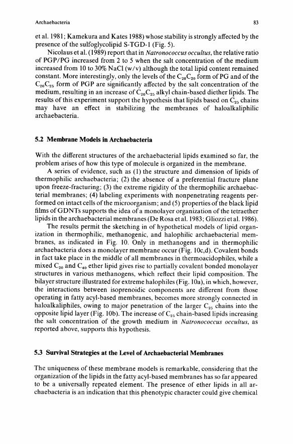
5.2 Membrane Models in Archaebacteria
With the different structures of the archae bacterial lipids examined so far, the problem arises of how this type of molecule is organized in the membrane.
A series of evidence, such as (1) the structure and dimension of lipids of thermophilic archaebacteria; (2) the absence of a preferential fracture plane upon freeze-fracturing; (3) the extreme rigidity of the thermophilic archaebacterial membranes; (4) labeling experiments with nonpenetrating reagents performed on intact cells of the microorganism; and (5) properties of the black lipid films of GDNTs supports the idea of a monolayer organization of the tetraether lipids in the archaebacterial membranes (De Rosa et al. 1983; Gliozzi et al. 1986).
The results permit the sketching in of hypothetical models of lipid organization in thermophilic, methanogenic, and halophilic archaebacterial membranes, as indicated in Fig. 10. Only in methanogens and in thermophilic archaebacteria does a monolayer membrane occur (Fig. lOc,d). Covalent bonds in fact take place in the middle of all membranes in thermoacidophiles, while a mixed C20 and C40 ether lipid gives rise to partially covalent bonded monolayer structures in various methanogens, which reflect their lipid composition. The bilayer structure illustrated for extreme halophiles (Fig. lOa), in which, however, the interactions between isoprenoidic components are different from those operating in fatty acyl-based membranes, becomes more strongly connected in haloalkaliphiles, owing to major penetration of the larger C25 chains into the opposite lipid layer (Fig. lOb). The increase of C25 chain-based lipids increasing the salt concentration of the growth medium in Natronococcus occu!tus, as reported above, supports this hypothesis.
5.3 Survival Strategies at the Level of Archaebacterial Membranes
The uniqueness of these membrane models is remarkable, considering that the organization of the lipids in the fatty acyl-based membranes has so far appeared to be a universally repeated element. The presence of ether lipids in all archaebacteria is an indication that this phenotypic character could give chemical

84 M. De Rosa et al.
a b
d
Fig. IOa-d. A sketch of membrane models in archaebacteria. a extreme halophiles; b alkalophilic halophiles; c methanogens; d thermoacidophiles
stability to the membrane in extreme environments. However, the comparison of properties of phospholipids containing ester links with their ether-linked analogues indicates that ether linkage has only a small effect on phospholipid packing (Paltauf 1983).
Different strategies have been adopted in different phenotypes of archaebacteria to control the compactness of the membrane, ensuring its optimal functionality in response to environmental stresses. In the alkaliphiles, which must cope with dual stress of high salt concentration and very high pH, an efficient control of the optimal membrane fluidity could be achieved by the extensive use of a C25 alkyl chain-based lipid, an alternative to those based on C20
alkyl chains. In a natural environment, in which water is close to the boiling point, a
biological membrane based on a lipidic monolayer represents one of the possible strategies to limit lipid mobility, maintaining the fluidity of the system at values compatible with the biological processes. In the classic membranes (Fig. II), monopolar lipids move into the layer to which they belong. This movement, temperature-dependent, conditions the activity ofthe membrane in a critical way. In a thermal environment, only the use of a bipolar lipid anchored on two sides

Archae bacteria
bilayer
monolayer
85
Fig. 11. Degree of freedom of monopolar and bipolar lipids in biological membranes. The arrows indicate the movement oflipids along the membrane
of the lipid monolayer permits the control of the lateral creep of the lipids (Fig. 10). Actually, in this manner the mobility of each lipid becomes very limited, because each one of them translates only as much as is permitted by the resultant forces acting on both anchorages, which generally have a low probability of developing in the same direction.
Acknowledgment. The authors thank Mr. Raffaele Turco for artwork.
References
Com ita PB, Gagosian RB, Pang H, Costello CE (1984) Structural elucidation of a unique macrocyclic membrane lipid from a new extremely thermophilic, deep-sea hydrothermal ventarchaebacterium Methanococcus jannaschii. J Bioi Chern 259: 15234-15241
De Rosa M, Gambacorta A (1986) Lipid biogenesis in archae bacteria. In: Kandler 0, Zillig W (eds) Archaebacteria '85. Fischer, Stuttgart, pp 278
De Rosa M, Gambacorta A (1988) The lipids of archaebacteria. Prog Lipid Res 27:153-175 De Rosa M, Gambacorta A, Nicolaus B (l980a) Regularity of isoprenoid biosynthesis in the ether
lipids of archae bacteria. Phytochemistry 19:791-793 De Rosa M, Gambacorta A, Nicolaus B, Sodano S, Bu' Lock JD (l980b) Structural regularities in
tetraether lipids of Caldariella and their biosynthetic and phyletic implications. Phytochemistry 19:833-836
De Rosa M, Gambacorta A, Nicolaus B, Sodano S (1982) Incorporation oflabelled glycerols into ether lipid in Caldariella aCidophiia. Phytochemistry 21: 595-599
De Rosa M, Gambacorta A, Nicolaus B, Chappe B, Albrecht P (1983) Isoprenoid ethers backbone of complex lipids of the archae bacterium Sulfolobus solfataricus. Biochim Biophys Acta 753:249-256

86 M. De Rosa et a1.
De Rosa M, Gambacorta A, Gliozzi A (1986a) Structure, biosynthesis and physicochemical properties of archae bacterial lipids. Microbiol Rev 50:70-80
De Rosa M, Gambacorta A, Lanzotti V, Trincone A, Harris JE, Grant WD (l986b) A range of ether core lipids from the methanogenic archae bacterium Methanosarcina barkeri. Biochim Biophys Acta 875:487-492
De Rosa M, Gambacorta A, Trincone A, Basso A, Zillig W, Holz I (1987) Lipids of Thermococcus celer, a sulfur-reducing archaebacterium: structure and biosynthesis. Syst Appl MicrobioI9:1-5
De Rosa M, Gambacorta A, Grant WD, Lanzotti V, Nicolaus B (1988) Polarlipids and glycine betaine from haloalkaliphilic archaebacteria. J Gen Microbiol 134:205-211
De Rosa M, Lanzotti V, Nicolaus B, Trincone A, Gambacorta A (1989) Lipids of archaebacteria: structural and biosynthetic aspects. In: Costa MS, Duarte JC, Williams RAD (eds) The microbiology of extreme environments and its potential for biotechnology. Elsevier Applied Science, London, pp 131
Ekiel I, Mash D, Smallbone BW, Kates M, Smith ICP (1981) The state of the lipids in the purple membrane of Halobacterium cutirubrum as seen by 31 P NMR. Biochem Biophys Res Commun 100: 105-113
Ekiel I, Sprott GD, Smith ICP (1986) Mevalonic acid is partially synthesized from aminoacids in Halobacterium cutirubrum: a 13C nuclear magnetic resonance study. J BacterioI166:559-564
Ferrante G, Ekiel I, Sprott DJ (1986) Structural characterization of the lipids of Methanococcus voltae including a novel N-acetylglucosamine I-P diether. J BioI Chern 36: 17062-17066
Ferrante G, Ekiel I, Sprott JD (1987) Structures of diether lipids of Methanospirillum hungatei containing novel head groups N,N-dimethylamino and N,N,N-dimethylaminopentane tetro1. Biochim Biophys Acta 921 :281-291
Ferrante G, Ekiel I, Girischandra BP, Sprott DJ (1988a) A novel core lipid isolated from the aceticlastic methanogen Methanothrix concilii GP6. Biochem Biophys Acta 963: 173-182
Ferrante G, Ekiel I, Girischandra BP, Sprott DJ (1988b) Structure of the major polar lipids isolated from the aceticlastic methanogen, Methanothrix concilii GP6. Biochim Biophys Acta 963:162-172
Fredrickson HL, Leeuw JW, Tas AC, van der Greef J, Lavos GF, Boon JJ (1989) Fast atom bombardment (tandem) mass spectrometric analysis of intact polar ether lipids extractable from the extremely halophilic archaebacterium Halobacterium cutirubrum. Biomed Mass Spectrom 18:96-105
Gliozzi A, Paoli G, De Rosa M, Gambacorta A (1983) Effect of isoprenoid cyclization on the transition temperature oflipids in thermophilic archaebacteria. Biochim Biophys Acta 735:234-242
Gliozzi A, Bruno S, Basak TK, De Rosa M, Gambacorta A (1986) Organization and dynamics of bipolar lipids from Sul{olobus sol{ataricus in bulk phases and in monolayer membranes. In: Kandler 0, Zillig W (eds) Archaebacteria' 85. Fischer, Stuttgart, pp 266
Grant WD, Larsen H (1989) Extremely halophilic archaebacteria. In: Staley JT, Bryant MP, Pfennig N, Holt JG (eds) Bergey's manual of systematic bacteriology, vol 3, Williams and Wilkins, London, pp 2216
Huber G, SpinIer C, Gambacorta A, Stetter KO (1989) Methallosphaera sedula sp. nov. represent a new genus of aerobic, metal-mobilizing, thermophilic archaebacteria. Syst Appl Microbiol 12:38-47
Kakinuma K, Yamagishi M, Fujimoto Y, Ikekawa N, Oshima T (1988) Stereochemistry of the biosynthesis of sn-2,3-0-diphytanyl glycerol, membrane lipid of archae bacterium Halobacterium halobium. J Am Chern Soc 110:4861-4863
Kamekura M, Kates M (1988) Lipids of halophilic archaebacteria. In: Rodriguez-Valera F (ed) Halophilic bacteria, vol II. CRC Press, Boca Raton, Florida, pp 25
Kates M, Kushwaha SC (1978) Biochemistry ofthe lipids of extremely halophilic bacteria. In: Caplan SR, Ginzburg M (eds) Energetics and structure of halophilic microorganisms. Elsevier North Holland Biomedical Press, Amsterdam, pp 461
Koga Y, Ohga M, Nishihara M, Morii H (1987) Distribution of a diphytanyl ether analog of phosphatidylserine and an ethanolamine-containing tetraether lipid methanogenic bacteria. Syst Appl MicrobioI9:176-l82
Konig H (1988) Archaebacteria. In: Rehm HJ (ed) Biotechnology, vol6. Verlag Chemie, Basel, pp 697 Kramer JKG, Saver FD, Blackwell BA (1987) Structure of the two new aminophospholipids from
Methanobacterium thermoautotrophicum. Biochem J 245:139-143

Archaebacteria 87
Kushwaha SC, Kates M, Sprott JD, Smith ICP (1981) Novel polar lipids from the methanogen Methanospirillum hungatei GPI. Biochim Biophys Acta 664: 156-173
Kushwaha SC, Juez Perez G, Rodriguez-Valera F, Kates M, Kushner OJ (1982) Survey oflipids of new group of extremely halophilic bacteria from salt ponds in Spain. Can J Microbiol28: 1365-1373
Langworthy TA (1979) Special features of Thermoplasma. In: Barile MF, Racin R (eds) The mycoplasma. Academic Press, New York, pp 495
Langworthy T A (1985) Lipids of Archaebacteria. In: Woese C, Wolfe RS (eds) The bacteria. Gustav Fischer, Stuttgart, pp 459
Langworthy TA, Pond JL (1986) Archaebacterial ether lipids and chemotaxonomy. In: Kandler 0, Zillig W (eds) Archaebacteria '85. Gustav Fischer, Stuttgart, pp 253
Lanzotti V, De Rosa M, Trincone A, Basso A, Gambacorta A, Zillig W (1987) Complex lipids from Desul[urococcus mobilis, a sulfur reducing archaebacterium. Biochim Biophys Acta 922:95-102
Lanzotti V, Nicolaus B, Trincone A, De Rosa M, Grant WD, Gambacorta A (l989a) A complex lipids with a cyclic phosphate from the archaebacterium Natronococcus occultus. Biochim Biophys Acta 1001:31-34
Lanzotti V, Trincone A, Nicolaus B, Zillig W, De Rosa M, Gambacorta A (1989b) Complex lipids of Pyrococcus and ANI, thermophilic members of archae bacteria belonging to Thermococcales. Biochim Biophys Acta 1004:44-48
Lanzotti V, Nicolaus B, Trincone A, De Rosa M, Grant WD, Gambacorta A (1989c) An isopranoid ether analogue of phosphatidic acid from a halophilic archaebacteria. Biochim Biophys Acta 1002:398-400
Luzzati V, Gambacorta A, De Rosa M, Gulik A (1987) Polar lipid of thermophilic prokaryotic organisms chemical and physical structure. In: Engelman OM, Rantez CR, Pollard TO (eds) Annual review of biophysics and biophysical chemistry 16. Annual Review Inc, Palo Alto, CA, p 25
Moldoveanu N, Kates M (1988) Biosynthetic studies of the polar lipids of Halobacterium cutirubrum formation of isoprenyl ether intermediates. Biochim Biophys Acta 960: 164-182
Morii H, Nishihara M, Ohga M, Koga Y (1986) A diphytanyl ether analog of phosphat idyl serine from methanogenic bacterium, Methanobrevibacter arboriphilus. J Lipid Res 27:724-730
Nicolaus B, Lanzotti V, Trincone A, De Rosa M, Grant WD, Gambacorta A (1989) Glycine-betaine and polar lipid composition in halophilic archae bacteria in response to growth in different salt concentration. FEMS Microbiol Lett 59:157-160
Nishihara M, Koga Y (1987) Extraction and composition of polar lipids from the archaebacterium, Methanobacterium thermoautotrophicum: effective extraction of tetraether lipids by an acidified solvent. J Biochem 10 I : 997 -1009
Nishihara M, Morii H, Koga Y (1989) Heptads of polar ether lipids of an archaebacterium Methanobacterium thermoautotrophicum: structure and biosynthetic relationship. Biochemistry 28:95-102
PaltaufF (1983) Ether lipids in biological and model membranes. In: Mangold HK, PaltaufF (eds) Ether lipids: biochemical and biomedical aspects. Academic Press, New York, pp 309
Poulter CD, Aoki T, Daniels L (1988) Biosynthesis of isoprenoid membranes in the methanogenic archaebacterium Methanospirillum hungatei. J Am Chern Soc 110:2620-2624
Thurl S, Schafer W (1988) Lipids from the sulfur dependent archaebacterium Thermoproteus tenax. Biochim Biophys Acta 961 :233-238
Torreblanca M, Rodriguez-Valera F, Juez G, Ventosa A, Kamekura M, Kates M (1986) Classification of non-alkaliphilic halobacteria based on numerical taxonomy and polar lipid composition and description of Haloarcula gen. nov. and Halo[erax gen. nov. Syst Appl Microbiol 8:89-99
Trincone A, Gambacorta A, De Rosa M, Scolastico C, Sydimov A, Potenza 0 (1989a) Mechanism of cyclopentane ring formation in tetraether lipids of Sul[olobus sol[ataricus. In: Da Costa MS, Duarte JC, Williams RAD (eds) Microbiology of extreme environments and the potential for biotechnology. Elsevier Applied Science, London, pp 180
Trincone A, Lanzotti V, Nicolaus B, Zillig W, De Rosa M, Gambacorta A (1989b) Comparative lipid composition of aerobically and anaerobically grown Desul[urolobus ambivalens an autotrophic thermophilic archaebacterium. J Gen Microbiol 135:2751-2757
Tsujimoto K, Y orimitsu S, Takahasi T, Ohashi M (1989) Revised structure ofa phospholipid obtained from Halobacterium halobium. Chern Commun 668-670
Woese CR (1987) Bacterial evolution. Microbiol Rev 51 :221-271