arabidopsis peroxisomes possess functionally redundant
TRANSCRIPT
Arabidopsis peroxisomes possess functionally redundantmembrane and matrix isoforms of monodehydroascorbatereductase
Cayle S. Lisenbee†,‡, Matthew J. Lingard‡ and Richard N. Trelease*
School of Life Sciences and Graduate Program in Molecular and Cellular Biology, PO Box 874501, Arizona State University,
Tempe, AZ 85287, USA
Received 22 April 2005; revised 23 June 2005; accepted 27 June 2005.*For correspondence: (fax þ1 480 965 6899; e-mail [email protected]).†Present address: Cancer Center, Mayo Clinic in Scottsdale, Scottsdale, AZ 85259, USA.‡These authors contributed equally to this study.
Summary
The H2O2 byproduct of fatty acid catabolism in plant peroxisomes is removed in part by a membrane-
associated antioxidant system that involves both an ascorbate peroxidase and a monodehydroascorbate
reductase (MDAR). Despite descriptions of 32-kDa MDAR polypeptides in pea and castor peroxisomal
membranes and cDNA sequences for several ‘cytosolic’ MDARs, the genetic and protein factors responsible for
peroxisomal MDAR function have yet to be elucidated. Of the six MDAR polypeptides in the Arabidopsis
proteome, named AtMDAR1 to AtMDAR6 in this study, 47-kDa AtMDAR1 and 54-kDa AtMDAR4 possess amino
acid sequences that resemble matrix (PTS1) and membrane peroxisomal targeting signals, respectively.
Epitope-tagged versions of these two MDARs and a pea 47-kDa MDAR (PsMDAR) sorted in vivo directly from
the cytosol to peroxisomes in Arabidopsis and BY-2 suspension cells, whereas AtMDAR2 and AtMDAR3
accumulated in the cytosol. The PTS1-dependent sorting of AtMDAR1 and PsMDAR to peroxisomes was
incomplete (inefficient?), but was improved for PsMDAR after changing its PTS1 sequence from –SKI to the
canonical tripeptide –SKL. A C-terminal transmembrane domain and basic cluster of AtMDAR4 were necessary
and sufficient for targeting directly to peroxisomes. MDAR activity in isolated Arabidopsis peroxisomes was
distributed among both water-soluble matrix and KCl-insoluble membrane subfractions that contained
respectively 47- and 54-kDa MDAR polypeptides. Notably, a 32-kDa MDAR was not identified. Combined with
membrane association and topological orientation findings, these results indicate that ascorbate recycling in
Arabidopsis (and probably other plant) peroxisomes is coordinated through functionally redundant MDARs
that reside in the membrane and the matrix of the organelle.
Keywords: Arabidopsis, monodehydroascorbate reductase, peroxisome, peroxisomal targeting signal, react-
ive oxygen species, tobacco BY-2 cell.
Introduction
One function of plant peroxisomes is the removal of toxic
reactive oxygen species, such as H2O2, that are produced
during the oxidative metabolism that takes place in the
matrix of the organelle. A portion of toxic H2O2 is removed
by a cooperative pair of ascorbate-dependent electron
transfer enzymes located at the peroxisomal membrane
(Corpas et al., 2001; Donaldson, 2002; del Rıo et al., 2002).
Specifically, ascorbate peroxidase (APX) initiates electron
transfer from two molecules of ascorbate to convert H2O2
into water. This reaction produces two molecules of
monodehydroascorbate (ascorbate free radical) which can
be recycled immediately to reduced ascorbate by monode-
hydroascorbate reductase (MDAR) via electron transfer from
NADH. Alternatively, monodehydroascorbate may dispro-
portionate spontaneously to fully oxidized dehydroascor-
bate, which is reduced back to ascorbate by the action of a
glutathione-dependent dehydroascorbate reductase
(Jimenez et al., 1997; del Rıo et al., 1998). The NADH-
dependent dehydrogenase activity of MDAR has been sug-
gested as an important mechanism for the regeneration of
900 ª 2005 Blackwell Publishing Ltd
The Plant Journal (2005) 43, 900–914 doi: 10.1111/j.1365-313X.2005.02503.x
NAD to support the b-oxidation of fatty acids in oilseed
glyoxysomes (specialized peroxisomes) (Donaldson, 2002;
Mullen and Trelease, 1996).
Compared with its peroxisomal APX counterpart, MDAR
is a much less characterized component of the peroxisomal
membrane in plants. The association of MDAR catalytic
activity with glyoxysomal or peroxisomal membranes has
been reported by several groups studying different plant
species (Bowditch and Donaldson, 1990; Bunkelmann and
Trelease, 1996; Ishikawa et al., 1998; Jimenez et al., 1997;
Karyotou and Donaldson, 2005; Lopez-Huertas et al., 1999;
Mittova et al., 2000). However, the specific peroxisomal
membrane protein(s) (PMPs) responsible for this activity has
been identified in only a few cases. For instance, in the
membranes of castor glyoxysomes Luster et al. (1988)
identified an integral 32-kDa polypeptide that exhibited
NADH:ferricyanide reductase activity, which was found later
to be responsible for the observed NADH-dependent reduc-
tion of monodehydroascorbate (Bowditch and Donaldson,
1990). Lopez-Huertas et al. (1999) provided more convincing
evidence for a similar 32-kDa PMP in pea leaf peroxisomes
by showing that this PMP had ferricyanide reductase activity
andwas recognized by antibodies raised against a cucumber
MDAR. Interestingly, these same antibodies were shown
very recently to recognize a 47-kDa castor polypeptide that
was associated with MDAR enzymatic activity in alkaline
carbonate washed glyoxysomal membranes (Karyotou and
Donaldson, 2005).
Despite this biochemical evidence, a gene coding for a
peroxisomal membrane-bound MDAR has yet to be identi-
fied and described. Genes coding for putative matrix
peroxisomal MDAR polypeptides have been cloned from
pea (Murthy and Zilinskas, 1994), cucumber (Sano and
Asada, 1994), tomato (Grantz et al., 1995) and Chinese
cabbage (Yoon et al., 2004), and databases include reports
of similar cDNAs/genes in rice (GenBank accession number
D85764), broccoli (AB125637), iceplant (AJ301553) and Ara-
bidopsis (AGI Locus At3g52880). The primary amino acid
sequences of all these predicted MDARs include a type 1
matrix peroxisomal targeting signal (PTS1) that consists of a
C-terminal tripeptide that resembles the canonical –SKL
motif (Mullen, 2002; Olsen and Harada, 1995). Curiously,
none of these MDARs have been evaluated for subcellular
localization, nor have any of their C-terminal sequences
been shown to function as a PTS.
Nearly all peroxisomal proteins are synthesized on cyto-
solic polyribosomes and then post-translationally targeted
to pre-existing and/or differentiating organelles. In addition
to the PTS1 mentioned above, many PMPs are targeted to
the peroxisomal membrane by a less well-characterized
membrane PTS (mPTS) that consists of a hydrophobic
transmembrane domain (TMD) and an adjacent cluster of
basic amino acids (Dyer et al., 1996; Hunt and Trelease, 2004;
Jones et al., 2001; Mullen and Trelease, 2000; Wang et al.,
2001). Coupled with the lack of secretory pathway targeting
determinants, the presence of any PTS predicts that nascent
matrix polypeptides or PMPs sort directly from their site of
synthesis in the cytosol to peroxisomes. This direct pathway
has been demonstrated for yeast PMP47 (Dyer et al., 1996)
and Arabidopsis peroxin 3 (Hunt and Trelease, 2004), both of
which possess a mPTS. However, recent findings suggest
that at least some PMPs follow an indirect sorting pathway
that includes the endoplasmic reticulum (ER). For example,
peroxisomal APX and Arabidopsis peroxin 16 appear in a
subdomain(s) of rough ER before reaching the peroxisomal
membrane in Arabidopsis and BY-2 cultured cells (Karnik
and Trelease, 2005; Lisenbee et al., 2003a,b; Mullen et al.,
1999, 2001). Considering the cooperative functional roles of
MDAR and APX, it is interesting that intracellular sorting
pathways have not been elucidated for any of the MDARs
identified previously.
Our goal in the present study was to establish a much-
needed link between the genetic and protein components
responsible for peroxisomal MDAR activity in plants. Obara
et al. (2002) predicted the existence of seven MDAR poly-
peptides in Arabidopsis from as many then-available gene
and cDNA sequences. In that work, they characterized two of
the polypeptides as chloroplast and mitochondrial isoforms
that were coded from a single alternatively transcribed open
reading frame (ORF). Subsequent sequence updates indi-
cate that this ORF resides within one of five MDAR loci in
Arabidopsis, from which only six MDAR polypeptides may
be predicted (assuming no other instances of alternative
gene regulation). Two of the four uncharacterized MDARs
are predicted 47- and 54-kDa polypeptides that possess a
PTS1 and an mPTS, respectively. We cloned all four
uncharacterized Arabidopsis MDARs and found from in vivo
immunofluorescence sorting and targeting analyses that
both of the putative peroxisomal MDARs sorted directly to
Arabidopsis and BY-2 suspension cell peroxisomes in a PTS-
dependentmanner. These protein products corresponded to
endogenous 47- and 54-kDa Arabidopsis polypeptides that
in biochemical assays were associated both structurally and
functionally with the peroxisomal matrix and membrane,
respectively. The details of these findings with respect to
enzyme function, protein sorting, organelle association and
topology now provide a more complete foundation upon
whichmodels of peroxisomal ascorbatemetabolismmay be
tested.
Results
Identification of AtMDAR1 and AtMDAR4 as representative
matrix- and membrane-localized peroxisomal MDARs
For the purposes of this study, Arabidopsis gene loci are
referred to by their AGI names and the proteins by the given
names AtMDAR1 to AtMDAR6. AtMDAR1 (At3g52880)
Peroxisomal monodehydroascorbate reductases in Arabidopsis 901
ª Blackwell Publishing Ltd, The Plant Journal, (2005), 43, 900–914

possesses a C-terminal –AKI tripeptide that resembles the
PTS1 signal for directing proteins to the peroxisomal matrix.
Similarly, AtMDAR4 (At3g27820) contains within a unique
C-terminal extension mPTS-like sequences that comprise a
predicted TMD followed immediately by five basic arginine
residues. Neither AtMDAR2 (At5g03630) nor AtMDAR3
(At3g09940) include sequence features that predict specific
subcellular (organellar) localizations. AtMDAR5 and AtM-
DAR6 (At1g63940) are the respective mitochondrial and
chloroplast MDARs that have been characterized previously
(Obara et al., 2002).
We searched the literature and protein sequence databas-
es for PTS-containing plant MDAR homologs with the goal
of identifying sequences that would validate the predicted
peroxisomal localizations and MDAR functions of AtMDAR1
and AtMDAR4. The results are listed in Table 1 and are
arranged in ascending order according to mass. Most of the
sequences corresponded to MDAR polypeptides that pos-
sessed a C-terminal PTS1 signal. All these sequences had
predicted masses of approximately 47 kDa and possessed
the NADP/FAD binding domains that are typical of flavopro-
teins such as MDAR that belong to the pyridine nucleotide–
disulphide oxidoreductase family (Pfam accession number
PF00070). In comparison, AtMDAR1 is at least 76% identical
to any one of the seven other PTS1-containing MDARs listed
in Table 1 (alignment not shown). AtMDAR1 also contains
three sequence motifs that correspond to the NADP/FAD
binding domains typified by the cucumber and pea poly-
peptides, both of which have been purified and shown to
exhibit NADH-dependent MDAR activity in vitro (Murthy
and Zilinskas, 1994; Sano et al., 1995). Furthermore, the
C-terminal –AKI tripeptide of AtMDAR1 matches the PTS1-
like motif –(S/A)K(I/V) (Mullen, 2002) that is shared among
this group of 47-kDa MDARs. The presence of functional
domains responsible for MDAR catalytic activity and for
targeting to peroxisomes supports the assignment of AtM-
DAR1 as an authentic peroxisomal matrix MDAR.
The biochemical and sequence data compiled in Table 1
indicated the existence of at least one other group of
peroxisomal MDARs. This group consisted of four predicted
approximately 52–54 kDa polypeptides that contained puta-
tive TMD regions at both ends of the proteins. AtMDAR4 is
70% identical to any one of the other group members
(alignment not shown), all of which possess the three NADP/
FAD binding motifs of the 47-kDa cucumber MDAR that also
were identified in AtMDAR1. The C-terminal-most TMD of
AtMDAR4 is defined on each end by conserved proline and
glycine residues and is situated adjacent to a tryptophan-
capped basic cluster –R(K/R)RRR that is shared by all five
polypeptides (including an incomplete sequence from
Table 1 Evidence for (putative) peroxisomal matrix and membrane MDAR proteins
OrganismPredictedsize (kDa)
SDS-PAGEsize (kDa)
(Predicted)location
GenBank no./AGI locus Reference
Pisum sativum n.d. 32c Membrane n.a. Lopez-Huertas et al. (1999)Ricinus communis n.d. 32d Membrane n.a. Luster et al. (1988)Hordeum vulgare 38.9a n.d. Membrane CAC69935 UnpublishedGlycine max n.d. 39/40e Unknown n.a. Dalton et al. (1992)Arabidopsis thaliana 46.5 47f Matrix At3g52880 This studyBrassica oleracea 46.5 n.d. Matrix BAD14934 UnpublishedBrassica campestris 46.5 n.d. Matrix AAK72107 Yoon et al. (2004)Oryza sativa 46.6 n.d. Matrix BAA77214 UnpublishedMesembryanthemum crystallinum 47.0b n.d. Matrix CAC82727 UnpublishedLycopersicon esculentum 47.0 n.d. Matrix AAC41654 Grantz et al. (1995)Ricinus communis n.d. 47g Membrane n.a. Karyotou and Donaldson (2005)Pisum sativum 47.3 47h Matrix AAA60979 Murthy and Zilinskas (1994); this studyCucumis sativus 47.4 47i,j Matrix BAA05408 Hossain and Asada (1985); Sano et al. (1995)Oryza sativa 51.9 n.d. Membrane AAL87166 UnpublishedZea mays 52.0 n.d. Membrane AY106646 Gardiner et al. (2004)Triticum aestivum 52.2 n.d. Membrane BT009417 UnpublishedArabidopsis thaliana 53.5 54k Membrane At3g27820 This study
Publicly available literature and sequence databases were searched for data supporting the existence of MDAR-like gene(s) and protein(s) affiliatedwith the ascorbate metabolism of plant peroxisomes. Listed in ascending order according to size are only those candidates for which the availableexperimental and/or sequence data strongly support peroxisomal functions. For MDAR-like sequences that have yet to be verified experimentally,the criteria for inclusion were the presence of amino acid sequences that resembled matrix or membrane peroxisomal targeting signals. Predictedsizes of polypeptides were calculated with the SAPS algorithm (Brendel et al., 1992); aincomplete sequence, bportion of sequence homologous tomatrix MDAR proteins. SDS-PAGE sizes, as reported in the references listed, were derived from the following sources: cimmunoblot, P. sativumleaf PMPs; dprotein stain, R. communis glyoxysomal membrane extracts; eprotein stain, purified proteins from G. max root nodule extracts;fimmunoblot, A. thaliana suspension cell peroxisomal matrix fractions; gimmunoblot, R. communis glyoxysomal membrane fractions; hproteinstain, E. coli expressed proteins; iprotein stain, purified proteins from C. sativus fruit extracts; jimmunoblot, E. coli expressed proteins;kimmunoblot, A. thaliana suspension cell peroxisomal membrane fractions. n.d., not determined; n.a., not available.
902 Cayle S. Lisenbee et al.
ª Blackwell Publishing Ltd, The Plant Journal, (2005), 43, 900–914
barley). Together the TMD and basic cluster form a C-
terminal mPTS, neither component of which was found in
any of the 47-kDa MDAR sequences. These comparisons
suggest the classification of AtMDAR4 as an authentic
peroxisomal membrane MDAR, and when combined with
those of the 47-kDa group reveal the presence of two
predominant MDAR isoforms in plant peroxisomes that in
Arabidopsis are represented by AtMDAR1 and AtMDAR4.
Arabidopsis peroxisomes possess separate matrix and
membrane MDARs
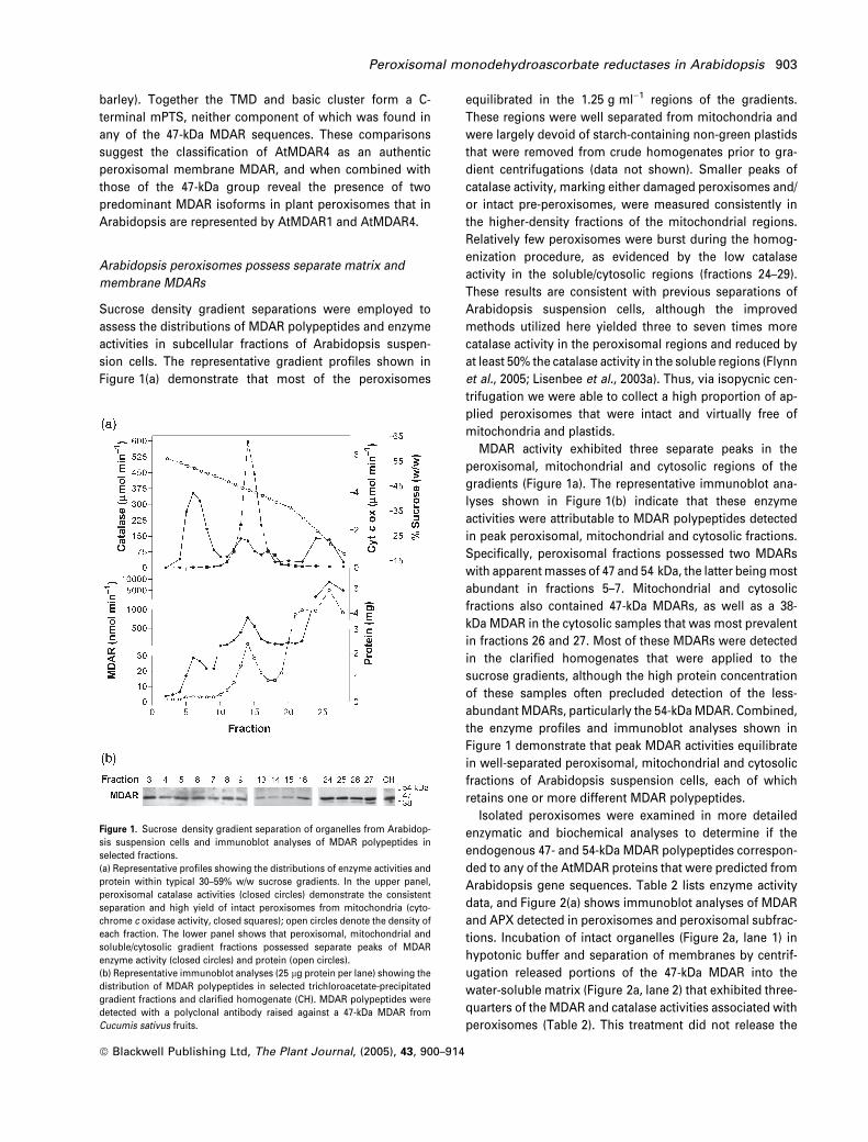
Sucrose density gradient separations were employed to
assess the distributions of MDAR polypeptides and enzyme
activities in subcellular fractions of Arabidopsis suspen-
sion cells. The representative gradient profiles shown in
Figure 1(a) demonstrate that most of the peroxisomes
equilibrated in the 1.25 g ml)1 regions of the gradients.
These regions were well separated from mitochondria and
were largely devoid of starch-containing non-green plastids
that were removed from crude homogenates prior to gra-
dient centrifugations (data not shown). Smaller peaks of
catalase activity, marking either damaged peroxisomes and/
or intact pre-peroxisomes, were measured consistently in
the higher-density fractions of the mitochondrial regions.
Relatively few peroxisomes were burst during the homog-
enization procedure, as evidenced by the low catalase
activity in the soluble/cytosolic regions (fractions 24–29).
These results are consistent with previous separations of
Arabidopsis suspension cells, although the improved
methods utilized here yielded three to seven times more
catalase activity in the peroxisomal regions and reduced by
at least 50% the catalase activity in the soluble regions (Flynn
et al., 2005; Lisenbee et al., 2003a). Thus, via isopycnic cen-
trifugation we were able to collect a high proportion of ap-
plied peroxisomes that were intact and virtually free of
mitochondria and plastids.
MDAR activity exhibited three separate peaks in the
peroxisomal, mitochondrial and cytosolic regions of the
gradients (Figure 1a). The representative immunoblot ana-
lyses shown in Figure 1(b) indicate that these enzyme
activities were attributable to MDAR polypeptides detected
in peak peroxisomal, mitochondrial and cytosolic fractions.
Specifically, peroxisomal fractions possessed two MDARs
with apparentmasses of 47 and 54 kDa, the latter beingmost
abundant in fractions 5–7. Mitochondrial and cytosolic
fractions also contained 47-kDa MDARs, as well as a 38-
kDa MDAR in the cytosolic samples that was most prevalent
in fractions 26 and 27. Most of these MDARs were detected
in the clarified homogenates that were applied to the
sucrose gradients, although the high protein concentration
of these samples often precluded detection of the less-
abundant MDARs, particularly the 54-kDaMDAR. Combined,
the enzyme profiles and immunoblot analyses shown in
Figure 1 demonstrate that peak MDAR activities equilibrate
in well-separated peroxisomal, mitochondrial and cytosolic
fractions of Arabidopsis suspension cells, each of which
retains one or more different MDAR polypeptides.
Isolated peroxisomes were examined in more detailed
enzymatic and biochemical analyses to determine if the
endogenous 47- and 54-kDa MDAR polypeptides correspon-
ded to any of the AtMDAR proteins that were predicted from
Arabidopsis gene sequences. Table 2 lists enzyme activity
data, and Figure 2(a) shows immunoblot analyses of MDAR
and APX detected in peroxisomes and peroxisomal subfrac-
tions. Incubation of intact organelles (Figure 2a, lane 1) in
hypotonic buffer and separation of membranes by centrif-
ugation released portions of the 47-kDa MDAR into the
water-soluble matrix (Figure 2a, lane 2) that exhibited three-
quarters of the MDAR and catalase activities associated with
peroxisomes (Table 2). This treatment did not release the
Figure 1. Sucrose density gradient separation of organelles from Arabidop-
sis suspension cells and immunoblot analyses of MDAR polypeptides in
selected fractions.
(a) Representative profiles showing the distributions of enzyme activities and
protein within typical 30–59% w/w sucrose gradients. In the upper panel,
peroxisomal catalase activities (closed circles) demonstrate the consistent
separation and high yield of intact peroxisomes from mitochondria (cyto-
chrome c oxidase activity, closed squares); open circles denote the density of
each fraction. The lower panel shows that peroxisomal, mitochondrial and
soluble/cytosolic gradient fractions possessed separate peaks of MDAR
enzyme activity (closed circles) and protein (open circles).
(b) Representative immunoblot analyses (25 lg protein per lane) showing the
distribution of MDAR polypeptides in selected trichloroacetate-precipitated
gradient fractions and clarified homogenate (CH). MDAR polypeptides were
detected with a polyclonal antibody raised against a 47-kDa MDAR from
Cucumis sativus fruits.
Peroxisomal monodehydroascorbate reductases in Arabidopsis 903
ª Blackwell Publishing Ltd, The Plant Journal, (2005), 43, 900–914
54-kDa MDAR or membrane-bound APX, and appreciable
amounts of MDAR activity were found repeatedly in the
resultingmembrane pellets. Subsequent incubation of these
membranes in KCl removed the remainder of the 47-kDa
MDAR (Figure 2a, lane 3), but not the 54-kDa MDAR or APX,
both of which remained associated with the KCl-insoluble
pellet (Figure 2a, lane 4) that received nearly all the mem-
brane-associated MDAR activity (Table 2). Cytosolic sam-
ples contained 38- and 47-kDa MDARs and the soluble form
of APX (Figure 2a, lane 5) that were detected with other
immunologically related proteins in the clarified homogen-
ates (Figure 2a, lane 6). Mitochondria isolated from the same
sucrose gradients exhibited 50 times more MDAR activity
than peroxisomes, most of which was associated with the
mitochondrial matrix, and not KCl-extracted membranes
that exhibited cytochrome c oxidase activity (Table 2).
Intact peroxisomes also were incubated with proteinase K
with or without Triton X-100 to elucidate the topological
orientation of the endogenous Arabidopsis MDARs. As
shown in Figure 2(b), all the MDAR polypeptides as well as
APX and catalase remained unchanged in control reactions
in which neither proteinase K or detergent was added (lane
1). Incubation of intact peroxisomes in protease alone (lane
2) resulted in the digestion of cytosolically oriented APX, but
not the 47- and 54-kDa MDAR proteins or catalase. However,
the latter three proteins were digested when the peroxi-
somes were treated first with Triton X-100 and then incuba-
ted in proteinase K (lane 3). Together, the in vitro results
presented in Table 2 and Figure 2 show that Arabidopsis
peroxisomes retain two unique MDAR polypeptides that are
distributed differentially among the membrane and matrix.
More specifically, the 54-kDaMDAR is an integral membrane
protein positioned on the matrix face of the membrane and,
like catalase, the 47-kDa MDAR is in the matrix. These
biochemical characteristics suggest that the endogenous
47- and 54-kDa peroxisomal MDARs are coded by the Arabi-
dopsis genesAtMDAR1 andAtMDAR4, respectively. Further-
more, the 47-kDa MDARs detected in mitochondria and the
cytosol are probably coded by AtMDAR5 and AtMDAR2/3.
Table 2 Enzyme activity and amount of protein in subfractions ofperoxisomes and mitochondria isolated in sucrose density gradi-ents
MDAR(nmol min)1)
Catalase(lmol min)1)
Cytochrome coxidase(lmol min)1)
Protein(mg)
Applied samplea 33000 14700 20.5 120Peroxisomesb 80.4 3500 0.12 1.50
Matrix 50.3 486 0 0.330Membrane 14.9 119 0 0.300KCl soluble 2.10 28.9 n.d. 0.040KCl insoluble 11.7 80.6 n.d. 0.110
Mitochondriac 5500 189 47.3 17.1Matrix 2010 115 0.165 6.20Membrane 310 44.4 16.0 5.85KCl soluble 200 50.0 0.01 0.93KCl insoluble 350 0 14.0 4.30
Cytosold 5380 583 0.24 39.1
Values are averages derived from two experiments. Each experimentutilized organelle fractions pooled from three separate sucrosegradients (e.g. Figure 1a): aclarified homogenate (1500 g superna-tant, 15 min) applied to the top of each 30–59% w/w sucrose gradientin a vTi 50 rotor tube (8.4 ml split equally among three gradients);bfractions 4–8; cfractions 13–15; dfractions 25–28. n.d., not deter-mined.
Figure 2. Suborganellar distribution, membrane association, and topologi-
cal orientation of MDAR polypeptides in Arabidopsis peroxisomes.
Proteins in Arabidopsis suspension cell peroxisomes (pooled from three
sucrose gradients, e.g. Figure 1) and peroxisomal subfractions were precipi-
tated in trichloroacetate, subjected to SDS-PAGE (50 lg protein per lane), and
then analyzed on blots for MDAR polypeptides that were detected with anti-
cucumber MDAR antibodies. Replicate immunoblots probed with anti-APX or
anti-catalase IgGs provided positive controls for membrane- and matrix-
localized proteins, respectively.
(a) Membrane association. Peroxisomes (lane 1) were subjected to hypotonic
burst in 25 mM HEPES-KOH (pH 7.5) and centrifuged to produce water-
solubilized protein supernatants (lane 2) and water-insoluble membrane
pellets. Peripheral membrane proteins were extracted from these pellets in
0.2 M KCl and extracts were centrifuged to generate a KCl-soluble supernatant
(lane 3) and KCl-insoluble pellet (lane 4). Cytosolic fractions (cleared of
membranes by centrifugation, lane 5) and clarified homogenate (CH) (sample
applied to the gradients, lane 6) also were examined.
(b) Membrane topology. Intact peroxisomes (lane 1) were treated with
proteinase K without (-) and with (þ) presolubilization in Triton X-100 (lanes 2
and 3, respectively).
904 Cayle S. Lisenbee et al.
ª Blackwell Publishing Ltd, The Plant Journal, (2005), 43, 900–914

AtMDAR1 sorts directly to peroxisomes by means of its
PTS1 signal
The results of our sequence analyses matched those of our
biochemical examinations in predicting the existence of
separate 47- and 54-kDa polypeptides, namely AtMDAR1
and AtMDAR4, which reside in the matrix and membrane of
Arabidopsis peroxisomes. To confirm/refute these findings
we cloned, epitope-tagged, and then overexpressed transi-
ently AtMDAR1–AtMDAR4 (Table 3) for in vivo localization
in Arabidopsis and tobacco BY-2 suspension cells.
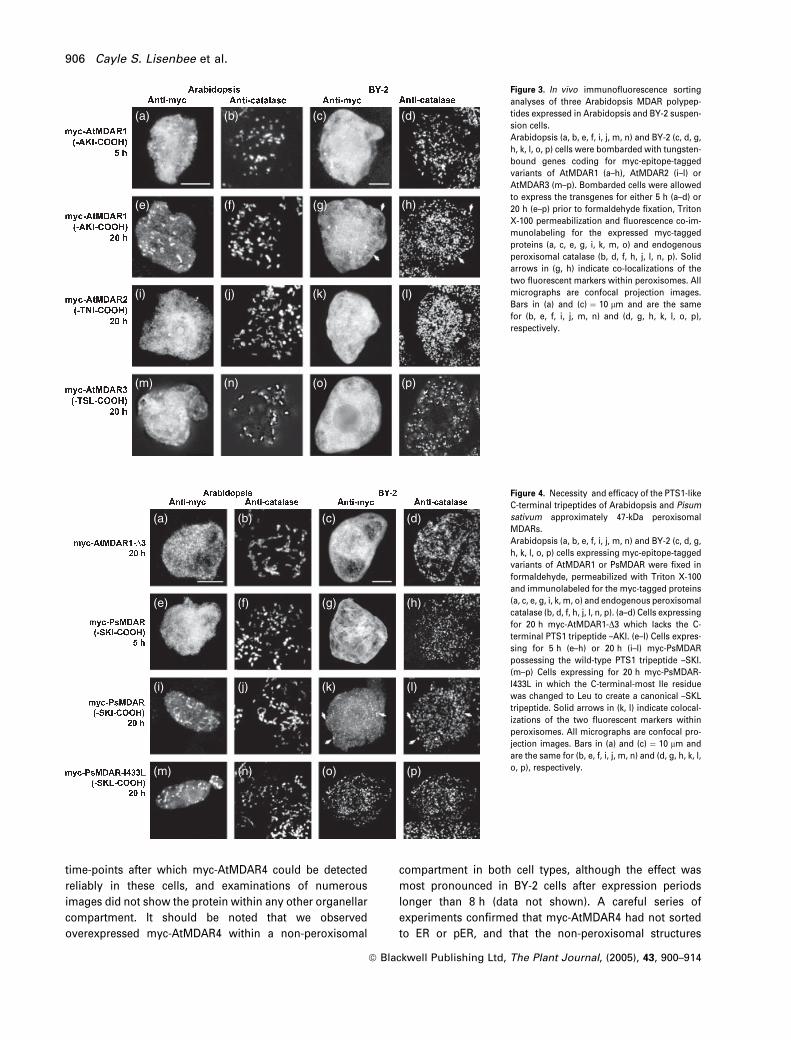
Figure 3(a–h) shows the results of in vivo sorting analyses
of transiently expressed myc-AtMDAR1. After 5 h of tran-
sient gene expression, application of anti-myc primary and
fluorophore-conjugated secondary antibodies revealed that
nearly all the overexpressed myc-AtMDAR1 was localized
in the cytosol of either Arabidopsis (Figure 3a) or BY-2
(Figure 3c) cells. Curiously, after 20 h transgene expression
most of themyc-AtMDAR1 had localized in Arabidopsis cells
to peroxisomes that also were marked with anti-catalase
antibodies (Figure 3e,f). Similar results were obtained with
BY-2 cells subjected to the same 20-h expression period
(Figure 3g,h), although the relative amount of expressed
myc-AtMDAR1 localized to peroxisomes wasmuch less than
in Arabidopsis cells. Multiple labeling experiments with
various organelle-specific markers were unable to detect
myc-AtMDAR1 in compartments other than the cytosol or
peroxisomes, and allowing cells to express the transgenes
for longer periods did not change the peroxisomal sorting
seen at 20 h (data not shown). Partial localization to peroxi-
someswas not observed in experiments withmyc-AtMDAR2
(Figure 3i–l) or myc-AtMDAR3 (Figure 3m–p), both of which
were detected only in the cytosol in both cell types after 20 h
expression. Cumulatively, these results demonstrate that
AtMDAR1 sorts directly to Arabidopsis and BY-2 cell peroxi-
somes, whereas AtMDAR2 and AtMDAR3 remain and prob-
ably function in the cytosol.
The sorting characteristics of AtMDAR1 prompted us to
analyze more closely the protein’s putative PTS1 tripeptide.
Figure 4(a–d) shows that removal of the –AKI tripeptide from
the C terminus of AtMDAR1 (Table 3) abolishes its sorting to
Arabidopsis and BY-2 cell peroxisomes after 20 h expres-
sion. This finding confirms the necessity of these three
residues for peroxisomal targeting, and suggests that the
analogous residues of the other seven 47-kDa MDARs listed
in Table 1 also confer localization to peroxisomes. We tested
this hypothesis with a partially characterized MDAR from
pea and found that the results of similar in vivo sorting
analyses mimicked those of AtMDAR1. More specifically,
after 5 h expression nearly all the myc-PsMDAR was detec-
ted in the cytosol of Arabidopsis and BY-2 cells (Figure
4e–h), whereas after 20 h expression a portion of myc-
PsMDAR was detected in peroxisomes (Figure 4i–l). We
were able to increase the sorting efficiency of this MDAR,
particularly in BY-2 cells, by changing the C-terminal tripep-
tide from-SKI to the more canonical-SKL. In both cell types,
Figure 4(m–p) shows very little myc-PsMDAR (expressed for
20 h) in the cytosol and nearly perfect colocalization with
endogenous catalase.
AtMDAR4 sorts directly to peroxisomal membranes and is
inserted such that its N terminus is exposed to the cytosol
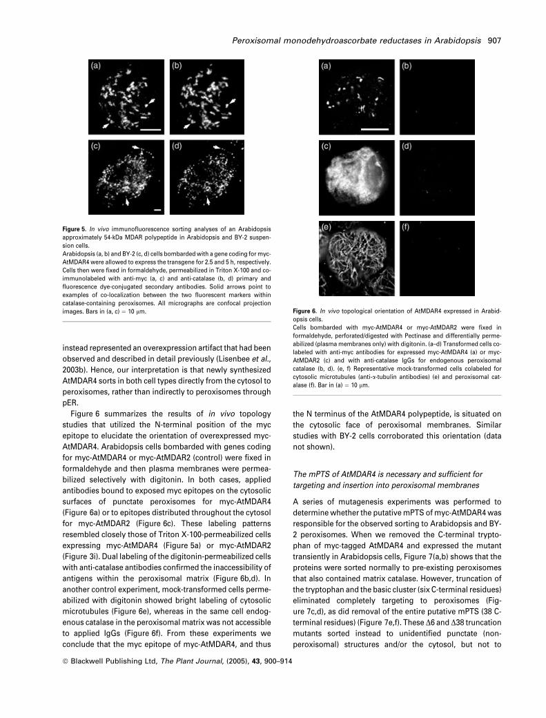
We also tested in vivo the hypothesis that transiently
expressed myc-AtMDAR4 sorts to peroxisomal membranes
in Arabidopsis and BY-2 suspension cells. Figure 5(a–d)
shows representative Arabidopsis (Figure 5a,b) and BY-2
(Figure 5c,d) cells expressing for 2.5 and 5 h, respectively,
myc-AtMDAR4 within peroxisomes that also contained the
marker enzyme catalase. These periods were the earliest
Table 3 Alignment of C-terminal amino acid sequences of MDAR protein variants created for sorting and targeting experiments
MDAR variant name C-terminal sequence
myc-AtMDAR1 -SFAAKImyc-AtMDAR1-D3 -SFA
myc-PsMDAR -SFASKImyc-PsMDAR-I433L -SFASKL
myc-AtMDAR2 -SFATNImyc-AtMDAR3 -SFATKFYSTSLmyc-AtMDAR4 -GFAHTVVSQQKVPEVKDIPSAEMVKQSASVVMIKKPLYVWHAATGVVVAASVAAFAFWYGRRRRRW
myc-AtMDAR4-D1 -GFAHTVVSQQKVPEVKDIPSAEMVKQSASVVMIKKPLYVWHAATGVVVAASVAAFAFWYGRRRRRmyc-AtMDAR4-D6 -GFAHTVVSQQKVPEVKDIPSAEMVKQSASVVMIKKPLYVWHAATGVVVAASVAAFAFWYGmyc-AtMDAR4-D38 -GFAHTVVSQQKVPEVKDIPSAEMVKQSAGFP-AtMDAR4(6) -LYKSSRRRRRWGFP-AtMDAR4(TMD) -LYKSSSVVMIKKPLYVWHAATGVVVAASVAAFAFWYGGFP-AtMDAR4(38) -LYKSSSVVMIKKPLYVWHAATGVVVAASVAAFAFWYGRRRRRW
Epitope-tagged A. thaliana and P. sativumMDAR proteins (bold type) were aligned using the CLUSTALW algorithm. PTS1-like tripeptides are single-underlined. mPTS-like TMDs predicted with the TMHMM program (version 2.0) and basic (positively charged) amino acid clusters are bold type anddouble-underlined, respectively.
Peroxisomal monodehydroascorbate reductases in Arabidopsis 905
ª Blackwell Publishing Ltd, The Plant Journal, (2005), 43, 900–914
time-points after which myc-AtMDAR4 could be detected
reliably in these cells, and examinations of numerous
images did not show the protein within any other organellar
compartment. It should be noted that we observed
overexpressed myc-AtMDAR4 within a non-peroxisomal
compartment in both cell types, although the effect was
most pronounced in BY-2 cells after expression periods
longer than 8 h (data not shown). A careful series of
experiments confirmed that myc-AtMDAR4 had not sorted
to ER or pER, and that the non-peroxisomal structures
(a) (b) (c) (d)
(e) (f) (g) (h)
(i) (j) (k) (l)
(m) (n) (o) (p)
Figure 3. In vivo immunofluorescence sorting
analyses of three Arabidopsis MDAR polypep-
tides expressed in Arabidopsis and BY-2 suspen-
sion cells.
Arabidopsis (a, b, e, f, i, j, m, n) and BY-2 (c, d, g,
h, k, l, o, p) cells were bombarded with tungsten-
bound genes coding for myc-epitope-tagged
variants of AtMDAR1 (a–h), AtMDAR2 (i–l) or
AtMDAR3 (m–p). Bombarded cells were allowed
to express the transgenes for either 5 h (a–d) or
20 h (e–p) prior to formaldehyde fixation, Triton
X-100 permeabilization and fluorescence co-im-
munolabeling for the expressed myc-tagged
proteins (a, c, e, g, i, k, m, o) and endogenous
peroxisomal catalase (b, d, f, h, j, l, n, p). Solid
arrows in (g, h) indicate co-localizations of the
two fluorescent markers within peroxisomes. All
micrographs are confocal projection images.
Bars in (a) and (c) ¼ 10 lm and are the same
for (b, e, f, i, j, m, n) and (d, g, h, k, l, o, p),
respectively.
(a) (b) (c) (d)
(e) (f) (g) (h)
(i) (j) (k) (l)
(m) (n) (o) (p)
Figure 4. Necessity and efficacy of the PTS1-like
C-terminal tripeptides of Arabidopsis and Pisum
sativum approximately 47-kDa peroxisomal
MDARs.
Arabidopsis (a, b, e, f, i, j, m, n) and BY-2 (c, d, g,
h, k, l, o, p) cells expressing myc-epitope-tagged
variants of AtMDAR1 or PsMDAR were fixed in
formaldehyde, permeabilized with Triton X-100
and immunolabeled for the myc-tagged proteins
(a, c, e, g, i, k, m, o) and endogenous peroxisomal
catalase (b, d, f, h, j, l, n, p). (a–d) Cells expressing
for 20 h myc-AtMDAR1-D3 which lacks the C-
terminal PTS1 tripeptide –AKI. (e–l) Cells expres-
sing for 5 h (e–h) or 20 h (i–l) myc-PsMDAR
possessing the wild-type PTS1 tripeptide –SKI.
(m–p) Cells expressing for 20 h myc-PsMDAR-
I433L in which the C-terminal-most Ile residue
was changed to Leu to create a canonical –SKL
tripeptide. Solid arrows in (k, l) indicate colocal-
izations of the two fluorescent markers within
peroxisomes. All micrographs are confocal pro-
jection images. Bars in (a) and (c) ¼ 10 lm and
are the same for (b, e, f, i, j, m, n) and (d, g, h, k, l,
o, p), respectively.
906 Cayle S. Lisenbee et al.
ª Blackwell Publishing Ltd, The Plant Journal, (2005), 43, 900–914
instead represented an overexpression artifact that had been
observed and described in detail previously (Lisenbee et al.,
2003b). Hence, our interpretation is that newly synthesized
AtMDAR4 sorts in both cell types directly from the cytosol to
peroxisomes, rather than indirectly to peroxisomes through
pER.
Figure 6 summarizes the results of in vivo topology
studies that utilized the N-terminal position of the myc
epitope to elucidate the orientation of overexpressed myc-
AtMDAR4. Arabidopsis cells bombarded with genes coding
for myc-AtMDAR4 or myc-AtMDAR2 (control) were fixed in
formaldehyde and then plasma membranes were permea-
bilized selectively with digitonin. In both cases, applied
antibodies bound to exposed myc epitopes on the cytosolic
surfaces of punctate peroxisomes for myc-AtMDAR4
(Figure 6a) or to epitopes distributed throughout the cytosol
for myc-AtMDAR2 (Figure 6c). These labeling patterns
resembled closely those of Triton X-100-permeabilized cells
expressing myc-AtMDAR4 (Figure 5a) or myc-AtMDAR2
(Figure 3i). Dual labeling of the digitonin-permeabilized cells
with anti-catalase antibodies confirmed the inaccessibility of
antigens within the peroxisomal matrix (Figure 6b,d). In
another control experiment, mock-transformed cells perme-
abilized with digitonin showed bright labeling of cytosolic
microtubules (Figure 6e), whereas in the same cell endog-
enous catalase in the peroxisomal matrix was not accessible
to applied IgGs (Figure 6f). From these experiments we
conclude that the myc epitope of myc-AtMDAR4, and thus
the N terminus of the AtMDAR4 polypeptide, is situated on
the cytosolic face of peroxisomal membranes. Similar
studies with BY-2 cells corroborated this orientation (data
not shown).
The mPTS of AtMDAR4 is necessary and sufficient for
targeting and insertion into peroxisomal membranes
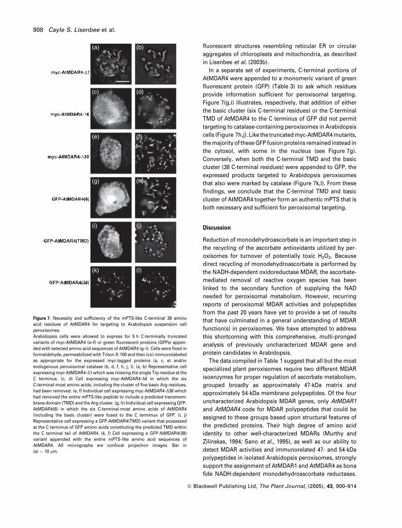
A series of mutagenesis experiments was performed to
determinewhether the putativemPTS ofmyc-AtMDAR4was
responsible for the observed sorting to Arabidopsis and BY-
2 peroxisomes. When we removed the C-terminal trypto-
phan of myc-tagged AtMDAR4 and expressed the mutant
transiently in Arabidopsis cells, Figure 7(a,b) shows that the
proteins were sorted normally to pre-existing peroxisomes
that also contained matrix catalase. However, truncation of
the tryptophan and the basic cluster (six C-terminal residues)
eliminated completely targeting to peroxisomes (Fig-
ure 7c,d), as did removal of the entire putative mPTS (38 C-
terminal residues) (Figure 7e,f). These D6 and D38 truncation
mutants sorted instead to unidentified punctate (non-
peroxisomal) structures and/or the cytosol, but not to
Figure 5. In vivo immunofluorescence sorting analyses of an Arabidopsis
approximately 54-kDa MDAR polypeptide in Arabidopsis and BY-2 suspen-
sion cells.
Arabidopsis (a, b) and BY-2 (c, d) cells bombardedwith a gene coding for myc-
AtMDAR4 were allowed to express the transgene for 2.5 and 5 h, respectively.
Cells then were fixed in formaldehyde, permeabilized in Triton X-100 and co-
immunolabeled with anti-myc (a, c) and anti-catalase (b, d) primary and
fluorescence dye-conjugated secondary antibodies. Solid arrows point to
examples of co-localization between the two fluorescent markers within
catalase-containing peroxisomes. All micrographs are confocal projection
images. Bars in (a, c) ¼ 10 lm. Figure 6. In vivo topological orientation of AtMDAR4 expressed in Arabid-
opsis cells.
Cells bombarded with myc-AtMDAR4 or myc-AtMDAR2 were fixed in
formaldehyde, perforated/digested with Pectinase and differentially perme-
abilized (plasmamembranes only) with digitonin. (a–d) Transformed cells co-
labeled with anti-myc antibodies for expressed myc-AtMDAR4 (a) or myc-
AtMDAR2 (c) and with anti-catalase IgGs for endogenous peroxisomal
catalase (b, d). (e, f) Representative mock-transformed cells colabeled for
cytosolic microtubules (anti-a-tubulin antibodies) (e) and peroxisomal cat-
alase (f). Bar in (a) ¼ 10 lm.
Peroxisomal monodehydroascorbate reductases in Arabidopsis 907
ª Blackwell Publishing Ltd, The Plant Journal, (2005), 43, 900–914
fluorescent structures resembling reticular ER or circular
aggregates of chloroplasts and mitochondria, as described
in Lisenbee et al. (2003b).
In a separate set of experiments, C-terminal portions of
AtMDAR4 were appended to a monomeric variant of green
fluorescent protein (GFP) (Table 3) to ask which residues
provide information sufficient for peroxisomal targeting.
Figure 7(g,i) illustrates, respectively, that addition of either
the basic cluster (six C-terminal residues) or the C-terminal
TMD of AtMDAR4 to the C terminus of GFP did not permit
targeting to catalase-containing peroxisomes in Arabidopsis
cells (Figure 7h,j). Like the truncatedmyc-AtMDAR4mutants,
themajority of theseGFP fusion proteins remained instead in
the cytosol, with some in the nucleus (see Figure 7g).
Conversely, when both the C-terminal TMD and the basic
cluster (38 C-terminal residues) were appended to GFP, the
expressed products targeted to Arabidopsis peroxisomes
that also were marked by catalase (Figure 7k,l). From these
findings, we conclude that the C-terminal TMD and basic
cluster of AtMDAR4 together form an authentic mPTS that is
both necessary and sufficient for peroxisomal targeting.
Discussion
Reduction of monodehydroascorbate is an important step in
the recycling of the ascorbate antioxidants utilized by per-
oxisomes for turnover of potentially toxic H2O2. Because
direct recycling of monodehydroascorbate is performed by
the NADH-dependent oxidoreductase MDAR, the ascorbate-
mediated removal of reactive oxygen species has been
linked to the secondary function of supplying the NAD
needed for peroxisomal metabolism. However, recurring
reports of peroxisomal MDAR activities and polypeptides
from the past 20 years have yet to provide a set of results
that have culminated in a general understanding of MDAR
function(s) in peroxisomes. We have attempted to address
this shortcoming with this comprehensive, multi-pronged
analysis of previously uncharacterized MDAR gene and
protein candidates in Arabidopsis.
The data compiled in Table 1 suggest that all but the most
specialized plant peroxisomes require two different MDAR
isoenzymes for proper regulation of ascorbate metabolism,
grouped broadly as approximately 47-kDa matrix and
approximately 54-kDa membrane polypeptides. Of the four
uncharacterized Arabidopsis MDAR genes, only AtMDAR1
and AtMDAR4 code for MDAR polypeptides that could be
assigned to these groups based upon structural features of
the predicted proteins. Their high degree of amino acid
identity to other well-characterized MDARs (Murthy and
Zilinskas, 1994; Sano et al., 1995), as well as our ability to
detect MDAR activities and immunorelated 47- and 54-kDa
polypeptides in isolated Arabidopsis peroxisomes, strongly
support the assignment of AtMDAR1 and AtMDAR4 as bona
fide NADH-dependent monodehydroascorbate reductases.
(a) (b)
(c) (d)
(e) (f)
(g) (h)
(i) (j)
(k) (l)
Figure 7. Necessity and sufficiency of the mPTS-like C-terminal 38 amino
acid residues of AtMDAR4 for targeting to Arabidopsis suspension cell
peroxisomes.
Arabidopsis cells were allowed to express for 5 h C-terminally truncated
variants of myc-AtMDAR4 (a–f) or green fluorescent proteins (GFPs) appen-
ded with selected amino acid sequences of AtMDAR4 (g–l). Cells were fixed in
formaldehyde, permeabilized with Triton X-100 and then (co)-immunolabeled
as appropriate for the expressed myc-tagged proteins (a, c, e) and/or
endogenous peroxisomal catalase (b, d, f, h, j, l). (a, b) Representative cell
expressingmyc-AtMDAR4-D1 which wasmissing the single Trp residue at the
C terminus. (c, d) Cell expressing myc-AtMDAR4-D6 in which the six
C-terminal-most amino acids, including the cluster of five basic Arg residues,
had been removed. (e, f) Individual cell expressing myc-AtMDAR4-D38 which
had removed the entire mPTS-like peptide to include a predicted transmem-
brane domain (TMD) and the Arg cluster. (g, h) Individual cell expressing GFP-
AtMDAR4(6) in which the six C-terminal-most amino acids of AtMDAR4
(including the basic cluster) were fused to the C terminus of GFP. (i, j)
Representative cell expressing a GFP-AtMDAR4(TMD) variant that possessed
at the C terminus of GFP amino acids constituting the predicted TMD within
the C terminal tail of AtMDAR4. (k, l) Cell expressing a GFP-AtMDAR4(38)
variant appended with the entire mPTS-like amino acid sequences of
AtMDAR4. All micrographs are confocal projection images. Bar in
(a) ¼ 10 lm.
908 Cayle S. Lisenbee et al.
ª Blackwell Publishing Ltd, The Plant Journal, (2005), 43, 900–914
Coupled with the finding that epitope-tagged versions of
AtMDAR1 and AtMDAR4 sorted to peroxisomes in vivo, we
are confident that the AtMDAR1 and AtMDAR4 genes code
for the Arabidopsis 47- and 54-kDa MDARs that appear to be
common components of nearly all plant peroxisomes.
None of the four uncharacterized Arabidopsis MDARs
appeared to represent the membrane-associated 32- and 47-
kDa MDARs in castor glyoxysomal membranes, the 32-kDa
MDAR in pea leaf peroxisomal membranes or the 39/40-kDa
MDARs in soybean root nodule extracts (Table 1). Our
biochemical results confirmed this notion by identifying
47- and 54-kDa MDAR polypeptides in isolated Arabidopsis
peroxisomes, each of which exhibited distinguishable mat-
rix and membrane associations, respectively, on blots that
were probed with anti-cucumber 47-kDa MDAR antibodies
(Sano et al., 1995). These results were surprising, because
the same antibodies recognized the seemingly very different
32-kDa pea and 47-kDa castor MDAR PMPs (Karyotou and
Donaldson, 2005; Lopez-Huertas et al., 1999). In the current
study, these antibodies also cross-reacted with a 38-kDa
Arabidopsis polypeptide that was most abundant in cytoso-
lic fractions. This polypeptide also was detected inconsis-
tently in peroxisome preparations, and when present always
behaved as a loosely attached, protease-sensitive compo-
nent of intact organelles. These findings may point to the
existence of as-yet unidentified peroxisomal MDARs in
Arabidopsis, and to the speculation that the 38-kDa protein
corresponds to the pea and castor 32-kDa MDARs and/or the
soybean 39/40-kDaMDARs. Such a hypothesis is discredited
by the fact that the 32-kDa MDARs exhibited much tighter
associations with peroxisomal/glyoxysomal membranes
than did the 38-kDa polypeptide (Bowditch and Donaldson,
1990; Jimenez et al., 1997; Lopez-Huertas et al., 1999; Luster
et al., 1988). Although none of the AtMDAR genes coded for
a 38-kDa polypeptide, another possibility is that the 47-kDa
AtMDAR2 and/or AtMDAR3 isoforms that were shown in this
study to remain in the cytosol are post-translationally
processed to a cytosolic 38-kDa MDAR. Our contention is
that the 38-kDa polypeptide is not another peroxisomal
MDAR, but is instead an immunorelated cytosolic oxidore-
ductase that may associate peripherally with the cytosolic
side of peroxisomal membranes, although the unreliable
behavior of this interaction probably indicates that it was an
artifact of the cell fractionations.
The inability of KCl to remove most of the 54-kDa
polypeptide (AtMDAR4) and enzyme activity from peroxi-
somalmembranes is consistent with previous results, and in
our opinion provides the most reliable measure of mem-
brane association in this system. For example, KCl treat-
ments also did not remove MDAR enzyme activities from
spinach glyoxysomal (Ishikawa et al., 1998) or pea leaf
peroxisomal (Jimenez et al., 1997) membranes. In a more
detailed analysis, Bowditch and Donaldson (1990) showed
that KCl-treated castor glyoxysomal membranes retained
80% of the total pretreatment MDAR activity. Notably, this
and another study (Bunkelmann and Trelease, 1996) also
revealed that 0.1 M Na2CO3 abolished 85% of the total MDAR
activity, prompting the authors to suggest that these condi-
tions cannot be used to assess membrane associations. In
support of this contention, our attempts at detecting MDAR
polypeptides within peroxisomal membranes that had been
extracted sequentially with KCl and then Na2CO3 yielded
results that were inconsistent with those derived from use of
KCl alone. Furthermore, these findings were not consistent
with predictions of the biochemical behavior of matrix-
associated catalase or the membrane association of an
AtMDAR4 polypeptide having two putative TMDs. None the
less, alkaline carbonate washes have been employed com-
monly in even the most recent studies of MDAR PMPs, but it
must be emphasized that none of these studies have
addressed the association of a 54-kDa MDAR. For instance,
Lopez-Huertas et al. (1999) showed that a 32-kDa pea leaf
MDAR isolated from carbonate-washed peroxisomal mem-
branes possessed NADH-dependent ferricyanide reductase
activity; the MDAR activity of this polypeptide was not
measured, due presumably to its carbonate inactivation.
Karyotou and Donaldson (2005) detected MDAR activity in
carbonate-washed castor glyoxysomal membranes, but this
activity was attributed to a 47-kDa polypeptide that until this
report had not been detected in castor. Considering our
consistent immunodetection within KCl-washed peroxi-
somal membranes of a 54-kDa MDAR under conditions that
preserved detectable MDAR activity, we conclude that
AtMDAR4, like peroxisomal APX, is an integral membrane
protein of Arabidopsis peroxisomes.
As predicted, the PTS sequences of both AtMDAR1 and
AtMDAR4 provided the targeting information necessary for
sorting transiently expressed, epitope-tagged versions of
these proteins to Arabidopsis and BY-2 peroxisomes. How-
ever, we were surprised to find that the C-terminal –AKI and
–SKI tripeptides of AtMDAR1 and PsMDAR, respectively,
may have functioned inefficiently in peroxisomal targeting.
Although this could have been due to overexpression from
the CaMV 35S promoter, others have concluded from similar
observations that residues outside the PTS1 signal function
as accessory sequences that enable peroxisomal targeting
by non-optimal PTS1 combinations (Bongcam et al., 2000;
Mullen, 2002; Mullen et al., 1997a,b). We did not test this
hypothesis directly for AtMDAR1 or PsMDAR, but did note
enhanced peroxisomal sorting upon changing the –SKI
tripeptide of PsMDAR to –SKL. Similarly, the TMD and the
adjacent basic amino acid cluster at the C terminus of
AtMDAR4 together, but not singly, were necessary and
sufficient for targeting to peroxisomes. Mullen and Trelease
(2000) arrived at a similar conclusion with respect to the
analogous TMD and basic cluster of peroxisomal membrane
APX. The sorting of several PMPs to peroxisomal
membranes depends upon a basic amino acid cluster
Peroxisomal monodehydroascorbate reductases in Arabidopsis 909
ª Blackwell Publishing Ltd, The Plant Journal, (2005), 43, 900–914
(Baerends et al., 2000; Brosius et al., 2002; Dyer et al., 1996;
Elgersma et al., 1997; Honsho and Fujiki, 2001; Hunt and
Trelease, 2004; Kammerer et al., 1998; Murphy et al., 2003;
Soukupova et al., 1999), and in some cases sorting occurs
indirectly through the ER when the basic cluster is juxta-
posed with a TMD (Baerends et al., 1996, 2000; Elgersma
et al., 1997; Mullen and Trelease, 2000). Interestingly, the
mPTS of AtMDAR4 is contained within an approximately 60
amino acid C-terminal extension that is strikingly similar to
the mPTS-containing C-terminal extensions of peroxisomal
membrane-bound APXs (Jespersen et al., 1997; Mullen and
Trelease, 2000). When protein sequence databases were
queriedwith the C-terminal extensions of either AtMDAR4 or
cottonseed peroxisomal APX, other plant peroxisomal APXs
and the 54-kDa MDARs were the only mPTS-containing
sequences identified (data not shown). Despite their similar
mPTSs, our early stage time-course results suggest that
AtMDAR4 does not sort to peroxisomes indirectly through
the pER subdomain that is utilized by peroxisomal APX
(Lisenbee et al., 2003a,b; Mullen et al., 1999).
Results presented in the current study also suggest that
AtMDAR4 is associated with and inserted into the peroxi-
somal membrane in a manner different from peroxisomal
APX, an integral type II (Ncytosol-Cmatrix) tail-anchored PMP
(Mullen and Trelease, 2000). For instance, 54 kDa AtMDAR4
in intact peroxisomes was inaccessible to applied proteases
in the absence of detergent, indicating that most of
the polypeptide faces the peroxisomal matrix and not the
cytosol. This ‘reverse’ orientation of AtMDAR4 places the
enzyme’s active site within the peroxisomal matrix, thus
indicating that the PMP is by definition not a C-terminal tail-
anchored protein with a large functional domain in the
cytosol (Wattenberg and Lithgow, 2001). A separate set of
topology experiments showed that the N terminus of
AtMDAR4 was accessible to applied IgGs under conditions
that prevented detection of matrix antigens. These data are
consistent with the most accurate TMD algorithms that
predict membrane-spanning regions at each end of the
polypeptide (Moller et al., 2001), as well as with the finding
that the 54-kDa AtMDAR4 behaved as a KCl-insoluble,
integral component of Arabidopsis peroxisomal mem-
branes. This topology clearly carries functional implications
for MDAR catalytic activity (see below), particularly in that
the N-terminal TMD of AtMDAR4 curiously includes one of
the FAD-binding sequence motifs that categorizes the pro-
tein in part as an authentic MDAR. None the less, it seems
clear that upon reaching the peroxisomal membrane, newly
synthesized AtMDAR1 is inserted into and resides within the
matrix, whereas nascent AtMDAR4 is integrated, such
that the bulk of the polypeptide is on the matrix face of
the membrane with the N terminus exposed to the cytosol.
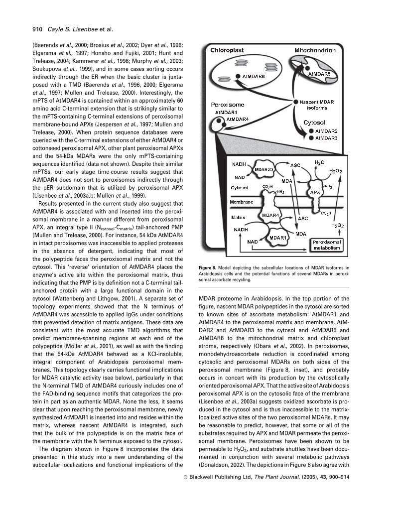
The diagram shown in Figure 8 incorporates the data
presented in this study into a new understanding of the
subcellular localizations and functional implications of the
MDAR proteome in Arabidopsis. In the top portion of the
figure, nascent MDAR polypeptides in the cytosol are sorted
to known sites of ascorbate metabolism: AtMDAR1 and
AtMDAR4 to the peroxisomal matrix and membrane, AtM-
DAR2 and AtMDAR3 to the cytosol and AtMDAR5 and
AtMDAR6 to the mitochondrial matrix and chloroplast
stroma, respectively (Obara et al., 2002). In peroxisomes,
monodehydroascorbate reduction is coordinated among
cytosolic and peroxisomal MDARs on both sides of the
peroxisomal membrane (Figure 8, inset), and probably
occurs in concert with its production by the cytosolically
orientedperoxisomalAPX. That the active site ofArabidopsis
peroxisomal APX is on the cytosolic face of the membrane
(Lisenbee et al., 2003a) suggests oxidized ascorbate is pro-
duced in the cytosol and is thus inaccessible to the matrix-
localized active sites of the two peroxisomal MDARs. It may
be reasonable to predict, however, that some or all of the
substrates required by APX and MDAR permeate the peroxi-
somal membrane. Peroxisomes have been shown to be
permeable to H2O2, and substrate shuttles have been docu-
mented in conjunction with several metabolic pathways
(Donaldson, 2002). The depictions in Figure 8 also agreewith
Figure 8. Model depicting the subcellular locations of MDAR isoforms in
Arabidopsis cells and the potential functions of several MDARs in peroxi-
somal ascorbate recycling.
910 Cayle S. Lisenbee et al.
ª Blackwell Publishing Ltd, The Plant Journal, (2005), 43, 900–914
the reported matrix-associated MDAR activities of pea leaf
peroxisomes (Jimenez et al., 1997) and castor glyoxysomes
(Bowditch and Donaldson, 1990), but it is notable that these
studies did not determine whether the activities originated
from a matrix protein and/or a PMP. In contrast, enzyme
latency studies showed that the active sites of spinach
peroxisomal MDAR were on the cytosolic side of the mem-
brane (Ishikawa et al., 1998). Donaldson (2002) has provided
the particularly relevant explanation that these observations
in spinach may represent a matrix-facing enzyme whose
substrates permeate the membrane at rates greater than the
enzyme’s turnover number. More recent work from this
groupalsosuggested thatAPXandMDARformacooperative
membrane-bound complex for the efficient removal of H2O2
(Karyotou and Donaldson, 2005), possibly to effect the direct
shuttling of substrates and electron equivalents across the
peroxisomal membrane. Regardless of the exact mechan-
ism, this new representationprovides themost complete and
detailed understanding to date of howAPX andMDARmight
be configured functionallywithin theperoxisomalmatrix and
membrane for the removal of reactive oxygen species.
Experimental procedures
Arabidopsis cell culture, sucrose gradient isolation and
subfractionation of organelles, enzyme assays and
membrane protein association and topology
Arabidopsis (Arabidopsis thaliana var. Landsberg erecta) sus-pension cells (50 ml cultures) were grown, maintained and har-vested according to Lisenbee et al. (2003a). For isolation oforganelles in sucrose gradients, protoplasts were prepared priorto cell disruption as follows. Cells collected by centrifugationfrom each 4-day culture were washed twice (5 min each at roomtemperature) with 25 ml of aqueous 0.4 M D-mannitol. The cellswere then resuspended in 25 ml of protoplasting solution [Ara-bidopsis culture medium (Lisenbee et al., 2003a) plus 0.4 M D-mannitol, 0.1% w/v Pectinase (Sigma-Aldrich, St Louis, MO, USA)and 1% w/v Cellulase Y-C (Karlan Research Products, Cotton-wood, AZ, USA)] and incubated with rocking inversion at 30�Cuntil the 20–30-cell clusters typical of Arabidopsis suspensioncultures were reduced to one to four cells per cluster (approxi-mately 2.5 h). The protoplasts were pelleted in a fixed-angleSorvall SS-34 rotor at 480 g for 10 min and washed subsequentlythree times in aqueous 0.4 M D-mannitol before final resuspen-sion in two pellet volumes of ice-cold homogenization medium(HM) (25 mM HEPES-KOH, pH 7.5, 0.7 M sucrose, 3 mM dithio-threitol (DTT) and 0.5 mM phenylmethylsulfonyl fluoride).
Resuspended cells (5–6 ml, equivalent to 1–1.5 flasks of 4-daycells) were disrupted with an ice-cold 15-ml Dounce (WheatonScience Products, Millville, NJ, USA) tissue grinder (pestle ‘A’)using 25–35 up-and-down movements until approximately 80% ofthe cells were ruptured as judged by optical microscopy. Homo-genates were centrifuged in a Sorvall HB-6 swing-out rotor at 1500 g
for 15 min at 4�C to pellet unbroken cells, starch-containing non-green plastids, nuclei and cell debris. The resulting supernatants(2.5–3 ml, equivalent to 1 flask of 4-day cells) were loaded onto 25-ml linear gradients (30–59% w/w sucrose in 25 mM HEPES-KOH, pH
7.5) underlaid with 5-ml cushions (59%w/w sucrose) in Beckman vTi50 Quick-Seal centrifuge tubes (Beckman Coulter, Fullerton, CA,USA). Each applied sample was overlaid with HM (0.4 M sucrose)before the gradient tubeswere heat-sealed and then centrifuged in avTi 50 rotor at 50 000 g for 75 min at 4�C. Peroxisomes equilibratedas a white band near the bottom of the gradients clearly separatedfrom the larger mitochondrial band at lower sucrose density.Fractions (1 ml) were collected by hand through a hole puncturedin the bottom of the tubes. The peroxisomes used for the topolo-gical studies presented in Figure 2(b) were isolated similarlyaccording to the procedure detailed in Lisenbee et al. (2003a).
Experiments designed to elucidate the organellar distribution andmembrane association of MDAR proteins in Arabidopsis peroxi-somes were conducted mainly as described by Lisenbee et al.(2003a). The following describes important detailed differences.Organelles (Figure 1) in fractions 4–8 (peroxisomes), 13–15 (mito-chondria) and 25–28 (cytosol) were pooled from each of threegradients. A duplicate set of pooled fractions from three othercombined gradients also was prepared. Pooled peroxisomes(approximately 10 ml) or mitochondria (approximately 10 ml) wereburst in 1.5 volumes of 25 mM HEPES-KOH, pH 7.5, with incubation(inversion rocking) for 40 min at 4�C. The suspensions werecentrifuged in a Beckman fixed-angle 70 Ti rotor at 150 000 g for45 min at 4�C to produce pelleted membranes and supernatants(water-solubilized proteins). The membrane pellets were resus-pended in 1 ml (peroxisomes) or 4 ml (mitochondria) of 0.2 M KCl in25 mM HEPES-KOH, pH 7.5, and incubated with intermittent mixingfor 60 min at 4�C. KCl-insoluble membranes were pelleted fromthese suspensions in a Beckman 90 Ti rotor at 150 000 g for 30 min,generating a supernatant with KCl-soluble proteins. The KCl-insol-uble membrane pellets were resuspended in 1 ml (peroxisomes) or2 ml (mitochondria) of 0.2 M KCl in 25 mM HEPES-KOH, pH 7.5, forenzyme and protein assays. Peroxisomes used for protease-diges-tion (proteinase K) topology experiments were treated as describedin detail in Lisenbee et al. (2003a).
Catalase and cytochrome c oxidase activities were assayed asdescribed by Ni et al. (1990) and Tolbert et al. (1968), respectively.MDAR enzyme activities were assayed essentially as described byBunkelmann and Trelease (1996). Briefly, the reaction was carriedout in a final 1-ml volume of 50 mM HEPES-KOH, pH 7.6, 2.5 mM
ascorbate (Sigma), 0.5 units ascorbate oxidase (Sigma), 5–100 llsample (up to 400 ll in very dilute samples) and 0.1 mM NADH(Sigma). The components were added sequentially to a quartzcuvette and the linear decrease in A340 was monitored for 1–2 min.These assays included measuring the potential rate of MDAR-independent NADH oxidation by omitting additions of ascorbateand ascorbate oxidase and subtracting this potential activity fromthe rate of MDAR-dependent NADH oxidation. Activity of thecommercially supplied ascorbate oxidase was verified using amodification of the manufacturer’s suggested assay, i.e., thedecrease in A265 of ascorbate was followed in a 1-ml reactioncontaining 50 mM HEPES-KOH, pH 7.6, ascorbate (A265 approxi-mately 0.907) and 0.5 units ascorbate oxidase.
Buoyant density measurement of sucrose gradient fractions,protein estimation, trichloroacetic acid (TCA) precipitation, sodiumdodecyl sulphate-polyacrylamide gel electrophoresis (SDS-PAGE)and immunoblot detection were carried out essentially as describedby Lisenbee et al. (2003a). The modifications used in this paper arethat a final concentration of 0.05% w/v deoxycholate was added toall fractions prior to protein precipitation at a final concentration of10% v/v TCA for 30 min at 4�C. Samples (25 lg protein) wereneutralized with solid Tris base and stored at 4�C as described.Samples were reduced by additions of freshly prepared 0.5 M or 1 M
DTT to a final concentration of 10 mM, and then boiled 8 min prior to
Peroxisomal monodehydroascorbate reductases in Arabidopsis 911
ª Blackwell Publishing Ltd, The Plant Journal, (2005), 43, 900–914
separation in 10% w/v precast Mini-Protean II polyacrylamide gels(Bio-Rad, Hercules, CA, USA). Electroblotting was performed for45 min (for two gels). Primary and secondary antibodies were usedas follows: rabbit anti-cucumber 47-kDa MDAR antiserum (1:1000)(Sano et al., 1995), rabbit anti-cucumber peroxisomal APX IgGs(1:1000) (Corpas et al., 1994), rabbit anti-cottonseed catalase IgGs(1:1000 or 1:2000) (Kunce et al., 1988) and goat anti-rabbit alkalinephosphatase conjugate (1:10 000) (Bio-Rad).
Acquisition, subcloning and mutagenesis of MDAR coding
sequences
Molecular biology reagents employed in standard recombinantDNA manipulations were purchased from Promega (Madison, WI,USA), New England Biolabs (Beverly, MA, USA) and Takara Bio-medicals (Otsu, Shiga, Japan). Mutations were incorporated intocoding sequences in polymerase chain reaction (PCR)-based site-directed mutagenesis reactions that included appropriate forwardand reverse mutagenic primers. Mutagenic primer sets were de-signed for either fragment-specific or whole-plasmid PCR amplifi-cations; the latter were carried out using the QuikChange site-directed mutagenesis kit according to the manufacturer’s instruc-tions (Stratagene, La Jolla, CA, USA). Custom oligonucleotideprimers were synthesized by Genetech Biosciences (Tempe, AZ,USA), and all (mutated) plasmid inserts were confirmed by auto-mated dye-terminator cycle sequencing (Arizona State UniversityDNA Laboratory, Tempe, AZ, USA). Sequence details and DNAsamples of all primer sets and plasmids used/created in this studyare available from the authors upon request.
Full-length expressed sequence tag cDNAs were obtained fromthe Arabidopsis Biological Resource Center (Ohio State University,Columbus, OH, USA) for A. thaliana ecotype Columbia genesAtMDAR1 (ABRC stock number U12996), AtMDAR2 (U14648) andAtMDAR3 (U21865). All three cDNAs were epitope-tagged asfollows. First, open reading frames (ORFs) were amplified fromtheir parent plasmids in PCR reactions that replaced start codonswith in-frame BamHI sites and appended XbaI sites after the stopcodons. PCR products were TA cloned into pCR2.1 (Invitrogen, SanDiego, CA, USA), digested with BamHI and XbaI, and then ligatedinto BamHI/XbaI-digested pRTL2/mycBX to yield pRTL2/myc-AtM-DAR1, pRTL2/myc-AtMDAR2 and pRTL2/myc-AtMDAR3. pRTL2/mycBX is a CaMV 35S promoter-driven plant expression cassettethat adds a single copy of the myc epitope to the 5¢ end of an ORF.The PsMDAR ORF was amplified from pSK/PsMDAR (Murthy andZilinskas, 1994) and myc epitope-tagged as described above forAtMDAR1, except that the start codon was replaced with an XbaI-compatible NheI site for subcloning into an analogous pRTL2/mycXplant expression cassette. A full-length cDNA of AtMDAR4 wasamplified from 4-day Arabidopsis suspension cell total RNA usingan RNeasy PlantMini Kit (Qiagen, Valencia, CA, USA) and the accessreverse transcription-polymerase chain reaction (RT-PCR) system(Promega), both according to the manufacturer’s instructions. Theforward primer corresponded to sequences downstream of theinitiation codon, which was replaced by an in-frame NheI site.The reverse primer corresponded to sequences upstream of andincluding the termination codon and introduced a unique XbaI sitewithin the 3¢ untranslated region. The cDNA products were TAcloned, sequenced to verify amplification of the correct MDAR ORF,and then subcloned into XbaI-digested pRTL2/mycX to yieldepitope-tagged pRTL2/myc-AtMDAR4.
Sequences coding for the C-terminal PTS signals of AtMDAR1,PsMDAR and AtMDAR4 were modified for targeting necessityexperiments as follows. pRTL2/myc-AtMDAR1-D3 was created from
pRTL2/myc-AtMDAR1 in a whole-plasmid PCR reaction that chan-ged the GCT codon encoding A432 to a TGA stop codon. Similarly,pRTL2/myc-PsMDAR-I433L was created from pRTL2/myc-PsMDARusing complementary primers that changed the ATT codon enco-ding I433 to a leucine-coding TTA codon. To create pRTL2/myc-AtMDAR4-D1, the AtMDAR4 ORF was PCR-amplified from pRTL2/myc-AtMDAR4 using the forward primer described above and amutagenic reverse primer that changed the final tryptophan-codingTGG codon to a TGA stop codon. The PCR products were TA cloned,digested with the appropriate restriction enzymes, and then ligatedinto pRTL2/mycX to yield pRTL2/myc-AtMDAR4-D1. Whole-plasmidPCR reactions were used to generate pRTL2/myc-AtMDAR4-D6 andpRTL2/myc-AtMDAR4-D38 from pRTL2/myc-AtMDAR4 with com-plementary primers that substituted TGA stop codons for thoseencoding amino acids R483 and S451, respectively.
Fusion of various portions of the mPTS of AtMDAR4 to the Cterminus of GFP for targeting sufficiency experiments was accom-plished as follows. pRTL2/GFP-AtMDAR4(6) was made by firstannealing complementary oligonucleotides containing sequencescoding for the RRRRRW basic cluster (six C-terminal residues) andstop codon of AtMDAR4. The resulting double-stranded fragmentswere phosphorylated with T4 polynucleotide kinase (New EnglandBiolabs) and then ligated through designed 5¢ NheI and 3¢ XbaIoverhangs into an XbaI-digested pRTL2/GFPX fusion cassette toyield pRTL2/GFP-AtMDAR4(6). pRTL2/GFPX was created fromsequential whole-plasmid PCR reactions that inserted both an in-frame XbaI site in place of the stop codon and a monomer-inducingA206Kmutation (Lisenbee et al., 2003b; Zacharias et al., 2002) into aplant optimized S65T variant of GFP (Haseloff et al., 1997). pRTL2/GFP-AtMDAR4(TMD) and pRTL2/GFP-AtMDAR4(38) were made byfirst PCR-amplifying from pRTL2/myc-AtMDAR4 the TMD (residues451–482) or the entire mPTS (residues 451–488) of AtMDAR4. Bothreactions included a mutagenic forward primer that introduced anin-frame NheI site after the codon encoding amino acid A450;mutagenic reverse primers either substituted a TGA stop codon andan XbaI site for that encoding R483 [pRTL2/GFP-AtMDAR4(TMD)] orappended an XbaI site after the natural stop codon [pRTL2/GFP-AtMDAR4(38)]. TA-cloned PCR products were digested with theappropriate restriction enzymes and then ligated into XbaI-digestedpRTL2/GFPX as described above.
BY-2 cell culture and microprojectile bombardment of
suspension cells
Detailed descriptions of how Nicotiana tabacum L., cv. Bright Yel-low 2 (BY-2) suspension-cultured cells were propagated in MSmedium and how Arabidopsis and BY-2 cells were transformed viamicroprojectile bombardments can be found in Lisenbee et al.(2003a) and Mullen et al. (1999), respectively. For transient trans-formations, cells were resuspended in the appropriate transforma-tion medium (MS medium without growth hormones), spread ontofilter papers in petri dishes pre-wetted with the same medium andequilibrated for 1 h at room temperature in the dark. Equilibratedcells were bombarded with DNA-coated tungsten particles and thenheld in covered petri dishes for 2–20 h (sometimes longer) to allowfor expression of the introduced transgene(s).
Immunofluorescence microscopy
Bombarded cells were scraped from filter papers, fixed in 4% w/vformaldehyde (prepared fresh from paraformaldehyde) (Ted Pella,Redding, CA, USA), and then perforated/digested in 0.1% w/v Pec-tolyase Y-23 (Karlan) and 0.1% w/v Cellulase RS (Arabidopsis only)
912 Cayle S. Lisenbee et al.
ª Blackwell Publishing Ltd, The Plant Journal, (2005), 43, 900–914
(Karlan) in preparation for immunofluorescence localization of theexpressed proteins (Mullen et al., 2001). Fixed and perforated cellswere immunolabeled according to our standard 1-ml volume pro-cedure described in Lisenbee et al. (2003a). Briefly, cells were per-meabilized first in 0.3% v/v Triton X-100 (Sigma) and were thenincubated for 1 h each in primary and fluorophore-conjugatedsecondary antibodies diluted in PBS. In experiments aimed atdetermining the topological orientation of proteins in vivo(Figure 6), Arabidopsis cells instead were perforated/digested in0.1% w/v Pectinase for 1 h at 30�C and then were permeabilizedselectively at plasma, but not organellar, membranes with25 lg ml)1 digitonin for 15 min at room temperature (Mullen et al.,2001). Primary antibody sources and concentrations were as fol-lows: mouse anti-myc monoclonal antibody 9E10 (1:500) (SantaCruz Biotechnology, Santa Cruz, CA, USA), mouse anti-chick braina-tubulin monoclonal antibody DM1A (1:500) (Accurate Chemicaland Scientific Corp., Westbury, NY, USA) and rabbit anti-cottonseedcatalase IgGs (1:500 or 1:2000) (Kunce et al., 1988). Secondaryimmunoreagents conjugated to various green, red or far-red fluor-ophores were purchased from Jackson ImmunoResearch Laborat-ories (Westgrove, PA, USA). Immunolabeled cells were examinedand photographed as described (Hunt and Trelease, 2004), and theresulting micrographs were adjusted for contrast and assembledinto plates with Adobe Photoshop software (Adobe Systems, SanJose, CA, USA).
Acknowledgements
Thanks are extended to Satoshi Sano (Kyoto Prefectural University)for donating ample supplies of anti-cucumber MDAR antibodiesand to Barbara Zilinskas (Rutgers University) for providing thePsMDAR cDNA clone. We also thank Robert Mullen (Guelph Uni-versity) for constructing the pRTL2/mycX and pRTL2/mycBX cas-settes and Scott Bingham (Arizona State University) for DNAsequencing and helpful advice on cloning techniques and proce-dures. Special thanks also are given to Sheetal Karnik for her helpfulinstructions and discussions pertaining to cell fractionation andbiochemical techniques. Heather Gustafson (Arizona State Univer-sity) maintained the suspension cultures and assisted in cell fracti-onations. Confocal microscopy was performed in the W.M. KeckBioImaging Laboratory at Arizona State University. This work wassupported by the National Science Foundation (grant no. MCB-0091826 to RNT) and in part by the William N. and Myriam Penn-ington Foundation. The Graduate Program in Molecular and Cellu-lar Biology at Arizona State University funded a ResearchAssistantship for CSL.
References
Baerends, R.J.S., Rasmussen, S.W., Hilbrands, R.E., van der Heide,
M., Faber, K.N., Reuvekamp, P.T.W., Kiel, J.A.K.W., Cregg, J.M.,
van der Klei, I.J. and Veenhuis, M. (1996) The Hansenula poly-morpha PER9 gene encodes a peroxisomal membrane proteinessential for peroxisome assembly and integrity. J. Biol. Chem.271, 8887–8894.
Baerends, R.J.S., Faber, K.N., Kram, A.M., Kiel, J.A.K.W., van der
Klei, I.J. and Veenhuis, M. (2000) A stretch of positively chargedamino acids at the N terminus of Hansenula polymorpha Pex3p isinvolved in incorporation of the protein into the peroxisomalmembrane. J. Biol. Chem. 275, 9986–9995.
Bongcam, V., Petetot, J.M., Mittendorf, V., Robertson, E.J., Leech,
R.M., Qin, Y.-M., Hiltunen, J.K. and Poirier, Y. (2000) Importanceof sequences adjacent to the terminal tripeptide in the import of a
peroxisomal Candida tropicalis protein in plant peroxisomes.Planta, 211, 150–157.
Bowditch, M.I. and Donaldson, R.P. (1990) Ascorbate free-radicalreduction by glyoxysomal membranes. Plant Physiol. 94, 531–537.
Brendel, V., Bucher, P., Nourbakhsh, I.R., Blaisdell, B.E. and Karlin,
S. (1992) Methods and algorithms for statistical analysis of pro-tein sequences. Proc. Natl Acad. Sci. USA, 89, 2002–2006.
Brosius, U., Dehmel, T. and Gartner, J. (2002) Two different target-ing signals direct human peroxisomal membrane protein 22 toperoxisomes. J. Biol. Chem. 277, 774–784.
Bunkelmann, J.R. and Trelease, R.N. (1996) Ascorbate peroxidase: aprominent membrane protein in oilseed glyoxysomes. PlantPhysiol. 110, 589–598.
Corpas, F.J., Bunkelmann, J. and Trelease, R.N. (1994) Identificationand immunochemical characterization of a family of peroxisomemembrane proteins (PMPs) in oilseed glyoxysomes. Eur. J. CellBiol. 65, 280–290.
Corpas, F.J., Barroso, J.B. and del Rıo, L.A. (2001) Peroxisomes as asource of reactive oxygen species and nitric oxide signal mole-cules in plant cells. Trends Plant Sci. 6, 145–150.
Dalton, D.A., Langeberg, L. and Robbins, M. (1992) Purification andcharacterization of monodehydroascorbate reductase from soy-bean root nodules. Arch. Biochem. Biophys. 292, 281–286.
Donaldson, R.P. (2002) Peroxisomal membrane enzymes. In PlantPeroxisomes (Baker, A. and Graham, I., eds). Dordrecht: KluwerAcademic Publishers, pp. 259–278.
Dyer, J.M., McNew, J.A. and Goodman, J.M. (1996) The sortingsequence of the peroxisomal integral membrane protein PMP47is contained within a short hydrophilic loop. J. Cell Biol. 133, 269–280.
Elgersma, Y., Kwast, L., van den Berg, M., Snyder, W.B., Distel, B.,
Subramani, S. and Tabak, H.F. (1997) Overexpression of Pex15p,a phosphorylated peroxisomal integral membrane protein re-quired for peroxisome assembly in S. cerevisiae, causes prolif-eration of the endoplasmic reticulum membrane. EMBO J. 16,7326–7341.
Flynn, C.R., Heinze, M., Schumann, U., Gietl, C. and Trelease, R.N.
(2005) Compartmentalization of the plant peroxin, AtPex10p,within subdomain(s) of ER. Plant Sci. 168, 635–652.
Gardiner, J., Schroeder, S., Polacco, M.L. et al. (2004) Anchoring9371 maize expressed sequence tagged unigenes to the bacterialartificial chromosome contig map by two-dimensional overgohybridization. Plant Physiol. 134, 1317–1326.
Grantz, A.A., Brummell, D.A. and Bennett, A.B. (1995) Ascorbatefree radical reductase mRNA levels are induced by wounding.Plant Physiol. 108, 411–418.
Haseloff, J., Siemering, K.R., Prasher, D.C. and Hodge, S. (1997)Removal of a cryptic intron and subcellular localization of greenfluorescent protein are required to mark transgenic Arabidopsisplants brightly. Proc. Natl Acad. Sci. USA, 94, 2122–2127.
Honsho, M. and Fujiki, Y. (2001) Topogenesis of peroxisomalmembrane protein requires a short, positively charged inter-vening-loop sequence and flanking hydrophobic segments.Study using human membrane protein PMP34. J. Biol. Chem.276, 9375–9382.
Hossain, M.A. and Asada, K. (1985) Monodehydroascorbate reduc-tase from cucumber is a flavin adenine dinucleotide enzyme.J. Biol. Chem. 260, 12920–12926.
Hunt, J.E. and Trelease, R.N. (2004) Sorting pathway and moleculartargeting signals for the Arabidopsis peroxin 3. Biochem. Bio-phys. Res. Commun. 314, 586–596.
Ishikawa, T., Yoshimura, K., Sakai, K., Tamoi, M., Takeda, T. and
Shigeoka, S. (1998) Molecular characterization and physiological
Peroxisomal monodehydroascorbate reductases in Arabidopsis 913
ª Blackwell Publishing Ltd, The Plant Journal, (2005), 43, 900–914
role of a glyoxysome-bound ascorbate peroxidase from spinach.Plant Cell Physiol. 39, 23–34.
Jespersen, H.M., Kjærsgard, I.V.H., Østergaard, L. and Welinder,
K.G. (1997) From sequence analysis of three novel ascorbateperoxidases from Arabidopsis thaliana to structure, function andevolution of seven types of ascorbate peroxidase. Biochem. J.326, 305–310.
Jimenez, A., Hernandez, J.A., del Rıo, L.A. and Sevilla, F. (1997)Evidence for the presence of the ascorbate-glutathione cycle inmitochondria and peroxisomes of pea leaves. Plant Physiol. 114,275–284.
Jones, J.M., Morrell, J.C. and Gould, S.J. (2001) Multiple distincttargeting signals in integral peroxisomal membrane proteins.J. Cell Biol. 153, 1141–1149.
Kammerer, S., Holzinger, A., Welsch, U. and Roscher, A.A. (1998)Cloning and characterization of the gene encoding the humanperoxisomal assembly protein Pex3p. FEBS Lett. 429, 53–60.
Karnik, S.K. and Trelease, R.N. (2005) Arabidopsis thaliana peroxin16 (AtPex16p) coexists at steady state in peroxisomes andendoplasmic reticulum. Plant Physiol. doi: 10.1104/105.061291.
Karyotou, K. and Donaldson, R.P. (2005) Ascorbate peroxidase, ascavenger of hydrogen peroxide in glyoxysomal membranes.Arch. Biochem. Biophys. 434, 248–257.
Kunce, C.M., Trelease, R.N. and Turley, R.B. (1988) Purification andbiosynthesis of cottonseed Gossypium hirsutum L. catalase.Biochem. J. 251, 147–155.
Lisenbee, C.S., Heinze, M. and Trelease, R.N. (2003a) Peroxisomalascorbate peroxidase resides within a subdomain of roughendoplasmic reticulum in wild-type Arabidopsis cells. PlantPhysiol. 132, 870–882.
Lisenbee, C.S., Karnik, S.K. and Trelease, R.N. (2003b) Overexpres-sion and mislocalization of a tail-anchored GFP redefines theidentity of peroxisomal ER. Traffic, 4, 491–501.
Lopez-Huertas, E., Corpas, F.J., Sandalio, L.M. and del Rıo, L.A.
(1999) Characterization of membrane polypeptides from pea leafperoxisomes involved in superoxide radical generation. Bio-chem. J. 337, 531–536.
Luster, D.G., Bowditch, M.I., Eldridge, K.M. and Donaldson, R.P.
(1988) Characterization of membrane-bound electron transportenzymes from castor bean glyoxysomes and endoplasmic reti-culum. Arch. Biochem. Biophys. 265, 50–61.
Mittova, V., Volokita, M., Guy, M. and Tal, M. (2000) Activities ofSOD and the ascorbate-glutathione cycle enzymes in subcellularcompartments in leaves and roots of the cultivated tomato and itswild salt-tolerant relative Lycopersicon pennellii. Physiol. Plant.110, 42–51.
Moller, S., Croning, M.D.R. and Apweiler, R. (2001) Evaluation ofmethods for the prediction of membrane spanning regions. Bio-informatics, 17, 646–653.
Mullen, R.T. (2002) Targeting and import of matrix proteins intoperoxisomes. In Plant Peroxisomes (Baker, A. and Graham,I., eds.). Dordrecht: Kluwer Academic Publishers, pp. 339–383.
Mullen, R.T. and Trelease, R.N. (1996) Biogenesis and membraneproperties of peroxisomes: does the boundary membrane serveand protect? Trends Plant Sci. 1, 389–394.
Mullen, R.T. and Trelease, R.N. (2000) The sorting signals for per-oxisomal membrane-bound ascorbate peroxidase are within itsC-terminal tail. J. Biol. Chem. 275, 16337–16344.
Mullen, R.T., Lee, M.S., Flynn, C.R. and Trelease, R.N. (1997a) Di-verse amino acid residues function within the type 1 peroxisomaltargeting signal. Plant Physiol. 115, 881–889.
Mullen, R.T., Lee, M.S. and Trelease, R.N. (1997b) Identification ofthe peroxisomal targeting signal for cottonseed catalase. Plant J.12, 313–322.
Mullen, R.T., Lisenbee, C.S., Miernyk, J.A. and Trelease, R.N. (1999)Peroxisomal membrane ascorbate peroxidase is sorted to amembranous network that resembles a subdomain of the endo-plasmic reticulum. Plant Cell, 11, 2167–2185.
Mullen, R.T., Lisenbee, C.S., Flynn, C.R. and Trelease, R.N. (2001)Stable and transient expression of chimeric peroxisomal mem-brane proteins induces an independent ‘zippering’ of peroxi-somes and an endoplasmic reticulum subdomain. Planta, 213,849–863.
Murphy, M.A., Phillipson, B.A., Baker, A. and Mullen, R.T. (2003)Characterization of the targeting signal of the Arabidopsis 22-kDintegral peroxisomal membrane protein. Plant Physiol. 133, 813–828.
Murthy, S.S. and Zilinskas, B.A. (1994) Molecular cloning andcharacterization of a cDNA encoding pea monodehydroascorbatereductase. J. Biol. Chem. 269, 31129–31133.
Ni, W., Trelease, R.N. and Eising, R. (1990) Two temporally syn-thesized charge subunits interact to form the five isoforms ofcottonseed (Gossypium hirsutum) catalase. Biochem. J. 269, 233–238.
Obara, K., Sumi, K. and Fukuda, H. (2002) The use of multipletranscription starts causes the dual targeting of Arabidopsisputative monodehydroascorbate reductase to both mitochondriaand chloroplasts. Plant Cell Physiol. 43, 697–705.
Olsen, L.J. and Harada, J.J. (1995) Peroxisomes and their assemblyin higher plants. Annu. Rev. Plant Physiol. 46, 123–146.
del Rıo, L.A., Sandalio, L.M., Corpas, F.J., Lopez-Huertas, E.,
Palma, J.M. and Pastori, G.M. (1998) Activated oxygen-medi-ated metabolic functions of leaf peroxisomes. Physiol. Plant.104, 673–680.
del Rıo, L.A., Corpas, F.J., Sandalio, L.M., Palma, J.M., Gomez, M.
and Barroso, J.B. (2002) Reactive oxygen species, antioxidantsystems and nitric oxide in peroxisomes. J. Exp. Bot. 53, 1255–1272.
Sano, S. and Asada, K. (1994) cDNA cloning of monodehydro-ascorbate radical reductase from cucumber: a high degree ofhomology in terms of amino acid sequence between thisenzyme and bacterial flavoenzymes. Plant Cell Physiol. 35, 425–437.
Sano, S., Miyake, C., Mikami, B. and Asada, K. (1995) Molecularcharacterization of monodehydroascorbate radical reductasefrom cucumber highly expressed in Escherichia coli. J. Biol.Chem. 270, 21354–21361.
Soukupova, M., Sprenger, C., Gorgas, K., Kunau, W.-H. and Dodt, G.
(1999) Identification and characterization of the human peroxinPEX3. Eur. J. Cell Biol. 78, 357–374.
Tolbert, N.E., Oeser, A., Kisaki, T., Hageman, R.H. and Yamazaki,
R.K. (1968) Peroxisomes from spinach leaves containing en-zymes related to glycolate metabolism. J. Biol. Chem. 243,5179–5184.
Wang, X., Unruh, M.J. and Goodman, J.M. (2001) Discrete targetingsignals direct Pmp47 to oleate-induced peroxisomes in Sac-charomyces cerevisiae. J. Biol. Chem. 276, 10897–10905.
Wattenberg, B. and Lithgow, T. (2001) Targeting of C-terminal (tail)-anchored proteins: understanding how cytoplasmic activities areanchored to intracellular membranes. Traffic, 2, 66–71.
Yoon, H.-S., Lee, H., Lee, I.-A., Kim, K.-Y. and Jo, J. (2004)Molecular cloning of the monodehydroascorbate reductasegene from Brassica campestris and analysis of its mRNA levelin response to oxidative stress. Biochim. Biophys. Acta, 1658,181–186.
Zacharias, D.A., Violin, J.D., Newton, A.C. and Tsien, R.Y. (2002)Partitioning of lipid-modified monomeric GFPs into membranemicrodomains of live cells. Science, 296, 913–916.
914 Cayle S. Lisenbee et al.
ª Blackwell Publishing Ltd, The Plant Journal, (2005), 43, 900–914