aprv: an update - krcs · aprv: an update chloe steinshouer, md pulmonary & sleep consultants...
TRANSCRIPT

APRV: An UpdateCHLOE STEINSHOUER, MDPULMONARY & SLEEP CONSULTANTS OF KANSAS04/06/2017

Disclosures
No conflicts of interest

Objectives
Attendees will be able to:
Define the mechanism of APRV Describe the application of APRV and understand basics of settings Discuss some recent literature supporting the use of APRV

Airway Pressure Release Ventilation (APRV)
Mode of mechanical ventilation utilizing elevated CPAP with intermittent timed pressure release
Pressure-limited, time-cycled mode Open lung mode of ventilation
Spontaneous breathing at any point in the cycle
Inverted I:E ratio Used primarily as salvage mode for oxygenation

APRV
APRV (Drӓger Evita, Savina and V series, Hamilton G5)
Bi-Vent (Maquet Servo-i), BiLevel (Engström Carestation, Puritan Bennett 840 & 980) APRV/Biphasic (Viasys Avea) DuoPAP (Hamilton)
Very similar to BIPAP mode (Europe)


Goals
Acute lung injury (ALI/ARDS) Low compliance and high pressure situations
Lung protective/Low Vt ventilation Minimize alveolar collapse/re-expansion Minimize alveolar overdistention
Heterogenous filling
Restore and maintain FRC with recruitment and PEEP

History
Created 1987 by Stock & Downs
Not commercially available until the mid 1990s
Maintained CPAP allowing spontaneous breaths without significant airway pressure fluctuation and a brief cyclic release phase for efficient ventilation (i.e., CPAP with release)
Simple definition allows much variability and definition for studies Limited comparison between studies due to variability Different settings and mechanics greatly change how the breath is “seen”
by the lungs
Downs JB, Stock MC (1987) Airway pressure release ventilation: a new concept in ventilatory support. Crit Care Med 15:459–461
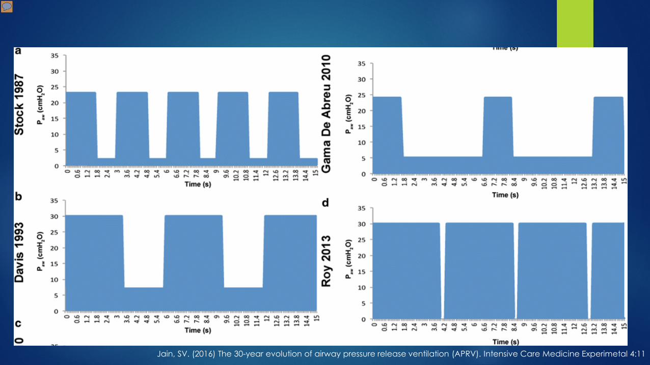
Jain, SV. (2016) The 30-year evolution of airway pressure release ventilation (APRV). Intensive Care Medicine Experimetal 4:11

Types of APRV
Fixed (F-APRV) and Personalized (P-APRV) Fixed: T-high makes up <90% of cycle time with fixed T-low that doesn’t
change with lung mechanics
Personalized: P-high set to desired P-plateau, T-high set to 90% of cycle time, T-low set based on lung mechanics/slope of expiratory flow curve, and P-low set at 0 (minimize convective expiratory gas flow resistance and maximize ventilation while maintaining PEEP) EEF/PEF ~0.75
Habashi NM (2005) Other approaches to open-lung ventilation: airway pressure release ventilation. Crit Care Med 33:S228–S240
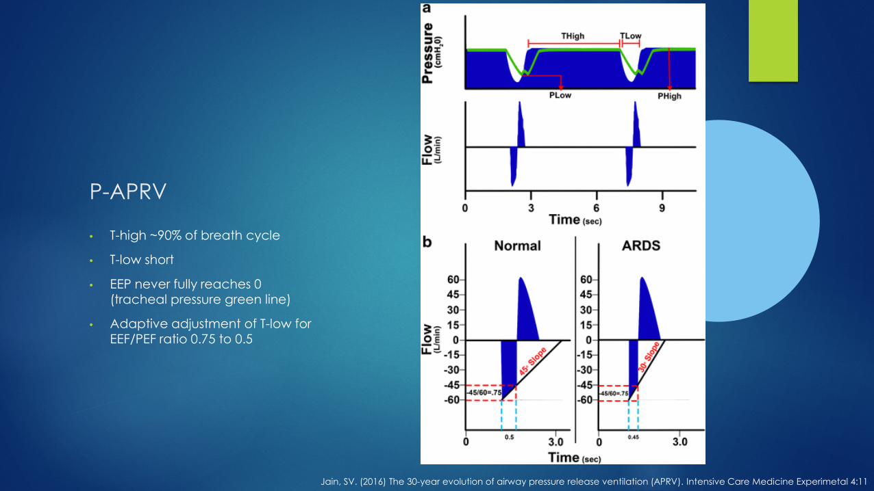
P-APRV
• T-high ~90% of breath cycle
• T-low short
• EEP never fully reaches 0 (tracheal pressure green line)
• Adaptive adjustment of T-low for EEF/PEF ratio 0.75 to 0.5
Jain, SV. (2016) The 30-year evolution of airway pressure release ventilation (APRV). Intensive Care Medicine Experimetal 4:11
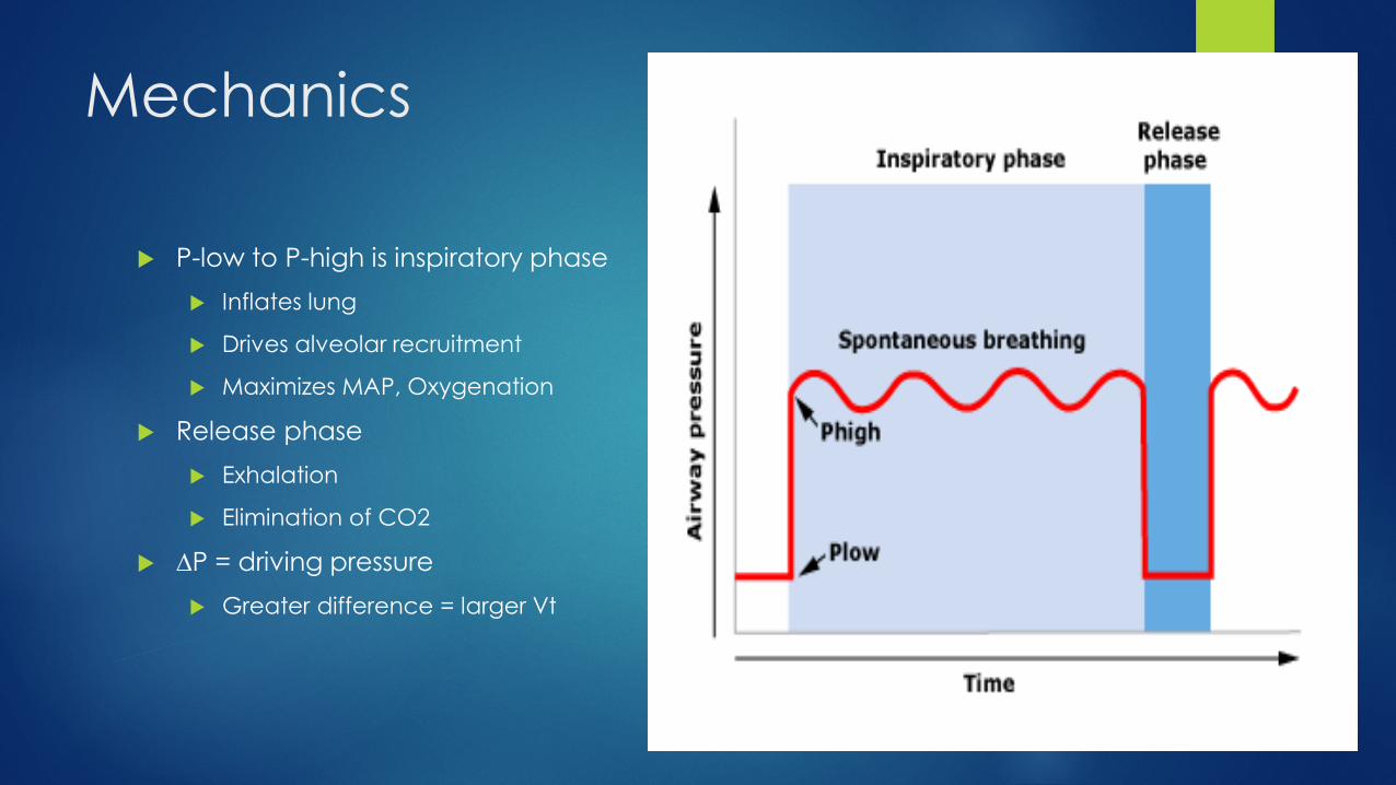
Mechanics
P-low to P-high is inspiratory phase Inflates lung
Drives alveolar recruitment
Maximizes MAP, Oxygenation
Release phase Exhalation
Elimination of CO2
∆P = driving pressure Greater difference = larger Vt

Mechanics
Spontaneous breathing at any point in cycle Elimination of CO2 Most at P-high due to duration
Use of high CPAP leads to restoration of lost FRC (in the setting of reduced compliance) Facilitates improvement of the
pressure volume curve and improved ventilatory parameters
Open lung approach Avoids repeated inflation/deflation
of alveoli

Spontaneous Breathing
Available at any point in cycle of APRV Patient controlled respiratory frequency and volumes Limits sedation needs Improved synchrony
Major difference between APRV and other IRV modes

To Add PS or Not To Add PS

To Add PS or Not To Add PS
NO

To Add PS or Not To Add PS
Spontaneous breathing only accounts for 10-30% of ventilation in APRV
Cyclical breathing with negative pressure effort (No PS) Decreased intrathoracic pressure, improved venous return, improved
cardiac output/index Higher O2 delivery and mixed venous saturations Improved vasopressor use
Improves dependent lung recruitment and VQ matching Preserves diaphragmatic strength Minimizes atelectasis in the near diaphragmatic and dependent
spaces
Kaplan LJ, Bailey H, Formosa V. Airway pressure release ventilation increases cardiac performance in patients with ALI./ARDS. Crit Care. 2001 Aug; 5(4):221-6.Modrykamien, A. Airway Pressure Release Vetilation: An alternative mode of mechanical ventilation in ARDS. Cleveland Clinic J of Med. 2011 Feb; 78(2): 101-10

Ventilation in the Dependent Lung
Diaphragm movement and ventilation in spontaneous, positive pressure, and paralyzed lung
Negative pressure breathing recruits best VQ matched lung

Pressure support
If you must use it, P-high must be decreased to maintain peak pressures <30-35 Set from P-low or drop P-high and add
Follow for evidence of overdistention, lung injury Increased risk of pneumothorax/pneumomediastinum With use of Pressure Support must follow:
Trigger, requires synchronization
T-low alterations
Variable volumes and volume loss/alveolar collapse
Kaplan LJ, Bailey H, Formosa V. Airway pressure release ventilation increases cardiac performance in patients with ALI./ARDS. Crit Care. 2001 Aug; 5(4):221-6.Modrykamien, A. Airway Pressure Release Vetilation: An alternative mode of mechanical ventilation in ARDS. Cleveland Clinic J of Med. 2011 Feb; 78(2): 101-10

Challenges
Control of parameters and proper waveform assessment for adjustment
Deviation from original concept of CPAP with timed release Non-standard usage and settings Use of Pressure Support

APRV
Benefits Improved oxygenation with
decreased peak airway pressure
Improved dead space ventilation
Improved alveolar recruitment and FRC
Decreased sedation needs
Improved VQ match and decreased shunting
Improved venous return and CO
Risks/Drawbacks Dyssynchrony
Loss of benefit with heavy sedation and/or paralysis
Auto-PEEP
Hemodynamic instability in susceptible patients
High airway resistance
Volutrauma/stretch injury

APRV Settings

APRV Starting Points
P-high At plateau or slightly above desired MAP
26-30cm H2O
Keep below 35cm H2O (as possible)
T-high 4-6 seconds
RR 8-15

APRV Starting Points
P-low 0
Can use low PEEP if using BIPAP style mode with longer T-low
T-low 0.4-0.8 seconds
Should terminate breath when EEF/PEF ratio 0.75 to 0.5
FiO2 1.0
PS NONE

APRV titration
Oxygenation FiO2 MAP
Increase P-high Decrease T-low
Hypercapnia Increase P-high Lengthen T-low (do not drop EEF/PEF ratio below 0.25) Increase T-high to allow more spontaneous breaths Decrease T-high to allow more releases
Hyperventilation Decrease P-high Increase T-high to decrease number of releases

APRV titration
Increased work of breathing Increase P-high to increase MAP/Recruitment
Decrease T-low to maximize FRC
Decrease P-high and increase T-high Maintains MAP but decreases overdistention

APRV Weaning
Wean FiO2 Goal is <0.5
Drop and stretch (transition to CPAP with spontaneous breathing) Simultaneous changes: Decrease P-high
Increments of 2cm H2O Goal <20 cm H2O
Increase T-high Decreased number of releases Goal >10 secods
Once at adequate pressures, transition to VCV or PCV with high PEEP

Current Status of APRV
Paucity of data Non-comparable studies
Non-standardized definitions and settings
Study types limited Almost entirely crossover or retrospective data
APRV is a divided name with very different mechanics and breath styles F-APRV inferior to P-APRV in protective mechanics for the lung
Rose L, Hawkins M (2008) Airway pressure release ventilation and biphasic positive airway pressure: a systematic review of definitional criteria. Intensive Care Med 34:1766–1773Facchin F, Fan E (2015) Airway pressure release ventilation and high-frequency oscillatory ventilation: potential strategies to treat severe hypoxemia and prevent ventilator-induced lung injury.
Respir Care 60:1509–1521
Current Status of APRV
Well proven to be non-inferior Putsenen et al. – improved sedation needs, increased oxygenation, CI,
and pulmonary compliance. No change in mortality or ventilator free days
Varpula et al. – similar mortality and ventilator free days, APRV group with higher disease acuity
Maxwell et al. – no change in mortality, ventilator free days, or complication rates despite APRV group with high baseline disease severity
Current Status of APRV
Possibly superior Hanna et al. – improved P/F ratio, procurement rate, and graft survival
in donor lungs
Davies et al. – improved oxygenation with decreased cerebral ischemia in TBI
Andrews et al. – decreased incidence of ARDS and mortality in trauma patients compared to traditional PPV
Hussain et al. – earlier hemodynamic stability in septic shock patients

Conclusions
APRV, when used optimally is an adaptive, flow directed, duration dependent mode of ventilation that can be modified for patients with multiple lung pathologies
It has similar mortality, ICU day, and ventilator day outcomes to traditional low Vt ventilation in ARDS (non-inferior), but has not been proven superior Slow adoption
The “definition” of the mode is still in flux and being debated Lack of familiarity with the physiology may result in maladjustment
and loss of the benefits of the mode NEEDS MORE RESEARCH
