apoptosis crma-sensitive death · pdf filetnf-a/cycloheximide (chx)-induced apoptosis....
TRANSCRIPT

Proc. Natl. Acad. Sci. USAVol. 92, p. 8318-8322, August 1995Medical Sciences
Tumor necrosis factor-induced apoptosis is mediated by aCrmA-sensitive cell death pathway
(programmed cell death/tumor necrosis factor a/interleukin lj-converting enzyme)
MASAYUKI MIURA*t, ROBERT M. FRIEDLANDER*tI, AND JUNYING YUAN*t*Cardiovascular Research Center, Massachusetts General Hospital-East, 149 13th Street, Charlestown, MA 02129; tDepartment of Medicine, Harvard MedicalSchool, Boston, MA 02115; and *Department of Neurosurgery, Massachusetts General Hospital, Harvard Medical School, Boston, MA 02114
Communicated by Jerome Gross, Massachusetts General Hospital-East, Charlestown, MA, May 5, 1995
ABSTRACT We report here that the activation of theinterleukin 1l3 (IL-1(3)-converting enzyme (ICE) family islikely to be one of the crucial events of tumor necrosis factor(TNF) cytotoxicity. The cowpox virus CrmA protein, a mem-ber of the serpin superfamily, inhibits the enzymatic activityof ICE and ICE-mediated apoptosis. HeLa cells overexpress-ing crmnA are resistant to apoptosis induced by Ice but not byIch-l, another member of the Ice/ced-3 family of genes. Wefound that the CrmA-expressing HeLa cells are resistant toTNF-a/cycloheximide (CHX)-induced apoptosis. Inductionof apoptosis in HeLa cells by TNF-a/CHX is associated withsecretion of mature IL-18, suggesting that an IL-18-pro-cessing enzyme, most likely ICE itself, is activated by TNF-a/CHX stimulation. These results suggest that one or moremembers of the ICE family sensitive to CrmA inhibition areactivated and play a critical role in apoptosis induced by TNF.
Interleukin 1(3-converting enzyme (ICE), a novel cysteineprotease, is homologous to the Caenorhabditis elegans celldeath gene product CED-3 (1). ICE was identified as theenzyme responsible for cleaving prointerleukin 113 (pro-IL-1(3)to generate mature biologically active IL-1f3 (2, 3). Thesequence Lys-Pro-Xaa4-Gln-Ala-Cys-Arg-Gly, encompassingthe active-site cysteine, and the amino acid residues that formthe P1 carboxylate-binding pocket are entirely conserved inCED-3 and ICE. The structural similarity between ICE andCED-3 suggests functional conservation of both proteins.Emerging evidence suggests that members of the Ice/ced-3gene family play an important role in controlling programmedcell death (apoptosis) (4-7). Expression ofIce and Ich-1 (Nedd-2), a new member of the Ice/ced-3 family, induces programmedcell death in a variety of cell lines, which is inhibited byexpression of bcl-2, a mammalian protooncogene that cansuppress apoptosis. The death of chicken dorsal root ganglianeurons induced by trophic factor deprivation in culture can besuppressed by microinjection of an expression construct ofcrnmA, a cowpox virus gene encoding a serpin that is a specificand potent inhibitor of ICE but a weak inhibitor of ICH-lL(4-6, 8, 9). Expression of crmA also protects Rat-1 cells fromapoptosis induced by serum removal (6). These results suggestthat members of the ICE family sensitive to CrmA inhibitionplay important roles in controlling cell death induced by avariety of stimuli.Tumor necrosis factor a (TNF-a) is a pleiotrophic tumori-
cidal cytokine (10). TNF-a affects the growth, differentiation,and function of a multitude of cell types and is a mediator ofinflammation and cellular immune responses (11-14). One ofthe striking functions of TNF-a is to induce apoptosis intransformed cells (15). In the case of nontransformed cells,TNF-a can also induce apoptosis in the presence of metabolicinhibitors (10). Here we report that TNF-a activates the
activity of the ICE family in HeLa cells and that TNF-a-induced apoptosis is suppressed by CrmA. Our results suggestthat TNF-a activates the ICE/CED-3-mediated cell deathpathway.
MATERIALS AND METHODSCells and Tissue Culture. HeLa cells were grown in Dul-
becco's modified Eagle's medium (DMEM) with 10% (vol/vol) fetal bovine serum. HeLa cells were transfected with thepHD1.2 crnmA expression vector (5) by calcium phosphateprecipitation. Two days after transfection, 600 ,ug-ml-1 ofG418 (GIBCO) was added for selection. Resistant colonieswere cloned by limiting dilution. To test the dosage responseofHeLa and HeLa/CrmA cells to TNF-a treatment, cells wereseeded in DMEM plus 10% (vol/vol) fetal calf serum in a24-well plate at a density of 4 x 10⁴ cells per well. After anovernight incubation, the cells were washed twice with serum-free DMEM. Drugs were then added to a total volume of 0.2ml of serum-free DMEM and incubated for 24 hr. Cells werethen trypsinized, and dead cells were scored on a hemocy-tometer by trypan blue exclusion (Sigma). At least 200 cellswere scored per well. Each concentration was tested in dupli-cate each time in three separate experiments.
Immunoblotting (Western Blotting). Cells were lysed inSDS sample buffer (50 mM Tris HCl, pH 6.8/100 mM dithio-threitol/2% SDS/0.1% bromphenol blue/10% glycerol), andcell lysates were subjected to SDS/15% PAGE. After theproteins were electroblotted to Immobilon nylon membrane(Millipore), the membrane was blocked with 4% nonfat milkin TBST (25 mM Tris-HCl, pH 7.5/150 mM NaCl/0.2%Tween). The membrane was incubated with the anti-CrmAantibody (5) at 5 ,ggml-' for 1 hr at room temperature andthen washed five times with TBST. The membrane was incu-bated with horseradish peroxidase-conjugated goat anti-rabbitIgG (1:1000 dilution; Amersham) for 30 min and then washedfive times with TBST. CrmA protein was detected with anenhanced chemiluminescence detection kit (Amersham).DNA Transfection. One day before transfection, cells were
seeded at a density of about 2 x 10⁵ per well in six-well dishes.For each well, 1 ,ug ofplasmidDNA and 10 ,ug of lipofectaminereagent were added according to a protocol from GIBCO/BRL. Cells were incubated for 3 hr in serum-free mediumcontaining DNA and lipofectamine; then the medium waschanged to DMEM containing 10% fetal bovine serum, andincubation was continued for 24 hr. The expression of chimericgene was detected as described (4).
Detection of IL-18 Production from HeLa Cells. HeLa cellswere grown in DMEM containing 10% fetal calf serumovernight, and then the medium was changed to serum-freeDMEM with or without drugs. After 24 hr, cells were scrapedoff and precipitated. Conditioned medium was collected,
Abbreviations: IL-1,B, interleukin 1,3; ICE, IL-113-converting enzyme;TNF, tumor necrosis factor; CHX, cycloheximide.
The publication costs of this article were defrayed in part by page chargepayment. This article must therefore be hereby marked "advertisement" inaccordance with 18 U.S.C. §1734 solely to indicate this fact.
8318
Proc. Natl. Acad. Sci. USA 92 (1995) 8319
dialyzed against distilled water at 4°C overnight, lyophilized,and then dissolved in distilled water. Cell precipitates were
extracted with the extraction buffer (20mM Hepes adjusted topH 7.4 with NaOH containing 10 mM KCl, 1.5 mM MgCl2, 0.5mM EDTA, 1% Nonidet P-40, and 10 ,tg of phenylmethyl-sulfonyl fluoride, 10 jig of E64, 2 jig of pepstatin, 1 ,ug ofleupeptin, and 0.5 Ag of aprotinin per ml). Insoluble materialwas removed by centrifugation. Proteins were separated bySDS/15% PAGE, and IL-113 was detected by immunoblottingwith an anti-human IL-1,3 antibody (1:300 dilution; Calbio-chem) as described above.
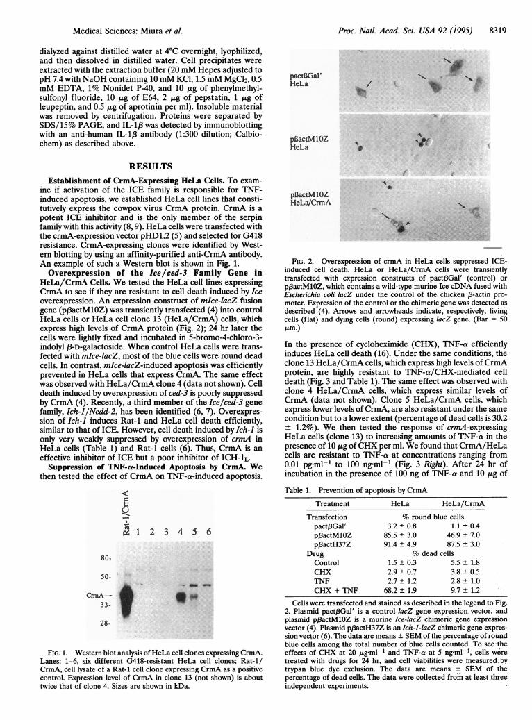
pactBGal'HeLa
plBactM IOZHeLa It
RESULTS
Establishment of CrmA-Expressing HeLa Cells. To exam-ine if activation of the ICE family is responsible for TNF-induced apoptosis, we established HeLa cell lines that consti-tutively express the cowpox virus CrmA protein. CrmA is a
potent ICE inhibitor and is the only member of the serpinfamily with this activity (8, 9). HeLa cells were transfected withthe crmA-expression vector pHD1.2 (5) and selected for G418resistance. CrmA-expressing clones were identified by West-ern blotting by using an affinity-purified anti-CrmA antibody.An example of such a Western blot is shown in Fig. 1.Overexpression of the Ice/ced-3 Family Gene in
HeLa/CrmA Cells. We tested the HeLa cell lines expressingCrmA to see if they are resistant to cell death induced by Iceoverexpression. An expression construct of mIce-lacZ fusiongene (p3actMlOZ) was transiently transfected (4) into controlHeLa cells or HeLa cell clone 13 (HeLa/CrmA) cells, whichexpress high levels of CrmA protein (Fig. 2); 24 hr later thecells were lightly fixed and incubated in 5-bromo-4-chloro-3-indolyl f3-D-galactoside. When control HeLa cells were trans-fected with mkce-lacZ, most of the blue cells were round deadcells. In contrast, mkce-lacZ-induced apoptosis was efficientlyprevented in HeLa cells that express CrmA. The same effectwas observed with HeLa/CrmA clone 4 (data not shown). Celldeath induced by overexpression of ced-3 is poorly suppressedby CrmA (4). Recently, a third member of the Ice/ced-3 genefamily, Ich-l/Nedd-2, has been identified (6, 7). Overexpres-sion of Ich-1 induces Rat-1 and HeLa cell death efficiently,similar to that of ICE. However, cell death induced by Ich-1 isonly very weakly suppressed by overexpression of crmA inHeLa cells (Table 1) and Rat-1 cells (6). Thus, CrmA is an
effective inhibitor of ICE but a poor inhibitor of ICH-iL.Suppression of TNF-a-Induced Apoptosis by CrmA. We
then tested the effect of CrmA on TNF-a-induced apoptosis.
X 1 2 3 4 5 6
80-
50-
CmA-33-
28 -
'S.m
FIG. 1. Western blot analysis ofHeLa cell clones expressing CrmA.Lanes: 1-6, six different G418-resistant HeLa cell clones; Rat-1/CrmA, cell lysate of a Rat-1 cell clone expressing CrmA as a positivecontrol. Expression level of CrmA in clone 13 (not shown) is abouttwice that of clone 4. Sizes are shown in kDa.
pBactM lOZHeLa/CrmA
a
FIG. 2. Overexpression of crmA in HeLa cells suppressed ICE-induced cell death. HeLa or HeLa/CrmA cells were transientlytransfected with expression constructs of pact3Gal' (control) or
pf3actMlOZ, which contains a wild-type murine Ice cDNA fused withEscherichia coli lacZ under the control of the chicken ,B-actin pro-moter. Expression of the control or the chimeric gene was detected as
described (4). Arrows and arrowheads indicate, respectively, livingcells (flat) and dying cells (round) expressing lacZ gene. (Bar = 50tLm.)
In the presence of cycloheximide (CHX), TNF-a efficientlyinduces HeLa cell death (16). Under the same conditions, theclone 13 HeLa/CrmA cells, which express high levels of CrmAprotein, are highly resistant to TNF-a/CHX-mediated celldeath (Fig. 3 and Table 1). The same effect was observed withclone 4 HeLa/CrmA cells, which express similar levels ofCrmA (data not shown). Clone 5 HeLa/CrmA cells, whichexpress lower levels of CrmA, are also resistant.under the samecondition but to a lower extent (percentage of dead cells is 30.2
1.2%). We then tested the response of crmA-expressingHeLa cells (clone 13) to increasing amounts of TNF-a in thepresence of 10 ,ug of CHX per ml. We found that CrmA/HeLacells are resistant to TNF-a at concentrations ranging from0.01 pgml-1 to 100 ng-ml-1 (Fig. 3 Right). After 24 hr ofincubation in the presence of 100 ng of TNF-a and 10 ,ug of
Table 1. Prevention of apoptosis by CrmA
Treatment HeLa HeLa/CrmATransfection % round blue cells
pactf3Gal' 3.2 ± 0.8 1.1 ± 0.4p,BactMlOZ 85.5 ± 3.0 46.9 ± 7.0p,BactH37Z 91.4 ± 4.9 87.5 ± 3.0
Drug % dead cellsControl 1.5 ± 0.3 5.5 ± 1.8CHX 2.9±0.7 3.8±0.5TNF 2.7 ± 1.2 2.8 ± 1.0CHX + TNF 68.2 ± 1.9 9.7 ± 1.2
Cells were transfected and stained as described in the legend to Fig.2. Plasmid pact,BGal' is a control lacZ gene expression vector, andplasmid pf3actMlOZ is a murine Ice-lacZ chimeric gene expressionvector (4). Plasmid pB3actH37Z is an Ich-1-lacZ chimeric gene expres-sion vector (6). The data are means ± SEM of the percentage of roundblue cells among the total number of blue cells counted. To see theeffects of CHX at 20 ug-ml-1 and TNF-ca at 5 ngml-1, cells weretreated with drugs for 24 hr, and cell viabilities were measured.bytrypan blue dye exclusion. The data are means ± SEM of thepercentage of dead cells. The data were collected from at least threeindependent experiments.
Medical Sciences: Miura et aL
..........

8320 Medical Sciences: Miura et al.
HeLA HrLaCrnA
CON
CHX
TNF
CH)+
TNF
0 I- 1- Q0 00 r- 0 0 0 0
Co '- o00 0
1o 0 00-C
TNF-a, pg/ml
80 -
70-coa 60-0
-o 50-(U
40-
30-20 -
10 -
I ,:
FIG. 3. TNF-induced cytotoxicity was suppressed by overexpression of CrmA. (Left) HeLa cells or HeLa/CrmA cells were treated with CHX(20 ,ug-ml-l; Sigma) alone, TNF-a (5 ng-ml-1; Sigma) alone, or a combination of both drugs. Cells were photographed 24 hr after drug treatment.Con, control. (Bar = 50 ,um.) (Right) Control HeLa and HeLa/CrmA (clone 13) cells were tested for their ability to resist increasing amounts ofTNF-a in the presence of 10 ,ug of CHX per ml; 1 jig of TNF-a has 1.1 x 10⁵ units of activity according to R & D Systems. The results were fromthree experiments, with each condition done in duplicate.
CHX per ml, 83% of control HeLa cells died compared with23% of HeLa/CrmA cells.
Activation of the ICE Family After TNF Stimulation. Wehave detected the expression of both Ice and Ich-1 in HeLacells (6). Since expression of crmA effectively prevents celldeath induced by TNF-a in the presence ofCHX, the cell deathpathway mediated by the ICE family members sensitive toCrmA inhibition may be activated by TNF-a stimulation andmay play a major role in the induction of HeLa cell death. Ifthis is the case, TNF-a stimulation may activate endogenousIL-1,-processing activity in HeLa cells. To date, pro-IL-i j3 isthe only known endogenous substrate of ICE. If ICE isactivated after TNF-a/CHX stimulation, the endogenous 33-kDa pro-IL-1i3 should be processed, and the mature 17.5-kDaIL-1,B should be secreted. To detect mature IL-1,B, we col-lected conditioned media from HeLa cells with or withoutTNF stimulation and analyzed the processing of pro-IL-1,3 byWestern blot (Fig. 4). Our data show that mature IL-1,B was
observed only after induction of apoptosis by TNF-a/CHX.HeLa/CrmA cells are much more resistant to TNF-a/CHX-induced cell death and secreted much less mature IL-1 3 underthe same condition, which can be observed as a very faint bandonly after long exposure. These results strongly suggest thatapoptosis induced by TNF stimulation is mediated by one ormore members of the ICE family that are sensitive to CrmAinhibition and can process pro-IL-13.
DISCUSSION
Our previous work demonstrated that overexpression of ICEinduces Rat-1 cells to undergo apoptosis (4) and that expres-sion of crmA can prevent chicken dorsal root ganglion (DRG)neurons from cell death induced by trophic factor deprivation(5). These results showed that ICE has the ability to induce celldeath and that inhibition of ICE activity can prevent pro-grammed cell death. Using pro-IL-i,B processing as an indi-cator, we demonstrate here that mature IL-1,B is producedwhen HeLa cells are induced to die by treatment with TNF-a
and CHX. Our work has shown that the enzyme(s) that cancleave pro-IL-113 to mature IL-1i3 is activated in apoptosisinduced by TNF-a. Since ICE is the only enzyme identified so
far that can process pro-IL-l,B, our data suggest that ICE isactivated during apoptosis.The amino acid sequence of CrmA protein placed it in the
serpin family whose other members are all inhibitors thattarget serine proteases (17). Titrations of ICE with the CrmA-glutathione S-transferase (GST) fusion protein showed that2.5 mol of CrmA-GST is required to inhibit 1 mol of theenzyme with a K1 of <4 x 10-12 M (9). Purified CrmA failedto inhibit representative members of several major families ofserine proteases: trypsin, chymotrypsin, cathepsin G, pig andhuman elastases, thrombin, plasmin, human tissue plasmino-gen activator, and human urinary plasminogen activator. Italso failed to inhibit papain, a cysteine protease (8). CrmA didnot inhibit any of these proteases even when the ratio of CrmAto the proteases was 100:1 (wt/wt) (8). More recently, CrmAwas tested for the ability to form complexes with severalrepresentatives of major protease families on native PAGEanalysis: ICE, neutrophil elastase, cathepsin G, Staphylococcusaureus V8 proteinase and subtilisin Carlsberg. In these assays,only the ICE-CrmA complex was detected (9). Thus, CrmA isa specific inhibitor of the ICE family. Our own results showedthat CrmA inhibits ICE-induced cell death much more effec-tively than ICH-1L-induced cell death, suggesting that in theICE family, CrmA prefers ICE to ICH-iL. It is possible thatother members of the ICE family that are more homologousto ICE than to ICH-iL may also be effectively inhibited byCrmA. Although ICE is the only enzyme identified so far thatcan cleave pro-IL-1,3 to generate mature IL-l,l3, we cannot ruleout that other members of the ICE family may be able to cleavepro-IL-1,3 as well. Thus, we conclude from our results that theCrmA-sensitive member(s) of the ICE family play a criticalrole in apoptosis induced by TNF, and at least one of them hasthe ability to cleave pro-IL-1,3.Mice that are deficient in ICE have been reported (18). The
ICE mutant mice develop apparently normally, with the major
HeLa
Proc. Natl. Acad. Sci. USA 92 (1995)

Proc. Natl. Acad. Sci. USA 92 (1995) 8321Medical Sciences: Miura et al.
kDa 147-
24-
17-It
2 3 4 5 6 7 8
__~~~~~~.. _
123451 2 3 4 5
43kDa- *.w.
3OkDa -
2OkDa - lo 4-- Mature IL-1Il;
14kDa -
FIG. 4. Processing of IL-1,3 after induction of apoptosis by TNF-a/CHX in HeLa cells. (Upper) A Western blot of HeLa cells probedwith anti-human IL-1,B (Calbiochem). Lanes: 1, purified maturehuman IL-1,B; 3-6, cell lysates (10 ,ug Qf protein per lane); 7-10,supernatant (5 ,g per lane); 3 and 7, serum-free control; 4 and 8,lipopolysaccharide (LPS) (10 ,ggml-'; Sigma) treatment; 5 and 9, LPS(10 ,ug-ml-1), CHX (20 ,ug.ml-1), and TNF-a (5 ng.ml-1) treatment;and 6 and 10, CHX (20 ,ug.ml-1) and TNF-a (5 ng ml-1) treatment.Cell viabilities were measured by trypan blue exclusion (97.4 ± 1.5%for the serum-free control, 97.4 ± 0.2% for LPS treatment, 56.3 +
2.2% for TNF-a/CHX treatment). Arrows and the arrowhead indi-cate, respectively, pro-IL-113 and mature IL-113. (Lower) A Westernblot of supernatant from HeLa (lanes 1-4) or HeLa/CrmA cells (lane5) treated with no addition (control, lane 1) or 20 ,ug of CHX per ml(lane 2) or S ng of TNF-a per ml (lane 3) or 20 ,ug of CHX and 5 ngof TNF-a per ml (lanes 4 and 5), probed with anti-human IL-1,3(Calbiochem). The amount of protein was measured by Bio-Rad assay.Three micrograms of protein was loaded onto each lane.
defect being inability to produce mature IL-1l3 after stimula-tion with lipopolysaccharide. This result suggests the redun-dency of the ICE family in controlling vertebrate apoptosis:multiple members of the ICE family may all contribute to thecontrol of cell death. Alternatively, the cell death defects of themutant ICE mice may be revealed only by counting the numberof cells at appropriate places. For example, the transgenic miceexpressing bcl-2 under the control of neural-specific enolase(NSE) promoter or 3-phosphoglycerate kinase (PGK) pro-moter develop essentially normally, and yet upon cell counting,they contain up to 40% and 50% more neurons in the facialnucleus and the ganglion cell layer of the retina (19). C. elegansced-3 or ced-4 mutants also develop and behave normallydespite the fact that they contain up to 20% more cells (20).Activation of ICE has been reported in mammary epithelialcells induced to die by antibodies to (31 integrins or byoverexpression of stromelysin 1, which degrades extracellularmatrix (21). Such a result suggests the participation of ICE inapoptotic pathways activated by different signals.The death of HeLa cells induced by TNF-a in the presence
of cycloheximide shows DNA fragmeritation, a typical featureof apoptosis (16). Expression of the E1B 19-kDa protein, aviral homolog of the Bc1-2 protein (22), by viral infection, bytransient expression, or in transformed cells completely andspecifically blocks the TNF-a-induced DNA fragmentationand cell death (16). Thus, TNF-a stimulation likely activates anendogenous pathway of programmed cell death.The reasons why CHX potentiates TNF cytotoxicity in
nontransformed cells is unclear. Most of the cell lines includingHeLa cells, NIH 3T3 cells, and TA1 cells are not killed by TNFalone but are killed by the combined actions ofTNF and CHX(23, 24). TNF-a is a pleiotrophic cytokine that may inducemore than one cellular response in a single cell line. The
presence of CHX may inhibit the synthesis of certain signalingmolecules and, thus, potentiates the killing activity of TNF.Alternatively, CHX may simply inhibit the synthesis of ageneral cell survival factor(s) and, thus, allow cells to becomemore sensitive to TNF cytotoxicity.HeLa cells express predominantly the p55 TNF receptor,
which is thought to be responsible for cell death signaling (25,26). The TNF p55 receptor triggers the activation of phospho-lipase A2, protein kinase C, sphingomyelinase, phosphatidyl-choline-specific phospholipase C, and NF-KB (27, 28). In TNFp55 receptor-knockout mice, TNF-mediated induction ofNF-KB is prevented in thymocytes (29). TNF p55 receptor-knockout mice are resistant to lethal doses of either lipopoly-saccharides or S. aureus enterotoxin B, suggesting that the TNFp55 receptor mediates the pathogenesis of hepatocyte necrosis(29). One possible explanation for the resistance of HeLa/CrmA cells to TNF-a is that they might have lost the TNF-areceptor. This is highly unlikely because HeLa/CrmA cellslose the expression of crmA easily if propagated for more thana few weeks in culture without selection; and when they losethe expression, they become completely sensitive to TNF-aagain (data not shown). In addition, three HeLa/CrmA clonestested show CrmA- expression level-dependent resistance toTNF-a.TNF-a is a major player in host inflammatory responses of
mammals (6). Upon injection of endotoxin (LPS) in models ofseptic shock, TNF, IL-1, and IL-6 are quickly induced (30). Insuch conditions, the secretion of IL-113 appears to be depen-dent upon TNF, since passive immunization with TNF mono-clonal antibodies during endotoxemia in vivo attenuates theappearance of IL-1,B (31). Our results here suggest that TNFmay play a role in activating members of the ICE family toprocess pro-IL-113. Expression of mitochondrial manganesesuperoxide dismutase has been shown to promote the survivalof tumor cells exposed to TNF (32), suggesting that generationof free radicals may play a role in cell death induced by TNF.There are several reports that TNF cytotoxicity is related tothe generation of free radicals and lipid peroxides (33, 34).These observations raised the possibility that members of theICE family may be activated directly or indirectly by freeradicals.
Note: After we submitted this paper, Tewari and Dixit (35) reportedthat expression of CrmA inhibited TNF-a- and anti-Fas-induced celldeath in MCF-7 cells.
We thank L. Bergeron for critical reading of the manuscript. Thiswork is supported in part by grants to J.Y. from Bristol-Myers Squibband from the National Institute of Aging. M.M. is supported by theMochida Memorial Foundation for Medical and Pharmaceutical Re-search and a postdoctoral fellowship from the National Institute ofHealth. R.M.F. is supported by a postdoctoral training fellowship fromthe National Institutes of Health.
1. Yuan, J., Shaham, S., Ledoux, S., Ellis, H. M. & Horvitz, H. R.(1993) Cell 75, 641-652.
2. Black, R. A., Kronheim, S. R. & Sleath, P. R. (1989) FEBS Lett.247, 386-390.
3. Kostura, M. J., Tocci, M. J., Limjuco, G., Chin, J., Cameron, P.,Hillman, A. G., Chartrain, N. A. & Schmidt, J. A. (1989) Proc.Natl. Acad. Sci. USA 86, 5227-5231.
4. Miura, M., Zhu, H., Rotello, R., Hartweig, E. A. & Yuan, J.(1993) Cell 75, 653-660.
5. Gagliardini, V., Fernandez, P.-A., Lee, R. K. K., Drexler,H. C. A., Rotello, R., Fishman, M. & Yuan, J. (1994) Science 263,826-828.
6. Wang, L., Miura, M., Bergeron, L., Zhu, H. & Yuan, J. (1994)Cell 78, 739-750.
7. Kumar, S., Kinoshita, M., Noda, M., Copeland, N. G. & Jenkins,N. A. (1994) Genes Dev. 8, 1613-1626.

8322 Medical Sciences: Miura et al.
8. Ray, C. A., Black, R. A., Kronheim, S. R., Greenstreet, T. A.,Sleath, P. R., Salvesen, G. S. & Pickup, D. J. (1992) Cell 69,597-604.
9. Komiyama, T., Ray, C. A., Pickup, D. J., Howard, A. D., Thorn-berry, N. A., Peterson, E. P. & Salvesen, G. (1994)J. Biol. Chem.269, 19331-19337.
10. Tracey, K. J. & Cerami, A. (1993) Annu. Rev. Cell Biol. 9,317-343.
11. Beutler, B. & Cerami, A. (1988) Annu. Rev. Biochem. 57,505-518.
12. Beutler, B. & Cerami, A. (1989)Annu. Rev. Immunol. 7,625-655.13. Paul, N. L. & Ruddle, N. H. (1988) Annu. Rev. Immunol. 6,
407-438.14. Vassalli, P. (1992) Annu. Rev. Immunol. 10, 411-452.15. Laster, S. M., Good, J. G. & Gooding, L. R. (1988) J. Immunol.
141, 2629-2634.16. White, E., Sabbatini, P., Debbas, M., Wold, W. S. M., Kusher,
D. I. & Gooding, L. (1992) Mol. Cell. Biol. 12, 2570-2580.17. Carrell, R. W. & Travis, J. (1985) Trends Biochem. Sci. 10, 20-24.18. Li, P., Allen, H., Banerjee, S., Franklin, S., Herzog, L., Johnston,
C., McDowell, J., Paskind, M., Rodman, L., Salfeld, J., Towne, E.,Tracey, D., Wardwell, S., Wei, F., Wong, W., Kamen, R. &Seshadri, T. (1995) Cell 80, 401-411.
19. Martinou, J.-C., Dubois-Dauphin, M., Staple, J. K., Rodriguez,Frankowski, H., Missotten, M., Albertini, P., Talabot, D., Catsi-cas, S., Pietra, C. & Huarte, J. (1994) Neuron 13, 1017-1030.
20. Ellis, H. M. & Horvitz, H. R. (1986) Cell 44, 817-829.21. Boudreau, N., Sympson, C. J., Werb, Z. & Bissell, M. J. (1995)
Science 267, 891-893.22. Chiou, S. K., Tseng, C. C., Rao, L. & White, E. (1994)J. Virol. 68,
6553-6566.
23. Reid, T., Torti, F. M. & Reingold, G. M. (1991) J. Biol. Chem.264, 4583-4589.
24. Reid, T., Ramesha, C. S. & Reingold, G. M. (1991)J. Biol. Chem.266, 16580-16586.
25. Engelmann, H., Brakebush, C., Avni, S., Sarov, I., Nophar, Y.,Hadas, E., Leitner, 0. & Wallach, D. (1990) J. Biol. Chem. 265,14497-14504.
26. Thoma, B., Grell, M., Pfizenmaier, K. & Scheurich, P. (1990) J.Exp. Med. 72, 1019-1023.
27. Wiegmann, K., Schutze, S., Kampen, E., Himmler, A., Machleidt,T. & Kronke, M. (1992) J. Biol. Chem. 267, 17997-18001.
28. Schutze, S., Potthoff, K., Machleidt, T., Berkovic, D., Wiegmann,K. & Kronke, M. (1992) Cell 71, 765-776.
29. Pfeffer, K., Matsuyama, T., Kungig, T. M., Wakeham, A., Kishi-hara, K., Shahinian, A., Wiegmann, K., Ohashi, P. S., Kronke, M.& Mak, T. W. (1993) Cell 73, 457-467.
30. Ayala, J. M., Yamin, T.-T., Egger, L.-A., Chin, J., Kostura, M. J.& Miller, D. K. (1994) J. Immunol. 153, 2592-2599.
31. Fong, Y., Tracey, K. J., Moldwer, L. L., Hesse, D. G., Manogue,K. B., Kenney, J. S., Lee, A. T., Kuo, G. C., Allison, A. C., Lowry,S. F. & Cerami, A. (1989) J. Exp. Med. 170, 1627-1633.
32. Hirose, K., Longo, D. L., Oppenheim, J. J. & Matsushima, K.(1993) Mol. Cell. Biol. 13, 3301-3310.
33. Hennet, T., Richter, C. & Peterhans, E. (1993) Biochem. J. 289,587-592.
34. Schulze-Osthoff, K., Bakker, A. C., Vanhaesebroeck, B., Beyaert,R., Jacob, W. A. & Fiers, W. (1992) J. Biol. Chem. 267, 5317-5323.
35. Tewari, M. & Dixit, V. M. (1995) J. Biol. Chem. 270, 3255-3260.
Proc. Natl. Acad. Sci. USA 92 (1995)