aortic graft infection- contemporary management of a ... pdfs... · aortic graft...
TRANSCRIPT

1
Aortic Graft Infection-Contemporary
Management of a Resurgent Problem
Peter F. Lawrence, MDProfessor and Chief
Division of Vascular SurgeryUniversity of California Los Angeles
Incidence of Aortic Graft Infection
Meta-analysis - 13 series with 11,526 aortic grafts1.6% incidence; highest with aortofemoral graft
Aortoenteric fistula/erosion - 0.75%Underestimates true incidenceProjected infections - 95,000 grafts x 1.6 =1,520/year
Sarfati - Epidemiology of Aortic Graft Infection in Gewertz Surgery of the Aorta
Aortic Graft Infection Morbidity/Mortality
High mortality: – One year survival - 65%; 5 year survival - 55%– Early mortality- sepsis, MSOF, hemorrhage, renal failure, MI– Late mortality- Graft related(recurrent infection), CV disease– Mortality declining
Morbidity– Limb loss - 20% – Pneumonia, renal failure, cardiac - 60%– Reoperation - 20%
Re-infection of new graft – 20-60%Occlusion of new graft - 25%
Evolution of Aortic Graft Infection
Incidence of Aortic Graft Infection
0
2
4
6
8
10
12
14
16
1952 1958 1970 1976 1978 1985 1995 2005
Year
1st graft
Graft inclusion technique
Aortofemoral graft
End-to-end preferable
Routine antibiotics
Vascular Surgeons
Stent grafts?
Per
cent
age
Infe
cted
2
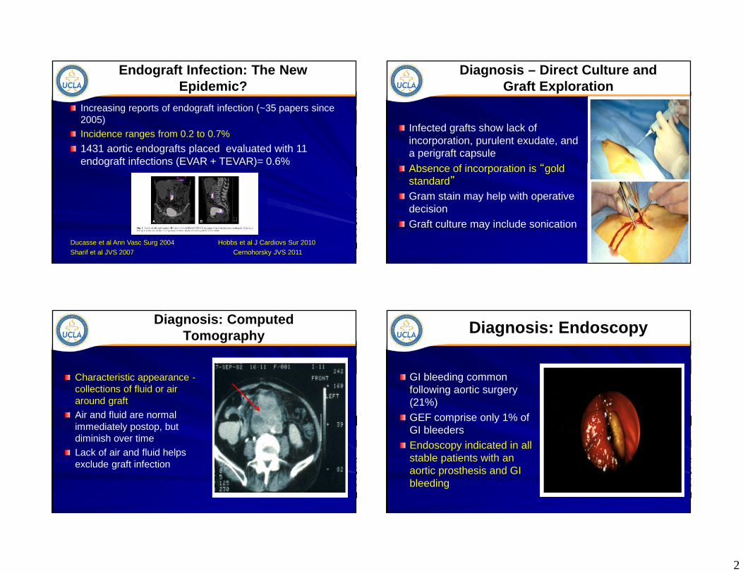
Endograft Infection: The New Epidemic?
Increasing reports of endograft infection (~35 papers since 2005)
Incidence ranges from 0.2 to 0.7%
1431 aortic endografts placed evaluated with 11 endograft infections (EVAR + TEVAR)= 0.6%
Ducasse et al Ann Vasc Surg 2004 Hobbs et al J Cardiovs Sur 2010
Sharif et al JVS 2007 Cernohorsky JVS 2011
Diagnosis – Direct Culture and Graft Exploration
Infected grafts show lack of incorporation, purulent exudate, and a perigraft capsule
Absence of incorporation is “gold standard”Gram stain may help with operative decision Graft culture may include sonication
Diagnosis: Computed Tomography
Characteristic appearance -collections of fluid or air around graftAir and fluid are normal immediately postop, but diminish over timeLack of air and fluid helps exclude graft infection
Diagnosis: Endoscopy
GI bleeding common following aortic surgery (21%)GEF comprise only 1% of GI bleedersEndoscopy indicated in all stable patients with an aortic prosthesis and GI bleeding
3
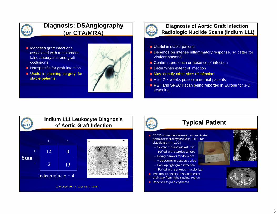
Diagnosis: DSAngiography(or CTA/MRA)
Identifies graft infections associated with anastomotic false aneurysms and graft occlusionsNonspecific for graft infectionUseful in planning surgery for stable patients
Diagnosis of Aortic Graft Infection: Radiologic Nuclide Scans (Indium 111)
Useful in stable patientsDepends on intense inflammatory response, so better for virulent bacteriaConfirms presence or absence of infectionDetermines extent of infectionMay identify other sites of infection+ for 2-3 weeks postop in normal patientsPET and SPECT scan being reported in Europe for 3-D scanning
Indium 111 Leukocyte Diagnosis of Aortic Graft Infection
12 0
2 13Scan
Indeterminate = 4
+
+
-
-
Lawrence, PF. J. Vasc Surg 1985
Typical Patient
57 YO woman underwent uncomplicated aorto-bifemoral bypass with PTFE for claudication in 2004– Severe rheumatoid arthritis,
– Rx’ed with steroids-24 ops– Heavy smoker for 45 years
– + troponins in post op period– Post op right groin infection– Rx’ed with sartorius muscle flap
Two month history of spontaneous drainage from right inguinal regionRecent left groin erythema

4
TreatmentLess Invasive Approaches
IV / topical antibioticsMuscle coverage without graft excisionDrainage with Abx irrigationVACReplacement with Abxbonded femoral graftNever “cure” infectionSeminars in Vascular Surgery 2011
Definitive Graft Infection Treatment –Excision Without Revascularization
Entire graft removal is conventional approach; revascularization not always requiredIf graft thrombosed, then removal alone is OKMay also work when indication was claudication or proximal anastomosis was E-SAortic aneurysms unlikely to tolerate graft removal alone
15/101 patients in one series not revascularized
Test to Determine Revascularization Need
Segmental pressures for multilevel diseaseAnkle pressure > 40 and ABI with graft occlusion/ compression Ankle pressure > 40 and ABI with angiographic balloon occlusion
Graft Excision with Extra-anatomic Revascularization
1st described by Blaisdell in 1961Gold standard for aortic infection involving more than isolated area of graftEarly results resulted in 40% mortality and 25% amputationRecent results with improved anesthesia and sequencing of procedures have 25% mortality

5
Staged Treatment of Aortic Graft Infection
Revascularization precedes graft excision by 1-2 daysEliminates period of prolonged ischemiaAllows for better hemodynamic stabilityRests surgical teamDoes not result in increased graft infection rate
Reilly J Vasc Surg 1987
Extraanatomic Bypass
This image cannot currently be displayed.
McCann Ann Surg 1993; Bunt Cardiovasc Surg 1993; Lawrence 1984
Graft Thrombosis: 10-20% at 5 yearsGraft Residual or reinfection: 5-20% at 5 years
Aortic stump disruption: 0-5%, but may occur years later
Revascularization with Autogenous Tissue (venous)
Jicha, Reilly, Goldstone JVS 1996
Prosthetic Insitu Replacement
Not appropriate when suture line is involved with bleedingMajor risk is recurrent infection
Debridement of infected aortic wall is critical
Most appropriate for patients with normal defenses and no extensive purulenceBest prosthesis is antibiotic bonded Dacron, using Rifampin with a gelatin bond
Lachapelle J Vasc Surg 1994

6
In Situ Replacement with Femoral Veins (NAIS)
Popularized by Claggett and colleagues at Southwestern“Neoaortoiliac system”
(NAIS)
NAIS Results
Study Patients (n)
Follow-Up (Months)
30-day Mortality
Major Amputatio
n
Clagett(1993) 21 23 10% 10%
Ehsan(2009) 48 56 2% 0%
Ali (2009) 144 32 10% 7%
NAIS Results
Reported 9% mortality and 5% amputation rate
Used with all organisms
Peripheral edema occurs, but usually controllable
Good durabilityLong procedure(10-12 hrs)
Clagett GP J Vasc Surg 1997
Insitu Revascularization with Allograft
Mean age = 65 ±±±± 9 years
Indication for allograft use:– Primary graft infection (n=125, 70%)
– Secondary aorto-enteric fistula (n=54, 30%)
62% of patients underwent 3 ±±±± 2
repeat operations before allograft
replacement
179 Patients
Fresh allograft:111 Patients
Cryopreserved allograft:
68 Patients
1988-2002

7
Kieffer E, et al
Allograft-related complications are significantly reduced by using
cryopreserved allografts rather than fresh allografts
Late mortality = 25.9% (allograft-related = 2.1%)– All 3 patient deaths were due to
allograft rupture at 9, 10, and 27 months.
– 2 patients received fresh allograft (66%)
Cyropreserved Allograft
Previous aneurysm concerns have been addressed with changes in preservation Options include Cryovein and Cryoartery
Expensive- are Cryoartery costs justified by better outcomes?
Advantages of CryoArtery vs. Cryovein for In-Line Reconstruction
Thicker wall vs. vein conduit– Durable material--less rupture risk
– Less risk for recurrent infection
Excellent fit: available as bifurcated conduitExpensive but cost-competitive– Does not require time in OR for
construction of neo-aortoiliac
segment
Duncan, et al, Allograft registry; JVS 2003
Uses of Allograft

8
Technique
• Need proximal and distal control above and below the infection
• Often requires supra-celiac clamping• Opening the retroperitoneum may
still result in significant bleeding• Necrotic tissue requires debridement• Sew up to the orifices of the renal
arteries• Occasionally need autogenous
transplant of renal arteries
Explanted Graft
Aortic Graft Infection: Single-Institution (UCLA) Experience
Vardanian AJ et al, Am Surgeon 2009
On behalf of the
Investigators
The Use of Cryopreserved AortoiliacAllograft for Aortic Reconstruction in the
United States

9
Results
220 Patients at 14 institutions (M:F = 1.6/1, Mean age = 65±12 yrs)
Indication for Use of CAA n (%)
Prosthetic graft infection 134 (61%)
Primary abdominal aortic infection 35 (16%)
Graft enteric fistula/erosion 33 (15%)
Infection pseudoaneurysm 9 (4%)
Other, including high risk of graft infection 9 (4%)
Type of Initial Aortic Procedure n (%)Open reconstruction 209 (95%)Endovascular 11 (5%)
Procedure Details
Operative Variable (N=220) n (%)
Graft Excision
Full excision 149 (68%)
Distal Anastomosis
Bilaterally to external iliac artery 139 (63%)
Bilaterally to femoral artery 66 (30%)
Unilateral to femoral and external iliac artery 15 (7%)
Concomitant Procedures with CAA Placement
Femoral artery to distal artery bypass 42 (19%)
Duodenal repair or colon resection 7 (3%)
Other vascular procedures 32 (15%)
Technique Early and Late Complications
Complication (n = 55) n (%)
Persistent sepsis 17 (8%)
CAA thrombosis/occlusion 9 (4%)
CAA rupture 8 (4%)
Recurrent CAA infection 8 (4%)
CAA pseudoaneurysm 6 (3%)
Fistula recurrence 4 (2%) Lower extremity compartment syndrome 1 (<1%)
Colonic perforation 1 (<1%)
Lower limb ischemia 1 (<1%)Mean follow-up = 30 ± 3 months Range = 1 to 160 months
92%86%
80%71%
10
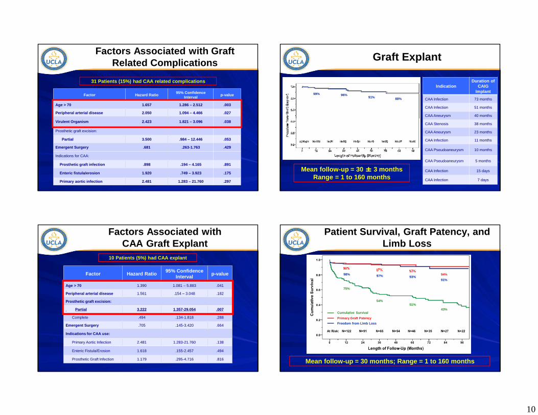
Factors Associated with Graft Related Complications
31 Patients (15%) had CAA related complications
Factor Hazard Ratio95% Confidence
Intervalp-value
Age > 70 1.657 1.286 – 2.512 .003
Peripheral arterial disease 2.050 1.094 – 4.466 .027
Virulent Organism 2.423 1.821 – 3.096 .038
Prosthetic graft excision:
Partial 3.500 .984 – 12.446 .053
Emergent Surgery .681 .263-1.763 .429
Indications for CAA:
Prosthetic graft infection .898 .194 – 4.165 .891
Enteric fistula/erosion 1.920 .749 – 3.923 .175
Primary aortic infection 2.481 1.283 – 21.760 .297
Graft Explant
IndicationDuration of
CAIGImplant
CAA Infection 73 months
CAA Infection 51 months
CAA Aneurysm 40 months
CAA Stenosis 38 months
CAA Aneurysm 23 months
CAA Infection 11 months
CAA Pseudoaneurysm 10 months
CAA Pseudoaneurysm 5 months
CAA Infection 15 days
CAA Infection 7 days
Mean follow-up = 30 ±±±± 3 months Range = 1 to 160 months
99% 96%91% 88%
Factors Associated with CAA Graft Explant
Factor Hazard Ratio95% Confidence
Intervalp-value
Age > 70 1.390 1.081 – 5.883 .041
Peripheral arterial disease 1.561 .154 – 3.048 .182
Prosthetic graft excision:
Partial 3.222 1.357-29.054 .007
Complete .494 .134-1.818 .288
Emergent Surgery .705 .145-3.420 .664
Indications for CAA use:
Primary Aortic Infection 2.481 1.283-21.760 .138
Enteric Fistula/Erosion 1.618 .155-2.457 .494
Prosthetic Graft Infection 1.179 .295-4.716 .816
10 Patients (5%) had CAA explant
Patient Survival, Graft Patency, and Limb Loss
71%
Cumulative Survival
Primary Graft Patency
Freedom from Limb Loss
75%
54%51%
43%
98% 97% 97%94%98% 97% 93%91%
Mean follow-up = 30 months; Range = 1 to 160 months

11
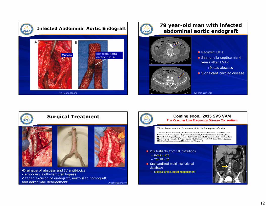
Infected Abdominal Aortic Endograft
Usually total graft involvement
Aneurysm or pseudoaneurysmabove infected graft
Often significant periaorticinflammation
Indium 111-labelled WBC scan
Mayo Clinic ExperienceInfected Abdominal Aortic Endograft
N=15 N=2 N=4
Frank purulence
Infected Abdominal Aortic Endograft
JVS 2013;58:371-379
12
Mucosa Bile from Aorto-enteric fistula
Infected Abdominal Aortic Endograft
JVS 2013;58:371-379
79 year-old man with infected abdominal aortic endograft
Recurrent UTIs
Salmonella septicemia 4 years after EVAR
Psoas abscessSignificant cardiac disease
JVS 2013;58:371-379
Surgical Treatment
•Drainage of abscess and IV antibiotics •Temporary axillo-femoral bypass •Staged excision of endograft, aorto-iliac homograft, and aortic wall debridement JVS 2013;58:371-379
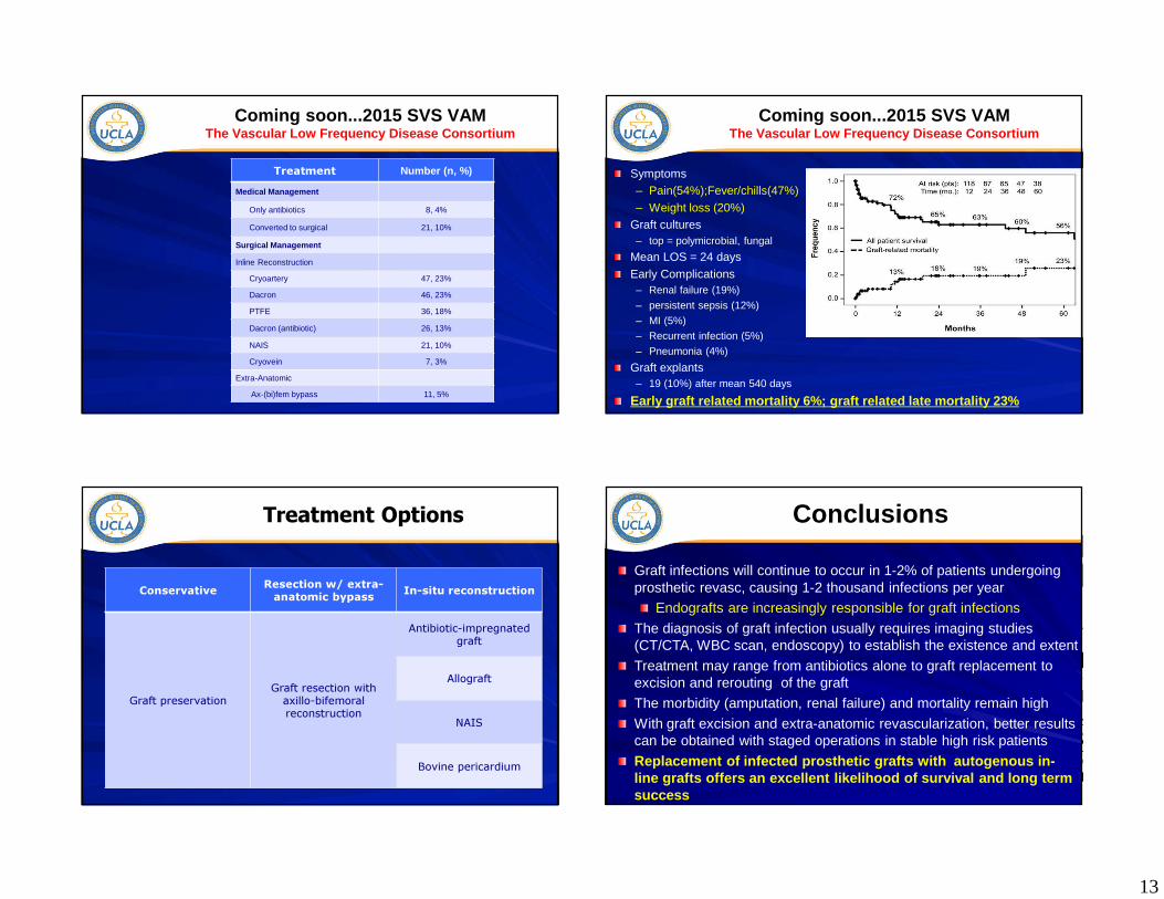
Coming soon...2015 SVS VAMThe Vascular Low Frequency Disease Consortium
202 Patients from 18 institutions– EVAR = 176– TEVAR = 26
Standardized multi-institutional database– Medical and surgical management
13
Coming soon...2015 SVS VAMThe Vascular Low Frequency Disease Consortium
Treatment Number (n, %)
Medical Management
Only antibiotics 8, 4%
Converted to surgical 21, 10%
Surgical Management
Inline Reconstruction
Cryoartery 47, 23%
Dacron 46, 23%
PTFE 36, 18%
Dacron (antibiotic) 26, 13%
NAIS 21, 10%
Cryovein 7, 3%
Extra-Anatomic
Ax-(bi)fem bypass 11, 5%
Coming soon...2015 SVS VAMThe Vascular Low Frequency Disease Consortium
Symptoms– Pain(54%);Fever/chills(47%)
– Weight loss (20%)Graft cultures– top = polymicrobial, fungal
Mean LOS = 24 days
Early Complications– Renal failure (19%)
– persistent sepsis (12%)
– MI (5%)– Recurrent infection (5%)
– Pneumonia (4%)
Graft explants– 19 (10%) after mean 540 days
Early graft related mortality 6%; graft related lat e mortality 23%
Treatment Options
Conservative Resection w/ extra-anatomic bypass In-situ reconstruction
Graft preservationGraft resection with axillo-bifemoralreconstruction
Antibiotic-impregnated graft
Allograft
NAIS
Bovine pericardium
Conclusions
Graft infections will continue to occur in 1-2% of patients undergoing prosthetic revasc, causing 1-2 thousand infections per year
Endografts are increasingly responsible for graft infections
The diagnosis of graft infection usually requires imaging studies (CT/CTA, WBC scan, endoscopy) to establish the existence and extent
Treatment may range from antibiotics alone to graft replacement to excision and rerouting of the graft
The morbidity (amputation, renal failure) and mortality remain high
With graft excision and extra-anatomic revascularization, better results can be obtained with staged operations in stable high risk patients
Replacement of infected prosthetic grafts with aut ogenous in-line grafts offers an excellent likelihood of survi val and long term success

14