aortic endothelium in hiv-1 infection
TRANSCRIPT
American Journal ofPathology, Vol. 149, No. 6, December 1996Copyrigbt X American Societyfor Investigative Pathology
Aortic Endothelium in HIV-1 Infection
Chronic Injury, Activation, and Increased LeukocyteAdherence
Christian Zietz,* Barbara Hotz,*Michael Sturzl,t Elisabeth Rauch,tRandolph Penning,t and Udo Lohrs*From the Departments ofPathology* and ForensicPathologyj Ludwig Maximilians University ofMunich,Munich, and the Department of Virology,t Max PlanckInstitutefor Biochemistry, Martinsried, Germany.
Clinical and serological studies provide evidencefor a pathogeneticaly relevant vasculopathy inacquired immune deficiency syndrome (AIDS);however, the morphological status of the endo-thelium under conditions ofhuman immunodeft-ciency virus (HIV)-l infection is only sparselydocumented. In this study we adapted an en facepreparation technique ofendotheliumfor use inimmunohistochemistry and investigated the aor-tic endothelium of pre-AIDS and AIDS patients(n = 32) in comparison with an HIV-negativegroup (n = 17). The control group showed aregularpattern ofevenly distributed aortic endo-thelial cells, whereas the endothelial ceUpatternin the HIV-1-infected patients was clearly dis-turbed. Simultaneously, the degree of leukocyteadherence on the aortic endothelium increasedsignificantly. These changes were accompaniedby an up-regulation ofthe vascular ceU adhesionmolecule-i (VCAM-1) and E-selectin (ELAM-1).The endothelium turnover increased, and one-half of the HIV-1-infected patients exhibitedHLA-DR (major histocompatibility complex classII) antigen in the aortic endothelium. Our resultsprovide evidence for a profound and repeatedinjury with regeneration and activation of theendothelium in HIV-1 infection. Injury as well asactivation ofthe endothelium impairs its normalregulatory properties. This could have conse-quences for the maintenance of the blood-brainbarrier; it might influence the immunologicalyimportant interaction of the endothelium with T
ceUs; and it might trigger Kaposi's sarcoma.(AmJ Pathol 1996, 149:1887-1898)
The endothelium is a regulatory organ that partici-pates in immune responses and plays a critical rolein the progression and outcome of infectious dis-eases. In this framework it can be important in thespread of virus, in T-cell function, and in the initiationof neoangiogenesis.1 - In human immunodeficiency(HIV)-1 infection and acquired immune deficiencysyndrome (AIDS), the endothelium is under the com-bined influence of deregulated immune effectorcells, increased concentrations of circulating anti-gens, and immune complexes.5 It is chronically ex-posed to imbalanced concentrations of cytokinesand other activating factors such as the HIV Tatprotein.6 6 Consequently, the normal function of theendothelium may be impaired under the conditionsof HIV-1 infection. Indeed, clinical indications for adisturbance of vascular function are becoming in-creasingly apparent. An HIV-associated ocular mi-croangiopathic syndrome is well established.9 14Other groups reported on an altered brain perfusionwith a reduced cerebral blood flow already in theearly stages of HIV-1 infection.1516 Joshi et al17demonstrated an artheriopathy in children with AIDS.Plasma levels of diverse endothelial cell markerssuch as von Willebrand factor antigen (vWF), tissue-type plasminogen activator, plasminogen activatorinhibitor, fibronectin, angiotensin-converting en-zyme, and endothelin increase in the course of HIV-1infection. 18-22 An elevated von Willebrand factorplasma value as a presumed marker of endothelialcell damage correlated inversely with the CD4+ cell
Supported by the Bundesministerium fur Forschung und Technik(FKZ FVP 11/89 VIII).Accepted for publication August 14, 1996.Address reprint requests to Dr. C. Zietz, Department of Pathology,
Ludwig Maximilians University of Munich, Thalkirchnerstrasse 36,80337 MOnchen, Germany.
1887
1888 Zietz et alAJP December 1996, Vol. 149, No. 6
counts.20'21 High von Willebrand factor levels arealso regarded as an adverse prognostic factor inAIDS.20 These data indicate an endothelium injuryand a systemic vasculopathy under the conditions ofHIV-1 infection.
In this study we evaluated the normal pattern ofthe aortic endothelium in a healthy control group andcompared it qualitatively and quantitatively with theendothelium of HIV-1 -infected patients. Presumably,more information is obtained by observing endothe-lial cells en face than from sections cut perpendicu-larly or obliquely to the surface. For this reason weused an endothelium en face preparation method.This technique offers far better possibilities for theassessment of endothelial alterations than conven-tional paraffin histology does. The adaption of thismethod to immunohistochemistry enabled us to de-termine endothelial parameters such as adhesionmolecules and major histocompatibility complex(MHC) class 11 antigens. Counting Ki-67-positivecells, we could compare the endothelium prolifera-tion rate of the healthy group with that of the HIV-1-infected patients.
Materials and Methods
Patient Characteristics and Autopsy Data
Extensive autopsy and histological examinations ofall patients included in this study were carried out.Serum testing before autopsy by enzyme-linked im-munosorbent assay and, if required, by additionalWestern blotting confirmed the HIV status. Fromdeath to autopsy all persons had been kept in acold-storage chamber at 40C. Patients with a higherdegree of atherosclerosis were not included in thisstudy and only macroscopically inconspicuous ar-eas of the aorta without fatty streaks or manifestatherosclerosis were chosen for en face preparation.The extent of overall atherosclerosis did not differbetween the groups. The material for en face prepa-ration was obtained from the ascending and theabdominal aorta. No differences could be observedin the degree of structural and immunohistochemicalresults between the ascending and the abdominalaorta. Surrounding areas of vascular ostia were omit-ted for en face preparation.
The anamnestically healthy persons in the HIV-negative control group (n = 17; 7 females and 10males; average age, 39 years with a range from 12 to57 years) died instantly due to suicide or a roadaccident without any period of intensive care. Theaverage time interval between death and collectionof specimens was 18 hours (4 to 30 hours) at 40C.
Postmortem investigation ruled out infections, neo-plasms and other diseases. Toxicological tests ofblood and urine by fluorescence polarization immu-noassay on barbiturates, benzodiazepines, mor-phine derivatives, amphetamines, tricyclic antide-pressant drugs, cocaine, Methadone, and cannabisexcluded drug abuse in these patients.
The HIV group (n = 32; 2 females and 30 males;average age of 42 years with a range from 7 to 60years) was composed of pre-AIDS patients (Centersfor Disease Control (CDC) stage A,23 n = 6) andpatients dying of AIDS (CDC stage C,23 n = 26). Theaverage time interval between death and collectionof specimens was 20 hours (11 to 24 hours) at 4°C.
All of the pre-AIDS patients (n = 6 males; averageage of 41 years with a range from 34 to 45 years)died an unnatural death (suicide or accident). Fromanamnestical data, these patients were not in med-ical care and not on medication. Most of them obvi-ously did not know of their infection while alive. Thefirst HIV-1 test was carried out at autopsy. Infections,neoplasms, and drug abuse were ruled out as in thecontrol group.
Four of the six pre-AIDS patients exhibited anHIV-associated generalized lymphadenopathy withfollicular hyperplasia.
The AIDS collective (n = 26; 2 females and 24males; average age of 42 years with a range from 7to 60 years) showed one or more opportunistic in-fections and/or Kaposi's sarcoma as well as typicalHIV-associated morphological changes to the lym-phatic system. Most AIDS patients were on intensivemedical care with multi-drug therapy for their infec-tions and/or Kaposi's sarcoma. The type and numberof opportunistic infections/malignancies proven byautopsy as well as the class and combination ofdrugs administered in the last weeks before deathdid not show a measurable influence on the degreeof the endothelial changes described in this study.
Preparation of Endothelium
The method of endothelium en face preparation bymeans of a nitrocellulose film was established morethan 50 years ago. For this study we used the tech-nique in Freudenberg's modified form.2425 For ob-taining endothelial cell monolayers with this method,the aorta was purged of blood for 2 minutes in phys-iological sodium chloride and then freed of fat andconnective tissue and opened longitudinally. Thespecimens were spread, mounted on a piece of corkwith the endothelial layer facing upward. Specimenswere fixed in 70% ethanol overnight at 4°C. Afterdehydration in graded ethanol, the specimens were
Aortic Endothelium in HIV-1 Infection 1889AJP December 1996, Vol. 149, No. 6
Table 1. Monoclonal Mouse Antibodies Used in tbis Study Applied to Antigens Listed
Source
Dianova-Immunotech (Hamburg, Germany), 1243Dako, M 775Dianova (Hamburg, Germany), M 400Dianova (Hamburg, Germany), DIA 505Dianova-Immunotech, 1244
Dilution of MAb
1/5*1/100*1/101/10*1/5*
*Microwave pretreatment.
dissected into pieces of 1 cm2 and immersed in a
mixture of ethanol/ether (1:1, v:v). A grease-freeglass slide was coated with a thin colloidon film(Cedukol, Kollodium, and glycerine from Merck(Darmstadt, Germany) and Pro Celloidin from Fluka(Buchs, Switzerland)) and dipped into a mixture ofethanol/ether (1:1, v:v). Pieces of the arterial wall (1cm2) were then placed on the colloidon film with theendothelium facing downward. The endothelium waspressed evenly onto the colloidon film. Sixty secondsafter placing the vessel on the coated slide the tu-nica media and adventitia were pulled away. Theendothelial cell monolayer, which adhered to thecolloidon film, was cut out, lifted off the slide, airdried, and stored at room temperature. In each case
at least six endothelial cell preparations were stainedwith hematoxylin and eosin (H&E), Giemsa stain, andchloroacetate esterase histochemical stain.26 De-pending on the availability of additional suitable en
face preparations, immunohistochemical studieswere performed using standard immunohistochemi-cal techniques27.28 and a panel of monoclonalmouse antibodies against human endothelial cell an-
tigens (Table 1). Antibodies to ELAM-1, VCAM-1,HLA-DR, and IL-1p were applied to demonstratedifferent endothelial activation parameters and ad-hesive leukocytes. An antibody to Ki-67 (MIB-1) wasused for visualization of cells undergoing mitosis.The avidin-biotin complex method was applied fordetection of VCAM-1, ELAM-1, and HLA-DR anti-gens (Vectastain, Vector Laboratories, Burlingame,CA). The alkaline phosphatase anti-alkaline phos-phatase method was chosen for IL-18 (FIB-3) andKi-67 (MIB-1) antigens (Dako, Copenhagen, Den-mark). 27,28
During all steps of the staining procedures the en
face preparations have to float loosely in reactionfluids and buffers to guarantee dyes and antibodiescomplete passage through the endothelial mono-
layer. Pretreatment and dilution of antibodies dif-fered between regular paraffin material and en facepreparations and had to be adapted specifically forthe individual antigens (Table 1). To ensure specific-ity and for control of background staining, controls
were included in all staining runs with the primaryantibody replaced by bovine serum albumin. Nostaining was observed in these controls.
To quantify alterations of the structural endothelialpattern, the following grading system was used: reg-ular structure, evenly distributed endothelial cellswithout phenotypical changes; low-grade distur-bance, major parts of the en face preparations with a
regular structure, with only small areas with distur-bance of pattern and minor phenotypical changes tothe endothelial cells; intermediate-grade distur-bance, some undisturbed areas beneath fields withclear alterations; high-grade disturbance, predomi-nance of a disturbed endothelial cell pattern, withfrequently phenotypical changes of the endothelialcells (Table 2).
The mean number of adhesive leukocytes was
determined as the percentage of adhering leuko-cytes compared with the total intimal cell count (<5,<10, <20, and >20%). The mean of all en facepreparations of each case was calculated (Table 3).The documentation of immunohistochemical stain-
ing was evaluated according to Remmele using an
immunoreactive score.29 The score was calculatedfrom the staining intensity in four categories (0 to 3),and the percentage of positive cells was calculatedin four categories (0 to 3) by multiplying stainingintensity and percentage of positive cells. Throughthis method of keeping a semiquantitative score, theimmunohistochemical results were rated negative(N), low (1+), intermediate (2+), or highly immuno-
Table 2. Aortic Endothelium Cell Pattern and Extent ofDisturbance
Pattern
NLDIDHD
Number of cases (n)
HIV-negative HIV-positivecontrol group patients
13211
0
2 (1)4 (2)
26 (3)
Disturbance of endothelial pattern in gradations: N, normalendothelial pattern; LD, low-grade disturbance; ID, intermediate-grade disturbance; HD, high-grade disturbance. n, number ofpre-AIDS patients.
Antigen
ELAM-1HLA-DRIL-1 (3Ki-67VCAM-1
MAb clone
1.2 B 6CR3/43FIB-3MIB-1lG1l
1890 Zietz et alAJP December 1996, Vol. 149, No. 6
Table 3. Evaluation of Leukocyte Adherence on theAortic Endothelium
Number of cases (n)
Leukocyte HIV-negative HIV-positiveadhesion control group patients
0-5% 8 1 (1)5-10% 6 1 (1)10-20% 3 17(2)>20% 0 13 (2)
Leukocyte adhesion is presented as the number of adherentcells as a percentage of the total number of intimal cells. n,number of pre-AIDS patients.
reactive (3+) (Table 4). The numbers of Ki-67-posi-tive nuclei were counted per 1-cm2 area of en facepreparations: 0 to 1 positive cells/cm2, 1 to 2 positivecells/cm2, 2 to 3 positive cells/cm2 and >3 positivecells/cm2 (Table 5).
ResultsWe compared the aortic endothelium of HIV-1-in-fected patients and HIV-negative control personswith regard to cellular pattern, proliferation, leuko-cyte adhesion, and activation of gene expression(VCAM-1, ELAM-1, HLA-DR, and IL-123). These dif-ferent parameters were qualitatively and quantita-tively evaluated to characterize the biological statusof the endothelium under the conditions of HIV-1infection.
Morphological Alteration of the AorticEndothelium under Conditions of HIV- 1Infection (Table 2)In 76% (13 of 17) of non-HIV-infected, healthy per-sons, the aortic endothelium formed a regular celllayer of flattened, fairly uniform mononuclear cells.Endothelial cells were oriented longitudinally alongthe vessel with their nuclei uniformly aligned in par-allel (Figure 1A). This regular pattern of the normalaortic endothelial cell layer was clearly disturbed in94% (30 of 32) of the HIV-infected patients. Mostobvious was an aberrant irregular cell pattern with
variability in cellularity as well as nuclear size, num-
ber, and staining (Figure 1, B and C). In detail, theaortic endothelium in HIV-1-infected patientsshowed two types of phenotypic changes: 1) thepresence of smaller cells with rounded nuclei andslightly increased chromatin (Figure 1C) and 2)multinucleated, sometimes bizarre-shaped endothe-lial cells (Figure 1, B and C) often located near smalldenuded areas. Frequently, nuclear pyknoses of en-dothelial cells were evident (Figure 1 D). One-half ofthe pre-AIDS patients (3 of 6) exhibited a high-gradedisturbance in the structure of the endothelium.
Increased Adherence of Leukocytes toAortic Endothelium under Conditions of HIV-1 Infection (Table 3)The number of nonintimal cells was less than 10% ofall intimal cells in 82% (14 of 17) of the non-HIV-infected persons. Thus, some mononuclear cells andgranulocytes were found to be regular constituentsof the normal endothelium. In 18% (3 of 17) of controlpatients, a degree of leukocyte adherence between10 and 20% of all intimal cells was revealed.
HIV-1-infected patients exhibited clearly in-creased numbers of nonendothelial cells in the aorticintima. In 94% (30 of 32) of the HIV-1-infected pa-
tients, the number of leukocytes exceeded 10% ofthe total number of intimal cells (Figure 2, A and B).Two patients of the pre-AIDS group showed lowernumbers of adherent leukocytes.
Only a few areas with clear structural changeswere lacking significant mononuclear cell adhesion(Figure 11B). Most en face preparations showed an
increased leukocyte adhesion in parallel with thestructural alterations of the aortic endothelium.
Using the chloroacetate esterase reaction26 in theHIV-1-infected group, the amount of granulocytes wasdetermined to be less than 5% of the nonintimal cellpopulation. Therefore, it can be concluded that themajority of adhesive cells on the endothelium must bemononuclear cells. Ongoing experiments with themacrophage-associated antibody MAC 387 (Dako-
Table 4. Antigen Excpression and Immunoreactive Score of the Aortic Endothelium
HIV-negative control group HIV-positive patients (n)N 1+ 2+ 3+ N 1+ 2+ 3+
VCAM-1 6 1 0 0 0 6(2) 8 9(2)ELAM-1 7 0 0 0 3(1) 2(1) 5 0HLA-DR 6 0 0 0 8 (2) 4 2 (1) 2IL-1 5 1 0 0 0 3 9 (1) 12 (4)
N, no positive cells (HLA-DR and IL-1p) or basic weak and patchy expression in single cells (VCAM-1 and ELAM-1); 1+, lowimmunoreactive score; 2+, intermediate immunoreactive score; 3+, high immunoreactive score. n, number of pre-AIDS patients.
Aortic Endothelium in HIV-1 Infection 1891AJP December 1996, Vol. 149, No. 6
Number of cases
HIV-negative HIV-positivecontrol patientsgroup (n)
60
0
0
0
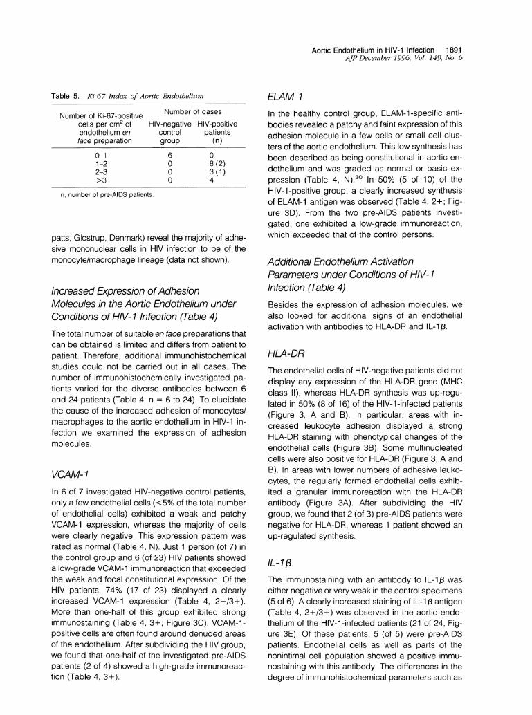
8 (2)3 (1)4
n, number of pre-AIDS patients.
patts, Glostrup, Denmark) reveal the majority of adhe-sive mononuclear cells in HIV infection to be of themonocyte/macrophage lineage (data not shown).
Increased Expression ofAdhesionMolecules in the Aortic Endothelium underConditions of HIV- 1 Infection (Table 4)The total number of suitable en face preparations thatcan be obtained is limited and differs from patient topatient. Therefore, additional immunohistochemicalstudies could not be carried out in all cases. Thenumber of immunohistochemically investigated pa-
tients varied for the diverse antibodies between 6and 24 patients (Table 4, n = 6 to 24). To elucidatethe cause of the increased adhesion of monocytes/macrophages to the aortic endothelium in HIV-1 in-fection we examined the expression of adhesionmolecules.
VCAM- 1
In 6 of 7 investigated HIV-negative control patients,only a few endothelial cells (<5% of the total numberof endothelial cells) exhibited a weak and patchyVCAM-1 expression, whereas the majority of cellswere clearly negative. This expression pattern was
rated as normal (Table 4, N). Just 1 person (of 7) inthe control group and 6 (of 23) HIV patients showeda low-grade VCAM-1 immunoreaction that exceededthe weak and focal constitutional expression. Of theHIV patients, 74% (17 of 23) displayed a clearlyincreased VCAM-1 expression (Table 4, 2+/3+).More than one-half of this group exhibited strongimmunostaining (Table 4, 3+; Figure 3C). VCAM-1-positive cells are often found around denuded areas
of the endothelium. After subdividing the HIV group,
we found that one-half of the investigated pre-AIDSpatients (2 of 4) showed a high-grade immunoreac-tion (Table 4, 3+).
ELAM-1In the healthy control group, ELAM-1-specific anti-bodies revealed a patchy and faint expression of thisadhesion molecule in a few cells or small cell clus-ters of the aortic endothelium. This low synthesis hasbeen described as being constitutional in aortic en-dothelium and was graded as normal or basic ex-pression (Table 4, N).30 In 50% (5 of 10) of theHIV-1-positive group, a clearly increased synthesisof ELAM-1 antigen was observed (Table 4, 2+; Fig-ure 3D). From the two pre-AIDS patients investi-gated, one exhibited a low-grade immunoreaction,which exceeded that of the control persons.
Additional Endothelium ActivationParameters under Conditions of HIV-1Infection (Table 4)Besides the expression of adhesion molecules, wealso looked for additional signs of an endothelialactivation with antibodies to HLA-DR and IL-1f3.
HLA-DR
The endothelial cells of HIV-negative patients did notdisplay any expression of the HLA-DR gene (MHCclass 11), whereas HLA-DR synthesis was up-regu-lated in 50% (8 of 16) of the HIV-1-infected patients(Figure 3, A and B). In particular, areas with in-creased leukocyte adhesion displayed a strongHLA-DR staining with phenotypical changes of theendothelial cells (Figure 3B). Some multinucleatedcells were also positive for HLA-DR (Figure 3, A andB). In areas with lower numbers of adhesive leuko-cytes, the regularly formed endothelial cells exhib-ited a granular immunoreaction with the HLA-DRantibody (Figure 3A). After subdividing the HIVgroup, we found that 2 (of 3) pre-AIDS patients werenegative for HLA-DR, whereas 1 patient showed anup-regulated synthesis.
IL-1,8The immunostaining with an antibody to IL-1,8 waseither negative or very weak in the control specimens(5 of 6). A clearly increased staining of IL-1 3 antigen(Table 4, 2+/3+) was observed in the aortic endo-thelium of the HIV-1-infected patients (21 of 24, Fig-ure 3E). Of these patients, 5 (of 5) were pre-AIDSpatients. Endothelial cells as well as parts of thenonintimal cell population showed a positive immu-nostaining with this antibody. The differences in thedegree of immunohistochemical parameters such as
Table 5. Ki-67 Index of Aortic Endothelium
Number of Ki-67-positivecells per cm2 ofendothelium enface preparation
0-11-22-3>3
1892 Zietz et alAJP December 1996, Vol. 149, No. 6
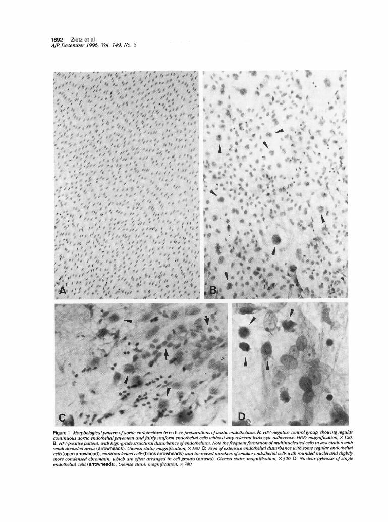
Figure 1. Morphologicalpattern ofaortic endothelium in en face preparations ofaortic endothelium. A: HIV-negative control group, showing regularcontinuous aortic endothelialpavement andfairly tniform endothelial cells without any relevant leukocyte adherence. H&E; magnification, X 120.B: HIV-positivepatient, with high-grade structural disturbance ofendothelium. Note thefrequentformation ofmultinucleated cells in association withsmall denuded areas (arrowheads). Giemsa stain; magnification, X 180. C: Area ofextensive endothelial disturbance with some regular endothelialcells (open arrowhead), multinucleated cells (black arrowheads) and increased numbers ofsmaller endothelial cells with rounded nuclei and slightlymore condensed chromatin, which are often arranged in cell groups (arrows). Giemsa stain; magnification, x320. D: Nuclear pyknosis ofsingleendothelial cells (arrowheads). Giemsa stain; magnification, X 740.
I
I
I
Aortic Endothelium in HIV-1 Infection 1893AJP December 1996, Vol. 149, No. 6
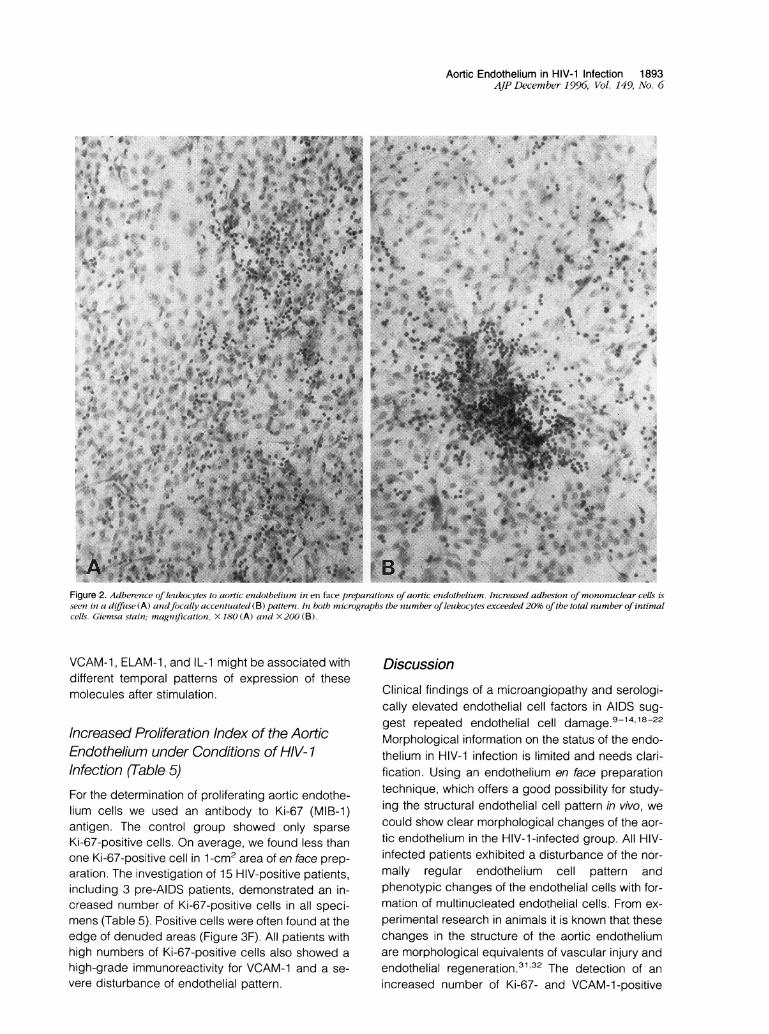
Figure 2. Adherence of leukocytes to aortic endothelium in en face preparations of aortic endothelium. Increased adhesion of mononuclear cells isseen in a diffuse (A) andfocally accentuated (B) pattern. In both micrographs the number ofleukocytes exceeded 20% ofthe total number ofintimalcells. Giemsa stain; magnification, x 180 (A) and X 200 (B).
VCAM-1, ELAM-1, and IL-1 might be associated withdifferent temporal patterns of expression of thesemolecules after stimulation.
Increased Proliferation Index of the AorticEndothelium under Conditions of HIV- 1Infection (Table 5)For the determination of proliferating aortic endothe-lium cells we used an antibody to Ki-67 (MIB-1)antigen. The control group showed only sparseKi-67-positive cells. On average, we found less thanone Ki-67-positive cell in 1-cm2 area of en face prep-aration. The investigation of 15 HIV-positive patients,including 3 pre-AIDS patients, demonstrated an in-creased number of Ki-67-positive cells in all speci-mens (Table 5). Positive cells were often found at theedge of denuded areas (Figure 3F). All patients withhigh numbers of Ki-67-positive cells also showed ahigh-grade immunoreactivity for VCAM-1 and a se-vere disturbance of endothelial pattern.
Discussion
Clinical findings of a microangiopathy and serologi-cally elevated endothelial cell factors in AIDS sug-gest repeated endothelial cell damage.9-14,18-22Morphological information on the status of the endo-thelium in HIV-1 infection is limited and needs clari-fication. Using an endothelium en face preparationtechnique, which offers a good possibility for study-ing the structural endothelial cell pattern in vivo, wecould show clear morphological changes of the aor-tic endothelium in the HIV-1-infected group. All HIV-infected patients exhibited a disturbance of the nor-mally regular endothelium cell pattern andphenotypic changes of the endothelial cells with for-mation of multinucleated endothelial cells. From ex-perimental research in animals it is known that thesechanges in the structure of the aortic endotheliumare morphological equivalents of vascular injury andendothelial regeneration.31'32 The detection of anincreased number of Ki-67- and VCAM-1-positive
1894 Zietz et al4/P December 1996( Vol. 149, No. 6
.. . t.
*
U ' .cI
*itF
Aortic Endothelium in HIV-1 Infection 1895AJP December 1996, Vol. 149, No. 6
cells around denuded areas of endothelium alsoindicates morphological signs of injury as demon-strated by Lindner and Collins.33
Demonstrating an up-regulation of the inducibleadhesion molecules VCAM-1 and ELAM-1 as well as
MHC class 11 antigens such as HLA-DR, this studyalso provided support for an activated endothelialcell status.4
Taking all of these results into account, it can beconcluded from our data that the aortic endotheliumof the HIV-infected patients exhibits clear signs ofinjury, regeneration, and activation. This was accom-
panied by increased numbers of adherent leuko-cytes on the endothelium.
Causes of the Endothelial Injury in HIV- 1Infection
Due to the immense antigen shift of the virus andtriggered by simple stress factors, pre-AIDS patientsalready exhibit ongoing and serologically measur-
able episodes of a repeatedly increased viral burdenwith HIV-1.3 This continuous immunological chal-lenge finds a morphological equivalent in HIV-asso-ciated lymphadenopathy, which we also found infour of our six pre-AIDS patients. In more advancedstages of the disease with more frequent episodes ofvirus-associated endothelial injury, one may expecta higher degree of endothelial changes. Therefore, itis interesting that neither in the evaluation of endo-thelial structure and leukocyte adherence nor in theimmunohistochemical results were we able to showessential differences between the HIV-1-infectedpersons in the CDC stage A and the patients withfull-blown AIDS (CDC stage C).23 The absence ofessential differences between the HIV groups may
be due to the high regeneration capacity of the en-
dothelium in the intervals between the repeated ep-
isodes of viremias. As demonstrated by Ki-67 immu-nohistochemistry, the HIV-positive patients exhibiteda high regeneration capacity of the endothelium,which is obviously similar in pre-AIDS and AIDS pa-
tients.There are several ways in which the repeated viral
burden of HIV-1 may effect the endothelium. Somegroups found a direct HIV infection of endothelialcells.3536 HIV infection of mononuclear cells withderangement of immunocompetent cells and distur-
bance of the local cytokine balance might even bemore important for the genesis of endothelialchanges.
It is well known that inflammatory cytokines acti-vate cultured endothelial cells to synthesize and ex-
press leukocyte adhesion molecules.4,37-41 Cyto-kines such as tumor necrosis factor-a, IL-1,B, andinterferon-y are elevated in the serum of HIV-1-in-fected patients.642 They may up-regulate the syn-
thesis of the adhesion molecules VCAM-1 andELAM-1 and, as a consequence, may be the cause
of an increased adhesion of leukocytes to the endo-thelium. Adherence of leukocytes to the endotheliumcan cause an additional increase of cytokine con-
centrations in locally restricted areas. This may resultin patches of adherent leukocytes, as was observedin this study. Toxic products released by monocytesare known to damage the endothelium. Indeed, inthe HIV group, we observed a positive correlationbetween the number of adherent cells and the de-gree of endothelial disturbance. Additionally, theHIV-1 Tat protein is known to induce adhesion mol-ecules on endothelial cells.8 Changes may also bedue to HIV-1-associated direct complement pathwayactivation and to other factors such as circulatingimmune complexes with HIV-1 antigens.5'43'44
Consequences of the Endothelial Injury inHIV-1 Infection
Injury and activation of the endothelial cells disruptthe normal regulatory mechanisms and result in mor-phological and functional alterations commonly de-fined as endothelial dysfunction.4 In this regard, our
findings might indicate a dysfunction of the aorticendothelium in HIV-1 infection, although we did notmeasure function. Is such an injured endothelium a
localized phenomenon in the aorta or are there ar-
guments for a systemic event? Indeed, clinical andmorphological studies of the eyes and the brainsfrom HIV-1-infected patients confirmed an increasedendothelial permeability and a vascular leaki-ness.45-48 In this context, it is interesting that braincapillaries as well as aortic endothelium in miceshow prolonged VCAM-1 expression after stimula-tion.49 A prolonged elevation of VCAM-1 combinedwith a vascular leakage triggered by a dysfunctionalendothelium may influence the leukocyte trafficking
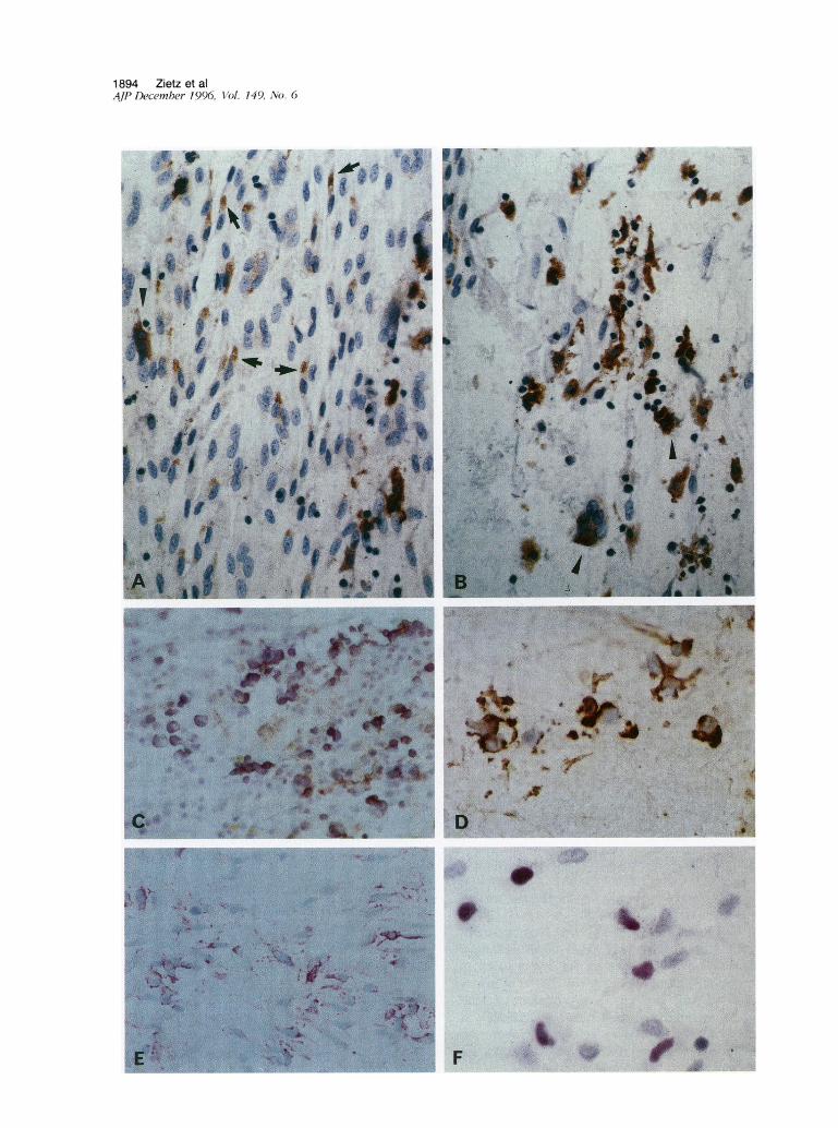
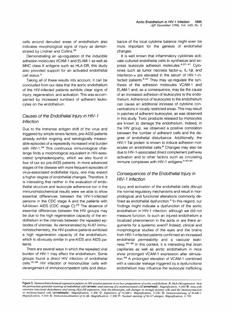
Figure 3. Immunohistochemical expression pattern in HIV-positivepatients in en face preparations ofaortic endothelium. A: HLA-DR expression. Notethepeninucleargranular staining ofendothelial cells (arrows) and staining ofa multinucleated cell (arrowhead). Magnification, X300. B: Area withextensive structural disturbance and strong HLA-DR expression. Note the phenotypic cell changes in strongly positive cells and the immunoreactionin multinucleated cells (arrowheads). Magnification, X300. C: Expression of VCAM-1. Magnification, X250. D: Immunostaining of ELAM-1.Magnification, X 600. E: Immunolocalization of IL-1,8. Magnification, X300. F: Nuclear staining ofKi-67 antigen. Magnification, X 700.
1896 Zietz et alAJP December 1996, Vol. 149, No. 6
to the central nervous system and the integrity of theblood-brain barrier in AIDS.48 This in turn may beimportant for the development of HIV-associated en-cephalopathy and dementia.
Injury, activation, and leukocyte adhesion of en-dothelial cells may play a role in the genesis ofHIV-associated microvascular proliferation and Ka-posi's sarcoma. With regard to Kaposi's sarcoma,the increased endothelial cell proliferation and fac-tors produced from the adherent monocytes mightparticipate in the initiation of the early angiomatoidKaposi's sarcoma.5053 Viruses of the herpes type inparticular can interact with the endothelium.1 Theassociation of Kaposi's sarcoma and a new humany-herpes virus, which has been given the trivial de-scriptive name Kaposi's sarcoma-associated her-pesvirus with its formal classification likely to be hu-man herpesvirus-8 is by now well documented.54
Most importantly, a chronically repeated injury ofthe endothelium might disrupt the normal immuno-logical features of endothelial cells such as phago-cytosis, antigen presentation, and induction of lym-phocyte proliferation and maturation.4 Endothelialcells are able to stimulate primary and secondaryT-cell responses and they offer co-stimulatory sig-nals such as VCAM-1 expression to functional T-cellactivation. 3,39,55 Whereas normal vascular endo-thelial cells enhance T-cell responses by augment-ing IL-2 concentrations,56 HIV-exposed endothelialcells were consistently defective in promoting IL-2secretion in vitro.5758
In conclusion, our study revealed injury, regener-ation, and activation of the aortic endothelium inHIV-positive patients. This may play a more activerole in the pathogenesis and progression of AIDSthan assumed so far, as it may have an affect on thecontribution of the endothelium to the immune net-work, on its regulatory properties for T-cell function,and on maintenance of the blood-brain barrier; itmay also be relevant for the initiation of Kaposi'ssarcoma.
Acknowledgments
We thank Karin Schneiderbanger and Beate Haar fortheir skillful technical assistance, Joachim Dieboldand Stephan lhrler for their critical reading of themanuscript, Liselotte Dohle for photographic assis-tance, and Thelma Coutts (Max Planck Institute forBiochemistry, Germany) for linguistic revision of themanuscript.
References
1. Beilke MA: Vascular endothelium in immunology andinfectious disease. Rev Infect Dis 1989, 11:273-283
2. Pober JS, Doukas J, Hughes CC, Savage CO, MunroJM, Cotran RS: The potential roles of vascular endo-thelium in immune reactions. Hum Immunol 1990, 28:258-262
3. Hughes CC, Savage CO, Pober JS: The endothelial cellas a regulator of T-cell function. Immunol Rev 1990,117:85-102
4. Rubanyi GM: The role of endothelium in cardiovascularhomeostasis and diseases. J Cardiovasc Pharmacol1993, 22:S1-S14
5. Krapf FE, Herrmann M, Leitmann W, Schwartlander B,Kalden JR: Circulating immune complexes in HIV-in-fected persons. Klin Wochenschr 1990, 68:299-305
6. Scott Algara D, Vuillier F, Marasescu M, de Saint MartinJ, Dighiero G: Serum levels of IL-2, IL-la, TNF-a, andsoluble receptor of IL-2 in HIV-1-infected patients.AIDS Res Hum Retroviruses 1991, 7:381-386
7. Sinicco A, Biglino A, Sciandra M, Forno B, Pollono AM,Raiteri R, Gioannini P: Cytokine network and acuteprimary HIV-1 infection. AIDS 1993, 7:1167-1172
8. Hofman FM, Wright AD, Dohadwala MM, Wong Staal F,Walker SM: Exogenous tat protein activates humanendothelial cells. Blood 1993, 82:2774-2780
9. Holland GN, Gottlieb MS, Yee RD, Schanker HM, PettitTH: Ocular disorders associated with a new severeacquired cellular immunodeficiency syndrome. Am JOphthalmol 1982, 93:393-402
10. Holland GN, Pepose JS, Pettit TH, Gottlieb MS, YeeRD, Foos RY: Acquired immune deficiency syndrome:ocular manifestations. Ophthalmology 1983, 90:859-873
11. Pepose JS, Holland GN, Nestor MS, Cochran AJ, FoosRY: Acquired immune deficiency syndrome: patho-genic mechanisms of ocular disease. Ophthalmology1985, 92:472-484
12. Freeman WR, Chen A, Henderly DE, Levine AM, LuttrullJK, Urrea PT, Arthur J, Rasheed S, Cohen JL, NeubergD, Leung RJ: Prevalence and significance of acquiredimmunodeficiency syndrome-related retinal microvas-culopathy. Am J Ophthalmol 1989, 107:229-235
13. Engstrom RE Jr, Holland GN, Hardy WD, MeiselmanHJ: Hemorheologic abnormalities in patients with hu-man immunodeficiency virus infection and ophthalmicmicrovasculopathy. Am J Ophthalmol 1990, 109:153-161
14. Turu AC, Civera AA, Latorre X: Ophthalmic manifesta-tions of acquired immunodeficiency syndrome: a studyof thirty-four patients. Ophthalmologica 1988, 197:113-119
15. Tatsch K, Schielke E, Bauer WM, Markl A, Einhaupl KM,Kirsch CM: Functional and morphological findings inearly and advanced stages of HIV infection: a compar-ison of 99mTc-HMPAO SPECT with CT and MRI stud-ies. Nuklearmedizin 1990, 29:252-258
Aortic Endothelium in HIV-1 Infection 1897AJP December 1996, Vol. 149, No. 6
16. Schielke E, Tatsch K, Pfister HW, Trenkwalder C, Leins-inger G, Kirsch CM, Matuschke A, Einhaupl KM: Re-duced cerebral blood flow in early stages of humanimmunodeficiency virus infection. Arch Neurol 1990,47:1342-1345
17. Joshi VV, Pawel B, Connor E, Sharer L, Oleske JM,Morrison S, Marin Garcia J: Arteriopathy in childrenwith acquired immune deficiency syndrome. PediatrPathol 1987, 7:261-275
18. Schved JF, Gris JC, Arnaud A, Martinez P, Sanchez N,Wautier JL, Sarlat C: von Willebrand factor antigen,tissue-type plasminogen activator antigen, and risk ofdeath in human immunodeficiency virus 1-related clin-ical disease: independent prognostic relevance of tis-sue-type plasminogen activator. J Lab Clin Med 1992,120:411-419
19. Drouet L, Scrobohaci ML, Janier M, Baudin B: Endo-thelial cells: target for the HIVI virus? Nouv Rev FrHematol 1990, 32:103-106
20. Lafeuillade A, Alessi MC, Poizot Martin I, Boyer Neu-mann C, Zandotti C, Quilichini R, Aubert L, Tamalet C,Juhan Vague I, Gastaut JA: Endothelial cell dysfunctionin HIV infection. J Acquired Immune Defic Syndr 1992,5:127-131
21. Janier M, Flageul B, Drouet L, Scrobohaci ML, VilletteJM, Palangie A, Cottenot F: Cutaneous and plasmavalues of von Willebrand factor in AIDS: a marker ofendothelial stimulation? J Invest Dermatol 1988, 90:703-707
22. Rolinski B, Geier SA, Sadri I, Klauss V, Bogner JR,Ehrenreich H, Goebel FD: Endothelin-1 immunoreactiv-ity in plasma is elevated in HIV-infected patients withretinal microangiopathic syndrome. Clin Investig 1994,72:288-293
23. Centers for Disease Control: 1993 revised classifica-tion system for HIV infection and expanded surveil-lance case definition for AIDS among adolescents andadults. MMWR Morb Mortal Wkly Rep 1992, 41:1-19
24. Freudenberg N, Riese K-H, Freudenberg MA: The Vas-cular Endothelial System. Stuttgart, Gustav FischerVerlag, 1983, pp 1-1 14
25. Riese KH, Freudenberg N, Haas W: En face preparationmethods for investigation of endothelial and mesothe-lia. Pathol Res Pract 1978, 162:327-336
26. Gomori G: Chloracylesters as histochemical sub-strates. J Histochem Cytochem 1953, 1:469-470
27. Hsu SM, Raine L, Fanger H: The use of antiavidinantibody and avidin-biotin-peroxidase complex in im-munoperoxidase techniques. Am J Clin Pathol 1981,75:816-821
28. Cordell JL, Falini B, Erber WN, Ghosh AK, Abdulaziz Z,MacDonald S, Pulford KA, Stein H, Mason DY: Immu-noenzymatic labeling of monoclonal antibodies usingimmune complexes of alkaline phosphatase andmonoclonal anti-alkaline phosphatase (APAAP com-plexes). J Histochem Cytochem 1984, 32:219-229
29. Remmele W, Hildebrand U, Hienz HA, Klein PJ, Vier-buchen M, Behnken LJ, Heicke B, Scheidt E: Compar-
ative histological, histochemical, immunohistochemi-cal, and biochemical studies on oestrogen receptors,lectin receptors, and Barr bodies in human breast can-cer. Virchows Arch A Pathol Anat Histopathol 1986,409:127-147
30. Page C, Rose M, Yacoub M, Pigott R: Antigenic heter-ogeneity of vascular endothelium. Am J Pathol 1992,141:673-683
31. Poole JCF, Sanders AG, Florey HW: The regenerationof aortic endothelium. J Pathol Bacteriol 1958, 75:133-143
32. Cotton RE, Harwood TR, Wartman WB: Regeneration ofaortic endothelium. J Pathol Bacteriol 1961, 81:175-180
33. Lindner V, Collins T: Expression of NF-KB and IKB-a byaortic endothelium in an arterial injury model. Am JPathol 1996, 148:427-438
34. Sonigo P, Courgnaud V, Castelot S, Fossati B, Le-meignan B, Leste-Lasserre T, Nerrienet E, Valere T,Pancino G: Evolution of the viral burden: a conse-quence of the adaptive strategies of persistent lentivi-ruses? Viral Quantification in HIV Infection. Edited byJM Andrieu. Paris, John Libbey Eurotext, 1991, pp113-118
35. Ward JM, O'Leary TJ, Baskin GB, Benveniste R, HarrisCA, Nara PL, Rhodes RH: Immunohistochemical local-ization of human and simian immunodeficiency viralantigens in fixed tissue sections. Am J Pathol 1987,127:199-205
36. Rhodes RH, Ward JM: Immunohistochemistry of hu-man immunodeficiency virus in the central nervoussystem and an hypothesis concerning the pathogene-sis of AIDS meningoencephalomyelitis. Prog AIDSPathol 1989, 1:167-179
37. Pober JS: Warner-Lambert/Parke-Davis award lecture:Cytokine-mediated activation of vascular endothelium.Am J Pathol 1988, 133:426-433
38. Bevilacqua MP, Pober JS, Wheeler ME, Cotran RS,Gimbrone MA: Interleukin-1 activation of vascularendothelium: effects on procoagulant activity and leu-kocyte adhesion. Am J Pathol 1985, 121:394-403
39. Pober JS, Cotran RS: Cytokines and endothelial cellbiology. Physiol Rev 1990, 70:427-451
40. Pober JS, Collins T, Gimbrone MA Jr, Libby P, ReissCS: Inducible expression of class 11 major histocompat-ibility complex antigens and the immunogenicity ofvascular endothelium. Transplantation 1986, 41:141-146
41. Collins T, Korman AJ, Wake CT, Boss JM, Kappes DJ,Fiers W, Ault KA, Gimbrone MA Jr, Strominger JL,Pober JS: Immune interferon activates multiple class 11major histocompatibility complex genes and the asso-ciated invariant chain gene in human endothelial cellsand dermal fibroblasts. Proc Natl Acad Sci USA 1984,81:4917-4921
42. Fuchs D, Hausen A, Reibnegger G, Werner ER, WernerFelmayer G, Dierich MP, Wachter H: Interferon-y con-centrations are increased in sera from individuals in-
1898 Zietz et alAJP December 1996, Vol. 149, No. 6
fected with human immunodeficiency virus type 1. JAcquired Immune Defic Syndr 1989, 2:158-162
43. Marschang P, GOrtler L, Totsch M, Thielens NM, ArlaudGJ, Hittmair A, Katinger H, Dierich MP: HIV-1 and HIV-2isolates differ in their ability to activate the complementsystem on the surface of infected cells. AIDS 1993,7:903-910
44. Grunfeld C, Pang M, Doerrler W, Shigenaga JK,Jensen P, Feingold KR: Lipids, lipoproteins, triglycer-ide clearance, and cytokines in human immunodefi-ciency virus infection and the acquired immunodefi-ciency syndrome. J Clin Endocrinol Metab 1992, 74:1045-1052
45. Gariano RF, Rickman LS, Freeman WR: Ocular examina-tion and diagnosis in patients with the acquired immuno-deficiency syndrome. West J Med 1993, 158:254-262
46. Zietz C, Speiser B, Rauch E, Lohrs U: Untersuchungenan Endothel und Gefasssystem bei HIV-lnfektion. VerhDtsch Ges Pathol 1992, 76:541
47. Rhodes RH: Evidence of serum-protein leakage acrossthe blood-brain barrier in the acquired immunodefi-ciency syndrome. J Neuropathol Exp Neurol 1991, 50:171-183
48. Petito CK, Cash KS: Blood-brain barrier abnormalitiesin the acquired immunodeficiency syndrome: immuno-histochemical localization of serum proteins in post-mortem brain. Ann Neurol 1992, 32:658-666
49. Fries JW, Williams AJ, Atkins RC, Newman W, Lips-comb MF, Collins T: Expression of VCAM-1 and E-selectin in an in vivo model of endothelial activation.Am J Pathol 1993, 143:725-737
50. Sturzl M, Brandstetter H, Roth WK: Kaposi's sarcoma: areview of gene expression and ultrastructure of KSspindle cells in vivo. AIDS Res Hum Retroviruses 1992,8:1753-1763
51. StOrzl M, Roth WK, Brockmeyer NH, Zietz C, Speiser B,Hofschneider PH: Expression of platelet-derivedgrowth factor and its receptor in AIDS-related Kaposisarcoma in vivo suggests paracrine and autocrinemechanisms of tumor maintenance. Proc Natl Acad SciUSA 1992, 89:7046-7050
52. Albini A, Barillari G, Benelli R, Gallo RC, Ensoli B:Angiogenic properties of human immunodeficiency vi-rus type 1 Tat protein. Proc Natl Acad Sci USA 1995,92:4838-4842
53. StOrzl M, Brandstetter H, Zietz C, Eisenburg B, RaivichG, Gearing DP, Brockmeyer NH: Identification of inter-leukin-1 and platelet-derived growth factor-B as majormitogens for spindle cells of Kaposi's sarcoma: a com-bined in vitro and in vivo analysis. Oncogene 1995,10:2007-2016
54. Chang Y, Cesarman E, Pessin MS, Lee F, Culpepper J,Knowles DM, Moore PS: Identification of herpesvirus-like DNA sequences in AIDS-associated Kaposi's sar-coma. Science 1994, 266:1865-1869
55. Pober JS, Cotran RS: Immunologic interactions of Tlymphocytes with vascular endothelium. Adv Immunol1991, 50:261-302
56. Guinan EC, Smith BR, Doukas JT, Miller RA, Pober JS:Vascular endothelial cells enhance T cell responses bymarkedly augmenting IL-2 concentrations. Cell Immu-nol 1989, 118:166-177
57. Teitel JM, Shore A, Read SE, Schiavone A: Immunefunction of vascular endothelial cells is impaired byHIV. J Infect Dis 1989, 160:551-552
58. Murray HW, Welte K, Jacobs JL, Rubin BY, Mertels-mann R, Roberts RB: Production of and in vitro re-sponse to interleukin 2 in the acquired immunodefi-ciency syndrome. J Clin Invest 1985, 76:1959-1964