antioxidant activity and neuroprotective role of
TRANSCRIPT
antioxidants
Review
Antioxidant Activity and Neuroprotective Role of DocosahexaenoicAcid (DHA) Supplementation in Eye Diseases That Can Lead toBlindness: A Narrative Review
María Lafuente 1,*, María Elena Rodríguez González-Herrero 1, Stéphanie Romeo Villadóniga 2
and Joan Carles Domingo 3
�����������������
Citation: Lafuente, M.; Rodríguez
González-Herrero, M.E.; Romeo
Villadóniga, S.; Domingo, J.C.
Antioxidant Activity and
Neuroprotective Role of
Docosahexaenoic Acid (DHA)
Supplementation in Eye Diseases
That Can Lead to Blindness: A
Narrative Review. Antioxidants 2021,
10, 386. https://doi.org/10.3390/
antiox10030386
Academic Editors: Ram Kannan and
Francisco J. Romero
Received: 11 January 2021
Accepted: 2 March 2021
Published: 5 March 2021
Publisher’s Note: MDPI stays neutral
with regard to jurisdictional claims in
published maps and institutional affil-
iations.
Copyright: © 2021 by the authors.
Licensee MDPI, Basel, Switzerland.
This article is an open access article
distributed under the terms and
conditions of the Creative Commons
Attribution (CC BY) license (https://
creativecommons.org/licenses/by/
4.0/).
1 Service of Ophthalmology, Hospital Universitario Virgen de la Arrixaca, El Palmar, E-30120 Murcia, Spain;[email protected]
2 Service of Ophthalmology, Complejo Hospitalario Universitario de Ferrol, Ferrol, E-15405 A Coruña, Spain;[email protected]
3 Department of Biochemistry and Molecular Biomedicine, Faculty of Biology, University of Barcelona,E-08028 Barcelona, Spain; [email protected]
* Correspondence: [email protected]; Tel.: +34-968-360900
Abstract: The objective of this narrative review is to provide updated evidence, based on data fromexperimental and clinical studies, of the prominent role of omega-3 polyunsaturated fatty acids(n-3 PUFAs) for a number of crucial mechanisms involved in counteracting cell damage induced byoxidative stress in eye diseases. This article is focused on the antioxidant and neuroprotective effectsof docosahexaenoic acid (DHA), which have been assessed in different experimental models andclinical studies, particularly in proliferative diabetic retinopathy, age-related macular degenerationand glaucoma that are the most common eye diseases leading to severe vision loss. The mechanismsinvolved in the role of DHA in protecting human retinal pigment epithelial cells from oxidativestress as well as the interaction with glutathione (GSH) are also described. The review is intended toprovide novel and salient findings supporting the rationale of the use of dietary supplementationwith high-dose DHA (1050 mg/day) in the form of triglyceride as a potent antioxidant compoundfor improving the eye health. However, the overall clinical evidence for the use of dietary strategiesbased on supplementation with n-3 PUFAs in eye diseases linked to oxidative stress other thanhigh-dose DHA triglyceride is both limited and inconsistent.
Keywords: omega-3 fatty acids; docosahexaenoic acid; glutathione; diabetic macular edema; glau-coma; oxidative stress; eye health
1. Introduction
Oxidative stress has been defined as a phenomenon resulting from an imbalancebetween production and accumulation of free radicals or reactive oxygen species (ROS) incells and tissues and the ability of biological system to detoxify these reactive metabolites(i.e., protective mechanisms by antioxidants) [1]. ROS generated by biological systems asmetabolic by-products include superoxide radicals, hydrogen peroxide, hydroxyl radicals,and singlet oxygen. Mitochondria under both pathological and physiological conditions arean important source of ROS. Superoxide radicals are produced by cellular respiration, bycyclooxygenases (COX) during the metabolism of arachidonic acid and by lipoxygenases(LOX) as well as by endothelial cells and inflammatory cells [2]. The oxidation productsor nitrosylated products linked to ROS reduce biological activity with loss of energymetabolism, transport, cell signaling and a variety of detrimental effects on crucial cellularfunctions. Proteasome degradation targeted by these altered products contributes to furtherreduction of cellular function [3]. Although cell organelles have intrinsic ROS scavengingcapacity, enzymatic components such as catalase (CAT), superoxide dismutase (SOD),
Antioxidants 2021, 10, 386. https://doi.org/10.3390/antiox10030386 https://www.mdpi.com/journal/antioxidants
Antioxidants 2021, 10, 386 2 of 13
glutathione peroxidase (GPx) and glutathione reductase (GR) are the mainly enzymaticcomponents involved in the cell enzymatic antioxidant defensive system [4].
On the other hand, ROS are important signaling molecules involved in the progressionof inflammation, with the production of oxidized proteins, glycated products and lipidperoxidation induced by oxidative stress resulting in an inflammatory response [5]. Keyinflammatory events in which ROS are involved mainly include increased vascular perme-ability, leukocyte extravasation, phagocytosis and angiogenesis. However, ROS-inducedeffects on inflammation are multi-faceted and still remain unclear [6], but overproductionof ROS may result in cell and tissue injury and contributes to chronic inflammation asthe underlying pathophysiological mechanism of a wide spectrum of neurodegenerative,cardiovascular and metabolic diseases.
2. Antioxidant Activity of Glutathione and Omega-3 Fatty Acids
Glutathione (GSH) is the most abundant low molecular weight thiol compoundsynthesized in cells and plays critical roles in protecting cells from oxidative damage andin maintaining redox homeostasis. GSH is also involved in other functions includingapoptosis, detoxification and modulation of cell proliferation [7]. The first step in thesynthesis of GSH is the production of γ-glutamylcysteine due to the combination ofglutamate with cysteine. The second step to produce GSH includes the addition of glycineto the dipeptide with the action of the glutathione synthetase enzyme. The thiol (sulfhydryl)group of cysteine plays a role in GSH-related conjugation and reduction reactions, whichare essential functions in the antioxidant activity of GSH. Although all cell types synthesizeGSH, the liver remains the main GSH source in the body. Hepatocytes supply GSH foundin plasma, which is used as a source of cysteine for GSH synthesis in other cells [7].
Glutathione is present in two forms: the disulfide-oxidized (glutathione disulfide,GSSG) form and the GSH thiol-reduced form. The redox-active thiol group modulatesthe antioxidant activity of GSH, becoming oxidized when target molecules are reducedby GSH. In response to different apoptotic stimuli, depletion of GSH is an early indicatorof programmed cell death. Maintenance of the GSSG and harmful nitrogen and reactiveoxygen species at very low levels together with GSH at a high level is attained by cellularredox homeostasis. The intracellular GSH:GSSG ratio is considered a marker of cellulartoxicity. In a resting state and in normal conditions, the GSH:GSSG molar ratio exceeds100:1, whereas it decreases to 10:1 (and even 1:1) in oxidative stress models [8].
Cellular enzymatic mechanisms by which oxygenated free radicals are scavenged areshown in Figure 1.
Antioxidants 2021, 10, x FOR PEER REVIEW 2 of 14
intrinsic ROS scavenging capacity, enzymatic components such as catalase (CAT), su-peroxide dismutase (SOD), glutathione peroxidase (GPx) and glutathione reductase (GR) are the mainly enzymatic components involved in the cell enzymatic antioxidant defen-sive system [4].
On the other hand, ROS are important signaling molecules involved in the progres-sion of inflammation, with the production of oxidized proteins, glycated products and lipid peroxidation induced by oxidative stress resulting in an inflammatory response [5]. Key inflammatory events in which ROS are involved mainly include increased vascular permeability, leukocyte extravasation, phagocytosis and angiogenesis. However, ROS-induced effects on inflammation are multi-faceted and still remain unclear [6], but overproduction of ROS may result in cell and tissue injury and contributes to chronic in-flammation as the underlying pathophysiological mechanism of a wide spectrum of neurodegenerative, cardiovascular and metabolic diseases.
2. Antioxidant Activity of Glutathione and Omega-3 Fatty Acids Glutathione (GSH) is the most abundant low molecular weight thiol compound
synthesized in cells and plays critical roles in protecting cells from oxidative damage and in maintaining redox homeostasis. GSH is also involved in other functions including apoptosis, detoxification and modulation of cell proliferation [7]. The first step in the synthesis of GSH is the production of γ-glutamylcysteine due to the combination of glu-tamate with cysteine. The second step to produce GSH includes the addition of glycine to the dipeptide with the action of the glutathione synthetase enzyme. The thiol (sulfhydryl) group of cysteine plays a role in GSH-related conjugation and reduction reactions, which are essential functions in the antioxidant activity of GSH. Although all cell types synthe-size GSH, the liver remains the main GSH source in the body. Hepatocytes supply GSH found in plasma, which is used as a source of cysteine for GSH synthesis in other cells [7].
Glutathione is present in two forms: the disulfide-oxidized (glutathione disulfide, GSSG) form and the GSH thiol-reduced form. The redox-active thiol group modulates the antioxidant activity of GSH, becoming oxidized when target molecules are reduced by GSH. In response to different apoptotic stimuli, depletion of GSH is an early indicator of programmed cell death. Maintenance of the GSSG and harmful nitrogen and reactive oxygen species at very low levels together with GSH at a high level is attained by cellular redox homeostasis. The intracellular GSH:GSSG ratio is considered a marker of cellular toxicity. In a resting state and in normal conditions, the GSH:GSSG molar ratio exceeds 100:1, whereas it decreases to 10:1 (and even 1:1) in oxidative stress models [8].
Cellular enzymatic mechanisms by which oxygenated free radicals are scavenged are shown in Figure 1.
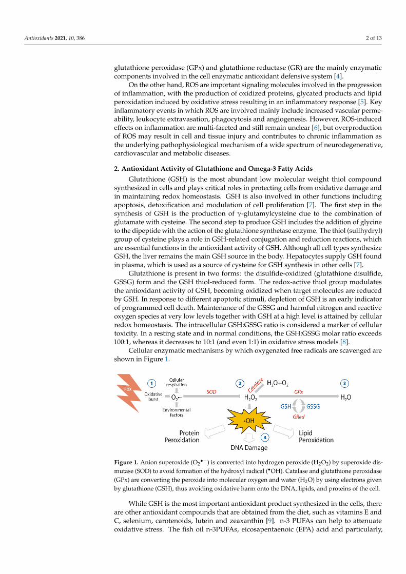
Figure 1. Anion superoxide (O2●−) is converted into hydrogen peroxide (H2O2) by superoxide dismutase (SOD) to avoid formation of the hydroxyl radical (●OH). Catalase and glutathione peroxidase (GPx) are converting the peroxide into molecular oxygen and water (H2O) by using electrons given by glutathione (GSH), thus avoiding oxidative harm onto the DNA, lipids, and proteins of the cell.
Figure 1. Anion superoxide (O2•−) is converted into hydrogen peroxide (H2O2) by superoxide dis-
mutase (SOD) to avoid formation of the hydroxyl radical (•OH). Catalase and glutathione peroxidase(GPx) are converting the peroxide into molecular oxygen and water (H2O) by using electrons givenby glutathione (GSH), thus avoiding oxidative harm onto the DNA, lipids, and proteins of the cell.
While GSH is the most important antioxidant product synthesized in the cells, thereare other antioxidant compounds that are obtained from the diet, such as vitamins E andC, selenium, carotenoids, lutein and zeaxanthin [9]. n-3 PUFAs can help to attenuateoxidative stress. The fish oil n-3PUFAs, eicosapentaenoic (EPA) acid and particularly,
Antioxidants 2021, 10, 386 3 of 13
docosahexaenoic (DHA) acid, have demonstrated the most promising and consistentbeneficial health effects, including anti-inflammatory, antiproliferative, antiangiogenicand antioxidant properties [10–12]. In common to other n-3 fatty acids, DHA can besynthesized from the plant-derived α-linoleic acid (ALA) found in components of thehuman diet, but the extent of conversion of ALA to DHA appears to be small. Seafoodand products derived from seafood are the primary dietary sources of DHA, so that DHAintake is heavily influenced by fish consumption. Dietary supplementation with DHAis an alternative supply to endogenous synthesis of DHA to achieve optimum levels ofn-3PUFAs in the body and maintain essential functions, mainly neuroprotective.
n-3 PUFAs have important physiological functions because of their presence as phos-pholipids in cell membranes, contributing to an optimum bilayer structure, which isnecessary for intercellular communication and other highly differentiated functions [13].n-3 PUFAs are primary precursors of bioactive lipid mediators, including eicosanoids,which have diverse paracrine and autocrine actions [10]. In nervous tissue, DHA ispresent in high concentrations, playing an essential role in neuroprotection and braindevelopment [14,15]. In the retina, the highest concentration of DHA is found in the outersegments of rod photoreceptors where DHA plays a crucial role in retinal function [16] (seeSection 3.1), which is in contrast to minor significance of EPA at ocular level.
Enhanced Antioxidant Response of DHA and GSH
Experimental studies have provided some lines of evidence of an interrelationshipin the antioxidant defense mechanisms exerted by DHA and GSH. In a model of humanfibroblast culture, supplementation with 30 µmol/L DHA was accompanied by a largeincrease in intracellular GSH content in association with an elevated catalytic activity ofglutathione reductase and glutathione-5-transferase [17]. Increased GSH contributed toreduce ROS as evaluated by a decreased accumulation of dicholorofluorescein inside cells.Apparently, this was the first report of a potent and specific effect of DHA for reducingoxidative stress in human fibroblasts [17].
In a rat pheochromocytoma cell line (PC12) culture, pretreatment with DHA (24 h) pro-tected the cells from H2O2-induced oxidative damage by different mechanisms, includingthe regulation of the nuclear factor erythroid 2 like 2 (NFE2L2) and its downstream targetprotein, heme-oxygenase-1 (HO-1), and an increase in intracellular levels of enzymaticantioxidants such as SOD and GPx [18]. Interestingly, it has been recently shown that n-3PUFAs may suppress pro-oxidant activity by upregulating genes encoding cytoprotec-tive antioxidant proteins such as heme oxygenase 1 (HO-1) and glutathione peroxidase(GPx) [19]. In a study of dopaminergic SM4741 cells, the toxic effect of paraquat relatedto an increase of intracellular ROS content in different organelles was ameliorated bypretreatment with DHA, which also increased glutathione reductase and the accumulationof the GSH pool by enhancing GSH homeostasis regulated by NFE2L2 protein levels [20].
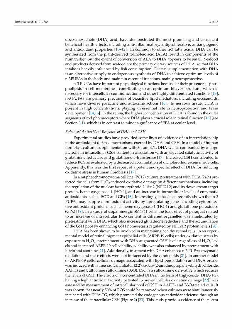
DHA has been shown to be involved in maintaining healthy retinal cells. In an experi-mental model of retinal pigment epithelial cells (ARPE-19 cells) under oxidative stress byexposure to H2O2, pretreatment with DHA augmented GSH levels regardless of H2O2 lev-els and increased ARPE-19 cell viability; viability was also enhanced by pretreatment withlutein and xanthine [21]. Additionally, treatment with DHA enhanced n-3 PUFAs enzymaticoxidation and these effects were not influenced by the carotenoids [21]. In another modelof ARPE-19 cells, cellular damage associated with lipid peroxidation and DNA breakswas induced with a free radical initiator (2,2′-azobis-(2-amidinopropane)-dihydrochloride,AAPH) and buthionine sulfoximine (BSO). BSO is a sulfoximine derivative which reducesthe levels of GSH. The effects of a concentrated DHA in the form of triglyceride (DHA-TG),having a high antioxidant activity patented to prevent cellular oxidation damage [22]) wasassessed by measurement of intracellular pool of GSH in AAPH- and BSO-treated cells. Itwas shown that nearly 50% of ROS could be removed when cultures were simultaneouslyincubated with DHA-TG, which promoted the endogenous antioxidant defense through anincrease of the intracellular GSH (Figure 2) [23]. This study provides evidence of the potent
Antioxidants 2021, 10, 386 4 of 13
effect of DHA-TG for decreasing oxidative stress involving GSH on pigment epithelial cellsof the human retina.
Antioxidants 2021, 10, x FOR PEER REVIEW 4 of 14
concentrated DHA in the form of triglyceride (DHA-TG), having a high antioxidant ac-tivity patented to prevent cellular oxidation damage [22]) was assessed by measurement of intracellular pool of GSH in AAPH- and BSO-treated cells. It was shown that nearly 50% of ROS could be removed when cultures were simultaneously incubated with DHA-TG, which promoted the endogenous antioxidant defense through an increase of the intracellular GSH (Figure 2) [23]. This study provides evidence of the potent effect of DHA-TG for decreasing oxidative stress involving GSH on pigment epithelial cells of the human retina.
Figure 2. Retinal ARPE-19 cells (×10 magnification) showing disappearance of oxidation of fluo-rescent probes by DHA triglyceride (DHA-TG) (right) as compared with control (left) as indicative of removal of intracellular ROS (reactive oxygen species).
3. Protective Effects of DHA against Oxidative Stress in Eye Structures 3.1. Retinal Cells and Photoreceptors
In the human body, the retina has the highest oxygen consumption per gram of tissue, requiring large amounts of adenosine triphosphate (ATP) to support cellular functions. This high metabolism makes the retina especially vulnerable to oxidative stress damage. Numerous studies have shown that ROS contributes to vascular endo-thelial dysfunction and retinal neural degeneration [24]. Moreover, inflammation in the retina and neuron degeneration has been linked to excessive ROS formation, which can directly modify cellular molecules and impair their function. The production of inflam-matory cytokines, such as interleukin-6 (IL-6), IL-1β and tumor necrosis factor-alpha (TNFα), as well as the activation of transcription factors, such as nuclear factor (NF)-kappa B (NF-κB) can be stimulated by ROS causing inflammation and cell death [25]. Retinal endothelial dysfunction caused by accumulation of ROS impairs the balance of nitric oxide (NO) metabolism and the responsiveness of smooth muscle cells and vascular endothelial cells to physiological stimuli. Reduced endothelium-dependent vasodilation and a prothrombotic and proinflammatory state are characteristic features of endothelial dysfunction [25].
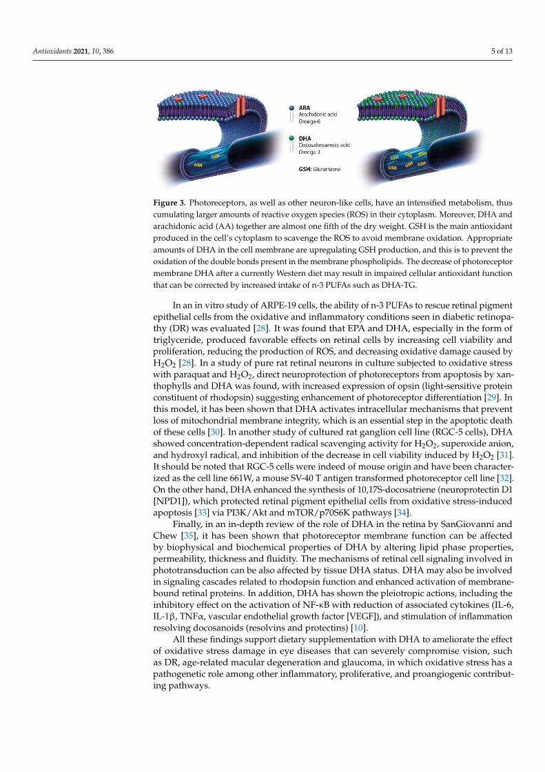
Lipids are structural components of cell membranes, provide energy storage, and act as signaling molecules. n-3 PUFAs are essential for neural development of the brain and different structures, including the retina. n-3 PUFAs cannot be efficiently synthesized by the human body, so that adequate amounts depend on the consumption of foods rich in these compounds, such as fish and fish oil products (Figure 3). DHA accounts for ap-proximately 20% of the retinal weight and affects the development and survival of neu-rons and retinal vascular cells [24]. DHA is the dominant fatty acid of retinal phospho-lipids and plays a significant role in maintaining retinal integrity [25], also related to the increased production of GSH (Figure 3).
Figure 2. Retinal ARPE-19 cells (×10 magnification) showing disappearance of oxidation of fluores-cent probes by DHA triglyceride (DHA-TG) (right) as compared with control (left) as indicative ofremoval of intracellular ROS (reactive oxygen species).
3. Protective Effects of DHA against Oxidative Stress in Eye Structures3.1. Retinal Cells and Photoreceptors
In the human body, the retina has the highest oxygen consumption per gram of tissue,requiring large amounts of adenosine triphosphate (ATP) to support cellular functions.This high metabolism makes the retina especially vulnerable to oxidative stress damage.Numerous studies have shown that ROS contributes to vascular endothelial dysfunctionand retinal neural degeneration [24]. Moreover, inflammation in the retina and neurondegeneration has been linked to excessive ROS formation, which can directly modifycellular molecules and impair their function. The production of inflammatory cytokines,such as interleukin-6 (IL-6), IL-1β and tumor necrosis factor-alpha (TNFα), as well asthe activation of transcription factors, such as nuclear factor (NF)-kappa B (NF-κB) canbe stimulated by ROS causing inflammation and cell death [25]. Retinal endothelialdysfunction caused by accumulation of ROS impairs the balance of nitric oxide (NO)metabolism and the responsiveness of smooth muscle cells and vascular endothelial cells tophysiological stimuli. Reduced endothelium-dependent vasodilation and a prothromboticand proinflammatory state are characteristic features of endothelial dysfunction [25].
Lipids are structural components of cell membranes, provide energy storage, and actas signaling molecules. n-3 PUFAs are essential for neural development of the brain anddifferent structures, including the retina. n-3 PUFAs cannot be efficiently synthesized by thehuman body, so that adequate amounts depend on the consumption of foods rich in thesecompounds, such as fish and fish oil products (Figure 3). DHA accounts for approximately20% of the retinal weight and affects the development and survival of neurons and retinalvascular cells [24]. DHA is the dominant fatty acid of retinal phospholipids and plays asignificant role in maintaining retinal integrity [25], also related to the increased productionof GSH (Figure 3).
Cell membranes of the rod outer segment of retinal photoreceptors have a high contentof DHA (50–70% of fatty acids) since DHA is a component of phospholipids that clusteraround rhodopsin, which is the protein that receives the light signal. When the light signalis received, a phototransduction cascade is initiated through activation of a conformationalchange of rhodopsin, which is facilitated by the presence of DHA within the membrane [26].On the other hand, the retina has an abundance of DHA-containing phospholipids (DHA-PL). In a model of lysophosphatidic acid acyltransferase 3 (LPAAT3)-knock-out mice,abnormalities in the retinal layers following loss of DHA-PL were observed, such asincomplete elongation of the outer segment and decreased thickness of the outer nuclearlayers, as well as disordered disc shape in photoreceptor cells [27].
Antioxidants 2021, 10, 386 5 of 13Antioxidants 2021, 10, x FOR PEER REVIEW 5 of 14
Figure 3. Photoreceptors, as well as other neuron-like cells, have an intensified metabolism, thus cumulating larger amounts of reactive oxygen species (ROS) in their cytoplasm. Moreover, DHA and arachidonic acid (AA) together are almost one fifth of the dry weight. GSH is the main anti-oxidant produced in the cell’s cytoplasm to scavenge the ROS to avoid membrane oxidation. Ap-propriate amounts of DHA in the cell membrane are upregulating GSH production, and this is to prevent the oxidation of the double bonds present in the membrane phospholipids. The decrease of photoreceptor membrane DHA after a currently Western diet may result in impaired cellular an-tioxidant function that can be corrected by increased intake of n-3 PUFAs such as DHA-TG.
Cell membranes of the rod outer segment of retinal photoreceptors have a high content of DHA (50–70% of fatty acids) since DHA is a component of phospholipids that cluster around rhodopsin, which is the protein that receives the light signal. When the light signal is received, a phototransduction cascade is initiated through activation of a conformational change of rhodopsin, which is facilitated by the presence of DHA within the membrane [26]. On the other hand, the retina has an abundance of DHA-containing phospholipids (DHA-PL). In a model of lysophosphatidic acid acyltransferase 3 (LPAAT3)-knock-out mice, abnormalities in the retinal layers following loss of DHA-PL were observed, such as incomplete elongation of the outer segment and decreased thickness of the outer nuclear layers, as well as disordered disc shape in photoreceptor cells [27].
In an in vitro study of ARPE-19 cells, the ability of n-3 PUFAs to rescue retinal pig-ment epithelial cells from the oxidative and inflammatory conditions seen in diabetic retinopathy (DR) was evaluated [28]. It was found that EPA and DHA, especially in the form of triglyceride, produced favorable effects on retinal cells by increasing cell viability and proliferation, reducing the production of ROS, and decreasing oxidative damage caused by H2O2 [28]. In a study of pure rat retinal neurons in culture subjected to oxida-tive stress with paraquat and H2O2, direct neuroprotection of photoreceptors from apoptosis by xanthophylls and DHA was found, with increased expression of opsin (light-sensitive protein constituent of rhodopsin) suggesting enhancement of photore-ceptor differentiation [29]. In this model, it has been shown that DHA activates intracel-lular mechanisms that prevent loss of mitochondrial membrane integrity, which is an essential step in the apoptotic death of these cells [30]. In another study of cultured rat ganglion cell line (RGC-5 cells), DHA showed concentration-dependent radical scav-enging activity for H2O2, superoxide anion, and hydroxyl radical, and inhibition of the decrease in cell viability induced by H2O2 [31]. It should be noted that RGC-5 cells were indeed of mouse origin and have been characterized as the cell line 661W, a mouse SV-40 T antigen transformed photoreceptor cell line [32]. On the other hand, DHA enhanced the synthesis of 10,17S-docosatriene (neuroprotectin D1 [NPD1]), which protected retinal pigment epithelial cells from oxidative stress-induced apoptosis [33] via PI3K/Akt and mTOR/p70S6K pathways [34].
Finally, in an in-depth review of the role of DHA in the retina by SanGiovanni and Chew [35], it has been shown that photoreceptor membrane function can be affected by biophysical and biochemical properties of DHA by altering lipid phase properties, per-meability, thickness and fluidity. The mechanisms of retinal cell signaling involved in phototransduction can be also affected by tissue DHA status. DHA may also be involved
Figure 3. Photoreceptors, as well as other neuron-like cells, have an intensified metabolism, thuscumulating larger amounts of reactive oxygen species (ROS) in their cytoplasm. Moreover, DHA andarachidonic acid (AA) together are almost one fifth of the dry weight. GSH is the main antioxidantproduced in the cell’s cytoplasm to scavenge the ROS to avoid membrane oxidation. Appropriateamounts of DHA in the cell membrane are upregulating GSH production, and this is to prevent theoxidation of the double bonds present in the membrane phospholipids. The decrease of photoreceptormembrane DHA after a currently Western diet may result in impaired cellular antioxidant functionthat can be corrected by increased intake of n-3 PUFAs such as DHA-TG.
In an in vitro study of ARPE-19 cells, the ability of n-3 PUFAs to rescue retinal pigmentepithelial cells from the oxidative and inflammatory conditions seen in diabetic retinopa-thy (DR) was evaluated [28]. It was found that EPA and DHA, especially in the form oftriglyceride, produced favorable effects on retinal cells by increasing cell viability andproliferation, reducing the production of ROS, and decreasing oxidative damage caused byH2O2 [28]. In a study of pure rat retinal neurons in culture subjected to oxidative stresswith paraquat and H2O2, direct neuroprotection of photoreceptors from apoptosis by xan-thophylls and DHA was found, with increased expression of opsin (light-sensitive proteinconstituent of rhodopsin) suggesting enhancement of photoreceptor differentiation [29]. Inthis model, it has been shown that DHA activates intracellular mechanisms that preventloss of mitochondrial membrane integrity, which is an essential step in the apoptotic deathof these cells [30]. In another study of cultured rat ganglion cell line (RGC-5 cells), DHAshowed concentration-dependent radical scavenging activity for H2O2, superoxide anion,and hydroxyl radical, and inhibition of the decrease in cell viability induced by H2O2 [31].It should be noted that RGC-5 cells were indeed of mouse origin and have been character-ized as the cell line 661W, a mouse SV-40 T antigen transformed photoreceptor cell line [32].On the other hand, DHA enhanced the synthesis of 10,17S-docosatriene (neuroprotectin D1[NPD1]), which protected retinal pigment epithelial cells from oxidative stress-inducedapoptosis [33] via PI3K/Akt and mTOR/p70S6K pathways [34].
Finally, in an in-depth review of the role of DHA in the retina by SanGiovanni andChew [35], it has been shown that photoreceptor membrane function can be affectedby biophysical and biochemical properties of DHA by altering lipid phase properties,permeability, thickness and fluidity. The mechanisms of retinal cell signaling involved inphototransduction can be also affected by tissue DHA status. DHA may also be involvedin signaling cascades related to rhodopsin function and enhanced activation of membrane-bound retinal proteins. In addition, DHA has shown the pleiotropic actions, including theinhibitory effect on the activation of NF-κB with reduction of associated cytokines (IL-6,IL-1β, TNFα, vascular endothelial growth factor [VEGF]), and stimulation of inflammationresolving docosanoids (resolvins and protectins) [10].
All these findings support dietary supplementation with DHA to ameliorate the effectof oxidative stress damage in eye diseases that can severely compromise vision, suchas DR, age-related macular degeneration and glaucoma, in which oxidative stress has apathogenetic role among other inflammatory, proliferative, and proangiogenic contribut-ing pathways.
Antioxidants 2021, 10, 386 6 of 13
3.2. Trabecular Meshwork and Intraocular Pressure
The trabecular meshwork is a specialized eye tissue that regulates the outflow of theaqueous humor and controls intraocular pressure (IOP). Cells of this biological filter arecrucial for maintaining homeostasis of the whole outflow system through which the aque-ous humor reaches the Schlemm canal. There is increasing evidence of the pathogeneticrole of oxidative DNA damage affecting the trabecular meshwork cells in the developmentof glaucomatous optic neuropathy [36–40], and its relation with glutathione S-transferasegenetic polymorphisms and lack of the GSTM1 gene, which catalyzes neutralization of freeradicals by conjugation with GSH [40,41].
Reduced ocular blood flow associated with red blood cell membrane abnormalitieshave been implicated in the pathogenesis of primary open-angle glaucoma (POAG). In astudy of lipid composition of red blood cell membranes in patients with POAG, reducederythrocyte levels of phosphatidyl-choline (PC) carrying docosahexaenoic acid (DHA) wasreported [42,43]. In Sprague-Dawley rat dams, induced IOP elevation associated witha deficient n-3 PUFAs diet caused dysfunction of retinal ganglion cell activity, and thecombination of these factors (dietary manipulation and IOP stress) showed a cumulativeeffect [44]. The authors concluded that sufficient dietary n-3 PUFAs improves retinalganglion cell function making it less susceptible to IOP insult, which may have implicationsfor glaucoma [44]. In a DBA/2J mouse model of hereditary glaucoma, supplementation ofn-3 PUFAs had neuroprotective effect in the retinas, with downregulation of TNFα andIL-18 [45].
Different clinical studies have investigated the implications of oxidative stress inpatients with POAG. In a study of 40 patients with POAG and 60 healthy controls, signif-icantly higher levels of plasma malondialdehyde (MDA) as a marker of oxidative stresswere found in patients with POAG [46]. In a sample of 21 patients with POAG and 34 age-and gender-matched control subjects, patients with glaucoma showed significantly lowerlevels of GSH suggesting a general impairment of the antioxidative defense [47]. In anotherstudy of 206 patients with POAG and 126 controls, lower systemic antioxidant capacityin glaucoma patients was demonstrated as measured by plasma levels of reactive oxygenmetabolites, antioxidant capacity, and thiol-antioxidant capacity [48]. In a study in whichDNA damage markers and the antioxidant status of serum and aqueous humor weremeasured from 28 patients with glaucoma and 27 patients with cataracts at the time ofsurgery, aqueous humor and serum levels 8-hydroxy-2′-deoxyguanosine (8-OHdG), anestablished marker for oxidative stress-induced DNA damage were significantly higher inglaucoma patients, whereas aqueous and serum total antioxidant status was significantlylower [49]. A further study that evaluated DNA damage in terms of 8-OHdG in specimensof trabecular meshwork from 42 patients with glaucoma and 42 controls of similar age andsex, also showed significantly higher levels of 8-OHdG in glaucoma patients [50]. Moreover,the GSTM1 genotype assessed by polymerase chain reaction in DNA samples showedthat the GSTM1-null genotype was significantly more common in patients with glaucoma,who also showed significantly higher levels of 8-OHdG as compared with patients withGSTM1-positive genotype [41].
Taken together, these studies suggest that dietary supplementation with DHA couldmodulate impaired antioxidant status associated with glaucomatous damage.
4. Eye Benefits of Dietary Supplementation with DHA4.1. Diabetic Retinopathy and Macular Edema
Severe vision loss in DR is seen in diabetic macular edema and proliferative DR,which is characterized by retinal neovascularization. The crucial factor leading to visionloss and eventually blindness is chronic hyperglycemia due to poorly controlled bloodglucose levels. Hyperglycemia leads to the activation of metabolic pathways that involveinflammation, oxidative damage, and neurovascular dysfunction. The duration of diabetesand poor glycemic control are key factors increasing the prevalence of DR. Approximately,
Antioxidants 2021, 10, 386 7 of 13
33% of diabetic patients present signs of DR and 10% have vision-threatening stages ofDR [24].
Oxidative stress forms part of the multifactorial pathogenesis of DR and diabeticmacular edema [51,52] and provides the rationale of dietary supplementation with DHAand other naturally occurring antioxidants, such as carotenoids and xantophylls. However,DHA exhibits a vision-regulating role ensuring proper functioning of rhodopsin pigmentpresent in the retinal photoreceptor cells. DHA is also a strong anti-inflammatory agentand inhibits the expression of VEGF, decreasing the production of superoxide radicalsinvolved in the VEGF signaling pathway [51]
Of the three n-3 PUFAs, ALA, EPA and DHA, which can be used for delaying/inhibitingthe various underlying mechanisms induced during hyperglycemia focusing on DR, DHAexhibits the widest range of anti-inflammatory mediators, besides decreasing the formationof free radicals and inducing expression of endogenous antioxidant enzymes and remark-ably preventing retinal angiogenesis by downregulation of the expression of angiogenicagents especially VEGF [51].
Real-life outcomes of dietary supplementation with high-dose DHA-TG in DR wereassessed in a prospective controlled study of 12 asymptomatic patients with nonprolifera-tive DR (n = 24 eyes) and 12 healthy controls (n = 24 eyes) [53]. Variables measured weremacular sensitivity and integrity, visual acuity, macular thickness by optical coherencetomography (OCT), plasma total antioxidant capacity (TAC), and DHA content in the ery-throcyte membrane. Participants in the experimental group received a high-rich DHA-TG(1050 mg/day) supplementation, whereas controls received no treatment but were blindedregarding the existence of an experimental group. Of note is that TAC in plasma wasmeasured using a commercially available kit, with uric acid equivalent used to calculatecooper-reducing equivalent values (µM Cu-reducing equivalent). High TAC values reflecthigh antioxidant capacity, i.e., greater protection. The duration of treatment was 90 days.Salient results included statistically significant differences in macular function in favorof the experimental group, with plasma TAC values and DHA content of the erythrocytemembrane increasing significantly in the experimental group only. In conclusion, supple-mentation with high-dose DHA-TG at an early stage of DR improved macular functionmeasured by microperimetry and increased the antioxidant status capacity.
Plasma TAC was also determined in a 3-year randomized single-blind controlledtrial of intravitreal injection of ranibizumab combined with oral supplementation withDHA-TG in diabetic patients with macular edema [54,55]. Briefly, all patients had type2 diabetes with decreased vision due to diabetic macular edema (DME) (1 mm thicknessin the central subfield OCT). In this trial, patients were randomized to treatment withranibizumab (loading dose 0.5 mg/0.05 m for the first 4 months followed on as as-neededtreatment) alone (control group) or combined with high-rich DHA-TG supplementation(1050 mg/day) (DHA group). The dietary supplement also contained 127 mg of EPA. TACwas expressed as µM Cu-reducing equivalent. At 2 years with 33 patients and 42 eyesanalyzed in the control group, and 34 eyes and 29 patients in the DHA group, the decreasein central subfield macular thickness was statistically significant in favor of the DHAsupplementation group, which was also observed at 3 years. Visual outcomes, however,were similar in both two study groups, either at 2- or 3-year assessments. As expected,differences in plasma TAC and erythrocyte membrane DHA content were all significant infavor of the DHA supplementation group. This study shows the beneficial effect of DHAsupplementation in terms of anatomical improvement in DME.
Findings of a substudy from the PREDIMED randomized clinical study (Preventionwith Mediterranean Diet) showed that in older and middle-age subjects with type 2 dia-betes, intake of at least 500 mg/day of dietary n-3 PUFAs (minimum two weekly servingsof oily fish) was associated with a 46% risk reduction of incident sight-threatening DR ascompared with patients not fulfilling this dietary recommendation [56]. In this study, how-ever, dietary intake of n-3 PUFAs was estimated using a validated dietary questionnairein which the consumption of 8 different types of seafoods were included, but information
Antioxidants 2021, 10, 386 8 of 13
regarding the consumption of specific amounts of EPA and DHA is not provided. Addi-tionally, differences in the risk of DR could be related to other lifestyle factors, as well as tothe fact that participants who met n-3 PUFAs consumption target tended to be youngerand showed a lower prevalence of hypertension (or antihypertensive treatment) and usedless insulin than participants not fulfilling the dietary target recommendation.
4.2. Age-Related Macular Degeneration
Similar studies of dietary supplementation with high-dose DHA-TG in age-relatedmacular degeneration (AMD) have not been carried out, but results reported from avariety of research are inconsistent regarding the effects on omega-3 fatty acids on visualacuity, progression and development of drusen or the presence of geographic atrophy [57].However, it has been reported that high erythrocyte content of EPA/DHA is a protectivefactor against AMD compared with permanently low EPA/DHA levels [58]. Results of thephase 3 randomized controlled AREDS2 study in which there was no overall additionalbenefit from adding n-3 PUFAS or lutein and zeaxanthin to the original AREDS formulation(vitamins C and E, beta-carotene, zinc oxide, cupric oxide) in reducing the risk of advancedAMD [59] merits a comment. In contrast to the use of a highly concentrated dose ofDHA-TG (1050 mg/day) in the randomized studies of diabetic macular edema [54,55],participants in the AREDS2 study received 10 mg lutein, 20 mg zeaxanthin, and 650 mgof EPA and 350 mg of DHA in the form of ethyl esters. It should be noted that the dailydose of DHA in AREDS2 study was markedly inferior to that used in the randomizedtrials of diabetic macular edema [54,55], and that the ethyl ester formulations of n-3 PUFAScreated in the process of transesterification have shown lower bioavailability than chemicaltriglyceride binding [60].
It is interesting to discuss the effect of DHA administered in combination withlutein/zeaxanthin. An experimental study in quails showed that supplementation withlutein/zeaxanthin prevented light-induced photoreceptor cell death [61]. In a clinicalstudy of 100 healthy participants (200 eyes) aged 40–70 years randomized in a 1:1 ratio toreceive daily lutein or lutein/DHA for 3 months, macular pigment optical density (MPOD)showed significantly higher values in the lutein/DHA group than in the lutein alone group,as well as higher lutein levels in plasma, so that this study highlights the relevance ofthe adjunctive role of DHA for a better lutein bioavailability [62]. In a 4-month study of49 women (aged 60–80 years) randomized to lutein (12 mg/day), DHA (800 mg/day),lutein + DHA, or placebo, lutein alone increased MPOD eccentrically, DHA supplementa-tion alone resulted in central increases in MPOD, and the combination of supplements hada combined effect on the MPOD spatial profile. In addition, DHA facilitated accumulationof lutein in the blood and macula [63].
The Veterans LAST (Lutein Antioxidant Supplementation Trial) was a prospective,12-month, randomized, double-masked, placebo-controlled trial aimed to assess whethernutritional supplementation with lutein or lutein together with antioxidants, vitamins,and minerals improves visual function and symptoms in atrophic AMD [64]. There werethree study groups: patients in group 1 received lutein 10 mg (L); in Group 2, a lutein10 mg/antioxidants/vitamins and minerals broad spectrum supplementation formula(L/A) (n-3 PUFAs were not included); and in Group 3, a maltodextrin placebo (P) over12 months. In patients treated with lutein alone or lutein together with other nutrients,there was a significant increase in eye macular pigment optical density. Visual acuity andcontrast sensitivity also improved. In a further analysis of the same study population, itwas observed that patients with lower baseline values of macular pigment optical densitywere those most likely to benefit from either the lutein or the lutein plus antioxidantsupplementation [65]. Accordingly, it may be inferred that if a deficiency in macularpigment optical density is accurately diagnosed, effective interventions should be able tore-establish this prophylactic barrier.
Antioxidants 2021, 10, 386 9 of 13
Other studies of n-3 PUFA supplementation in AMD besides AREDS and AREDS2have been carried out. In a large study of 263 patients over 55 years of age with earlylesions of age-related maculopathy and randomized to receive either 840 mg/day DHAand 270 mg/day EPA from fish oil capsules or the placebo (olive oil capsules) for 3 years,no differences were observed in the occurrence of choroidal neovascularization in the studyeye [66]. In a prospective study over more than 2 decades of follow-up of 75,889 womenfrom the Nurses’ Health Study and 38,961 men from the Health Professionals Follow-upStudy, the intake of EPA + DHA ≥ 350 mg/day (697 mg/day for the fifth quintile) or fattyfish ≥ 2 servings/week showed a moderate reduction in the risk of visually significantAMD, but was inconclusive regarding protection against development of advancementAMD [67].
4.3. Glaucoma
Antioxidant supplementation in patients with glaucoma has been associated withbeneficial effects as extensively reported in a recent comprehensive review of 19 clinicaltrials. Data, however, are difficult to interpret because of differences among studies oftypes of glaucoma patients, study outcomes, and antioxidant substrates [68].
There is limited information on the use of high-dose DHA supplementation in glau-coma. A prospective randomized open-label controlled study in patients with pseudoexfo-liative glaucoma (PEX) was conducted to assess improvement of antioxidant protectionassociated with high-rich dietary supplementation with DHA triglyceride [69]. Patientswere assigned to active supplementation (DHA-TG 1050 mg/day) (n = 23) or to a controlgroup (n = 24) without dietary intervention. The duration of treatment was 6 months.Biochemical analyses in this trial included plasma TAC (µM Cu-reducing equivalent val-ues), plasma MDA, and erythrocyte membrane content of DHA (% total fatty acids). Inthe DHA group, mean IOP in both eyes decreased significantly at the end of treatment.In addition, as compared with baseline TAC levels and DHA content in the erythrocytemembrane increased significantly, whereas MDA decreased significantly in the DHA grouponly. Although the duration of treatment was limited to 6 months, decreases in IOP aresuggestive of the clinical value of reducing oxidative stress with a high-rich DHA supple-ment in patients with PEX. In other studies, oral supplementation with high-dose DHAtriglyceride achieved a reduction in oxidative stress markers and a significant improvementin symptoms and signs of eye dryness present in glaucoma patients [70,71].
5. Conclusions
Eyes are by far the most important organs of sense, and loss of vision in particularaffecting the central fovea, has a profound impact on all aspects of an individual’s life.The identification of underlying cellular and molecular pathogenetic mechanisms in eyediseases has been a crucial advancement in the development of effective treatment strategiesfor preventing potential causes of blindness. The involvement of local oxidative stress andoxidative DNA damage in the underlying mechanisms of retinal diseases or glaucoma thatcan lead to blindness supports the rationale of the use of antioxidants in the managementof these conditions. Randomized controlled studies of dietary supplementation with high-dose DHA in the form of triglyceride have provided evidence of the significant beneficialeffects of n-3 PUFAs in the care of patients with DR, macular edema, and optic neuropathyassociated with glaucoma. However, the overall clinical evidence for the use of dietarystrategies based on supplementation with PUFAs in eye diseases linked to oxidative stressother than high-dose DHA triglyceride is both limited and inconsistent.
Author Contributions: Conceptualization, M.L. and J.C.D.; methodology, M.L., M.E.R.G.-H., S.R.V.and J.C.D.; writing—original draft preparation, J.C.D.; writing—review and editing, M.L. and J.C.D.All authors have read and agreed to the published version of the manuscript.
Funding: This research received no external funding.
Antioxidants 2021, 10, 386 10 of 13
Acknowledgments: The authors thank Marta Pulido for editing the manuscript and editorial as-sistance. M. Lafuente was working at the Department of Ophthalmology, Hospital UniversitarioMorales Meseguer, E-30008 Murcia, Spain, while she performed the clinical trial cited in [54,55].
Conflicts of Interest: The authors declare no conflict of interest.
Abbreviations
AA arachidonic acidAAPH 2,2′-azobis-(2-amidinopropane)-dihydrochlorideALA α-linoleic acidAMD age-related macular degenerationATP adenosine triphosphateBSO buthionine sulfoximineCAT catalaseCOX cyclooxygenasesDHA docosahexaenoic acidDHA-PL DHA containing phospholipidsDHA-TG DHA triglycerideDME diabetic macular edemaDR diabetic retinopathyEPA eicosapentaenoic acidGPx glutathione peroxidaseGR glutathione reductaseGSH glutathioneGSSG glutathione disulfideH2O waterH2O2 hydrogen peroxideIL interleukinIOP intraocular pressureLOX lipoxygenasesLPAAT3 lysophosphatidic acid acyltransferase 3MDA malondialdehydeMPOD macular pigment optical densityn-3 PUFAs omega-3 polyunsaturated fatty acidsNFE2L2 nuclear factor erythroid 2 like 2NF-κB nuclear factor (NF)-kappa BNO nitric oxideNPD1 neuroprotectin D1OCT optical coherence tomography8-OHdG 8-hydroxy-2′-deoxyguanosinePC phosphatidyl-cholinePEX pseudoexfoliative glaucomaPOAG primary open-angle glaucomaROS reactive oxygen speciesSOD superoxide dismutaseTAC total antioxidant capacityTNFα tumor necrosis factor-alphaVEGF vascular endothelial growth factor
References1. Pizzino, G.; Irrera, N.; Cucinotta, M.; Pallio, G.; Mannino, F.; Arcoraci, V.; Squadrito, F.; Altavilla, D.; Bitto, A. Oxidative stress:
Harms and benefits for human health. Oxid Med. Cell. Longev. 2017, 2017, 8416763. [CrossRef] [PubMed]2. Al-Gubory, K.H.; Garrel, C.; Faure, P.; Sugino, N. Roles of antioxidant enzymes in corpus luteum rescue from reactive oxygen
species-induced oxidative stress. Reprod. Biomed. Online 2012, 25, 551–560. [CrossRef]3. Aiken, C.T.; Kaake, R.M.; Wang, X.; Huang, L. Oxidative stress-mediated regulation of proteasome complexes. Mol. Cell. Proteom.
2011, 10, R110.006924. [CrossRef] [PubMed]4. Deponte, M. Glutathione catalysis and the reaction mechanisms of glutathione-dependent enzymes. Biochim. Biophys. Acta 2013,
1830, 3217–3266. [CrossRef] [PubMed]
Antioxidants 2021, 10, 386 11 of 13
5. Mittal, M.; Siddiqui, M.R.; Tran, K.; Reddy, S.P.; Malik, A.B. Reactive oxygen species in inflammation and tissue injury. Antioxid.Redox Signal. 2014, 20, 1126–1167. [CrossRef] [PubMed]
6. Forrester, S.J.; Kikuchi, D.S.; Hernandes, M.S.; Xu, Q.; Griendling, K.K. Reactive oxygen species in metabolic and inflammatorysignaling. Circ. Res. 2018, 122, 877–902. [CrossRef] [PubMed]
7. Forman, H.J.; Zhang, H.; Rinna, A. Glutathione: Overview of its protective roles, measurement, and biosynthesis. Mol. Asp. Med.2009, 30, 1–12. [CrossRef]
8. Zitka, O.; Skalickova, S.; Gumulec, J.; Masarik, M.; Adam, V.; Hubalek, J.; Trnkova, L.; Kruseova, J.; Eckschlager, T.; Kizek, R.Redox status expressed as GSH:GSSG ratio as a marker for oxidative stress in paediatric tumour patients. Oncol. Lett. 2012, 4,1247–1253. [CrossRef]
9. Visioli, F.; Galli, C. The role of antioxidants in the Mediterranean diet. Lipids 2001, 36, S49–S52. [CrossRef]10. Calder, P.C. Omega-3 fatty acids and inflammatory processes. Nutrients 2010, 2, 355–374. [CrossRef]11. Shahidi, F.; Ambigaipalan, P. Omega-3 polyunsaturated fatty acids and their health benefits. Annu. Rev. Food Sci. Technol. 2018, 9,
345–381. [CrossRef] [PubMed]12. Gammone, M.A.; Riccioni, G.; Parrinello, G.; D’Orazio, N. Omega-3 polyunsaturated fatty acids: Benefits and endpoints in sport.
Nutrients 2018, 11, 46. [CrossRef]13. Wall, R.; Ross, R.P.; Fitzgerald, G.F.; Stanton, C. Fatty acids from fish: The anti-inflammatory potential of long-chain omega-3 fatty
acids. Nutr. Rev. 2010, 68, 280–289. [CrossRef] [PubMed]14. Valentine, R.C.; Valentine, D.L. Omega-3 fatty acids in cellular membranes: A unified concept. Prog. Lipid Res. 2004, 43, 383–402.
[CrossRef]15. Mayurasakorn, K.; Williams, J.J.; Ten, V.S.; Deckelbaum, R.J. Docosahexaenoic acid: Brain accretion and roles in neuroprotection
after brain hypoxia and ischemia. Curr. Opin. Clin. Nutr. Metab. Care 2011, 14, 158–167. [CrossRef]16. Jeffrey, B.G.; Weisinger, H.S.; Neuringer, M.; Mitchell, D.C. The role of docosahexaenoic acid in retinal function. Lipids 2001, 36,
859–871. [CrossRef] [PubMed]17. Arab, K.; Rossary, A.; Flourié, F.; Tourneur, Y.; Steghens, J.P. Docosahexaenoic acid enhances the antioxidant response of human
fibroblasts by upregulating gamma-glutamyl-cysteinyl ligase and glutathione reductase. Br. J. Nutr. 2006, 95, 18–26. [CrossRef]18. Clementi, M.E.; Lazzarino, G.; Sampaolese, B.; Brancato, A.; Tringali, G. DHA protects PC12 cells against oxidative stress and
apoptotic signals through the activation of the NFE2L2/HO-1 axis. Int. J. Mol. Med. 2019, 43, 2523–2531. [CrossRef]19. Bang, H.Y.; Park, S.A.; Saeidi, S.; Na, H.K.; Surh, Y.J. Docosahexaenoic acid induces expression of heme oxygenase-1 and
NAD(P)H:quinone oxidoreductase through activation of Nrf2 in human mammary epithelial cells. Molecules 2017, 22, 969.[CrossRef] [PubMed]
20. Lee, H.J.; Han, J.; Jang, Y.; Kim, S.J.; Park, J.H.; Seo, K.S.; Jeong, S.; Shin, S.; Lim, K.; Heo, J.Y.; et al. Docosahexaenoic acid preventsparaquat-induced reactive oxygen species production in dopaminergic neurons via enhancement of glutathione homeostasis.Biochem. Biophys. Res. Commun. 2015, 457, 95–100. [CrossRef]
21. Leung, H.H.; Galano, J.M.; Crauste, C.; Durand, T.; Lee, J.C. Combination of lutein and zeaxanthin, and DHA regulatedpolyunsaturated fatty acid oxidation in H2O2-stressed retinal cells. Neurochem. Res. 2020, 45, 1007–1019. [CrossRef]
22. Brudy Technology, S.L. Results shown in the European Patent EP 1 962 825 B1 Related to the Use of DHA for Treating a PathologyAssociated with Cellular Oxidative Damage. European Patent EP 1 962 825 B1, 2 April 2014.
23. Gasso, F.; Bogdanov, P.; Domingo, J. Docosahexaenoic acid improves endogen antioxidant defense in Arpe-19 cells. Investig.Ophthalmol. Vis. Sci. 2008, 49, 5932.
24. Suzumura, A.; Terao, R.; Kaneko, H. Protective effects of molecular signaling of n-3 fatty acids on oxidative stress and inflammationin retinal diseases. Antioxidants 2020, 9, 920. [CrossRef] [PubMed]
25. Ruan, Y.; Jiang, S.; Musayeva, A.; Gericke, A. Oxidative stress and vascular dysfunction in the retina: Therapeutic strategies.Antioxidants 2020, 9, 761. [CrossRef] [PubMed]
26. Senapati, S.; Gragg, M.; Samuels, I.S.; Parmar, V.M.; Maeda, A.; Park, P.S. Effect of dietary docosahexaenoic acid on rhodopsincontent and packing in photoreceptor cell membranes. Biochim. Biophys. Acta Biomembr. 2018, 1860, 1403–1413. [CrossRef]
27. Shindou, H.; Koso, H.; Sasaki, J.; Nakanishi, H.; Sagara, H.; Nakagawa, K.M.; Takahashi, Y.; Hishikawa, D.; Iizuka-Hishikawa,Y.; Tokumasu, F.; et al. Docosahexaenoic acid preserves visual function by maintaining correct disc morphology in retinalphotoreceptor cells. J. Biol. Chem. 2017, 292, 2054–12064. [CrossRef]
28. Saenz de Viteri, M.; Hernandez, M.; Bilbao-Malavé, V.; Fernandez-Robredo, P.; González-Zamora, J.; Garcia-Garcia, L.; Ispizua,N.; Recalde, S.; Garcia-Layana, A. A Higher proportion of eicosapentaenoic acid (EPA) when combined with docosahexaenoicacid (DHA) in omega-3 dietary supplements provides higher antioxidant effects in human retinal cells. Antioxidants 2020, 9, 828.[CrossRef] [PubMed]
29. Chucair, A.J.; Rotstein, N.P.; Sangiovanni, J.P.; During, A.; Chew, E.Y.; Politi, L.E. Lutein and zeaxanthin protect photoreceptorsfrom apoptosis induced by oxidative stress: Relation with docosahexaenoic acid. Investig. Ophthalmol. Vis. Sci. 2007, 48, 5168–5177.[CrossRef] [PubMed]
30. Rotstein, N.P.; Politi, L.E.; German, O.L.; Girotti, R. Protective effect of docosahexaenoic acid on oxidative stress-induced apoptosisof retina photoreceptors. Investig. Ophthalmol. Vis. Sci. 2003, 44, 2252–2259. [CrossRef]
31. Shimazawa, M.; Nakajima, Y.; Mashima, Y.; Hara, H. Docosahexaenoic acid (DHA) has neuroprotective effects against oxidativestress in retinal ganglion cells. Brain Res. 2009, 1251, 269–275. [CrossRef]
Antioxidants 2021, 10, 386 12 of 13
32. Krishnamoorthy, R.R.; Clark, A.F.; Daudt, D.; Vishwanatha, J.K.; Yorio, T. A forensic path to RGC-5 cell line identification: Lessonslearned. Investig. Ophthalmol. Vis. Sci. 2013, 54, 5712–5719. [CrossRef]
33. Mukherjee, P.K.; Marcheselli, V.L.; Serhan, C.N.; Bazan, N.G. Neuroprotectin D1: A docosahexaenoic acid-derived docosatrieneprotects human retinal pigment epithelial cells from oxidative stress. Proc. Natl. Acad Sci. USA 2004, 101, 8491–8496. [CrossRef]
34. Faghiri, Z.; Bazan, N.G. PI3K/Akt and mTOR/p70S6K pathways mediate neuroprotectin D1-induced retinal pigment epithelialcell survival during oxidative stress-induced apoptosis. Exp. Eye Res. 2010, 90, 718–725. [CrossRef]
35. SanGiovanni, J.P.; Chew, E.Y. The role of omega-3 long-chain polyunsaturated fatty acids in health and disease of the retina. Prog.Retin. Eye Res. 2005, 24, 87–138. [CrossRef]
36. Izzotti, A.; Bagnis, A.; Saccà, S.C. The role of oxidative stress in glaucoma. Mutat. Res. 2006, 612, 104–114. [CrossRef]37. Pinazo-Durán, M.D.; Zanón-Moreno, V.; García-Medina, J.J.; Gallego-Pinazo, R. Evaluation of presumptive biomarkers of
oxidative stress, immune response and apoptosis in primary open-angle glaucoma. Curr. Opin. Pharmacol. 2013, 13, 98–107.[CrossRef] [PubMed]
38. Mohanty, K.; Dada, R.; Dada, T. Oxidative DNA damage and reduced expression of DNA repair genes: Role in primary openangle glaucoma (POAG). Ophthalmic Genet. 2017, 38, 446–450. [CrossRef]
39. Caballero, M.; Liton, P.B.; Epstein, D.L.; Gonzalez, P. Proteasome inhibition by chronic oxidative stress in human trabecularmeshwork cells. Biochem. Biophys. Res. Commun. 2003, 308, 346–352. [CrossRef]
40. Saccà, S.C.; Pascotto, A.; Camicione, P.; Capris, P.; Izzotti, A. Oxidative DNA damage in the human trabecular meshwork: Clinicalcorrelation in patients with primary open-angle glaucoma. Arch. Ophthalmol. 2005, 123, 458–463. [CrossRef] [PubMed]
41. Huang, W.; Wang, W.; Zhou, M.; Chen, S.; Zhang, X. Association of glutathione S-transferase polymorphisms (GSTM1 andGSTT1) with primary open-angle glaucoma: An evidence-based meta-analysis. Gene 2013, 526, 80–86. [CrossRef]
42. Acar, N.; Berdeaux, O.; Juaneda, P.; Grégoire, S.; Cabaret, S.; Joffre, C.; Creuzot-Garcher, C.P.; Bretillon, L.; Bron, A.M. Red bloodcell plasmalogens and docosahexaenoic acid are independently reduced in primary open-angle glaucoma. Exp. Eye Res. 2009, 89,840–853. [CrossRef]
43. Ren, H.; Magulike, N.; Ghebremeskel, K.; Crawford, M. Primary open-angle glaucoma patients have reduced levels of blooddocosahexaenoic and eicosapentaenoic acids. Prostaglandins Leukot. Essent. Fatty Acids 2006, 74, 157–163. [CrossRef] [PubMed]
44. Nguyen, C.T.; Vingrys, A.J.; Bui, B.V. Dietaryω-3 deficiency and IOP insult are additive risk factors for ganglion cell dysfunction.J. Glaucoma 2013, 22, 269–277. [CrossRef] [PubMed]
45. Kalogerou, M.; Kolovos, P.; Prokopiou, E.; Papagregoriou, G.; Deltas, C.; Malas, S.; Georgiou, T. Omega-3 fatty acids protectretinal neurons in the DBA/2J hereditary glaucoma mouse model. Exp. Eye Res. 2018, 167, 128–139. [CrossRef]
46. Yildirim, O.; Ates, N.A.; Ercan, B.; Muslu, N.; Unlü, A.; Tamer, L.; Atik, U.; Kanik, A. Role of oxidative stress enzymes inopen-angle glaucoma. Eye 2005, 19, 580–583. [CrossRef]
47. Gherghel, D.; Griffiths, H.R.; Hilton, E.J.; Cunliffe, I.A.; Hosking, S.L. Systemic reduction in glutathione levels occurs in patientswith primary open-angle glaucoma. Investig. Ophthalmol. Vis. Sci. 2005, 46, 877–883. [CrossRef]
48. Tanito, M.; Kaidzu, S.; Takai, Y.; Ohira, A. Status of systemic oxidative stresses in patients with primary open-angle glaucoma andpseudoexfoliation syndrome. PLoS ONE 2012, 7, e49680. [CrossRef]
49. Sorkhabi, R.; Ghorbanihaghjo, A.; Javadzadeh, A.; Rashtchizadeh, N.; Moharrery, M. Oxidative DNA damage and total antioxidantstatus in glaucoma patients. Mol. Vis. 2011, 17, 41–46.
50. Izzotti, A.; Saccà, S.C.; Cartiglia, C.; De Flora, S. Oxidative deoxyribonucleic acid damage in the eyes of glaucoma patients. Am. J.Med. 2003, 114, 638–646. [CrossRef]
51. Behl, T.; Kaur, I.; Kotwani, A. Implication of oxidative stress in progression of diabetic retinopathy. Surv. Ophthalmol. 2016, 61,187–196. [CrossRef]
52. Behl, T.; Kotwani, A. Omega-3 fatty acids in prevention of diabetic retinopathy. J. Pharm. Pharmacol. 2017, 69, 946–954. [CrossRef]53. Rodríguez González-Herrero, M.E.; Ruiz, M.; López Román, F.J.; Marín Sánchez, J.M.; Domingo, J.C. Supplementation with a
highly concentrated docosahexaenoic acid plus xanthophyll carotenoid multivitamin in nonproliferative diabetic retinopathy:Prospective controlled study of macular function by fundus microperimetry. Clin. Ophthalmol. 2018, 12, 1011–1020. [CrossRef]
54. Lafuente, M.; Ortín, L.; Argente, M.; Guindo, J.L.; López-Bernal, M.D.; López-Román, F.J.; García, M.J.; Domingo, J.C.; Lajara, J.Combined intravitreal ranibizumab and oral supplementation with docosahexaenoic acid and antioxidants for diabetic macularedema: Two-year randomized single-blind controlled trial results. Retina 2017, 37, 1277–1286. [CrossRef]
55. Lafuente, M.; Ortín, L.; Argente, M.; Guindo, J.L.; López-Bernal, M.D.; López-Román, F.J.; García, M.J.; Domingo, J.C.; Lajara, J.Three-year in a single-blind controlled trial of intravitreal ranibizumab and oral supplementation with docosahexaenoic acid andantioxidants for diabetic macular edema. Retina 2019, 39, 1083–1090. [CrossRef]
56. Sala-Vila, A.; Díaz-López, A.; Valls-Pedret, C.; Cofán, M.; García-Layana, A.; Lamuela-Raventós, R.M.; Castañer, O.; Zanon-Moreno, V.; Martinez-Gonzalez, M.A.; Toledo, E.; et al. Prevención con Dieta Mediterránea (PREDIMED) Investigators. Dietarymarineω-3 fatty acids and incident sight-threatening retinopathy in middle-aged and older individuals with type 2 diabetes:Prospective investigation from the PREDIMED Trial. JAMA Ophthalmol. 2016, 134, 1142–1149. [CrossRef]
57. Walchuk, C.; Suh, M. Nutrition and the aging retina: A comprehensive review of the relationship between nutrients and their rolein age-related macular degeneration and retina disease prevention. Adv. Food Nutr. Res. 2020, 93, 293–332. [CrossRef]
Antioxidants 2021, 10, 386 13 of 13
58. Ulanczyk, Z.; Grabowicz, A.; Cecerska-Heryc, E.; Sleboda-Taront, D.; Krytkowska, E.; Mozolewska-Piotrowska, K.; Safranow, K.;Kawa, M.P.; Dołegowska, B.; Machalinska, A. Dietary and lifestyle factors modulate the activity of the endogenous antioxidantsystem in patients with age-related macular degeneration: Correlations with disease severity. Antioxidants 2020, 9, 954. [CrossRef]
59. Age-Related Eye Disease Study 2 Research Group. Lutein + zeaxanthin and omega-3 fatty acids for age-related maculardegeneration: The Age-Related Eye Disease Study 2 (AREDS2) randomized clinical trial. JAMA 2013, 309, 2005–2015. [CrossRef]
60. Cholewski, M.; Tomczykowa, M.; Tomczyk, M. A comprehensive review of chemistry, sources and bioavailability of omega-3fatty acids. Nutrients 2018, 10, 1662. [CrossRef] [PubMed]
61. Thomson, L.R.; Toyoda, Y.; Langner, A.; Delori, F.C.; Garnett, K.M.; Craft, N.; Nichols, C.R.; Cheng, K.M.; Dorey, C.K. Elevatedretinal zeaxanthin and prevention of light-induced photoreceptor cell death in quail. Investig. Ophthalmol. Vis. Sci. 2002, 43,3538–3549.
62. Zanon-Moreno, V.; Domingo, J.C.; Sanz Gonzalez, S.; Raga-Cervera, J.; Salazar-Corral, J.; Pinazo-Duran, M.D. Feasibility studyof a docosahexaenoic acid optimized nutraceutical formulation on the macular levels of lutein in a healthy Mediterraneanpopulation. Ophthalmic Res. 2020. [CrossRef]
63. Johnson, E.J.; Chung, H.; Caldarella, S.M.; Snodderly, D.M. The influence of supplemental lutein and docosahexaenoic acid onserum, lipoproteins, and macular pigmentation. Am. J. Clin. Nutr. 2008, 87, 1521–1529. [CrossRef]
64. Richer, S.; Stiles, W.; Statkute, L.; Pulido, J.; Frankowski, J.; Rudy, D.; Pei, K.; Tsipursky, M.; Nyland, J. Double-masked, placebo-controlled, randomized trial of lutein and antioxidant supplementation in the intervention of atrophic age-related maculardegeneration: The Veterans LAST study (Lutein Antioxidant Supplementation Trial). Optometry 2004, 75, 216–230. [CrossRef]
65. Richer, S.; Devenport, J.; Lang, J.C. LAST II: Differential temporal responses of macular pigment optical density in patientswith atrophic age-related macular degeneration to dietary supplementation with xanthophylls. Optometry 2007, 78, 213–219.[CrossRef] [PubMed]
66. Souied, E.H.; Delcourt, C.; Querques, G.; Bassols, A.; Merle, B.; Zourdani, A.; Smith, T.; Benlian, P. Nutritional AMD Treatment 2Study Group. Oral docosahexaenoic acid in the prevention of exudative age-related macular degeneration: The Nutritional AMDTreatment 2 study. Ophthalmology 2013, 120, 1619–1631. [CrossRef]
67. Wu, J.; Cho, E.; Giovannucci, E.L.; Rosner, B.A.; Sastry, S.M.; Willett, W.C.; Schaumberg, D.A. Dietary intakes of eicosapentaenoicacid and docosahexaenoic acid and risk of age-related macular degeneration. Ophthalmology 2017, 124, 634–643. [CrossRef]
68. Garcia-Medina, J.J.; Rubio-Velazquez, E.; Lopez-Bernal, M.D.; Cobo-Martinez, A.; Zanon-Moreno, V.; Pinazo-Duran, M.D.;Del-Rio-Vellosillo, M. Glaucoma and antioxidants: Review and update. Antioxidants 2020, 9, 1031. [CrossRef]
69. Romeo Villadóniga, S.; Rodríguez García, E.; Sagastagoia Epelde, O.; Álvarez Díaz, M.D.; Domingo Pedrol, J.C. Effects of oralsupplementation with docosahexaenoic acid (DHA) plus antioxidants in pseudoexfoliative glaucoma: A 6-month open-labelrandomized trial. J. Ophthalmol. 2018, 2018, 8259371. [CrossRef] [PubMed]
70. Galbis-Estrada, C.; Pinazo-Durán, M.M.; Cantú-Dibildox, J.; Marco-Ramírez, C.; Díaz-Llópis, M.; Benítez-del-Castillo, J. Patientsundergoing long-term treatment with antihypertensive eye drops responded positively with respect to their ocular surfacedisorder to oral supplementation with antioxidants and essential fatty acids. Clin. Interv. Aging 2013, 8, 711–719. [CrossRef]
71. Tellez-Vazquez, J. Omega-3 fatty acid supplementation improves dry eye symptoms in patients with glaucoma: Results of aprospective multicenter study. Clin. Ophthalmol. 2016, 10, 617–626. [CrossRef]