identification, stress tolerance, and antioxidant activity
TRANSCRIPT
HAL Id: hal-01399824https://hal.univ-reunion.fr/hal-01399824
Submitted on 21 Nov 2016
HAL is a multi-disciplinary open accessarchive for the deposit and dissemination of sci-entific research documents, whether they are pub-lished or not. The documents may come fromteaching and research institutions in France orabroad, or from public or private research centers.
L’archive ouverte pluridisciplinaire HAL, estdestinée au dépôt et à la diffusion de documentsscientifiques de niveau recherche, publiés ou non,émanant des établissements d’enseignement et derecherche français ou étrangers, des laboratoirespublics ou privés.
Identification, stress tolerance, and antioxidant activityof lactic acid bacteria isolated from tropically grown
fruits and leavesAmandine Fessard, Emmanuel Bourdon, Bertrand Payet, Fabienne Remize
To cite this version:Amandine Fessard, Emmanuel Bourdon, Bertrand Payet, Fabienne Remize. Identification, stresstolerance, and antioxidant activity of lactic acid bacteria isolated from tropically grown fruits andleaves. Canadian Journal of Microbiology, NRC Research Press, 2016, 62, pp.550-561. �10.1139/cjm-2015-0624�. �hal-01399824�

Identification, stress tolerance and antioxidant activity of lactic acid bacteria isolated from
tropically-grown fruits and leaves
Amandine FESSARD(1)
, Emmanuel BOURDON(2)
, Bertrand PAYET(3)
, Fabienne REMIZE(1)*
Affiliations :
(1)Université de La Réunion, UMR QualiSud, ESIROI, 2 rue J. Wetzell, Parc Technologique
Universitaire, Sainte Clotilde, France
(2)Université de La Réunion, UMR DéTROI – Inserm U1188 Diabète athérothrombose Thérapies
Réunion Océan Indien, Plateforme CYROI, Saint Denis de La Réunion, France
(3)Université de La Réunion, EA LCSNSA, rue René Cassin, Saint Denis de La Réunion, France
*Corresponding author: Fabienne Remize, Université de La Réunion, UMR QualiSud, ESIROI, 2 rue J.
Wetzell, Parc Technologique Universitaire, Sainte Clotilde, France
[email protected]; +262 692 20 07 85

ABSTRACT
Out of six samples of tropically grown fruits and leaves, ten lactic acid bacteria belonging
Leuconostoc, Weissella and Lactobacillus species were isolated and identified by 16S gene
sequencing and (GTG)5 fingerprinting. Acidification kinetics determined from BHI broth cultures
showed genus-related patterns. In particular, Weissella cibaria appeared to act as a potent acidifier.
Tolerance to acid, oxidative or salt stress of isolates was highly variable and strain dependent. Isolate
S14 (Leuconostoc pseudomesenteroides) growth was not affected by the presence of 0.05% H2O2
while Lactobacillus spp. isolates (S17 and S29) were the most tolerant to pH 4.5. The growth of four
isolates, S5 (Leuconostoc mesenteroides), S14 and S10 (Leuc. pseudomesenteroides) and S27
(W. cibaria) was not affected by 5% NaCl. Nutritional beneficial properties were examined through
measurement of antioxidant activities of short-term fermented pineapple juice, such as LDL
oxidation and polyphenol content, and through exopolysaccharide formation from sucrose. Two
isolates, S14 and S27 increased antioxidant capacity of pineapple juice. The robust capacity of W.
cibaria and of Leuc. pseudomesenteroides for vegetable lactic fermentation aimed to ameliorate food
nutritional and functional quality was highlighted.
Keywords: diversity, Weissella spp., Leuconostoc spp., antioxidant, stress adaptation

INTRODUCTION
Lactic acid bacteria (LAB) cover many different species, with very diverse physiological traits and
ecology. They can be used as starters, probiotics, for ingredient biosynthesis or for bio-preservation.
Lactic acid fermentation of fruits and vegetables has regained attention over the last decade due to
the multiple health advantages related to the consumption of fruits and vegetables compared to
animal products and to the fast growing demand of healthy foods by increasingly conscious
consumers. When applied to table olives, cabbage for sauerkraut or kimchi production or small fruits
for pickles, this simple and valuable biotechnology results in a safety increase, in prolonged shelf-life
and in changes of nutritional and sensorial properties (Bourdichon et al. 2012; Hugenholtz 2013;
Juodeikiene et al. 2012; Rodriguez et al. 2009). In particular, the effects of lactic acid fermentation on
antioxidant activity of fruits and vegetables were investigated (Di Cagno et al. 2011, 2009; Filannino
et al. 2013; Noumo et al. 2013; Othman et al. 2009; Park et al. 2011; Sun et al. 2009; Wu et al. 2011).
Depending on the studies, a more or less marked decrease or increase in antioxidant activity and
changes in phenolic compounds have been reported. The differential effects of lactic acid
fermentation observed between studies can be explained by the diversity of substrates,
fermentation protocols and analytical methods used in these studies, but also by the diversity of LAB.
Edible fruits and leaves in warm-climate countries are often traditionally preserved as lactic
fermented foods (Franz et al. 2014; Lan et al. 2013; Ng et al. 2011; Nguyen et al. 2013). In these
cases, home-made products result from spontaneous fermentation or back-slopping (Lan et al. 2013;
Liu et al. 2011; Oguntoyinbo et al. 2007) and fermentative bacteria originate from fruit phyllosphere
and kitchen equipment. Spontaneous fermentation results from a competition between
microorganisms where the most adapted ones will prevail. Hence, food fermentation processes are
associated with several stresses, depending on the growth medium composition and other
environmental conditions (Giraffa 2004; Serrazanetti et al. 2013). For these reasons, autochthonous
bacteria, well-adapted to their ecological niche, are recognized as the most efficient to perform
fermentation.

In this study, we first aimed to assess the biotechnological diversity of LAB isolates from the surface
of papaya, tomato or sliced cabbage grown under tropical warm-climate. A second objective was to
select LAB for their future use as starter for healthy fermented fruit or vegetable juices. The Victoria
cultivar of pineapple, produced in South West areas of Indian Ocean, recognized for its sensorial
properties, was chosen as fermentation substrate. As bacteria ability to resist to acid juice and to
maintain or increase antioxidant activity level is critical for fruit or vegetable fermentation, relevant
phenotypic traits as growth yield, acidification rate, stress tolerance, antioxidant activity and
exopolysaccharide formation were determined.
MATERIALS AND METHODS
Biological materials
LAB were isolated from tomatoes (Lycopersicon esculantum), sliced white cabbage (Brassica oleacera
var. capitata) and papaya (Carica papaya) grown in Reunion Island and isolates were stored at -80°C.
These food materials were obtained from local markets and stored for 24 h at 4°C in sterile bags
before analysis. Sliced cabbage was directly used, whereas tomato and papaya were cut: a 1 cm-thick
slice in the median part of the fruit was cut with a sterile scalpel and 25 g were sampled from the
center of the slice to the external arc of circle. For isolation, 25 g sample was mixed with 25 g
buffered peptone water in a stomacher for 1 min, and then serial decimal dilutions in the same
buffer were spread on selective agar. All isolates share the ability to grow on MRS agar with
cycloheximide and are catalase negative. Lactobacillus plantarum DSM2601 was used as reference
strains for all experiments.
A single batch of commercial pasteurized pineapple juice (Pur Jus Ananas Victoria de la Réunion,
ALBIUS) was used for all experiments.
Culture conditions
Isolates and DSM2601 were grown on MRS agar at 37°C for 72 h.

For isolate reactivation, one or two colonies or a loop of -80°C stock were suspended in 9 mL of BHI
(Brain Heart Infusion) and incubated at 37°C for 48 h. Afterwards cultures were homogenized by
vortex and 0.2 mL of the medium was used to inoculate 9 mL of BHI. This pre-culture was incubated
at 37°C for 72 h. After 72 h, another tube of BHI broth was inoculated from this pre-culture and
incubated at 37°C for 24 h. In BHI, cell population was estimated by measuring absorbance at 660 nm
wavelength.
An adequate volume of juice was poured in autoclaved containers. The inoculation volume
corresponded to an initial OD at 660 nm of 0.05. For short-term cultures, a volume of 40 mL of
inoculated juice was incubated in 250 mL Erlenmeyer flasks at 25°C in a shaking incubator at 150 rpm
for 48 h. Pasteurized juice was used as control. For each isolate and for both controls, the experiment
was performed three times from independent pre-cultures.
Identification
DNA extraction was performed using Instagen protocol (Instagen Matrix, Biorad). The supernatant
containing DNA was stored at -20°C.
Two primers (Eurogentec), RD1m 5’-GGM-TAC-CTT-ACG-AYT-TC-3’ and FD1m 5’-AGA-GTT-TGA-TCH-
TGG-CTC-AG-3’, were used for DNA amplification. The reaction mixture contained buffer 1x, dNTP 2
mM, each primer 0.3 µM, MgCl2 2.5 mM, and Taq polymerase 0.02 U.µL-1
(Diamond, Eurogentec).
The PCR reaction was performed in a 45 µL-volume with 10 µL of DNA solution. The PCR program
comprised 35 cycles of denaturation for 40 s at 94°C, annealing for 40 s at 55°C and elongation for 1
min at 72°C. The cycles were preceded by a denaturation step at 94°C for 3 min and followed by an
elongation step at 72°C for 10 min. PCR was performed with a Bio-Rad S100 Thermal Cycler. PCR
products were separated by electrophoresis (60 min at 100 V) on 0.8% agarose gel in TAE 0.5x. DNA
was detected with a GelDoc UV-illuminator system (Bio-Rad) after staining with ethidium bromide.
The 1 kb DNA ladder (Invitrogen) was used.
PCR product purification was performed with GenElute PCR Clean-up Kit (Sigma-Aldrich). PCR
product was sequenced by Sanger method with FD1m primer (Eurofins). The obtained sequence was
compared to Ribosomal Database Project (Cole et al. 2014) and NCBI Nucleotide with BLASTN
program. Alignments were performed with CLUSTAL OMEGA program (EMBL) (Sievers et al. 2011)
and trees were built up with JalView (Waterhouse et al. 2009).
A rep-PCR method based on (GTG)5 primer was used to differentiate species. Reference strains
DSM2601 (Lb. plantarum), DSM 20174 (Lb. plantarum Type strain), DSM 20188 (Leuconostoc
citreum), DSM 5625 (Leuconostoc pseudomesenteroides), DSM 20193 (Leuc. pseudomesenteroides
Type strain), DSM 15830 (Weissella koreensis Type strain), DSM 20196 (Weissella confusa Type
strain), DSM 5625 (Weissella cibaria), DSM 15878 (W. cibaria Type strain) were used as control.
PCR was performed according to Versalovic et al. (1994) with some modifications. The PCR reaction
was performed in 50µL containing 10µL of DNA solution. The reaction mixture contains 1X buffer,
dNTP 2mM, (GTG)5 primer 5’-GTG-GTG-GTG-GTG-GTG-3’ 2µM (Eurogentec), MgCl2 4mM and Taq
polymerase 0.0375 U.µL-1
(5 PRIME). The (GTG)5 PCR program comprised 30 cycles of denaturation
for 1 min at 94°C, annealing for 1 min at 40°C and elongation for 3 min at 72°C. The cycles were
preceded by a denaturation step at 94°C for 5 min and followed by an elongation step at 72°C for 10
min. PCR was performed with a Bio-Rad S100 Thermal Cycler. PCR products were separated by
electrophoresis in a 0.8% agarose gel (20 X 20) for 6 hours at 80 V in 1x TAE. DNA was detected as
previously described by comparison with 1 kb and 100 bp DNA ladders (Nippon Genetics).
Electrophoretic profiles were analyzed with Phoretix 1D Pro software (Totallab, UK). Nearest
Neighbor algorithm and Pearson coefficient correlation were used to build the dendrogram.
Exopolysaccharide (EPS) production
Screening for EPS producing phenotype was carried out from colony aspect after growth at 30°C
during 72 h on MRS medium supplemented with 40 g.L-1
of sucrose. The phenotype was rated
according to mucoid appearance with attributes: (-) non mucoid/non EPS-producing; (+) EPS-
producing; (++) high EPS producing and (+++) very high EPS producing. The colony aspect of EPS
producing isolates was described as liquid or creamy.
Acidification kinetics

Bacterial isolates were reactivated for 48 h at 37°C in BHI broth. They were inoculated in 150 mL of
BHI broth. Volume of inoculation was calculated corresponding to an initial OD at 660 nm of 0.05.
Cultures were incubated at 25°C in a water bath under agitation at 100 rpm with a magnetic stirrer. A
pH probe connected to an automated acquisition system (Consor multi-parameter analyser C3060)
was plunged in each culture. The medium pH values were acquired for 72 h with a time interval of 5
min with CINAC Software. Acidification curve was obtained by plotting pH as a function of time. Each
culture was performed in duplicate. Lag time λ was the time corresponding to an initial pH variation
below 0.1. Minimal pH pHmin was the final constant pH value. Maximum acidification rate VM, time to
reach the maximum acidification rate without the lag phase tM and pH at maximal acidification rate
pHM were determined from dpH/dt = f(t) curves obtained with Regressi Software.
Adaptation
Bacterial isolates were grown for 48 h at 37°C in BHI broth pH 6.8 and 0.2 mL fraction of this bacterial
suspension was inoculated into 9-mL assay broths. The control medium used was BHI broth pH 6.8.
For acid stress adaptation, BHI broth acidified to pH 4.5 with HCl 2M was used. For osmotic stress
adaptation, BHI broth containing 5% NaCl was used. For oxidative stress, BHI broth containing 0.025
% or 0.05 % H2O2 or 0.07 % H2O2 was used. OD at 660 nm was measured just after inoculation (OD0)
and after incubation at 37°C for 48 h (OD48). Results were expressed as Log (OD48/OD0).
All adaptation assays were performed in triplicate for each isolate.
Analysis of pineapple juice
All chemicals were purchased from Sigma Aldrich. Reducing sugar content was determined according
to Miller method (Miller 1959). Cell counts were performed after inoculation and after 48 h of
incubation by MRS platting. Seven different assays were used to evaluate the antioxidant activity.
Each test was performed in independent triplicates for each sample and control.
Oxygen Radical Absorbance Capacity (ORAC) analysis
Oxygen Radical Absorbance Capacity (ORAC) analysis was used to determine antioxidant capacity of
5000-fold diluted, fermented or non-fermented, pineapple juices. The antioxidant activity of the
sample was determined from its capacity to reduce fluorescein oxidation by peroxyl radicals
generated with AAPH (2,2’-Azobis [2-methyl- propionamidin]dihydrochloride). Briefly, in a black 96-
well plate, 25 µL of sample were mixed with 150 µL of 0.0838 µM fluorescein prepared in 1 M
phosphate buffer pH 7.4. After 15 min of incubation at 37°C in the TECAN INFINITE M200
spectrofluorimeter, 25 µL of 153 mM AAPH were added in each well. Decrease in fluorescence
corresponding to fluorescein oxidation was measured at excitation and emission wavelengths of 485
and 530 nm respectively. Trolox at concentrations of 20, 50 and 100 µM was used as standard. The
area under the curve (AUC) was determined automatically for each sample and for each Trolox
concentration. Standard curve was established by plotting AUC versus Trolox concentration.
Antioxidant activity was expressed as µM Trolox equivalent.
Erythrocytes hemolysis
In this assay, antioxidant activity of a sample corresponds to its capacity to inhibit erythrocyte
oxidation by radicals generated from AAPH. Erythrocytes were separated from the plasma by
centrifugation at 1000 x g for 5 min. The supernatant was removed and the same volume of 0.15 M
NaCl solution was added. This washing step was repeated three times. Afterwards, erythrocyte
solution was reconstituted by adding a volume of NaCl solution equivalent to initial plasma volume.
In a 96-well plate, 100 µL of 50-fold diluted erythrocytes were mixed with 10,000-fold diluted
samples in 0.15 M NaCl. Then, 40 µL of 0.5 M AAPH solution was added in each well. The microplate
was incubated at 37°C and hemolysis was followed by the decrease of absorbance at 450 nm
(FLUOSTAR – BMG France). Omega-5 software was used to acquire absorbance values. Results are
expressed in half time of hemolysis (HT50), determined from GraphPad Prism 2.01 with a Boltzman
sigmoidal nonlinear regression. The equation was: Y = Absi + ���������
����
���������
where Y is the absorbance
at 450 nm, Absi the initial absorbance at 450 nm, Absf the final absorbance at 450 nm, and V-50 the
half time hemolysis, expressed in min, and slope is the steepness of the curve.
OD 280nm measurement

Pineapple juice fermented or not was diluted 10-fold in ultrapure water, before the measurement of
OD at 280 nm. Gallic acid was used as standard. Results are expressed as g.L-1
gallic acid equivalent
(GAE).
Determination of total phenolic content
The Folin-Ciocalteau method with gallic acid (GA) as standard was used and samples were 100-fold
diluted. In a 96-well plate, 15 µL of Folin reagent was mixed with 30 µL of each sample or standard.
After incubation for 4 min at ambient temperature, 60µL of Na2CO3 700 mM was added to stop the
reaction. Then 195 µL of distilled water was added. After incubation for 1 h at room temperature,
absorbance at 760 nm was measured. Results were expressed as g.L-1
gallic acid equivalent (GAE).
2,2′-azinobis-(3-ethylbenzothiazoline-6-sulfonic acid) (ABTS) free radical scavenging activity
ABTS is oxidized by potassium persulfate K2S2O8 to generate cation radical ABTS°+ which absorbs at
734 nm. Antioxidant compounds degrade this radical, resulting in a decrease of absorbance at 734
nm. To generate the cation radical ABTS°+, 5 mL of a solution of 7 mM ABTS was added to 5 mL of
2.45 mM potassium persulfate. ABTS°+ solution was then diluted in methanol in order to obtain an
absorbance of 0.7 at 734 nm. Samples were 5-fold diluted in ultrapure water. In a transparent 96-
well plate, 280 µL of ABTS°+ was mixed with 20µL of sample. The blank was obtained with 20 µL of
ultrapure water. After 5 min of incubation at 30°C in the Tecan Infinite M200 spectrofluorimeter,
absorbance at 734 nm was measured. Trolox at concentrations 50 µM to 250 µM was used as
standard. The % of inhibition was expressed for Trolox and for samples with the formula: % of
inhibition = 100 – 100∗��������
��������.
The curve indicating % of inhibition versus Trolox concentrations was used as standard curve.
Antioxidant activity was expressed in mM TE.
1,1-Diphenyl-2-picrylhydrazyl (DPPH) free radical scavenging activity
Diphenyl-2-picrylhydrazyl (DPPH) assay was used to determine the free radical scavenging activity of
fermented and non-fermented pineapple juice. In a 96-well plate, 150 µL of DPPH solution (0.2 mM
in methanol) was mixed with 50 µL of 5-fold diluted sample. The mixture was then kept at room

temperature for 30 min in the dark. The absorbance of the mixture was measured at 517 nm using
microplate reader (Fluostar). Results were expressed as a % of inhibition of sample versus control.
Trolox was used as positive control.
Low-density lipoprotein (LDL) oxidation
LDL (1.019 –1.055 g.mL-1
) were isolated by sequential ultracentrifugation of pooled plasma from
normolipidemic subjects (Beckman centrifuge). After dialysis against PBS pH 7.4, LDL
were assayed for protein content by the bicinchoninic acid method and stored at 4°C in the dark for
no longer than 15 days.
LDL oxidation was determined by measuring conjugated diene formation. Oxidation of LDL was
conducted with 50 µL of 1.96 µg.µL-1
of LDL protein in the presence of 30 µL samples (10,000-fold
dilution). Negative control consisted of 30 µL of distilled water. Reaction was started by adding 20 µL
of CuSO4 solution (80 µM). Conjugated diene absorbance was measured by spectrophotometry at
234 nm every 5 min during 2 h at 37°C by using a microplate reader (Fluostar). Results are expressed
in half time oxidation (V-50), obtained from GraphPad Prism 2.01 with a Boltzman sigmoidal
nonlinear regression, as for erythrocyte hemolysis assay. V-50 corresponds to the half time oxidation,
expressed in min.
Statistics
All values are expressed as mean ± standard deviation.
Variance analysis and Student t-test have been used to compare the different treatments. If a
significant difference was pointed out, Bonferroni test was used to test the difference between each
treatment and control. Principal component analysis was performed with Pearson(n) matrix.
The statistical analysis was performed using GraphPad Prism 2.01 (GraphPad Software, Inc) or
XLSTAT (Addinsoft). Degrees of significance are indicated as follow: * p < 0.05, ** p < 0.01, *** p <
0.001, ns = not significant.

RESULTS
Isolation and molecular identification
LAB population was determined from the different samples of papaya, tomato and sliced cabbage,
purchased from local markets (Table1). The surface of the two fruits was characterized by LAB
population comprised between 5.1 and 5.8 log10 cfu.g-1
, while sliced cabbage contained higher LAB
population with 9.2 to 10.0 log10 cfu.g-1
. Isolates were recovered from these samples on the basis of
colony morphological differences and microscopy differences. Out of 64 LAB isolates, 10 isolates
from six samples were chosen based on morphological differences and identified by 16S rRNA coding
region partial sequencing (Table 1). They belong to three different genera. Leuconostoc spp. was the
most frequent. Isolates from papaya covered the three genera.
In order to distinguish species with highly identical 16S rRNA genomic sequences, a rep-PCR was
applied. Similar profiles were obtained for isolates S9 and S13, S17 and S29, S5 and S6 and S12 and
S27 respectively. Their comparison to reference strain profiles increased the accuracy of species
assignment (Table 1 and Figure S1).
EPS producing isolates
Three isolates exhibited a negative EPS producing phenotype: S17, S29 and DSM2601. The isolates
S5, S6, S9 and S10 resulted in high (+++) creamy colony phenotypes. The isolates S12 and S27
resulted in poor (+) liquid colony phenotypes. The isolates S13 and S14 did not succeed to grow
under the test conditions.
Acidification kinetics
The 10 isolates were grown in BHI, a nutrient-rich broth, for 72h and pH was monitored every 5 min.
Two profiles of acidification were observed by plotting dpH/dt as a function of time (Figure 1). W.
cibaria and Lactobacillus spp. isolates exhibited a single acidification peak whereas Leuconostoc spp.
isolates showed two peaks of acidification. From those profiles, several parameters were extracted:
lag time, time for the first acidification peak (tM), maximal acidification rate (VM), pH at maximal rate
(pHM) and minimal pH observed during the culture (Table 2). These parameters were used as

variables for a principal component analysis, with isolates as observations (Figure 2). The two
projection axes, F1 and F2, were representative of variables and accounted for 83.71%. Interestingly,
the isolates grouped according to their genus. Leuconostoc spp. exhibited a long lag phase, a low VM
and a high minimal pH. Both W. cibaria and Lactobacillus spp. exhibited a short lag phase and a low
minimal pH, but W. cibaria showed a high VM contrarily to Lactobacillus spp.
One or two isolates of each genus were grown under the same conditions, but in MRS broth (Table
2). In this medium, the double peak profile was not observed for Leuconostoc spp. isolate. In MRS
broth, maximal acidification rates were 2.4 to 4.5 fold higher than in BHI and minimal pH, pHM and
lag time were lower. Except for lag time, these parameters followed the same tendencies between
isolates in BHI and in MRS broth.
Growth and adaptation to sub-lethal stress
An assay to evaluate isolate adaptation to different stress was standardized. The control condition
showed that two isolates, S9 and S13, both Leuconostoc citreum, exhibited a poor growth in BHI over
48 h at 30°C, with an OD variation of less than 0.2 (Figure 3a). For other isolates, 48 h OD variation
without stress was comprised between 0.4 and 0.9.
For these eight isolates, the stress adaptation assay was applied to evaluate adaptation to low pH, to
high sodium chloride level or to oxidative stress, in respect to control condition (Figure 3b, c).
Oxidative stress adaptation was tested with two concentrations of hydrogen peroxide, 0.025% and
0.05% (Figure 3b). The lowest H2O2 level resulted for S12 and S27, both W. cibaria isolates, in a 40%
decrease of 48h-growth compared to non-stressing condition, whereas other isolates growth did not
significantly decrease. The effect of 0.05% H2O2 level was much more marked: growth of all isolates
was affected but in different proportion. For instance, the effect of 0.05% H2O2 was more
pronounced on the growth of S12 and DSM2601 (p-value<0.001) whereas the effect on S14 was not
significant. A level of 0.07% H2O2 was tested, and none of the isolates was able to adapt to this
concentration. Two isolates, S5 and S6, which belong to Leuc. mesenteroides, were particularly
sensitive to pH, whereas S17 and S29, identified as Lb. paraplantarum/pentosus were the most

tolerant (Figure 3c). For W. cibaria, isolate S12, contrarily to isolate S27, was not able to grow in the
presence of 5% NaCl (Figure 3c). The highest salt tolerance was observed for S5 (Leuc.
mesenteroides), S10 and S14 (Leuc. pseudomesenteroides), DSM2601 and S27 (W. cibaria).
From the obtained data, four isolates demonstrated the best ability to adapt tested stress
individually: S10 and S14 (Leuc. pseudomesenteroides), S17 (Lactobacillus paraplantarum/pentosus)
and S27 (W. cibaria).
Pineapple antioxidant capacity modulation
Each isolate was inoculated into pasteurized pineapple juice and incubated for 48 h at 25°C with
agitation. Pineapple juice exhibited an initial pH of 3.6 and an initial reducing sugar level of 94.4 ±
10.0 g.L-1
. After 48h, no significant decrease in pH was observed and a significant growth was
observed only for isolates S5, S12 and S17.
Antioxidant capacity was evaluated from seven different assays: ORAC, ABTS, hemolysis test,
OD280nm measurement, DPPH, Folin-Ciocalteau, and LDL oxidation. Pasteurized juice was used as
control.
From ORAC, ABTS, erythrocyte hemolysis, DPPH and OD 280nm assays, no significant difference
could be observed between samples fermented with different isolates, neither between fermented
juices versus control condition. The ORAC value for pineapple juice was 187 ± 11 mM TE. For ABTS
assay, pineapple juice value was 57.0 ± 3.2 mM TE. Hemolysis test showed a HT50 (time for half-
hemolysis) value of 103 ± 49 min for 100-fold diluted juice, and DPPH assay resulted in a value of
42.6 ± 2.1 g GAE.L-1
for control juice. The measurement of OD at 280 nm of pineapple juice resulted in
300 ± 18 g GAE.L-1
.
Contrary to the above, Folin-Ciocalteau and LDL oxidation assays showed a number of variations
(Figure 4).
Folin-Ciocalteau assay resulted in 1.15 ± 0.05 g GAE.L-1
for pineapple juice. Pineapple juice fermented
with isolate S14 (Leuc. pseudomesenteroides) exhibited a Folin-Ciocalteau value of 1.43 ± 0.25 g

GAE.L-1
. The two values significantly differed with a p-value of 0.06. Other fermented juices did not
differ from the control.
From LDL oxidation assay, the determination of V50, the time for half-LDL oxidation, was used to
compare samples and control. Distilled water was used as control and showed a V50 value of 56.6 ±
0.8 min. A 104-fold diluted juice did not exhibit a significant difference compared to water, but the
use of 103-fold diluted juice resulted in an absence of measurable oxidation of LDL by copper. Only
the juice fermented with S27 (W. cibaria) and 104-fold diluted exhibited a significant delayed
oxidation versus control with a V50 value of 59.7 ± 1.6 min. The other diluted fermented juice did not
differ from the control.
DISCUSSION
The interest in fruit or vegetable foods, and particularly in lactic fermented foods, has considerably
increased over the last decade as these foods can significantly contribute to a healthy diet. In the
present study, lactic acid bacteria were isolated from papaya, tomato or sliced cabbage samples,
identified by molecular method and characterized for phenotypic traits related to technological
properties relevant for food fermentation. A special focus was made on antioxidant activity, choosing
an acidic fruit juice and a short incubation time for testing.
The sampled fruits and vegetables exhibited very diverse levels of LAB, with population ranging
between 5.1 and 10.0 cfu.g-1
. The highest population was observed for sliced cabbage, a ready-to-eat
vegetable, for which cutting resulted in increased surface and nutrient availability. We limited our
study to 10 isolates chosen from morphological diversity criterion, and those isolates were showed to
belong to five different species and three genera. Whereas Weissella spp. and Leuconostoc spp. are
frequently isolated from fermented foods, starters used in vegetable food fermentation essentially
belong to Lactobacillus spp. Interestingly, isolates exhibited very different fermentation kinetics
parameters. The use of acidification kinetics as a tool to monitor fermentation performance has been
developed more than 25 years ago (Spinnler and Corrieu 1989) and has been widely applied to

characterize dairy starters (Cachon et al. 2002; Latrille et al. 1992; Pinheiro De Souza Oliveira et al.
2009; Xanthopoulos et al. 2001). However, this tool has never been previously used to characterize
Leuconostoc spp. or Weissella spp. Our study revealed that Weissella cibaria can act as a potent
acidifier. No significant decrease of pH was observed during short-term fermentation of pineapple
juice, probably as a consequence of the low initial pH (3.6). Dairy bacteria grown in milk exhibited VM
values that range between 0.2 and 1.2 UpH.h-1
and TM between 3 and 10h (Cachon et al. 2002;
Latrille et al. 1992; Pinheiro De Souza Oliveira et al. 2009; Xanthopoulos et al. 2001). In our study, the
values of VM on MRS broth ranged between 0.10 and 0.15 UpH.h-1
whatever the species, which is by
far much lower than VM observed in dairy studies. MRS broth is recommended for LAB culture: as
expected, even lower VM were observed in BHI. Interestingly, acidification kinetics in BHI showed
genus related patterns, hereby characterized by VM, tM, pHM, minimal pH and lag time. Differences
between slow and rapid fermentative isolates and between high and low acidifiers were easily
pointed out in this medium.
The comparison of growth yield between un-stressing and stressing conditions is critical since
fermentation generally results from bacterial competition in a stressing environment (Serrazanetti et
al. 2013). Indeed, most fruits and many vegetables exhibit a low pH. Moreover, addition of salt in
vegetable preparations is often used as a selective agent. Stress adaptation ability widely differed
between isolates, even within the same species. Interestingly, S12 and S27, both W. cibaria, shared
similar (GTG)5 profile, but showed different stress adaptation ability. On the contrary, S17 and S29,
both Lb. paraplantarum/pentosus, shared similar (GTG)5 profile and showed same adaptation
patterns. Several mechanisms are involved in LAB stress tolerance: glutathione system (Kim et al.
2012; Zhang and Li 2013), the so-called general stress response or acid tolerance response (ATR) (van
de Guchte et al. 2002), and the ability to detoxify reactive oxygen species, thanks to antioxidant
enzymes (An et al. 2011; Bruno-Bárcena et al. 2010; Ramesh et al. 2011). Although LAB are catalase
negative, other enzymes, such as superoxide dismutase, peroxidases or NADH oxidases might be

involved in detoxification. These key-activities deserve to be determined in the most resistant
isolates.
Recently, a relationship between redox regulation mechanisms and production of
exopolysaccharides (EPS) was shown in Lactobacillus casei (Zhang and Li 2013). Moreover, many EPS,
produced by Leuconostoc spp., were shown to exhibit antioxidant activities by themselves (Li et al.
2014; Pan and Mei 2010). As Weissella spp. is a high EPS producer, investigation of the activities of
enzymes involved in intracellular redox balance and oxidative stress detoxification is of particular
importance for this genus.
The contribution of a short fermentation time to the antioxidant capacity of pineapple juice was
poor. Only two antioxidant capacity tests, Folin-Ciocalteau and LDL oxidation assays, showed
significant differences between fermented juice and control. Noteworthy enough, this fermentation
step did not result in a loss of antioxidant capacity of juice. Other studies have pointed differences in
antioxidant capacity after fermentation. For instance, in olives, in green or red smoothies or in
pomegranate juice, fermentation with Lb. plantarum, alone or associated with W. cibaria or
Pediococcus pentosaceus, decreased the antioxidant capacity (Di Cagno et al. 2011; Filannino et al.
2013; Othman et al. 2009). In other studies, over a very short time (8-17h) of fermentation of
tempeh (soybean seeds) or tomato juice with Lb. plantarum, the antioxidant capacity was
maintained (Di Cagno et al. 2009; Starzyńska-Janiszewska et al. 2014). Conversely, fermentation of
white cabbage over 7 days, with Lb. plantarum and/or Leuc. mesenteroides, showed a two-fold
increase in the antioxidant capacity measured with ORAC (Martinez-Villaluenga et al. 2012). Similarly,
the antioxidant activity of soymilk increased by three-fold after 24h fermentation with a Lb.
rhamnosus starter (Marazza et al. 2012). The relationship between antioxidant capacity modulation
and changes in composition or enzyme activities deserve to be extensively investigated.
Based on stress tolerance, four isolates appeared resistant: S10, S14, S17 and S27. They were
distributed over the four species identified. All four isolates revealed very different acidification
kinetics. Among those, only S14 and S27 modulated significantly the juice antioxidant capacity. The

applied method is thus relevant to identify potential starters which may be used to perform mild or
strong, slow or rapid food fermentation. A larger screening of isolates, completed with enzymatic
activity screening, could highlight relationships between stress resistance, ability to modulate
antioxidant capacity and enzyme activities.
Isolate S14 belong to Leuc. pseudomesenteroides species and isolate S27 to W. cibaria. These species
are commonly identified from fermented vegetables, including cabbage and olive (Di Cagno et al.
2013). However, their use as starters is not yet described. In addition, the physiological properties of
Weissella spp. are still poorly known, but recently niche specific features were highlighted at
genomic level (Lynch et al. 2015). The S27 isolate showed in this study a high potential for its use as
starter for fermented fruit or vegetable juices. An experimental plan with different fruit juices and
different fermentation durations is warranted in order to determine the safety and the sensorial
properties of the resulting food.
ACKNOWLEDGEMENTS
This study was supported by grants from the structure Fédérative Environnement, Biodiversité et
Santé, University of La Reunion. AF PhD is supported by a fellowship from the “Conseil Regional de La
Réunion et l’Europe”. We thank Pr Teeshan Bahorun, Centre of Excellence for Biomedical and
Biomaterials Research, University of Mauricius, for careful reading.
REFERENCES
An, H., Zhai, Z., Yin, S., Luo, Y., Han, B., Hao, Y., 2011. Coexpression of the superoxide dismutase and
the catalase provides remarkable oxidative stress resistance in Lactobacillus rhamnosus. J. Agric.
Food Chem. 59, 3851–6. doi:10.1021/jf200251k
Bourdichon, F., Casaregola, S., Farrokh, C., Frisvad, J.C., Gerds, M.L., Hammes, W.P., Harnett, J., Huys,
G., Laulund, S., Ouwehand, A., Powell, I.B., Prajapati, J.B., Seto, Y., Ter Schure, E., Van Boven, A.,

Vankerckhoven, V., Zgoda, A., Tuijtelaars, S., Hansen, E.B., 2012. Food fermentations:
microorganisms with technological beneficial use. Int. J. Food Microbiol. 154, 87–97.
doi:10.1016/j.ijfoodmicro.2011.12.030
Bruno-Bárcena, J.M., Azcárate-Peril, M.A., Hassan, H.M., 2010. Role of antioxidant enzymes in
bacterial resistance to organic acids. Appl. Environ. Microbiol. 76, 2747–53. doi:10.1128/AEM.02718-
09
Cachon, R., Jeanson, S., Adarf, M., Divies, C., 2002. Characterization of lactic starters based on
acidification and reduction activities. Lait 82, 281–288.
Cole, J.R., Wang, Q., Fish, J.A., Chai, B., McGarrell, D.M., Sun, Y., Brown, C.T., Porras-Alfaro, A., Kuske,
C.R., Tiedje, J.M., 2014. Ribosomal Database Project: data and tools for high throughput rRNA
analysis. Nucleic Acids Res. 42, D633–42. doi:10.1093/nar/gkt1244
Di Cagno, R., Coda, R., De Angelis, M., Gobbetti, M., 2013. Exploitation of vegetables and fruits
through lactic acid fermentation. Food Microbiol. 33, 1–10. doi:10.1016/j.fm.2012.09.003
Di Cagno, R., Minervini, G., Rizzello, C.G., De Angelis, M., Gobbetti, M., 2011. Effect of lactic acid
fermentation on antioxidant, texture, color and sensory properties of red and green smoothies. Food
Microbiol. 28, 1062–71. doi:10.1016/j.fm.2011.02.011
Di Cagno, R., Surico, R.F., Paradiso, A., De Angelis, M., Salmon, J.-C., Buchin, S., De Gara, L., Gobbetti,
M., 2009. Effect of autochthonous lactic acid bacteria starters on health-promoting and sensory
properties of tomato juices. Int. J. Food Microbiol. 128, 473–83.
doi:10.1016/j.ijfoodmicro.2008.10.017
Filannino, P., Azzi, L., Cavoski, I., Vincentini, O., Rizzello, C.G., Gobbetti, M., Di Cagno, R., 2013.
Exploitation of the health-promoting and sensory properties of organic pomegranate (Punica

granatum L.) juice through lactic acid fermentation. Int. J. Food Microbiol. 163, 184–92.
doi:10.1016/j.ijfoodmicro.2013.03.002
Franz, C.M.A.P., Huch, M., Mathara, J.M., Abriouel, H., Benomar, N., Reid, G., Galvez, A., Holzapfel,
W.H., 2014. African Fermented Foods and Probiotics. Int. J. Food Microbiol. 190, 84–96.
doi:10.1016/j.ijfoodmicro.2014.08.033
Giraffa, G., 2004. Studying the dynamics of microbial populations during food fermentation. FEMS
Microbiol. Rev. 28, 251–60. doi:10.1016/j.femsre.2003.10.005
Hugenholtz, J., 2013. Traditional biotechnology for new foods and beverages. Curr. Opin. Biotechnol.
24, 155–9. doi:10.1016/j.copbio.2013.01.001
Juodeikiene, G., Bartkiene, E., Viskelis, P., Urbonaviciene, D., Eidukonyte, D., Bobinas, C., 2012.
Fermentation processes using lactic acid bacteria producing bacteriocins for preservation and
improving functional properties of food products, Advances in Applied Biotechnology. InTech, Rijeka,
Croatia.
Kim, J.E., Eom, H.-J., Kim, Y., Ahn, J.E., Kim, J.H., Han, N.S., 2012. Enhancing acid tolerance of
Leuconostoc mesenteroides with glutathione. Biotechnol. Lett. 34, 683–7. doi:10.1007/s10529-011-
0815-1
Lan, C.-H., Son, C.-K., Ha, H.P., Florence, H., Binh, L.T., Mai, L.-T., Tram, N.T.H., Khanh, T.T.M., Phu,
T.V., Dominique, V., Yves, W., 2013. Tropical traditional fermented food, a field full of promise.
Examples from the Tropical Bioresources and Biotechnology programme and other related French-
Vietnamese programmes on fermented food. Int. J. Food Sci. Technol. 48, 1115–1126.
doi:10.1111/ijfs.12064

Latrille, E., Picque, D., Perret, B., Corrieu, G., 1992. Characterizing acidification kinetics by measuring
pH and electrical conductivity in batch thermophilic lactic fermentations. J. Ferment. Bioeng. 74, 32–
38. doi:10.1016/0922-338X(92)90264-U
Li, W., Ji, J., Chen, X., Jiang, M., Rui, X., Dong, M., 2014. Structural elucidation and antioxidant
activities of exopolysaccharides from Lactobacillus helveticus MB2-1. Carbohydr. Polym. 102, 351–9.
doi:10.1016/j.carbpol.2013.11.053
Liu, S., Han, Y., Zhou, Z., 2011. Lactic acid bacteria in traditional fermented Chinese foods. Food Res.
Int. 44, 643–651. doi:10.1016/j.foodres.2010.12.034
Lynch, K.M., Lucid, A., Arendt, E.K., Sleator, R.D., Lucey, B., Coffey, A., 2015. Genomics of Weissella
cibaria with an examination of its metabolic traits. Microbiology 161, 914–30.
doi:10.1099/mic.0.000053
Marazza, J. a., Nazareno, M. a., de Giori, G.S., Garro, M.S., 2012. Enhancement of the antioxidant
capacity of soymilk by fermentation with Lactobacillus rhamnosus. J. Funct. Foods 4, 594–601.
doi:10.1016/j.jff.2012.03.005
Martinez-Villaluenga, C., Peñas, E., Sidro, B., Ullate, M., Frias, J., Vidal-Valverde, C., 2012. White
cabbage fermentation improves ascorbigen content, antioxidant and nitric oxide production
inhibitory activity in LPS-induced macrophages. LWT - Food Sci. Technol. 46, 77–83.
doi:10.1016/j.lwt.2011.10.023
Miller, G.L., 1959. Use of dinitrosalicylic acid reagent for determination of reducing sugar. Anal.
Chem. 31, 426–428. doi:10.1021/ac60147a030

Ng, C.-C., Wang, C.-Y., Wang, Y.-P., Tzeng, W.-S., Shyu, Y.-T., 2011. Lactic acid bacterial fermentation
on the production of functional antioxidant herbal Anoectochilus formosanus Hayata. J. Biosci.
Bioeng. 111, 289–93. doi:10.1016/j.jbiosc.2010.11.011
Nguyen, D.T.L., Van Hoorde, K., Cnockaert, M., De Brandt, E., Aerts, M., Binh Thanh, L., Vandamme,
P., 2013. A description of the lactic acid bacteria microbiota associated with the production of
traditional fermented vegetables in Vietnam. Int. J. Food Microbiol. 163, 19–27.
doi:10.1016/j.ijfoodmicro.2013.01.024
Noumo, T.N., Tatsadjieu, L.N., Montet, D., Moses, F.M.C., 2013. Effect of pure culture fermentation
on biochemical composition of Moringa oleifera lam leaves powders. Food Nutr. Sci. 04, 851–859.
doi:10.4236/fns.2013.48111
Oguntoyinbo, F.A., Sanni, A.I., Franz, C.M.A.P., Holzapfel, W.H., 2007. In vitro fermentation studies
for selection and evaluation of Bacillus strains as starter cultures for the production of okpehe, a
traditional African fermented condiment. Int. J. Food Microbiol. 113, 208–18.
doi:10.1016/j.ijfoodmicro.2006.07.006
Othman, N. Ben, Roblain, D., Chammen, N., Thonart, P., Hamdi, M., 2009. Antioxidant phenolic
compounds loss during the fermentation of Chétoui olives. Food Chem. 116, 662–669.
doi:10.1016/j.foodchem.2009.02.084
Pan, D., Mei, X., 2010. Antioxidant activity of an exopolysaccharide purified from Lactococcus lactis
subsp. lactis 12. Carbohydr. Polym. 80, 908–914. doi:10.1016/j.carbpol.2010.01.005
Park, J.-M., Shin, J.-H., Gu, J.-G., Yoon, S.-J., Song, J.-C., Jeon, W.-M., Suh, H.-J., Chang, U.-J., Yang, C.-
Y., Kim, J.-M., 2011. Effect of antioxidant activity in kimchi during a short-term and over-ripening
fermentation period. J. Biosci. Bioeng. 112, 356–9. doi:10.1016/j.jbiosc.2011.06.003

Pinheiro De Souza Oliveira, R., Perego, P., Converti, A., De Oliveira, M.N., 2009. Effect of inulin on
growth and acidification performance of different probiotic bacteria in co-cultures and mixed culture
with Streptococcus thermophilus. J. Food Eng. 91, 133–139. doi:10.1016/j.jfoodeng.2008.08.013
Ramesh, V., Kumar, R., Singh, R.R.B., Kaushik, J.K., Mann, B., 2011. Comparative evaluation of
selected strains of lactobacilli for the development of antioxidant activity in milk. Dairy Sci. Technol.
92, 179–188. doi:10.1007/s13594-011-0048-z
Rodriguez, H., Curiel, J.A., Landete, J.M., de las Rivas, B., Lopez de Felipe, F., Gomez-Cordoves, C.,
Mancheno, J.M., Munoz, R., 2009. Food phenolics and lactic acid bacteria. Int. J. Food Microbiol. 132,
79–90. doi:10.1016/j.ijfoodmicro.2009.03.025
Serrazanetti, D., Gottardi, D., Montanari, C., Gianotti, A., 2013. Dynamic stresses of lactic acid
bacteria associated to fermentation processes, in: Intech Open Science (Ed.), Lactic Acid Bacteria – R
& D for Food, Health and Livestock Purposes. p. chapter 23.
Sievers, F., Wilm, A., Dineen, D., Gibson, T.J., Karplus, K., Li, W., Lopez, R., McWilliam, H., Remmert,
M., Söding, J., Thompson, J.D., Higgins, D.G., 2011. Fast, scalable generation of high-quality protein
multiple sequence alignments using Clustal Omega. Mol. Syst. Biol. 7, 539. doi:10.1038/msb.2011.75
Spinnler, H.E., Corrieu, G., 1989. Automatic method to quantify starter activity based on pH
measurement. J. Dairy Res. 56, 755–764.
Starzyńska-Janiszewska, A., Stodolak, B., Mickowska, B., 2014. Effect of controlled lactic acid
fermentation on selected bioactive and nutritional parameters of tempeh obtained from unhulled
common bean (Phaseolus vulgaris) seeds. J. Sci. Food Agric. 94, 359–66. doi:10.1002/jsfa.6385
Sun, Y.-P.P., Chou, C.-C.C., Yu, R.-C.C., 2009. Antioxidant activity of lactic-fermented Chinese cabbage.
Food Chem. 115, 912–917. doi:10.1016/j.foodchem.2008.12.097

Van de Guchte, M., Serror, P., Chervaux, C., Smokvina, T., Ehrlich, S.D., Maguin, E., 2002. Stress
responses in lactic acid bacteria. Antonie Van Leeuwenhoek 82, 187–216.
doi:10.1023/A:1020631532202
Versalovic, J., Schneider, M., De Bruijn, F.J., Lupski, J.R., 1994. Genomic fingerprinting of bacteria
using repetitive sequence-based polymerase chain reaction. Methods Mol. Cell. Biol. 5.
Waterhouse, A.M., Procter, J.B., Martin, D.M.A., Clamp, M., Barton, G.J., 2009. Jalview Version 2--a
multiple sequence alignment editor and analysis workbench. Bioinformatics 25, 1189–91.
doi:10.1093/bioinformatics/btp033
Wu, S.-C., Su, Y.-S., Cheng, H.-Y., 2011. Antioxidant properties of Lactobacillus-fermented and non-
fermented Graptopetalum paraguayense E. Walther at different stages of maturity. Food Chem. 129,
804–809. doi:10.1016/j.foodchem.2011.05.025
Xanthopoulos, V., Petridis, D., Tzanetakis, N., 2001. Characterization and classification of
Streptococcus thermophilus and Lactobacillus delbrueckii subsp. bulgaricus strains isolated from
traditional Greek yogurts. J. Food Sci. 66, 747–752. doi:10.1111/j.1365-2621.2001.tb04632.x
Zhang, Y., Li, Y., 2013. Engineering the antioxidative properties of lactic acid bacteria for improving its
robustness. Curr. Opin. Biotechnol. 24, 142–7. doi:10.1016/j.copbio.2012.08.013

TABLES

Table 1: Origin and identification of isolates
Isolate Origin Numeration
(cfu.g-1
)
16S partial
sequence (bp)
% ID Species (GTG)5 profile
S5 Papaya 1.33 105 430 99% LN774434.1 Leuc. mesenteroides/pseudomesenteroides Leuc.
mesenteroides
S6 Papaya 1.33 105 939 99% KJ477421.1 Leuc. mesenteroides/pseudomesenteroides Leuc.
mesenteroides
S9 Papaya 1.33 105 941 99% AB904775.1; HF562952.1 Leuc. holzapfelii/citreum Leuc. citreum
S10 Papaya 1.33 105 945 99% GQ351323.1 Leuc. mesenteroides/pseudomesenteroides ND*
S13 Tomato 6.64 105 915 99% KF879147.1; AB904775.1 Leuc. holzapfelii/citreum Leuc. citreum
S14 Sliced
cabbage
1.57 109 921 99% KF879168.1 Leuc. mesenteroides/pseudomesenteroides ND
S12 Papaya 1.33 105 935 99% AB761300.1 W. cibaria/confusa W. cibaria
S27 Sliced
cabbage
1.07 1010
879 100% HF562959 W. cibaria/confusa W. cibaria
S17 Papaya 1.30 105 879 99% KJ690749.1; KJ690750.1 Lb. plantarum/paraplantarum/pentosus Lb. paraplantarum
/pentosus
S29 Tomato 6.64 105 614 99% KC914585.1;
KP189214.1
Lb. plantarum/paraplantarum/pentosus Lb. paraplantarum
/pentosus
DSM
2601
Pickled
cabbage
- Lb. plantarum
* ND: not determined

Table 2: Parameters of acidification (mean ± standard deviation)
Isolate Lag time (h) pH min VM (mU pH.h-1
) pHM tM (h)
Growth in BHI (initial pH 6.8)
S5 11.42 ± 3.42 5.9 ± 0.1 32 ± 1 6.3 ± 0.2 4.29 ± 0.41
S6 10.92 ± 0.59 5.8 ± 0.1 33 ± 1 6.3 ± 0.1 4.25 ± 0.24
S9 5.13 ± 0.77 5.9 ± 0.1 38 ± 5 6.4 ± 0.1 4.00 ± 0.59
S10 4.17 ± 0.59 6.2 ± 0.2 38 ± 7 6.4 ± 0.1 3.00 ± 0.47
S13 19.58 ± 0.59 6.2 ± 0.4 26 ± 9 6.5 ± 0.3 4.75 ± 1.71
S14 17.75 ± 0.59 6.1 ± 0.1 31 ± 6 6.4 ± 0.1 5.58 ± 0.59
S12 6.58 ± 2.24 4.9 ± 0.1 66 ± 3 6.1 ± 0.1 5.13 ± 0.12
S27 2.04 ± 1.24 4.9 ± 0.1 50 ± 1 6.1 ± 0.6 6.25 ± 1.36
S17 10.96 ± 0.06 5.6 ± 0.1 43 ± 1 6.2 ± 0.1 11.29 ± 0.35
S29 11.54 ± 1.00 5.7 ± 0.1 43 ± 7 5.7 ± 0.1 10.79 ± 0.41
DSM2601 8.13 ± 0.65 4.5 ± 0.1 33 ± 1 5.8 ± 0.1 12.38 ± 1.47
Growth in MRS broth (initial pH 5.7)
S5 4.92 4.1 118 5.1 5.33
S10 6.67 4.2 105 5.0 8.33
S27 3.17 3.7 156 4.8 10.08
DSM2601 3.75 3.7 150 4.8 10.50

FIGURE LEGENDS
Figure 1
Acidification profile (dpH/dt) obtained from three isolates grown in BHI (initial pH 6.8) at 37°C. Black
line: S12 W. cibaria; Grey line: S17 Lb. plantarum/pentosus; Bold line: S13 Leuc. holzapfelii/citreum
Figure 2
Principal component analysis of isolates (observations). Variables are acidification profile
parameters: Tm, Vm, pHm, pHmin and lag time.
Figure 3
Growth of isolates over 48h at 30°C. A: OD 600nm variation in BHI; B: relative OD 600nm variation in
the presence of oxidative stress. Grey bar: H2O2 0.025%; black bar: H2O2 0.05%. C: relative OD 600nm
variation in the presence of acid (pH 4.5) or salt (5% NaCl) stress. Grey bar: pH 4.5; black bar: NaCl
5%. *p<0.05; **p<0.01; ***p<0,001 compared to control condition (BHI, 100%).
Figure 4
Antioxydant activity of pineapple juice after 48h incubation at 25°C, without or with indicated isolate.
A: Folin-Ciocalteau values, expressed as GAE g.L-1
; B: LDL oxidation kinetics for isolate S27. V50 values
are shown by arrows. **p<0.01 compared to control condition (juice).
Figure S1
Dendrogram for Weissella spp isolates and reference strains using (GTG)5 genotyping.
See materials and methods for cluster analysis and tree building.
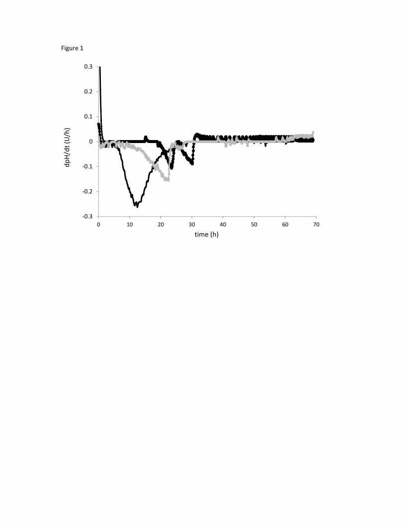
Figure 1
-0.3
-0.2
-0.1
0
0.1
0.2
0.3
0 10 20 30 40 50 60 70
dp
H/d
t (U
/h)
time (h)

Figure 2

Figure 3
A
B C
*
*****
*
*
***
*
*
***
**
***
* *
**** *
***
* *****

Figure 4
A
B
**