antimicrobial polymers with metal nanoparticles
TRANSCRIPT

Int. J. Mol. Sci. 2015, 16, 2099-2116; doi:10.3390/ijms16012099
International Journal of
Molecular Sciences ISSN 1422-0067
www.mdpi.com/journal/ijms
Review
Antimicrobial Polymers with Metal Nanoparticles
Humberto Palza
Departamento de Ingeniería Química y Biotecnología, Facultad de Ciencias Físicas y Matemáticas,
Universidad de Chile, Beauchef 850, Santiago 8320000, Chile; E-Mail: [email protected];
Tel.: +56-22-978-4085; Fax: +56-22-699-1084
Academic Editor: Antonella Piozzi
Received: 24 November 2014 / Accepted: 9 January 2015 / Published: 19 January 2015
Abstract: Metals, such as copper and silver, can be extremely toxic to bacteria at
exceptionally low concentrations. Because of this biocidal activity, metals have been
widely used as antimicrobial agents in a multitude of applications related with agriculture,
healthcare, and the industry in general. Unlike other antimicrobial agents, metals are stable
under conditions currently found in the industry allowing their use as additives. Today
these metal based additives are found as: particles, ions absorbed/exchanged in different
carriers, salts, hybrid structures, etc. One recent route to further extend the antimicrobial
applications of these metals is by their incorporation as nanoparticles into polymer
matrices. These polymer/metal nanocomposites can be prepared by several routes such as
in situ synthesis of the nanoparticle within a hydrogel or direct addition of the metal
nanofiller into a thermoplastic matrix. The objective of the present review is to show
examples of polymer/metal composites designed to have antimicrobial activities, with a
special focus on copper and silver metal nanoparticles and their mechanisms.
Keywords: antimicrobial metals; polymer nanocomposites; copper; silver
1. Introduction
Non-essential metals, such as silver, can be toxic to bacteria, having biocidal activities at exceptionally
low concentrations, while essential metals, such as copper, can also be lethal above some threshold
despite their relevance in the biochemistry of organisms [1]. Because of this biocidal activity, metals
have been widely used for centuries as antimicrobial agents in agriculture, healthcare, and industry
in general. Metal, oxide, or salt compounds based on copper and silver are among the most widely
OPEN ACCESS

Int. J. Mol. Sci. 2015, 16 2100
applied antimicrobial agents in this context [2]. However, the use of these metals in industrial
applications presents several challenges associated with the nature of the metal itself. Consequently,
one of their first applications was in the form of salt-based additives, for instance as silver nitrate,
avoiding its highly expensive metal form. Metal copper otherwise is cheaper than silver but presents
corrosion processes at standard conditions. The processing and manipulation of metal-based materials,
such as alloys, is another issue that should be resolved. Therefore, these metals are still mostly used as
additives in several applications such as wood preservation, antifouling paints, and antibacterial
textiles, among others. However, with the recent developments in materials science, their uses can be
extended today to metal surfaces and coatings, chelates, and nanomaterials [1]. Metal nanoparticles are
stressed due to both their enhanced antimicrobioal behaviour as compared with traditional materials
and their capacity to be embedded into polymer matrices.
Polymer/metal composites emerge as a route to further extend the applications of biocide metals
as a large percentage of hospital acquired infections (HAIs) are spread through surface contacts
or catheters mostly made of plastics. For instance, around 80%–95% of hospital-acquired urinary tract
infections originate from urinary catheters [3,4]. This is even more relevant taking into account the
steady growth of the polymer market, especially that based on polyolefins (i.e., polypropylene and
polyethylene), in healthcare applications. This growth is due to its properties such as: chemical,
radiation, and heat resistance, stiffness, clarity, barrier behavior for gases and liquids, impact, flexibility
and moderate cost [5]. For instance, polypropylene (PP) products are extensively used in medical
devices, packaging products, and delivery systems for solid and liquid pharmaceuticals [5]. About
a quarter of disposable and a third of non-disposable medical devices are PP-based, while the global
growth rate for PP in the healthcare industry is about 8.7% per year [6]. Polyamide is another example
of the relevance of polymers in healthcare applications as wound sutures, artificial tendons, and
medical packaging are made from this thermoplastic matrix [7].
Antimicrobial polymers therefore are highly demanded as a strategy to avoid HAIs and they can be
prepared either by embedding a biocide agent into the polymer bulk, for instance, during their processing
or by applying surface coatings [8–11]. For instance, either electrochemical or plasma based methods
have been recently applied to produce antimicrobial polymer/metal composite coatings [9,12–15].
A different approach is the polymerization of monomer-containing biocide groups or the grafting of
antimicrobial agents into the polymers [11]. The polymerization of the biocide polymer on the surface
of commercial polymers by atom transfer radical polymerization has been also reported [16,17].
From all these methods, the direct addition of the biocide agent into the polymers has received
considerable attention especially for thermoplastics such as polyolefins [18]. The main reason is that
this method can be easily implemented in the standard processing units already designed to prepare
particulate filled polymer composites and that are used extensively in the industry [19]. In this context,
polymer/metal composites prepared by melt blending are perceived as a useful way to produce biocidal
polymers [1]. Moreover, metals do not suffer degradation under the standard processing conditions of
thermoplastic polymers (~200 °C) [18,20]. This approach can be further extended to antimicrobial
metal nanoparticles as it seems that similar to other nanoparticles applications, their addition into
polymer matrices could be the fastest route to take commercial advantage of their enhanced properties.
Int. J. Mol. Sci. 2015, 16 2101
2. Antimicrobial Metals
2.1. General Aspects
Metal can be extremely toxic to most bacteria and yeast at exceptionally low concentrations [1].
Because of this biocide activity, some particular metals have been used as antimicrobial agents since
ancient times. For instance, vessels made of Cu and Ag have been used for water disinfection and food
preservation since the time of the Persian kings [1]. This practice was later adopted by the Phoenicians,
Greeks, Romans and Egyptians. Despite this long evidence, today the specific mechanisms explaining
the toxicity of metals are not yet fully elucidated as several variables are involved. However,
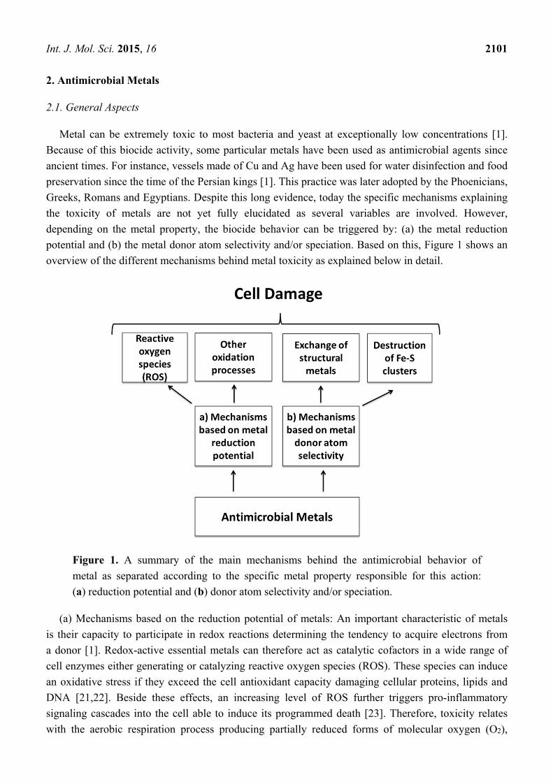
depending on the metal property, the biocide behavior can be triggered by: (a) the metal reduction
potential and (b) the metal donor atom selectivity and/or speciation. Based on this, Figure 1 shows an
overview of the different mechanisms behind metal toxicity as explained below in detail.
Figure 1. A summary of the main mechanisms behind the antimicrobial behavior of
metal as separated according to the specific metal property responsible for this action:
(a) reduction potential and (b) donor atom selectivity and/or speciation.
(a) Mechanisms based on the reduction potential of metals: An important characteristic of metals
is their capacity to participate in redox reactions determining the tendency to acquire electrons from
a donor [1]. Redox-active essential metals can therefore act as catalytic cofactors in a wide range of
cell enzymes either generating or catalyzing reactive oxygen species (ROS). These species can induce
an oxidative stress if they exceed the cell antioxidant capacity damaging cellular proteins, lipids and
DNA [21,22]. Beside these effects, an increasing level of ROS further triggers pro-inflammatory
signaling cascades into the cell able to induce its programmed death [23]. Therefore, toxicity relates
with the aerobic respiration process producing partially reduced forms of molecular oxygen (O2),

Int. J. Mol. Sci. 2015, 16 2102
such as hydrogen peroxide (H2O2) and superoxide (O2•−). With the presence of some metals,
Fenton reactions (see Equation 1) occur intensifying oxygen toxicity by catalyzing the electron transfer
from a donor biomolecule to H2O2 producing hydroxide (OH−) and the highly reactive hydroxyl
radical (OH•):
Fe2+ + H2O2 → Fe3+ + OH− + OH• (1)
The presence of external metals can therefore increase the aforementioned reactions producing an
excess of ROS derivatives and, as a consequence, an oxidative stress in the cell. Depending on the
specific metal involved, at least three mechanisms have been proposed for the ROS increase during
metal poisoning in bacteria [1]: (1) acceleration of Fenton reactions [24,25]; (2) disruption of Fe
bonded cellular donor ligands, for instance [4Fe–4S] clusters of proteins, resulting in the release of
additional Fenton-active Fe [1]; and (3) thiol-mediated reduction leading to the generation of ROS via
intermediate S radical chemistry [1].
Regarding Cu-induced cellular toxicity, several mechanisms have been proposed based on the
formation of ROS by free Cu ions as both cupric and cuprous ions can participate in redox
reactions [26]. In the presence of either superoxide or other reducing agents such as ascorbic acid,
Cu2+ can be reduced to Cu+ catalyzing the formation of hydroxyl radicals from hydrogen peroxide via
the Haber-Weiss reactions [27]:
O2•− + Cu2+ → O2 + Cu+ (2)
Cu+ + H2O2 → Cu2+ + OH− + OH• (3)
The hydroxyl radical is the most powerful oxidizing radical reacting with practically every
biological molecule [28]. It can initiate oxidative damage by abstracting the hydrogen both from an
amino-bearing carbon to form a carbon centered protein radical and from an unsaturated fatty acid to
form a lipid radical.
Although most of the mechanisms explaining the biocidal activity of metals are directly based on
the presence of ROS, there are others such as those associated with either the direct redox processes on
molecules, for instance the oxidation of cellular thiols, or the indirect formation of ROS [1]. Some
metal atoms can form covalent bonds with S leading to the formation of protein disulphides and to the
depletion of antioxidant reserves, particularly glutathione, within microbial cells [1]. Metals could further
catalyse site-specific damage to cellular proteins by an oxidation process causing loss of catalytic
activity and triggering an active process of protein degradation. Finally, there are reports about
genotoxicity where, for instance, lethal DNA damage in Escherichia coli can be catalyzed by
Fe-mediated Fenton chemistry [1].
(b) Mechanisms based on the donor atom selectivity and/or speciation of metals: metal ions in
general bind to some atoms of donor ligands, such as O, N and S, through strong and selective
interactions [1,29]. Indeed, external metal ions or their complexes can replace original metals present
in biomolecules leading to cellular dysfunction [1,30]. This phenomenon is called ionic mimicry or
molecular mimicry, depending on whether metal ions or metal complexes are involved. In this way,
some metals can promote the destruction of Fe–S clusters, for instance from bacterial dehydratases
that is particularly vulnerable to site-specific inactivation by toxic metals [1,29–31]. Metals can also
replace non-catalytic metal-binding sites inhibiting enzyme activity [1]. Cupric ions (Cu2+) in particular

Int. J. Mol. Sci. 2015, 16 2103
are able to form organic complexes with sulfur-, nitrogen- or oxygen-containing functional groups
present in the microorganism. This may result in defects in the conformational structure of nucleic
acids and proteins, besides changes in oxidative phosphorylation and osmotic balance. Finally, bacteria
and yeast exposed to toxic doses of some metals upregulate genes involved in the elimination of ROS
generating oxidative stress [1].
Independent on the aforementioned, another possible classification for the mechanisms can be
based on where the biocidal metal acts: in the cell membrane or in the intracellular region. The former
is based on the fact that bacterial membranes contain macromolecules with highly electronegative
chemical groups that serve as sites of adsorption for metal ions [32]. Because of their ability to
coordinate metals, it has been postulated that the membrane is the site at which some metals exert
bactericidal toxicity [1]. Other evidence suggests that some metals, particularly Ag, disrupt the activity
of the bacterial electron transport chain. Ag+ can dissipate the chemiosmotic potential of the membrane
by causing proton leakage through the membrane [1]. One of the best-known consequences of copper
excess is the peroxidative damage to membrane lipids by the reaction of lipid radicals and oxygen [26].
Regarding the intracellular region, metal toxic mechanisms based on either ROS or atoms selectively
can occur here where DNA and most proteins are presented. Notably, these two sites for metal toxic
action are likely related as recently reported [33]. Under this hypothesis, the cell membrane is first
damaged by the metal ions, allowing their subsequent further intake into the intracellular region.
2.2. Metal Nanoparticles
During the last years, nanotechnology has produced a new route to take advantage of the
antimicrobial behavior of metals by synthesizing highly active metal nanoparticles [29]. Biocidal metal
nanoparticles can be either immobilized or coated onto surfaces towards application in several fields
such as medical instruments and devices, water treatment, and food processing, among others [30].
However, their combination with polymers forming composites is stressed for a better, and easier,
utilization of the antimicrobial activity of these nanoparticles [30].
Nanoparticles can dissolve faster in a given solution volume as compared with larger particles
releasing therefore a higher amount of metal ions [34]. Therefore, based on the mechanisms
aforementioned based on the presence of metal ion, nanoparticles should present stronger antimicrobial
effects than either microparticles or metal surfaces. For instance, the copper nanoparticle corrosion in
distilled water is quite different as compared with microparticles [35]. The Cu2+ transformation ratio
of microparticles increases slowly with the immersion time and levels off eventually, meanwhile in
nanoparticles this transformation ratio increases sharply with the immersion time, reaching a peak
rapidly, and then decreasing increases sharply with the immersion time, reaching a peak rapidly, and
then decreasing. However, new toxic mechanisms, depending on the cellular characteristics at the
nano-scale, emerge by taking into account the role of the particle size itself [26]. The best example
relate with the direct incorporation of nanoparticles into the cell via endocytotic mechanisms.
Afterward the cellular uptake of ions increases as ionic species are subsequently released within the
cells by nanoparticle dissolution, a process often referred as “the Trojan horse mechanism” [36–38].
This high intracellular concentration gained after nanoparticle dissolution within the cell likely results

Int. J. Mol. Sci. 2015, 16 2104
in massive oxidative stress. A summary of the possible mechanisms associated with the antimicrobial
behavior of metal nanoparticles are displayed in Figure 2, and will be discussed below in detail.
Figure 2. A summary of the mechanisms associated with the antimicrobial behaviour of
metal nanoparticles: (1) “Trojan-horse effect” due to endocytosis processes; (2) attachment
to the membrane surface; (3) catalyzed radical formation; and (4) release of metal ions.
2.2.1. Silver Nanoparticles
Silver is generally used in the nitrate form to induce antimicrobial effects, but when silver nanoparticles
are used, there is a huge increase in the surface area available opening new approaches [22]. The most
pronounced effect of silver nanoparticles is on the cellular metabolic activity and the membrane
inflicting damage to the cells and potentially resulting in a myriad of secondary effects, such as
generation of ROS and DNA damage [39]. The potency of silver nanoparticles to induce cell damage
compared to silver ions is cell type and size-dependent. However, the exact mechanism which silver
nanoparticles cause antimicrobial effect is not clearly known although there are several theories about
their biocidal action on bacteria. Silver nanoparticles have the ability to anchor to the bacterial cell
wall and subsequently penetrate it, causing structural changes in the cell membrane such as
in permeability, and afterward cell death [22]. There is also a formation of “pits” on the cell surface
increasing the accumulation of silver nanoparticles on the cell surface [40]. The formation of free
radicals by silver nanoparticles may also be considered to explain cell death. Electron spin resonance
spectroscopy results suggest the formation of free radicals by silver nanoparticles when they are
in contact with the bacteria. These radicals can damage the cell membrane and make it porous leading
to cell death [22,41,42]. Despite all these mechanisms, the release of silver ions by nanoparticles
is also proposed as the main toxic mechanism [43].

Int. J. Mol. Sci. 2015, 16 2105
2.2.2. Copper Nanoparticles
Micrometric metal copper did not cause cell damage as compared with highly biocidal copper
nanoparticles at the same mass [34]. However, when the cells were exposed to the same surface area,
the cell membrane damage was similar showing the relevance of this parameter. Cu nanoparticles can
be more easily oxidized when interacting with cell membranes containing higher O2 concentration
as compared with the cell media [34]. In the same article, it was concluded that the cell membrane
damage does not relate directly with ionic species but rather with the metal release process at the
particle-cell surface interface. However, another work focused on copper oxide (CuO) nanoparticles
presented other antecedents [2]. The soluble ions released from the nanoparticles caused cytotoxicity
by interacting either directly with the cellular membrane or intracellularly. The biocidal effect of the
ions released from CuO nanoparticles was partially explained in amino acid-rich medium through
formation of copper-peptide complexes, rather than by the solid nanoparticles. However, the effect of
ions from CuO nanoparticles is different as compared with the effect of ions from copper salts, either
nitrate or sulfate, at identical soluble concentrations [2]. These results stress the complex behavior of
antimicrobial metal nanoparticles as either the particle itself or their ions can participate in the biocide
mechanisms. Anyway, independent of that, in the end, metal ions are the active biocidal agent
and therefore the same mechanisms detailed in Figure 1 finally explain the antimicrobial effect of
metal nanoparticles.
3. Polymer/Metal Composites
3.1. A General Overview
One of the best methods to further extend the range of applications of antimicrobial metals
is by their addition into a polymer obtaining a composite material. However, the best methodology
or technology is far to be solved, as it will depend on both the final application and the polymer matrix
used. For instance, metals can be either incorporated on the surface of a polymer or embedded into the
matrix. In particular, copper has been impregnated on the surface of cotton fibers, latex, and other
polymeric materials [44]. These materials present broad-spectrum antimicrobial (i.e., antibacterial,
antiviral, and antifungal) and antimite activities. Copper can further be incorporated by plasma
immersion ion implantation producing antibacterial polyethylene surface [9]. The production of copper
alginate-cotton cellulose (CACC) composite fibers by immersing cotton fibers in aqueous solution of
sodium alginate is another approach producing biocidal materials [45]. The process includes the ionic
crosslinking of alginate chains within the cotton cellulose fibers with Cu2+ ions. Regarding silver,
substituted zeolites are one of the most widely-used particles in antimicrobial polymer additives,
particularly for food applications [18]. Sodium ions present in zeolites are substituted by silver ions
meaning that these particles act as a carrier for antimicrobial ions. The same methodology applies for
copper, zinc, and other antimicrobial metals exchanged in zeolites that are later embedded in polymer
matrices [46]. Despite these approaches, during the last years the use of metal nanoparticles has
gained interest triggered by the amazing properties appearing at the nanoscale, for instance enhanced
antimicrobial behavior [40]. In polymer science, the impact of nanotechnology comes from the

Int. J. Mol. Sci. 2015, 16 2106
development and preparation of nanocomposites defined as hybrid materials containing nanometric
inorganic fillers embedded into a polymeric matrix.
Polymeric nanocomposites are opening up a new generation of macromolecular materials with low
densities and multifunctional properties [47]. The main advantage of nanocomposites is the extremely
low amount of filler needed to achieve the desired requirements that can be one or even two orders
of magnitude lower than conventional micro-fillers [7,47]. Therefore, in the next sections we will
focus on polymer/metal nanocomposites, in particular those using copper and silver as the active agent.
We stress however that independent of the metal-based particle used as additive (e.g., metal, nanoparticle,
oxide, salt, complex, exchanged zeolite, etc.), the mechanism for the antimicrobial behavior of the
polymer composite will correspond with the release of metal ions. In this way, the same mechanisms
discussed below for polymer/metal nanocomposites can be extended to any polymer/metal
antimicrobial material.
Two general approaches can be distinguished for the preparation of polymer/metal nanocomposites
depending on where the nanoparticles are synthesized (Figure 3): (1) in situ by using the polymer
matrix as the reaction medium; and (2) ex situ, meaning that the particle is synthesized before their
incorporation into the polymer and in this way the matrix is just the dispersion medium. The former
approach is mostly used for polymer hydrogel nanocomposites where the presence in the macromolecules
of both several functional groups and a water-rich medium improves the metal stabilization
and dispersion. The second approach is currently used in thermoplastic composites where the high
viscoelastic matrix at the melt state improves the dispersion of the nanoparticles.
Figure 3. Two main routes producing antimicrobial polymer/metal nanocomposites:
(a) Polymer as reaction mediun for in-situ synthesis of nanoparticles; and (b) Polymer as a
dispersion mediun of pre-synthesized nanoparticles.
Int. J. Mol. Sci. 2015, 16 2107
3.2. Polymer/Silver Composites
Silver nanoparticles are the most used antimicrobial filler in polymeric nanocomposites [48].
In particular, extensive research has focused on the development of hydrogel composites where silver
nanoparticles are in situ synthesized as displayed in Figure 3a. In general, all these systems used
silver-salt precursors, such as nitrate, together with a reducing agent, in presence of the polymer gel
network acting as a nanoreactor where the nanoparticles are formed. For instance, swollen poly
(acrylamide-co-acrylic acid) hydrogels can be a medium for the formation of silver nanoparticles
with sizes around 25–30 nm [49]. These nanocomposites demonstrated excellent antibacterial activity
depending on the nanoparticle size as this parameter changes the surface area that is in contact with the
bacterial species. Superabsorbent hydrogel-silver nanocomposite based on poly(vinyl alcohol) and
sodium alginate has also been prepared using free radical polymerization [50]. Highly stable silver
nanoparticles were in situ synthesized in the hydrogel networks by reduction of silver nitrate with
sodium borohydride as a reducing agent. The silver nanocomposite hydrogel showed very good
antibacterial activity on gram-positive and gram-negative microorganisms. Semi interpenetrating
polymer network (IPN) hydrogels, in which poly(vinyl pyrrolidone) chains were physically
dispersed throughout poly (acrylamide) gel networks, were synthesized and used as nanoreactors
for producing and stabilizing metal nanoparticles [51]. In particular, metal nanoparticles were
entrapped throughout hydrogel networks via polyvinylpyrrolidone chains with sizes around 4 nm.
These semi-IPN hydrogel-silver nanocomposites presented antibacterial behaviors. The same authors
later on developed antimicrobial silver-based composites using semi interpenetrating hydrogel networks
based on crosslinked poly(acrylamide) prepared through a redox-solution polymerization in the
presence of carbohydrate polymers [52]. In this case, three different polymers were studied: gum
acacia, carboxymethylcellulose, and starch. The method allows the synthesis of nanoparticles with
sizes between 5 and 20 nm depending on the polymer. All the nanocomposite hydrogels presented
similar antimicrobial behavior. Hydrogel networks based on N-isopropylacrylamide and sodium
acrylate were also prepared by redox-polymerization and used to synthesize highly stable and
uniformly distributed silver nanoparticles [53]. Similar to previous works, the hydrogel network is a
carrier where the in situ reduction of silver nitrate in the presence of sodium borohydride as a reducing
agent occurred. These hybrid hydrogels with different sizes of silver nanoparticles can be effectively
employed as antibacterial material.
The in situ synthesis of silver nanoparticles can also be extended to non-hydrogel based
polymers. A simple technique based on the in situ synthesis of silver nanoparticles by dissolving silver
1,5-cyclooctadiene-hexafluoroacetylacetonate in amorphous polystyrene has been reported [54]. The
metal precursor can thermally decompose producing silver atoms that diffuse into the polymer and
form clusters. These silver-doped polystyrenes provide high antibacterial activity. A similar approach
was used to add silver nanoparticles into polyamide matrices producing novel antimicrobial materials [7].
In this case, the nanoparticles were first produced in a masterbatch by thermal reduction of silver
acetate during melt processing of the matrix. In a second extrusion step, the masterbatch was diluted
with pure polyamide. Notably, the antimicrobial behavior of these nanocomposites was compared with
polyamide/silver microcomposites. Polyamide filled with just 0.06 wt % silver nanoparticles is able to
eliminate the bacteria completely within 24 h whereas microcomposite containing 1.9 wt % of silver

Int. J. Mol. Sci. 2015, 16 2108
kills only about 80% of the bacteria during the same time. The rate of the silver ion release from the
nanocomposites is about one order of magnitude higher in comparison to the microcomposites,
because of the much larger specific surface area of the nanoparticles [7]. Similar in situ routes were
further extended by these authors to polyurethane composites and compared with composites based on
silver nanoparticles synthesized in invertible polyester prior to their incorporation in the matrix
producing smaller particles [55]. At a constant weight percentage of silver in the polymer, the composite
with the ex situ silver nanoparticles exhibits a silver ion release which is about two orders of magnitude
higher than the release from the composite with the in situ silver nanoparticles [55]. This observation is
explained by the higher specific surface area of the smaller particles and the higher coefficient of
diffusion for silver ions. The polymer composite with a concentration of only 0.07 wt % of ex situ
silver nanoparticles exhibits a high enough release of silver ions to achieve antimicrobial properties.
There are other methods able to produce antimicrobial polymer/silver nanocomposites. For instance,
natural sodium alginate polymer acting as both reducing and stabilizing agent can also be used to
synthesis silver nanoparticles [56]. The nanoparticles capped with alginate displayed antimicrobial
behavior and they were further blended with varying amounts of chitosan forming polyelectrolyte
complexes that were casted into stable films. The blended film demonstrated excellent antibacterial
activity. Silver nanoparticles can also be synthesized in water by reduction of silver salts in the
presence of poly(acrylates) of different molecular weights [57]. These results clearly showed that
the reduction method and the polymer chain length played key roles in the achievement of
a few-nanometer-sized nanoparticles. The nanoparticle dispersions were then used to functionalize
cotton, wool, and polyester samples in order to obtain antimicrobial textiles for biomedical applications.
Addition of silver nanoparticles on polymer surfaces by plasma processes is another method
reported for the production of antimicrobial materials [58,59]. Deposition of silver nanoparticles onto
surface-functional porous poly(ethylene glycol dimethacrylate-co-acrylonitrile) microspheres has also
been reported with excellent biocide behaviours [60]. Antimicrobial coatings can further be developed
based on plasma polymerized polyacrylic acid (PPAA) deposited on a polyethylene terephthalate
mesh [14]. This method allows the entrapment of silver nanoparticle as the carboxylic groups of
PPAA can act as anchor as well as capping and stabilizing agents for silver nanoparticles synthesized
by a reduction method [14]. Other antimicrobial polymer coatings for a standard metallic orthopaedic
substrate containing chitosan, Bioglass® microparticles, and silver nanoparticles, were recently fabricated
using a single-step electrophoretic deposition (EPD) technique [13]. The low released concentration of
Ag ions (<2.5 ppm) was efficiently antibacterial against Staphyloccocus aureus up to 10 days. Polymer
coatings with silver nanoparticles can also be electrosynthesised on the surface of titanium based
implants by using polyacrylate-based hydrogel starting from poly(ethylene glycol diacrylate)-co-acrylic
acid [15]. These hydrogel coatings present swelling capabilities. Silver ion release was properly tuned
in order to assure antibacterial activity against the most common pathogens in implant infections while
preserving osteoblasts response at the implant interface [15].
3.3. Polymer/Copper
Antimicrobial polymer composites based on copper nanoparticles have been much less studied than
those based on silver. The reasons might be related to the higher stability and efficiency of silver as

Int. J. Mol. Sci. 2015, 16 2109
compared with copper, or, likely more important, to less available knowledge concerning copper
antimicrobial effects. One of the firsts antecedents regarding the development of polymer/copper metal
nanocomposites used polyvinylmethyl ketone, poly-(vinyl chloride), and polyvinylidenefluoride as the
polymer matrices. These composites were able to show antifungal and bacteriostatic properties
depending on the specific properties of the polymeric matrix [61]. The results of the biostatic activity
correlate very well with the copper ions released directly into the yeast-free culture broth exposed to
the nanocomposites for 4 h. It was also reported that the Cu2+ release rate increases with the increasing
of mass fraction of copper nanoparticles. Another approach is the attachment on the copper
nanoparticle surface of acrylic groups that can be later on copolymerized with other acrylic monomers.
These hybrid materials can become an integral part of the polymer backbone and they have a strong
potential for use in antibacterial or marine antifouling coatings [62]. Coatings based on polymer/copper
nanocomposites provide an interesting route to develop antimicrobial materials [9,12]. Beside
plasma-based methodologies previously discussed, poly(ethylene glycol diacrylate) hydrogel thin films
can be modified with copper nanoparticles. These coatings were firmly attached on metal substrates by
means of electrochemical polymerization technique and present antimicrobial behavior [12].
A relevant set of antimicrobial polymer/copper nanocomposites are based on natural occurring
biopolymers such as cellulose. Nanocomposites based on both vegetable and bacterial cellulose matrices
were prepared by in situ and ex situ methods [63]. The results showed that the chemical nature and
morphology of the copper nanofillers have great effect on the antibacterial activity, with an increase in
the antibacterial activity when the copper content is increased in the composites. The cellulosic matrices
also showed an effect on the antibacterial efficiency of the nanocomposites, with vegetal cellulose
fibers acting as the most effective substrate [63]. Another study also reports copper nanoparticles in
cellulose by modification of its fibers by periodate-induced oxidation followed by covalent attachment
of the biopolymer chitosan [64]. The borohydride-induced reduction yielded copper nanoparticle-loaded
fibers with an average diameter of particles around 30 nm that showed biocidal action.
Regarding other thermoplastic polymer matrices, the mechanism summarized in Figure 3b was used
to produce polyethylene/copper metal nanocomposites by melt blending for intrauterine devices having
excellent bioactive properties [65,66]. The introduction of a porous structure can improve the cupric
ion release rate of these composites [67]. Polypropylene has also been mixed with copper metal
nanoparticles by melt blending in order to produce antimicrobial plastic materials [68,69]. The biocide
kinetics can be controlled by the nanofiller content and composites with nanoparticle concentrations higher
than 10 v/v % eliminated 99% of the bacteria in less than 2 h. Copper oxide nanoparticles were also
embedded in polypropylene showing stronger antimicrobial behavior than metal copper nanoparticles [69].
A different route is the preparation of hybrid copper nanoparticle fillers such as bentonite supported
copper nanoparticles [70]. In an aqueous solution of copper sulfate, the sodium cations within the
bentonite interlayers were exchanged for Cu2+. These exchanged copper cations were reduced by adding
hydrazinium hydrate. Such aqueous bentonite/metal hybrid nanoparticle dispersions were blended
with cationic polymethylmethacrylate (PMMA) latex to produce hybrid nanocomposites containing
exfoliated polymer grafted organoclay together with bentonite supported metal nanoparticles. The
PMMA/bentonite/copper hybrid nanocomposites exhibited high antimicrobial activity.

Int. J. Mol. Sci. 2015, 16 2110
4. Mechanisms for Antimicrobial Polymer/Metal Nanocomposites
The toxic mechanisms of antimicrobial materials coming from the mixture of an antimicrobial agent
and a non-active polymer, are as similar as the mechanism of the agent itself [18,48]. In polymer/metal
nanocomposites therefore the main toxic mechanism relates with the nanoparticles meaning,
as previously discussed, two possible routes depending on the species considered as the active agent:
(1) the metal nanoparticle or (2) the metal ions released from the particles [22]. However, a growing
number of reports indicate that the ion release is the driving force behind the antimicrobial
properties of antibacterial nanoparticles [1,2]. In fact, most of the analyses regarding antimicrobial
metal nanoparticles focused on the metal ion release instead of the particle absorbed by the
bacteria [30,62,71]. This was confirmed by the results coming from polymer/metal nanocomposites
where the antimicrobial effect of these materials related with the metal ion releases rather than with
the leaching of the particle [68,69]. The presence of a polymer film covering the nanoparticles as found
previously by X-ray photoelectron spectroscopy (XPS) analysis in thermoplastic polymer/copper
composites, confirms the hypothesis that the ion release is the main mechanism in this case [68].
However, the exact route for the action of the nanoparticle will depend on the specific characteristics
of the polymer used, as in antimicrobial hydrogels the particle release has been stated as the main
process for this behavior.
We first focus on the mechanisms for nanocomposites based on thermoplastics, or dense polymers
in general, as representing the most complex system from the material point of view as summarized in
Figure 4. Afterward, the discussion will be straightforward for nanocompositesbased on hydrogels.
As aforementioned, in dense polymers with embedded metal nanoparticles, the ion release is the main
mechanism behind their biocide activity. In this context, particles present in the surface-region of the
composites seem to be the first approach to understand the experimental data. However, XPSanalysis
shows that these nanoparticles are not present on the surface of these samples [68] or at concentrations
lower than the bulk of the material [61]. Therefore, the only suitable mechanism for the release of
metal ions is the corrosion of particles present in the bulk of the polymer owing to the diffusion of
water molecules coming from the bacteria medium into the surface of particles [7]. Even highly
non-polar matrices such as polyethylene or polypropylene allow the diffusion of water molecules
through [72]. The polymer particle interface can further increase water diffusion through holes or
micron-scale defects, allowing fast Knudsen diffusion. This mechanism is directly extended to more
polar matrices. When a polyvinylmethyl ketone was used as the polymer matrix, the minimum depth
where the nanocomposite becomes hydrated, and eventually releases the soluble copper species, was
estimated around 50 nm, which was about 1/10 of the total film thickness in that case [61]. When water
with dissolved oxygen reaches the metal particles presented in the polymer bulk, the standard corrosion
process occurs [7,68,69]. Afterward, ions coming from corrosion or dissolution process can diffuse-out
through the polymer matrix and finally be released. This mechanism is confirmed for metal copper
nanoparticles by comparing the X-ray diffraction analysis of the original composite with the diffraction
of the same sample but immersed in water. In the latter case, new diffractions peaks appear related
with a Cu2O layer formed on the surface of the particle [68,69]. All these processes, especially those
related with the dissolution step, can be accelerated by the presence of the bacteria in the near-region of
the composite due to its organic compounds, pH change, and high surface affinity [2]. In hydrogels the

Int. J. Mol. Sci. 2015, 16 2111
release of nanoparticles from the matrix due to the much larger free volume of these polymers as
compared with dense thermoplastic matrices should be further considered [52,53]. Moreover, the
diffusion issues of water into/out-to the matrix can be avoided due to the nature of the hydrogel.
Therefore, in hydrogels the mechanisms are much closer to pure metal nanoparticles.
Figure 4. Mechanisms for the antimicrobial behavior of polymer/metal nanocomposites
based on thermoplastic matrices: (1) adsorbtion of bacteria on the polymer surface
triggering the diffusion of water through the polymer matrix due to the medium surrounded
the bacteria; (2) water with dissolved oxygen reaches the surface of embedded metal
nanoparticles allowing dissolution or corrosion processes and in this way metal ions are
realized; (3) metal ions reach the composite surface damaging the bacteria membrane;
(4) Afterward, metal ions can diffuse into the bacteria. The details of the specific mechanisms
are explained in Figure 1. Although this figure represents a polymer nanocomposite, it can
be extrapolated to any polymer/metal materials with the biocide agent embedded in the matrix.
5. Conclusions
The addition of metal-based particles into polymers is a versatile route to take advantage of their
strong antimicrobial properties producing novel biocide materials and allowing a further extension of
the range of applications. In this context metal nanoparticles emerge for the production of a broad
range of polymer nanocomposites with a high release of metal ions and therefore antimicrobial
behaviors. Although there are several examples related with copper and silver nanoparticles embedded
into different polymer matrices, as discussed through this manuscript, further research is needed to
support the development of novel bioactive polymeric materials to be used in hospital equipment or in
prostheses, avoiding for example hospital acquired infections. In this context, we stress technologies
based either on commercial matrices with embedded metal nanoparticles or on polymer/metal coatings,

Int. J. Mol. Sci. 2015, 16 2112
as they can be easily implemented at industrial scale or in implant materials, respectively. Future areas
of investigations may include scaling up processes, optimization of the filler dispersion and therefore
of the ion release, novel metal-based antimicrobial nanoparticles, different polymer matrices such as
elastomers, in situ studies about the antimicrobial behaviour of the novel materials, and toxicity,
among others.
Acknowledgments
The author gratefully acknowledges the financial support of CONICYT, project FONDECYT 1110078.
Conflicts of Interest
The author declares no conflict of interest.
References
1. Lemire, J.A.; Harrison, J.J.; Turner, R.J. Antimicrobial activity of metals: Mechanisms, molecular
targets and applications. Nat. Rev. Microbiol. 2013, 11, 371–384.
2. Gunawan, C.; Teoh, W.Y.; Marquis, C.P.; Amal, R. Cytotoxic origin of copper(II) oxide
nanoparticles: Comparative studies with micron-sized particles, leachate, and metal salts. ACS Nano
2011, 5, 7214–7225.
3. Curtis, L.T. Prevention of hospital-acquired infections: Review of non-pharmacological interventions.
J. Hosp. Infect. 2008, 69, 204–219.
4. Cheadle, W.G. Risk factors for surgical site infection. Surg. Infect. 2006, 7, S7–S11.
5. Sastri, V.S. Plastic in Medical Devices: Properties, Requirements and Applications; Elsevier:
Burlington, MA, USA, 2010.
6. NGP Europe. Polyolefins for Use in Medical Devices. Available online: http://www.ngphrama.eu.com
(accessed on 6 January 2012).
7. Damm, C.; Munstedt, H.; Rosch, A. The antimicrobial efficacy of polyamide 6/silver-nano- and
microcomposites. Mater. Chem. Phys. 2008, 108, 61–66.
8. Kenawy, E.R.; Worley, S.D.; Broughton, R. The chemistry and applications of antimicrobial
polymers: A state-of-the-art review. Biomacromolecules 2007, 8, 1359–1384.
9. Zhang, W.; Zhang, Y.H.; Ji, J.H.; Zhao, J.; Yan, Q.; Chu, P.K. Antimicrobial properties of copper
plasma-modified polyethylene. Polymer 2006, 47, 7441–7445.
10. Jones, D.S.; Djokic, J.; Gorman, S.P. The resistance of polyvinylpyrrolidone-Iodine-poly
(ε-caprolactone) blends to adherence of Escherichia coli. Biomaterials 2005, 26, 2013–2020.
11. Yuan, Y.L.; Ai, F.; Zang, X.P. Polyurethane vascular catheter surface grafted with zwitterionic
sulfobetaine monomer activated by ozone. Colloid Surf. B 2004, 35, 1–5.
12. Cometa, S.; Iatta, R.; Ricci, M.A.; Ferretti, C.; de Giglio, E. Analytical characterization and
antimicrobial properties of novel copper nanoparticles-loaded electrosynthesised hydrogel coatings.
J. Bioact. Compat. Polym. 2013, 28, 508–522.

Int. J. Mol. Sci. 2015, 16 2113
13. Pishbin, F.; Mouriño, V.; Gilchrist, J.B.; McComb, D.W.; Kreppel, S.; Salih, V.; Ryan, M.P.;
Boccaccini, A.R. Single-step electrochemical deposition of antimicrobial orthopaedic coatings
based on a bioactive glass/chitosan/nano-silver composite system. Acta Biomater. 2013, 9,
7469–7479.
14. Kumar, V.; Jolivalt, C.; Pulpytel, J.; Jafari, R.; Arefi-Khonsari, F. Development of silver
nanoparticle loaded antibacterial polymer mesh using plasma polymerization process. J. Biomed.
Mater. Res. A 2013, 101, 1121–1132.
15. De Giglio, E.; Cafagna, D.; Cometa, S.; Allegretta, A.; Pedico, A.; Giannossa, L.C.; Sabbatini, L.;
Mattioli-Belmonte, M.; Iatta, R. An innovative, easily fabricated, silver nanoparticle-based titanium
implant coating: Development and analytical characterization. Anal. Bioanal. Chem. 2013, 405,
805–816.
16. Huang, J.; Murata, H.; Koepsel, R.R.; Russell, A.J.; Matyjaszewski, K. Antibacterial polypropylene
via surface-initiated atom transfer radical polymerization. Biomacromolecules 2007, 8, 1396–1399.
17. Lee, S.B.; Koepsel, R.R.; Morley, S.W.; Matyjaszewski, K.; Sun, Y.; Russel, A.J. Permanent,
nonleaching antibacterial surfaces 1: Synthesis by atom transfer radical polymerization.
Biomacromolecules 2004, 5, 877–882.
18. Appendini, P.; Hotchkiss, J.H. Review of antimicrobial food packaging. Innov. Food Sci.
Emerg. Technol. 2002, 3, 113–126.
19. Pehlivan, H.; Balkose, D.; Ulku, S.; Tihminlioglu, F. Characterization of pure and silver
exchanged natural zeolite filled polypropylene composite films. Compos. Sci. Technol. 2005, 65,
2049–2058.
20. De Azeredo, H.M.C. Nanocomposites for food packaging applications. Food Res. Int. 2009, 42,
1240–1253.
21. Shleeva, S.; Tkac, J.; Christenson, A.; Ruzgas, T.; Yaropolov, A.I.; Whittaker, J.W.; Gorton, L.
Direct electron transfer between copper-containing proteins and electrodes. Biosens. Bioelectron.
2005, 20, 2517–2554.
22. Prabhu, S.; Poulose, E.K. Silver nanoparticles: Mechanism of antimicrobial action, synthesis,
medical applications, and toxicity effects. Int. Nano Lett. 2012, 32, 2–10.
23. Sintubin, L.; de Windt, W.; Dick, J.; Mast, J.; van der Ha, D.; Verstraete, W.; Boon, N. Lactic
acid bacteria as reducing and capping agent for the fast and efficient production of silver
nanoparticles. Appl. Microbiol. Biotechnol. 2009, 84, 741–749.
24. Valko, M.; Morris, H.; Cronin, M.T.D. Metals, toxicity and oxidative stress. Curr. Med. Chem.
2005, 12, 1161–1208.
25. Stohs, S.J.; Bagchi, D. Oxidative mechanisms in the toxicity of metal ions. Free Radic. Biol. Med.
1985, 18, 321–336.
26. Gaetke, L.M.; Chow, C.K. Copper toxicity, oxidative stress, and antioxidant nutrients. Toxicology
2003, 189, 147–163.
27. Bremner, I. Manifestations of copper excess. Am. J. Clin. Nutr. 1988, 67, 1069S–1073S.
28. Buettner, G.R.; Jurkiewicz, B.A. Catalytic metals, ascorbate and free radicals: Combinations to
avoid. Radiat. Res. 1996, 145, 532–541.
29. Grass, G.; Rensing, C.; Solioz, M. Metallic copper as an antimicrobial surface. Appl. Environ. Microbiol.
2011, 77, 1541–1548.
Int. J. Mol. Sci. 2015, 16 2114
30. Ruparelia, J.P.; Chatterjee, A; Duttagupta, S.P.; Mukherji, S. Strain specificity in antimicrobial
activity of silver and copper nanoparticles. Acta Biomater. 2008, 4, 707–716.
31. Xu, F.F.; Imlay, J.A. Silver (I), mercury (II), cadmium (II), and zinc (II) target exposed enzymic
iron-sulfur clusters when they toxify Escherichia coli. Appl. Environ. Microbiol. 2012, 78,
3614–3621.
32. Zhang, Y.M.; Rock, C.O. Membrane lipid homeostasis in bacteria. Nat. Rev. Microbiol. 2008, 6,
222–233.
33. Mathews, S.; Hans, M.; Mücklich, F.; Solioz, M. Contact killing of bacteria on copper is
suppressed if bacterial-metal contact is prevented and is induced on iron by copper ions.
Appl. Environ. Microbiol. 2013, 79, 2605–2611.
34. Karlsson, H.L.; Cronholm, P.; Hedberg, Y.; Tornberg, M.; de Battice, L.; Svedhem, S.;
Wallinder, I.O. Cell membrane damage and protein interaction induced by copper containing
nanoparticles—Importance of the metal release process. Toxicology 2013, 313, 59–69.
35. Xia, X.; Xie, C.; Cai, S.; Yang, Z.; Yang, X. Corrosion characteristics of copper microparticles
and copper nanoparticles in distilled water. Corros. Sci. 2006, 48, 3924–3932.
36. Studer, A.M.; Limbach, L.K.; van Duc, L.; Krumeich, F.; Athanassiou, E.K.; Gerber, L.C.;
Moch, H.; Stark, W.J. Nanoparticle cytotoxicity depends on intracellular solubility: Comparison
of stabilized copper metal and degradable copper oxide nanoparticles. Toxicol. Lett. 2010, 197,
169–174.
37. Wang, Z.; Li, N.; Zhao, J.; White, J.C.; Qu, P.; Xing, B. CuO nanoparticle interaction with human
epithelial cells: Cellular uptake, location, export, and genotoxicity. Chem. Res. Toxicol. 2012, 25,
1512–1521.
38. Cronholm, P.; Midander, K.; Karlsson, H.L.; Elihn, K.; Odnevall Wallinder, I.; Möller, L. Effect
of sonication and serum proteins on copper release from copper nanoparticles and the toxicity
towards lung epithelial cells. Nanotoxicology 2011, 5, 269–281.
39. Park, M.V.D.Z.; Neigh, A.M.; Vermeulen, J.P.; de la Fonteyne, L.J.J.; Verharen, H.W.;
Briedé, J.J.; van Loveren, H.; de Jong, W.H. The effect of particle size on the cytotoxicity,
inflammation, developmental toxicity and genotoxicity of silver nanoparticles. Biomaterials 2011,
32, 9810–9817.
40. Sondi, I.; Salopek-Sondi, B. Silver nanoparticles as antimicrobial agent: A case study on E. coli
as a model for Gram-negative bacteria. J. Colloid Interface Sci. 2004, 275, 177–182.
41. Kim, J.S.; Kuk, E.; Yu, K.; Kim, J.H.; Park, S.J.; Lee, H.J.; Kim, S.H.; Park, Y.K.; Park, Y.H.;
Hwang, C.Y.; et al. Antimicrobial effects of silver nanoparticles. Nanomedicine 2007, 3, 95–101.
42. Danilcauk, M.; Lund, A.; Saldo, J.; Yamada, H.; Michalik, J. Conduction electron spin resonance
of small silver particles. Spectrochim. Acta Part A 2009, 63, 189–191.
43. Feng, Q.L.; Wu, J.; Chen, G.Q.; Cui, F.Z.; Kim, T.N.; Kim, J.O. A mechanistic study of the
antibacterial effect of silver ions on Escherichia coli and Staphylococcus aureus. J. Biomed.
Mater. Res. 2008, 52, 662–668.
44. Borkow, G.; Gabbay, J. Copper as a biocidal tool. FASEB J. 2004, 18, 1728–1775.
45. Grace, M.; Chand, N.; Bajpai, S.K. Copper alginate-cotton cellulose (CACC) fibers with excellent
antibacterial properties. J. Eng. Fibers Fabr. 2009, 4, 24–34.

Int. J. Mol. Sci. 2015, 16 2115
46. Kaali, P.; Prez-Madrigal, M.M.; Strmberg, E.; Aune, R.E.; Czél, G.; Karlsson, S. The influence of
Ag+, Zn2+ and Cu2+ exchanged zeolite on antimicrobial and long term in vitro stability of medical
grade polyether polyurethane. Exp. Polym. Lett. 2011, 5, 1028–1040.
47. Paul, D.R.; Robeson, L.M. Polymer nanotechnology: Nanocomposites. Polymer 2008, 49,
3187–3204.
48. Muñoz-Bonilla, A.; Fernández-García, M. Polymeric materials with antimicrobial activity.
Prog. Polym. Sci. 2012, 37, 281–339.
49. Thomas, V.; Yallapu, M.M.; Sreedhar, B.; Bajpai, S.K. A versatile strategy to fabricate
hydrogel-silver nanocomposites and investigation of their antimicrobial activity. J. Colloid Int. Sci.
2007, 315, 389–395.
50. Ghasemzadeh, H.; Ghanaat, F. Antimicrobial alginate/PVA silver nanocomposite hydrogel,
synthesis and characterization. J. Polym. Res. 2014, 21, 355–269.
51. Murthy, P.S.K.; Mohan, Y.M.; Varaprasad, K.; Sreedhar, B.; Raju, K.M. First successful design
of semi-IPN hydrogel-silver nanocomposites: A facile approach for antibacterial application.
J. Colloid Int. Sci. 2008, 318, 217–224.
52. Vimala, K.; Sivudu, K.S.; Mohan, Y.M.; Sreedhar, B.; Raju, K.M. Controlled silver nanoparticles
synthesis in semi-hydrogel networks of poly(acrylamide) and carbohydrates: A rational methodology
for antibacterial application. Carbohydr. Polym. 2009, 75, 463–471.
53. Mohan, Y.M.; Lee, K.; Premkumar, T.; Geckeler, K.E. Hydrogel networks as nanoreactors:
A novel approach to silver nanoparticles for antibacterial applications. Polymer 2007, 48, 158–164.
54. Palomba, M.; Carotenuto, G.; Cristino, L.; di Grazia, M.A.; Nicolais, F.; de Nicola, S.
Activity of antimicrobial silver polystyrene nanocomposites. J. Nanomater. 2012, 2012, 185029,
doi:10.1155/2012/185029.
55. Triebel, C.; Vasylyev, S.; Damm, C.; Stara, H.; Özpınar, C.; Hausmann, S.; Peukert, W.;
Münstedt, H. Polyurethane/silver-nanocomposites with enhanced silver ion release using
multifunctional invertible polyesters. J. Mater. Chem. 2011, 21, 4377–4383.
56. Sharma, S.; Sanpui, P.; Chattopadhyay, A.; Ghosh, S.S. Fabrication of antibacterial silver
nanoparticle—Sodium alginate-chitosan composite films. RSC Adv. 2012, 2, 5837–5843.
57. Falletta, E.; Bonini, M.; Fratini, E.; Lo Nostro, A.; Pesavento, G.; Becheri, A.; Lo Nostro, P.;
Canton, P.; Baglioni, P. Clusters of poly(acrylates) and silver nanoparticles: Structure and
applications for antimicrobial fabrics. J. Phys. Chem. C 2008, 112, 11758–11766.
58. Jiang, H.; Manolache, S.; Wong, A.C.L.; Denes, F.S. Plasma-enhanced deposition of silver
nanoparticles onto polymer and metal surfaces for the generation of antimicrobial characteristics.
J. Appl. Polym. Sci. 2004, 93, 1411–1422.
59. Del Nobile, M.A.; Cannarsi, M.; Altieri, C.; Sinigalia, M.; Favia, P.; Iacoviello, G.; D’Agostino, R.
Effect of Ag-containing nano-composite active packaging system on survival of Alicyclobacillus
acidoterrestris. J. Food Sci. 2004, 69, E379–E384.
60. Kim, J.W.; Lee, J.E.; Kim, S.J.; Lee, J.S.; Ryu, J.H.; Kim, J.; Han, S.H.; Chang, I.S.; Suh, K.D.
Synthesis of silver/polymer colloidal composites from surface-functional porous polymer
microspheres. Polymer 2004, 45, 4741–4747.

Int. J. Mol. Sci. 2015, 16 2116
61. Cioffi, N.; Torsi, L.; Ditarantano, N.; Tantalillo, G.; Ghibelli, L.; Sabbatini, L.; Bleve-Zacheo, T.;
D’Alessio, M.; Zambonin, P.G.; Traversa, E. Copper nanoparticle/polymer composites with
antifungal and bacteriostatic properties. Chem. Mater. 2005, 17, 5255–5262.
62. Anyaogu, K.C.; Fedorov, A.V.; Neckers, D.C. Synthesis, characterization, and antifouling potential
of functionalized copper nanoparticles. Langmuir 2008, 24, 4340–4346.
63. Pinto, R.J.; Daina, S.; Sadocco, P.; Pascoal Neto, C.; Trindade, T. Antibacterial activity of
nanocomposites of copper and cellulose. Antibacterial Activity of Nanocomposites of Copper and
Cellulose. Biomed. Res. Int. 2013, 2013, doi:10.1155/2013/280512.
64. Mary, G.; Bajpai, S.K.; Chand, N. Copper(II) ions and copper nanoparticles-loaded chemically
modified cotton cellulose fibers with fair antibacterial properties. J. Appl. Polym. Sci. 2009, 113,
757–766.
65. Cai, S.; Xia, X.; Xie, C. Corrosion behaviour of copper/LDPE nanocomposites in simulated uterine
solution. Biomaterials 2005, 26, 2671–2678.
66. Xu, T.; Lei, H.; Cai, S.Z.; Xia, X.P.; Xie, C.S. The release of cupric ion in simulated uterine: New
material nano-Cu/low-density polyethylene used for intrauterine devices. Contraception 2004, 70,
153–157.
67. Zhang, W.; Xia, X.; Qi, C.; Xie, C.; Cai, S. A porous Cu/LDPE composite for copper-containing
intrauterine contraceptive devices. Acta Biomater. 2012, 8, 897–903.
68. Palza, H.; Gutiérrez, S.; Delgado, K.; Salazar, O.; Fuenzalida, V.; Avila, J.; Figueroa, G.;
Quijada, R. Toward tailor-made biocide materials based on polypropylene/copper nanoparticles.
Macromol. Rapid Commun. 2010, 31, 563–569.
69. Delgado, K.; Quijada, R.; Palma, R.; Palza, H. Polypropylene with embedded copper metal or
copper oxide nanoparticles as a novel plastic antimicrobial agent. Lett. Appl. Microbiol. 2011, 53,
50–54.
70. Weickmann, H.; Tiller, J.C.; Thomann, R.; Mulhaupt, R. Metallized organoclays as new
intermediates for aqueous nanohybrid dispersions, nanohybrid catalysts and antimicrobial
polymer hybrid nanocomposites. Macromol. Mater. Eng. 2005, 290, 875–883.
71. Ren, G.; Hub, D.; Cheng, E.W.C.; Vargas-Reus, M.A.; Reipd, P.; Allaker, R.P. Characterisation
of copper oxide nanoparticles for antimicrobial applications. Int. J. Antimicrob. Agents 2009, 33,
587–592.
72. Ton-That, T.M.; Jungnickel, B.J. Water diffusion into transcrystalline layers on polypropylene.
J. Appl. Polym. Sci. 1999, 74, 3275–3285.
© 2015 by the authors; licensee MDPI, Basel, Switzerland. This article is an open access article
distributed under the terms and conditions of the Creative Commons Attribution license
(http://creativecommons.org/licenses/by/4.0/).