antibody repertoire diversification through vh gene ... · antibody repertoire diversification...
TRANSCRIPT
Antibody repertoire diversification through VH genereplacement in mice cloned from an IgA plasma cellRashmi Kumara, Martina P. Bachb,c, Federica Mainoldia, Mikako Maruyad, Satoshi Kishigamie,1, Hassan Jumaab,c,Teruhiko Wakayamae,1, Osami Kanagawaf, Sidonia Fagarasand, and Stefano Casolaa,2
aThe Institute of Molecular Oncology (IFOM) of the Italian Foundation for Cancer Research (FIRC), Milan 20139, Italy; bInstitute of Immunology, UniversityClinic Ulm, 89081 Ulm, Germany; cDepartment of Molecular Immunology, Albert-Ludwigs University of Freiburg and Max Planck Institute of Immunobiologyand Epigenetics, 79108 Freiburg, Germany; dCenter for Integrative Medical Sciences, RIKEN Yokohama Institute, Yokohama, Kanagawa 230-0045, Japan;eRIKEN Center for Developmental Biology, Kobe, Hyogo 650-0047, Japan; and fAkashi City Hospital, Akashi 673-8501, Japan
Edited by Frederick W. Alt, Program in Cellular and Molecular Medicine, Boston Children’s Hospital, Harvard Medical School, and Howard Hughes MedicalInstitute, Boston, MA, and approved December 19, 2014 (received for review September 18, 2014)
In mammals, VDJ recombination is responsible for the establish-ment of a highly diversified preimmune antibody repertoire.Acquisition of a functional Ig heavy (H) chain variable (V) generearrangement is thought to prevent further recombination at theIgH locus. Here, we describe VHQ52NT; Vκgr32NT Ig monoclonalmice reprogrammed from the nucleus of an intestinal IgA+ plasmacell. In VHQ52NT mice, IgA replaced IgM to drive early B-cell de-velopment and peripheral B-cell maturation. In VHQ52NT animals,over 20% of mature B cells disrupted the single productive, non-autoimmune IgH rearrangement through VH replacement and ex-changed it with a highly diversified pool of IgH specificities. VHreplacement occurred in early pro-B cells, was independent of pre–B-cell receptor signaling, and involved predominantly one adjacentVH germ-line gene. VH replacement was also identified in 5% ofperipheral B cells of mice inheriting a different productive VH rear-rangement expressed in the form of an IgM H chain. In summary,editing of a productive IgH rearrangement through VH replace-ment can account for up to 20% of the IgH repertoire expressedby mature B cells.
B cells | VH replacement | antibody repertoire | IgA | nuclear cloning
Afunctional immune system relies on the ability of B-lym-phocytes to recognize foreign antigens in a highly specific
fashion through the Ig receptor (also called B-cell receptor, orBCR). Each BCR consists of two identical Ig heavy (H) and light(L) chains, which are expressed on the surface of the B cell to-gether with an Igα/Igβ heterodimer to form a signaling unit. Inmost vertebrates, the ability of the immune system to generatea highly diversified repertoire of antibody specificities relies onthe stochastic assembly of variable (V), diversity (D), and joining(J) gene segments that encode for the antigen-binding domain ofIgH and IgL chains of the BCR. This process, called V(D)J re-combination, is mediated by the ordered recruitment at Ig loci ofthe RAG-1/2 endonucleases, followed by nonhomologous end-joining factors that catalyze the joining of the cleaved DNAsegments (1). The latter processes are often accompanied bytrimming and insertion of n-nucleotides at junctional ends. Alltogether these mechanisms contribute to generate a highly di-versified pool of Ig specificities.Ig receptor editing provides B cells with the opportunity to
exchange BCR specificity through secondary VDJ recombination.Whereas IgL chain editing was shown to play a central role in theneutralization of autoimmune BCR specificities expressed by newlygenerated immature B cells (2), the contribution of IgH receptorediting to antibody repertoire diversification has remained largelyunappreciated. Two mechanisms promote secondary IgH rear-rangements. In VH-to-JH direct joining, RAG proteins cleave atRecombination Signal Sequences (RSSs) of VH and JH elementslying, respectively, upstream and downstream of the original VHrearrangement, followed by microhomology-driven joining of the Iggene segments. This mechanism has been mainly observed in IgHknock-in mice carrying nonproductive IgH rearrangements (3, 4).
VH replacement relies, instead, on evolutionary conserved crypticRSSs embedded within the framework region 3 of most VH germ-line genes (5). During VH replacement, cryptic recombinationsignal sequences (cRSS) within the V gene of a preexisting VHrearrangement are engaged in a RAG-mediated recombinationreaction together with RSSs of an upstream VH germ-line gene, inaccordance with the 12/23 rule (reviewed in ref. 6). VH replacementgenerates a novel VH rearrangement that carries a different Vgene and shares with the original one part of its Complemen-tary Determining Region 3 (CDR3). Studies in B-lymphomacell lines were the first to identify VH replacement as a mecha-nism to edit both in-frame (IF) and out-of-frame (OF) IgH rear-rangements (7, 8). Later analyses with IgH knock-in miceconfirmed in vitro studies, unraveling the potential of VH re-placement to rescue progenitor B cells carrying nonproductiveVH rearrangements (4, 9). VH replacement has also beenproposed to diversify the preimmune repertoire of productiveIgH specificities in both human and mice (10–12). Bioinformaticanalyses of IgH V gene repertoires obtained through next-genera-tion sequencing have shown limitations to identify VH replace-ments (13). Studies on IgH transgenic mice have in part overcomesuch limitations (9, 11, 14–17). However, the targeting strategy togenerate most IgH knock-in mice may severely limit the in-terpretation of VH replacement data obtained from such models.Indeed, in most IgH knock-in mice, prerearranged VH genesreplace the four JH segments of the IgH locus. This atypical
Significance
Antibodies produced by B cells provide a protective barrier to ourorganism against the penetration and dissemination of micro-organisms. Each antibody recognizes a specific antigen throughvariable (V) region domains of pairs of immunoglobulin (Ig)heavy (H) and light (L) chains. In mammals, VDJ recombinationgenerates a highly diversified preimmune pool of VH and VL
domains. Acquisition of a functional VH rearrangement is thoughtto prevent further VDJ recombination at the IgH locus. Instead,mice cloned from a terminally differentiated B cell unravel theability of VDJ recombination to revise a functionally rearrangedVH gene through VH replacement. We show that up to 20% ofthe antibody V gene repertoire of mature B-lymphocytes can begenerated through VH replacement.
Author contributions: S.C. designed research; R.K., M.P.B., and F.M. performed research;M.M., S.K., T.W., O.K., and S.F. contributed new reagents/analytic tools; R.K., M.P.B., F.M.,H.J., and S.C. analyzed data; and R.K. and S.C. wrote the paper.
The authors declare no conflict of interest.
This article is a PNAS Direct Submission.1Present address: Faculty of Life and Environmental Science, University of Yamanashi,Kofu, Yamanashi 400-8510, Japan.
2To whom correspondence should be addressed. Email: [email protected].
This article contains supporting information online at www.pnas.org/lookup/suppl/doi:10.1073/pnas.1417988112/-/DCSupplemental.
E450–E457 | PNAS | Published online January 21, 2015 www.pnas.org/cgi/doi/10.1073/pnas.1417988112
chromosomal configuration may affect the rate and nature ofsecondary IgH rearrangements. Intergenic Control Region 1(IGCR1), which is crucial for ordered and lineage-specific VDJrecombination (18), represents one example of a cis regulatoryelement that is excised from the IgH locus during physiologicV-to-DJ recombination, but is retained in most IgH knock-in animals.Recent advancements in ES gene targeting strategies have
allowed the establishment of next-generation IgH knock-in micewhere the insertion of a particular VH rearrangement into the JHlocus is coupled to Cre recombinase-assisted deletion of the in-tervening region between DH-proximal VH genes and the JHlocus (4). This elegant approach relies on multiple targetingsteps that are time consuming and may preclude germ-linetransmission of targeted ES cells. Instead, somatic cell nucleartransfer (SCNT) technology applied to B-lymphocytes allowsthe rapid generation of IgH monoclonal mice carrying VH re-arrangements placed in their physiologic location (19).Here, we applied SCNT to establish a novel mouse strain
(VHQ52NT; Vκgr32NT) starting from the nucleus of a terminallydifferentiated IgA+ intestinal plasma cell (PC). Cloned miceallowed the investigation of B-cell development and IgH reper-toire diversification under conditions where a single, productiveIgH rearrangement consisting of a DH-proximal Q52 VH genewas expressed from an IgA class-switched IgH locus. We couldshow that a BCR specificity selected in the lamina propria (LP)of the small intestine and expressed in the form of IgA couldeffectively drive early B-cell development and instruct peripheralB-cell subset differentiation. VHQ52NT mice allowed the study ofthe contribution of VH replacement to the diversification of theIgH antibody repertoire in mice starting with a single productivenonautoimmune IgH specificity. Surprisingly, our results indicatethat up to 20% of IgH specificities expressed in the pool ofmature B cells can be generated through VH replacement.
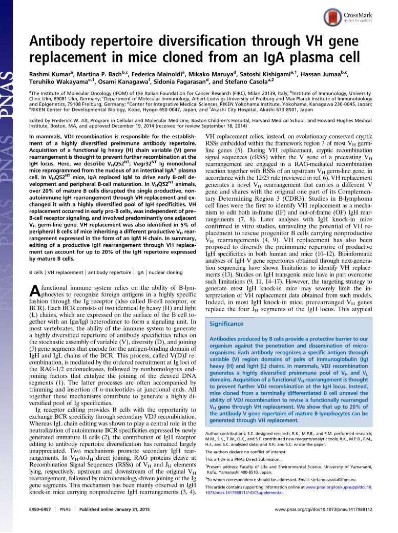
ResultsNuclear Reprogramming of Intestinal PCs. We applied SCNT toreprogram terminally differentiated IgA+ PCs isolated from theLP of the small intestine of mice housed under specific patho-gen-free conditions. Nuclear transferred ES (ntES) cell lineswere established from independent IgA cloned embryos. Deri-vation of ntES lines from IgA PCs was confirmed by genomicPCR amplification of Ig H and L chain V gene rearrangements.Chimeric mice were obtained through blastocyst injection of onerepresentative IgA ntES cell line. Southern blotting analysis andPCR amplification of tail-tip genomic DNA of chimeric offspringconfirmed germ-line transmission of cloned Ig V gene rear-rangements (Fig. 1A and Fig. S1 A and B). These data indicatethat PCs can undergo nuclear reprogramming to become plu-ripotent stem cells.
IgA Can Replace IgM to Drive B-Cell Development. IgA transnuclearmice allowed us to test whether an IgA BCR selected by an in-testinal PC could replace IgM to drive B-cell development. IgAmonoclonal mice inherited a productive, unmutated, VH rear-rangement consisting of the DH-proximal VHQ52.a27.79 genejoined to DFL16.1 and JH3 segments. The VL gene rearrange-ment consisted of Vκgr32 joined to Jκ4 (Fig. 1B and Fig. S1B).Mice inheriting prerearranged VH and VL genes were called, re-spectively, VHQ52
NT and Vκgr32NT.VHQ52NT;Vκgr32NT heterozygous (HT) mice were analyzed on
the Rag1-deficient genetic background to study B-cell developmentunder conditions of Ig monospecificity. VHQ52
NT; Vκgr32NT; Rag1−/−
mice showed normal numbers of CD19+ B cells, all expressingsurface IgA (sIgA), in spleen (SP) and lymph nodes (LNs) (Fig. 1 CandD). Immunophenotypic analysis revealed the presence in the SPof mature follicular/B-2 (FO; CD19+CD21+CD23+) and marginalzone (MZ; CD19+CD21hiCD23lo) B cells (Fig. 1E). In contrast, B-1
B cells (CD19hiCD23lo) were largely missing in the peritoneal cavityof IgA monoclonal mice (Fig. 1E). These results indicate thata BCR specificity selected by an intestinal PC and expressed in theform of IgA can drive early B-cell development and promote dif-ferentiation into mature FO and MZ B cells.
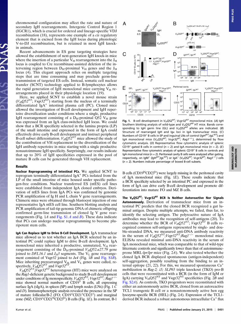
The VHQ52NT; Vκgr32NT BCR Is Neither Autoreactive Nor Signals
Autonomously. Derivation of transnuclear mice from an in-testinal PC predicts that the cloned BCR recognized a gut lu-minal antigen. Despite multiple attempts, we failed, thus far, toidentify the selecting antigen. The polyreactive nature of IgAantibodies may lead to the recognition of self-antigens (20). Todetermine whether the BCR of VHQ52NT; Vκgr32NT B cells re-cognized common self-antigens represented by single- and dou-ble-stranded DNA, we measured anti-DNA antibody reactivityin the serum of VHQ52NT;Vκgr32NT;Rag1−/− monoclonal mice.ELISAs revealed minimal anti-DNA reactivity in the serum ofIgA monoclonal mice, which was comparable to that of wild-typelittermate controls and significantly lower than that of autoimmune-prone MRL-lpr/lpr mice (Fig. 2A). We also tested whether thecloned IgA BCR displayed spontaneous (antigen-independent)self-aggregation, possibly resulting from the binding to an in-ternal epitope (21, 22). For this, we measured spontaneous Ca+
mobilization in Rag-2; λ5; SLP65 triple knockout (TKO) pro-Bcells that were reconstituted with a BCR (in the form of IgM orIgA) carrying VHQ52NT and Vκgr32NT specificities (Fig. 2B andFig. S2A). As controls, TKO progenitors were reconstituted witheither an autonomously active BCR, cloned from an autoreactiveTCL1 transgenic B cell or a nonautonomously active hen egglysozyme-specific BCR (HEL) (Fig. 2A). Expression of the TCL1-derived BCR induced a robust autonomous intracellular Ca+ flux
Fig. 1. B-cell development in VHQ52NT; Vκgr32NT monoclonal mice. (A) IgHSouthern blotting analysis of wild-type and VHQ52NT HT mice. Bands corre-sponding to IgH germ line (GL) and VHQ52NT alleles are indicated. (B)Structure of rearranged IgH and Igκ loci in IgA transnuclear mice. (C)Numbers of CD19+ B cells in SP and inguinal LNs of control (IgHWT;IgLWT) andIgA monoclonal mice (VHQ52NT; Vκgr32NT; Rag1−/−), determined by flowcytometric analysis. (D) Representative flow cytometric analysis of splenicCD19+-gated B cells in control (n = 2) and IgA monoclonal mice (n = 2). (E)Representative flow cytometric analysis of splenic CD19+ B cells in controls andIgAmonoclonal mice (n = 2). Peritoneal cavity B cells were analyzed after gating,respectively, on IgM+ (IgHWT;IgLWT) or IgA+ (VHQ52
NT; Vκgr32NT; Rag1−/−) cells(n = 2). Numbers indicate percentage of boxed B-cell subsets.
Kumar et al. PNAS | Published online January 21, 2015 | E451
IMMUNOLO
GYAND
INFLAMMATION
PNASPL
US
in TKO progenitors upon tamoxifen-dependent activation of anERT2–SLP65 fusion protein (21). In sharp contrast, BCR spe-cificities from both IgA transnuclear and HEL-specific B cellsfailed to trigger spontaneous Ca+ mobilization in response toSLP65 activation (Fig. 2 C and D and Fig. S2 B and C). Theseresults, together with the observations that IgA monoclonal mice(both VHQ52NT-HT and VHQ52NT-HT; Vκgr32NT-HT animals) agedin a comparable fashion to wild-type littermate controls lackedsigns of systemic autoimmunity and displayed a normal (or, atmost, lower) proportion of sIgλ+B cells (Fig. S2D), render it unlikelythat the IgA BCR expressed by transnuclear B cells is self-reactive.
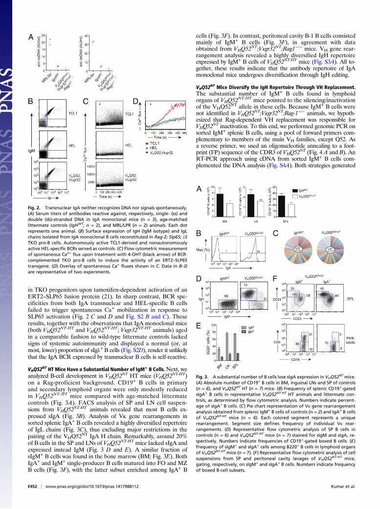
VHQ52NT HT Mice Have a Substantial Number of IgM+ B Cells.Next, we
analyzed B-cell development in VHQ52NT HT mice (VHQ52
NT-HT)on a Rag-proficient background. CD19+ B cells in primaryand secondary lymphoid organs were only modestly reducedin VHQ52NT-HT mice compared with age-matched littermatecontrols (Fig. 3A). FACS analysis of SP and LN cell suspen-sions from VHQ52
NT-HT animals revealed that most B cells ex-pressed sIgA (Fig. 3B). Analysis of Vκ gene rearrangements insorted splenic IgA+ B cells revealed a highly diversified repertoireof IgL chains (Fig. 3C), thus excluding major restrictions in thepairing of the VHQ52NT IgA H chain. Remarkably, around 20%of B cells in the SP and LNs of VHQ52
NT-HT mice lacked sIgA andexpressed instead IgM (Fig. 3 D and E). A similar fraction ofsIgM+ B cells was found in the bone marrow (BM; Fig. 3E). BothIgA+ and IgM+ single-producer B cells matured into FO and MZB cells (Fig. 3F), with the latter subset enriched among IgA+ B
cells (Fig. 3F). In contrast, peritoneal cavity B-1 B cells consistedmainly of IgM+ B cells (Fig. 3F), in agreement with dataobtained from VHQ52NT;Vκgr32NT;Rag1−/− mice. VH gene rear-rangement analysis revealed a highly diversified IgH repertoireexpressed by IgM+ B cells of VHQ52
NT-HT mice (Fig. S3A). All to-gether, these results indicate that the antibody repertoire of IgAmonoclonal mice undergoes diversification through IgH editing.
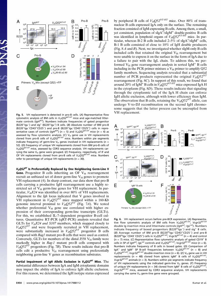
VHQ52NT Mice Diversify the IgH Repertoire Through VH Replacement.
The substantial number of IgM+ B cells found in lymphoidorgans of VHQ52NT-HT mice pointed to the silencing/inactivationof the VHQ52NT allele in these cells. Because IgM+ B cells werenot identified in VHQ52
NT;Vκgr32NT;Rag-1−/− animals, we hypoth-esized that Rag-dependent VH replacement was responsible forVHQ52
NT inactivation. To this end, we performed genomic PCR onsorted IgM+ splenic B cells, using a pool of forward primers com-plementary to members of the main VH families, except Q52. Asa reverse primer, we used an oligonucleotide annealing to a foot-print (FP) sequence of the CDR3 of VHQ52
NT (Fig. 4 A and B). AnRT-PCR approach using cDNA from sorted IgM+ B cells com-plemented the DNA analysis (Fig. S4A). Both strategies generated
Fig. 2. Transnuclear IgA neither recognizes DNA nor signals spontaneously.(A) Serum titers of antibodies reactive against, respectively, single- (ss) anddouble (ds)-stranded DNA in IgA monoclonal mice (n = 3), age-matchedlittermate controls (IgHWT; n = 2), and MRL/LPR (n = 2) animals. Each dotrepresents one animal. (B) Surface expression of IgH (IgM isotype) and IgLchains isolated from IgA monoclonal B cells reconstituted in Rag-2; Slp65; λ5TKO pro-B cells. Autonomously active TCL1-derived and nonautonomouslyactive HEL-specific BCRs served as controls. (C) Flow cytometric measurementof spontaneous Ca2+ flux upon treatment with 4-OHT (black arrow) of BCR-complemented TKO pro-B cells to induce the activity of an ERT2–SLP65transgene. (D) Overlay of spontaneous Ca+ fluxes shown in C. Data in B–Dare representative of two experiments.
Fig. 3. A substantial number of B cells lose sIgA expression in VHQ52NT mice.(A) Absolute number of CD19+ B cells in BM, inguinal LNs and SP of controls(n = 6), and VHQ52NT HT (n = 7) mice. (B) Frequency of splenic CD19+-gatedsIgA+ B cells in representative VHQ52NT-HT HT animals and littermate con-trols, as determined by flow cytometric analysis. Numbers indicate percent-age of sIgA+ B cells. (C) Pie chart representation of Vκ gene rearrangementanalysis obtained from splenic IgM+ B cells of controls (n = 2) and IgA+ B cellsof VHQ52NT-HT mice (n = 6). Each colored segment represents a uniquerearrangement. Segment size defines frequency of individual Vκ rear-rangements. (D) Representative flow cytometric analysis of SP B cells incontrols (n = 6) and VHQ52NT-HT mice (n = 7) stained for sIgM and sIgA, re-spectively. Numbers indicate frequencies of CD19+-gated boxed B cells. (E)Frequency of sIgM+ and sIgA+ cells among B220+ B cells in lymphoid organsof VHQ52NT-HT mice (n = 7). (F) Representative flow cytometric analysis of cellsuspensions from SP and peritoneal cavity lavages of VHQ52NT-HT mice,gating, respectively, on sIgM+ and sIgA+ B cells. Numbers indicate frequencyof boxed B-cell subsets.
E452 | www.pnas.org/cgi/doi/10.1073/pnas.1417988112 Kumar et al.

PCR products that were subsequently cloned and sequenced. Se-quence analyses revealed the consistent replacement of the originalVHQ52.a27.79 gene of VHQ52
NT with other germ-line VH genes(Fig. 4C, Fig. S4B, and Table S1). All VH replacements shared withthe original VHQ52NT rearrangement part of the CDR3 FP se-quence and the JH3 segment (Fig. 4D). VH replacements werefurther diversified by de novo n-nucleotide addition (Fig. 4D andTable S1). Importantly, CDR3 sequence analysis indicated thatmost VH replacements in VHQ52NT-HT mice were unique, thusunderscoring the polyclonal origin of IgM+ B cells (Fig. 4E, Fig.S4C, and Table S1). In accordance with the sIgA-negative phe-notype, VH replacements in IgM+ B cells of VHQ52NT-HT micewere predominantly OF (Table S1). This result is compatible with
a scenario whereby the occurrence of OF VH replacements causedthe inactivation of VHQ52
NT, which in turn prompted further VDJrecombination on the second IgH chromosome favoring the gen-eration of IgM+ B cells. To estimate the frequency of secondary VHrearrangements in an unbiased fashion, we performed IgH South-ern blotting analysis on genomic DNA from VHQ52
NT homozygoussplenic B cells. We compared the intensity of the bands corre-sponding to the VHQ52
NT allele between B cells and liver cells thatwere used as negative control (Fig. S4D). Quantification of the datarevealed that around 22% of splenic IgA+ B cells disrupted oneVHQ52NT allele (Fig. 4F).To validate our results, we used VHQ52NT as a reporter allele
to monitor VH replacement in B1-8f IgH knock-in mice (23).Flow cytometric analysis of splenocytes of double IgH knock-inmice revealed a major population of B cells expressing concomi-tantly sIgM and sIgA (Fig. 4G). Importantly, we also detecteda distinct subset of IgM-only B cells that ranged between 3% and5% of splenic B cells (Fig. 4G and Fig. S4E). VH rearrangementanalysis in sorted IgM-only B cells identified OF VH replacementsthat disrupted the VHQ52NT allele (Table S1). We rarelyidentified sIgA-only B cells, possibly because inactivation ofB1-8f through VH replacement is rendered unlikely by its chro-mosomal location (JH locus) and the lack of a cRSS (23). In sum-mary, using two independent mouse models starting with a highlyrestricted repertoire of productive IgH specificities, we found that3–20% of their mature B cells diversified antibody specificitythrough VH replacement.
VH Replacements Are Already Detected in Pro-B Cells. To investigatethe temporal onset of VH replacement, we first evaluated earlyB-cell development in VHQ52NT-HT mice. Flow cytometric anal-ysis revealed reduced numbers of Igκ−B220lo BM B-cell pro-genitors in VHQ52NT mice in comparison with controls (Fig. 5A).The impairment was mainly caused by fewer CD25+ pre-B cells,whereas CD43+ pro-B cells were largely comparable in numberto controls (Fig. 5B). To test whether VH replacement had al-ready occurred in early B-cell progenitors, we sorted B220loIgκ−CD43+ pro-B cells from VHQ52NT-HT mice and performedVH replacement analysis. Sequencing of PCR products revealeda considerable number of unique VH replacements, which werealso detected in their CD25+ pre–B-cell progeny (Fig. S5A). Vgene use in VH replacements cloned from pro-B cells indicateda similar pattern to that of SP IgM+ B cells (Fig. 5 C and D, Fig.S5 A and C, and Table S1). Ninety-seven percent of unique VHreplacements sequenced from pro-B cells were nonproductive(Fig. 5E). In contrast, close to 10% of VH replacements in pre-Bcells were IF (Fig. S5B). These results suggest that OF VHreplacements block the development of pro-B cells unless res-cued by functional secondary IgH rearrangements.
VH Replacement Occurs Independently of Pre-BCR Expression/Signaling.The lower number of pre-B cells in VHQ52
NT mice may resultfrom impaired expression and/or signaling of a pre-BCR com-posed of an IgA H chain (24). Altered pre-BCR function may, inturn, prolong RAG-mediated recombination at the IgH locus,hence facilitating VH replacement. To test this hypothesis, weanalyzed VHQ52NT-HT;Vκgr32NT-HT double Ig insertion mice.The latter animals displayed a significant reduction in both pro-Bcells and pre-B cells, as a result of the premature expression ofpairing IgH/IgL chains (Fig. 6 A and B). Remarkably, flowcytometric analysis of splenic cell suspensions from IgH/L in-sertion mice revealed a fraction of sIgM+ B cells comparable tothat of VHQ52NT-only animals (Fig. 6 C and D). Molecular ana-lysis confirmed the inactivation of VHQ52NT through OF VHreplacements in IgM+ B cells of VHQ52
NT-HT;Vκgr32NT-HT mice(Fig. 6 E and F and Table S1). These results indicate that pre-BCRexpression and/or signaling is not required to initiate VH replace-ment of a productive IgH rearrangement.
Fig. 4. VHQ52NT is edited through VH replacement. (A) CDR3 nucleotidesequence of VHQ52NT. Cryptic heptamer and n-nucleotides are indicated,respectively, in red and gray. (B) PCR strategy to identify VH replacements.Arrows indicate PCR primers. Colored blocks symbolize VH families. An oli-gonucleotide annealing to a unique sequence (FP) within the CDR3 ofVHQ52NT was used as a reverse primer. (C) Pie representation of VH gene usein VH replacements cloned from splenic IgM+ B cells of VHQ52NT-HT mice (n =4). Numbers indicate frequency of rearrangements carrying the indicated VH
genes. PCR primers used for the analysis are indicated. (D) CDR3 nucleotidesequence of representative VH replacements cloned from IgM+ B cells ofVHQ52NT-HT mice using the VHX24.a1.84 donor VH gene. First line showsCDR3 sequence of the original VHQ52NT rearrangement. Underlined areVHQ52NT FP sequences indicated, respectively, as FP1 and FP2. De novoadded n-nucleotides are labeled in brown. cRSSs are indicated in red. (E)Frequency of unique VH replacements cloned from IgM+ splenic B cells ofVHQ52NT-HT mice, as assessed through CDR3 sequence analysis. Replacementsusing the same VH germ-line gene were grouped. (F) Southern blottingquantification of VHQ52NT gene copy number in splenic IgA+ B cells ofVHQ52NT homozygous (HO) mice. Data were normalized for DNA input andrepresented as relative to VHQ52NT copy number in liver cells of transnuclearmice. (G) Representative flow cytometric analysis of splenic CD19+ B cells ofVHQ52NT;B1-8f double IgH insertion mice (n = 4). Number within dot plotrefers to frequency of IgM-only B cells.
Kumar et al. PNAS | Published online January 21, 2015 | E453
IMMUNOLO
GYAND
INFLAMMATION
PNASPL
US
VHQ52NT Is Preferentially Replaced by One Neighboring Germ-Line V
Gene. Progenitor B cells inheriting an OF VH rearrangementrecruit an unbiased set of donor germ-line VH genes to promoteVH replacement (4). In sharp contrast, our data show that pro-Bcells carrying a productive IgH rearrangement use a highly re-stricted set of VH germ-line genes for VH replacement. In par-ticular, VHX24 was identified in over 80% of VH replacements.Alignment to the Igh locus indicated that V genes involved inVH replacement in VHQ52NT mice mapped within a 160-Kbgenomic interval proximal to VHQ52NT (Fig. 7A). We testedwhether preferential VH gene use correlated with higher ex-pression of their corresponding germ-line transcripts (GLTs).For this, we established IL-7–dependent progenitor B-cell cul-tures. Quantitative RT-PCR (qRT-PCR) analysis revealed thatGLTs for VHX24 and S107 members, which mapped closely toVHQ52NT and were frequently recruited in VH replacement,were substantially increased in VHQ52
NT progenitor B cellscompared with Rag1 mutant pro-B cells that were used as control.Conversely, GLTs for members of the distal J558 VH family weremarkedly higher in Rag-1 mutant pro-B cells compared withVHQ52
NT progenitors (Fig. 7B). These results indicate that pro-Bcells edit a productive VH rearrangement using preferentiallyneighboring germ-line V genes as recombination substrates.
Partial Impairment of IgH Allelic Exclusion in VHQ52NT Mice. The
substantial differences between IgA and IgM cytoplasmic domainsmay impact the ability of IgA to enforce IgH allelic exclusion.For this reason, we determined the IgH isotype status expressed
by peripheral B cells of VHQ52NT-HT mice. Over 80% of trans-
nuclear B cells expressed IgA only on the surface. The remainingfraction consisted of IgM-expressing B cells. Among them, a small,yet consistent, population of sIgA+/sIgM+ double-positive B cellswas identified in lymphoid organs of VHQ52
NT-HT mice. In par-ticular, whereas B-2 B cells included 2–5% of sIgA+/sIgM+ cells,B-1 B cells consisted of close to 10% of IgH double producers(Fig. 8 A and B). Next, we investigated whether sIgM-only B cellsincluded cells that retained the original VH rearrangement butwere unable to express it on the surface in the form of IgA due toa failure to pair with the IgL chain. To address this, we per-formed VH gene rearrangement analysis in sorted IgM+ B cellsincluding in the PCR primer mixture a VH primer to amplify Q52family members. Sequencing analysis revealed that a substantialnumber of PCR products represented the original VHQ52NT
rearrangement (Fig. 8C). In support of this result, we found thataround 20% of IgM+ B cells in VHQ52
NT-HTmice expressed IgA Hin the cytoplasm (Fig. 8D). These results indicate that signalingthrough the cytoplasmic tail of the IgA H chain can enforceIgH allelic exclusion, although with lower efficiency than IgM.The observation that B cells, retaining the VHQ52NT allele, canundergo V-to-DJ recombination on the second IgH chromo-some suggests that the latter process can be uncoupled fromVH replacement.
Fig. 5. VH replacement is detected in pro-B cells. (A) Representative flowcytometric analysis of BM cells in VHQ52NT-HT mice and age-matched litter-mate controls (IgHWT). Numbers indicate frequencies of gated progenitor(B220+Igκ−) and sIg+ (B220+Igκ+) B cells. (B) Absolute number of BM pro-B(B220+Igκ−CD43+CD25−) and pre-B (B220+Igκ−CD43−CD25+) cells in repre-sentative cases of controls (IgHWT) (n = 5) and VHQ52NT-HT mice (n = 4) asassessed by flow cytometric analysis. (C) VH gene use in VH replacementscloned from pro-B cells of VHQ52NT-HT mice. Numbers within pie segmentsindicate frequency of germ-line VH genes involved in VH replacements (n =52). (D) Frequency of unique VH replacements cloned from BM pro-B cells ofVHQ52NT-HT mice, assessed by CDR3 sequence analysis. VH replacements car-rying the same VH gene were grouped. (E) Frequency, respectively, of IF andOF VH replacements cloned from pro-B cells of VHQ52NT-HT mice. Numbersrefer to percentage of unique VH replacements (n = 30).
Fig. 6. VH replacement occurs before pre-BCR expression. (A) Representa-tive flow cytometric analysis of BM cells from VHQ52NT-HT; Vκgr32NT-HT
double Ig insertion mice and littermate controls (IgHwt; IgLwt). Numbersindicate frequency of boxed progenitors (B220loIgκ−) and sIg+ B cells.(B) Average number of BM pro-B (B220loIgκ−CD43+CD25−) and pre-B(B220loIgκ−CD43−CD25+) cells in VHQ52NT-HT; Vκgr32NT-HT (n = 4) and control(n = 7) mice. (C) Representative flow cytometric analysis of gated CD19+ Bcells in SP of IgHwt; IgLwt controls and VHQ52NT-HT; Vκgr32NT-HT mice (n = 6).Numbers indicate frequency of B cells in boxed gates. (D) Comparison ofIgA+ and IgM+ SP B-cell frequencies between VHQ52NT-HT (n = 8) andVHQ52NT-HT; Vκgr32NT-HT double-insertion mice (n = 6). (E) VH gene use in VHreplacements (n = 48) cloned from splenic IgM+ B cells of VHQ52NT-HT;Vκgr32NT-HT animals (n = 3). Numbers within pie segments indicate frequencyof VH replacements using the indicated germ-line VH genes. (F) Frequencyof unique VH replacements (n = 34) cloned from IgM+ B cells of VHQ52NT-HT;Vκgr32NT-HT mice, assessed by CDR3 sequence analysis. VH replacementscarrying the same VH germ-line gene were grouped.
E454 | www.pnas.org/cgi/doi/10.1073/pnas.1417988112 Kumar et al.

DiscussionHere, we describe the cloning of mice from the nucleus of anIgA-secreting PC. The derivation of pluripotent ES cells throughreprogramming of a PC unravels the epigenetic plasticity ofterminally differentiated B cells, extending previous work (19).IgA monoclonal mice were established from a PC residing in
the LP of the small intestine, possibly as a result of the recognitionof a luminal antigen (25). Although the selecting antigen remainsyet undefined, several evidences exclude that the cloned IgA BCRis autoreactive (20). First, serum antibodies in VHQ52NT; Vκgr32NT
monoclonal mice lacked reactivity to common self-antigensrepresented by single- and double-stranded DNA. Second, Rag2;λ5; SLP65 TKO pro-B cells reconstituted with a BCR carryingVHQ52NT; Vκgr32NT specificities lacked autonomous calcium mo-bilization, hence excluding spontaneous self-aggregation (26). Third,Igκ/λ light chain ratio was comparable between IgA transnuclearand littermate controls. Fourth, IgA cloned mice lacked signs ofsystemic autoimmunity and aged similarly to littermate controls.Mice expressing a prerearranged VH rearrangement from an
IgA class-switched locus generated close-to-normal numbers ofmature B cells, the majority of which expressed IgA. In mono-clonal VHQ52NT; Vκgr32NT mice, splenic mature B cells consistedof both FO and MZ B cells. Instead, peritoneal cavity B-1 B cellswere largely missing. These results are in accordance with, andextend, previous data (24) indicating that IgA can replace IgM todrive early B-cell development and instruct peripheral B-cellmaturation. Whereas VH gene use may influence the FO versusMZ B-cell fate (27), our data reveal that BCR extrinsic factorscritically contribute to this decision. Also, the development ofFO and MZ B cells at the expense of B-1 B cells may reflect the
expression by transnuclear B cells of a BCR specificity enablingselectively the development of a B2/MZ B-cell progenitor (28,29). Weak tonic BCR signaling may also contribute to thepreferential development of FO and MZ B cells in IgA trans-nuclear mice (30).We anticipated that transnuclear mice on the Rag-proficient
background retained expression of the VHQ52NT specificityamong mature B cells. Remarkably, instead, a substantial frac-tion of SP B cells in VHQ52NT mice lost expression of the originalVH rearrangement. In particular, in VHQ52NT HT mice, over20% of peripheral B cells expressed IgM instead of IgA. Anallele-specific PCR strategy revealed that in the latter cellsthe VHQ52NT allele was consistently inactivated through OF VHreplacements. These results are compatible with a scenariowhereby an initial OF VH replacement, disrupting VHQ52NT,prompts further VDJ recombination on the second IgH chromo-some, allowing the rescue of those B cells that acquire a productiveVH rearrangement. IF VH replacements were also identified, hencecontributing to the diversification of the IgA+ B-cell pool. CDR3sequence analysis of VH replacements revealed a highly diversifiedpool of secondary IgH rearrangements. Within the yet-limited cov-erage of the IgH repertoire of VHQ52
NT mice, these results suggestthat transnuclear B cells undergoing VH replacement do notrepresent a rare population that has undergone substantial clonalexpansion. Southern blotting analysis performed on VHQ52
NT ho-mozygous B cells revealed that ∼20% of peripheral IgA+ B cellsinactivated one VHQ52
NT allele. Because VH-to-JH direct joiningevents were rarely found in VHQ52
NT B cells, we conclude that VHreplacement represents the preferred mechanism for the di-versification of the productive, preimmune, IgH V gene repertoire.VH replacement was proposed to occur in pro-B as well as in
immature B cells, where it may contribute to neutralize BCRautoreactivity (4, 12, 15, 31). In VHQ52NT mice, VH replace-ments were already found in Ig−B220loCD43+ BM pro-B cells.Because VH replacement was accompanied by de novo additionof n-nucleotides, our data indicate that this process likely occursin an early pro-B cell that expresses Tdt (32). The high frequencyof VH replacements seen in VHQ52NT mice could result from
Fig. 7. VH replacement of VHQ52NT employs adjacent VH donor genes. (A)Schematic view of the DH-proximal portion of the IgH locus mapping up-stream of the VHQ52NT rearrangement. Colored blocks indicate functionalgerm-line VH genes ordered based on their physical distance from VHQ52NT.Numbers above blocks indicate unique VH replacements using the indicatedVH genes. (B) qRT-PCR determination of VH GLTs for the indicated VH familiesin pro–B-cell cultures established from Rag1-mutant and VHQ52NT-HO mice.Levels of GLTs are relative to those of the housekeeping gene Rplp0.
Fig. 8. IgH allelic inclusion is observed in a small fraction of VHQ52NT matureB cells. (A) Representative flow cytometric determination of sIgA/IgM doubleproducers within, respectively, gated B2 (CD19+B220+) and B1 (B220loCD19hi) Bcells in the peritoneal cavity of VHQ52NT-HT mice (n = 5). Numbers indicatefrequency of boxed sIgA/sIgM double producers. (B) Average frequency ofsIgA/sIgM double producers among the indicated B-cell subsets in the perito-neal cavity of VHQ52NT-HT mice (n = 5). (C) VH gene use in purified IgM+ splenicB cells of VHQ52NT-HT mice, assessed by genomic PCR using primers annealingto V genes of the main VH families including Q52. Numbers indicate frequencyof IgH rearrangements (n = 56) consisting of the indicated VH genes. (D) In-tracellular flow cytometric determination of IgA H expression in the indicatedsubset of SP B cells in controls (IgHWT) and VHQ52NT-HT mice.
Kumar et al. PNAS | Published online January 21, 2015 | E455
IMMUNOLO
GYAND
INFLAMMATION
PNASPL
US

impaired pro-B to pre-B cell transition. However, the analysis ofVHQ52NT; Vκgr32NT double Ig insertion mice, largely lacking theprogenitor B-cell compartment, showed frequencies of IgM+ Bcells that were similar to those of VHQ52NT-only animals. Hence,whereas a contribution of the pre-BCR to the induction of IgHediting cannot be formally excluded, our results, in accordancewith previous evidences (4), support a scenario whereby VHreplacement of a productive IgH rearrangement starts in an earlypro-B cell before pre-BCR expression/signaling.The loss of a large genomic segment of the CH locus resulting
from IgA class switch recombination could contribute to the highrate of VH replacements observed in VHQ52NT mice, throughthe loss of putative, yet unidentified, negative cis regulatoryelements. To address this question, we crossed VHQ52NT mice toB1-8f IgH knock-in mice. In Ig double-insertion mice, VHQ52NT
served as a reporter allele to monitor VH replacement in B cellstransiting through early B-cell development as a result of B1-8fexpression. Whereas most B cells were IgH allelically included,a distinct subset of them ranging between 3% and 5% lost theVHQ52NT allele through VH replacement. The different fre-quency of B cells undergoing VH replacement in VHQ52NT; B1-8f mice compared with VHQ52NT-only animals renders unlikelythe existence of regulatory elements embedded in the CH locusthat influence VH replacement. Instead, these results point todifferences between IgA and IgM H chains, or levels of theircorresponding transcripts (33), in the regulation of IgH locusaccessibility to the recombination machinery.Analysis of V-gene use in VH replacements of VHQ52NT B
cells revealed a different scenario from the one described formice carrying OF VH rearrangements (4). Indeed, IgA trans-nuclear mice showed a highly restricted set of VH genes engagedin VH replacement. In particular, VHX24.a1.84 contributed toover 80% of all VH replacements. Notably, all VH germ-linegenes recruited in VH replacement mapped in close proximityto the VHQ52NT rearrangement. The biased use of VH genes inVH replacement could reflect the limited time to which the pro-ductive VHQ52NT rearrangement is exposed to the VDJ recom-bination machinery. This condition may facilitate the targetingof the RAG proteins to neighboring V genes, as previouslysuggested (34). The evidence that GLTs for VH genes proximalto VHQ52NT were more abundant in transnuclear pro-B cellscompared with controls supports this hypothesis. Higher germ-linetranscription of DH-proximal V genes coupled to their preferentialrecruitment in VH replacement may result from the lack of IGCR1on the VHQ52
NT allele (18). VH replacement may hence representa mechanism that acquires relevance for the editing of productiveVH rearrangements carrying DH-proximal V genes. The failure ofVHQ52
NT B cells to recruit V genes of distal VH families may de-pend on VH replacement occurring in a developmental windowthat precedes IgH locus contraction (35). The identification ofa small subset (1–2%) of IgM+ B cells in ovalbumin-specific IgG1and hematoagglutinin-specific IgG2b transnuclear mice (both usingdistal J558 VH genes) suggests that secondary IgH rearrangementsmay also occur in these animals (36, 37).The IgA H chain cytoplasmic domain is longer and differs
substantially in the primary sequence from that of IgM (38). Wewondered whether such differences affected the ability of theIgA H chain to signal IgH allelic exclusion. Flow cytometric datarevealed that over 80% of B cells in VHQ52
NT mice expressed IgAonly. The remaining fraction consisted of sIgM+ B cells. Around20% of the latter cells (accounting for 4% of total peripheral Bcells) expressed cytoplasmic IgA H, which was confirmed by suc-cessful PCR amplification of the VHQ52
NT rearrangement. More-over, around 2% of B2 B cells and over 10% of peritoneal cavityB-1 B cells expressed both IgA and IgM on the surface. From theseresults, we conclude that the IgA cytoplasmic tail is able to instructIgH allelic exclusion, although with lower efficiency than IgM.Similar data were reported for the IgG1 H chain (36).
In summary, this study unravels the contribution of VH re-placement to the diversification of the productive preimmune IgHrepertoire of mice starting with a restricted pool of antibodyspecificities. Using two independent Ig transgenic models, weshow that 3% or more (reaching a maximum of 20%) of mature Bcells edited their initially productive IgH rearrangement throughVH replacement. Our results are in agreement and complementdata presented in the accompanying paper by Sun et al. (39).The concomitant presence in HT VHQ52NT mice of di-
versified pools of, respectively, IgM- and IgA-only B cellsoffers the unprecedented opportunity to study in a competitivesetup the properties of IgA and IgM BCRs to control homeo-stasis of B cells and their recruitment into T-cell–dependentand –independent immune responses, especially in mucosa-associated lymphoid tissues.
Materials and MethodsAnimal Care. Animals were housed under specific pathogen-free conditionsat the IFOM-IEO Campus and maintained according to protocols approvedby the IFOM Institutional Animal Care and Use Committee and the ItalianMinistry of Health. Rag1−/− mice were purchased from Jackson Laboratories.B1-8f mice were previously described (23). Experiments were performedwith 8–16-wk-old animals.
Generation of IgA Transnuclear Mice. Small intestine LP cells of C57BL/6 XB10D2 F1 mice were prepared as previously described (40) and stained withfluorescent-labeled anti-B220, anti–MHC-II, and anti-IgA antibodies to iso-late B220−MHC-II−IgA+ PCs using a MoFlo (Beckman Coulter) cell sorter.SCNT procedures have been previously published (41) and are summarized inSI Materials and Methods.
Flow Cytometry and Cell Sorting. Single-cell suspensions from lymphoidorgans were stained using fluorescent- or biotin-conjugated monoclonalantibodies against mouse CD19 (ID3), CD21 (8D9), CD23 (B3B4), CD45R/B220(R3A3-6B2), CD25 (7D4), TCRβ (H57-59), CD5 (53-7.3), and IgA (mA-6E1),all from eBioscience. Anti-CD43 (S7) was from BD Biosciences. Anti-IgM(R33.24.12) and anti-Igκ (R33-18-10) antibodies were conjugated in house.For intracellular staining, B cells were fixed, permeabilized in Cytofix/Cyto-perm (BD Biosciences), and stained with IgA-specific antibody. Samples wereacquired on a FACSCalibur (BD Biosciences) and data analyzed with FlowJosoftware (Tree Star). Cell sorting was performed on FACSAria (BD Bio-sciences) after size exclusion of dead cells.
Analysis of Ig V Gene Rearrangements. Genomic DNA or total RNA wasextracted using the All Prep DNA/RNA mini kit according to the manu-facturer’s protocol (Qiagen). For VH replacement analysis, genomic DNA wasPCR amplified using a mixture of VH family-specific primers (42) in combi-nation with a VHQ52NT FP-specific primer. PCR conditions are listed in SIMaterials and Methods. qRT-PCR analysis of VH replacements was per-formed using cDNA prepared from purified IgM+ B cells using a mixture ofVH family-specific primers (42) combined with an oligonucleotide comple-mentary to Cα. VH rearrangements occurring on the second IgH chromo-some in IgM+ VHQ52NT-HT B cells were amplified by RT-PCR using VH family-specific primers together with an oligonucleotide annealing to Cμ. Igκ Vgene rearrangements were amplified by RT-PCR using a mixture of de-generate forward primers annealing to most Vκ genes (30), in combinationwith a Cκ reverse primer. PCR products were cloned and subjected to Sangersequencing. V gene analyses were performed using IGBLAST. Primersequences are listed in Table S2.
Quantification of VH GLTs. qRT-PCR was performed in triplicate with SYBRGreen-I Master Mix using primer combinations for GLTs listed in Table S2.Values were calculated using the comparative CT method. To normalize forcDNA input, a segment of the housekeeping Rplp0 gene was amplified withprimers listed in Table S2.
Southern Blotting Analysis. Southern blotting was performed on genomicDNA isolated from CD19+ splenic B cells, as previously described (43) andsummarized in SI Materials and Methods.
BCR Complementation of Rag2; λ5; SLP65 TKO Pro-B Cells. Rag2; λ5; SLP65 TKOpro-B cells expressing a tamoxifen-inducible ERT2–SLP65 fusion protein werepreviously described (26). SLP65 signaling function was restored incubating
E456 | www.pnas.org/cgi/doi/10.1073/pnas.1417988112 Kumar et al.

TKO pro-B cells with 1 μM of 4-Hydroxytamoxifen (4-OHT, Sigma-Aldrich).VHQ52NT and Vκgr32NT specificities were cloned, respectively, into pMIZCCand pMIZYN retroviral vectors, as previously described (26). Retroviral vec-tors for TCL1-derived IgH and IgL chains were previously described (22). TheHEL-specific HyHEL 10 antibody was described in ref. 44. Primers used forcloning of VHQ52NT and Vκgr32NT IgH and IgL chains are listed in Table S2.Retrovirus production and transduction of TKO pro-B cells was performed aspreviously described (26).
Measurement of Intracellular Calcium Flux. BCR reconstitution of Rag2; λ5;SLP65 TKO pro-B cells was revealed by staining cells with goat anti-mouseIgM (Jackson ImmunoResearch), anti-mouse Igκ (Southern Biotech), or ratanti-mouse IgA (e-Bioscience) antibodies, respectively. Ca2+ mobilizationwas measured as described before (26). Briefly, 5 × 10⁶ transduced pro-B cellsexpressing ERT2–SLP65 were incubated with 5 μg/mL of Indo1 (Molecularprobes) and 0.5 μg/mL of Pluronic F-127 (Molecular Probes) in Iscove mediumsupplemented with 1% serum (Vitromex) at 37 °C for 45 min. After loading,cells were centrifuged, resuspended in Iscove medium with 1% FCS, and
stimulated with 1 mM of 4-OHT. Calcium flux was measured on an LSR II flowcytometer (Becton Dickinson).
Enzyme Linked Immunosorbent Assay. Titration of serum anti-DNA antibodieswas performed by ELISA, as previously described (45). Procedures are sum-marized in SI Materials and Methods.
ACKNOWLEDGMENTS. We thank F. Alberghini for sharing preliminary dataand the remaining members of the S.C. group for helpful discussions. Weacknowledge N. Hövelmeyer and Ari Waisman for sharing reagents andS. P. Mahrt for suggestions. T. Kurosaki and K. Kometani kindly shared un-published data. This study was supported by grants from the RIKEN ResearchCenter for Allergy and Immunology through the International Research Col-laboration Award (to S.C. and S.F.), the Italian Association for Cancer Re-search (to S.C.), and the European Research Council through the ItalianMinistry of Health (Progetto IDEAS-Fondo per gli Investimenti Ricerca diBase). S.C. was supported by the Italian Foundation for Cancer Research Foun-dation and the Giovanni Armenise-Harvard Foundation. R.K. was supported byan European Molecular Biology Organization long-term fellowship.
1. Bassing CH, Swat W, Alt FW (2002) The mechanism and regulation of chromosomal V(D)J recombination. Cell 109(Suppl):S45–S55.
2. Nemazee D (2006) Receptor editing in lymphocyte development and central toler-ance. Nat Rev Immunol 6(10):728–740.
3. Koralov SB, Novobrantseva TI, Hochedlinger K, Jaenisch R, Rajewsky K (2005) Direct invivo VH to JH rearrangement violating the 12/23 rule. J Exp Med 201(3):341–348.
4. Koralov SB, Novobrantseva TI, Königsmann J, Ehlich A, Rajewsky K (2006) Antibodyrepertoires generated by VH replacement and direct VH to JH joining. Immunity 25(1):43–53.
5. Radic MZ, Zouali M (1996) Receptor editing, immune diversification, and self-tolerance. Immunity 5(6):505–511.
6. Zhang Z, Burrows PD, Cooper MD (2004) The molecular basis and biological signifi-cance of VH replacement. Immunol Rev 197:231–242.
7. Kleinfield R, et al. (1986) Recombination between an expressed immunoglobulinheavy-chain gene and a germline variable gene segment in a Ly 1+ B-cell lymphoma.Nature 322(6082):843–846.
8. Reth M, Gehrmann P, Petrac E, Wiese P (1986) A novel VH to VHDJH joining mech-anism in heavy-chain-negative (null) pre-B cells results in heavy-chain production.Nature 322(6082):840–842.
9. Lutz J, Müller W, Jäck HM (2006) VH replacement rescues progenitor B cells with twononproductive VDJ alleles. J Immunol 177(10):7007–7014.
10. Zhang Z, et al. (2003) Contribution of Vh gene replacement to the primary B cellrepertoire. Immunity 19(1):21–31.
11. Cascalho M, Wong J, Wabl M (1997) VH gene replacement in hyperselected B cells ofthe quasimonoclonal mouse. J Immunol 159(12):5795–5801.
12. Davila M, et al. (2007) Multiple, conserved cryptic recombination signals in VH genesegments: Detection of cleavage products only in pro B cells. J Exp Med 204(13):3195–3208.
13. Meng W, et al. (2014) Trials and tribulations with VH replacement. Front Immunol 5:10.14. Taki S, Meiering M, Rajewsky K (1993) Targeted insertion of a variable region gene
into the immunoglobulin heavy chain locus. Science 262(5137):1268–1271.15. Chen C, Nagy Z, Prak EL, Weigert M (1995) Immunoglobulin heavy chain gene re-
placement: A mechanism of receptor editing. Immunity 3(6):747–755.16. Chen C, et al. (1995) The site and stage of anti-DNA B-cell deletion. Nature 373(6511):
252–255.17. Li J, Geissal ED, Li W, Stollar BD (2005) Repertoire diversification in mice with an IgH-
locus-targeted transgene for the rearranged VH domain of a physiologically selectedanti-ssDNA antibody. Mol Immunol 42(12):1475–1484.
18. Guo C, et al. (2011) CTCF-binding elements mediate control of V(D)J recombination.Nature 477(7365):424–430.
19. Hochedlinger K, Jaenisch R (2002) Monoclonal mice generated by nuclear transferfrom mature B and T donor cells. Nature 415(6875):1035–1038.
20. Shimoda M, Inoue Y, Azuma N, Kanno C (1999) Natural polyreactive immunoglobulinA antibodies produced in mouse Peyer’s patches. Immunology 97(1):9–17.
21. Meixlsperger S, et al. (2007) Conventional light chains inhibit the autonomous sig-naling capacity of the B cell receptor. Immunity 26(3):323–333.
22. Dühren-von Minden M, et al. (2012) Chronic lymphocytic leukaemia is driven byantigen-independent cell-autonomous signalling. Nature 489(7415):309–312.
23. Lam KP, Kühn R, Rajewsky K (1997) In vivo ablation of surface immunoglobulin onmature B cells by inducible gene targeting results in rapid cell death. Cell 90(6):1073–1083.
24. Duchez S, et al. (2010) Premature replacement of mu with alpha immunoglobulinchains impairs lymphopoiesis and mucosal homing but promotes plasma cell matu-ration. Proc Natl Acad Sci USA 107(7):3064–3069.
25. Benckert J, et al. (2011) The majority of intestinal IgA+ and IgG+ plasmablasts in thehuman gut are antigen-specific. J Clin Invest 121(5):1946–1955.
26. Köhler F, et al. (2008) Autoreactive B cell receptors mimic autonomous pre-B cellreceptor signaling and induce proliferation of early B cells. Immunity 29(6):912–921.
27. Kaplinsky J, et al. (2014) Antibody repertoire deep sequencing reveals antigen-independent selection in maturing B cells. Proc Natl Acad Sci USA 111(25):E2622–E2629.
28. Montecino-Rodriguez E, Leathers H, Dorshkind K (2006) Identification of a B-1 B cell-specified progenitor. Nat Immunol 7(3):293–301.
29. Tung JW, Mrazek MD, Yang Y, Herzenberg LA, Herzenberg LA (2006) Phenotypicallydistinct B cell development pathways map to the three B cell lineages in the mouse.Proc Natl Acad Sci USA 103(16):6293–6298.
30. Casola S, et al. (2004) B cell receptor signal strength determines B cell fate. Nat Im-munol 5(3):317–327.
31. Liu J, et al. (2013) Regulation of VH replacement by B cell receptor-mediated signalingin human immature B cells. J Immunol 190(11):5559–5566.
32. Wasserman R, Li YS, Hardy RR (1997) Down-regulation of terminal deoxynucleotidyltransferase by Ig heavy chain in B lineage cells. J Immunol 158(3):1133–1138.
33. Lutz J, et al. (2011) Pro-B cells sense productive immunoglobulin heavy chain re-arrangement irrespective of polypeptide production. Proc Natl Acad Sci USA 108(26):10644–10649.
34. Yancopoulos GD, Alt FW (1986) Regulation of the assembly and expression ofvariable-region genes. Annu Rev Immunol 4:339–368.
35. Kosak ST, et al. (2002) Subnuclear compartmentalization of immunoglobulin lociduring lymphocyte development. Science 296(5565):158–162.
36. Dougan SK, et al. (2012) IgG1+ ovalbumin-specific B-cell transnuclear mice show classswitch recombination in rare allelically included B cells. Proc Natl Acad Sci USA109(34):13739–13744.
37. Dougan SK, et al. (2013) Antigen-specific B-cell receptor sensitizes B cells to infectionby influenza virus. Nature 503(7476):406–409.
38. Word CJ, Mushinski JF, Tucker PW (1983) The murine immunoglobulin alpha geneexpresses multiple transcripts from a unique membrane exon. EMBO J 2(6):887–898.
39. Sun A, et al. (2015) VH replacement in primary immunoglobulin repertoire di-versification. Proc Natl Acad Sci USA 112:E458–E466.
40. Fagarasan S, Kinoshita K, Muramatsu M, Ikuta K, Honjo T (2001) In situ class switchingand differentiation to IgA-producing cells in the gut lamina propria. Nature413(6856):639–643.
41. Wakayama T, Perry AC, Zuccotti M, Johnson KR, Yanagimachi R (1998) Full-termdevelopment of mice from enucleated oocytes injected with cumulus cell nuclei.Nature 394(6691):369–374.
42. Ehlich A, Martin V, Müller W, Rajewsky K (1994) Analysis of the B-cell progenitorcompartment at the level of single cells. Curr Biol 4(7):573–583.
43. Casola S (2004) Conditional gene mutagenesis in B-lineage cells. Methods Mol Biol271:91–109.
44. Goodnow CC, et al. (1988) Altered immunoglobulin expression and functional si-lencing of self-reactive B lymphocytes in transgenic mice. Nature 334(6184):676–682.
45. Hao Z, et al. (2008) Fas receptor expression in germinal-center B cells is essential for Tand B lymphocyte homeostasis. Immunity 29(4):615–627.
Kumar et al. PNAS | Published online January 21, 2015 | E457
IMMUNOLO
GYAND
INFLAMMATION
PNASPL
US