anti-inflammatory effect of miglustat in bronchial ... · maria cristina dechecchi a,⁎, elena...
TRANSCRIPT

(2008) 555–565www.elsevier.com/locate/jcf
Journal of Cystic Fibrosis 7
Anti-inflammatory effect of miglustat in bronchial epithelial cells☆
Maria Cristina Dechecchi a,⁎, Elena Nicolis a, Caroline Norez a,b, Valentino Bezzerri a,Monica Borgatti c, Irene Mancini c, Paolo Rizzotti a, Carla M.P. Ribeiro d, Roberto Gambari c,
Frederic Becq b, Giulio Cabrini a
a Laboratory of Molecular Pathology, Laboratory of Clinical Chemistry and Haematology, University Hospital of Verona, Verona, Italyb Institut de Physiologie et Biologie Cellulaires, Université de Poitiers, CNRS, Poitiers, France
c Department of Biochemistry and Molecular Biology, University of Ferrara, Italyd Department of Medicine and Cystic Fibrosis Pulmonary Research and Treatment Center, University of North Carolina, Chapel Hill, NC, USA
Received 4 March 2008; received in revised form 6 June 2008; accepted 26 June 2008Available online 23 September 2008
Abstract
The role of CFTR deficiency in promoting inflammation remains unclear. Perez et al. [A. Perez, A.C. Issler, C.U. Cotton, T.J. Kelley, A.S.Verkman and P.B. Davis, CFTR inhibition mimics the cystic fibrosis inflammatory profile. Am J Physiol Lung Cell Mol Physiol 2007; 292:L383–L395.] recently demonstrated that the inhibition of function of w/t CFTR produces an inflammatory profile that resembles that observed in CFpatients, whereas we found that correction of F508del-CFTR function with MPB-07 down-modulates the inflammatory response to P. aeruginosain CF bronchial cells [M.C. Dechecchi, E. Nicolis, V. Bezzerri, A. Vella, M. Colombatti, B.M. Assael, et al., MPB-07 reduces the inflammatoryresponse to Pseudomonas aeruginosa in cystic fibrosis bronchial cells. Am J Respir Cell Mol Biol 2007; 36, 615–624.]. Since both evidencesupport a link between CFTR function and inflammation, we extended our investigation to other F508del-CFTR correctors, such as miglustat(Norez, 2006), an approved drug for Gaucher disease, in comparison with the galactose analogue NB-DGJ. We report here that miglustat but notNB-DGJ restores F508del-CFTR function in CF bronchial epithelial IB3-1 and CuFi-1 cells. Miglustat and NB-DGJ reduce the inflammatoryresponse to P. aeruginosa in both CF and non-CF bronchial cells, indicating that the anti-inflammatory effect is independent of the correction ofF508del-CFTR function. Miglustat also inhibits the inflammatory response induced by the supernatant of mucopurulent material obtained from thelower airway tract of cystic fibrosis patients with chronic bacterial colonization (Ribeiro, 2005). Both compounds do not interfere with theadherence of P. aeruginosa to the cells and reduce the expression of IL-8 not only after challenge with P. aeruginosa but also after exposure toTNF alpha or IL-1 beta, suggesting an effect on transduction proteins downstream and in common with different receptors for pathogens. Finally,miglustat has no major effects on overall binding activity of transcription factors NF-κBNF-kB and AP-1. Since miglustat is an approved drug, itcould be investigated as a novel anti-inflammatory molecule to ameliorate lung inflammation in CF patients.© 2008 European Cystic Fibrosis Society. Published by Elsevier B.V. All rights reserved.
Keywords: Cystic fibrosis; Information response; pseudomonas aeruginosa; CFTR function; correctors
☆ Data from this manuscript have been presented at: 21st Annual NorthAmerican Cystic Fibrosis Conference, Anaheim California, 3-6 October, 2007and XII Italian Cystic Fibrosis Congress-III Congress SIFC, Milan, Italy, 30thNovember–2nd December 2007.⁎ Corresponding author. Laboratory of Molecular Pathology-Laboratory of
Clinical Chemistry and Haematology, University Hospital of Verona, Verona,Italy, Azienda Ospedaliera di Verona-Piazzale Stefani 1, 37126, Verona, Italy.Tel.: +39 0458122191; fax: +39 0458122840.
E-mail address: [email protected] (M.C. Dechecchi).
1569-1993/$ - see front matter © 2008 European Cystic Fibrosis Society. Publishedoi:10.1016/j.jcf.2008.06.002
1. Background
Airway epithelial cells have their own repertoire of innateimmune functions. Once pathogens invade the airways, theepithelium expresses chemokines and cytokines to recruit andactivate neutrophils, thus contributing to initiate the overallinflammatory response in the lung. Airway inflammation is ahallmark of cystic fibrosis (CF) lung disease that is character-ized by persistent bacterial infection, prevalently by Pseudo-monas aeruginosa (P. aeruginosa), increased number ofneutrophils and elevated levels of cytokines in the airway
d by Elsevier B.V. All rights reserved.

556 M.C. Dechecchi et al. / Journal of Cystic Fibrosis 7 (2008) 555–565
fluids [1–3]. There is a consensus that in CF the inflammatoryresponse to infection is dysregulated and excessive, however itremains enigmatic how abnormalities in the cystic fibrosistransmembrane conductance regulator (CFTR) can be responsiblefor the exuberant and persistent pulmonary infection andinflammation. The most prominent hypotheses propose thatdefective CFTR causes a decrease in the airway surface liquid(ASL) which makes more difficult to efficiently clear infectedsecretions from the lung [4], leading to a chronic obstruction andbacterial infection with Gram-negative organisms growing inbiofilms, especially P. aeruginosa, thus triggering a dramaticinflammatory response. It has been controversial whether thishyperinflammatory milieu is only the result of the chronicinfection or it is primary to the CFTR defect [5]. This controversyis difficult to resolve since most of the studies have beenperformed comparing cell cultures developed from normal andCF individuals which differ at many genetic loci besides the CFgene, thus increasing the variability and making unlikely toascribe responses only to defective CFTR. Recently, the CFTR-specific inhibitor CFTRinh-172was used byPerez et al., to create aCF model with its own control to test whether the absence ofCFTR-dependent Cl-conductance by itself was sufficient toproduce inflammatory changes observed inmatched cell lines [7].They demonstrated that the inhibition of function of CFTRmimics the CF inflammatory profile, supporting the hypothesisthat lack of CFTR activity accounts for the onset of theinflammatory cascade in the CF lung. On the other hand, wehave previously shown that in CF bronchial cells, the correctionof F508del-CFTR function with the corrector benzo(c)quinolizi-nium (MPB)-07 strongly reduces the inflammatory responseinduced by P. aeruginosa [8]. These complementary lines ofevidence suggest that the pro-inflammatory circuitry in CFairways could be initiated from those surface epithelial cellslacking CFTR function. Therefore, in the present work we havefurther explored the possibility to control the inflammatoryresponse to P. aeruginosa in CF bronchial cells through thepharmacological modulation of CFTR defect. These studies are inline with the concept that knowledge of the pharmacotherapy fordefective CFTR is on an exponential increase as high-throughputscreening of chemically different compounds is revealing a greatnumber of activators [6,9,10] and correctors of defectiveprocessing [10–12].
In this respect, glycosidases are involved in the biosynthesis ofthe oligosaccharide chains and quality control mechanisms of theN-linked glycoproteins and glycolipids and have been implicatedin the development of various diseases including viral infection,cancer and genetic disorders [13]. The iminosugar N-butyldeox-ynojirimycin (miglustat), an inhibitor of glycolipid biosynthesis[14] as been proposed for treating type I Gaucher disease, aninherited glycosphingolipid (GSL) lysosomal storage disease[15]. In addition to the inhibitory effect on the glycolipid biosyn-thesis, miglustat is a potent α-glucosidase inhibitor and may alsoworks as a pharmaceutical chaperone [16].
We have recently demonstrated that miglustat restoresfunctional F508del-CFTR channels in human and mice epithelialCF cells [17]. There is no information regarding the effects ofmiglustat on the inflammatory response in CF epithelial cells.
In the present study, airway inflammatory response to P.aeruginosa was investigated by measuring the expression ofinterleukin (IL)-8 and intercellular adhesionmolecule-1 (ICAM-1) induced by the P. aeruginosa laboratory strain PAO1 in CFbronchial cells treated with miglustat, in comparison with thegalactose analogue N-butyldeoxygalactonojirimycin (NB-DGJ). The effect of miglustat was also studied in CF cellsstimulated by pooled supernatants of mucopurulent material(SMM) collected from the lower airway tract of the lungs of CFpatients with chronic bacterial colonization obtained at the timeof lung transplantation [18–19]. In addition to IB3-1 and CuFi-1,the possible anti-inflammatory effect of miglustat was analyzedin non-CF NuLi-1 cells. The effects of miglustat on theexpression of IL-8 and ICAM-1 were compared with theanalysis of the activity of two transcription factors (NF-κB andAP-1).
Since miglustat is an approved drug, it could represent aninteresting new molecule to ameliorate lung inflammation in CFpatients. Therefore, analysis of its effects on bronchial CF andnon-CF cells is mandatory to reach a general commitment aboutthe use of miglustat in novel therapeutic approaches based onability to restore functional CFTR channels and to reduceinflammation.
2. Methods
2.1. Cell lines and bacteria
IB3-1 is a human bronchial epithelial cell line, immortalisedwith adeno12/SV40, derived from a CF patient with a F508del/W1282X mutant genotype [20]. These cells have been obtainedfrom LGC Promochem, Europe and were grown in LHC-8 basalmedium (Biofluids Inc., Rockville, MO) supplemented with 5%FBS. All culture flasks and plates were coated with a solutioncontaining 35 μg/ml bovine collagen (Becton–Dickinson,Franklin Lakes, NJ), 1 μg/ml BSA (Sigma, St. Louis, MO)and 1 μg/ml human fibronectin (Becton–Dickinson) asdescribed [20]. CuFi-1 and NuLi-1 cells, a generous gift of A.Klingelhutz, P. Karp and J. Zabner (University of Iowa, IowaCity) derived from human bronchial epithelium from a CFpatient (CuFi-1, F508del/F508del CFTR mutant genotype) or anon-CF subject (NuLi-1, wild type CFTR) and have beentransformed by the reverse transcriptase component of telomer-ase, hTERT, and human papillomavirus type 16 (HPV-16) E6and E7 genes [21]. These cell lines were grown on humanplacental collagen type VI (Sigma, St. Louis, MO) coated flasksin BEGM medium (Cambrex Bio Science Walkersville, MD),as described [20]. PAO1, a prototypic laboratory strain of P.aeruginosa, kindly provided by A. Prince (Columbia Uni-versity, NewYork) was grown in trypticase soy broth (TSB) oragar (TSA) (Difco, Detroit MI).
2.2. CFTR function assay
2.2.1. Single-cell fluorescence imagingIB3-1 and CuFi-1 cells were incubated with 200 μM
miglustat or NB-DGJ (Toronto Research Chemicals, North

557M.C. Dechecchi et al. / Journal of Cystic Fibrosis 7 (2008) 555–565
York, ON,Canada) for 2, 4 and 24 h.CFTR functionwas assessedby single-cell fluorescence imaging, using the potential-sensitiveprobe DiSBAC2[3] (Molecular Probes, Eugene, OR) as pre-viously reported [8]. CFTR-dependent Cl-channel was stimulatedby a cAMP elevating cocktail: 20 μM forskolin plus 100 μMIBMX and 50 μM genistein. The thiazolidinone CFTR inhibitorCFTRinh-172, kindly provided by A. S.Verkman (University ofCalifornia, San Francisco) [22], was added to a final concentrationof 10 μM.
2.2.2. Iodide effluxCuFi-1 cells were grown in multiwell plates and incubated
with miglustat ranging from 1 nM to 100 μM for 24 h. Iodideefflux was performed as described [23]. Cells cultured inmultiwell plates were washed twice with efflux buffer containing(in mM) 136.9 NaCl, 5.4 KCl, 0.3 KH2PO4, 0.3 NaH2PO4, 1.3CaCl2, 0.5 MgCl2, 0.4 MgSO4, 5.6 glucose and 10 HEPES, pH7.4 and incubated in efflux buffer containing Na125I (1 μCiNa125I/ml, NEN, Boston, MA) during 1 h at 37 °C. Cells werethenwashedwith effluxmedium to remove extracellular 125I. Theloss of intracellular 125I was determined by removing the mediumwith efflux buffer every 1 min for up to 10 min. The first threealiquots were used to establish a stable baseline in efflux bufferalone. Amedium containing the appropriate drugwas used for theremaining aliquots. Residual radioactivity was extracted with0.1 N NaOH/0.1% SDS. The fraction of initial intracellular 125Ilost during each time point was collected and time-dependentrates of 125I efflux calculated from: ln(It1
125It1 /125It2) / (t1− t2)
where 125It is the intracellular 125I at time t, and t1 and t2successive time points. Curves were constructed by plotting rateof 125I versus time. All comparisons were based on maximalvalues for the time-dependent rates (k=peak rates, min−1)excluding the points used to establish the baseline (k peak–kbasal, min−1). The rate of activation was determined aftermathematical fitting. The EC50 was calculated using the softwareGraphPad Prism version 4.0 for Windows (Graphpad Software).
2.3. Inflammatory response
Cells were seeded at density ranging from 50,000 to 100,000cells/cm2. After adhesion, IB3-1 cells were starved in serumfree LHC-8 for 18 h. Before the experiment, bacteria fromovernight cultures on TSA plates were grown in 20 ml TSBbroth at 37 °C with shaking until there was an OD at 660 nm ofabout 1×10⁹ CFU/ml, determined by dilution plating. Mono-layers of cells were treated or not with 200 μMmiglustat or NB-DGJ, dissolved in water, for 24 h and then infected with PAO1as described [8], or stimulated by the pro-inflammatorycytokines TNF alpha [10 ng/ml] or IL-1 beta [50 ng/ml](Human recombinant, Sigma, St. Louis, MO), for 4 h at 37 °C.Monolayer of cells were incubated for 4 h with supernatants ofmucopurulent material (SMM) pooled from samples collected
Fig. 1. Miglustat restores F508del-CFTR function in IB3-1 and CuFi-1bronchial cells. IB3-1 cells grown on round glass coverslips were incubated withmiglustat or NB-DGJ (200 μM) for 2 and 4 h (A) or 24 h (B) and then mountedon the perfusion chamber and perfused with Cl-free solution containingDiSBAC2 [3] to allow the equilibration of the dye within cell membranes. Thearrows indicate the time of the addition of the stimuli or the inhibitor.Fluorescence coming from each single cell was analyzed. Typical time coursesare shown. Data represents the mean±SEM of the relative fluorescencecollected from all the cells of the field (n=9 A, n=11 B). Representative of 6 (A)or 4 (B) independent experiments are shown C. CuFi-1 cells grown on roundglass coverslips were incubated with miglustat or NB-DGJ as indicated for Aand B and then mounted on the perfusion chamber and the experimentperformed as indicated above. Data represents the mean±SEM of the relativefluorescence collected from all the cells of the field (n=9). Representative of 6independent experiments is shown.
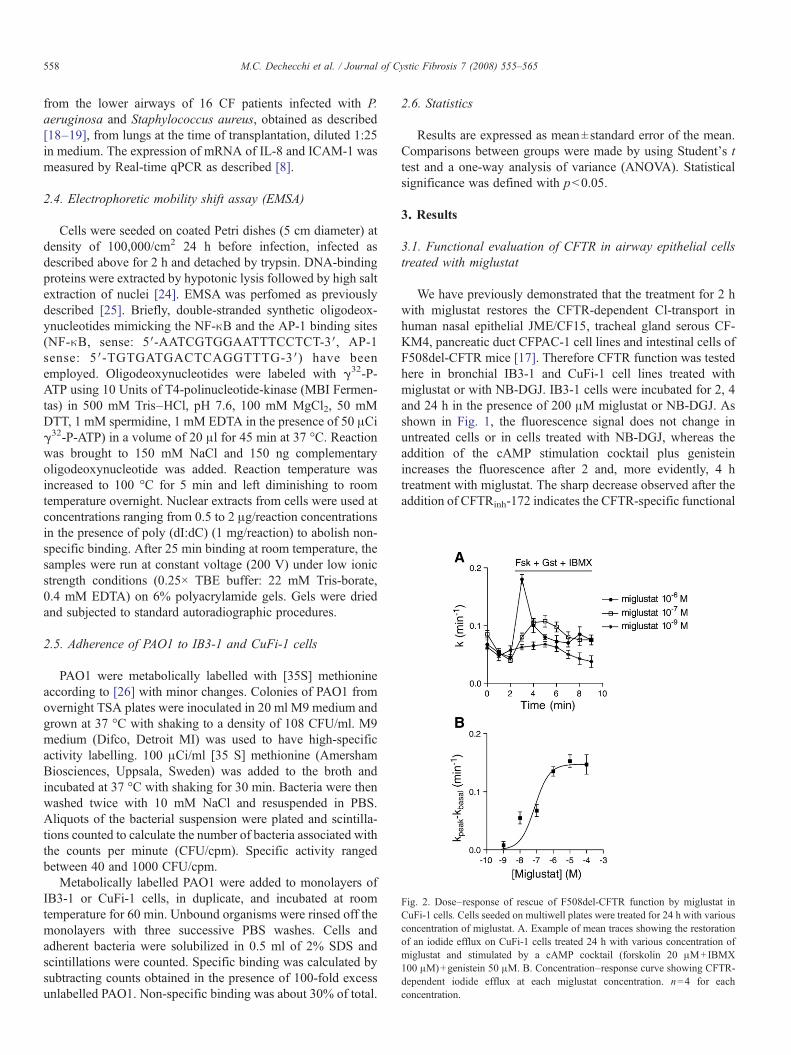
Fig. 2. Dose–response of rescue of F508del-CFTR function by miglustat inCuFi-1 cells. Cells seeded on multiwell plates were treated for 24 h with variousconcentration of miglustat. A. Example of mean traces showing the restorationof an iodide efflux on CuFi-1 cells treated 24 h with various concentration ofmiglustat and stimulated by a cAMP cocktail (forskolin 20 μM+IBMX100 μM)+genistein 50 μM. B. Concentration–response curve showing CFTR-dependent iodide efflux at each miglustat concentration. n=4 for eachconcentration.
558 M.C. Dechecchi et al. / Journal of Cystic Fibrosis 7 (2008) 555–565
from the lower airways of 16 CF patients infected with P.aeruginosa and Staphylococcus aureus, obtained as described[18–19], from lungs at the time of transplantation, diluted 1:25in medium. The expression of mRNA of IL-8 and ICAM-1 wasmeasured by Real-time qPCR as described [8].
2.4. Electrophoretic mobility shift assay (EMSA)
Cells were seeded on coated Petri dishes (5 cm diameter) atdensity of 100,000/cm2 24 h before infection, infected asdescribed above for 2 h and detached by trypsin. DNA-bindingproteins were extracted by hypotonic lysis followed by high saltextraction of nuclei [24]. EMSA was perfomed as previouslydescribed [25]. Briefly, double-stranded synthetic oligodeox-ynucleotides mimicking the NF-κB and the AP-1 binding sites(NF-κB, sense: 5′-AATCGTGGAATTTCCTCT-3′, AP-1sense: 5′-TGTGATGACTCAGGTTTG-3′) have beenemployed. Oligodeoxynucleotides were labeled with γ32-P-ATP using 10 Units of T4-polinucleotide-kinase (MBI Fermen-tas) in 500 mM Tris–HCl, pH 7.6, 100 mM MgCl2, 50 mMDTT, 1 mM spermidine, 1 mM EDTA in the presence of 50 μCiγ32-P-ATP) in a volume of 20 μl for 45 min at 37 °C. Reactionwas brought to 150 mM NaCl and 150 ng complementaryoligodeoxynucleotide was added. Reaction temperature wasincreased to 100 °C for 5 min and left diminishing to roomtemperature overnight. Nuclear extracts from cells were used atconcentrations ranging from 0.5 to 2 μg/reaction concentrationsin the presence of poly (dI:dC) (1 mg/reaction) to abolish non-specific binding. After 25 min binding at room temperature, thesamples were run at constant voltage (200 V) under low ionicstrength conditions (0.25× TBE buffer: 22 mM Tris-borate,0.4 mM EDTA) on 6% polyacrylamide gels. Gels were driedand subjected to standard autoradiographic procedures.
2.5. Adherence of PAO1 to IB3-1 and CuFi-1 cells
PAO1 were metabolically labelled with [35S] methionineaccording to [26] with minor changes. Colonies of PAO1 fromovernight TSA plates were inoculated in 20 ml M9 medium andgrown at 37 °C with shaking to a density of 108 CFU/ml. M9medium (Difco, Detroit MI) was used to have high-specificactivity labelling. 100 μCi/ml [35 S] methionine (AmershamBiosciences, Uppsala, Sweden) was added to the broth andincubated at 37 °C with shaking for 30 min. Bacteria were thenwashed twice with 10 mM NaCl and resuspended in PBS.Aliquots of the bacterial suspension were plated and scintilla-tions counted to calculate the number of bacteria associated withthe counts per minute (CFU/cpm). Specific activity rangedbetween 40 and 1000 CFU/cpm.
Metabolically labelled PAO1 were added to monolayers ofIB3-1 or CuFi-1 cells, in duplicate, and incubated at roomtemperature for 60 min. Unbound organisms were rinsed off themonolayers with three successive PBS washes. Cells andadherent bacteria were solubilized in 0.5 ml of 2% SDS andscintillations were counted. Specific binding was calculated bysubtracting counts obtained in the presence of 100-fold excessunlabelled PAO1. Non-specific binding was about 30% of total.
2.6. Statistics
Results are expressed as mean±standard error of the mean.Comparisons between groups were made by using Student's ttest and a one-way analysis of variance (ANOVA). Statisticalsignificance was defined with pb0.05.
3. Results
3.1. Functional evaluation of CFTR in airway epithelial cellstreated with miglustat
We have previously demonstrated that the treatment for 2 hwith miglustat restores the CFTR-dependent Cl-transport inhuman nasal epithelial JME/CF15, tracheal gland serous CF-KM4, pancreatic duct CFPAC-1 cell lines and intestinal cells ofF508del-CFTR mice [17]. Therefore CFTR function was testedhere in bronchial IB3-1 and CuFi-1 cell lines treated withmiglustat or with NB-DGJ. IB3-1 cells were incubated for 2, 4and 24 h in the presence of 200 μM miglustat or NB-DGJ. Asshown in Fig. 1, the fluorescence signal does not change inuntreated cells or in cells treated with NB-DGJ, whereas theaddition of the cAMP stimulation cocktail plus genisteinincreases the fluorescence after 2 and, more evidently, 4 htreatment with miglustat. The sharp decrease observed after theaddition of CFTRinh-172 indicates the CFTR-specific functional

Fig. 3. Miglustat reduces the expression of mRNA of IL-8 and ICAM-1, stimulated by PAO1 in IB3-1 and CuFi-1 cells. Cells were incubated for 24 h in the presence of200 μM miglustat and infected with PAO1 (50 CFU/cell) for 4 h as described in the Materials and methods section. The mRNA induction (relative to non-infected cells) is obtained by comparing the ratio IL-8/GAPDH (A, C) and ICAM-1/GAPDH (B,D) between non-infected and infected cells. Results are expressed asmean±SEM of duplicate wells. Representative of 9 independents experiments (A) or 6 (B, C and D) in duplicate are shown.
Fig. 4. Miglustat inhibits the inflammatory response to SMM in IB3-1 cells.Cells were incubated for 24 h in the presence of 200 μM miglustat and thenstimulated by SMM diluted 1:25 in medium, for 4 h. The IL-8 mRNA induction(relative to not infected cells) is obtained as indicated in the legend of Fig. 3.Results are expressed as mean±SEM of duplicate wells. Representative of 2independent experiments, in duplicate, is shown.
559M.C. Dechecchi et al. / Journal of Cystic Fibrosis 7 (2008) 555–565
activation after treatment with miglustat. It should be noted thatno correction is observed after 24 h of treatment with miglustat(Fig. 1B). The results obtained in CuFi-1 cells are reported inFig. 1C. In agreement with IB3-1 cells, no changes of thefluorescent signal are obtained in untreated cells or in cellstreated with NB-DGJ. As demonstrated by the increase of thefluorescence after the addition of the cAMP stimulation cocktailplus genistein and by the decrease after the addition of theinhibitor, a correction of F508del-CFTR function is observedafter 2, 4 and more evident, 24 h of treatment. Collectively,these results indicate a fast rescue of F508del-CFTR functionboth in IB3-1 and CuFi-1 CF cells.
In order to evaluate the half maximal effective concentrationof miglustat, CFTR-dependent Cl-transport was studied inCuFi-1 cells by iodide efflux. Cells were incubated in thepresence of miglustat ranging from 1 nM to 100 μM and iodideefflux was performed as described above. Fig. 2A shows thecorrection of CFTR function by miglustat in CuFi-1 cellsstarting from 10 nM, being 76 nM the half maximal effectiveconcentration (Fig. 2B).

560 M.C. Dechecchi et al. / Journal of Cystic Fibrosis 7 (2008) 555–565
3.2. Effect of miglustat on the expression of inflammatorymediators in IB3-1 cells and CuFi-1 cells stimulated by PAO1
The principal aim of this study was to evaluate the effect ofcorrectors of F508del-CFTR function on the modulation of theexpression of pro-inflammatory mediators, induced by P.aeruginosa infection in respiratory epithelial cells. IL-8 is anessential component of the host defense system, being releasedfrom respiratory epithelial cells in response to bacteria, toregulate transepithelial migration and activation of neutrophils[27]. ICAM-1 is constitutively expressed on the cell surface of awide variety of cells and it is modulated in response to different
Fig. 5. A. Increase of NF-κB and AP-1 binding activity following PAO1 infection. Nuamounts (μg/reactions) of nuclear proteins were mixed with 32P-labelled NF-κB and Agel-shift. Asterisks indicate free 32P-labelled NF-κB and AP-1 probes. B,C. Miglustatcultured in the absence (upper gels in each panel) or in the presence (lower gels in eachPAO1 for 2 h, as indicated. Nuclear extracts from these treated cells, as indicated, weκBNF-kB (B) and AP-1 (C) target 32P-labelled oligodeoxynucleotides and EMSA perinteractions obtained using 0.5 μg /reaction nuclear factors, performed using the Bbinding activities relative to control protein extracts isolated from untreated contromiglustat, no PAO1; grey bars: miglustat; black bars: PAO1; dashed bars: PAO1+m
inflammatory stimuli, including bacteria [28]. As we havepreviously demonstrated that overnight incubation of CFbronchial cells with the corrector MPB-07 down-modulatesboth the mRNA levels and protein expression of IL-8 andICAM-1, stimulated by PAO1 [8], the effect of miglustat wasstudied in the same experimental model in IB3-1 and CuFi-1cells using quantitative real-time RT-PCR as analyticalmethodology. Miglustat significantly inhibits the PAO1 inducedaccumulation of both IL-8 (pb0.0001) and ICAM-1(pb0.0009)mRNAs in IB3-1 cells (Fig. 3A and B). Also in CuFi-1 cellsmiglustat significantly inhibits IL-8 (pb0.003) and ICAM-1(pb0.03) mRNA accumulation (Fig. 3C and D). Considering
clear extracts were isolated from uninfected and PAO1 infected cells. IncreasingP-1 target oligonucleotides and protein/DNA complexes (arrowed) analysed by
has no major effect on NF-κB and AP-1 overall binding activity. IB3-1 cells werepanel) of 200 μMmiglustat for 24 h and then infected or not with 50 CFU/cell ofre isolated and increasing amounts of them (μg/reactions) were mixed with NF-formed. In the right side of panels B and C quantitative analysis of DNA–proteinio-Rad Gel-Doc densitometry system is shown. The results reported representl cells (−) (average±SD from three independent experiments). Open bars=noiglustat.

Fig. 6. Both miglustat and NB-DGJ reduce the PAO1 stimulated IL-8 mRNAexpression in CF and non-CF bronchial cells. Cells were incubated with miglustatorNB-DGJ (200μM) for 24h as described in Fig. 3. ThemRNA induction (relativeto non-infected cells) is obtained by comparing the ratio IL-8/GAPDH betweennon-infected and infected cells. Results are expressed as mean±SEM of duplicatewells. Representative of 3(A), 4(B) and 5(C) independent experiments in duplicateare shown.
561M.C. Dechecchi et al. / Journal of Cystic Fibrosis 7 (2008) 555–565
that chronic lung infection in the airways of CF patients issustained in the advanced stages by non-motile P. aeruginosagrowing in biofilms, we tested the effect of a potent pro-inflammatory stimulus deriving from the bronchial lumina ofCF patients undergoing lung transplantation [18–19]. Pooledsupernatants of mucopurulent material (SMM), obtained fromhuman CF lungs infected with P. aeruginosa and S. aureus[18–19], were tested in IB3-1 cells upon treatment withmiglustat. The significant inhibition of the expression of IL-8stimulated by SMM, shown in Fig. 4, strengthens the potentialanti-inflammatory effect of miglustat in reducing the P.aeruginosa-dependent transcription of two critical genesinvolved in leukocyte chemotaxis, such as IL-8 and ICAM-1.
3.3. DNA-binding activity of NF-κB and AP-1 by P. aeruginosain cells treated with miglustat
The inhibitory effect observed on IL-8 and ICAM-1transcription in CF airway epithelial cell lines opens thepossibility that miglustat could intervene in the signallingcascade between the receptors recognized by P. aeruginosa andthe transcription of IL-8 and ICAM-1, besides correctingF508del-CFTR. Attention was therefore drawn on the tran-scription factors NF-κBNF-kB and AP-1, which play a centralrole in the immune response to P. aeruginosa infection inrespiratory epithelial cells [29] and have already demonstratedto be activated in IB3-1 and CuFi-1 cells upon infection withPAO1 [8]; accordingly, putative target consensus sequences ofthese two transcription factors have been found within thepromoter sequences of both IL-8 and ICAM-1 genes (TESSsoftware analysis) (www.cbill.upenn.edu/cgi_bin/tess/tess). Inorder to determine whether miglustat treatment modulates theoverall NF-κBNF-kB and AP-1 binding activity the experimentshown in Fig. 5 was performed. IB3-1 cells were cultured in theabsence or in the presence of miglustat and then infected or notwith PAO1 for 2 h. Nuclear extracts were isolated, increasingamounts of them were mixed with NF-κBNF-kB and AP-1target 32P-labelled oligodeoxynucleotides and EMSA wasperformed. The results clearly allow these conclusions:(a) both NF-κBNF-kB and AP-1 binding activity significantlyincreases following P. aeruginosa infection of IB3-1 cells(pb0.05) (see data reported in Fig. 5A) and (b) no significantmajor alteration of this binding activity occurs in bothuninfected or PAO1 infected cells treated with miglustat (seedata reported in Fig. 5, B and C). These results wereconsistently obtained in three independent experiments. There-fore miglustat has no major effect on overall binding activity ofthe transcription factors NF-κBNF-kB and AP-1.
3.4. Effect of NB-DGJ on PAO1 stimulated IL-8 MRNAexpression in IB3-1, CuFi-1 and NuLi-1 cells
To verify whether the anti-inflammatory effect of miglustatobserved in IB3-1 and CuFi-1 cells might be related to thecorrection of F508del-CFTR function, cells were also treatedwith NB-DGJ, which does not restore CFTR-dependent Cl-transport (Fig. 1). As shown in Fig. 6A and B, also NB-DGJ
significantly reduces the expression of IL-8 mRNA stimulatedby PAO1 in IB3-1 (pb0.003) and CuFi-1 cells (pb0.04). Theseexperiments were extended to NuLi-1 cells, which express wildtype CFTR protein. As shown in Fig. 6C miglustat significantlydecreases the amount of IL-8 mRNA also in NuLi-1 cells(pb0.02). Moreover, NB-DGJ produces a significant inhibitionof the transcription of IL-8 stimulated by PAO1 (pb0.04)although to a lower extent of that obtained with miglustat(pb0.03). On the whole, these results indicate that the anti-

Fig. 8. Miglustat and NB-DGJ reduce the expression of IL-8 mRNA stimulatedby PAO1, or TNF alpha or IL-1 beta in IB3-1 and CuFi-1 cell lines. Cells wereincubated for 24 h with miglustat or NB-DGJ (200 μM) and then infected withPAO1 as above described or stimulated with TNF alpha (10 ng/ml) or IL-1 beta(50 ng/ml) for 4 h. Stimulated IL-8 mRNA expression was calculated asindicated in the legend of Fig. 3 and it is expressed as % of untreated cells. A. Inuntreated IB3-1 cells the stimulated IL-8 mRNA expression was 407±80 byPAO1, 51±6 by TNF alpha and 148±15 by IL-1 beta (n=4). B. In untreatedCuFi-1 cells the stimulated IL-8 mRNA expression was 52±13 by PAO1, 6±1by TNF alpha and 9±1 by IL-1 beta (n=4). Representative of 2 independentexperiments in duplicate are shown.
Fig. 7. Miglustat does not reduce the binding of PAO1 to IB3-1 and CuFi-1 cells.500,000 IB3-1 (A) or CuFi-1 (B) cells on Petri dishes, in duplicate, were treatedfor 24 h with 200 μM miglustat and then washed with PBS before performingadhesion experiments. Different amounts of 35S-PAO1, expressed as CFU/well,were added to the wells and incubated as described in Materials and methodssection. Data reported in the figure are the specific binding calculated bysubtracting counts obtained in the presence of 100-fold excess of non-labelledPAO1 and are expressed as CFU/well. Representative of two independentexperiments are reported in the figure.
562 M.C. Dechecchi et al. / Journal of Cystic Fibrosis 7 (2008) 555–565
inflammatory action of miglustat is independent of thecorrection of F508del-CFTR in IB3-1 and CuFi-1 cells.
3.5. Adherence of PAO1 to IB3-1 and CuFi-1 cells treated withmiglustat
Many pathogenic microorganisms use glycoconiugatereceptors to establish contact with the host tissues [30] andpharmacological agents that inhibit the biosynthesis of thesereceptors may have a major impact on the pathogenesis ofinfection. Miglustat was shown to decrease the GSL content, toinhibit the P-fimbriated bacterial attachment and, as aconsequence, to impair the mucosal inflammatory response inmice [31]. P. aeruginosa flagellin can interact with both Toll-like receptor 5 (TLR5) and the cell surface glycolipid,asialoGM1, to activate the innate immune response [29].Therefore the anti-inflammatory effect of miglustat observed inIB3-1 and CuFi-1 cells could reflect the reduced expression ofthe glycolipid receptors for PAO1. To evaluate this possibilitythe adherence of metabolically labelled [35S] methionine-PAO1was measured in IB3-1 and CuFi-1 cells, treated or not withmiglustat. Fig. 7A and B show a dose dependent increase of the
PAO1 binding to the cells and no significant differences due tothe treatment with miglustat. These results indicate thatincubation with miglustat for 24 h does not affect the adherenceof PAO1 to IB3-1 and CuFi-1 cells, suggesting that the hugeinhibition of P. aeruginosa-dependent IL-8 and ICAM-1transcription after treatment with miglustat is not dependentupon reduction of bacterial-host cell interactions.
3.6. Effect of miglustat and NB-DGJ on the expression of IL-8mRNA stimulated by PAO1, TNF alpha or IL-1 beta in IB3-1and CuFi-1 cells
The results shown so far indicate that miglustat and NB-DGJhave an anti-inflammatory effect in bronchial epithelial cells,suggesting that they could inhibit the transmembrane signallingbetween the receptors for pathogens and the transcription of theinflammatory genes. To obtain preliminary insights on whichtransduction pathway is inhibited by miglustat and NB-DGJ, theexpression of IL-8 mRNA stimulated by either PAO1 or the pro-inflammatory cytokines TNF alpha and IL-1 beta, was studiedboth in IB3-1 and in CuFi-1 cells. As depicted in Fig. 8A,miglustat and NB-DGJ significantly inhibit the expression of

563M.C. Dechecchi et al. / Journal of Cystic Fibrosis 7 (2008) 555–565
IL-8 mRNA not only after stimulation by PAO1 but also byTNF alpha or IL-1 beta , in IB3-1 cells. Fig. 8B confirms thesefindings also in CuFi-1 cells. Interestingly, miglustat produces asignificantly stronger reduction of IL-1 beta elicited IL-8mRNA expression than that given by NB-DGJ, in IB3-1 andCuFi-1 cells, whereas no differences are observed both uponinfection with PAO1 and induction by TNF alpha. Therefore,miglustat and NB-DGJ inhibit the pro-inflammatory signallingdownstream the receptors for PAO1 and for these pro-inflammatory cytokines.
4. Conclusions
We recently demonstrated that miglustat, an inhibitor of theER alpha-1,2-glucosidase, restores F508del-CFTR function inhuman nasal epithelial JME/CF15 cells by preventing theinteraction between F508del-CFTR and calnexin [17]. Foldingand trafficking of proteins to their correct cellular locationdepend on molecular chaperones which can have differenteffects on each particular substrate and particular cell type [32].As a consequence, the effectiveness of small correctors ofprotein trafficking defect may depend on the cellular environ-ment and may be cell type specific. The rescue of CFTRfunction by miglustat has been observed in different cells, suchas nasal JME/CF15, tracheal gland serous CFKM4, pancreaticduct CFPAC-1 cell lines, intestinal cells of F508del-CFTR mice[17]. Here we report a correcting effect in the CF bronchial IB3-1 and CuFi-1 cells (Figs. 1 and 2), although we observed a lackof rescue of CFTR function in IB3-1 cells treated with miglustatfor 24 h (Fig. 1B). Experience from the newly discoveredF508del-CFTR correctors starts indicating that the potency ofaction can be quite different, or sometimes even absent, whentested in different cell models [32]. In respect to miglustat, itshould be recalled that it corrects F508del-CFTR by inhibitingthe α-glucosidase of the ER [17], an effect which can vary uponthe cells line model and the culture conditions. Moreover, CuFi-1 cells are homozygous for the F508del-CFTR mutationwhereas IB3-1 cells are compound heterozygous for theF508del-CFTR mutation (genotype F508del/W1282X). As theamount of F508del proteins in IB3-1 cells may be quite differentcompared to that of CuFi-1 cells this may also contribute to thevariability of the correcting effect, which anyway requiresfurther investigation.
In this study we have shown that miglustat: i) is a corrector ofF508del-CFTR function in CF bronchial IB3-1 and CuFi-1cells; ii) has an anti-inflammatory effect in bronchial cellsindependently of the correction of F508del-CFTR; iii) has noeffect on NF-κB and AP1 overall binding activity; iv) inhibitsthe pro-inflammatory signalling downstream both the receptorsfor pathogens and key pro-inflammatory cytokines.
Therefore, besides confirming the correction of F508del-CFTR function, we have demonstrated that miglustat reducesthe inflammatory response elicited by P. aeruginosa or SMM inCF bronchial IB3-1 and CuFi-1 cells (Figs. 3 and 4), supportingthe hypothesis that the pharmacological modulation of CFTRdefect may reduce the excessive lung inflammatory response inCF cells, consistent with previous findings showing an anti-
inflammatory effect of the corrector MPB-07 [8]. However, alsothe galactose analog of miglustat, NB-DGJ, which is not acorrector of F508del-CFTR function, reduces the inflammatoryresponse in CF cells and both the compounds have an anti-inflammatory effect in non-CF bronchial NuLi-1 cells (Fig. 6).Therefore, althoughmiglustat rescues functional F508del-CFTRin CF bronchial cells, it affects the inflammatory response to P.aeruginosa through a mechanism which, at least partly, isindependent of the correction of F508del-CFTR function.
How this occurs is a matter of speculation. Both analogues,miglustat and NB-DGJ, are inhibitors of the first step in GSLbiosynthesis which is the transfer of glucose to ceramide by theceramide-specific glucosyl-transferase (CerGlcT) [30], whereasmiglustat, but not NB-DGJ, also inhibits ER alpha-glucosidase[16]. The fact that correction of F508del-CFTR function isobtained in cells treated with miglustat but not with NB-DGJ,supports that the correcting effect is specifically related to ERalpha-glucosidase inhibition, thus preventing the interaction ofF508del-CFTR with calnexin [17]. On the other hand the anti-inflammatory effect observed in cells treated with either miglustator NB-DGJ suggests a mechanism involving the inhibition ofCerGlcT, an activity shared by both compounds. Nevertheless,depending on cell type and pro-inflammatory stimulus, miglustatseems to be more effective than NB-DGJ (Figs. 3C and 8). Thus,also ER alpha-glucosidase could play a role in pro-inflammatorysignaling. CerGlcT is crucial for the synthesis of GSL and it is thetarget for treating GSL lysosomal storage disease in thetherapeutic approach termed substrate reduction therapy [16].Gaucher disease is the most frequently occurring GSL storagedisease, characterized by accumulation of glucosylceramidecausing heterogeneous clinical features, among which sustainedinflammatory reaction associated with elevated levels of pro-inflammatory cytokines [33]. It has been suggested that GSLmayact as signalling molecule to dysregulate the immune system inGaucher disease [34]. Interestingly, mouse models of GSLstorage diseases treated by substrate reduction therapy withmiglustat, display a marked reduction in systemic inflammation[35], strengthening the link between the accumulation ofGLS andthe extent of the inflammatory response. Very importantly, theaccumulation of the sphingolipid ceramide has been recentlyidentified as one of the key regulators of inflammation andinfection in CF airways [36]. We can thus argue that miglustatreduces the inflammatory response by interfering with ceramidesignalling. The transcription factors NF-κBNF-kB and AP-1 playa central role in the immune response toP. aeruginosa infection inrespiratory epithelial cells [29]. Interestingly, an alternativepathway, independent of NF-κB, operating through prostaglandinE2 receptor, has been demonstrated to regulate IL-8 secretion inIB3-1 cells [37]. A likely inhibition of the overall binding activityof NF-κBNF-kB and AP-1 by miglustat has been ruled out fromthe data reported in Fig. 5, suggesting that it could interfere withother pathways in signal transduction. Future studies on thepotential effect of miglustat on the complex array of transcriptionfactors regulating IL-8, ICAM-1 and other genes of the innateimmune response are needed to ascertain this point.
The anti-inflammatory effect of miglustat and NB-DGJcould reflect the reduced expression of the glycolipid receptors

564 M.C. Dechecchi et al. / Journal of Cystic Fibrosis 7 (2008) 555–565
for P. aeruginosa due to a decrease in the cellular GSL contentas a consequence of the inhibition of CerGlcT activity. Here weshow that miglustat does not reduce the adherence of P.aeruginosa to the IB3-1 cells (Fig. 7), thus making unlikely thatthe anti-inflammatory effect of miglustat is due to interferencewith the receptors for P. aeruginosa. Moreover, since miglustatstrongly reduces the pro-inflammatory signals elicited also byTNF alpha and IL-1 beta (Fig. 8), these compounds couldpossibly modulate directly or indirectly the activity of some ofthe different kinases or adapters downstream the receptors forpathogens, in common with TNF alpha and IL-1 beta receptorsthrough a mechanism that needs further investigation. From thepractical point of view, the results presented in this paper showthat miglustat has two therapeutically relevant effects in CFcells: it rescues the F508del-CFTR function and reduces theinflammatory response to P. aeruginosa. Interestingly, bothcorrection of the cystic fibrosis defect and inhibition of anti-inflammatory responses are of great importance in the therapyof CF patients. Accordingly, a drug displaying these two effectsdeserves great attention. Furthermore, the effects of miglustaton human subjects are well known. In fact, miglustat is an orallybioavailable drug approved in Europe and USA for thetreatment of Gaucher disease and other GSL storage diseases.Human studies show that it is well tolerated at 100 or 300 mgonce or three times daily [38]. The pharmacokinetic propertiesof miglustat, studied at the dosage used in the treatment of type1 Gaucher's disease [15] indicate that the drug is rapidlyabsorbed after oral administration, with a peak plasmaconcentration of 0.86 μM/mL at 2.5 h. In clinical trials, for adosage of 100 mg three times daily, steady-state plasmaconcentration of 1.5 μM/mL was attained within 4–6 weeks[15,39]. Although studies in murine models indicate that lungtissue concentrations reach equimolar levels with those inplasma [40], to the best of our knowledge no data on humanlung tissue concentrations have been studied after oraltreatment. Therefore, further investigation in animal modelswill be necessary to start understanding the effective anti-inflammatory concentration in vivo. Moreover, analysis ofmarkers of inflammation in CF patients undergoing clinicaltrials with oral miglustat as a CFTR corrector (http://clinicaltrials.gov/) will likely help elucidating this importantissue, which is difficult to predict simply from in vitro studies.
All this considered, miglustat may represent a promisingcandidate for the pharmacotherapy of CF and an interestingresearch tool to investigate the immune response of airwaycells.
5. Conflict of interest statement
All authors disclose any financial and personal relationshipswith other people or organizations that could inappropriatelyinfluence this work.
Acknowledgements
We are grateful to A. Tamanini for the helpful discussions, A.Prince for the P. aeruginosa laboratory strain PAO1, M.G. Giri
for the statistical analysis, F. Quiri for excellent technicalassistance, In Vitro Model and Cell Culture Care of theUniversity of Iowa for providing NuLi-1 and CuFi-1 cells andA.S. Verkman for CFTRinh-172.
This research was supported by the Italian Cystic FibrosisResearch Foundation (grant FFC # 16/2006) with the contribu-tion of “Associazione Veneta per la lotta alla Fibrosi Cistica”,Fondazione Cariverona— Bando 2005 — Malattie rare e dellapovertà (to GC) and by Vaincre la Mucoviscidose (to CN andFB).
References
[1] Govan JRW, Deretic V. Microbial pathogenesis in cystic fibrosis: mucoidPseudomonas aeruginosa and Burkholderia cepacia. Microbiol Rev1996;60:539–74.
[2] Bonfield TL, Panuska JR, Konstan MW, Hilliard JB, Hilliard JB, GhnaimH, et al. Inflammatory cytokines in cystic fibrosis lungs. Am J Respir CritCare Med 1995;152:2111–8.
[3] Dakin CJ, Numa AH, Wang H, Morton JR, Vertzyas CC, Henry RL.Inflammation, infection and pulmonary function in infants and youngchildren with cystic fibrosis. Am J Respir Crit Care Med 2002;165:904–10.
[4] Matsui H, Grubb BR, Tarran R, Randell SH, Gatzy JT, Davis CW, et al.Evidence for periciliary liquid layer depletion, not abnormal ioncomposition, in the pathogenesis of cystic fibrosis airway disease. Cell1998;95:1005–15.
[5] Machen TE. Innate immune response in CF airway epithelia: hyperin-flammatory? Am J Physiol Cell Physiol 2006;291:C218–30.
[6] Ma T, Vetrivel L, Yang H, Pedemonte N, Zegarra-Moran O, Galietta LJV,et al. High-affinity activators of cystic fibrosis transmembrane conductanceregulator (CFTR) chloride conductance identified by throughput screen-ing. J Biol Chem 2002;277:37235–41.
[7] Perez A, Issler AC, Cotton CU, Kelley TJ, Verkman AS, Davis PB. CFTRinhibition mimics the cystic fibrosis inflammatory profile. Am J PhysiolLung Cell Mol Physiol 2007;292:L383–95.
[8] Dechecchi MC, Nicolis E, Bezzerri V, Vella A, Colombatti M, Assael BM,et al. MPB-07 reduces the inflammatory response to Pseudomonasaeruginosa in cystic fibrosis bronchial cells. Am J Respir Cell Mol Biol2007;36:615–24.
[9] Pedemonte N, Diena T, Caci E, Nieddu E, Mazzei M, Ravazzolo R, et al.Antihypertensive 1,4-dihydropyridines as correctors of the cystic fibrosistransmembrane conductance regulator channel gating defect caused bycystic fibrosis mutations. Mol Pharmacol 2005;68:1736–46.
[10] Van Goor F, Straley KS, Cao D, Gonzàles J, Hadida S, Hazlewood, et al.Rescue of ΔF508 CFTR trafficking and gating in human cystic fibrosisairway primary cultures by small molecules. Am J Physiol Lung Cell MolPhysiol 2006;290:L1117–30.
[11] Pedemonte N, Lukacs GL, Du K, Caci E, Zegarra-Moran O, Galietta LJV, etal. Small-molecule correctors of defective DF508-CFTR cellular processingidentified by high-throughput screening. J Clin Invest 2005;115:2564–71.
[12] Wang Y, Bartlett MC, Loo TW, Clarke DM. Specific rescue of the cysticfibrosis transmembrane conductance regulator processing mutants usingpharmacological chaperones. Mol Pharmacol 2006;70:297–302.
[13] Asano N. Glycosidase inhibitors: update and perspectives on practical use.Glycobiology 2003;13:93R–104R.
[14] Platt FM, Neises GR, Dwek RA, Butters TD. N-butyldeoxynojirimycin is anovel inhibitor of glycolipid biosynthesis. J Biol Chem 1994;269:8362–5.
[15] Cox T, Lachmann R, Hollak C, Aerts J, van Weely S, Hrebicek M, et al.Novel oral treatment of Gaucher's disease with N-butyldeoxynojirimycin(OGT 918) to decrease substrate biosynthesis. Lancet 2000;355:1481–5.
[16] Butters TD, Dwek RA, Platt FM. Imino sugar inhibitors for treating thelysosomal Glycoshingolipidoses. Glycobiology 2005;15:43R–52R.
[17] Norez C, Noel S, Wilke M, Bijvelds M, Jorna H, Melin P, et al. Rescue offunctional delF508-CFTR channels in cystic fibrosis epithelial cells by theα-glucosidase inhibitor miglustat. FEBS Lett 2006;580:2081–6.

565M.C. Dechecchi et al. / Journal of Cystic Fibrosis 7 (2008) 555–565
[18] Ribeiro CMP, Paradiso AM, Carew MA, Shears SB, Boucher RC. Cysticfibrosis airway epithelial Ca2+i signaling. The mechanism for the largeragonist-mediated Ca2+i signals in human cystic fibrosis airway epithelia. JBiol Chem 2005;280:10202–9.
[19] Ribeiro CMP, Paradiso AM, Schwab U, Perez-Vilar J, Jones L, O'Neal W,et al. Chronic airway infection/inflammation induces a Ca2+i-dependenthyperinflammatory response in human cystic fibrosis airway epithelia. JBiol Chem 2005;280:17798–806.
[20] Zeitlin PL, Lu L, Rhim J, Cutting G, Stetten G, Kieffer KA, et al. A cysticfibrosis bronchial epithelial cell line: immortalization by adeno-12-SV40infection. Am J Respir Cell Mol Biol 1991;4:313–9.
[21] Zabner J, Karp P, Seiler M, Phillips Sl, Mitchell CJ, Saavedra M, et al.Development of cystic fibrosis and non-cystic fibrosis airway cell lines.Am J Physiol Lung Cell Mol Physiol 2003;284:L844–54.
[22] Ma T, Thiagarajah JR, Yang H, Sonawane ND, Folli C, Galietta LJV, et al.Thiazolidinone CFTR inhibitor identified by high-throughput screeningblocks cholera-toxin induced intestinal fluid secretion. J Clin Invest2002;110:1651–8.
[23] Norez C, Antigny F, Becq F, Vandebrouck C. Maintaining low Ca2+ levelin the endoplasmic reticulum restores abnormal endogenous F508del-CFTR trafficking in airway epithelial cells. Traffic 2006;7:562–73.
[24] Andrews NC, Faller DV. A rapid micropreparation technique for extractionof DNA-binding proteins from limiting numbers of mammalian cells.Nucleic Acids Res 1991;19:2499.
[25] Borgatti M, Lampronti I, Romanelli A, Pedone C, Saviano M, Bianchi N,et al. Transcription factor decoy molecules based on a peptide nucleic acid(PNA)–DNA chimera mimicking Sp1 binding sites. J BiolChem2003;278:7500–9.
[26] Saiman L, Prince A. Pseudomonas aeruginosa pili bind to asialoGM1which is increased on the surface of cystic fibrosis epithelial cells. J ClinInvest 1993;92:1875–1880.25.
[27] Diamond G, Legarda D, Ryan LK. The innate immune response of therespiratory epithelium. Immunol Rev 2000;173:27–38.
[28] Roebuck KA, Finnegan A. Regulation of intercellular adhesion molecule-1(CD54) gene expression. J Leukoc Biol 1999;66:876–88.
[29] McNamara N, Gallup M, Sucher A, Maltseva I, McKemy D, Bausbaum C.AsialoG and TLR5 cooperate in flagellin-induced nucleotide signalling toactivate Erk1/2. Am J Respir Cell Mol Biol 2006;34:653–60.
[30] Dwek RA, Butters TD, Platt FM, Zitzmann N. Targeting glycosilation as atherapeutic approach. Nat Rev Drug Discov 2002;1:65–75.
[31] Svensson M, Frendeus B, Butters T, Platt F, Dwek R, Svanborg C.Glycolipid depletion in antimicrobial therapy. Mol Microbiol2003;47:453–61.
[32] Amaral M. Therapy through chaperones: sense or antisense? Cysticfibrosis as a model disease. J Inherit Metab Dis 2006;29:477–87.
[33] Beutler E, Grabowski GA. Gaucher disease. In: Scriver CR, Beaudet AL,Sly WS, Valle D, editors. The metabolic and molecular bases of inheriteddisease. New York, New York, USA: McGraw-Hill; 2001. p. 3635–68.
[34] Mizukami H, Mi Y, Wada R, Kono M, Yamashita T, Liu Y, et al. Systemicinflammation in glucocerebrosidase-deficient mice with minimal gluco-sylceramide storage. J Clin Invest 2002;109:1215–21.
[35] Jeyakumar M, Thomas R, Elliot-Smith E, van der Spoel AC, d'Azzo A,Hugh Perry V, et al. Central nervous system inflammation is a hallmark ofpathogenesis in mouse models of GM1 and GM2 gangliosidosis. Brain2003;126:974–87.
[36] Teichgraber V, et al. Ceramide accumulation mediates inflammation, celldeath and infection susceptibility in cystic fibrosis. NatMed 2008;14:382–91.
[37] Vij N, Amoako MO, Mazur S, Zeitlin PL. CHOP transcription factormediates IL-8 signaling in cystic fibrosis bronchial epithelial cells. Am JRespir Cell Mol Biol 2008;38:176–84.
[38] Cox TM, et al. Advisory Council to the European Working Group onGaucher disease. J Inherit Metab Dis 2003;26:513–26.
[39] McCormack PL, Goa KL. Miglustat. Drugs 2003;63:2427–34.[40] Treiber A, Morand O, Clozel M. The pharmacokinetics and tissue
distribution of the glucosylceramide synthase inhibitor miglustat in the rat.Xenobiotica 2007;37:298–314.