anthrax, matrix biology, and angiogenesis: capillary
TRANSCRIPT

Brigham Young UniversityBYU ScholarsArchive
All Theses and Dissertations
2017-06-01
Anthrax, Matrix Biology, and Angiogenesis:Capillary Morphogenesis Gene 2 MediatesActivity and Uptake of Type IV Collagen-DerivedAnti-Angiogenic PeptidesJordan Grant FinnellBrigham Young University
Follow this and additional works at: https://scholarsarchive.byu.edu/etd
Part of the Biochemistry Commons
This Thesis is brought to you for free and open access by BYU ScholarsArchive. It has been accepted for inclusion in All Theses and Dissertations by anauthorized administrator of BYU ScholarsArchive. For more information, please contact [email protected], [email protected].
BYU ScholarsArchive CitationFinnell, Jordan Grant, "Anthrax, Matrix Biology, and Angiogenesis: Capillary Morphogenesis Gene 2 Mediates Activity and Uptake ofType IV Collagen-Derived Anti-Angiogenic Peptides" (2017). All Theses and Dissertations. 6849.https://scholarsarchive.byu.edu/etd/6849

Anthrax, Matrix Biology, and Angiogenesis: Capillary Morphogenesis Gene 2
Mediates Activity and Uptake of Type IV Collagen-Derived
Anti-Angiogenic Peptides
Jordan Grant Finnell
A thesis submitted to the faculty of Brigham Young University
in partial fulfillment of the requirements for the degree of
Master of Science
Kenneth A. Christensen, Chair Jeffery S. Tessem
Barry M. Willardson
Department of Chemistry and Biochemistry
Brigham Young University
Copyright © 2017 Jordan Grant Finnell
All Rights Reserved

ABSTRACT
Anthrax, Matrix Biology, and Angiogenesis: Capillary Morphogenesis Gene 2 Mediates Activity and Uptake of Type IV Collagen-Derived
Anti-Angiogenic Peptides
Jordan Grant Finnell Department of Chemistry and Biochemistry, BYU
Master of Science
Capillary Morphogenesis Gene 2 (CMG2) is a type I transmembrane, integrin-like receptor. It was originally identified as one of several genes upregulated during capillary formation. It was subsequently identified as one of two physiological anthrax toxin receptors, where CMG2 serves as a cell-surface receptor for anthrax toxin and mediates entry of the toxin into cells via clathrin-dependent endocytosis. Additionally, loss-of-function mutations in CMG2 cause the genetic disorder hyaline fibromatosis syndrome (HFS), where the core symptom is dysregulation of extracellular matrix homeostasis (ECM), including excessive accumulation of proteinaceous hyaline material; HFS clearly indicates that CMG2 plays an essential function in ECM homeostasis and repair. Most often, these situational roles have been evaluated as separate intellectual and experimental entities; consequently, whereas details have emerged for each respective situational role, there has been little attempt to synthesize knowledge from each situational role in order to model a holistic map of CMG2 function and mechanism of action in normal physiology. The work presented in this thesis is an example of such a synthesis. Interactions between CMG2 and type IV collagen (Col IV) were evaluated, to better understand this putative interaction and its effect on CMG2 function in angiogenesis. Using an overlapping library peptide array of the Col IV α1 and α2 chains, it was found that CMG2-binding peptides were enriched within the NC1 domains. This finding was corroborated via another epitope mapping peptide array, where we found a major epitope for CMG2-binding within the α2 NC1 domain (canstatin). Identification of CMG2 interactions with Col IV NC1 domains (including canstatin) was both surprising and intriguing, as these domains are potent endogenous inhibitors of angiogenesis. To further evaluate the physiological relevance of interactions with Col IV NC1 domains, a canstatin-derived peptide from the original array was synthesized and used for further studies. This peptide (here known as S16) binds with high affinity (KD = 440 ± 160 nM) to the extracellular, ligand-binding CMG2 vWA domain; specificity was confirmed through competition studies with anthrax toxin PA, and through demonstration of divalent cation-dependent binding. CMG2 was found to be the relevant endothelial receptor for S16. CMG2 in fact mediates endocytic uptake of peptide S16, as demonstrated by flow cytometry, and colocalization studies. S16 further inhibits migration of endothelial cells. These findings demonstrate that CMG2 is a functional receptor for Col IV NC1 domain fragments. CMG2 may exert a pro-angiogenic effect through endocytosis and clearance of anti-angiogenic NC1 domain fragments. Additionally, this is the first demonstration of CMG2-mediated uptake of an endogenous matrix fragment, and suggests a mechanism by which CMG2 regulates ECM and basement membrane homeostasis, thereby establishing a functional connection between the receptor’s role in matrix biology and angiogenesis. Keywords: angiogenesis, CMG2/ANTXR2, matrix biology, peptide array, type IV collagen

ACKNOWLEDGEMENTS
Funding for the work described in this thesis was provided by the National Institutes of
Health [grant number R21EY024734 (K.A.C.)], the BYU Department of Chemistry and
Biochemistry (K.A.C.), and the Simmons Center for Cancer Research. Gratitude is expressed,
first, to Dr. Kenneth A. Christensen for his outstanding guidance, support, and mentorship. Also
to Dr. Barry M. Willardson and Dr. Jeffrey S. Tessem for their guidance, counsel, and assistance,
both formal and informal.
iv
TABLE OF CONTENTS
LIST OF FIGURES........................................................................................................................vi
1. Towards a unified understanding of disparate situational roles of capillary .......................... 1
1.1. Introduction ..................................................................................................................... 1
1.2. CMG2 architecture and structure .................................................................................... 3
1.3. CMG2 function in anthrax intoxication .......................................................................... 6
1.4. Insight into CMG2 function from HFS and Ankylosing Spondylitis ............................. 8
1.4.1. CMG2 in Hyaline Fibromatosis Syndrome ............................................................ 8
1.4.2. CMG2 in ankylosing spondylitis .......................................................................... 10
1.5. CMG2 as a regulator of angiogenesis ........................................................................... 13
1.6. Synthesizing disparate roles towards a unified understanding of CMG2 function ...... 17
1.6.1. CMG2 as a mediator of ECM homeostasis and remodeling, through endocytic.. 18
1.6.2. CMG2 as a linker between ECM and the actin cytoskeleton, regulating cell ...... 22
1.6.3. CMG2 as a component of signaling complexes regulating gene transcription .... 24
1.7. Final comments ............................................................................................................. 28
2. Capillary Morphogenesis Gene 2 mediates anti-angiogenic function and endocytic ........... 29
2.1. Abstract ......................................................................................................................... 29
2.2. Introduction ................................................................................................................... 30
2.3. Materials and Methods .................................................................................................. 33
2.3.1. Proteins, antibodies, and other reagents ................................................................ 33
2.3.2. Membrane-based peptide array ............................................................................. 35
2.3.3. PEPperPRINT overlapping peptide micro-array and epitope analysis ................. 35
2.3.4. CMG2-PA FRET assay......................................................................................... 36
2.3.5. Bio-layer interferometry ....................................................................................... 37

v
2.3.6. Cell lines and culturing technique......................................................................... 38
2.3.7. Endothelial cell binding assays by flow cytometry .............................................. 38
2.3.8. Confocal microscopy to track ligand endocytosis ................................................ 39
2.3.9. Wound-scratch migration assay ............................................................................ 40
2.3.10. Cell proliferation assay ......................................................................................... 40
2.3.11. CellAsic endothelial migration assay.................................................................... 41
2.3.12. Statistical analysis and data visualization ............................................................. 42
2.4. Results ........................................................................................................................... 42
2.4.1. Peptide array demonstrates that CMG2 binds preferentially to the Col IV .......... 42
2.4.2. Canstatin-derived peptide S16 binds with high affinity to CMG2, via the .......... 44
2.4.3. CMG2 is the relevant endothelial surface receptor for S16, and mediates ........... 46
2.4.4. Peptide S16 inhibits endothelial cell migration, but not proliferation .................. 49
2.5. Discussion ..................................................................................................................... 50
2.6. Figures........................................................................................................................... 55
3. Conclusion ............................................................................................................................ 62
References..................................................................................................................................... 63

vi
LIST OF FIGURES
Figure 2-1: CMG2-binding peptides are enriched within the anti-angiogenic Col4 NC1 ............ 56
Figure 2-2: In vitro characterization of CMG2 interaction with peptide S16 ............................... 57
Figure 2-3: CMG2 is the relevant cell surface receptor for S16 ................................................... 58
Figure 2-4: CMG2 mediates endocytosis of S16 .......................................................................... 59
Figure 2-5: Peptide S16 inhibits migration of endothelial cells through binding CMG2 ............. 61

1
1. Towards a unified understanding of disparate situational roles of capillary
morphogenesis gene 2
1.1. Introduction
Capillary Morphogenesis Gene 2 (CMG2) is an integrin-like type I transmembrane
receptor. As the name denotes, this receptor was originally identified as one of several genes
upregulated in endothelial cells undergoing capillary-like tubule formation1, suggesting a role in
vascular formation and angiogenesis. CMG2 was later described as one of two physiological
anthrax toxin receptors2, earning it the additional title of anthrax toxin receptor 2 (ANTXR2). The
other anthrax toxin receptor, known as ANTXR1 or tumor endothelial marker 8 (TEM8)3, shares
an overall 40% amino acid identity with CMG2; these two proteins are each other’s closest
homologs throughout the genome. Notably, TEM8 was also originally implicated as having a role
vascular formation, specifically tumor vascularization4. Considering the historical frame during
which TEM8 and CMG2 were discovered as anthrax toxin receptors—20013 and 20032,
respectively—their function in anthrax intoxication took center stage. This led to the rapid
elucidation of the cellular mechanisms by which anthrax toxin infects cells; but it also resulted in
a temporary overshadowing of the apparent roles of CMG2 and TEM8 in normal physiology,
including angiogenesis.

2
CMG2 is also the culprit behind another human disease, known as hyaline fibromatosis
syndrome (HFS). Unlike anthrax, this is not an infectious disease, but rather a rare genetic disorder.
In 2003, two independent groups reported that putative loss-of-function mutations in CMG2 were
the causative agent behind both infantile systemic hyalinosis (ISH) and juvenile hyaline
fibromatosis (JHF)5-6, and it was later realized that both diseases represented different points along
the disease spectrum denoted HFS7. In addition to HFS, recent work has identified mutations in
CMG2 as associated with the arthritic disease known as ankylosing spondylitis8-10. These genetic
disorders suggest that CMG2 plays an essential function in extracellular matrix (ECM)
homeostasis, and provides clues as to what this role may be; however, the exact nature of this
essential function remains to be determined.
Regarding the role of CMG2 in angiogenesis, we have fortunately not been left without
progress. Additional studies have validated a role of CMG2 in endothelial cell function and
angiogenesis11-12. Others have clearly demonstrated the efficacy of CMG2-targeting towards an
inhibition of pathologic angiogenesis12-16. These findings suggest that pharmacological inhibition
of CMG2 function would be a novel and effective anti-angiogenic therapy, applicable for the
treatment of cancer and eye disease, among others. Despite this new insight, the mechanism(s) by
which CMG2 acts as an angiogenic regulator remains elusive.
In this review, we summarize the current state of research and understanding of CMG2
biological function, in both health and disease. Specifically, we attempt to derive insight into the
“normal” biological role of CMG2 in matrix homeostasis and angiogenesis, using observations
of CMG2 function (or malfunction) in disease. There have been limited attempts to reconcile the
disparate roles of CMG2, in health and disease; but disease, after all, nearly always represents a
3
hijacking, or adulteration, of existing biological mechanisms; thus, the function of CMG2 in
disease will shed light on the function of CMG2 in health.
1.2. CMG2 architecture and structure
The CMG2 gene yields 4 distinct protein isoforms, generated through alternative splicing.
These are the full length CMG2489; CMG2488, which is identical to CMG2489 excepting the c-
terminal 13 residues; CMG2386, which lacks the extracellular Ig-like domain; and CMG2322,
which lacks both transmembrane and cytosolic domains, and thus is predicted to be secreted2.
The role that each of these distinct isoforms play is not known in detail.
A comprehensive analysis of CMG2 protein structure is outside the scope of this review;
nonetheless, for the sake of clarity, we will present an overview (for a detailed description of
CMG2 structure, see Lacy et al.17 and Deuquet et al.18). As most studies have utilized the full-
length variant, CMG2489, we will briefly discuss the structure and domain topology of this
isoform. Overall, CMG2 is a type I membrane glycoprotein. At the N-terminus lies a signal
peptide, which targets CMG2 to the endoplasmic reticulum during synthesis. The signal peptide
is followed by a von Willebrand factor type A domain (vWA domain), then an extracellular Ig-
like domain. CMG2 then traverses the membrane via a single transmembrane helix, and
terminates with a 148-residue cytosolic tail. CMG2 is a highly-conserved protein, sharing 84%
sequence homology from human to mouse. CMG2 also shares significant homology (overall
40%) with TEM8, particularly within the vWA (60%) and cytosolic domains.
The vWA domain mediates binding to extracellular ligands. As with vWA domains from
integrins and other matrix-binding molecules, the CMG2 vWA domain contains a metal-ion
dependent adhesion site (MIDAS), where the receptor coordinates a divalent cation (likely Ca2+

4
or Mg2+). The metal-bound receptor is then ready to engage ligands, which can interact with high
affinity by completing the metal coordination with an electron-rich side chain (such as aspartate
or glutamate). Several mutations within the vWA domain lead to CMG2 loss of function (these
will be discussed alongside HFS). The CMG2 vWA structure (including the MIDAS) has been
solved, both on its own17 and in complex with the receptor-binding subunit of the anthrax toxin,
protective antigen (PA)19. Whereas the interaction between CMG2 and PA has been thoroughly
characterized20 (as will be discussed below), less is known of interactions between the CMG2
vWA domain and ECM proteins. Bell et al. showed binding between the CMG2 vWA domain
and type IV collagen, laminin-111, and fibronectin1; but details and physiological relevance of
these interactions remain the subject of active research.
The precise function of the Ig-like domain of CMG2 remains a mystery. A few mutations
within the Ig-like domain result in HFS through CMG2 loss-of-function18, suggesting that the
integrity of the domain is essential for proper protein function. This domain contains two
disulfide bridges, both of which are required for proper folding of CMG2, and effective
trafficking of the protein from the ER to the plasma membrane—HFS mutations within the Ig-
like domain disrupt proper disulfide formation, and lead to ER retention and degradation via
ERAD21. CMG2386 lacks the Ig-like domain, and is sequestered within the endoplasmic
reticulum, the function of which has not been characterized. Taken together, these findings
suggest the Ig-like domain is essential for effective targeting of CMG2 to the cell surface. CMG2
is endogenously glycosylated22, and the only potential glycosylation sites are within the Ig-like
domain; though the function of this glycosylation remains unclear.
CMG2 contains a single membrane-spanning helix of 23 residues. The TM helix is
suspected to mediate receptor dimerization (or possibly higher order oligomers) for both CMG2

5
and TEM821, 23. This oligomerization is required for entry of the anthrax toxin into the cell24-25,
but whether CMG2 oligomerization occurs in normal biological function is unknown. An HFS
causing mutation (L329R) is found in the center of the TM helix. This could result in a loss-of-
function because of insertion of a positive charge within a membrane that would likely impact
protein stability and proper localization. Alternatively, it could affect CMG2 oligomerization,
which may impact proper CMG2 function. It should also be noted that the stability of CMG2
within the membrane is further facilitated by two cytosolic, juxtamembrane cysteines, which are
both sites of palmitoylation26.
The cytosolic domain of CMG2 appears to serve in various capacities. This domain is
predicted to be intrinsically disordered (does not undergo a traditional hydrophobic collapse)27,
owing in part to its large proportion (40%) of charged residues. This likely makes it amenable to
various post-translational modifications, and subsequent interaction with different partners. Little
is sure at this point, but the cytosolic domain does interact with various components of the
endocytic machinery required for anthrax uptake; it also contains a putative actin-binding region,
which may directly or indirectly associate with the actin cytoskeleton. Both features will be
discussed below.
CMG2 expression in humans is fairly ubiquitous, and has been detected in human heart,
skeletal muscle, small and large intestine, spleen, kidney, liver, placenta, lung, and peripheral
blood, with no expression observed in the brain and thymus2. Expression of CMG2 has
additionally been found within both normal and tumor-stromal vasculature, with no observed
expression within the tumor itself11. CMG2 and TEM8 both show temporally and spatially
regulated expression during chick development28, suggesting that the receptors may play an
important role in developmental processes.
6
1.3. CMG2 function in anthrax intoxication
Anthrax infection is caused by a tripartite toxin produced by bacillus anthracis. The toxin
consists of two enzymatic components: edema factor (EF), an adenylate cyclase that impairs
phagocytosis in neutrophils29-30; and lethal factor (LF), a zinc-dependent protease that cleaves
MEK (mitogen-activated protein kinase kinase, MAPKK), leading to lysis of macrophages31-33.
The third component of the anthrax toxin is anthrax protective antigen (PA). PA is an 83
kDa protein; it is non-enzymatic, but is the receptor-binding subunit, and escorts LF and EF from
the extracellular space to the cytoplasm25. PA can bind to both CMG2 and TEM82-3, through their
respective vWA domain. PA binds with high affinity to the anthrax toxin receptors (with highest
affinity for CMG2, KD ≈ 170 pM34) by engaging the MIDAS, and completing the metal
coordination by donating an aspartate side chain, with additional contacts occur within the vWA
domain but outside the MIDAS19. Upon binding to the host receptor, full length PA (PA-83) is
cleaved by furin, leaving PA-63 bound to the cell35. Receptor-bound PA-63 is then able to
oligomerize, forming a heptamer or octamer of PA-CMG2/TEM8 complexes19, 24. The removal of
PA-20 further exposes a large hydrophobic surface area, which serves as binding sites for LF and
EF on the PA-63 heptamer36.
This CMG2/TEM8-PA-LF/EF complex will then be taken up through clathrin-dependent
endocytosis25. This process involves several coordinated steps. First, binding and heptamerization
of the PA/CMG2 complex activates src-like kinase, triggering phosphorylation of cytosolic
tyrosine residues on CMG237. Second, a ubiquitination complex (including ß-arrestin and the E3
ubiquitin ligase Cbl) recognizes phosphorylated CMG2 and ubiquitinates lysine residues on
CMG226. This ubiquitination leads to recruitment of the endocytic complex, including clathrin,
actin and the adaptor protein AP-138. This triggers uptake of the receptor/toxin complex into
7
endosomes. Upon maturation and acidification of the endosomes, the PA heptamer rearranges and
forms a pore through the endosomal membrane39. The acidification also causes unfolding of LF
and EF, which are then able to traverse the PA-pore and enter the cytosol where they can act on
their enzymatic substrates.
Despite the earlier discovery of TEM8 as an anthrax toxin receptor3, multiple studies have
suggested that, at least in mice, CMG2 may be the primary anthrax toxin receptor. Using mice that
were either deficient in CMG2 or TEM8, it was demonstrated that anthrax lethality in mice is
mostly mediated by CMG240. This could be because PA shows at least one order of magnitude
higher affinity for CMG2 than TEM8, both in cells40, and using recombinant purified protein34.
This being said, the observation of anthrax resistance in CMG2-/- mice should be interpreted with
prudence, as the CMG2-null mouse model from this study does not represent a complete knockout:
the transmembrane domain was deleted, suggesting that the CMG2 (same as for TEM8-/-) will be
secreted into the extracellular milieu. This secreted domain could still be capable of interacting
with ligands, including anthrax toxin, potentially preventing ligands from interacting with cellular
receptors.
The work summarized above has enabled us to understand the critical and essential role of
CMG2 in anthrax infection. This understanding has informed work towards development of new
vaccines and treatment for anthrax. Additionally, and importantly, the high-affinity interaction
between non-toxic PA and CMG2 has also provided a safe and accessible means to investigate the
normal physiological role of CMG2, including in angiogenesis; the insight gained from this
approach will be summarized in a later section.

8
1.4. Insight into CMG2 function from HFS and Ankylosing Spondylitis
1.4.1. CMG2 in Hyaline Fibromatosis Syndrome
The first apparent case report of what is now recognized as hyaline fibromatosis syndrome
was in 187341-42. Since that time, there have been roughly 150 described cases of HFS in the
medical literature, showing no ethnic or geographic predisposition18. It is largely a disease of the
connective tissue. HFS was previously described as two separate disorders, infantile systemic
hyalinosis (ISH) and juvenile hyaline fibromatosis (HFS). The distinction between the two
classifications is that ISH represents the more severe disease phenotype (with fatality occurring
often during infancy), and is accompanied by recurrent respiratory infections and diarrhea7. The
cause remained unknown for some time, until the advent of genome sequencing technologies; in
2003, genome sequencing from both ISH and JHF patients revealed putative loss-of-function
mutations in CMG25-6. Since then, loss-of-function mutations in CMG2 have been identified in
every case of HFS18, 43, precluding the possibility of genetic heterogeneity for the disease. These
mutations have been found to span the CMG2 gene, with most occurring within exons.
Of the mutations that map to exons, Deuquet et al. defined 4 classes18. Class I HFS-
inducing mutations occur within the extracellular ligand-binding vWA domain, including
mutations that disrupt the metal chelation ability of the CMG2 MIDAS (such as T118K, where
insertion of a lysine in the MIDAS coulombically opposes metal chelation). Primary fibroblasts
from two HFS patients with vWA-domain mutations were unable to adhere to a laminin matrix5,
suggesting that the binding capacity of CMG2 to laminin is essential for adhesion of certain cell
types. And we have shown that two ISH-causing MIDAS mutations (D50N and T118K) result in
a 1000-fold reduced affinity for PA (T118K) or completely abolish the interaction (D50N,
unpublished data). And of the seven identified HFS mutations that result in amino acid

9
substitutions within the vWA-domain, six of these result in the more severe ISH18. Taken together,
these data suggest that interactions between CMG2 and its ligands (ECM components) are critical
for appropriate CMG2 function.
Other mutations, denoted Class II, within the extracellular Ig-like domain decrease the
folding stability of CMG221-22. Several of these disrupt disulfide bond formation within the vWA
and Ig-like domains. Due to poor folding, these are often retained in the ER and trafficked through
the ERAD pathway for degradation. Excitingly, it was demonstrated using patient primary
fibroblasts that proteasome inhibitors could restore CMG2 cell-surface expression and alleviate
the phenotype, suggesting that proteasome inhibition may be a feasible treatment strategy for this
rare but personally devastating disease.21-22 This defective folding class also includes a mutation
within the transmembrane helix (L329R)5; insertion of a charged residue within the membrane is
expected to prevent appropriate plasma membrane targeting, and possibly interfere with CMG2
oligomerization.
Two other general classes of mutations remain. Class III consists of frameshift mutations
that result in premature stop codons. Several of these mutations have been shown to lead to
unstable mRNAs, that are rapidly degraded via the nonsense-mediated mRNA decay pathway21,
44. Class IV mutations map to the cytosolic tail of CMG26, 45-46. These mutations do not affect
membrane targeting or protein stability—the short cytosolic tail is predicted to be intrinsically
disordered; rather it is presumed that they affect intracellular interactions and signaling.
Interestingly, one of the mutations (Y381C, leading to JHF) occurs in a tyrosine shown to be 1 of
4 phosphorylated during anthrax intoxication, and that this phosphorylation was required for
CMG2-mediated endocytosis of the toxin6, 37. This suggests that CMG2 phosphorylation may play
10
a role in endogenous function, potentially by signaling activation or endocytosis of CMG2 and
endogenous ligands, similar to anthrax intoxication.
HFS has several hallmark symptoms, all of which suggest that CMG2 plays a vital role in
homeostasis and repair of the ECM. For those with ISH, life expectancy does not span past
childhood; for JHF, seldom do patients exceed early adulthood. Throughout the course of the
disease, patients are intellectually normal. But, newborns and children begin to show thickening
of the dermis. They develop subdermal tissue nodules. These appear to develop preferentially over
sights of frequent mechanical stress or pressure (microtrauma), including the knees, fingertips, and
perioral, perianal, and perinasal locations42, suggesting a role for CMG2 in ECM repair after
microtrauma. These nodules begin with a cellular composition of fibroblast-like cells within an
eosinophilic matrix. But as the nodules age, they have been observed to shift to a more
proteinaceous, acellular composition47. The exact composition of these nodules has not been
definitively determined, but findings suggest that they consist largely of collagens, including type
I and type VI47-48. It is likely that other basement membrane and ECM components (including
glycoproteins and glycosaminoglycans) exist within these plaques. And considering that CMG2 is
an ECM receptor1, 5, the build-up of extracellular matrix components upon CMG2 loss-of-function
suggests that CMG2 may be involved in cellular uptake, clearance and lysosomal degradation of
ECM fragments. This remains, however, a question yet to be investigated.
1.4.2. CMG2 in ankylosing spondylitis
Ankylosing spondylitis (AS) is an auto-inflammatory arthritic disease, affecting roughly 1
out of 200 in the Caucasian population49. AS mostly affects the spine, where in severe cases new
bone formation (ankylosis) can occur leading to fusion of the vertebra; but the arthritis can affect
11
other joints, including the sacroiliac joint, and the shoulders. Inflammation can also extend to the
heart, lungs and kidneys8. The disease is strongly associated with MHC class I molecule, HLA-
B27 (in the UK, HLA-B27 is present in 90-95% of cases of ankylosing spondylitis)50. Yet, less
than 5% of individuals positive for HLA-B27 develop AS, suggesting that the disease pathogenesis
is more complex, and likely involves other genes. This realization has triggered a search for non-
MHC genes with strong associations for AS. And several have been identified, including IL23R
and ERAP1 (endoplasmic reticulum aminopeptidase 1)51, and recently, CMG2.
CMG2 linkage to AS was first reported in a genome-wide association study (GWAS)8;
specifically, this report identified a SNP (re4333130) within a non-coding region of CMG2 as
associated with the disease. Subsequently, several other GWAS confirmed the disease association
with this SNP, and identified novel SNPs within CMG29-10. Together, these association studies
confidently identify a link between CMG2 SNPs and AS. Interestingly, all identified SNPs lie
within putative gene regulatory regions, rather than coding regions, suggesting that defective
CMG2 expression may be the contribution of these SNPs to AS pathology.
These association studies provide no functional insight to the role of CMG2 in AS; for this,
the genetic analysis must be succeeded by molecular and cell biology. One study suggested a role
of microRNA in the regulation of CMG2 in AS52. The authors sought to characterize the role of
miR-124 in AS, and discovered that miR-124 possessed sequence complementarity for CMG2.
With a cohort of AS patients, they observed that AS patients had higher levels of miR-124, and
lower levels of CMG2 in peripheral blood (compared to healthy controls), suggesting that down-
regulation of CMG2 may contribute to spondylitis pathology. Further, when overexpressed in
Jurkat cells, miR-124 was indeed capable of silencing CMG2 expression. Findings from another
study suggest that CMG2 mRNA is upregulated, and protein is down-regulated in patients with

12
AS53. This seemingly counter-intuitive finding could indicate that, in AS, translation of the CMG2
mRNA cannot proceed efficiently. This could cause a build-up of CMG2 mRNA, as the cell
continues to signal for increased CMG2 expression, to no avail. However, the findings within that
study are tenuous, and were not sufficiently validated to afford a reliable interpretation, as CMG2
protein levels were not directly measured, but indirectly using a lipopolysaccharide (LPS)
stimulation assay.
Together, these findings suggest that a down-regulation of CMG2 contributes to AS
pathology; and while more work must be done to understand this role, previous discoveries
regarding CMG2 can shed light on this interesting question. Low-density lipoprotein receptor-
related protein 6 (LRP6) was identified by Wei et al. as a requisite co-receptor for anthrax toxin
internalization54. This suggests an interaction between LRP6 and CMG2 (or TEM8). And as LRP6
is also a receptor within the Wnt/β-catenin pathway affecting, among other things, bone formation,
this interaction may indicate that CMG2 plays some unknown role in bone morphogenesis. This
interpretation, however, must be taken cautiously, as the putative interaction between LRP6 and
the anthrax toxin receptors is controversial; Young et al. published work where they found that
LRP5/6 were not required for anthrax toxin internalization55. Then, a subsequent report found that,
while not being required for anthrax intoxication, LRP6 does functionally interact with both
CMG2 and TEM856.
There are also potential functional connections to be made between HFS and AS. In AS,
impaired bone homeostasis, specifically excessive bone deposition, is the disease hallmark;
patients also experience osteoporosis (severe low bone mineral density) and an increased risk of
fracture57. In HFS, patients suffer from osteopenia and osteoporosis (moderate to severe low bone
mineral density) with an increased susceptibility to fracture47, indicating that loss of CMG2 affects
13
bone homeostasis in AS and HFS, possibly through interaction with LRP5/6, as noted above. In
both diseases, patients suffer from painful and abnormal joint contractures, and an arthritic
decrease in joint space5, 57. In both cases, patients may suffer from inflammatory bowel syndrome6,
57. The rationale behind these phenotypic connections remains to be investigated. One potential
explanation centers on type I collagen homeostasis. It has been discussed previously that
compositional studies of HFS patient nodules demonstrate abnormal type I collagen deposition47-
48. As bone is composed predominantly of minerals (such as hydroxyapatite) and type I collagen,
abnormal collagen deposition within bone could lead to abnormal bone formations, with low bone
mineral density (if collagen deposition were to outpace hydroxyapatite formation), in agreement
with the ankyloses and osteoporosis in AS. In relation to the observed inflammatory bowel
syndrome, abnormal matrix and basement membrane homeostasis could result in decreased
integrity of the gut wall, increasing the permeability and exacerbating intestinal inflammation. This
has been observed as protein-losing enteropathy in HFS patients6, and a similar hypothesis has
been proposed for AS patients and their families, where increased intestinal permeability is
observed relative to healthy controls, independent of HLA-B2758. If both diseases experience
diminished CMG2 activity (definite in HFS5 and suggested in AS52), one could envision a model
wherein decreased CMG2 expression or function leads to dysregulation of collagen homeostasis,
either through increased deposition of collagen, or decreased degradation and clearance. In
addition to type I collagen, other matrix and ECM components are likely also affected.
1.5. CMG2 as a regulator of angiogenesis
Angiogenesis is the process of sprouting new blood vessels from existing vasculature; as
opposed to vasculogenesis, which is the developmental process of forming de novo vessels from

14
endothelial progenitor cells. Whilst angiogenesis is critical for certain healthy-state functions—
including wound healing—pathological angiogenesis is essential to the development and
progression of many significant diseases. Aberrant blood vessel formation is the leading cause of
blindness in the developed world59-60. Angiogenesis is also required for exponential tumor growth
and metastasis61-62. Effective inhibition of angiogenesis would not only starve a tumor of needed
nutrients and deny it a highway for metastasis; it would do so by targeting non-cancerous
endothelial cells, potentially side-stepping the classic issue of drug resistance acquired through
genomic instability within the transformed cells. Considering the severe consequences—and
potential benefits from effective inhibition—there is an urgent need to understand the cellular and
molecular processes underlying this vessel growth, so as to identify potential therapeutic targets.
VEGF and VEGFR-2 are among the most common targets for anti-angiogenic therapies, and have
been for several decades; but the efficacy of these treatments is underwhelming in most cases63.
Although targeting the VEGF axis may halt certain angiogenic pathways, others seem to arise
rapidly to compensate; in some cases, the cancer cells and vasculature will develop “resistance”
by drastically upregulating VEGF and VEGFR-2 expression. This is to say that, while inhibition
of angiogenesis is still seen as a promising strategy for treating cancer and eye disease, the eventual
success of that strategy relies on the identification of novel anti-angiogenic targets.
The anthrax toxin receptors have emerged as potential targets for anti-angiogenic therapies
in both eye disease and cancer16, with recent evidence suggesting that targeting of CMG2 may be
more efficacious than TEM864 (also, Rogers et al. unpublished). In 2007, Rogers and Christensen
et al. demonstrated for the first time that targeting of the anthrax toxin receptors was a viable
strategy for angiogenesis inhibition12. In that study, the authors used an anthrax toxin PA mutant
known as PA-SSSR, which possesses a mutant furin-cleavage site, rendering it unable to undergo

15
proteolytic processing and endocytosis, thus leaving it bound to the CMG2/TEM8 at the cell
surface. PA-SSSR drastically inhibited vessel formation, up to 58% of control, in a mouse corneal
neovascularization assay65. This effect was dependent on the capacity of PA-SSSR to bind
CMG2/TEM8, as PA-SSSR mutations within the receptor binding site abolished the inhibitory
capacity of PA. PA-SSSR treatment further resulted in reduced tumor volume. This effect was
observed to be the result of reduced endothelial cell migration, rather than proliferation. It is
suspected that this phenotype is the result of PA-SSSR competitively inhibiting the interaction of
CMG2/TEM8 with basement membrane and ECM proteins. Despite its efficacy, PA is seen by
most as an impractical anti-angiogenic therapeutic, owing in large part to its immunogenicity—in
fact, it is actively being investigated as an antigen in vaccines against anthrax66-67. Nonetheless,
these findings served as rationale for further studies to understand the mechanism by which CMG2
and TEM8 regulate angiogenesis, and to identify means to therapeutically target these receptors.
In 2010, Reeves et al. made the notable discovery that CMG2 is expressed in both tumor
and normal vasculature within the breast, and that CMG2 expression colocalizes with that of type
IV collagen, a major component of vascular basement membrane and putative CMG2 ligand1, 11.
The authors observed that targeted silencing of CMG2 resulted in impaired endothelial
proliferation and capillary morphogenesis, but observed no effect on cellular migration. This
finding appears at odds with those of Rogers et al.,12 where migration, but not proliferation was
affected by PA. There are a few potential explanations for this discrepancy. First, targeted
silencing, and downregulation of CMG2 expression may affect endothelial cell function differently
than competitive inhibition of CMG2 binding to extracellular ligands. Second, Rogers et al. used
human microvascular endothelial cells (HMVECs), whereas Reeves et al. used human umbilical
vein endothelial cells (HUVECs); differences in phenotype could arise from cell-type differences.

16
Third, the migration assays used in each paper (transwell migration and scratch-healing assay) are
different, and suffer from their respective drawbacks, sharing those of reproducibility and
sensitivity; thus, assay differences could have influenced the different conclusions. It is worth
noting that subsequent studies demonstrated that different CMG2-binding pharmaceutical agents
were capable of inhibiting either endothelial migration or proliferation, and that generally,
inhibition of either of these functions results in inhibition of capillary network formation13, 15, 68;
and one of these small molecules was even capable of modest inhibition of angiogenesis in the
mouse cornea15.
Other CMG2-targeting anti-angiogenic strategies do not aim to inhibit CMG2, but rather
to use it as a cellular Trojan horse, effectively infecting tumor and/or stromal cells with anthrax
toxin (or a more potent/selective analogue). By mutating the furin recognition sequence to that of
a tumor-associated protease (such as urokinase activator or matrix metalloproteinase), several PA
constructs have been engineered that exhibit high selectivity for tumor cells69-71. Once bound to
CMG2 and cleaved at the cell surface, these PA mutants function as they normally would, binding
to lethal factor and translocating this toxic enzyme to the cytosol of tumor cells. Liu et al.64
recently showed that the ability of these tumor selective PA-LF complexes to treat solid tumors
was not so much an effect on the primary cancer cells directly, but rather the result of CMG2-
dependent “infection” of the endothelial stroma, and subsequent decrease in endothelial
proliferation, resulting in destabilization and degradation of tumor vasculature. This then lead to a
significant decrease in tumor volume. Systemic administration of several doses of PA-LF was
enabled using an immunosuppressive regimen (as discussed earlier, PA is a highly immunogenic
molecule).

17
It has become clear that targeting of CMG2 is an effective strategy for anti-angiogenic
therapy, both in corneal and tumor models. This efficacy appears to arise from reduced endothelial
cell proliferation and/or migration. But what extra- and intra-cellular molecules interact with
CMG2 to effect this role in angiogenesis has remained unclear. Recent work from our group
(Tsang et al., data unpublished) is using Bio-ID72 to characterize intracellular interacting partners
with CMG2. Work presented in this thesis has identified CMG2 as a receptor for known anti-
angiogenic fragments of the type IV collagen NC1 domains. We have shown that CMG2 can
mediate the anti-angiogenic effect of these extracellular molecules, as well as their endocytosis
and clearance. These findings together represent a start to mapping the physiological CMG2
“interactome” and understanding the biological relevance of those interactions, particularly to
angiogenesis.
1.6. Synthesizing disparate roles towards a unified understanding of CMG2 function
Whereas CMG2 was originally identified as a player in capillary morphogenesis1, the
limelight quickly shifted towards its role in disease: predominantly in anthrax intoxication2, and
also in hyaline fibromatosis syndrome5-6. Today, anthrax intoxication via CMG2 and TEM8 is
among the most well characterized infectious pathways, with nearly every step of infection defined
on a molecular level73-76.
These three primary situational roles for CMG2—as a 1) anthrax receptor, 2) culprit gene
in HFS, and 3) involved in regulating angiogenesis—have traditionally been evaluated as separate
intellectual and experimental entities. There have been some connections established—notably
including the use of anthrax toxin PA to validate/exploit CMG2 as an anti-angiogenic target12, 16.
But, to the authors’ knowledge, there has been little hypothesis-driven experimentation to unify

18
all three of these disparate situational roles. Such an approach would drastically improve our basic
understanding of CMG2 biology, and would provide novel insight and potential therapeutic
strategies for the treatment of HFS, AS, anthrax intoxication and for the inhibition of angiogenesis.
Based on current data, we herein propose three plausible functional—rather than
situational—models for CMG2: 1) as a mediator of endocytosis and clearance of
degraded/damaged matrix fragments; 2) as a linkage between the extracellular matrix and the
intracellular actin cytoskeleton; and 3) as a component of complexes that signal to the nucleus.
While these propositions represent some degree of speculation, they are based on existing
knowledge and nonetheless provide an intellectual framework from which to devise testable
hypotheses. It is critical to note that these models are not proposed as mutually exclusive; rather,
it is our opinion, based on current data for CMG2 and analogy to TEM8, that CMG2 function is
best defined as a synthesis of the three above models. Below, we will elaborate on each of these
functions, and justify them from the position of anthrax intoxication, hyalinosis and ankylosing
spondylitis, angiogenesis, and other observed physiologic functions of CMG2.
1.6.1. CMG2 as a mediator of ECM homeostasis and remodeling, through endocytic
uptake and clearance of ECM fragments
This model casts CMG2 in a more “mechanical role”, as a mediator for uptake and
clearance of degraded or damaged fragments of ECM components. It is apparent from HFS that
human CMG2 is biologically essential for extracellular matrix homeostasis. The logic goes that
when CMG2 loss-of-function mutations occur (as in HFS), the receptor can no longer facilitate
clearance of ECM fragments, leading to their systemic deposition and build-up in nodules, as
experienced by HFS patients. Relatively little is known regarding cellular uptake and lysosomal

19
degradation of ECM components (as opposed to the broad knowledge available regarding
extracellular processing by MMPs and other proteinases). A few pathways have been identified,
involving binding and subsequent cellular uptake of ECM through different receptors, including:
heterodimeric complexes of integrin-ß1 (phagocytic, ESCRT- or caveolin-1-mediated), Endo-180
(clathrin-mediated) and dystroglycan receptor77; but these do not yet fully explain the complete
picture of cellular processing of ECM. There are several lines of evidence which suggest that this
may be an endogenous function of CMG2. Notably, build-up of aberrant extracellular matrix
components at sites of microtrauma (such as around the joints) in HFS patients suggests that
CMG2 plays a critical role in clearance of damaged matrix fragments78. And perhaps most
obviously, CMG2 mediates endocytic uptake of its best characterized ligand, anthrax toxin PA79.
Endogenous ligands of CMG2 include ECM proteins. As pathogens almost always exploit existing
biological mechanisms, it follows that CMG2 likely mediates endocytic uptake of endogenous
ligands, including ECM components and fragments thereof. Further TEM8 (the closest homolog
of CMG2) has been show to recycle back and forth from endosomes to the cell surface80, indicating
that receptor endocytosis occurs in normal biology; the same may be expected of CMG2. Yang et
al. also demonstrated that anti-TEM8 antibodies were internalized via TEM881.
Additional support for this model can be drawn from phenotypic observations in CMG2-/-
mice. In two different knockout strains, the only major observed phenotype was an inability of
pregnant CMG2-/- females to progress through parturition82-83. It was determined, independently
in both studies, that this defect in parturition was the result of extensive collagen and other matrix
deposition and decellularization of the myometrium. While interesting that CMG2-null mice did
not systemically phenocopy human HFS, the uterine phenotype of massive hyaline deposition
closely resembles patient tissues in HFS. The difference between human and murine phenotypes

20
could be explained temporally. HFS is characterized by a progressive deposition of hyaline
material. HFS symptoms do not manifest until several months to a few years after birth. It could
be that this is the time required for sufficient hyaline deposition to occur. In mice, only tissues that
undergo rapid and regular remodeling (such as the uterus) would be predicted to be affected in
their short lifetime. Regardless, the parturition-defective phenotype with collagen deposition
within the uterus supports a role for CMG2 in ECM remodeling and homeostasis, potentially
through endocytic uptake and degradation of ECM components82.
Reeves et al. suggested that the observed uterine fibrotic phenotype could be explained
through CMG2-dependent MMP regulation83: that loss of CMG2 led to a concomitant decrease in
MMP activity, resulting in build-up of extracellular hyaline material. They observed an apparent
increase in MT1-MMP (MMP14) function upon interaction with CMG2; however, this increase in
activity was modest, and has not been thoroughly reproduced84. By coimmunoprecipitation, they
observed an interaction between CMG2 and MT1-MMP; this occurred, however, only when both
proteins were overexpressed in 293T cells, raising questions as to if they interact at physiologically
relevant protein concentrations. Thus, more work should be done to definitively identify any
functional relationship between CMG2 and MMPs, though such a connection is plausible.
This model of matrix homeostasis and remodeling draws a clear connection between
hyalinosis and anthrax (through endocytosis), but how might it influence the role of CMG2 in
angiogenesis? An early step in the angiogenic process is vascular basement membrane remodeling,
where MMP and other proteinase activity leads to degradation of the surrounding ECM (type I
collagen and fibronectin) and the basement membrane (type IV collagen and laminin). These
fragments must then be cleared by the surrounding cells, allowing endothelial migration and
capillary formation85. In this thesis, the first evidence is presented of CMG2 mediating endocytic
21
uptake of an endogenous matrix fragment. Intriguingly, this fragment is derived from type IV
collagen NC1 domains; these domains are well characterized as potent, endogenous inhibitors of
angiogenesis85-86. These NC1 fragments are produced during the above described process of
proteolytic basement membrane remodeling during angiogenesis. They can then signal through
cellular receptors, as a sort of negative-feedback loop for angiogenesis. As CMG2 endocytoses
and clears these anti-angiogenic peptides, it removes them from the extracellular space; this not
only removes an anti-angiogenic molecule, it also physically clears the way for endothelial
motility. Thus, CMG2 activity results in a pro-angiogenic phenotype. The anti-angiogenic effect
of PA-SSSR treatment agrees with this model12. If PA-SSSR were to competitively engage and
sequester CMG2, the receptor would no longer mediate uptake of the anti-angiogenic fragments,
and thus the peptides would remain in the extracellular space and continue to inhibit angiogenesis,
both through sterics and signaling.
Several questions need to be addressed to evaluate and expand upon this model. For
example, during anthrax intoxication, PA must oligomerize prior to endocytic uptake24, 35. This
oligomerization of PA is accompanied by oligomerization of CMG2 at the cell surface,
demonstrating that receptor oligomerization is required for efficient uptake of the anthrax toxin.
Is oligomerization also required for endocytic uptake of endogenous proteins? Additionally, much
of the cellular machinery required for anthrax intoxication has been delineated26, 37-38; does this
same machinery facilitate post-translational modification of CMG2 and subsequent endocytosis of
ECM components and fragments?
Of note, an isoform of CMG2 (CMG2386) was found to localize to the endoplasmic
reticulum of endothelial cells, colocalizing with Hsp47, a collagen-specific chaperone1, 87. This
could suggest that, in addition to collagen degradation, CMG2 may also be involved in the proper

22
folding/assembly of collagen. Improper folding assembly could also contribute to aberrant
collagen deposition in HFS. This is an intriguing hypothesis that remains to be further investigated.
1.6.2. CMG2 as a linker between ECM and the actin cytoskeleton, regulating cell
adhesion and migration
The original identification of CMG2 observed that the extracellular vWA domain of CMG2
interacted with ECM proteins; but they also identified block homology of the cytosolic CMG2 tail
with the WH1 domain of WASP, a protein family fundamental for actin reorganization1. Physical
linkage between the ECM and the cytoskeleton could mechanistically explain the role of CMG2
in cell migration and adhesion. Both CMG2 and TEM8 are clearly characterized as ECM
interactors1, 88. And both have been identified as interacting with actin. For TEM8, that data is
robust. It has been clearly established that TEM8 couples with the actin cytoskeleton89-91, and
current evidence suggests that the interaction can be either direct between TEM8 and actin81, 91-92,
or involve binding to other adaptors in the actin cytoskeleton81. For CMG2, the role of binding
with actin has been less studied, but it has been demonstrated that actin dynamics are essential for
TEM8- and CMG2-dependent anthrax toxin uptake38, 91. Intriguingly, binding to actin has been
shown to regulate inside-out TEM8 signaling, as actin binding to the TEM8 cytosolic tail
influences the extracellular conformation and subsequent ligand binding, specifically actin binding
decreased the affinity of TEM8 for PA81, 90-91. This inside-out signaling is also a hallmark of
integrin regulation93; but whether actin binding influences the affinity of CMG2 for extracellular
ligands remains unknown.
Endothelial cell migration is a key step of vessel formation, and requires coupling of the
cytoskeleton with the ECM94. Several publications have shown a role of CMG2 in regulating cell

23
migration. Most have focused on endothelial cells, where targeting of CMG2 with either PA12 or
CMG2-binding small molecules13, 15, 68 inhibited endothelial migration. Unpublished data from our
lab using CMG2-binding small molecules (that have been screened to select against TEM8-
binding), provide additional support that CMG2 targeting is sufficient to inhibit endothelial
migration. And as will be presented in this thesis, anti-angiogenic type IV collagen NC1 fragments
inhibit endothelial migration through binding to the vWA domain of CMG2. A recent study
demonstrated that CMG2 knockdown inhibits migration of human uterine smooth muscle cells
(HUSMCs)84, suggesting that the role of CMG2 in cell migration extends beyond endothelial cells
(this observation suggests an additional mechanism for parturition defects in CMG2-null mice82-
83). TEM8 has also been identified as a positive regulator of endothelial migration on type I
collagen88. This could represent functional redundancy between CMG2 and TEM8; alternatively,
the two receptors could respectively mediate migration on differing substrates (for example, type
IV collagen for CMG2 and type I collagen for TEM8).
Another important cellular function is adhesion and spreading on ECM substrates, which
also require linkage between the ECM and the actin cytoskeleton. Several studies have
demonstrated a clear positive role for TEM8 in both cellular adhesion and spreading88, and have
shown that this process is dependent on TEM8 endosomal recycling80 and on interactions with
actin89. Again, for CMG2, less has been studied here. But recent work from our lab (Tsang et al.,
unpublished) demonstrates that CMG2 overexpression increases HEK293 cell adhesion to several
substrates, including PA, type IV collagen, fibronectin, and laminin, and that this increased
adhesion is inhibited by PA treatment, indicating that the adhesion is the result of interactions
between the ECM substrates and CMG2 vWA domain. Connected to this, we are also currently
investigating the role of CMG2 in regulating actin cytoskeletal dynamics, a hypothesis supported

24
by analogy with TEM892, and the observation that CMG2 is required for actin-mediated
contractility of HUSMCs84.
1.6.3. CMG2 as a component of signaling complexes regulating gene transcription
Unfortunately, much less is known on the role of CMG2 in regulating gene transcription,
as compared to the receptor’s function in endocytosis and cellular adhesion and migration. That
being said, we can draw early insights (leading to testable hypotheses) using a few studies that
have linked Wnt signaling with the anthrax toxin receptors, and other studies that have shown clear
signaling functions of TEM8.
The interplay between Wnt signaling and anthrax toxin receptors began as controversial.
Wei et al. originally reported that LRP6 (a Wnt co-receptor) was necessary for anthrax
intoxication54. Despite being received with excitement, attempts to reproduce the requirement of
LRP6 in anthrax intoxication demonstrated that LRP6 (and homolog LRP5) were not required for
anthrax lethality55, 95. A subsequent study by Abrami et al. helped to clarify this contradiction: both
CMG2 and TEM8 directly associate with LRP6, and this has a positive effect on Wnt signaling,
but this interaction is not required for, though does accelerate anthrax intoxication56. RNAi against
either CMG2 or TEM8 led to reduced levels of LRP6 (owing to increased proteasomal
degradation), and this resulted in a destabilization of ß-catenin during Wnt signaling. Intriguingly
overexpression of either CMG2 or TEM8 also led to reduced LRP6 levels, suggesting a
“Goldilocks” effect in their regulation of LRP6. In 2011, a study by Verma et al. confirmed a role
of TEM8 in regulating canonical Wnt signaling28. Using the embryonic chicken chorioallantoic
membrane (CAM), which does not express CMG2, but expresses TEM8 in a temporally regulated
fashion, the authors demonstrate that TEM8 expression amplifies Wnt signaling through

25
stabilizing ß-catenin and leads to increased expression of Wnt-induced reporter genes.
Additionally, as this assay was examining vessel development within the CAM, these findings
implicate TEM8 as an important regulator of Wnt-dependent developmental angiogenesis. An
interesting question is raised by this apparently positive relationship between CMG2/TEM8 and
LRP5/6, as loss-of-function mutations in CMG2 (HFS), TEM8 (GAPO, discussed below), and
LRP596 all lead to disorders displaying osteopenia (low bone mineral density); could CMG2 and
TEM8 be important co-receptors in Wnt-dependent bone formation96?
A role for TEM8 in developmental angiogenesis is further supported by the discovery of
TEM8 mutations associated with infantile hemangioma97. A mutation in TEM8 was found to
enhance the interaction of a novel signaling complex composed of ß1 integrin, TEM8, VEGFR2
and NFAT, and this enhanced interaction decreased ß1 integrin-dependent NFAT activation and
gene regulation; this provides an additional example of TEM8 functioning in gene regulation. The
result of this decreased signaling was an increase in VEGFR2 activation and signaling, resulting
in, among other things, rapid proliferation of hemangioma endothelium as compared to control97.
Recently, loss-of-function mutations in TEM8 have been identified as the causative agent
of GAPO syndrome98, which name is an acronym for the hallmark symptoms: growth retardation,
alopecia, pseudoanodontia (teeth develop but fail to erupt), and in most but not all cases,
progressive optic atrophy99. The disease results in significantly reduced lifespan, with death
generally occurring within an individual’s third or fourth decade. Identification of TEM8 loss-of-
function mutations was the first demonstration of the underlying genetic causes of GAPO
syndrome, and has since been validated in other case studies100. Remarkably, the underlying cause
of GAPO syndrome symptoms is dysregulation of extracellular matrix homeostasis98,
demonstrating that TEM8 deficiency, like that of CMG2 in HFS, results in impaired ECM
26
homeostasis. This further suggests an apparent functional redundancy of CMG2 and TEM8. The
distinction (and relationship) between the two anthrax toxin receptors is, at the moment, poorly
understood. Importantly though, GAPO and HFS have consistent differences. From close
inspection of the literature, GAPO appears to directly affect developmental processes, whereas
HFS seems to regulate repair processes (with ECM-composed matrix nodules most often
appearing at sites of microtrauma). The precise rationale for this distinction is unknown; but it may
suggest that CMG2 and TEM8 perform similar functions, in different situations, with TEM8
controlling development and CMG2 influencing repair after injury, and the idea remains largely
speculative. Indeed, many cell types have been identified as expressing both CMG2 and TEM8,
and how these two receptors might function together must further be investigated. For example,
Abrami et al. observed that RNAi against TEM8 had no effect on CMG2 mRNA levels, but led to
a drastic decrease in CMG2 protein levels, suggesting some functional cooperation between the
two receptors56.
A novel TEM8-null mouse model101 has enabled many exciting discoveries of TEM8
function regarding GAPO and infantile hemangioma and can inform the development of critical
questions in understanding CMG2 and TEM8 biology. The model was generated by inducing a
frameshift deletion of TEM8 exons 2-8, and replacing exon 1 with a TM-LacZ reporter under the
endogenous TEM8 promoter, allowing the visualization of TEM8 expression patterns101. These
mice recapitulated much of the GAPO phenotype, including growth retardation, vascular defects,
and overall excessive deposition of fibrillary ECM components.
In agreement with infantile hemangioma, the authors reported increased levels of VEGF-
A and VEGFR2 signaling. TEM8-null mice also displayed decreased expression of VEGFR1 and
ß1 integrin, and increased activity of HIF-1alpha and TGF-ß. And, strikingly, they observed that

27
TEM8 deletion results in increased synthesis of the fibrillar ECM components type I and type VI
collagen and fibronectin, and decreased synthesis of vascular basement membrane components
type IV and XVIII collagen and laminin alpha-5 (changes seen both on the mRNA and protein
levels); this finding suggests that TEM8 negatively regulates production of fibrillar matrix, but
positively regulates formation of vascular basement membrane. A subsequent report clearly
showed that, whereas some of the increased type I collagen and fibronectin synthesis resulted from
the increased VEGF-A signaling102-103 observed upon TEM8 knockdown, overexpression of the
predominant TEM8 isoform in mutant fibroblasts was sufficient to reduce type I collagen and
fibronectin transcripts, without any effect on VEGF-A expression104; the observed VEGF-A-
independent regulation of matrix synthesis may occur through interaction between TEM8 and
connective tissue growth factor (CTGF).
This is the first observation that an anthrax toxin receptor can regulate synthesis of matrix
proteins, and could be a critical point in understanding both GAPO and HFS. A role of CMG2 in
regulating matrix synthesis has not been demonstrated, but would be supported by observations
that certain CMG2 isoforms localize to the ER and may participate in collagen folding and
assembly1 (as discussed above), and that CMG2 colocalizes in vivo with type IV collagen11. It was
also observed that TEM8 knockout prevents proper ECM degradation, through a loss of MMP2
activity101 resulting from impaired endothelial-fibroblast paracrine communication. Intriguingly,
this finding appears to parallel the CMG2-dependent activity of MMP2, as observed by Reeves et
al.83. Finally, it will be interesting to investigate any cross-talk occurring between CMG2/TEM8-
depedent activation of Wnt signaling and the pathways identified by Besschetnova et al.101, 104; for
example, activation of Wnt signaling is required for TGF-ß-mediated fibrosis105, and TGF-ß is
upregulated upon TEM8 loss.

28
As described above, there is sufficient evidence to comfortably suspect an involvement of
CMG2 in various gene regulatory pathways, including the Wnt/ß-catenin pathway and others
identified in TEM8-null mice. Addressing this question promises to yield exciting results and
increased understanding of the physiological roles of CMG2.
1.7. Final comments
Compared to its role in disease, much less is known of the originally identified function of
CMG2 as a player in angiogenesis. The function of CMG2 and interacting cellular machinery in
mediating anthrax intoxication is thoroughly characterized. From HFS (and AS to a lesser degree),
it is known that CMG2 is essential from proper regulation of ECM; apparently the same is true of
TEM8, as learned from GAPO syndrome. Several studies have implicated CMG2 as an important
regulator of pathological angiogenesis. But, in the 16 years of study, little has been identified of
the molecular mechanisms whereby CMG2 regulates these important physiological processes
(with the notable exception of anthrax intoxication). To improve understanding of the biological
roles of CMG2, we proposed herein novel functional models for CMG2, as inspired by evidence
of the situational roles of CMG2, and by analogy to the more thoroughly studied TEM8. These
models are not proposed as an attempt to establish scientific dogma; but rather, it is our goal that
they will serve as a catalyst for designing and conducting hypothesis-driven work that will
accelerate our understanding of CMG2 biology, and improve capabilities to therapeutically target
this receptor in cancer, eye disease, anthrax, hyalinosis, and other diseases where it may be
involved.

29
2. Capillary Morphogenesis Gene 2 mediates anti-angiogenic function and endocytic uptake
of type IV collagen NC1 domain peptide fragments
2.1. Abstract
Capillary Morphogenesis Gene 2 (CMG2) is a type I transmembrane, integrin-like receptor. In
addition to its function as an anthrax toxin receptor (where its role is well characterized), reports
have repeatedly demonstrated that CMG2 plays a role in the regulation of angiogenesis. But the
mechanism by which CMG2 regulates angiogenesis remains elusive. Previous studies demonstrate
that CMG2 interacts with type IV collagen (Col IV), a key component of vascular basement
membrane; but the role of this interaction in vessel formation has not been investigated. To study
this interaction, we designed a peptide array representing the Col IV α1 and α2 chains. From this
array, we report here the novel observation that CMG2 is a receptor for peptide fragments of the
Col IV non-collagenous domain-1 (NC1). These C-terminal NC1 domains (arresten for Col IVα1
and canstatin for Col IVα2) are well characterized as endogenous anti-angiogenic molecules, but
their mechanism of action is not fully defined. This observation was validated by a second peptide
array that was used to map a binding epitope. We selected top hits from the initial array, and
subsequently identified a canstatin-derived peptide that binds to the CMG2 ligand-binding (vWA)
domain with high affinity (peptide S16, KD = 440 ± 160 nM), and found this interaction to be
30
competitive with anthrax toxin PA. CMG2 was validated as the relevant endothelial receptor for
S16. We demonstrate that CMG2 mediates endocytic uptake of S16. Peptide S16 inhibits
endothelial migration in two orthogonal assays, whereas S16 had no effect on endothelial cell
proliferation. This report represents the first identification of CMG2 as a functional receptor for
Col IV NC1 domains and provides important mechanistic insight regarding angiogenic regulation
by CMG2. Additionally, considering the essential role of CMG2 in extracellular matrix (ECM)
homeostasis, the observation that CMG2 mediates endocytic uptake of an ECM-derived peptide
suggests a mechanism by which CMG2 can control ECM degradation and cellular clearance.
2.2. Introduction
CMG2 (also known as anthrax toxin receptor 2, ANTXR2) is an integrin-like single-pass
transmembrane receptor that was originally identified because it is upregulated in endothelial cells
forming capillary-like tubes in vitro1. Like integrins, CMG2 contains an extracellular von
Willebrand factor type A (vWA) domain that chelates a divalent metal ion in a metal ion dependent
adhesion site (MIDAS) and binds various extracellular matrix proteins. The initial report
qualitatively showed CMG2 binding to type IV collagen, fibronectin, and laminin1; however,
affinity, specific binding sites, and cellular relevance of these extracellular interactions have not
been extensively examined.
CMG2, along with its close homolog TEM8, is best known for its role as an anthrax toxin
receptor. These two cellular receptors bind the anthrax toxin subunit protective antigen (PA, itself
non-toxic), and mediate entry of toxin enzymatic subunits, lethal factor and edema factor, into the
cell2, 19. Engineered mice lacking full-length CMG2 or TEM8 challenged with B. anthracis spores
indicate that CMG2 is the major receptor of anthrax toxin40; this observation is corroborated by

31
affinity studies, which demonstrate that CMG2 has higher affinity for PA than does TEM817, 106.
Fortuitously, interaction of CMG2 with PA has been used to confirm the role of CMG2 in
angiogenesis. PA inhibits angiogenesis and reduces tumor volume in vivo and blocks endothelial
cell migration ex vivo12. Further, knockdown of CMG2 in endothelial cells inhibits angiogenic
phenotypes, including proliferation and tubule formation11. Several additional studies have
demonstrated the relevance of CMG2-targeting to the inhibition of angiogenesis13, 15-16, 68.
Despite its role in angiogenesis, little is known of the physiologic function of CMG2.
Intriguingly, loss-of-function mutations in CMG2 cause a severe disorder known as hyaline
fibromatosis syndrome (HFS; a disease spectrum that includes infantile systemic hyalinosis-ISH-
and juvenile hyaline fibromatosis-JHF)7, 18, 21-22, characterized by aberrant accumulation of hyaline
material under skin and in other organs. These disorders generally lead to death in infancy (ISH)
or during early adulthood (JHF). From HFS, it is clear that CMG2 plays an essential role in
maintenance of ECM, possibly through endocytosis and clearance of matrix degradation
products77. There has previously been no functional connection between the role of CMG2 in ECM
homeostasis and the role of CMG2 in regulating angiogenesis. However, vascular basement
membrane (VBM, composed predominantly of type IV collagen and laminins, as well as other
glyco-components) remodeling is an essential step in angiogenesis85. It may be that CMG2
regulation of ECM/VBM degradation is intimately connected to angiogenic regulation by CMG2;
indeed, here we present the first evidence that such is the case.
VBM remodeling generates fragments of matrix proteins, many of which exhibit
angiogenic activity85. One class of VBM fragments, namely the type IV collagen (Col IV) C-
terminal non-collagenous (NC1) domains, has been well characterized as potently anti-
angiogenic86, 107-108. They appear to act as a form of negative feedback for angiogenesis: as

32
angiogenesis proceeds, the VBM is remodeled to allow vessel outgrowth and formation; but this
remodeling process generates Col IV NC1 fragments that act through cell surface receptors to
suppress further angiogenesis. In addition to the anti-angiogenic function of these NC1 domains,
they are essential for proper Col IV assembly (where NC1 trimer formation drives triple helix
formation) and extracellular network formation (where adjacent NC1 trimers can associate to form
a hexamer)109. While there are 6 distinct Col IV chains that can trimerize in 3 different
combinations, the most abundant Col IV isoform (and the isoform found within the VBM) is
composed of two α1 chains and an α2 chain. The NC1 domains of the α1 and α2 chains are arresten
and canstatin, respectively; both potently inhibit angiogenesis107-108.
Since their initial discovery, there have been strides to elucidate the mechanism by which
canstatin and arresten inhibit angiogenesis, including identification of specific integrin receptors
and downstream signaling pathways110-112. But still, questions remain regarding the end fate of
these NC1 domains, including potential receptor-mediated endocytic and degradation pathways.
In addition to biological questions, significant issues remain regarding NC1 pharmacological
utility. Initial excitement of their promise as therapeutic candidates has been diminished by several
key issues, including difficulty of expression, low stability and propensity for aggregation113.
Here, we report characterization of a novel interaction between CMG2 and fragments of
canstatin and arresten. This unexpected finding was initially demonstrated via overlapping-library
peptide arrays. Using a high-resolution overlapping peptide array, we report a binding epitope of
CMG2 to Col IV NC1 domains. Top array hits were then synthesized for further binding analysis
and characterization of anti-angiogenic effect. Of the fragments analyzed, only a canstatin-derived
15-mer peptide (denoted as S16) exhibited high affinity for CMG2 and potent anti-angiogenic
activity, suggesting that this small peptide can mimic the anti-angiogenic behavior of full-length
33
NC1 domains. In addition, we find that CMG2 mediates endocytosis and lysosomal delivery of
this peptide fragment. Together, these findings demonstrate that CMG2 is an important component
in mediating the anti-angiogenic function of Col IV NC1 fragments. Additionally, CMG2-
mediated endocytosis and degradation of ECM and BM fragments has been long suspected; but,
to the authors’ knowledge, this report represents the first demonstration of CMG2-mediated uptake
of an endogenous matrix fragment. And uptake and degradation of anti-angiogenic matrix
fragments provides a potential functional explanation for the pro-angiogenic behavior of CMG2.
2.3. Materials and Methods
2.3.1. Proteins, antibodies, and other reagents
CMG2-avi and PA were expressed and purified as previously described 34. To a pGEX
4T1 (Amersham Biosciences) derived plasmid encoding CMG2 vWA residues 40-217, with R40C
and C175A mutations, an avitag sequence was added (GLNDIFEAQKIEWHE) for in vivo
biotinylation. This construct was then transformed into BL21 T7 Express E. coli (New England
Biolabs) and CMG2 was expressed as a GST-fusion protein via fermentation in a 5L bioreactor.
Upon induction with 0.5mM IPTG, D-biotin (Amresco) was added to 50 uM to facilitate
biotinylation. We found that BirA overexpression was not necessary to achieve near stoichiometric
biotinylation. Cells were lysed via French press and sonication, and CMG2-avi was purified with
Glutathione Superflow Agarose (Pierce). Purity was confirmed as >85% by SDS-PAGE and
colloidal Coomassie staining 114. Glutathione was removed and CMG2-avi exchanged into HBS-
T with 50% glycerol using Sephadex G50 (Amersham Biosciences) on an Äkta Start
chromatography system. Purified protein was stored at -80 °C. CMG2-GST (without avitag) was
expressed and purified in a similar manner.

34
Recombinant PA was expressed from pET-22b (Novagen) into the periplasm and purified
from the periplasmic lysate via anion exchange chromatography (Q-sepharose, Amersham
Biosciences), using 20 mM Tris-HCl pH 8.0 with 20 mM NaCl (Buffer A) and Buffer A + 1 M
NaCl. >85% purity was confirmed by SDS-PAGE; PA was stored in TBS with 50% glycerol at -
80°C. Endotoxin was removed from PA samples prior to cell-based assays by passing twice
through poly-lysine coated cellulose beads (ThermoFisher), per manufacturer protocols. PA was
verified as endotoxin free by the limulus amebocyte lysate coagulation test, per manufacturer
protocols. Two constructs were expressed: PA-SSSR (contains mutations within the furin
cleavage sequence that prevents processing and cellular uptake; remains bound to the cell surface)
and PA-E733C (for fluorescent conjugation), as PA lacks endogenous cysteine residues.
CMG2-GST (AlexaFluor™ 488, DyLight™ 800) and PA-E733C (Alexa Fluor™ 546)
were conjugated via maleimide chemistry. Maleimide-dyes were purchased from ThermoFisher.
Protein was reduced by reacting with 5 mM TCEP (GoldBio) at room temperature for 30 minutes.
Maleimide-dyes were then added (dye:protein ratio, 5:1), and incubated for 2 hours at room
temperature, or overnight at 4C. Conjugated protein was then purified from free dye by size
exclusion (Sephadex G50). Unfortunately, we found that the presence of TCEP in the labeling
reaction limited the degree of labeling to about 50%. Nonetheless, this labeling was sufficient for
the assays described herein. Conjugated proteins were stored in HBST, 50% glycerol at -80C.
Peptide S16 was synthesized by and purchased from GenScript and Biomatik. Peptide EPG
was synthesized by GenScript. S16 conjugated at the N-terminus to HiLite™-488 was synthesized
by and purchased from Anaspec. Transferrin-AF633 was purchased from ThermoFisher. Dextran-
CascadeBlue (10,000 MW) was purchased from ThermoFisher.

35
2.3.2. Membrane-based peptide array
An overlapping peptide array (15 residue peptides, with 5 overlapping residues) composed
of the Col IV α1 (P02462) and α2 (P08572), fibronectin (P02751), and anthrax toxin PA-63
(P13423) sequences was created by direct synthesis of peptides onto a nitrocellulose membrane.
The membrane was first blocked in TBS-T with 1% milk. It was then probed by incubation with
250 nM CMG2-biotin (without GST) in blocking buffer with Mg2+ and Ca2+. Bound CMG2-biotin
was detected by incubation with avidin-biotin-HRP and subsequently developed by ECL on film.
Spot intensity was qualitatively and blindly scored by visual image inspection.
2.3.3. PEPperPRINT overlapping peptide micro-array and epitope analysis
A peptide array containing the Col IV α1 and α2 NC1 domains as 15-mer peptides, with a
13-residue overlap, was printed by PEPperPRINT. Incubation of this array was performed
according to manufacturer protocol. Assay buffer was 50 mM HEPES, 150 mM NaCl, pH 7.2,
0.05% Tween-20, 2 mM CaCl2 and 1 mM MgCl2. All incubations were performed on an orbital
shaker at 140 RPM. Briefly, the array was blocked by incubation in assay buffer with 1% milk. It
was then stained with an anti-HA-680 antibody, as a 1:1000 dilution in assay buffer with 0.1%
milk. The array was washed by three 30s incubations in assay buffer, dipped in salt-free Tris
buffer, dried in a gentle air stream, and then imaged using a LI-COR Odyssey CLx. This pre-
staining enabled visualization of the HA-control border, and demonstrated that the anti-HA
antibody did not bind to any of the array peptides. The array was then re-equilibrated in assay
buffer, and incubated with 2 uM CMG2-GST-800 in assay buffer with 0.1% milk. The array was
washed by three 20s washes in assay buffer, dipped, dried, and imaged as above.

36
PepSlide® Analyzer (Sicasys) was used to analyze the raw TIF image of the probed array.
Spot-by-spot quantification was performed with local background subtraction. This
quantification data was used for further analysis.
CMG2-binding epitopes were identified using the quantification data and visual
inspection of the array image. To be scored as an epitope, a peptide hit must be flanked by
adjacent hits. The strongest intensity hit within an epitope was selected as the best representation
of that epitope. False epitopes (identified as irregularly shaped noise or background) were
recognized upon visual array inspection, and removed from further analysis. Epitope mapping
molecular graphics were then generated using the UCSF Chimera115-116 package from the
Computer Graphics Laboratory, University of California, San Francisco (supported by NIH P41
RR-01081).
GibbsCluster-2.0117-118 was used to perform clustering analysis on hits from the
PEPperPRINT array. Briefly, hits with intensity above one-third of maximum were selected.
These hits were verified by visual image inspection to remove any “false” hits. True hits from
the entire array (65 in total) were submitted for clustering analysis. As no consensus sequence for
CMG2 binding has been identified, parameters were systematically varied to evaluate the effect
of motif length and insertions and deletions on the quality of the alignment. Seq2Logo119 was
used to generate a visual representation of the clustering analysis.
2.3.4. CMG2-PA FRET assay
Top hits from the above peptide array screen (including S16) were synthesized and
validated for CMG2-specific binding using a FRET assay to detect competition with PA, as
described previously68. Briefly, 10 nM each of CMG2-488 and PA-546 were incubated in the
37
presence of peptide of varying concentration. With no competing ligand, CMG2 and PA remain
bound and energy is transferred from the excited CMG2-488 (donor quenching) to PA-546
(acceptor stimulation). As increasing concentrations of competing ligand are added, the ratio of
energy transfer decreases. All samples were allowed to incubate for 4 hours at room temperature
to approximate equilibrium. This method provided an IC50 for S16 binding to CMG2.
2.3.5. Bio-layer interferometry
An Octet RED96 Bio-layer interferometer (BLI) was used to characterize the interaction
of peptide S16 with CMG2. Assay buffer was 50 mM HEPES, 150 mM NaCl, pH 7.2, 0.1%
Tween-20, 1 mg/mL BSA, 2 mM CaCl2, 1 mM MgCl2, 0.02% NaN3. First, streptavidin
biosensors (ForteBio) were coated with 5-10 ug/mL CMG2-avi overnight at 4C. 8 sensors were
loaded and run in parallel (6 sensors for S16 binding, 2 for reference control). Binding assays
were performed the following day using the Octet 8.2 Data Acquisition software. Assays were
performed at 30°C and a 1000 rpm plate shake speed. The CMG2-avi loaded sensors were
equilibrated in assay buffer (1200 sec) followed by an association step with a serial dilution (1-
90 uM) of peptide (300-1200 sec), and a dissociation in assay buffer (600-1800 sec). Binding
data for S16 concentrations below 1 uM were not obtainable, due to the small peptide size and
the corresponding low signal/noise ratio at sub-micromolar concentrations. Following
acquisition, data was processed and analyzed in the Octet Data Analysis 8.2 software. Processing
involved subtraction of reference controls, y-axis alignment and inter-step correction. A global
fit to a 1:1 binding model was performed on the processed data, to obtain a single KD (and
kinetic parameters) across all concentrations. Data presented here represents the average and
standard deviation of calculated KD from three independent experiments.

38
2.3.6. Cell lines and culturing technique
To investigate processes relevant to angiogenesis, two endothelial cell lines were used for
this report. EOMA (CRL-2586™) are a murine endothelial cell line from hemangioma120.
EA.hy926 (CRL-2922™) are an immortalized vascular endothelial cell, generated from the
fusion of human umbilical vein cells with lung carcinoma cells121. Both cell lines are routinely
used to study vascular endothelial cell biology. Both were cultured in Dulbecco’s Modified
Eagle’s Medium (DMEM) with L-glutamine and sodium pyruvate and 10% fetal bovine serum
(FBS). Cells were grown at 37°C in a humidified environment with 5% CO2. Cells were
subcultured at 80-95% confluency by tryspinization (0.25% trypsin, 1 mM EDTA)
CHO-K1 (generously provided by Dr. Scott Weber, WT CHO express neither CMG2 nor
TEM8) and derivative CHO-CMG2 and CHO-TEM8 (generously provided by Dr. Michael
Rogers) cell lines were cultured in Minimum Essential Medium (MEM)α, with 5% FBS, 10 mM
HEPES and 50 ug/mL gentamycin to prevent microbial contamination. Stably transfected CHO-
CMG2 and CHO-TEM8 were kept under selection with 500 ug/mL Hygromycin B. Cells were
grown at 37°C in a humidified environment with 5% CO2. As above, cells were subcultured at
80-95% confluency.
2.3.7. Endothelial cell binding assays by flow cytometry
Cells were seeded into 12- or 24-well plates and assays were conducted at 30-70%
confluency. Cells were conditioned in low-serum media (appropriate medium, with 1% FBS, and
200 nM endotoxin-free PA-SSSR for certain conditions) to synchronize receptor recycling. S16-
488 (2 uM) and transferrin-633 (10 ug/mL) were added to appropriate wells, with or without 200
nM endotoxin-free PA-SSSR. Cells were incubated at 37°C with staining solution for indicated

39
times to allow for ligand binding/uptake. For cold treatment assays, well plate with staining
solution was left on ice for indicated time. After incubation, cells were washed three times with
serum-free media to remove unbound S16-488 and Tf-633. Cells were then trypsinized for 5-10
minutes at 37C, and mixed to form a single-cell suspension. Cells were then spun down for 5
minutes at 300 xg, to remove trypsin and wash into cytometry buffer (PBS, 1% BSA, 5 mM
glucose). Cell suspensions were transferred to a 96-well plate, and analysis was performed using
a 6-color Attune Acoustic Focusing Cytometer, equipped with 488nm and 633nm lasers.
Controls for each experiment included unstained, single stains, DMSO (S16 vehicle), and
endotoxin-free PA-SSSR alone. Cytometry data was analyzed in FlowJo®.
2.3.8. Confocal microscopy to track ligand endocytosis
Cell preparation and staining for confocal microscopy was as described above for flow
cytometry, except that cells were seeded in appropriate optical plates. Staining with S16-488 (2
uM) and transferrin-633 (10 ug/mL), and dextran-CascadeBlue (250 ug/mL) was allowed to
proceed at 37°C for 1-3 hrs. After staining, cells were washed three times with serum-free media.
Imaging media (SDM79, 7.5 mM glucose, ProLong™ live cell antifade reagent) was added to
dishes, and incubated at 37°C for at least 1 hour. Live-cell images were then acquired on a Leica
DMi8 confocal microscope, in resonant scanning mode, using 405nm, 488nm and 633nm lasers.
Images were acquired with the Leica Application Suite X (LAS X), deconvoluted in Huygens
Essential, and further analyzed in LAS X to examine S16-488 and Tf-633 intracellular
colocalization.

40
2.3.9. Wound-scratch migration assay
EOMA cells were seeded in 96-well plate and cultured in DMEM supplemented with
10% FBS. Cells were incubated at 37ºC in a humidified environment with 5% CO2 until
completely confluent. A 200uL pipette tip was used to draw a vertical line on the confluent cell
layer to create a wound area, followed by three washes with PBS to remove dead cells and
debris. Cells were then treated under different conditions (i.e. Complete medium, serum free
medium, and complete medium with 1uM peptide). Pictures were taken every four hours, and the
change in wound area was quantified using ImageJ.
2.3.10. Cell proliferation assay
15,000 EOMA cells were seeded into each well in a 96-well plate and incubated 1 hour to
ensure cells attached to the well. Treatment with different peptides at a final concentration 1uM
was prepared in DMEM with 10% FBS. After cells were completely seeded, untreated medium
was replaced by the treatment media. Negative controls were generated by adding 80% ethanol
into the well and incubating at room temperature for 5 minutes. Ethanol was than replaced by
normal DMEM with 10% FBS. Cells were then incubated at 37°C with 5% CO2 for 24 hours,
followed by addition of 20uL CellTiter-Blue Reagent (Promega #G8080) into each well and
incubation for 4 hours. Fluorescence signal (Ex: 560nm / Em: 59nm) was obtained from a
BioTek Synergy H2 plate reader. All readings were normalized to the non-treated, full serum
control to obtain percent proliferation. ANOVA analysis was used to determine statistical
difference between treatments and control.
41
2.3.11. CellAsic endothelial migration assay
The gradient migration assay protocol followed the description in the CellASIC® ONIX
M04G-02 Microfluidic Gradient Plate User Guide. To prime the gradient plate, PBS from well 6
and well 8 (including liquid inside the PTFE ring) was removed. PBS from well 1 and 7 (leave
liquid inside the PTFE ring) was also removed. 10 uL of complete medium supplemented with
25mM HEPES was added into the PTFE ring of well 6 then the plate was sealed onto the F84
manifold. The default priming program for M04G plate (0.25psi for 2 minutes on well 6) on the
CellASIC ONIX Microfluidic FG software was run to flow solution into the cell culture chamber.
Once the plate was primed, it was detached from the manifold and kept at 37°C until cell loading.
All media used in the plate (excepting the cell suspension) was filtered through a 0.2 um syringe
filter to prevent clogging of system and gradient-perfusion barriers.
A suspension of 3E6 cells/mL was prepared freshly in complete medium supplemented
with 25mM HEPES. Solution in the PTFE ring from well 1 and 6 were removed. 50uL of
completed medium with HEPES were added into both well 7 and 8 to prevent cell loading prior
sealing onto the CellASIC manifold.
Following cell loading and seeding, a stable gradient of 0 – 10% FBS was generated by
perfusing DMEM with either 0% or 10% FBS on either side of the culture chamber. Depending
on condition, inhibitory compounds were added to both perfusion media in order to have uniform
concentration of inhibitor throughout cell chamber. Migration of individual cells was monitored
by taking images every 10 minutes, over a 12-hour period.
For data analysis, the manual tracking plugin within Image J was used to track individual
cells. Fifty cells were manually tracked and data was transferred into the Chemotaxis and
Migration Tools 2.0, available from the ibidi website. The values for endpoint-x displacement (no

42
chemoattractant gradient), endpoint-y displacement (chemoattractant gradient), accumulated and
Euclidean displacement were obtained. The average displacement was calculated for each variable
that was measured, along with the standard error of the mean (n=50). ANOVA was used to
determine statistical significance between the 10% to 10% and 10% to 0% assays as well as the
20uM S16 treatment, and PA-SSSR treatment.
2.3.12. Statistical analysis and data visualization
Unless otherwise indicated, data was analyzed and visualized using KaleidaGraph 4.1
(Synergy Software). ANOVA was conducted to determine statistical significance.
2.4. Results
2.4.1. Peptide array demonstrates that CMG2 binds preferentially to the Col IV NC1
domains
We sought to investigate the putative interaction of the CMG2 vWA domain with the VBM
component type IV collagen, to understand the relevance of this interaction to the role of CMG2
in angiogenesis. First, we designed a peptide array, where peptides were directly synthesized onto
a nitrocellulose membrane. These array peptides represented the linear sequences of human Col
IV α1 and α2 chains, divided into 15-mer peptides in a 10-residue sliding window. This array was
probed using CMG2-biotin and read out with avidin-HRP, and spots were qualitatively scored
with an intensity 0(min) to 5(max). Analysis of the binding data demonstrated, clearly though
unexpectedly, that CMG2-binding to Col IV occurs predominantly within the anti-angiogenic Col
IV NC1 domains (Fig 1A-B). When considering only hits with intensity of 2 or above to avoid
weak or non-specific signal, the array shows only 1.4% of synthesized peptides within the

43
collagenous domain of Col IV α1 bind to CMG2, whereas 50% of peptides within arresten (NC1
domain) bound (Fig 1A). Similarly, we see for Col IV α2 that only 8.9% of collagenous peptides
bound, whereas 48% of peptides within the canstatin NC1 domain exhibited affinity for CMG2
(Fig 1B). Noting that, in this array, the entire peptide library was generated in a 10-residue sliding
window, the prevalence of CMG2-binding peptides within the NC1 domain strongly suggests that
CMG2 may be a novel receptor for the Col IV NC1 domains.
Considering the established role of CMG2 in angiogenesis1, 11, 16, the observation of CMG2
interacting with peptides derived from the anti-angiogenic NC1 domains was too intriguing to
ignore. To validate this finding and further characterize the interaction between CMG2 and
arresten/canstatin, we synthesized a higher resolution peptide array, with the NC1 domains arrayed
as 15-mer peptides in a 2-residue sliding window. The array was probed using CMG2-GST-800
and anti-HA-680 (for control border peptides), and imaged on a LI-COR Odyssey CLx (Fig 1C).
Image data was quantified using PepSlide Analyzer; this image and quantification were used or
downstream analyses.
With the high-resolution overlapping library, we sought to map an epitope for CMG2
binding to Col IV NC1 domains. Binding epitopes were identified from quantitation and visual
inspection as a string of peptides exhibiting binding to CMG2. For these strings, the highest
binding peptide was selected, and the 15-mer sequence of this peptide was called the epitope. False
epitopes (noise and array aberrations) were identified by visual array inspection as irregular, and
removed from analysis. Using UCSF Chimera115, we mapped the binding epitopes onto a NC1
hexamer structure (PDB 1LI1) solved by Than et al.122. The hexamer structure shown consists of
two interacting NC1 heterotrimers, each composed 2 α1 and 1 α2 chain. This is the physiologically
relevant NC1 hexamer formation. The generated epitope map (Fig 1D) shows a large contiguous
44
epitope of CMG2 for canstatin (Fig 1D, left), but a relatively sparse epitope for CMG2 binding to
arresten (Fig 1D, right). This epitope model suggests that, in addition to binding synthesized
fragments of arresten/canstatin, CMG2 likely interacts with intact Col IV through the NC1
domains, particularly the α2 NC1 domain (canstatin). Identification of this interaction led us to ask
what affect CMG2 binding to canstatin-derived peptides might have on endothelial cell behavior,
and how that might influence angiogenic phenotypes.
We further used the PEPperPRINT array quantification to identify highest binding
peptides, and these, after removal of false hits, were subjected to clustering by GibbsCluster-2.0,
with the goal of uncovering a CMG2-binding motif. In contrast to many integrins, where the RGD-
tripeptide motif is well established amongst ligands123, no such motif has yet been reported for
CMG2 ligands. Our relatively small peptide sampling (< 70 peptides) surprisingly produced a
consistent motif element (Fig 1E). The most noticeable element from these consensus sequence
analyses is a hydrophobic/aliphatic residue followed two residues by an acidic enrichment (either
aspartate or glutamate). This pattern was observed consistently within simulations of varying motif
length and allowed insertions and deletions (Fig 1E). This acidic enrichment is not unexpected for
CMG2-ligands, and supports the relevance and accuracy of this peptide array, as most high-affinity
CMG2 ligands would be expected to coordinate a divalent cation within the CMG2 MIDAS.
2.4.2. Canstatin-derived peptide S16 binds with high affinity to CMG2, via the MIDAS
Being one of the highest intensity hits from the original peptide array, peptide S16 was
synthesized and binding to CMG2 was analyzed by bio-layer interferometry (BLI) using CMG2-
avi loaded streptavidin biosensors (Fig 2A). Analysis of data from BLI revealed a KD of 440 ±

45
170 nM (mean ± SD, n = 3 independent experiments). This interaction was characterized by a
slow on-rate (≈ 2 × 10² M-1s-1) and slow off-rate (≈ 1 × 10⁻⁴ s-1).
Interestingly, the observed association was inverted (association resulted in negative
signal, and dissociation gave a positive signal). While this does not impact the analysis, it is
necessary to consider why this inverted binding signal might occur. In BLI, the readout (via
white-light interferometry) is directly related to the change in effective size or thickness of the
biosensor tip. Generally, as molecules bind to the sensor tip, this increases the size of the sensor,
and results in a shifting interference pattern that yields a positive binding signal. However, if a
relatively small molecule is binding to a surface, and causes a large conformational change that
results in reduced hydrodynamic radius of a larger, loaded ligand, this binding event would be
recognized as a negative signal, since upon binding the effective size of the sensor actually
decreased. In the case of S16, this inverted signal could result from an “induced-fit” type
interaction between CMG2 and S16, where binding induces a significant conformational change
in CMG2. While possible, no other evidence exists to support a conformational change within
the CMG2 vWA domain upon ligand binding. Alternatively, binding of a large enough particle
could shift the interference pattern in the opposite direction as expected, and thus another
explanation (and more accessible to evaluate) is that large S16 aggregates are binding to CMG2.
Indeed, both dynamic light scattering of S16 and fluorescence anisotropy using S16-488
demonstrated that S16 does form large aggregates at concentrations > 10 uM (data not shown),
which is within the range for our BLI assays. Further, when S16 dilutions are prepared under
conditions that encourage monomerization, binding is not observed on BLI (data not shown).
However, this does not necessarily indicate that CMG2 binds only to S16 aggregates. Our BLI
assay may not have sufficient sensitivity to detect the binding of the small S16 monomer (1600
46
Da). In support of this hypothesis, data below demonstrates that S16-488 prepared to minimize
peptide aggregation is capable of binding to the surface of endothelial cells in a CMG2-
dependent manner (Fig 3A).
Specificity of the interaction between CMG2 and S16 was further confirmed using a
PA/CMG2 competition FRET-based assay68 (Fig 2B). This assay demonstrates that S16 interacts
with CMG2-488 in competition with PA-546, as increasing S16 concentrations reduced the
FRET between CMG2-488 and PA-546 (IC50 = 1.9 uM, 95% CI: 1.3 – 2.6 uM). This value is in
close agreement with the BLI-observed KD. As one assay is a detection of direct binding, and the
other involves competition with a high affinity interaction, we do not expect the values to be in
exact agreement. The ability of S16 to bind to CMG2 in competition with PA demonstrates that
S16 interacts with the known ligand binding surface of the CMG2 vWA domain. As further
support of specificity, EDTA inhibits the interaction of S16 and CMG2, as determined by BLI
(Fig 2C), indicating that the interaction requires a Mg2+ or Ca2+ to be present in the MIDAS of
CMG2. Indeed, S16 contains an aspartate (that is preceded by several aliphatic residues) that
could participate in metal coordination. In addition to the FRET assay, BLI was used to verify
that S16 binds in competition with PA; PA-saturated CMG2-avi loaded biosensors demonstrate
significantly reduced binding to S16 (Fig 2D).
2.4.3. CMG2 is the relevant endothelial surface receptor for S16, and mediates
endocytosis of S16
Having determined that S16 bound to CMG2 with high specificity and affinity (Fig 2), we
then sought to verify that full length CMG2, on the surface of endothelial cells, bound to S16.
HUVEC-derived EA.hy926 cells were co-incubated with an S16 conjugate (S16-HiLyte488, at 2

47
uM) and transferrin-AlexaFluor633 for various durations. Knowing that S16 binds to CMG2 in
competition with PA (Fig 1C, E), we postulated that, if cell surface binding to S16 occurred
through interaction with CMG2, we should see less S16-488 signal when co-incubated with high
concentrations of PA-SSSR (non-endocytosable PA construct). Following incubation, cells were
washed and harvested by trypsinization. Without PA-SSSR, EA.hy926 cells showed a time-
dependent increase in S16-488 signal, as analyzed by flow cytometry (Fig 3A). When co-incubated
with 200 nM PA-SSSR, a positive slop was observed, but there was a drastic and statistically
significant decrease compared with cells without PA (60% decrease at 10 hrs., Fig 3A). To verify
that the effect of PA-SSSR was a result of specific receptor binding, and not off-target effects, we
included Tf-633 in all experiments (positive control for both cell-binding and receptor mediated
endocytosis, the relevance of which will be discussed below). In contrast to S16-488, Tf-633 signal
did not change when cells were treated with PA-SSSR (Fig 3B), confirming that the decrease in
S16-488 binding upon PA-SSSR treatment was the result of a receptor-specific effect, with PA
inhibiting the interaction between S16-488 and CMG2. It is necessary to note that, as EA.hy926
cells are expected to express both CMG2 and TEM8, we cannot exclude, with current data, the
possibility that cell binding is mediated in part by TEM8, though there is clearly a high-affinity
interaction between S16 and CMG2 (Fig 2A).
Knowing that CMG2 is essential for proper regulation and homeostasis of the extracellular
matrix, and that loss-of-function mutations in CMG2 lead to excessive accumulation of
collagenous hyaline material5-6, 18, it has been hypothesized that CMG2 mediates endocytic uptake
of extracellular matrix fragments, and that these fragments may then be trafficked for lysosomal
degradation. This hypothesis is strengthened by the well documented and elucidated role of CMG2
in mediating endocytic uptake of the anthrax toxin2, 26, 37-38, considering that pathogens most often

48
are opportunistic organisms which subvert pre-existing biological mechanisms. To the authors’
knowledge, there has been no investigation of CMG2-mediated endocytic uptake of ECM or VBM
fragments. With this in mind, we sought to identify whether S16 (a fragment of Col IV α2 NC1
domain) was endocytosed through CMG2. This question was further spurred when considering
that cellular S16 binding (Fig 3) was investigated immediately following trypsinization for cell
harvesting; as trypsinization will cleave off cell surface receptors and any bound ligands, we
posited that the observed S16-488 signal was accounted for by internalized S16-488. To validate
this, additional assays were conducted to specifically track cellular uptake. First, we compared
S16-488 and Tf-633 binding signal to EA.hy926 when treated at 37°C, or on ice for the same time.
Cells kept on ice are unable to undergo endocytosis124. Following incubation, both treatments were
harvested identically via washing and trypsinization. And, indeed, we see that cold treatment
nearly completely abolishes the observed signal for both S16-488 (Fig 4A) and Tf-633 (Fig 4B),
indicating that the previously observed signal arises from internalized S16-488 and Tf-633.
Internalization of S16-488 could occur through receptor-mediated endocytosis following
CMG2 binding, or by non-specific fluid-phase pinocytosis. While we considered the latter to be
unlikely, considering the relatively low concentrations of S16-488 used (2 uM, 4 ug/mL), we
turned to confocal microscopy to further interrogate the mechanism of S16 internalization. As
transferrin is internalized via transferrin-receptor-mediated endocytosis, co-localization of S16-
488 with Tf-633 demonstrates that both are within endosomes (Fig 4C). In addition to the punctate
endosomal colocalization (white arrows), strong colocalization is seen within apparent perinuclear
lysosomes. The figure shown is representative of n = 20 cells imaged from two independent
experiments. Compared to S16-488 and Tf-633, significantly less uptake was observed after
incubation with a 10,000 MW dextran-CascadeBlue (ThermoFisher), indicating that the process
49
of pinocytosis is slow within EA.hy926 cells (data not shown). Considering the weak signal from
250 ug/mL dextran-CascadeBlue, which will only be taken in by fluid-phase uptake, the strong
signal observed for S16-488 at 4 ug/mL rules out the possibility of fluid-phase S16 uptake.
Together, flow cytometry and confocal microscopy clearly demonstrate that CMG2 is the
relevant endothelial cell receptor for S16-488, and that CMG2 in fact mediates endocytic uptake
and apparent lysosomal delivery of this canstatin-derived peptide.
2.4.4. Peptide S16 inhibits endothelial cell migration, but not proliferation
Chemotactic endothelial cell migration is an essential step in the process of angiogenesis
that requires coordination of growth-factor mediated signaling pathways with functional
connections between the actin cytoskeleton and underlying basement membrane and ECM94.
Previous work clearly shows that binding of certain angioinhibitors to CMG2—including PA and
other small molecules—inhibits endothelial cell migration, but not proliferation in microvascular
endothelial cells12, 15. We sought to determine whether peptide S16 was functionally active,
specifically, whether it affected angiogenic phenotypes in cell culture, including migration and
proliferation. S16 treatment results in drastic inhibition of EOMA cell migration, as seen by two
orthogonal assays. In the traditional wound scratch assay, S16 exhibited strong inhibition of
endothelial cell migration over the untreated, full-serum control (Fig 3A). Impressively, there was
no statistical difference in migration between the S16-treated and serum-free control, indicating
that S16 completely blocked endothelial migration. Of note, fibronectin-derived peptide EPG
(identified from original peptide array, binds to CMG2, KD ≈ 2 uM, data not shown) did not inhibit
migration in the wound scratch assay (Fig 5A), suggesting that the two may interact with CMG2
by different modes. Indeed, EPG-CMG2 binding was also found to be insensitive to EDTA (data

50
not shown), suggesting that the interaction occurs away from the MIDAS, in contrast to S16-
CMG2 binding. Neither S16 nor EPG, showed inhibition of endothelial cell proliferation (Fig 5B).
The wound scratch assay is standard in assessing cell migration, but is limited in relevance,
owing to several issues, including reproducibility and the lack of a chemoattractant gradient. To
further assess the effect of S16 on endothelial chemotactic migration, an orthogonal migration
assay was used. Microfluidic migration platforms provide greater control, and allow for more
insight into cellular processes, by tracking more parameters of cell motility. The CellAsic Onix
microfluidic migration platform allows migration to be assayed within a stable, unidimensional
and fairly linear chemoattractant gradient. In this assay, S16 significantly inhibits migration of
endothelial cells towards a serum gradient (Fig 5C). Surprisingly, and in keeping with results from
the wound scratch assay, the observed inhibition by S16 is nearly complete, and is as strong as
migratory inhibition by PA-SSSR (Fig 5D). Together, both migration assays demonstrate that S16
potently inhibits anti-angiogenic phenotypes.
2.5. Discussion
CMG2 has recently been shown to be an important regulator of angiogenesis11-12, 16, but there
has been little mechanistic insight to explain how this regulation occurs. Here, we made the
novel identification of an interaction between the extracellular ligand-binding CMG2 vWA
domain and Col IV NC1 domains (Fig 1). As these NC1 domains are themselves potent anti-
angiogenic molecules, binding to CMG2—an emerging regulator of angiogenesis—is
particularly interesting, and likely relevant to the role of each in angiogenesis. In order to better
understand the functional connection of CMG2-NC1 interactions and their relevance to the
regulation of angiogenesis, we conducted further experiments using a canstatin-derived 15-
51
residue peptide, denoted S16. This peptide binds with high affinity to CMG2 vWA domain, at
the MIDAS in a manner competitive with anthrax toxin PA. CMG2 (and possibly TEM8) is
indeed the relevant cell surface receptor for S16 binding to endothelial cells, as shown by PA-
SSSR competition flow cytometry assays, where a 60% inhibition in S16-488 fluorescence
signal was observed upon co-incubation with PA-SSSR (Fig 3A). While this decrease is notable,
one might expect that co-incubation with a competitive ligand such as PA-SSSR, which binds to
CMG2 with high affinity (KD ≈ 170 pM) should result in complete loss of S16 binding. There
could be several reasons for the deviation of our results from the expected outcome. First, S16
may be taken up slowly by EA.hy926 cells via pinocytosis; though, evidence from confocal
microscopy suggests that any S16-488 uptake by pinocytosis would be slow, and should be a
minimal contribution to the total signal observed. Second, while PA does interact with CMG2
with much higher affinity than does S16, the assay concentration of S16 (2 uM) were 10-fold
higher than those of PA-SSSR (200 nM). The higher S16 concentration could result in increased
binding of S16. Finally, the incomplete reduction by PA-SSSR treatment may indicate that S16
binds also to other cell surface receptors. Regardless, it is clear that much of the interaction of
S16 with endothelial cells is mediated by CMG2.
In addition to binding S16, CMG2 mediates the endocytic uptake of this Col IV-derived peptide
fragment. This is the first observation of CMG2-mediated uptake of an endogenously relevant
matrix fragment; but it does echo evidence from HFS that CMG2 is essential for ECM
homeostasis and repair5-6, 18, and established knowledge of the role of CMG2 in mediating
clathrin-dependent endocytosis of the anthrax toxin2, 38 A role for CMG2 in mediating uptake
and cellular degradation of ECM/VBM fragments77 could explain the excessive hyaline build-up
arising from CMG2 loss-of-function mutations in HFS. This finding is, however, preliminary.

52
More work must be done to understand the scope of this role for CMG2, and whether this
receptor is capable of mediating endocytic uptake of any matrix fragment to which it binds, or if
there is some additional specificity. Further, assays should be designed that examine the ability
of CMG2 to mediate uptake of proteolytically generated matrix fragments, rather than
synthesized peptides. It also remains to be seen whether endocytic uptake of matrix fragments
requires similar steps as endocytosis of anthrax toxin, namely: ligand binding and receptor
clustering, phosphorylation of intracellular tyrosine residues on CMG2 by src-like kinases,
binding to ß-arrestin, ubiquitination by Cbl, and subsequent recruitment of AP-1, clathrin and the
actin cytoskeleton18, 25-26, 37-38.
As S16 is an apparently angiogenically active peptide (derived from the anti-angiogenic
canstatin), endocytosis and clearance by CMG2 suggests a partial mechanism whereby CMG2
may exert its pro-angiogenic phenotype. For example, if CMG2 binds to, endocytoses, and
traffics to lysosome anti-angiogenic peptides from the extracellular space, these will no longer be
able to exert their anti-angiogenic effect through interactions with integrins and other functional
receptors. Thus, one role of CMG2 may be to remove anti-angiogenic molecules from the
extracellular space, and removal of a negative stimulus results in a positive response (pro-
angiogenic). With that said, CMG2 also likely regulates angiogenesis through controlling
endothelial cell migration, as discussed below.
Peptide S16 potently inhibits vascular endothelial cell migration, but does not affect cell
proliferation. This finding agrees with previous work showing that various exogenous ligands of
the CMG2 vWA domain inhibit endothelial cell migration, without affecting proliferation12-13, 15.
However, these findings stand in apparent conflict with work by Reeves et al., where shRNA-
mediated knockdown of CMG2 resulted in a decrease in HUVEC proliferation, with no
53
discernible effect on migration11. But, what appears to be conflict may be representations of the
different functional roles of CMG2 in vascular biology. It is plausible to imagine that CMG2-
dependent regulation of endothelial cell proliferation requires the involvement of CMG2 in
various signaling pathways and that this requires the presence and post-translational modification
of the CMG2 cytosolic tail. As knockdown will reduce total CMG2 expression, it may infringe
on this proliferative ability. Interaction with exogenous ligands, such as PA and S16, is not
expected to affect CMG2 expression, and thus may not be expected to inhibit endothelial
proliferation. These ligands do, however, competitively inhibit binding of the extracellular
CMG2 vWA domain to endogenous ligands (components of the ECM/VBM). This competitive
inhibition of ECM/VBM binding could lead to decoupling of a CMG2-mediated ECM-
cytoskeleton linkage. Such linkages are an essential element of vascular endothelial cell
migration94, and decoupling of these linkages inhibits chemotactic migration. While interactions
between CMG2 and the actin cytoskeleton have not yet been thoroughly investigated, there are
several reasons they are likely to exist. CMG2 contains a putative actin-interacting peptide
within its cytosolic tail1. Indeed, anthrax intoxication was shown to be dependent on the actin
cytoskeleton38. Further, TEM8 (the closest structural and functional homolog of CMG2) interacts
both directly and indirectly with the actin cytoskeleton89, 92, and these interactions lead to an
ECM-actin coupling that enables endothelial cell adhesion and migration88. This rationale will
support further work to understand the role that CMG2 plays in coupling the ECM/VBM with
the actin cytoskeleton, and the influence of this coupling on endothelial cell migration.
Inspired by the findings discussed above, novel anti-angiogenic strategies have emerged that
target CMG213, 15-16, 68. One such strategy that we have developed and pursued in this report
involves the identification of CMG2 ligand-derived small peptides that are capable of binding

54
the receptor with high affinity and exerting anti-angiogenic behavior. In addition to the
information regarding basic CMG2 biology, this report also demonstrates the feasibility of this
small-peptide approach to developing CMG2-targeted anti-angiogenic therapeutics. Future work
should strive to identify and affinity-mature tighter binding peptides, that can be investigated as
they were here, and subsequently evaluated for in vivo efficacy in eye disease and tumor
angiogenesis models. Additionally, as peptide S16 was derived from canstatin, this reductionist
approach of finding a bio-active, minimally-sized peptide may yield potent anti-angiogenic
peptides with pharmacological advantages over full length collagen NC1 domains.

55
2.6. Figures
7S-
Triple helical region Arresten
7S- Triple helical region Canstatin
B
A
180°
C D
E

56
Figure 2-1: CMG2-binding peptides are enriched within the anti-angiogenic Col4 NC1 domains
A peptide array was constructed by synthesizing 15-mer peptides with a 10-residue sliding window, covering the sequences of Col IV-A1 (A) and Col IV-A2 (B). This array was probed with 250 nM CMG2-biotin and read out using avidin-HRP. After imaging the membrane, the spots were scored from 0 (no observed binding) to 5 (maximal binding). Domain topology is shown below. There is a greater proportion of CMG2-binding peptides within the c-terminal NC1 domains than the collagenous and 7S domains. (C-E) PEPperPRINT peptide array validates and clarifies the interaction of CMG2 with NC1-domain derived peptides, and identifies a putative CMG2-binding motif. (C) Image of array, showing binding of anti-HA-680 (red) to HA border control peptides, and binding of CMG2-800 (green) to peptides within the array. Array, as seen, is composed of Col IV α1 and α2 chains, laminin-111 and PA-63. (D) Molecular representation of CMG2-Col IVNC1 epitope. Binding data, quantified by PepSlide Analyzer was used to identify CMG2 binding epitopes within Col IV NC1 domains. False epitopes, as identified by visual array inspection, were removed from analysis. Graphics generated in UCSF Chimera. Protein in blue, epitope sequences are shown in red. Left shows canstatin-exposed face of NC1 hexamer (PDB 1LI1), demonstrating a large, uniform epitope for CMG2 to canstatin. Right displays face with arresten exposed. Epitopes are present, but are more sparse. These data may suggest that the predominant Col IV NC1 ligand for CMG2 is cansatin. (E) Identification of a potential CMG2-binding motif. Top hits (65 peptides, after removing false hits) from entire array were pooled and subjected to clustering analysis with GibbsCluster-2.0. Left, 5-residue motif, with no insertions or deletions allowed. Right, 7-residue motif with up to 3 insertions and deletions allowed. Consistent motif consists of an enriched acidic position, preceded two positions by a hydrophobic/aliphatic residue.

57
A B
C D
Figure 2-2: In vitro characterization of CMG2 interaction with peptide S16
(A) Representative trace showing association and subsequent dissociation of S16 binding to CMG2-GST-biotin-coated streptavidin biosensors. KD = 440 ± 170 nM (average of 3 independent experiments). (B) FRET inhibition assay confirms the specificity of S16 (PAI) binding to CMG2. PAI disrupts the interaction of CMG2-488 and anthrax toxin PA-546, as demonstrated by FRET, with an IC50 of 1.9 uM. (C-D) Peptide S16 interacts with the MIDAS domain of CMG2. (C) Mg and/or Ca are required for CMG2 binding to S16. CMG2-GST-biotin coated streptavidin sensors were equilibrated either in buffer with 2 mM CaCl2 and 1 mM MgCl2, or 10 mM EDTA, and then interaction with S16 was probed by BLI (n=3). (D) CMG2-GST-biotin coated streptavidin biosensors were allowed to equilibrate with 500 nM PA until saturation, and then binding to S16 was probed by BLI. In agreement with the FRET assay, S16 and PA bind competitively to CMG2, further confirming S16 binding to the MIDAS domain.
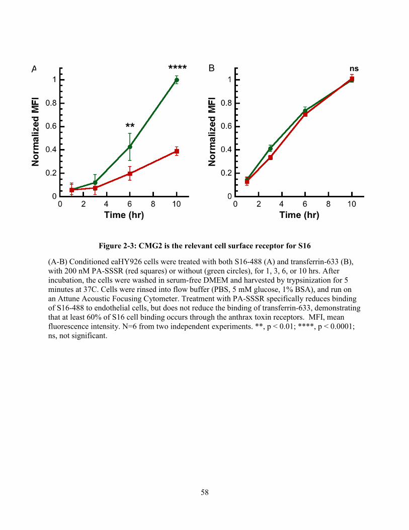
58
A B
Figure 2-3: CMG2 is the relevant cell surface receptor for S16
(A-B) Conditioned eaHY926 cells were treated with both S16-488 (A) and transferrin-633 (B), with 200 nM PA-SSSR (red squares) or without (green circles), for 1, 3, 6, or 10 hrs. After incubation, the cells were washed in serum-free DMEM and harvested by trypsinization for 5 minutes at 37C. Cells were rinsed into flow buffer (PBS, 5 mM glucose, 1% BSA), and run on an Attune Acoustic Focusing Cytometer. Treatment with PA-SSSR specifically reduces binding of S16-488 to endothelial cells, but does not reduce the binding of transferrin-633, demonstrating that at least 60% of S16 cell binding occurs through the anthrax toxin receptors. MFI, mean fluorescence intensity. N=6 from two independent experiments. **, p < 0.01; ****, p < 0.0001; ns, not significant.
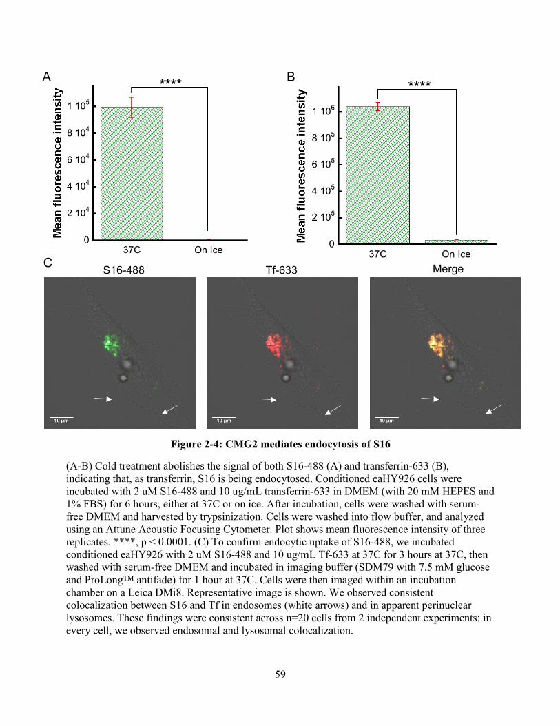
59
S16-488 Tf-633 Merge
A B
C
Figure 2-4: CMG2 mediates endocytosis of S16
(A-B) Cold treatment abolishes the signal of both S16-488 (A) and transferrin-633 (B), indicating that, as transferrin, S16 is being endocytosed. Conditioned eaHY926 cells were incubated with 2 uM S16-488 and 10 ug/mL transferrin-633 in DMEM (with 20 mM HEPES and 1% FBS) for 6 hours, either at 37C or on ice. After incubation, cells were washed with serum-free DMEM and harvested by trypsinization. Cells were washed into flow buffer, and analyzed using an Attune Acoustic Focusing Cytometer. Plot shows mean fluorescence intensity of three replicates. ****, p < 0.0001. (C) To confirm endocytic uptake of S16-488, we incubated conditioned eaHY926 with 2 uM S16-488 and 10 ug/mL Tf-633 at 37C for 3 hours at 37C, then washed with serum-free DMEM and incubated in imaging buffer (SDM79 with 7.5 mM glucose and ProLong™ antifade) for 1 hour at 37C. Cells were then imaged within an incubation chamber on a Leica DMi8. Representative image is shown. We observed consistent colocalization between S16 and Tf in endosomes (white arrows) and in apparent perinuclear lysosomes. These findings were consistent across n=20 cells from 2 independent experiments; in every cell, we observed endosomal and lysosomal colocalization.

60
A B
C D

61
Figure 2-5: Peptide S16 inhibits migration of endothelial cells through binding CMG2, with no effect on proliferation
(A) Wound scratch assay. EOMA cells were seeded at high confluency, and then a wound was created by scratch. This wound was imaged, then the cells were incubated in DMEM with 10% FBS for 4 hours, after which the wounds were again imaged. The degree of wound closure represents the degree of cell migration. Samples were treated either with canstatin-derived S16, fibronectin-derived EPG, or no serum. Peptide EPG has only a marginal effect on migration. S16 results in 60% inhibition of migration, and is not statistically different from the serum-free (negative) control. Error bars are SD of n=3 replicates. (B) Proliferation of EOMA cells was monitored. Treatment with S16 or EPG resulted in no change in proliferation relative to the untreated control. In contrast, cells fixed in ethanol showed a complete loss of proliferation. Error bars are SD of n=8 replicates from two independent experiments. (C-D) To validate the effect of S16 on endothelial migration, assays were performed with the CellASIC Onix microfluidic platform, using plates that enable monitoring of chemotaxis through the generation of a stable serum gradient in the imaging chamber. A serum gradient was generated (0-10% FBS), and chemotactic migration (in the y-direction) was monitored for 4 hrs. The gradient was reversed, and cells S16 (C) or PA-SSSR (D) was flowed at a uniform concentration to the cell chamber. Migration was monitored in the presence of these compounds for 4 hrs. Chemotactic migration under the different conditions were then compared. Error bars are SEM of n=50 cells for each condition. *, p < 0.05; ***, p < 0.001; ****, p < 0.0001; ns, not significant.

62
3. Conclusion
Capillary Morphogenesis Gene 2 (CMG2) has diverse situational roles: it has been well
characterized as the major anthrax toxin receptor2, 40; it is apparently essential for proper
homeostasis and repair of the extracellular matrix5-6, 18; and CMG2 is emerging as an important
regulator in pathological angiogenesis1, 11-12, 16. While progress has been made on each of these
fronts separately, most studies to date focus on one, sometime two, of these aspects, and the
broader question of the functional role of CMG2 has remained elusive.
Careful analysis of the existing literature reveals interesting overlap between these disparate
situational roles. For example, CMG2 is essential for proper ECM homeostasis and repair, and
ECM remodeling is a key step in angiogenesis. It is possible that CMG2 regulation of
angiogenesis is mediated, in part, by its role in ECM homeostasis. Indeed, data presented herein
supports this hypothesis. A novel type IV collagen NC1 domain-derived peptide (S16) was
identified that binds with high affinity to the CMG2 vWA domain (KD ≈ 400 nM). This peptide
interacts with human immortalized endothelial cells through binding to CMG2, and inhibits
endothelial cell migration, a critical process for angiogenesis94. Additionally, CMG2 mediates
endocytic uptake of peptide S16, as identified by flow cytometry and confocal microscopy. This
observation of S16 endocytosis presents an interesting model whereby CMG2 may regulate
angiogenesis through endocytic clearance of angiogenically active matrix fragments. As it has
here, hypothesis-driven synthesis of the disparate biological roles of this should continue to
accelerate and improve understanding of the physiological function and relevance of capillary
morphogenesis gene 2.

63
References
1. Bell, S. E.; Mavila, A.; Salazar, R.; Bayless, K. J.; Kanagala, S.; Maxwell, S. A.; Davis, G. E., Differential gene expression during capillary morphogenesis in 3D collagen matrices: regulated expression of genes involved in basement membrane matrix assembly, cell cycle progression, cellular differentiation and G-protein signaling. J Cell Sci 2001, 114 (Pt 15), 2755-73. 2. Scobie, H. M.; Rainey, G. J. A.; Bradley, K. A.; Young, J. A. T., Human capillary morphogenesis protein 2 functions as an anthrax toxin receptor. P Natl Acad Sci USA 2003, 100 (9), 5170-5174. 3. Bradley, K. A.; Mogridge, J.; Mourez, M.; Collier, R. J.; Young, J. A. T., Identification of the cellular receptor for anthrax toxin. Nature 2001, 414 (6860), 225-229. 4. St Croix, B.; Rago, C.; Velculescu, V.; Traverso, G.; Romans, K. E.; Montgomery, E.; Lal, A.; Riggins, G. J.; Lengauer, C.; Vogelstein, B.; Kinzler, K. W., Genes expressed in human tumor endothelium. Science 2000, 289 (5482), 1197-1202. 5. Dowling, O.; Difeo, A.; Ramirez, M. C.; Tukel, T.; Narla, G.; Bonafe, L.; Kayserili, H.; Yuksel-Apak, M.; Paller, A. S.; Norton, K.; Teebi, A. S.; Grum-Tokars, V.; Martin, G. S.; Davis, G. E.; Glucksman, M. J.; Martignetti, J. A., Mutations in capillary morphogenesis gene-2 result in the allelic disorders juvenile hyaline fibromatosis and infantile systemic hyalinosis. Am J Hum Genet 2003, 73 (4), 957-966. 6. Hanks, S.; Adams, S.; Douglas, J.; Arbour, L.; Atherton, D. J.; Balci, S.; Bode, H.; Campbell, M. E.; Feingold, M.; Keser, G.; Kleijer, W.; Mancini, G.; McGrath, J. A.; Muntoni, F.; Nanda, A.; Teare, M. D.; Warman, M.; Pope, F. M.; Superti-Furga, A.; Futreal, P. A.; Rahman, N., Mutations in the gene encoding capillary morphogenesis protein 2 cause juvenile hyaline fibromatosis and infantile systemic hyalinosis. Am J Hum Genet 2003, 73 (4), 791-800. 7. El-Kamah, G. Y.; Fong, K.; El-Ruby, M.; Affifi, H. H.; Clements, S. E.; Lai-Cheong, J. E.; Amr, K.; El-Darouti, M.; McGrath, J. A., Spectrum of mutations in the ANTXR2 (CMG2) gene in infantile systemic hyalinosis and juvenile hyaline fibromatosis. Brit J Dermatol 2010, 163 (1), 213-215. 8. Australo-Anglo-American Spondyloarthritis, C.; Reveille, J. D.; Sims, A. M.; Danoy, P.; Evans, D. M.; Leo, P.; Pointon, J. J.; Jin, R.; Zhou, X.; Bradbury, L. A.; Appleton, L. H.; Davis, J. C.; Diekman, L.; Doan, T.; Dowling, A.; Duan, R.; Duncan, E. L.; Farrar, C.; Hadler, J.; Harvey, D.; Karaderi, T.; Mogg, R.; Pomeroy, E.; Pryce, K.; Taylor, J.; Savage, L.; Deloukas, P.; Kumanduri, V.; Peltonen, L.; Ring, S. M.; Whittaker, P.; Glazov, E.; Thomas, G. P.; Maksymowych, W. P.; Inman, R. D.; Ward, M. M.; Stone, M. A.; Weisman, M. H.; Wordsworth, B. P.; Brown, M. A., Genome-wide association study of ankylosing spondylitis identifies non-MHC susceptibility loci. Nat Genet 2010, 42 (2), 123-7.
64
9. Evans, D. M.; Spencer, C. C.; Pointon, J. J.; Su, Z.; Harvey, D.; Kochan, G.; Oppermann, U.; Dilthey, A.; Pirinen, M.; Stone, M. A.; Appleton, L.; Moutsianas, L.; Leslie, S.; Wordsworth, T.; Kenna, T. J.; Karaderi, T.; Thomas, G. P.; Ward, M. M.; Weisman, M. H.; Farrar, C.; Bradbury, L. A.; Danoy, P.; Inman, R. D.; Maksymowych, W.; Gladman, D.; Rahman, P.; Spondyloarthritis Research Consortium of, C.; Morgan, A.; Marzo-Ortega, H.; Bowness, P.; Gaffney, K.; Gaston, J. S.; Smith, M.; Bruges-Armas, J.; Couto, A. R.; Sorrentino, R.; Paladini, F.; Ferreira, M. A.; Xu, H.; Liu, Y.; Jiang, L.; Lopez-Larrea, C.; Diaz-Pena, R.; Lopez-Vazquez, A.; Zayats, T.; Band, G.; Bellenguez, C.; Blackburn, H.; Blackwell, J. M.; Bramon, E.; Bumpstead, S. J.; Casas, J. P.; Corvin, A.; Craddock, N.; Deloukas, P.; Dronov, S.; Duncanson, A.; Edkins, S.; Freeman, C.; Gillman, M.; Gray, E.; Gwilliam, R.; Hammond, N.; Hunt, S. E.; Jankowski, J.; Jayakumar, A.; Langford, C.; Liddle, J.; Markus, H. S.; Mathew, C. G.; McCann, O. T.; McCarthy, M. I.; Palmer, C. N.; Peltonen, L.; Plomin, R.; Potter, S. C.; Rautanen, A.; Ravindrarajah, R.; Ricketts, M.; Samani, N.; Sawcer, S. J.; Strange, A.; Trembath, R. C.; Viswanathan, A. C.; Waller, M.; Weston, P.; Whittaker, P.; Widaa, S.; Wood, N. W.; McVean, G.; Reveille, J. D.; Wordsworth, B. P.; Brown, M. A.; Donnelly, P.; Australo-Anglo-American Spondyloarthritis, C.; Wellcome Trust Case Control, C., Interaction between ERAP1 and HLA-B27 in ankylosing spondylitis implicates peptide handling in the mechanism for HLA-B27 in disease susceptibility. Nat Genet 2011, 43 (8), 761-7. 10. Karaderi, T.; Keidel, S. M.; Pointon, J. J.; Appleton, L. H.; Brown, M. A.; Evans, D. M.; Wordsworth, B. P., Ankylosing spondylitis is associated with the anthrax toxin receptor 2 gene (ANTXR2). Ann Rheum Dis 2014, 73 (11), 2054-8. 11. Reeves, C. V.; Dufraine, J.; Young, J. A. T.; Kitajewski, J., Anthrax toxin receptor 2 is expressed in murine and tumor vasculature and functions in endothelial proliferation and morphogenesis. Oncogene 2010, 29 (6), 789-801. 12. Rogers, M. S.; Christensen, K. A.; Birsner, A. E.; Short, S. M.; Wigelsworth, D. J.; Collier, R. J.; D'Amato, R. J., Mutant anthrax toxin B moiety (protective antigen) inhibits angiogenesis and tumor growth. Cancer Res 2007, 67 (20), 9980-5. 13. Cao, S.; Cryan, L.; Habeshian, K. A.; Murillo, C.; Tamayo-Castillo, G.; Rogers, M. S.; Clardy, J., Phenolic compounds as antiangiogenic CMG2 inhibitors from Costa Rican endophytic fungi. Bioorg Med Chem Lett 2012, 22 (18), 5885-8. 14. Cryan, L. M.; Ackroyd, P. C.; Cao, S. G.; Clardy, J.; Christensen, K. A.; Rogers, M. S., Antiangiogenic small molecule antagonists of the anthrax toxin receptor CMG2. Cancer Res 2013, 73 (8). 15. Cryan, L. M.; Bazinet, L.; Habeshian, K. A.; Cao, S.; Clardy, J.; Christensen, K. A.; Rogers, M. S., 1,2,3,4,6-Penta-O-galloyl-beta-D-glucopyranose inhibits angiogenesis via inhibition of capillary morphogenesis gene 2. J Med Chem 2013, 56 (5), 1940-5. 16. Cryan, L. M.; Rogers, M. S., Targeting the anthrax receptors, TEM-8 and CMG-2, for anti-angiogenic therapy. Front Biosci (Landmark Ed) 2011, 16, 1574-88. 17. Lacy, D. B.; Wigelsworth, D. J.; Scobie, H. M.; Young, J. A. T.; Collier, R. J., Crystal structure of the von Willebrand factor A domain of human capillary morphogenesis protein 2: An anthrax toxin receptor. P Natl Acad Sci USA 2004, 101 (17), 6367-6372. 18. Deuquet, J.; Lausch, E.; Superti-Furga, A.; van der Goot, F. G., The dark sides of capillary morphogenesis gene 2. Embo J 2012, 31 (1), 3-13. 19. Santelli, E.; Bankston, L. A.; Leppla, S. H.; Liddington, R. C., Crystal structure of a complex between anthrax toxin and its host cell receptor. Nature 2004, 430 (7002), 905-908.
65
20. Scobie, H. M.; Young, J. A., Interactions between anthrax toxin receptors and protective antigen. Curr Opin Microbiol 2005, 8 (1), 106-12. 21. Deuquet, J.; Lausch, E.; Guex, N.; Abrami, L.; Salvi, S.; Lakkaraju, A.; Ramirez, M. C.; Martignetti, J. A.; Rokicki, D.; Bonafe, L.; Superti-Furga, A.; van der Goot, F. G., Hyaline fibromatosis syndrome inducing mutations in the ectodomain of anthrax toxin receptor 2 can be rescued by proteasome inhibitors. EMBO Mol Med 2011, 3 (4), 208-21. 22. Deuquet, J.; Abrami, L.; Difeo, A.; Ramirez, M. C. M.; Martignetti, J. A.; van der Goot, F. G., Systemic Hyalinosis Mutations in the CMG2 Ectodomain Leading to Loss of Function Through Retention in the Endoplasmic Reticulum. Hum Mutat 2009, 30 (4), 583-589. 23. Go, M. Y.; Kim, S.; Partridge, A. W.; Melnyk, R. A.; Rath, A.; Deber, C. M.; Mogridge, J., Self-association of the transmembrane domain of an anthrax toxin receptor. J Mol Biol 2006, 360 (1), 145-56. 24. Milne, J. C.; Furlong, D.; Hanna, P. C.; Wall, J. S.; Collier, R. J., Anthrax protective antigen forms oligomers during intoxication of mammalian cells. J Biol Chem 1994, 269 (32), 20607-12. 25. Abrami, L.; Liu, S. H.; Cosson, P.; Leppla, S. H.; van der Goot, F. G., Anthrax toxin triggers endocytosis of its receptor via a lipid raft-mediated clathrin-dependent process. Journal of Cell Biology 2003, 160 (3), 321-328. 26. Abrami, L.; Leppla, S. H.; van der Goot, F. G., Receptor palmitoylation and ubiquitination regulate anthrax toxin endocytosis. J Cell Biol 2006, 172 (2), 309-20. 27. Marsh, J. A.; Forman-Kay, J. D., Sequence determinants of compaction in intrinsically disordered proteins. Biophys J 2010, 98 (10), 2383-90. 28. Verma, K.; Gu, J.; Werner, E., Tumor endothelial marker 8 amplifies canonical Wnt signaling in blood vessels. Plos One 2011, 6 (8), e22334. 29. Leppla, S. H., Anthrax Toxin Edema Factor - a Bacterial Adenylate-Cyclase That Increases Cyclic-Amp Concentrations in Eukaryotic Cells. P Natl Acad Sci-Biol 1982, 79 (10), 3162-3166. 30. Obrien, J.; Friedlander, A.; Dreier, T.; Ezzell, J.; Leppla, S., Effects of Anthrax Toxin Components on Human-Neutrophils. Infect Immun 1985, 47 (1), 306-310. 31. Duesbery, N. S.; Webb, C. P.; Leppla, S. H.; Gordon, V. M.; Klimpel, K. R.; Copeland, T. D.; Ahn, N. G.; Oskarsson, M. K.; Fukasawa, K.; Paull, K. D.; Vande Woude, G. F., Proteolytic inactivation of MAP-kinase-kinase by anthrax lethal factor. Science 1998, 280 (5364), 734-737. 32. Friedlander, A. M., Macrophages Are Sensitive to Anthrax Lethal Toxin through an Acid-Dependent Process. J Biol Chem 1986, 261 (16), 7123-7126. 33. Pellizzari, R.; Guidi-Rontani, C.; Vitale, G.; Mock, M.; Montecucco, C., Anthrax lethal factor cleaves MKK3 in macrophages and inhibits the LPS/IFN gamma-induced release of NO and TNF alpha. Febs Lett 1999, 462 (1-2), 199-204. 34. Wigelsworth, D. J.; Krantz, B. A.; Christensen, K. A.; Lacy, D. B.; Juris, S. J.; Collier, R. J., Binding stoichiometry and kinetics of the interaction of a human anthrax toxin receptor, CMG2, with protective antigen. J Biol Chem 2004, 279 (22), 23349-23356. 35. Gordon, V. M.; Klimpel, K. R.; Arora, N.; Henderson, M. A.; Leppla, S. H., Proteolytic activation of bacterial toxins by eukaryotic cells is performed by furin and by additional cellular proteases. Infect Immun 1995, 63 (1), 82-7. 36. Petosa, C.; Collier, R. J.; Klimpel, K. R.; Leppla, S. H.; Liddington, R. C., Crystal structure of the anthrax toxin protective antigen. Nature 1997, 385 (6619), 833-8.

66
37. Abrami, L.; Kunz, B.; van der Goot, F. G., Anthrax toxin triggers the activation of src-like kinases to mediate its own uptake. P Natl Acad Sci USA 2010, 107 (4), 1420-1424. 38. Abrami, L.; Bischofberger, M.; Kunz, B.; Groux, R.; van der Goot, F. G., Endocytosis of the Anthrax Toxin Is Mediated by Clathrin, Actin and Unconventional Adaptors. Plos Pathog 2010, 6 (3). 39. Milne, J. C.; Collier, R. J., Ph-Dependent Permeabilization of the Plasma-Membrane of Mammalian-Cells by Anthrax Protective Antigen. Molecular Microbiology 1993, 10 (3), 647-653. 40. Liu, S.; Crown, D.; Miller-Randolph, S.; Moayeri, M.; Wang, H.; Hu, H.; Morley, T.; Leppla, S. H., Capillary morphogenesis protein-2 is the major receptor mediating lethality of anthrax toxin in vivo. Proc Natl Acad Sci U S A 2009, 106 (30), 12424-9. 41. Murray, J., On three peculiar cases of Molluscum Fibrosum in Children in which one or more of the following conditions were observed: hypertrophy of the gums, enlargement of the ends of the fingers and toes, numerous connecive-tissue tumours on the scalp, &c. Med Chir Trans 1873, 56, 235-254 1. 42. Whitfield, A.; Robinson, A. H., A Further Report on the Remarkable Series of Cases of Molluscum Fibrosum in Children communicated to the Society by Dr. John Murray in 1873. Med Chir Trans 1903, 86, 293-302. 43. Fong, K.; El-Kamah, G.; Clements, S.; Lai-Cheong, J.; Mellerio, J.; El-Darouti, M.; McGrath, J., Loss-of-function mutations in the ANTXR2 (CMG2) gene underlie both juvenile hyaline fibromatosis and infantile systemic hyalinosis. Brit J Dermatol 2009, 161, 119-119. 44. Rebbapragada, I.; Lykke-Andersen, J., Execution of nonsense-mediated mRNA decay: what defines a substrate? Curr Opin Cell Biol 2009, 21 (3), 394-402. 45. Hakki, S. S.; Ataoglu, T.; Avunduk, M. C.; Erdemli, E.; Gunhan, O.; Rahman, N., Periodontal treatment of two siblings with juvenile hyaline fibromatosis. J Clin Periodontol 2005, 32 (9), 1016-1021. 46. Hatamochi, A.; Sasaki, T.; Kawaguchi, T.; Suzuki, H.; Yamazaki, S., A novel point mutation in the gene encoding capillary morphogenesis protein 2 in a Japanese patient with juvenile hyaline fibromatosis. Br J Dermatol 2007, 157 (5), 1037-9. 47. Stucki, U.; Spycher, M. A.; Eich, G.; Rossi, A.; Sacher, P.; Steinmann, B.; Superti-Furga, A., Infantile systemic hyalinosis in siblings: clinical report, biochemical and ultrastructural findings, and review of the literature. Am J Med Genet 2001, 100 (2), 122-9. 48. Tanaka, K.; Ebihara, T.; Kusubata, M.; Adachi, E.; Arai, M.; Kawaguchi, N.; Utsunomiya, J.; Miki, Y.; Hiramoto, M.; Hattori, S.; Irie, S., Abnormal collagen deposition in fibromas from patient with juvenile hyaline fibromatosis. J Dermatol Sci 2009, 55 (3), 197-200. 49. Braun, J.; Bollow, M.; Remlinger, G.; Eggens, U.; Rudwaleit, M.; Distler, A.; Sieper, J., Prevalence of spondylarthropathies in HLA-B27 positive and negative blood donors. Arthritis Rheum 1998, 41 (1), 58-67. 50. Sheehan, N. J., The ramifications of HLA-B27. J R Soc Med 2004, 97 (1), 10-4. 51. Wellcome Trust Case Control, C.; Australo-Anglo-American Spondylitis, C.; Burton, P. R.; Clayton, D. G.; Cardon, L. R.; Craddock, N.; Deloukas, P.; Duncanson, A.; Kwiatkowski, D. P.; McCarthy, M. I.; Ouwehand, W. H.; Samani, N. J.; Todd, J. A.; Donnelly, P.; Barrett, J. C.; Davison, D.; Easton, D.; Evans, D. M.; Leung, H. T.; Marchini, J. L.; Morris, A. P.; Spencer, C. C.; Tobin, M. D.; Attwood, A. P.; Boorman, J. P.; Cant, B.; Everson, U.; Hussey, J. M.; Jolley, J. D.; Knight, A. S.; Koch, K.; Meech, E.; Nutland, S.; Prowse, C. V.; Stevens, H. E.; Taylor, N. C.; Walters, G. R.; Walker, N. M.; Watkins, N. A.; Winzer, T.; Jones, R. W.; McArdle, W. L.;

67
Ring, S. M.; Strachan, D. P.; Pembrey, M.; Breen, G.; St Clair, D.; Caesar, S.; Gordon-Smith, K.; Jones, L.; Fraser, C.; Green, E. K.; Grozeva, D.; Hamshere, M. L.; Holmans, P. A.; Jones, I. R.; Kirov, G.; Moskivina, V.; Nikolov, I.; O'Donovan, M. C.; Owen, M. J.; Collier, D. A.; Elkin, A.; Farmer, A.; Williamson, R.; McGuffin, P.; Young, A. H.; Ferrier, I. N.; Ball, S. G.; Balmforth, A. J.; Barrett, J. H.; Bishop, T. D.; Iles, M. M.; Maqbool, A.; Yuldasheva, N.; Hall, A. S.; Braund, P. S.; Dixon, R. J.; Mangino, M.; Stevens, S.; Thompson, J. R.; Bredin, F.; Tremelling, M.; Parkes, M.; Drummond, H.; Lees, C. W.; Nimmo, E. R.; Satsangi, J.; Fisher, S. A.; Forbes, A.; Lewis, C. M.; Onnie, C. M.; Prescott, N. J.; Sanderson, J.; Matthew, C. G.; Barbour, J.; Mohiuddin, M. K.; Todhunter, C. E.; Mansfield, J. C.; Ahmad, T.; Cummings, F. R.; Jewell, D. P.; Webster, J.; Brown, M. J.; Lathrop, M. G.; Connell, J.; Dominiczak, A.; Marcano, C. A.; Burke, B.; Dobson, R.; Gungadoo, J.; Lee, K. L.; Munroe, P. B.; Newhouse, S. J.; Onipinla, A.; Wallace, C.; Xue, M.; Caulfield, M.; Farrall, M.; Barton, A.; Biologics in, R. A. G.; Genomics Study Syndicate Steering, C.; Bruce, I. N.; Donovan, H.; Eyre, S.; Gilbert, P. D.; Hilder, S. L.; Hinks, A. M.; John, S. L.; Potter, C.; Silman, A. J.; Symmons, D. P.; Thomson, W.; Worthington, J.; Dunger, D. B.; Widmer, B.; Frayling, T. M.; Freathy, R. M.; Lango, H.; Perry, J. R.; Shields, B. M.; Weedon, M. N.; Hattersley, A. T.; Hitman, G. A.; Walker, M.; Elliott, K. S.; Groves, C. J.; Lindgren, C. M.; Rayner, N. W.; Timpson, N. J.; Zeggini, E.; Newport, M.; Sirugo, G.; Lyons, E.; Vannberg, F.; Hill, A. V.; Bradbury, L. A.; Farrar, C.; Pointon, J. J.; Wordsworth, P.; Brown, M. A.; Franklyn, J. A.; Heward, J. M.; Simmonds, M. J.; Gough, S. C.; Seal, S.; Breast Cancer Susceptibility, C.; Stratton, M. R.; Rahman, N.; Ban, M.; Goris, A.; Sawcer, S. J.; Compston, A.; Conway, D.; Jallow, M.; Newport, M.; Sirugo, G.; Rockett, K. A.; Bumpstead, S. J.; Chaney, A.; Downes, K.; Ghori, M. J.; Gwilliam, R.; Hunt, S. E.; Inouye, M.; Keniry, A.; King, E.; McGinnis, R.; Potter, S.; Ravindrarajah, R.; Whittaker, P.; Widden, C.; Withers, D.; Cardin, N. J.; Davison, D.; Ferreira, T.; Pereira-Gale, J.; Hallgrimsdo'ttir, I. B.; Howie, B. N.; Su, Z.; Teo, Y. Y.; Vukcevic, D.; Bentley, D.; Brown, M. A.; Compston, A.; Farrall, M.; Hall, A. S.; Hattersley, A. T.; Hill, A. V.; Parkes, M.; Pembrey, M.; Stratton, M. R.; Mitchell, S. L.; Newby, P. R.; Brand, O. J.; Carr-Smith, J.; Pearce, S. H.; McGinnis, R.; Keniry, A.; Deloukas, P.; Reveille, J. D.; Zhou, X.; Sims, A. M.; Dowling, A.; Taylor, J.; Doan, T.; Davis, J. C.; Savage, L.; Ward, M. M.; Learch, T. L.; Weisman, M. H.; Brown, M., Association scan of 14,500 nonsynonymous SNPs in four diseases identifies autoimmunity variants. Nat Genet 2007, 39 (11), 1329-37. 52. Xia, Y.; Chen, K.; Zhang, M. H.; Wang, L. C.; Ma, C. Y.; Lin, Y. L.; Zhao, Y. R., MicroRNA-124 involves in ankylosing spondylitis by targeting ANTXR2. Mod Rheumatol 2015, 25 (5), 784-9. 53. Zhang, Z.; Yu, K.; Dai, D.; Yuan, F.; Liang, F.; Liu, N.; Xi, Y.; Sun, Y. Y., A complex role of anthrax toxin receptor 2 polymorphisms and capillary morphogenesis protein 2 in ankylosing spondylitis pathogenesis. Clin Rheumatol 2016, 35 (9), 2243-50. 54. Wei, W.; Lu, Q.; Chaudry, G. J.; Leppla, S. H.; Cohen, S. N., The LDL receptor-related protein LRP6 mediates internalization and lethality of anthrax toxin. Cell 2006, 124 (6), 1141-1154. 55. Young, J. J.; Bromberg-White, J. L.; Zylstra, C.; Church, J. T.; Boguslawski, E.; Resau, J. H.; Williams, B. O.; Duesbery, N. S., LRP5 and LRP6 are not required for protective antigen-mediated internalization or lethality of anthrax lethal toxin. Plos Pathog 2007, 3 (3), e27. 56. Abrami, L.; Kunz, B.; Deuquet, J.; Bafico, A.; Davidson, G.; van der Goot, F. G., Functional interactions between anthrax toxin receptors and the WNT signalling protein LRP6. Cell Microbiol 2008, 10 (12), 2509-19.

68
57. Braun, J.; Sieper, J., Ankylosing spondylitis. Lancet 2007, 369 (9570), 1379-90. 58. Martinez-Gonzalez, O.; Cantero-Hinojosa, J.; Paule-Sastre, P.; Gomez-Magan, J. C.; Salvatierra-Rios, D., Intestinal permeability in patients with ankylosing spondylitis and their healthy relatives. Br J Rheumatol 1994, 33 (7), 644-7. 59. Qazi, Y.; Maddula, S.; Ambati, B. K., Mediators of ocular angiogenesis. J Genet 2009, 88 (4), 495-515. 60. Crawford, T. N.; Alfaro, D. V., 3rd; Kerrison, J. B.; Jablon, E. P., Diabetic retinopathy and angiogenesis. Curr Diabetes Rev 2009, 5 (1), 8-13. 61. Hanahan, D.; Weinberg, R. A., Hallmarks of Cancer: The Next Generation. Cell 2011, 144 (5), 646-674. 62. Folkman, J., Tumor angiogenesis: therapeutic implications. N Engl J Med 1971, 285 (21), 1182-6. 63. Sitohy, B.; Nagy, J. A.; Dvorak, H. F., Anti-VEGF/VEGFR therapy for cancer: reassessing the target. Cancer Res 2012, 72 (8), 1909-14. 64. Liu, S.; Liu, J.; Ma, Q.; Cao, L.; Fattah, R. J.; Yu, Z.; Bugge, T. H.; Finkel, T.; Leppla, S. H., Solid tumor therapy by selectively targeting stromal endothelial cells. Proc Natl Acad Sci U S A 2016, 113 (28), E4079-87. 65. Rogers, M. S.; Birsner, A. E.; D'Amato, R. J., The mouse cornea micropocket angiogenesis assay. Nat Protoc 2007, 2 (10), 2545-50. 66. Williamson, E. D.; Hodgson, I.; Walker, N. J.; Topping, A. W.; Duchars, M. G.; Mott, J. M.; Estep, J.; Lebutt, C.; Flick-Smith, H. C.; Jones, H. E.; Li, H.; Quinn, C. P., Immunogenicity of recombinant protective antigen and efficacy against aerosol challenge with anthrax. Infect Immun 2005, 73 (9), 5978-87. 67. Leffel, E. K.; Bourdage, J. S.; Williamson, E. D.; Duchars, M.; Fuerst, T. R.; Fusco, P. C., Recombinant protective antigen anthrax vaccine improves survival when administered as a postexposure prophylaxis countermeasure with antibiotic in the New Zealand white rabbit model of inhalation anthrax. Clin Vaccine Immunol 2012, 19 (8), 1158-64. 68. Rogers, M. S.; Cryan, L. M.; Habeshian, K. A.; Bazinet, L.; Caldwell, T. P.; Ackroyd, P. C.; Christensen, K. A., A FRET-Based High Throughput Screening Assay to Identify Inhibitors of Anthrax Protective Antigen Binding to Capillary Morphogenesis Gene 2 Protein. Plos One 2012, 7 (6). 69. Liu, S. H.; Aaronson, H.; Mitola, D. J.; Leppla, S. H.; Bugge, T. H., Potent antitumor activity of a urokinase-activated engineered anthrax toxin. P Natl Acad Sci USA 2003, 100 (2), 657-662. 70. Liu, S. H.; Redeye, V.; Kuremsky, J. G.; Kuhnen, M.; Molinolo, A.; Bugge, T. H.; Leppla, S. H., Intermolecular complementation achieves high-specificity tumor targeting by anthrax toxin. Nat Biotechnol 2005, 23 (6), 725-730. 71. Schafer, J. M.; Peters, D. E.; Morley, T.; Liu, S. H.; Molinolo, A. A.; Leppla, S. H.; Bugge, T. H., Efficient Targeting of Head and Neck Squamous Cell Carcinoma by Systemic Administration of a Dual uPA and MMP-Activated Engineered Anthrax Toxin. Plos One 2011, 6 (5). 72. Roux, K. J.; Kim, D. I.; Raida, M.; Burke, B., A promiscuous biotin ligase fusion protein identifies proximal and interacting proteins in mammalian cells. J Cell Biol 2012, 196 (6), 801-10. 73. Abrami, L.; Reig, N.; van der Goot, F. G., Anthrax toxin: the long and winding road that leads to the kill. Trends Microbiol 2005, 13 (2), 72-8.

69
74. Young, J. A.; Collier, R. J., Anthrax toxin: receptor binding, internalization, pore formation, and translocation. Annu Rev Biochem 2007, 76, 243-65. 75. van der Goot, G.; Young, J. A., Receptors of anthrax toxin and cell entry. Mol Aspects Med 2009, 30 (6), 406-12. 76. Friebe, S.; van der Goot, F. G.; Burgi, J., The Ins and Outs of Anthrax Toxin. Toxins 2016, 8 (3). 77. Rainero, E., Extracellular matrix endocytosis in controlling matrix turnover and beyond: emerging roles in cancer. Biochem Soc Trans 2016, 44 (5), 1347-1354. 78. Urbina, F.; Sazunic, I.; Murray, G., Infantile systemic hyalinosis or juvenile hyaline fibromatosis? Pediatr Dermatol 2004, 21 (2), 154-9. 79. Banks, D. J.; Barnajian, M.; Maldonado-Arocho, F. J.; Sanchez, A. M.; Bradley, K. A., Anthrax toxin receptor 2 mediates Bacillus anthracis killing of macrophages following spore challenge. Cell Microbiol 2005, 7 (8), 1173-85. 80. Gu, J.; Faundez, V.; Werner, E., Endosomal recycling regulates Anthrax Toxin Receptor 1/Tumor Endothelial Marker 8-dependent cell spreading. Exp Cell Res 2010, 316 (12), 1946-57. 81. Yang, M. Y.; Chaudhary, A.; Seaman, S.; Dunty, J.; Stevens, J.; Elzarrad, M. K.; Frankel, A. E.; St Croix, B., The cell surface structure of tumor endothelial marker 8 (TEM8) is regulated by the actin cytoskeleton. Biochim Biophys Acta 2011, 1813 (1), 39-49. 82. Peters, D. E.; Zhang, Y.; Molinolo, A. A.; Miller-Randolph, S.; Szabo, R.; Bugge, T. H.; Leppla, S. H.; Liu, S. H., Capillary morphogenesis protein-2 is required for mouse parturition by maintaining uterine collagen homeostasis. Biochem Bioph Res Co 2012, 422 (3), 393-397. 83. Reeves, C. V.; Wang, X.; Charles-Horvath, P. C.; Vink, J. Y.; Borisenko, V. Y.; Young, J. A.; Kitajewski, J. K., Anthrax toxin receptor 2 functions in ECM homeostasis of the murine reproductive tract and promotes MMP activity. Plos One 2012, 7 (4), e34862. 84. Vink, J. Y.; Charles-Horvath, P. C.; Kitajewski, J. K.; Reeves, C. V., Anthrax toxin receptor 2 promotes human uterine smooth muscle cell viability, migration and contractility. Am J Obstet Gynecol 2014, 210 (2), 154 e1-8. 85. Kalluri, R., Basement membranes: Structure, assembly and role in tumour angiogenesis. Nature Reviews Cancer 2003, 3 (6), 422-433. 86. Monboisse, J. C.; Oudart, J. B.; Ramont, L.; Brassart-Pasco, S.; Maquart, F. X., Matrikines from basement membrane collagens: A new anti-cancer strategy. Biochimica Et Biophysica Acta-General Subjects 2014, 1840 (8), 2589-2598. 87. Dafforn, T. R.; Della, M.; Miller, A. D., The molecular interactions of heat shock protein 47 (Hsp47) and their implications for collagen biosynthesis. J Biol Chem 2001, 276 (52), 49310-9. 88. Hotchkiss, K. A.; Basile, C. M.; Spring, S. C.; Bonuccelli, G.; Lisanti, M. P.; Terman, B. I., TEM8 expression stimulates endothelial cell adhesion and migration by regulating cell-matrix interactions on collagen. Exp Cell Res 2005, 305 (1), 133-44. 89. Werner, E.; Kowalczyk, A. P.; Faundez, V., Anthrax toxin receptor 1/tumor endothelium marker 8 mediates cell spreading by coupling extracellular ligands to the actin cytoskeleton. J Biol Chem 2006, 281 (32), 23227-36. 90. Go, M. Y.; Chow, E. M.; Mogridge, J., The cytoplasmic domain of anthrax toxin receptor 1 affects binding of the protective antigen. Infect Immun 2009, 77 (1), 52-9. 91. Garlick, K. M.; Batty, S.; Mogridge, J., Binding of filamentous actin to anthrax toxin receptor 1 decreases its association with protective antigen. Biochemistry-Us 2012, 51 (6), 1249-56.

70
92. Garlick, K. M.; Mogridge, J., Direct interaction between anthrax toxin receptor 1 and the actin cytoskeleton. Biochemistry-Us 2009, 48 (44), 10577-81. 93. Ginsberg, M. H.; Du, X.; Plow, E. F., Inside-out integrin signalling. Curr Opin Cell Biol 1992, 4 (5), 766-71. 94. Lamalice, L.; Le Boeuf, F.; Huot, J., Endothelial cell migration during angiogenesis. Circ Res 2007, 100 (6), 782-94. 95. Ryan, P. L.; Young, J. A. T., Evidence against a Human Cell-Specific Role for LRP6 in Anthrax Toxin Entry. Plos One 2008, 3 (3). 96. Wang, Y.; Li, Y. P.; Paulson, C.; Shao, J. Z.; Zhang, X.; Wu, M.; Chen, W., Wnt and the Wnt signaling pathway in bone development and disease. Front Biosci (Landmark Ed) 2014, 19, 379-407. 97. Jinnin, M.; Medici, D.; Park, L.; Limaye, N.; Liu, Y.; Boscolo, E.; Bischoff, J.; Vikkula, M.; Boye, E.; Olsen, B. R., Suppressed NFAT-dependent VEGFR1 expression and constitutive VEGFR2 signaling in infantile hemangioma. Nat Med 2008, 14 (11), 1236-46. 98. Stranecky, V.; Hoischen, A.; Hartmannova, H.; Zaki, M. S.; Chaudhary, A.; Zudaire, E.; Noskova, L.; Baresova, V.; Pristoupilova, A.; Hodanova, K.; Sovova, J.; Hulkova, H.; Piherova, L.; Hehir-Kwa, J. Y.; de Silva, D.; Senanayake, M. P.; Farrag, S.; Zeman, J.; Martasek, P.; Baxova, A.; Afifi, H. H.; St Croix, B.; Brunner, H. G.; Temtamy, S.; Kmoch, S., Mutations in ANTXR1 cause GAPO syndrome. Am J Hum Genet 2013, 92 (5), 792-9. 99. Tipton, R. E.; Gorlin, R. J., Growth retardation, alopecia, pseudo-anodontia, and optic atrophy--the GAPO syndrome: report of a patient and review of the literature. Am J Med Genet 1984, 19 (2), 209-16. 100. Salas-Alanis, J. C.; Scott, C. A.; Fajardo-Ramirez, O. R.; Duran, C.; Moreno-Trevino, M. G.; Kelsell, D. P., New ANTXR1 Gene Mutation for GAPO Syndrome: A Case Report. Mol Syndromol 2016, 7 (3), 160-3. 101. Besschetnova, T. Y.; Ichimura, T.; Katebi, N.; St Croix, B.; Bonventre, J. V.; Olsen, B. R., Regulatory mechanisms of anthrax toxin receptor 1-dependent vascular and connective tissue homeostasis. Matrix Biol 2015, 42, 56-73. 102. Amemiya, T.; Sasamura, H.; Mifune, M.; Kitamura, Y.; Hirahashi, J.; Hayashi, M.; Saruta, T., Vascular endothelial growth factor activates MAP kinase and enhances collagen synthesis in human mesangial cells. Kidney Int 1999, 56 (6), 2055-63. 103. Wu, T.; Zhang, B.; Ye, F.; Xiao, Z., A potential role for caveolin-1 in VEGF-induced fibronectin upregulation in mesangial cells: involvement of VEGFR2 and Src. Am J Physiol Renal Physiol 2013, 304 (6), F820-30. 104. Hu, K.; Olsen, B. R.; Besschetnova, T. Y., Cell autonomous ANTXR1-mediated regulation of extracellular matrix components in primary fibroblasts. Matrix Biol 2016. 105. Akhmetshina, A.; Palumbo, K.; Dees, C.; Bergmann, C.; Venalis, P.; Zerr, P.; Horn, A.; Kireva, T.; Beyer, C.; Zwerina, J.; Schneider, H.; Sadowski, A.; Riener, M. O.; MacDougald, O. A.; Distler, O.; Schett, G.; Distler, J. H., Activation of canonical Wnt signalling is required for TGF-beta-mediated fibrosis. Nat Commun 2012, 3, 735. 106. Scobie, H. M.; Thomas, D.; Marlett, J. M.; Destito, G.; Wigelsworth, D. J.; Collier, R. J.; Young, J. A.; Manchester, M., A soluble receptor decoy protects rats against anthrax lethal toxin challenge. J Infect Dis 2005, 192 (6), 1047-51. 107. Kamphaus, G. D.; Colorado, P. C.; Panka, D. J.; Hopfer, H.; Ramchandran, R.; Torre, A.; Maeshima, Y.; Mier, J. W.; Sukhatme, V. P.; Kalluri, R., Canstatin, a novel matrix-derived inhibitor of angiogenesis and tumor growth. J Biol Chem 2000, 275 (2), 1209-15.

71
108. Nyberg, P.; Xie, L.; Sugimoto, H.; Colorado, P.; Sund, M.; Holthaus, K.; Sudhakar, A.; Salo, T.; Kalluri, R., Characterization of the anti-angiogenic properties of arresten, an alpha1beta1 integrin-dependent collagen-derived tumor suppressor. Exp Cell Res 2008, 314 (18), 3292-305. 109. Khoshnoodi, J.; Pedchenko, V.; Hudson, B. G., Mammalian collagen IV. Microsc Res Tech 2008, 71 (5), 357-70. 110. Panka, D. J.; Mier, J. W., Canstatin inhibits Akt activation and induces Fas-dependent apoptosis in endothelial cells. J Biol Chem 2003, 278 (39), 37632-6. 111. Magnon, C.; Galaup, A.; Mullan, B.; Rouffiac, V.; Bouquet, C.; Bidart, J. M.; Griscelli, F.; Opolon, P.; Perricaudet, M., Canstatin acts on endothelial and tumor cells via mitochondrial damage initiated through interaction with alphavbeta3 and alphavbeta5 integrins. Cancer Res 2005, 65 (10), 4353-61. 112. Sudhakar, A.; Nyberg, P.; Keshamouni, V. G.; Mannam, A. P.; Li, J.; Sugimoto, H.; Cosgrove, D.; Kalluri, R., Human alpha1 type IV collagen NC1 domain exhibits distinct antiangiogenic activity mediated by alpha1beta1 integrin. J Clin Invest 2005, 115 (10), 2801-10. 113. Gunda, V.; Verma, R. K.; Pawar, S. C.; Sudhakar, Y. A., Developments in purification methods for obtaining and evaluation of collagen derived endogenous angioinhibitors. Protein Expr Purif 2014, 94, 46-52. 114. Dyballa, N.; Metzger, S., Fast and sensitive colloidal coomassie G-250 staining for proteins in polyacrylamide gels. J Vis Exp 2009, (30). 115. Pettersen, E. F.; Goddard, T. D.; Huang, C. C.; Couch, G. S.; Greenblatt, D. M.; Meng, E. C.; Ferrin, T. E., UCSF Chimera--a visualization system for exploratory research and analysis. J Comput Chem 2004, 25 (13), 1605-12. 116. Sanner, M. F.; Olson, A. J.; Spehner, J. C., Reduced surface: an efficient way to compute molecular surfaces. Biopolymers 1996, 38 (3), 305-20. 117. Andreatta, M.; Lund, O.; Nielsen, M., Simultaneous alignment and clustering of peptide data using a Gibbs sampling approach. Bioinformatics 2013, 29 (1), 8-14. 118. Andreatta, M.; Alvarez, B.; Nielsen, M., GibbsCluster: unsupervised clustering and alignment of peptide sequences. Nucleic Acids Res 2017. 119. Thomsen, M. C.; Nielsen, M., Seq2Logo: a method for construction and visualization of amino acid binding motifs and sequence profiles including sequence weighting, pseudo counts and two-sided representation of amino acid enrichment and depletion. Nucleic Acids Res 2012, 40 (Web Server issue), W281-7. 120. Obeso, J.; Weber, J.; Auerbach, R., A hemangioendothelioma-derived cell line: its use as a model for the study of endothelial cell biology. Lab Invest 1990, 63 (2), 259-69. 121. Ahn, K.; Pan, S.; Beningo, K.; Hupe, D., A permanent human cell line (EA.hy926) preserves the characteristics of endothelin converting enzyme from primary human umbilical vein endothelial cells. Life Sci 1995, 56 (26), 2331-41. 122. Than, M. E.; Henrich, S.; Huber, R.; Ries, A.; Mann, K.; Kuhn, K.; Timpl, R.; Bourenkov, G. P.; Bartunik, H. D.; Bode, W., The 1.9-A crystal structure of the noncollagenous (NC1) domain of human placenta collagen IV shows stabilization via a novel type of covalent Met-Lys cross-link. Proc Natl Acad Sci U S A 2002, 99 (10), 6607-12. 123. Plow, E. F.; Haas, T. K.; Zhang, L.; Loftus, J.; Smith, J. W., Ligand binding to integrins. J Biol Chem 2000, 275 (29), 21785-21788. 124. Tomoda, H.; Kishimoto, Y.; Lee, Y. C., Temperature effect on endocytosis and exocytosis by rabbit alveolar macrophages. J Biol Chem 1989, 264 (26), 15445-50.

72