aniridia due to a novel microdeletion affecting regulatory
TRANSCRIPT
Journal of Genetics, Vol. 97, No. 2, June 2018, pp. 555–562 © Indian Academy of Scienceshttps://doi.org/10.1007/s12041-018-0925-9
RESEARCH NOTE
Aniridia due to a novel microdeletion affecting PAX6 regulatory enhancers:case report and review of the literature
ANDREAS SYRIMIS1, NAYIA NICOLAOU1, ANGELOS ALEXANDROU2, IOANNIS PAPAEVRIPIDOU2,MICHAEL NICOLAOU1, ELENI LOUKIANOU3, VIOLETTA CHRISTOPHIDOU-ANASTASIADOU1,4,STAVROS MALAS5, CAROLINA SISMANI2 and GEORGE A. TANTELES1∗
1Department of Clinical Genetics, The Cyprus Institute of Neurology and Genetics, 2370Nicosia, Cyprus2Department of Cytogenetics and Genomics, The Cyprus Institute of Neurology and Genetics, 2370Nicosia, Cyprus3Department of Ophthalmology, Nicosia General Hospital, 2370Nicosia, Cyprus4Department of Clinical Genetics, Archbishop Makarios III Medical Centre, 2370Nicosia, Cyprus5Department of Developmental and Functional Genetics, The Cyprus Institute of Neurology and Genetics,2370Nicosia, Cyprus*For correspondence. E-mail: [email protected].
Received 23 May 2017; revised 20 July 2017; accepted 28 August 2017; published online 25 May 2018
Abstract. Aniridia is a rare congenital ocular malformation that follows an autosomal dominant mode of inheritance. Mostpatients carry pathogenic point mutations in the paired box 6 gene (PAX6), but some carry deletions involving the 11p13 region,encompassing partly or completelyPAX6 or the region downstream.We identified a novel deletion,∼564 kb in size located about 46.5kb downstream of PAX6 in a family with bilateral aniridia and foveal hypoplasia using array-CGH and multiplex ligation-dependentprobe amplification.We also review all of the reported deletions downstreamofPAX6 in patients with aniridia and/or other congenitalmalformations and define the overlapping region that leads to aniridia when deleted.
Keywords. aniridia; array-CGH; multiplex ligation-dependent probe amplification; PAX6 deletion.
Introduction
Classic aniridia (MIM: 106210) is a rare congenitalpanocular malformation characterized by complete orpartial iris hypoplasia due to heterozygous PAX6 muta-tions (Nelson et al. 1984; Samant et al. 2016). It can beaccompanied by foveal hypoplasia, strabismus and opticnerve hypoplasia, generally leading to impaired visualacuity, while late-onset manifestations may include nys-tagmus, glaucoma, cataract and corneal abnormalities(Valenzuela and Cline 2004). It can occur either as anisolated malformation or as part of a syndrome, such asWilms tumour, aniridia, genital anomalies, and mentalretardation (WAGR, MIM: 194072), which is caused by11p13 deletions involving PAX6 and the adjacent WT1locus (Miller et al. 1964). Moreover, patients with aniridiamay have nonocular sensory and neurological abnormal-ities, such as reduced olfaction and hearing difficulties(Hingorani et al. 2012).
The prevalence of aniridia ranges from 1 in 50,000 to1 in 100,000 live births (Gronskov et al. 2001). Approx-imately two thirds of all cases are familial following anautosomal dominant mode of inheritance with completepenetrance and variable expressivity, while the remainingcases are sporadic (Gronskov et al. 2001). Heterozygousloss-of-functionmutations inPAX6 are identified in about94% of patients with aniridia (Robinson et al. 2008).PAX6encodes a transcription factor that plays a crucial role inearly ocular morphogenesis and development of the cen-tral nervous system. It is expressed in nasal structures, gut,pancreas, pituitary, brain and spinal cord during embry-onic development. It regulates the expression of otherdevelopmental regulatory genes, cell adhesion molecules,structural proteins and also of PAX6 itself (Simpson andPrice 2002). The majority of PAX6 pathogenic mutations,including nonsense, splice-site and frame-shift mutations,cause the production of truncated transcripts resulting inhaploinsufficiency. Some patients though carry deletions
555
556 Andreas Syrimis et al.
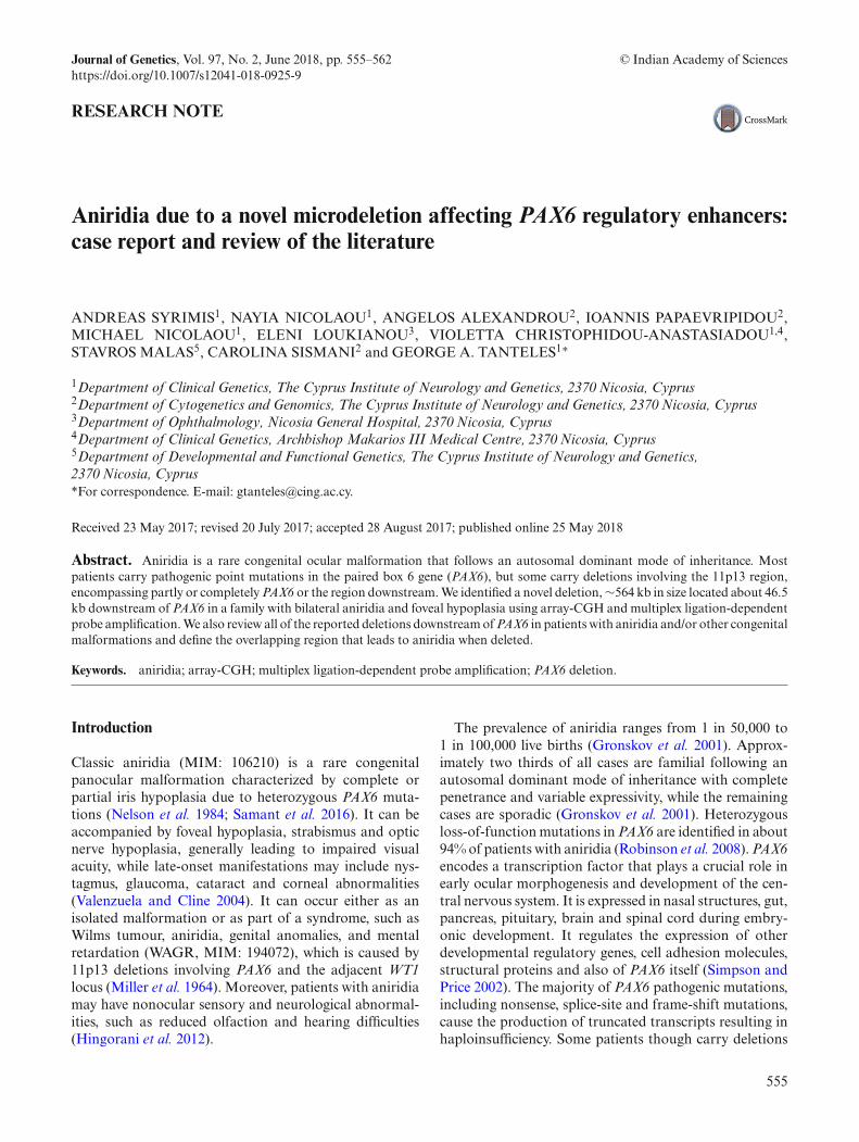
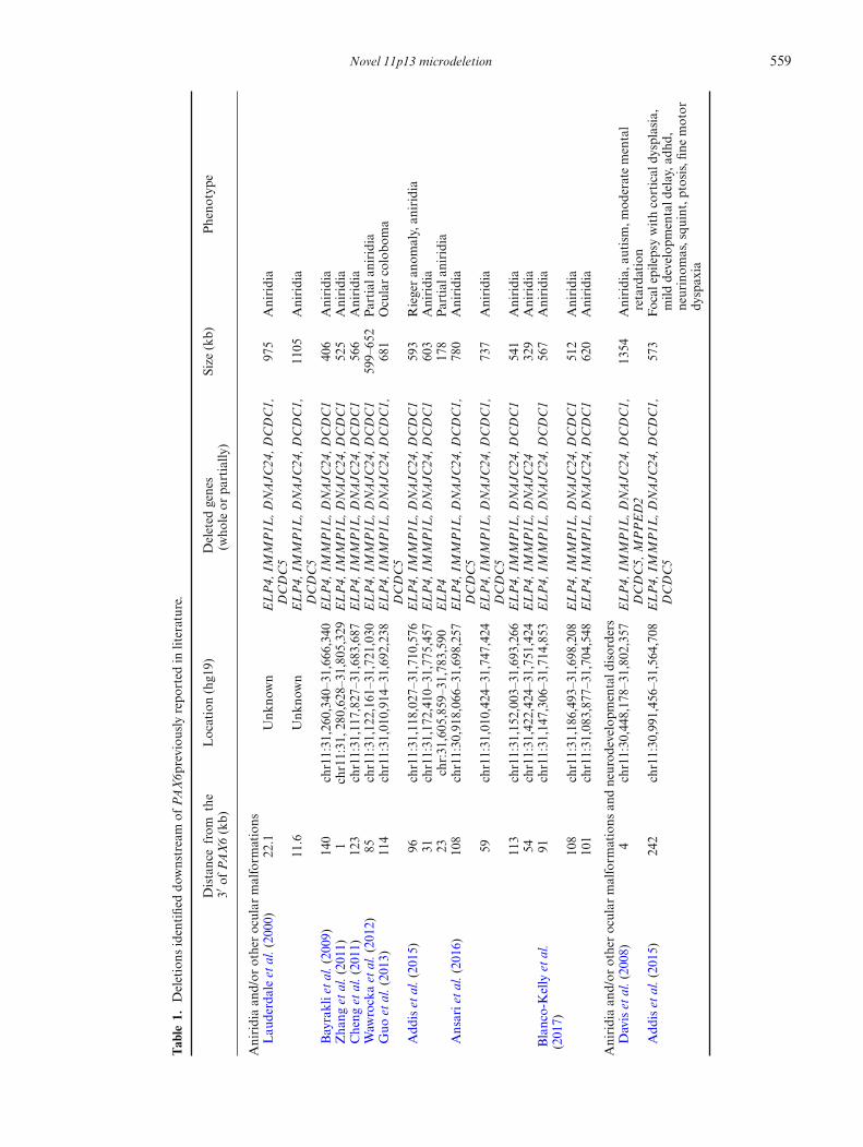
Figure 1. Ophthalmological findings in the index patient and her son. (a) Slit-lamp photographs of the index patient (top) and herson (bottom) showing complete hypoplasia of the iris in both patients. Also lens dislocation in the left eye of the index patient isshown as well as opacities in both of her lenses. (b) The fundus photographs of the index patient (top) and her son (bottom) showingfoveal hypoplasia. (c) The optical coherence tomography (OCT) of the macula confirming foveal hypoplasia in the index patient (left)and her son (right). L, left eye; R, right eye.
at 11p13, which are thought to be rare causes of aniridia,leading to partialPAX6 deletion, completePAX6 deletion,or deletion of the 3′ PAX6 regulatory region leaving itsentire coding region completely intact (Crolla andHeynin-gen 2002). In this study, we report the identification ofa novel microdeletion affecting the 3′ PAX6 regulatoryregion in a patient with familial aniridia and review thepreviously described phenotypes associated with similarmicrodeletions.
Materials and methods
Informed consent was obtained from all family mem-bers. Bidirectional Sanger sequencing of PAX6 was per-formed on an ABI 3130XL genetic analyzer (AppliedBiosystems, Foster City, USA). Array-CGH analysis wasperformed using the Cytochip ISCA array (ver. 1.0,BlueGnome, Cambridge, UK) with 180,000 oligos in a4x180k format. Fluorescent ratios were calculated usingthe Bluefuse Multi software (ver. 4.2, BlueGnome).
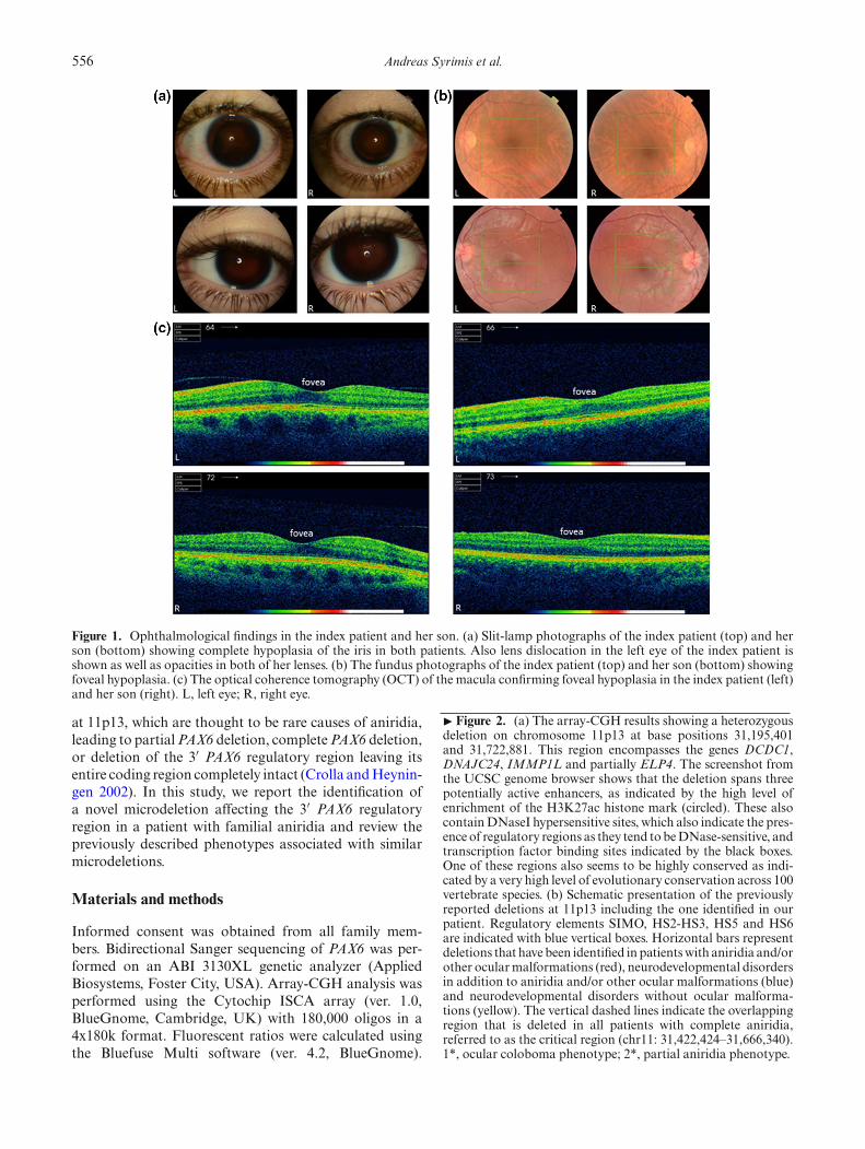
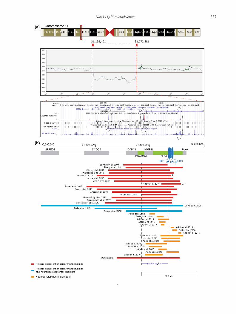
� Figure 2. (a) The array-CGH results showing a heterozygousdeletion on chromosome 11p13 at base positions 31,195,401and 31,722,881. This region encompasses the genes DCDC1,DNAJC24, IMMP1L and partially ELP4. The screenshot fromthe UCSC genome browser shows that the deletion spans threepotentially active enhancers, as indicated by the high level ofenrichment of the H3K27ac histone mark (circled). These alsocontainDNaseI hypersensitive sites, which also indicate the pres-ence of regulatory regions as they tend tobeDNase-sensitive, andtranscription factor binding sites indicated by the black boxes.One of these regions also seems to be highly conserved as indi-cated by a very high level of evolutionary conservation across 100vertebrate species. (b) Schematic presentation of the previouslyreported deletions at 11p13 including the one identified in ourpatient. Regulatory elements SIMO, HS2-HS3, HS5 and HS6are indicated with blue vertical boxes. Horizontal bars representdeletions that have been identified in patientswith aniridia and/orother ocularmalformations (red), neurodevelopmental disordersin addition to aniridia and/or other ocular malformations (blue)and neurodevelopmental disorders without ocular malforma-tions (yellow). The vertical dashed lines indicate the overlappingregion that is deleted in all patients with complete aniridia,referred to as the critical region (chr11: 31,422,424–31,666,340).1*, ocular coloboma phenotype; 2*, partial aniridia phenotype.
Novel 11p13 microdeletion 557
.
558 Andreas Syrimis et al.
Multiplex ligation-dependentprobeamplification (MLPA)was conducted using the SALSA probemix P219-B3(MRC-Holland, Amsterdam, The Netherlands). Frag-ment separation by capillary electrophoresis was per-formed on anABI 3130xl genetic analyzer.MLPAanalysiswas performed using the Coffalyser.Net software (ver. 1.4,MRC-Holland). Quantitative real time PCR was carriedout on the CFX96Real-TimeC1000 Thermal Cycler (Bio-rad,Hercules,USA) using the SsoFast Evagreen Supermix(Biorad). Several primers were initially designed to spanthe region adjacent to the deletion breakpoints at inter-vals of 5000 bp and then new primers were designed at 700bp intervals. Data analysis was performed using the CFXManager Software (Biorad).
Results
A 44-year-old female was referred to the Clinical Geneticsclinic with the diagnosis of aniridia. Her 13-year-old sonwas also affected. Review of her family history revealedthat she was one of the four siblings born to noncon-sanguineous parents. One of her maternal uncles wasdiagnosed with renal cancer at the age of 50 years. Familyhistory was otherwise noncontributory for aniridia.The probandwas an IUGRbabyborn close to termwith
a birth weight of 1.9 kg. Bilateral aniridia was identified inearly infancy. Her development was unremarkable. At theage of 35, she was incidentally diagnosed with type 2 dia-betes mellitus. At the age of 38, she underwent surgeryfor a multinodular goiter. Histology revealed an 8-mmpapillary thyroid carcinoma of the isthmus (T1N0M0).She developed postural tremor at the age of 42. She alsohad a history of hyperlipidaemia and hypovitaminosis D.On examination, her occipitofrontal circumference (ofc)was 55 cm (50th–75th centile), her weight was 79 kg(91st–98th centile) and her height was 154 cm (2nd–9thcentile). She was obese with a BMI 33.3 kg/m2. She wasfacially nondysmorphic. She had a short neck. She hadupper chest and shoulder freckling. She had a 2/6 softsystolic heart murmur. Neurology examination was non-focal. On ophthalmology evaluation, she had horizontalnystagmus and uncorrected visual acuity of 0.2 in eacheye, which increased to 0.4 when corrected with myopiclenses. The intraocular pressure was 20 mmHg in the righteye and 21 mmHg in the left eye. Slit lamp examina-tion showed near total aniridia in both eyes (figure 1a).The intraocular lens in the left eye was superiorly sub-luxated with stretched zonules and with opacities in botheyes (cataract). Gonioscopy showed rudimentary iris tis-sue. Fundoscopy revealed grade 3 foveal hypoplasia,whichwas confirmed by optical coherence tomography (OCT) ofthe macula (figure 1, b–c).Examination of her son at the age of 13 years and 10
months revealed an ofc of 55.5 cm (25th–50th centile), aweight of 76 kg (98th–99.6th centile) and height of 166
cm (50th–75th centile). He had bilateral aniridia. He hada single left-sided irregular hyperpigmented macule overthe upper abdomen. Cardiovascular examination revealeda 2–3/6 systolic heart murmur. He was overweight witha BMI of 27.6 kg/m2. Neurology examination was non-focal. Abdominal ultrasound scan was unremarkable. Hehad horizontal nystagmus and uncorrected visual acuityof 0.5 in each eye, which increased to 0.9 with hyperme-tropic lenses. The intraocular pressure was 16 mmHg inthe right eye and 17 mmHg in the left eye. Slit lamp exam-ination showed near total aniridia in both eyes (figure 1a).The intraocular lens was clear without any opacities andwithout any lens dislocation in either eye. Fundoscopyrevealed grade 1 foveal hypoplasia without any other reti-nal abnormality, which was confirmed with OCT of themacula (figure 1, b–c). Abdominal ultrasound scan wasunremarkable.Bidirectional Sanger sequencing of the coding and
flanking regions of PAX6 revealed no mutations in theproband and her son. Array-CGH detected a novelheterozygous 3′ deletion of ∼577 kb in size located37-kb downstream of PAX6 in the 11p13 region (chr11:31,195,401–31,772,881; figure 2a). This finding was con-firmed by MLPA in both proband and her son. Sub-sequently, qRT-PCR refined the deletion size to about564 kb located ∼46.5-kb downstream of PAX6 (chr11:31,200,388–31,764,028), encompassing the genesDCDC1,DNAJC24, IMMP1L and partially ELP4. This regionalso encompasses three sites that show enrichment for theH3K27ac histone mark, which indicates the presence ofpotentially active enhancers. In addition, one of these sitesthat show enrichment for the H3K27ac mark also show ahigh level of evolutionary conservation across 100 verte-brate species, further supporting the presence of regulatoryelements (figure 2b).
Discussion and review of the literature
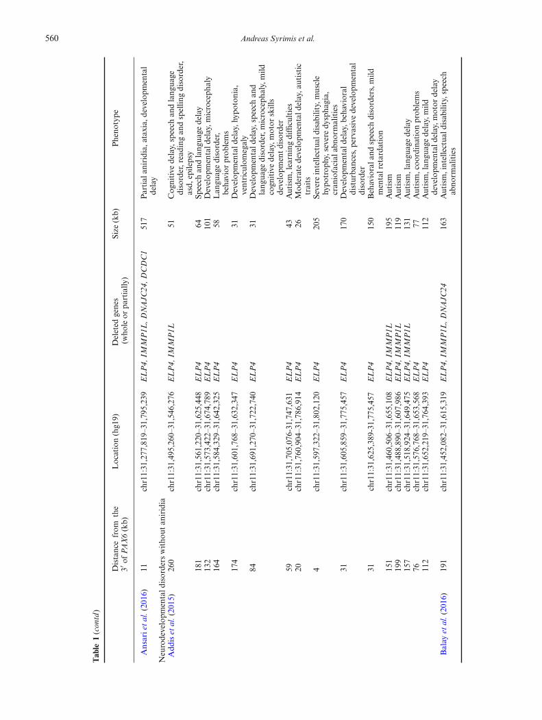
In this study, we identified a novel 564 kb deletion down-stream of PAX6 in a family with bilateral aniridia andfoveal hypoplasia. Deletions of the 3′ regulatory regionsdownstream of PAX6 abrogate its expression leading toaniridia due to PAX6 haploinsufficiency, a phenomenonknown as position effect (Fantes et al. 1995). Twentypatients with a similar deletion downstream of PAX6werereported in the literature to date. The phenotypes of thesepatients, the breakpoints as well as the genes within thedeleted region are summarized in table 1. All these patientshad aniridia, except a patientwith a deletion 114-kb down-stream of PAX6, who had ocular coloboma (Guo et al.2013). A patient with a larger deletion (1.3 Mb) encom-passingMPPED2,DCDC5,DCD1, IMMP1L,DNAJC24and ELP4 was also reported to have autism spectrumdisorder and intellectual disability in addition to aniridia(Davis et al. 2008).
Novel 11p13 microdeletion 559
Table
1.Deletions
identifieddo
wnstream
ofPA
X6p
reviou
slyrepo
rted
inliterature.
Distancefrom
the
3′of
PAX6(kb)
Location(hg19)
Deleted
genes
(who
leor
partially
)Size
(kb)
Pheno
type
Aniridiaan
d/or
otherocular
malform
ations
Lau
derdaleet
al.(2000)
22.1
Unk
nown
ELP4,
IMMP1L
,DNAJC
24,D
CDC1,
DCDC5
975
Aniridia
11.6
Unk
nown
ELP4,
IMMP1L
,DNAJC
24,D
CDC1,
DCDC5
1105
Aniridia
Bayraklietal.(2009)
140
chr11:31,260,340–31,666,340
ELP4,
IMMP1L
,DNAJC
24,D
CDC1
406
Aniridia
Zha
nget
al.(2011)
1chr11:31,2
80,628–31,805,329
ELP4,
IMMP1L
,DNAJC
24,D
CDC1
525
Aniridia
Cheng
etal.(2011)
123
chr11:31,117,827–31,683,687
ELP4,
IMMP1L
,DNAJC
24,D
CDC1
566
Aniridia
Waw
rockaet
al.(2012)
85chr11:31,122,161–31,721,030
ELP4,
IMMP1L
,DNAJC
24,D
CDC1
599–
652
Partial
aniridia
Guo
etal.(2013)
114
chr11:31,010,914–31,692,238
ELP4,
IMMP1L
,DNAJC
24,D
CDC1,
DCDC5
681
Ocularcolobo
ma
Add
iset
al.(2015)
96chr11:31,118,027–31,710,576
ELP4,
IMMP1L
,DNAJC
24,D
CDC1
593
Riegeran
omaly,an
iridia
31chr11:31,172,410–31,775,457
ELP4,
IMMP1L
,DNAJC
24,D
CDC1
603
Aniridia
23chr:31,605,859–31,783,590
ELP4
178
Partial
aniridia
Ansarietal.(2016)
108
chr11:30,918,066–31,698,257
ELP4,
IMMP1L
,DNAJC
24,D
CDC1,
DCDC5
780
Aniridia
59chr11:31,010,424–31,747,424
ELP4,
IMMP1L
,DNAJC
24,D
CDC1,
DCDC5
737
Aniridia
113
chr11:31,152,003–31,693,266
ELP4,
IMMP1L
,DNAJC
24,D
CDC1
541
Aniridia
54chr11:31,422,424–31,751,424
ELP4,
IMMP1L
,DNAJC
2432
9Aniridia
Blanco-Kellyet
al.
(2017)
91chr11:31,147,306–31,714,853
ELP4,
IMMP1L
,DNAJC
24,D
CDC1
567
Aniridia
108
chr11:31,186,493–31,698,208
ELP4,
IMMP1L
,DNAJC
24,D
CDC1
512
Aniridia
101
chr11:31,083,877–31,704,548
ELP4,
IMMP1L
,DNAJC
24,D
CDC1
620
Aniridia
Aniridiaan
d/or
otherocular
malform
ations
andneurod
evelop
mentald
isorders
Daviset
al.(2008)
4chr11:30,448,178–31,802,357
ELP4,
IMMP1L
,DNAJC
24,D
CDC1,
DCDC5,
MPPED2
1354
Aniridia,
autism
,mod
eratemental
retardation
Add
iset
al.(2015)
242
chr11:30,991,456–31,564,708
ELP4,
IMMP1L
,DNAJC
24,D
CDC1,
DCDC5
573
Focal
epilepsywithcortical
dysplasia,
mild
developm
entald
elay,a
dhd,
neurinom
as,squ
int,ptosis,fi
nemotor
dyspax
ia
560 Andreas Syrimis et al.
Table
1(contd)
Distancefrom
the
3′of
PAX6(kb)
Location(hg19)
Deleted
genes
(who
leor
partially
)Size
(kb)
Pheno
type
Ansarietal.(2016)
11chr11:31,277,819–31,795,239
ELP4,
IMMP1L
,DNAJC
24,D
CDC1
517
Partial
aniridia,ataxia,
developm
ental
delay
Neurodevelopm
entald
isorders
witho
utan
iridia
Add
iset
al.(2015)
260
chr11:31,495,260–31,546,276
ELP4,
IMMP1L
51Cog
nitive
delay,speech
andlang
uage
disorder,reading
andspellin
gdisorder,
asd,
epilepsy
181
chr11:31,561,220–31,625,448
ELP4
64Sp
eech
andlang
uage
delay
132
chr11:31,573,422–31,674,789
ELP4
101
Develop
mentald
elay,m
icroceph
aly
164
chr11:31,584,329–31,642,325
ELP4
58Lan
guagedisorder,
behavior
prob
lems
174
chr11:31,601,768–31,632,347
ELP4
31Develop
mentald
elay,h
ypoton
ia,
ventriculomegaly
84chr11:31,691,270–31,722,740
ELP4
31Develop
mentald
elay,speechan
dlang
uage
disorder,m
icroceph
aly,mild
cogn
itivedelay,motor
skills
developm
entdisorder
59chr11:31,705,076-31,747,631
ELP4
43Autism,learningdifficulties
20chr11:31,760,904–31,786,914
ELP4
26Mod
eratedevelopm
entald
elay,a
utistic
traits
4chr11:31,597,322–31,802,120
ELP4
205
Severe
intellectua
ldisab
ility,m
uscle
hypo
trop
hy,severedy
spha
gia,
cran
iofacial
abno
rmalities
31chr11:31,605,859–31,775,457
ELP4
170
Develop
mentald
elay,b
ehavioral
disturba
nces,p
ervasive
developm
ental
disorder
31chr11:31,625,389-31,775,457
ELP4
150
Behaviorala
ndspeech
disorders,mild
mentalretarda
tion
151
chr11:31,460,506–31,655,108
ELP4,
IMMP1L
195
Autism
199
chr11:31,488,890–31,607,986
ELP4,
IMMP1L
119
Autism
157
chr11:31,518,924–31,649,475
ELP4,
IMMP1L
131
Autism,lan
guagedelay
76chr11:31,576,768–31,653,568
ELP4
77Autism,coo
rdinationprob
lems
112
chr11:31,652,219–31,764,393
ELP4
112
Autism,lan
guagedelay,mild
developm
entald
elay,m
otor
delay
Balay
etal.(2016)
191
chr11:31,452,082–31,615,319
ELP4,
IMMP1L
,DNAJC
2416
3Autism,intellectua
ldisab
ility,speech
abno
rmalities
Novel 11p13 microdeletion 561
To further assess the prevalence of our identifieddeletion, we searched the DECIPHER database forpatientswith similar deletions downstreamofPAX6, yield-ing 12 additional aniridia patients (Firth et al. 2009).One of these patients in addition was reported to havecongenital cataracts and nystagmus, while another onewas also described to have global developmental delay.Another patient reported in DECIPHER had Riegeranomaly. Therefore, the prevalence of 11p13 microdele-tion in affected individuals seems to be high, althoughclinical manifestations may vary depending on the size ofthe deletion, the location of the breakpoints and the genesinvolved.All deletions downstream of PAX6 previously reported
to cause either aniridia or other ocular malformations,including the one identified in our study, encompassa common overlapping region 244 kb in size (chr11:31,422,424–31,666,340; figure 2b). This was previouslycharacterized by Ansari et al. (2016) as the ‘criticalregion’ for aniridia. This region partially encompassesDNAJC24, IMMP1L and introns 1–7 of ELP4. A dele-tion including only part of the critical region was foundin a patient with partial aniridia, suggesting that a par-tial deletion of this critical region possibly leads toa milder ocular phenotype (figure 2b) (Addis et al.2015).
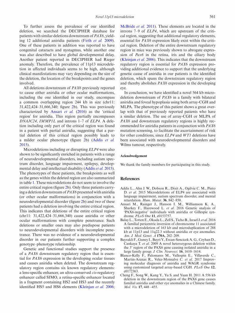
Microdeletions including or disrupting ELP4 were alsoshown to be significantly enriched in patients with a rangeof neurodevelopmental disorders, including autism spec-trum disorder, language impairment, epilepsy, develop-mental delay and intellectual disability (Addis et al. 2015).The phenotypes of these patients, the breakpoints as wellas the genes within the deleted region are also summarizedin table 1. These microdeletions do not seem to involve theentire critical region (figure 2b). Only three patients carry-ingadeletiondownstreamofPAX6presentedwithaniridia(or other ocular malformations) in conjunction with aneurodevelopmental disorder (figure 2b) and two of thesepatients had a deletion involving the entire critical region.This indicates that deletions of the entire critical region(chr11: 31,422,424–31,666,340) cause aniridia or otherocular malformations with complete penetrance. Suchdeletions or smaller ones may also predispose patientsto neurodevelopmental disorders with incomplete pene-trance. There was no evidence of a neurodevelopmentaldisorder in our patients further supporting a complexgenotype–phenotype relationship.Genetic and functional studies support the presence
of a PAX6 downstream regulatory region that is essen-tial for PAX6 expression in the developing ocular tissuesand causes aniridia when deleted. The downstream reg-ulatory region contains six known regulatory elements:a lens-specific enhancer, an ultra-conserved cis-regulatoryenhancer called SIMO, a retina-specific enhancer locatedin a fragment containing HS2 and HS3 and the recentlyidentified HS5 and HS6 elements (Kleinjan et al. 2006;
McBride et al. 2011). These elements are located in theintrons 7–9 of ELP4, which are upstream of the criti-cal region, suggesting that additional regulatory elements,essential for PAX6 expression likely exist within the criti-cal region. Deletion of the entire downstream regulatoryregion in mice was previously shown to abrogate expres-sion of Pax6 in the retina, iris and the ciliary body(Kleinjan et al. 2006). This indicates that the downstreamregulatory region is essential for PAX6 expression pro-viding additional evidence to support that the underlyinggenetic cause of aniridia in our patients is the identifieddeletion, which spans the downstream regulatory regionand thereby abolishes PAX6 expression in the developingeye.In conclusion, we have identified a novel 564 kb micro-
deletion downstream of PAX6 in a family with bilateralaniridia and foveal hypoplasia using both array-CGH andMLPA. The phenotype of this patient shows a great over-lap with that of previously reported patients who havea similar deletion. The use of array-CGH or MLPA ofPAX6 and downstream regulatory regions is highly rec-ommended for aniridia patients in conjunction withPAX6mutation screening, to facilitate the ascertainment of riskfor other conditions, since ELP4 andWT1 deletions havebeen associated with neurodevelopmental disorders andWilms tumour, respectively.
Acknolwedgement
We thank the family members for participating in this study.
References
Addis L., Ahn J. W., Dobson R., Dixit A., Ogilvie C. M., PintoD. et al. 2015 Microdeletions of ELP4 are associated withlanguage impairment, autism spectrum disorder, and mentalretardation. Hum. Mutat. 36, 842–850.
Ansari M., Rainger J., Hanson I. M., Williamson K. A.,Sharkey F., Harewood L. et al. 2016 Genetic snalysis of‘PAX6-negative’ individuals with aniridia or Gillespie syn-drome. PLoS One 11, e0153757.
BalayL.,TottenE.,OkadaL.,Zell S., TichoB., Israel J. et al. 2016A familial pericentric inversion of chromosome 11 associatedwith a microdeletion of 163 kb and microduplication of 288kb at 11p13 and 11q22.3 without aniridia or eye anomalies.Am. J. Med. Genet. A 170A, 202–209.
Bayrakli F.,Guney I., BayriY., Ercan-SencicekA.G.,CeyhanD.,Cankaya T. et al. 2009 A novel heterozygous deletion withinthe 3’ region of the PAX6 gene causing isolated aniridia in alarge family group. J. Clin. Neurosci. 16, 1610–1614.
Blanco-Kelly F., Palomares M., Vallespin E., Villaverde C.,Martin-Arenas R., Velez-Monsalve C. et al. 2017 Improv-ing molecular diagnosis of aniridia and WAGR syndromeusing customized targeted array-based CGH. PLoS One 12,e0172363.
Cheng F., Song W., Kang Y., Yu S. and Yuan H. 2011 A 556 kbdeletion in the downstream region of the PAX6 gene causesfamilial aniridia and other eye anomalies in a Chinese family.Mol. Vis. 17, 448 –455.
562 Andreas Syrimis et al.
Crolla J. A. and van Heyningen V. 2002 Frequentchromosome aberrations revealed by molecular cytoge-netic studies in patients with aniridia. Am. J. Hum. Genet. 71,1138–1149.
Davis L. K., Meyer K. J., Rudd D. S., Librant A. L., Epping E.A., SheffieldV.C. et al. 2008Pax6 3′ deletion results in aniridia,autism and mental retardation. Hum. Genet. 123, 371–378.
Fantes J., Redeker B., Breen M., Boyle S., Brown J., FletcherJ. et al. 1995 Aniridia-associated cytogenetic rearrangementssuggest that a position effect may cause themutant phenotype.Hum. Mol. Genet. 4, 415–422.
Firth H. V., Richards S. M., Bevan A. P., Clayton S., Corpas M.,Rajan D. et al. 2009 DECIPHER: database of chromosomalimbalance and phenotype in humans dsing ensembl resources.Am. J. Hum. Genet. 84, 524–533.
GronskovK., Olsen J. H., SandA., PedersenW., CarlsenN., BakJylling A. M. et al. 2001 Population-based risk estimates ofWilms tumor in sporadic aniridia. A comprehensive mutationscreening procedure of PAX6 identifies 80% of mutations inaniridia. Hum. Genet. 109, 11–18.
GuoH., Dai L., HuangY., Liao Q. and Bai Y. 2013 A large noveldeletion downstream of PAX6 gene in a Chinese family withocular coloboma. PLoS One 8, e83073.
Hingorani M., Hanson I. and van Heyningen V. 2012 Aniridia.Eur. J. Hum. Genet. 20, 1011–1017.
Kleinjan D. A., Seawright A., Mella S., Carr C. B., Tyas D. A.,Simpson T. I. et al. 2006 Long-range downstream enhancersare essential for Pax6 expression. Dev. Biol. 299, 563–581.
Lauderdale J. D., Wilensky J. S., Oliver E. R., Walton D. S. andGlaser T. 2000 3′ deletions cause aniridia by preventing PAX6gene expression. Proc. Natl. Acad. Sci. USA 97, 13755–13759.
McBride D. J., Buckle A., van Heyningen V. and Kleinjan D.A. 2011 DNaseI hypersensitivity and ultraconservation revealnovel, interdependent long-range enhancers at the complexPax6 cis-regulatory region. PLoS One 6, e28616.
Miller R. W., Fraumeni J. F., Jr. and Manning M. D. 1964Association of Wilms’s tumor with aniridia, hemihypertro-phy and other congenital malformations.N. Engl. J. Med. 270,922–927.
Nelson L. B., Spaeth G. L., Nowinski T. S., Margo C. E. andJacksonL. 1984Aniridia.A review.Surv.Ophthalmol. 28, 621–642.
Robinson D. O., Howarth R. J., Williamson K. A., van Heynin-gen V., Beal S. J. and Crolla J. A. 2008 Genetic analysis ofchromosome 11p13 and the PAX6 gene in a series of 125cases referred with aniridia. Am. J. Med. Genet. A 146A,558–569.
Samant M., Chauhan B. K., Lathrop K. L. and Nischal K. K.2016 Congenital aniridia: etiology, manifestations and man-agement. Expert Rev. Ophthalmol. 11, 135–144.
Simpson T. I. and Price D. J. 2002 Pax6; a pleiotropic player indevelopment. BioEssays 24, 1041–1051.
Valenzuela A. and Cline R. A. 2004 Ocular and nonocularfindings in patients with aniridia. Can. J. Ophthalmol. 39,632–638.
Wawrocka A., Budny B., Debicki S., Jamsheer A., Sowinska A.andKrawczynskiM.R. 2012 PAX6 3′ deletion in a familywithaniridia. Ophthalmic Genet. 33, 44–48.
Zhang X., Zhang Q., Tong Y., Dai H., Zhao X., Bai F. et al.2011 Large novel deletions detected in Chinese families withaniridia: correlation between genotype and phenotype. Mol.Vis. 17, 548–557.
Corresponding editor: Rajiva Raman