anatomy rhs 241 lecture 12 - ksu
TRANSCRIPT
The Upper Limb III
The Brachial Plexus
AnatomyRHS 241
Lecture 12Dr. Einas Al-Eisa

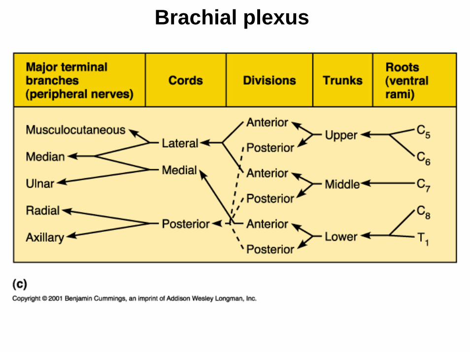
Brachial plexus
• Network of nerves supplying the upper limb
• Compression of the plexus results in motor & sensory changes within the upper limb
• The upper limb is innervated by ventral rami(just like the lower limb, and most muscles of the thoracic and abdominal walls)


Which structures are supplied by the dorsal rami of spinal nerves?

Spinal nerves
Brachial plexus

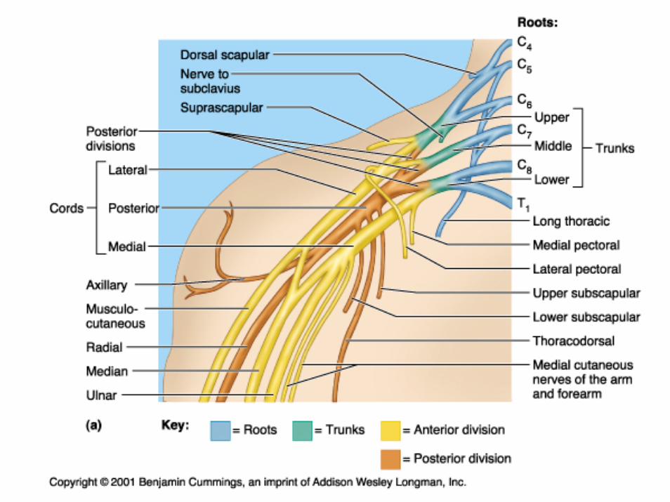
Brachial plexus componentsRoots (5)
• The ventral rami of cervical spinal nerves (C5-C8, and T1)
• Lies within the interscalene triangle(boundaries: scalenus anterior to the front, scalenus medius to the back, & the superior surface of the 1st rib below)

Brachial plexus componentsTrunks (3)
• supraclavicular
• Upper (superior) trunk: formed by the union of roots C5 & C6
• Middle trunk: the lateral extension of the C7 root
• Lower (inferior) trunk: formed by the union of roots C8 & T1

Brachial plexus componentsDivisions (6)
• Each of the trunks of the brachial plexus divides into anterior & posterior divisions.
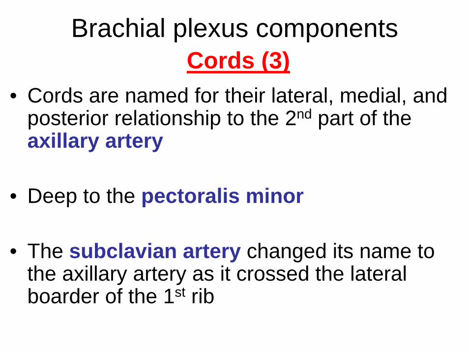
Brachial plexus componentsCords (3)
• Cords are named for their lateral, medial, and posterior relationship to the 2nd part of the axillary artery
• Deep to the pectoralis minor
• The subclavian artery changed its name to the axillary artery as it crossed the lateral boarder of the 1st rib


Brachial plexus componentsCords (3)
• Lateral cord: formed by the union of anterior divisions of the superior & middle trunks (C5, C6, & C7)
• Medial cord: formed by the anterior division of the inferior trunk (C8 & T1)
• Posterior cord: formed by the union of the three posterior divisions (C5 to T1)
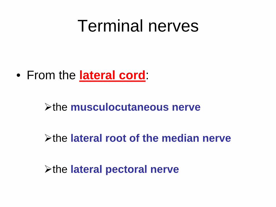
Terminal nerves
• From the lateral cord:
the musculocutaneous nerve
the lateral root of the median nerve
the lateral pectoral nerve

Terminal nerves
• From the medial cord:
the ulnar nerve (C8, T1)
the medial root of the median nerve
the medial pectoral nerve

Terminal nerves
• From the posterior cord:
the axillary nerve (C5, C6)
the radial nerve (C5-T1)
the thoracodorsal nervethe upper & lower subscapular nerves
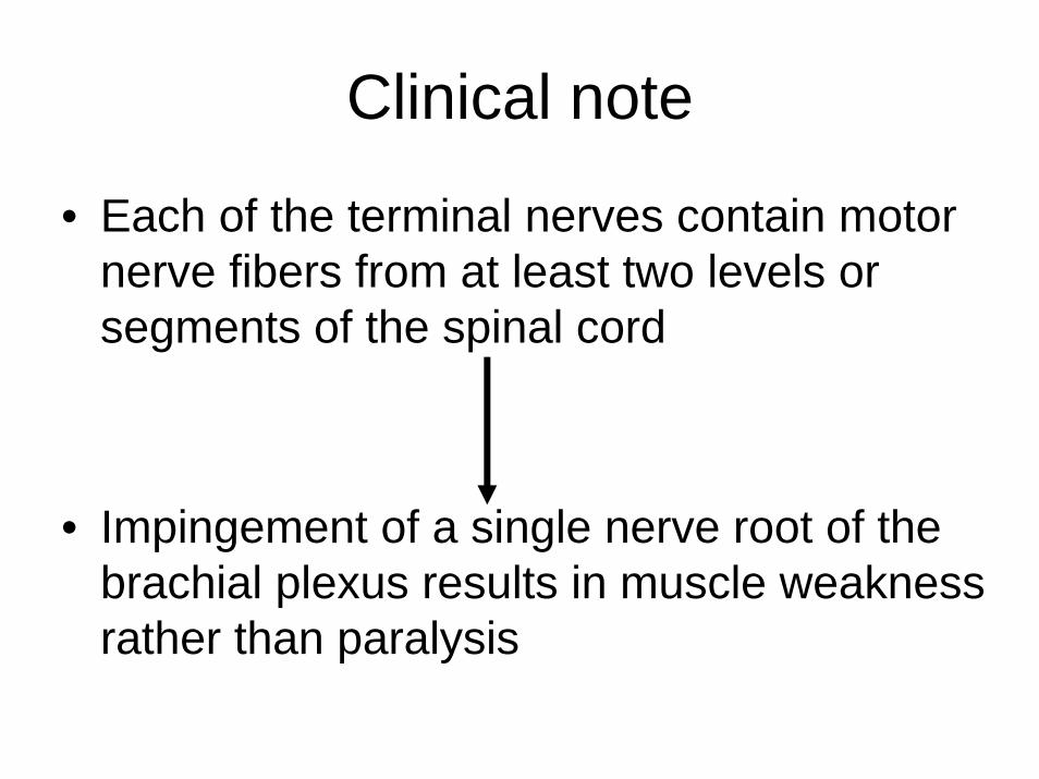
Clinical note
• Each of the terminal nerves contain motor nerve fibers from at least two levels or segments of the spinal cord
• Impingement of a single nerve root of the brachial plexus results in muscle weakness rather than paralysis

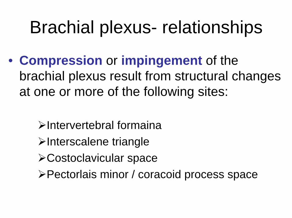
Brachial plexus- relationships
• Compression or impingement of the brachial plexus result from structural changes at one or more of the following sites:
Intervertebral formainaInterscalene triangleCostoclavicular spacePectorlais minor / coracoid process space

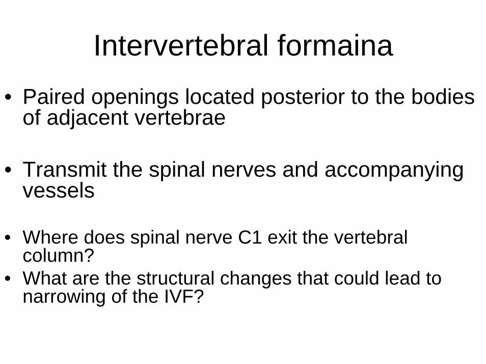
Intervertebral formaina• Paired openings located posterior to the bodies
of adjacent vertebrae
• Transmit the spinal nerves and accompanying vessels
• Where does spinal nerve C1 exit the vertebral column?
• What are the structural changes that could lead to narrowing of the IVF?
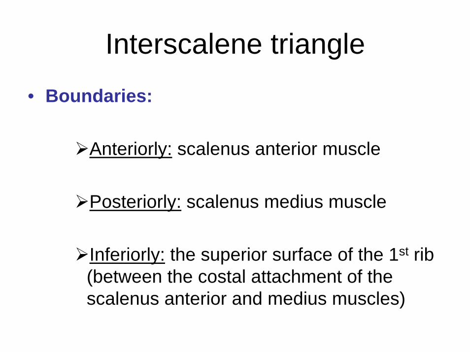
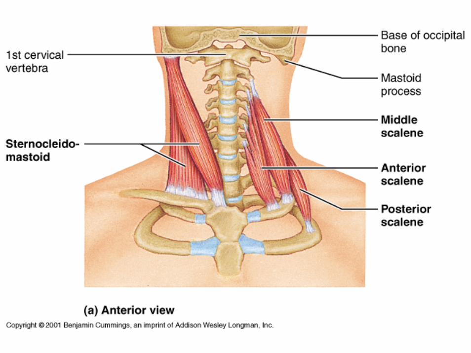
Interscalene triangle
• Boundaries:
Anteriorly: scalenus anterior muscle
Posteriorly: scalenus medius muscle
Inferiorly: the superior surface of the 1st rib (between the costal attachment of the scalenus anterior and medius muscles)

Interscalene triangle
• Content:
The roots of the brachial plexus (C5-T1)
Subclavian artery (which becomes the axillary artery as it crosses the lateral boarder of rib1)
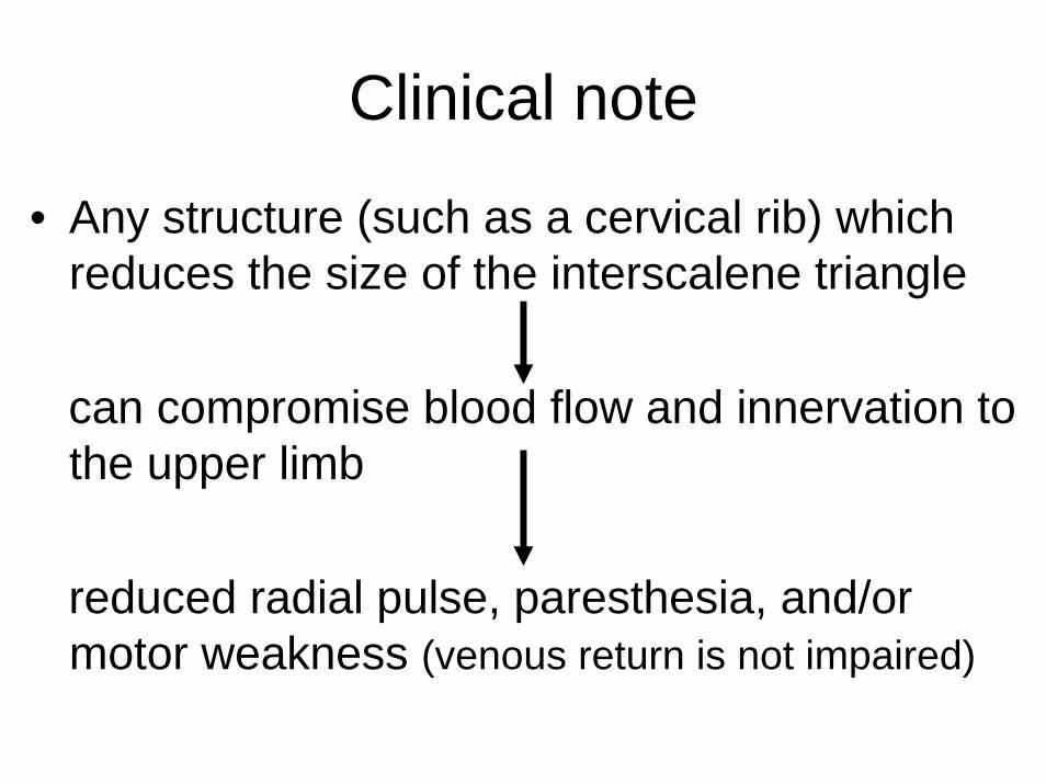
Clinical note
• Any structure (such as a cervical rib) which reduces the size of the interscalene triangle
can compromise blood flow and innervation to the upper limb
reduced radial pulse, paresthesia, and/or motor weakness (venous return is not impaired)


Q: What would you ask a patient to do to reduce the dimensions of
the interscalene triangle?
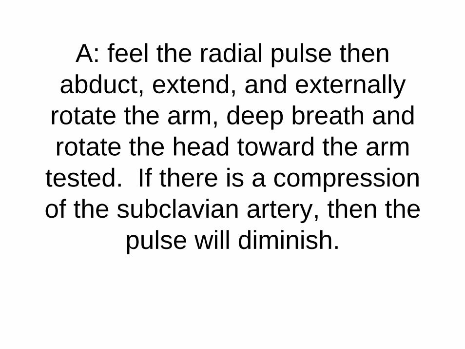
A: feel the radial pulse then abduct, extend, and externally
rotate the arm, deep breath and rotate the head toward the arm
tested. If there is a compression of the subclavian artery, then the
pulse will diminish.

Costoclavicular space
• Boundaries:
Clavicle: approximately middle third
First rib: approximately middle third

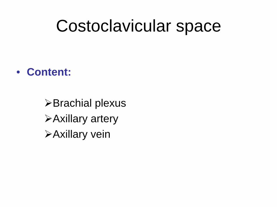
Costoclavicular space
• Content:
Brachial plexusAxillary arteryAxillary vein

The pectoralis minor/ coracoidprocess space
• Pectoralis minor (from ribs 3-5 to coracoid process)
• The brachial plexus lies posterior to the pectoralis minor, inferior to the coracoidprocess, and inferomedial to the head of the humerus
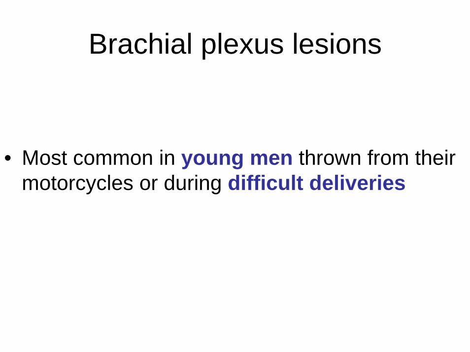
Brachial plexus lesions
• Most common in young men thrown from their motorcycles or during difficult deliveries

Brachial plexus lesions
Closed injuries:Can occur in 2 ways:
1. Violent lateral flexion of the neck with depression of the shoulder, or forced abduction of the arm
2. At birth during difficult deliveries
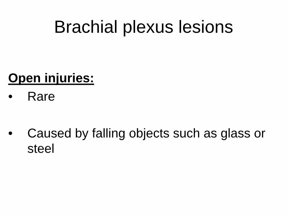
Brachial plexus lesions
Open injuries:• Rare
• Caused by falling objects such as glass or steel

Patterns of brachial plexus lesions
Supraclavicularlesions
Infraclavicularlesions

Supraclavicular lesions
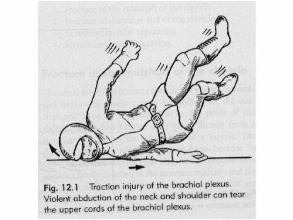
Trauma:
• Mechanism of injury: blows to the head and shoulder cause violent lateral flexion of the cervical spine and depression of the shoulder tear the upper cords
• Example: motorcyclists landing on the head and shoulder

Supraclavicular lesions
Obstetric palsy:• When the upper cords are damaged at birth
weak deltoid, elbow flexors, wrist extensors, supinator
“waiter’s tip” position of the arm (Erb’s palsy)

Erb’s palsy

Erb’s palsy

Infraclavicular lesions
Trauma:Mechanism of injury:
– When the arm is violently abducted– Anterior dislocation of the shoulder
injury to the lower part of the brachial plexus
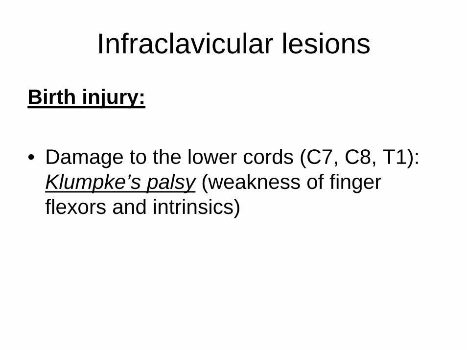
Infraclavicular lesions
Birth injury:
• Damage to the lower cords (C7, C8, T1): Klumpke’s palsy (weakness of finger flexors and intrinsics)
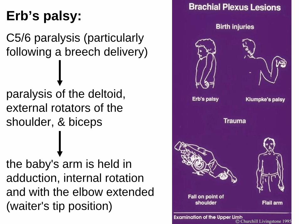
Erb’s palsy: C5/6 paralysis (particularly following a breech delivery)
paralysis of the deltoid, external rotators of the shoulder, & biceps
the baby's arm is held in adduction, internal rotation and with the elbow extended (waiter's tip position)

Klumpke's palsy: C7, C8 and T1 palsy
flexed elbow & paralyzed hand