terminal nerves of the brachial plexus - ksu...
TRANSCRIPT
Terminal Nerves of the Brachial Plexus
AnatomyRHS 241
Lecture 17Dr. Einas Al-Eisa
Musculocutaneous nerve
• C5,6,7
• Arises from the lateral cord
• Enters the arm by piercing the coracobrachialis
• Courses deep to the biceps brachii muscle
Musculocutaneous nerve
• Motor distribution: innervates all muscles of the anterior compartment of arm (biceps brachii, coracobrachialis, brachialis)
• Sensory distribution: the lateral cutaneousnerve of the forearm……….
Musculocutaneous nervelesion sites / clinical conditions
• As the nerve courses within the axilla: uncommon
• Motor changes: weakened flexion & supinationof the forearm
• Cutaneous changes: lateral forearm (part of the C6 dermatome)
Median nerve
• C5 – T1
• Arises as two roots from the lateral & medial cords
• Courses within the medial (protected) side of the arm
Median nerve
• Crosses the elbow joint just medial to the biceps tendon
• Courses within the anterior compartment of the forearm (deep to the flexor digitorumsuperficialis)
• Enters the hand by passing through the carpal tunnel
Median nerve
• Motor distribution: Arm: none
Forearm: innervates all muscles of the anterior compartment except the flexor carpi ulnaris and ulnar half of the flexor digitorum profundus
Hand: thenar muscles and the lateral two lumbricals

Median nerve
• Sensory distribution:
Arm and forearm: none
Hand: lateral 2/3rds of the palm, plus the lateral 3 ½fingers
Median nerveEntrapment sites
• Elbow level: as the nerve enters the forearm by passing between the two heads of pronatorteres
• Hand: as the nerve courses through the carpal tunnel
Median nerveclinical conditions
• Ape hand:due to unopposed pull by the extensors & long abductor of the thumb (innervated by..??)
the thumb is pulled dorsally and laterally to lie near the plane of the palm
wasting of the thenar muscles gives the lateral half of the hand a flattened’ ape-like appearance
Median nerveclinical conditions
• Carpal tunnel syndrome:
Painful hand condition caused by overuse (e.g., younger factory workers) or fluid/hormone changes (e.g., pregnancy)
Pain in the lateral side of the hand
Median nerveclinical conditions
• Carpal tunnel syndrome:
Muscle weakness (most noticeably of the thenarmuscles and lateral two lumbricals)
Severe cases are treated surgically by releasing the flexor retinaculum
Ulnar nerve
• C7 – T1• Arises from the medial cord
• Descends within the medial side of the arm
• Lies posterior to the medial epicondyle as it leaves the arm (compressing the nerve here is described as hitting one’s funny bone)
Ulnar nerve
• Enters the forearm by passing between the humeral and ulnar heads of the flexor carpiulnaris
• Enters the hand by coursing superficial to the flexor retinaculum
Ulnar nerve
• Motor distribution: Arm: none
Forearm: innervates the flexor carpi ulnaris and the ulnar ½ of flexor digitorum profundus
Hand: all muscles of the hypothenar eminence, all interossei, the 4th & 5th lumbricals, and the adductor pollicis
Ulnar nerve
• Sensory distribution:
Arm and forearm: none
Hand: skin of the medial 1/3rd of the palm, plus the 5th finger and ulnar half on dorsal and palmaraspects of the ring finger, plus all joints it crosses
Ulnar nerveEntrapment sites
• Elbow level: as the nerve enters the forearm by passing between the two heads of the flexor carpi ulnaris
• Wrist: as a component of Guyon’s tunnel

Ulnar nerveclinical conditions
• Claw hand:Clawing of the ring and 5th fingers
Clawed finger = one in which the MPJ is hyper-extended and both the PIPJ & DIPJ are pulled into flexion
Most severe when entrapments of the ulnar nerve occur at the wrist level
Explain Why……….?
Radial nerve
• C5 – T1• Arises from the posterior cord
• Courses laterally & inferiorly to enter the posterior compartment of the arm
• In the arm, the nerve lies within the spiral groove of the humerus
Radial nerve
• Lies anterior to the lateral condyle of the humerus as it enters the forearm
• Divides here into the superficial branch (sensory/cutaneous) and deep branch (motor)
• The deep branch enters the forearm by passing between the heads of the supinatormuscle
Radial nerve
• Motor distribution: Innervates all muscles within the posterior compartments of the arm & forearm (e.g., extensors of the elbow and wrist)

Radial nerve
• Sensory distribution: cutaeous
Arm: lower half- lateral armForearm: central posteriorHand: lateral half of the dorsum3 ½ fingers dorsally

Radial nerveEntrapment sites
• Arm: as the nerve lies in the spiral groove of the humerus (e.g., mid-humeral fractures)
• Proximal forearm: as its deep branch passes between the heads of the supinator muscle
• Axillary damage of the radial nerve can result from extended use of long crutches
Radial nerveclinical conditions
• Wrist drop:
Due to lost innervation to the extensors of the wrist
Sensation of the dorsal skin of the hand (lateral side) is unchanged with lesions of only the deep branch of the radial nerve
Axillary nerve
• C5 – C6• Arises from the posterior cord
• Courses laterally to pass through the quadrangular space
• Closely related to the surgical neck of humerus
Axillary nerve
• Motor distribution: Innervates the deltoid & teres minor muscles
• Sensory distribution:Cutaneous to the skin over the lower half of the deltoid muscle (the vaccination area)
Axillary nerveEntrapment sites
• As the nerve passes through the quadrangular spaceto innervate muscles of the posterior shouder
• Loss of function usually occurs with anterior dislocation of the shoulder
• The nerve is stretched by the downward and medial displacement of the proximal end of humerus
Axillary nerveclinical conditions
Weakened / lost abduction of the arm (particularly beyond 30o
Sensory changes within the vaccination area
Secondary motor nerves arising from the brachial plexus
Long thoracic nerve:• Arises from roots C5-C7• Descends on the surface of the serratus
anetrior muscle (near the mid-axillary line)• Motor to the serratus anetrior
• Damage of this nerve: “winging of the scapula”
Secondary motor nerves arising from the brachial plexus
Suprascapular nerve:• Arises from roots C5-C6 (superior trunk)
• Passes across the superior boarder of scapula
• Innervate the supraspinatus & infraspinatus
Secondary motor nerves arising from the brachial plexus
Thoracodorsal nerve:• Arises from roots C6-C8 (posterior cord)
• Descends vertically to innervate the lattisimus dorsi
Secondary motor nerves arising from the brachial plexus
Lateral pectoral nerve:• Arises from roots C5-C7 (lateral cord)
• Passes anetriorly to innervate pectoralismajor (superior half) and medial fibers of pectoralis minor

Secondary motor nerves arising from the brachial plexus
Medial pectoral nerve:• Arises from roots C8-T1 (medial cord)
• Passes anetriorly to innervate pectoralismajor (inferior half) and pectoralis minor
Cranial nerves
• Take their origin from the brain which is located in the cranium “cranial nerves”
• Leave the CNS via foramina in the cranium
• Numbered in a way that indicates the order in which they leave the brain
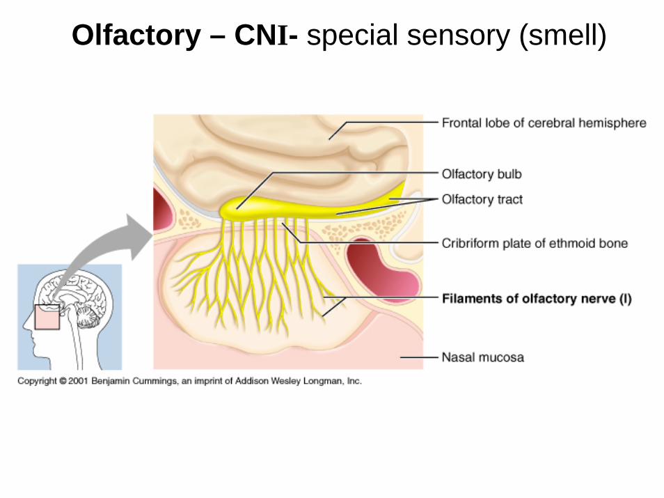
Olfactory – CNI- special sensory (smell)
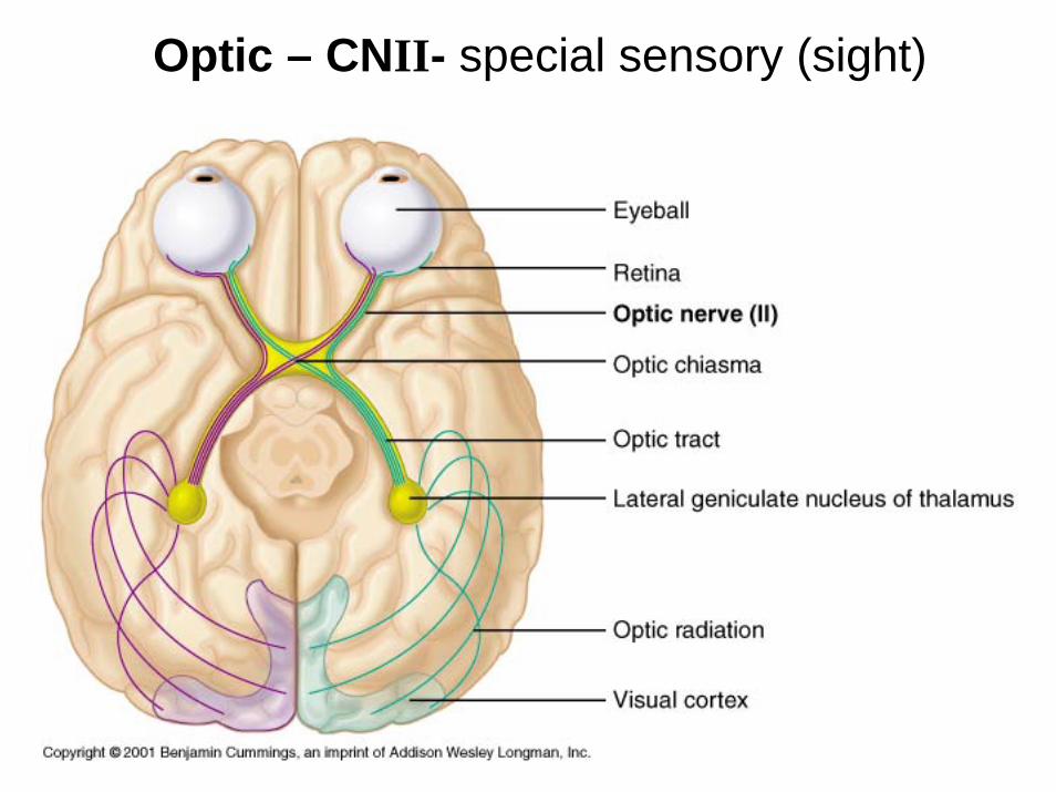
Optic – CNII- special sensory (sight)
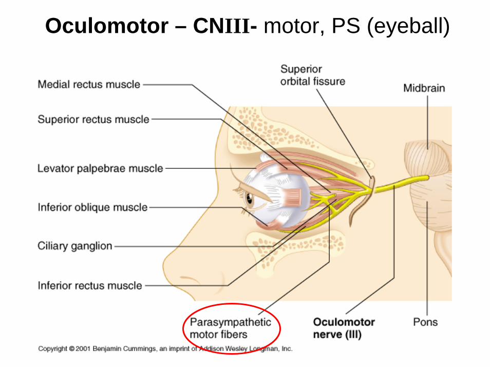
Oculomotor – CNIII- motor, PS (eyeball)
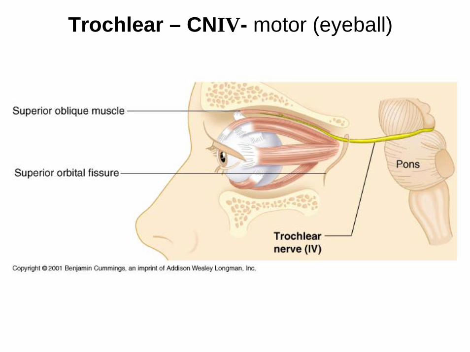
Trochlear – CNIV- motor (eyeball)
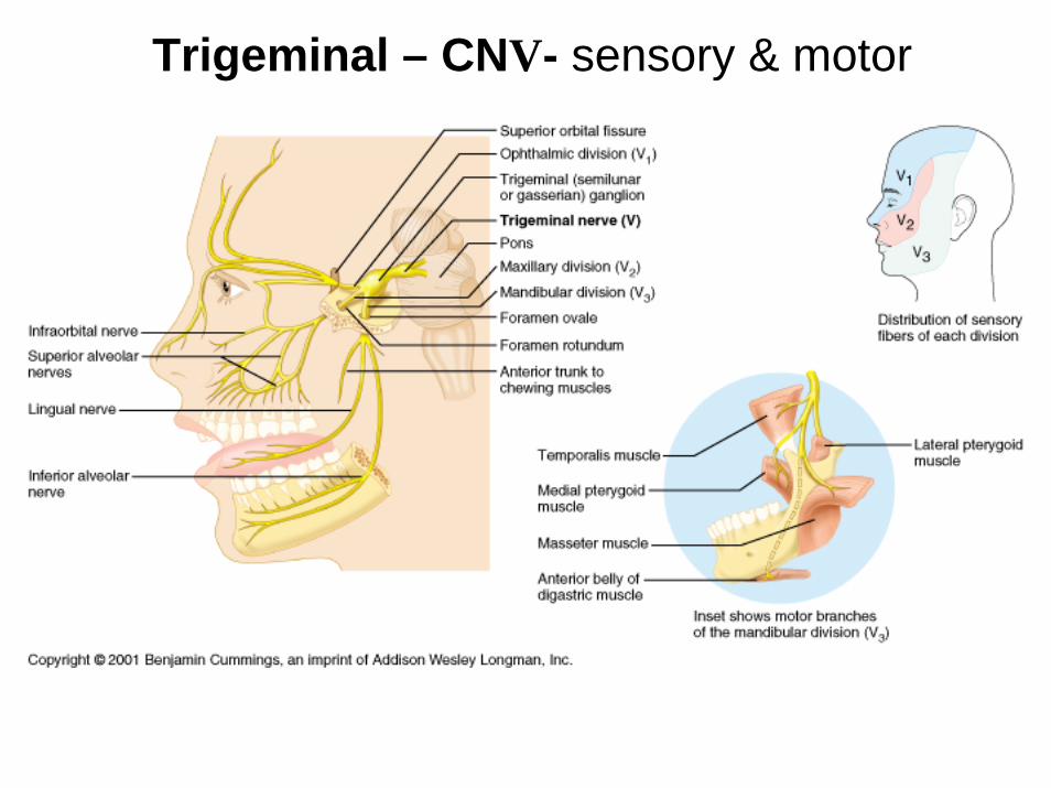
Trigeminal – CNV- sensory & motor
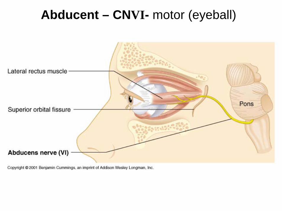
Abducent – CNVI- motor (eyeball)
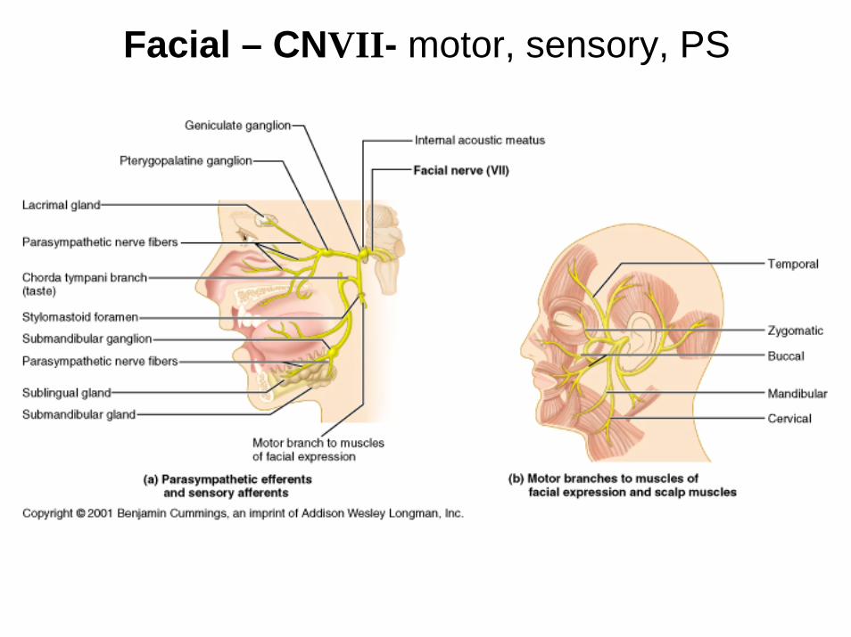
Facial – CNVII- motor, sensory, PS
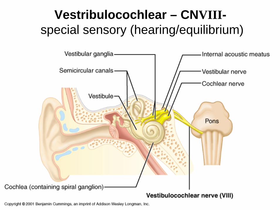
Vestribulocochlear – CNVIII-special sensory (hearing/equilibrium)
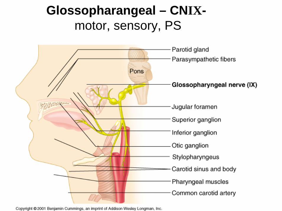
Glossopharangeal – CNIX-motor, sensory, PS
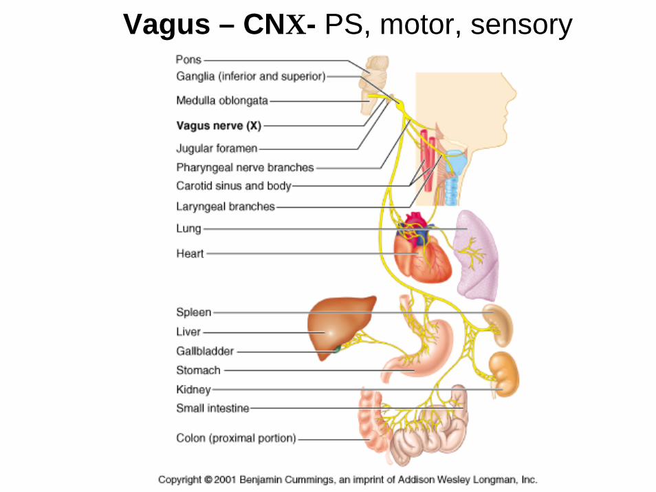
Vagus – CNX- PS, motor, sensory

Accessory – CNXI- motor
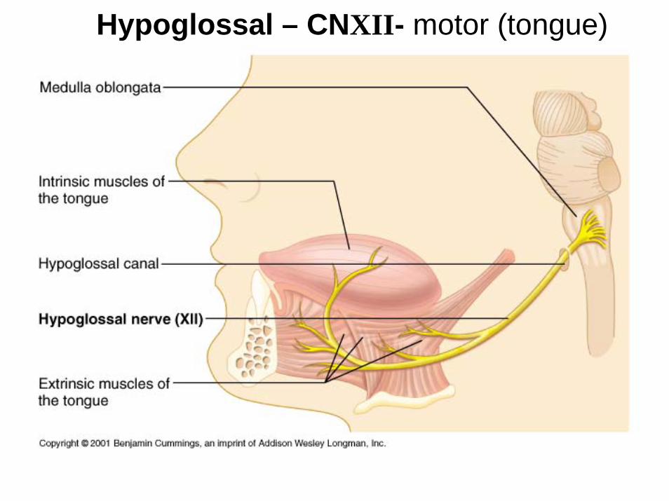
Hypoglossal – CNXII- motor (tongue)
Clinical questions on the UL
• Q: Where in the arm is the radial nerve at most risk of injury?
• A: risk of crushing injury where it lies next to the bone in the spiral groove at the back of the humeral shaft

Clinical questions on the UL
• Q: How does the motor nerve supply to the primary pronators & supinators of the forearm differ?
• A: the primary supinators (the supinatormuscle) is innervated as an extensor via the radial nerve. The primary pronators(pronator quadratus & pronator teres) are innervated as flexors via the median nerve

Clinical questions on the UL
• Q: If the musculocutaneous nerve is severed, is elbow flexion still possible?
• A: yes, weak elbow flexion is still possible. The muscle responsible for this action is brachioradialis (since the biceps and brachialis would be paralyzed)
Clinical questions on the UL
• Q: what would be the motor deficit if the median nerve was cut at the elbow?
• A: loss of pronation, weakness in wrist flexion, loss of thumb mobility, and ulnardeviation of the hand
Clinical questions on the UL
• What is “student elbow”?
• Friction bursitis affecting the superficial olecranon bursa

Clinical questions on the UL
• What motions of the elbow would be hindered if the ulnar nerve were severed in the arm?
• None, because the ulnar nerve does not innervate any prime movers of the elbow
Clinical questions on the UL
• Where in the forearm and wrist are the ulnar nerve and median nerve most at risk of entrapment?
• A patient can pick up a suitcase but can’t pick up a dime. Speculate on the underlying mechanical problem.
Clinical questions on the UL
• Q: what are the palpable bony landmarks of the distal radius and ulna?
• Q: describe how the radial, ulnar, and median nerves enter the forearm
• Q: if the radial nerve is severed at the wrist, what would be the motor deficit?