anatomy - meditron.ch · radiographic school. the phantom contains a real human skeleton as well as...
TRANSCRIPT

Anatomical Models 3D printed Specimen Medical Simulators Anatomical Charts
ANATOMY

1
2
3 4
1 Dental manikinA simple and economical chair manikin for auxiliary training of assistants and hygienists in the dental office. Unit includes DENTOFORM ® model for technique training, Aluminal skull, and soft outer Plassein head. Universal ball-joint allows for rotation of head into a variety of positions. Includes chair mount.
Weight: 2.8 kg
Ref.no. R17000
2 Dental manikin bench mountThis small portable bench-clamp is popular with dental assistant and dental hygiene classes, as well as for dentistry students. It has a lightweight universal ball-joint for simulating neck movement. Use with Dental Manikin R17000.
Weight: 1 kg
Ref.no. R17000A
Dental Radiography Head PhantomThis radiography phantom has removable jaws and tongue allowing for a variety of applications for training and research.
Features:
Each tooth is individually modeled and has a three-layer structure of enamel, dentin and pulp cavity.
Each hard tissue (enamel, dentin, cortical bone and cancellous bone) has a particular HU number and X-ray absorption rate.
Jaws and tongue are detachable to allow access to the oral cavity, pharyngeal cavity and maxillary sinus. Sensors, simulated lesions, or residue can be set in these cavities.
Carotid arteries are prepared as lumens to accommodate simulated calcifications.
Anatomy:
Synthetic skull with nasal cavity, maxillary sinus, mandible alveolar, and maxillary alveolar; cervical vertebrae and hyoid bone, teeth with enamel, dentin and pulp cavity.
Tongue, oral cavity, pharyngeal cavity and carotid arteries3 Head with closed mouth
Ref.no. R16525
4 Head with open mouth Ref.no. R16526

1
22
1 X-ray phantom headHuman skull, safely embedded in plastic for easy use. The jaws are slightly open to allow dental panoramic images of the teeth. The neck includes some cervical vertebrae depending on the ordered type. An embedded tread allows the use with a tripod. The jaw may have dental gaps, dental repairs, broken teeth, replaced teeth or other individual features. For detailed information about the available phantoms please contact our sales team.
X-ray phantom head with cervical vertebrae, transparent
Ref.no. 7300
X-ray phantom head with cervical vertebrae, opaque Ref.no. 7310
X-ray phantom head, transparent Ref.no. 7320
X-ray phantom head, opaque Ref.no. 7330
2 Tripod for x-ray phantom headVery strong tripod for use with the x-ray phantom head. Foldable but stable, holding the head safely in place. With rotatable head for precise positioning of the phantom in the x-ray machine
Ref.no. 7350
1
2
3
4
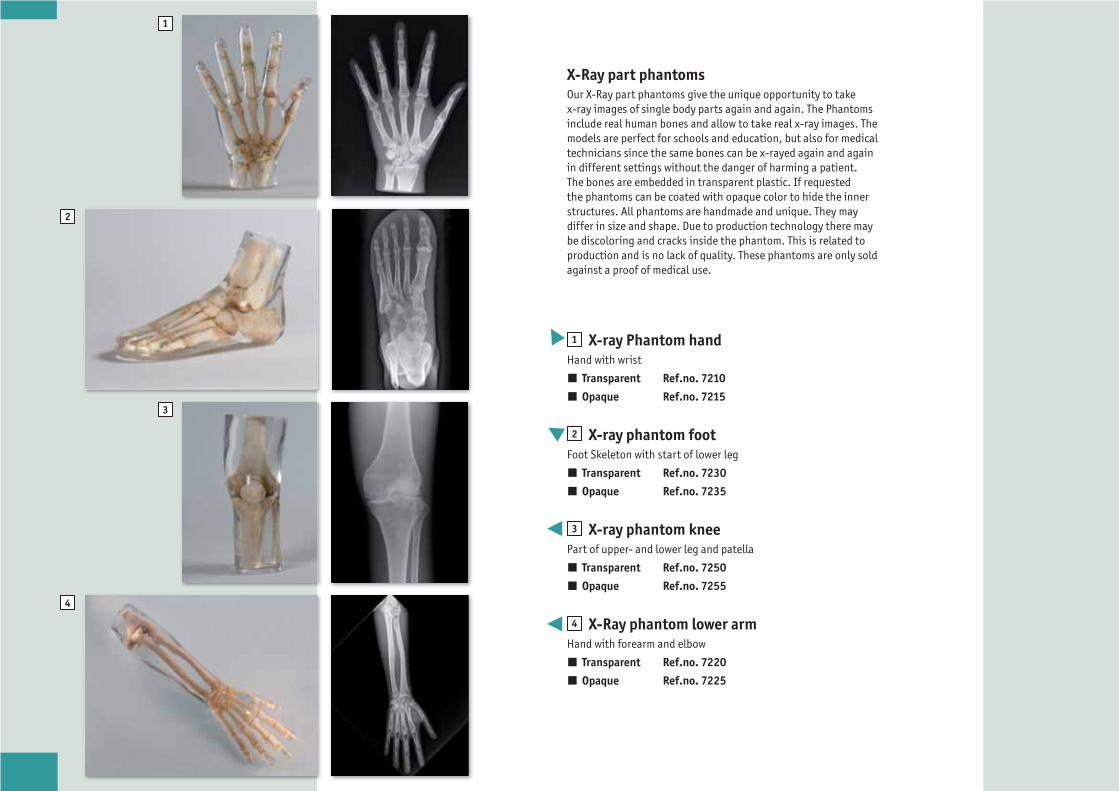
X-Ray part phantomsOur X-Ray part phantoms give the unique opportunity to take x-ray images of single body parts again and again. The Phantoms include real human bones and allow to take real x-ray images. The models are perfect for schools and education, but also for medical technicians since the same bones can be x-rayed again and again in different settings without the danger of harming a patient. The bones are embedded in transparent plastic. If requested the phantoms can be coated with opaque color to hide the inner structures. All phantoms are handmade and unique. They may differ in size and shape. Due to production technology there may be discoloring and cracks inside the phantom. This is related to production and is no lack of quality. These phantoms are only sold against a proof of medical use.
1 X-ray Phantom handHand with wrist
Transparent Ref.no. 7210
Opaque Ref.no. 7215
2 X-ray phantom footFoot Skeleton with start of lower leg
Transparent Ref.no. 7230
Opaque Ref.no. 7235
3 X-ray phantom kneePart of upper- and lower leg and patella
Transparent Ref.no. 7250
Opaque Ref.no. 7255
4 X-Ray phantom lower armHand with forearm and elbow
Transparent Ref.no. 7220
Opaque Ref.no. 7225

i
5 X-ray phantom elbowPart of upper and lower arm
Transparent Ref.no. 7260
Opaque Ref.no. 7265
6 X-ray phantom pelvisPelvis with 2 lumbar vertebrae and femur stumps
Transparent Ref.no. 7240
Opaque Ref.no. 7245
7 X-ray phantom shoulderHumerus with shoulder girdle
Transparent Ref.no. 7340
Opaque Ref.no. 7345
8 X-ray phantom spineComplete spine with simulated discs
Transparent Ref.no. 7290
Opaque Ref.no. 7295
All models embedded in
transparent plastic.

2
3
4 5
Sectional X-ray phantoms with artificial bonesThis series of sectional phantoms offers x-ray imaging with always identical images without anatomical differences between two models. This means you can use several identical phantoms or replace a broken or lost phantom by exactly the same. This is especially useful in case for example several technicians shall make identical images on different machines or an educational institute wants to make their own complex teaching papers. The phantoms are available in transparent or opaque versions, allowing to choose the suitable version. You may for example use the transparent phantom for teaching since it is easier to position and then change to the opaque phantom for examination purposes.
Head phantom1 Transparent
Ref.no. R16700
2 Opaque Ref.no. R16701
PelvisIncludes lumbar / sacral spine, pelvic bony anatomy and proximal femurs.
3 Opaque Ref.no. R16704
ThoraxIncludes a thoracic skeleton with embedded heart and lungs to provide realistic imaging. The scapulae are rotated outside of the lung fields for proper PA chest imaging.
4 Transparent Ref.no. R16702
5 Opaque Ref.no. R16703

1
3
45
6
7
9
10
12
Right ElbowMovable. Normal flexion range allows for AP/lateral and partial flexion views with one phantom.
1 Transparent Ref.no. R16705
2 Opaque Ref.no. R16706
Right HandFlat, unbent fingers.
3 Transparent Ref.no. R16707
4 Opaque Ref.no. R16708
Left HandGrasping position.
5 Transparent Ref.no. R16709
6 Opaque Ref.no. R16710
Right KneeFreely movable patella and joint allows for realistic positioning of the knee for AAP/lateral, oblique, sunrise and tunnel views.
7 Transparent Ref.no. R16711
8 Opaque Ref.no. R16712
Right footNormal position.
9 Transparent Ref.no. R16713
10 Opaque Ref.no. R16714
Left footOblique position.
11 Transparent Ref.no. R16715
12 Opaque Ref.no. R16716

1 Full Body X-Ray PhantomThis model is unique in the world and provides excellent training opportunities for positioning and alignment techniques in projection radiography. It should be part of the basic equipment of any radiographic school. The phantom contains a real human skeleton as well as outlines of larynx, lung, heart and kidneys (organs will create a shadow on the image), which allows taking real X-ray images like in a patient. Using a real skeleton provides even smallest guiding structures which is impossible with a plastic skeleton. During assembly of this phantom we pay special attention to the correct size of joint spaces. All joints are moveably mounted allow positioning in all normal x-ray positions (e.g. frog position, pro- and supination of lower arm). The arms can be moved upwards which makes the phantom suitable for use in all kinds of osseous examinations under CT. Each phantom is hand-made one of a kind; it may differ in size and appearance. Depending on the individual phantom it may have some pathologies, outer shape may differ depending on size of the skeleton. The new version was re-designed in co-operation with a well-known German school for radiographers and fits all needs for education in radiography. This phantom is only sold against proof of medical use. Life size.
Ref.no. 7200
2 Radiographic positioning doll, plastic skeletonThis model offers all features of model 7200 but includes a plastic skeleton and is due to this only suitable for positioning training.
Including a transport and storage case
Ref.no. 7201 (not pictured)
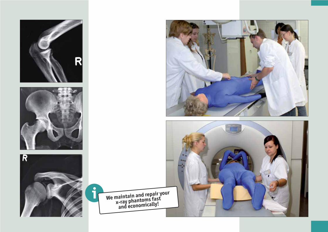
i We maintain and repair your
x-ray phantoms fast
and economically!
2
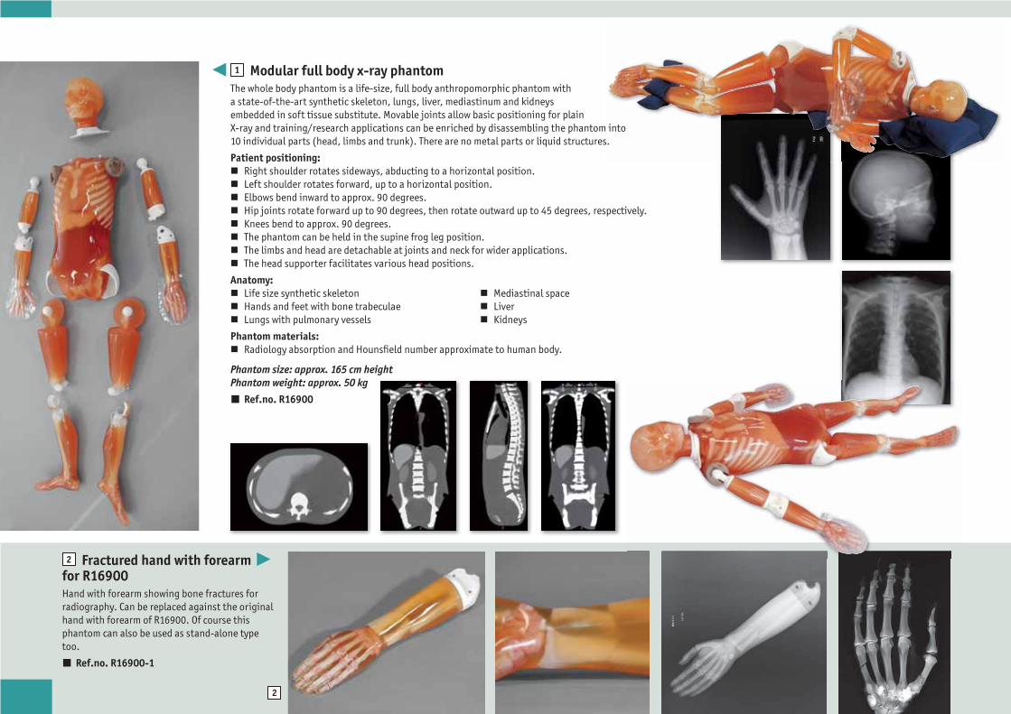
1 Modular full body x-ray phantomThe whole body phantom is a life-size, full body anthropomorphic phantom with a state-of-the-art synthetic skeleton, lungs, liver, mediastinum and kidneys embedded in soft tissue substitute. Movable joints allow basic positioning for plain X-ray and training/research applications can be enriched by disassembling the phantom into 10 individual parts (head, limbs and trunk). There are no metal parts or liquid structures.
Patient positioning: Right shoulder rotates sideways, abducting to a horizontal position. Left shoulder rotates forward, up to a horizontal position. Elbows bend inward to approx. 90 degrees. Hip joints rotate forward up to 90 degrees, then rotate outward up to 45 degrees, respectively. Knees bend to approx. 90 degrees. The phantom can be held in the supine frog leg position. The limbs and head are detachable at joints and neck for wider applications. The head supporter facilitates various head positions.
Anatomy: Life size synthetic skeleton Hands and feet with bone trabeculae Lungs with pulmonary vessels
Mediastinal space Liver Kidneys
Phantom materials: Radiology absorption and Hounsfield number approximate to human body.
Phantom size: approx. 165 cm height Phantom weight: approx. 50 kg
Ref.no. R16900
2 Fractured hand with forearm for R16900Hand with forearm showing bone fractures for radiography. Can be replaced against the original hand with forearm of R16900. Of course this phantom can also be used as stand-alone type too.
Ref.no. R16900-1
1
2
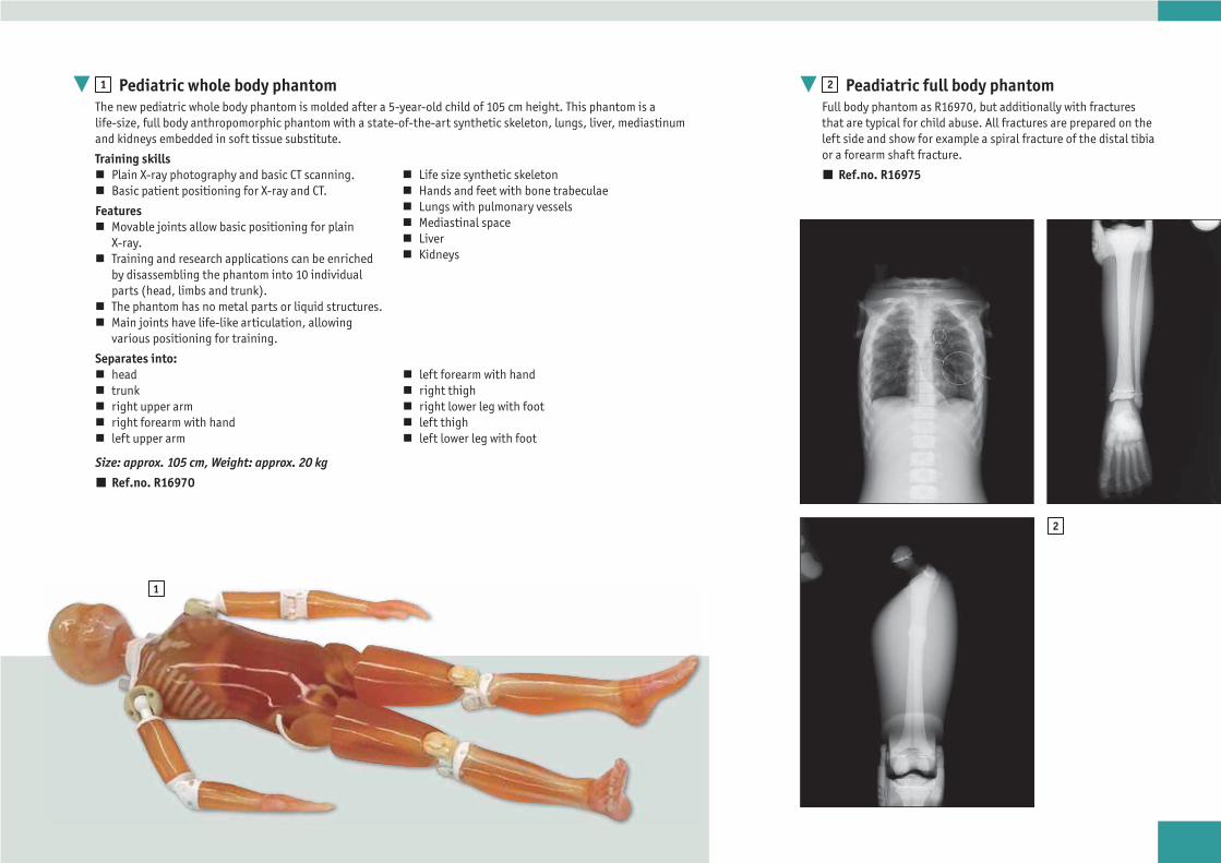
1 Pediatric whole body phantomThe new pediatric whole body phantom is molded after a 5-year-old child of 105 cm height. This phantom is a life-size, full body anthropomorphic phantom with a state-of-the-art synthetic skeleton, lungs, liver, mediastinum and kidneys embedded in soft tissue substitute.
Training skills Plain X-ray photography and basic CT scanning. Basic patient positioning for X-ray and CT.
Features Movable joints allow basic positioning for plain
X-ray. Training and research applications can be enriched
by disassembling the phantom into 10 individual parts (head, limbs and trunk).
The phantom has no metal parts or liquid structures. Main joints have life-like articulation, allowing
various positioning for training.
Separates into: head trunk right upper arm right forearm with hand left upper arm
left forearm with hand right thigh right lower leg with foot left thigh left lower leg with foot
Size: approx. 105 cm, Weight: approx. 20 kg
Ref.no. R16970
2 Peadiatric full body phantomFull body phantom as R16970, but additionally with fractures that are typical for child abuse. All fractures are prepared on the left side and show for example a spiral fracture of the distal tibia or a forearm shaft fracture.
Ref.no. R16975 Life size synthetic skeleton Hands and feet with bone trabeculae Lungs with pulmonary vessels Mediastinal space Liver Kidneys

1
2
1 Newborn Whole Body x-ray phantomNewborn whole body phantom is the world‘s first full body phantom for neonatal radiography with correct anatomical structure and movable limbs. Neonatal radiography is an important tool in NICU (Neonatal Intensive Care Unit). Patient positioning and immobilization are essential features. This phantom provides opportunities for hands-on training and experiments to minimize radiation exposure to newborn babies.
Features Limbs rotate 360 degrees at shoulders and hip joints. Left hand is clenched and right hand is open. Life size whole body newborn baby. Original human tissue substitute. No metal parts or liquid structures. Meconium aspiration syndrome can be made per custom order.
Anatomy
Skull, spine, ribs, pelvis, scapulae, clavicles, humeri, radius, ulnae, bones of hands, femora, fibulae, tibiae and bones of feet, lungs and mediastinum
Training Skills
Specifications
Set Includes: 1 newborn whole body phantom, 1 storage case, 1 set of sample X-ray images, 1 instruction manual
Size: 42 cm (representing a baby of 50 cm height) weight: 2.8 kg
Ref.no. R16980
Immobilization
Manual immobilization Immobilization with fixtures Autopsy imaging
Radiography
Upright AP (anteroposterior) Supine AP Upright lateral Supine lateral
2 Body plates for adult x-ray phantomsBody Plates for R16900 or R16950 to simulate a person with a BMI of 30.
Ref.no. R16900-2
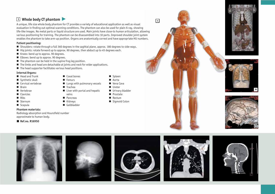
111 Whole body CT phantom
A unique, life size whole body phantom for CT provides a variety of educational application as well as visual evaluation in finding out optimal scanning conditions. The phantom can also be used for plain X-ray, showing life-like images. No metal parts or liquid structure are used. Main joints have close-to human articulation, allowing various positioning for training. The phantom can be disassembled into 10 parts. Improved shoulder joint system enables the phantom to take arm-up position. Organs are anatomically correct and have appropriate HU numbers.
Patient positioning: Shoulders: rotate through a full 360 degrees in the sagittal plane, approx. 180 degrees to side-ways. Hip joints: rotate forward up to approx. 90 degrees, then abduct up to 45 degrees each. Knees: bend up to approx. 90 degrees. Elbows: bend up to approx. 90 degrees. The phantom can be held in the supine frog leg position. The limbs and head are detachable at joints and neck for wider applications. The head supporter facilitates various head positions.
Internal Organs: Head and Trunk Synthetic skull Cervical vertebrae Brain Vertebrae Clavicles Ribs Sternum Scapula
Coxal bones Femurs Lungs with pulmonary vessels Trachea Liver with portal and hepatic
veins Pancreas Kidneys Gallbladder
Spleen Aorta Vena Cava Ureter Urinary bladder Prostate Rectum Sigmoid Colon
Phantom materials:Radiology absorption and Hounsfield number approximate to human body.
Ref.no. R16950
3
1
2
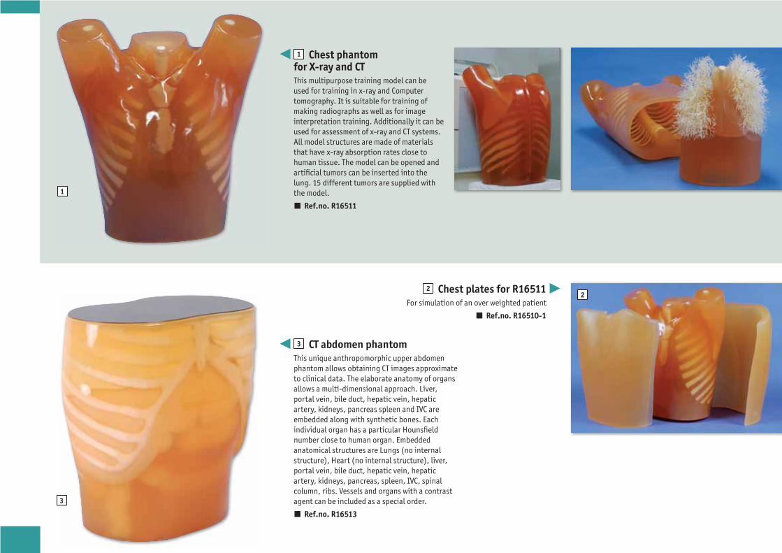
1 Chest phantom for X-ray and CTThis multipurpose training model can be used for training in x-ray and Computer tomography. It is suitable for training of making radiographs as well as for image interpretation training. Additionally it can be used for assessment of x-ray and CT systems. All model structures are made of materials that have x-ray absorption rates close to human tissue. The model can be opened and artificial tumors can be inserted into the lung. 15 different tumors are supplied with the model.
Ref.no. R16511
2 Chest plates for R16511For simulation of an over weighted patient
Ref.no. R16510-1
3 CT abdomen phantomThis unique anthropomorphic upper abdomen phantom allows obtaining CT images approximate to clinical data. The elaborate anatomy of organs allows a multi-dimensional approach. Liver, portal vein, bile duct, hepatic vein, hepatic artery, kidneys, pancreas spleen and IVC are embedded along with synthetic bones. Each individual organ has a particular Hounsfield number close to human organ. Embedded anatomical structures are Lungs (no internal structure), Heart (no internal structure), liver, portal vein, bile duct, hepatic vein, hepatic artery, kidneys, pancreas, spleen, IVC, spinal column, ribs. Vessels and organs with a contrast agent can be included as a special order.
Ref.no. R16513
1
2
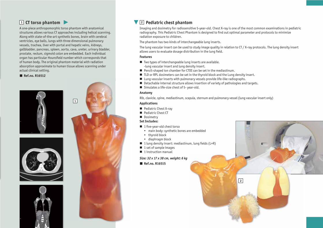
1 CT torso phantomA one-piece anthropomorphic torso phantom with anatomical structures allows various CT approaches including helical scanning. Along with state-of-the-art synthetic bones, brain with cerebral ventricles, eye balls, lungs with three dimensional pulmonary vessels, trachea, liver with portal and hepatic veins, kidneys, gallbladder, pancreas, spleen, aorta, cava, ureter, urinary bladder, prostate, rectum, sigmoid colon are embedded. Each individual organ has particular Hounsfield number which corresponds that of human body. The original phantom material with radiation absorption approximate to human tissue allows scanning under actual clinical setting.
Ref.no. R16512
2 Pediatric chest phantomImaging and dosimetry for radiosensitive 5-year-old. Chest X-ray is one of the most common examinations in pediatric radiography. This Pediatric Chest Phantom is designed to find out optimal parameter and protocols to minimize radiation exposure to children.
The phantom has two kinds of interchangeable lung inserts.
The lung vascular insert can be used to study image quality in relation to CT / X-ray protocols. The lung density insert allows users to evaluate dosage distribution in the lung field.
Features
Two types of interchangeable lung inserts are available. -lung vascular insert and lung density insert.
Pencil-shaped ion chamber for CTDI can be set in the mediastinum. TLD or RPL dosimeters can be set in the thyroid block and the Lung density insert. Lung vascular inserts with pulmonary vessels provide life-like radiographs. Detachable internal structure allows insertion of variety of pathologies and targets. Simulates a life-size chest of 5- year-old.
Anatomy
Rib, clavicle, spine, mediastinum, scapula, sternum and pulmonary vessel (lung vascular insert only)
Applications
Pediatric Chest X-ray Pediatric Chest CT Dosimetry
Set Includes:
1 five-year-old chest torso • main body: synthetic bones are embedded • thyroid block • diaphragm block
1 lung density insert: mediastinum, lung fields (L+R) 1 set of sample images 1 instruction manual
Size: 32 x 17 x 38 cm, weight: 6 kg
Ref.no. R16515

1
2
1 Head and Neck Phantom for CT, X-ray and Radiation TherapyHead and neck phantom with realistic anatomy. This highly realistic head and neck phantom was designed to simulate clinical imaging and dose exposure in computed tomography including dual energy CT, X-ray imaging and radiation therapy. The model provides a realistic simulation of all tissues and realistic attenuation values.
This phantom provides a detailed simulation of patient exposure and provides new opportunities for testing and optimizing image quality and dose, dose verification at low and high energy exposure and for training of medical and technical staff.
The phantom is manufactured based on a real CT data set and includes anatomic details for all tissues. The model is a handmade unique piece, which can differ slightly in size and design. The phantom can be provided as one-piece anthropomorphic phantom or in a sectional design and it can include openings for dosimeters. Pathologic features (e.g., masses, vascular pathologies) can be included upon request into the phantom.
Ref.no. R14000
2 Breast Phantom for Mammography and Breast TomosynthesisBreast phantom with adipose and glandular tissue. This breast phantom was designed to simulate breast imaging in mammography and breast tomosynthesis. It represents a compressed breast of 4 cm thickness that can be placed under the compression paddle.
This phantom provides a realistic simulation of breast imaging. It was developed for testing and optimizing dose and image quality and for training of medical and technical staff.
The phantom is manufactured from virtual data* containing adipose and glandular tissue. The models are handmade unique piece, which can differ slightly in size and design. The phantom can be provided as one-piece anthropomorphic phantom or in a sectional design. Dosimeter openings and pathologic features can be included upon request.
Ref.no. R14300
* References Breast Phantom:
1. Graff, C.G., "A new open-source multi-modality digital breast phantom," Proc. SPIE 9783, 978309 (2016).
2. Ikejimba, L.C., Graff, C.G., Rosenthal, S., Badal, A., Ghammraoui, B., Lo, J.Y. and Glick, S.J., “A novel physical anthropomorphic breast phantom for 2D and 3D x-ray imaging,” Medical Physics 44(2), 407-416 (2017).
NEW!
NEW!
2
11
2
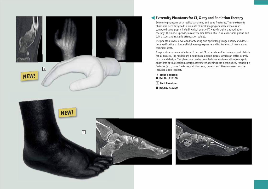
Extremity Phantoms for CT, X-ray and Radiation TherapyExtremity phantoms with realistic anatomy and bone fractures. These extremity phantoms were designed to simulate clinical imaging and dose exposure in computed tomography including dual energy CT, X-ray imaging and radiation therapy. The models provide a realistic simulation of all tissues including bone and soft tissues and realistic attenuation values.
The phantoms were developed for testing and optimizing image quality and dose, dose verification at low and high energy exposure and for training of medical and technical staff.
The phantoms are manufactured from real CT data sets and include anatomic details for all tissues. The models are a handmade unique pieces, which can differ slightly in size and design. The phantoms can be provided as one-piece anthropomorphic phantoms or in a sectional design. Dosimeter openings can be included. Pathologic features (e.g., bone fractures, calcifications, bone or soft tissue masses) can be included upon request.
1 Hand Phantom Ref.No. R14100
2 Foot Phantom
Ref.no. R14200
NEW!
NEW!
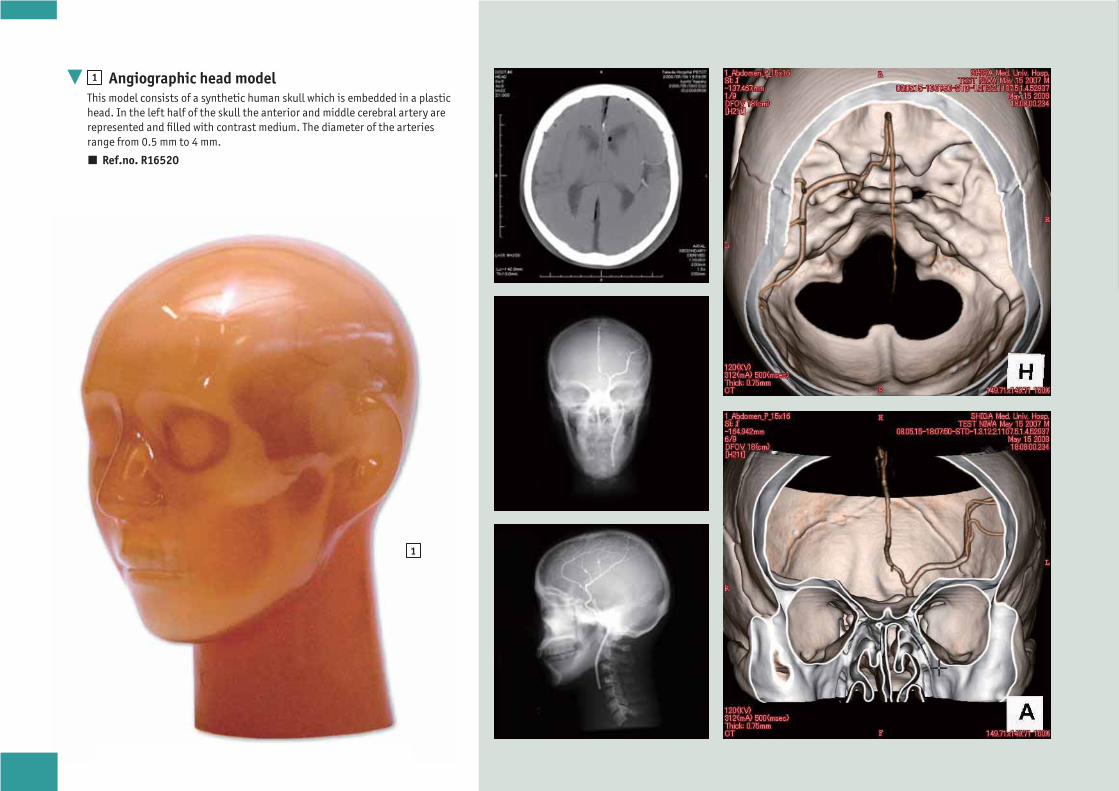
1
1 Angiographic head modelThis model consists of a synthetic human skull which is embedded in a plastic head. In the left half of the skull the anterior and middle cerebral artery are represented and filled with contrast medium. The diameter of the arteries range from 0.5 mm to 4 mm.
Ref.no. R16520

1
2
1 Lung cancer screening modelThis Phantom is a CT phantom developed to facilitate optimizing the radiation dose and other scanning conditions for lung cancer screening CT examination with Helical CT or MDCT, which is aiming at early detection of lung cancers. As the screening is usually done on healthy people, the necessity of minimizing the exposure while maximizing the image quality is considered to be particularly high. The phantom is designed to set conditions for detection of small early lung cancers such as GGA, which are difficult to be found by plain X-ray. Anthropologic structure of the phantom provides life-like images allowing operators visual evaluation, while quantitative evaluation on radiation dose and density curve of the image can be done stimulatory with a single scanning.
The model consists of a life size torso with arm up position and has the following internal structures:
Bones Simulated tumors on sections of three lung area: Apical portion of the lungs Bifurcation of the trachea Base of lungs Dose meter hole (13 mm dia., on the central axis of the phantom) 1 8-step linearity phantom 8 steps of 30mm dia. density samples are embedded
Ref.no. R16532
2 Radiation therapy phantomThis phantom is developed for the treatment planning and machine adjustment in the radiation therapy. The body consists of 3 cm slices with a 3 x 3 cm hole matrix for inserting glass dosimeters. The model material has a natural radioparency allowing the correct adjustment of the machines. This makes it ideal for planning and machine adjustment. The phantom has a holding and fixation frame which allows to position the phantom exactly.
Ref.no. R16531
