anatomy of spinal nerves in the first turkish illustrated anatomy
TRANSCRIPT

COVER EDITORIAL
Anatomy of spinal nerves in the first Turkish illustrated anatomyhandwritten textbook
Murat Çetkin1& Mustafa Orhan1
& İlhan Bahşi1 & Begümhan Turhan2
Received: 26 May 2016 /Accepted: 30 May 2016 /Published online: 10 June 2016# Springer-Verlag Berlin Heidelberg 2016
BTeşrih-ül Ebdan ve Tercümânı Kıbale-i Feylesûfan^ is thefirst handwritten anatomy textbook with illustrations writtenin Turkish in 17th century by Şemseddîn-i İtâḳî. BTeşrih^ hasdifferent meanings such as anatomy, skeleton, and cutting acorpse into pieces [1]. BTeşrih-ül Ebdan ve Tercümânı Kıbale-i Feylesûfan ^ means dissection of the body and scholars’birth knowledge [2]. Since this is the first handwritten text-book in Turkish, it has great importance in the development ofmedicine in Ottoman Empire. This book was written whileGrand Vizier Recep Pasha was in power, and it was dedicatedto the Sultan of that period, Murat the IVth [3, 4]. It is thoughtthat the book was written in 1632 [4–7].
Şemseddîn-i İtâḳî was born in Shirvan (North Azerbaijan)in 1570s. Şemseddîn-i İtâḳî was interested in mathematics,philosophy, medicine, hadith, cannon law, logic, and astrono-my [4, 7]. İtâḳî suffered hardships because of the wars andinternal conflicts in Shirvan. He lost a lot of his family, and hehad to leave Shirvan in 1604 when it was annexed by Persia[8]. After İtâḳî left his homeland, he travelled to several coun-tries for a long time but he did not receive much appreciation[4, 8]. He arrived in Istanbul during the reign of Sultan Muradthe IVth (1623–1640) and was introduced to Sadrazam(Grand Vizier) Recep Pasha. He was given the academic titleBHaremeyn Payesi^ by the Grand Vizier. He published hisbook with the help of Recep Pasha. In the introduction of
the book, İtâḳî acknowledges the contributions of the GrandVizier [4, 7].
Not many textbooks about anatomy existed in the IslamicWorld and the Ottoman Empire until İtâḳî’s book [9]. In othermedical textbooks, anatomy occupies only a few pages indifferent sections [4]. İtâḳî’s book is a pioneer in its area asit is written in Turkish, and it is supported with illustrations[4]. In addition to Turkish, the book contains mostly Arabicand rarely Persian terms as well [4, 6, 7]. Some editions of thisbook which was written in the 17th century were reprinted inthe 18th century. Seven different editions are known today [4].
The book contains illustrations similar to Ahmed İbnMansur’s (14th century) book called Teşrih-i Ebdan. Someof the content was quoted from Avicenna’s CanonMedicinae and İbn Nafis’s Şerh-i Teşrihül Kanun. In additionto these illustrations, illustrations from other European basedworks like Andreas Vesalius’s De Humani Corporis Fabricaalso exist. Some of the illustrations were drawn by the authorhimself. These illustrations were drawn by the author himselfusually depict the nervous system [3, 4, 6, 10].
The purpose of this study is to analyze the anatomy of thespinal nerves as stated in the book BTeşrih-ül Ebdan veTercümânı Kıbale-i Feylesûfan^ written in the OttomanPeriod, to evaluate the knowledge level about spinal nerveanatomy in 17th century and compare that knowledge withcurrent knowledge of anatomy.
In this study, Şemseddîn-i İtâḳî’s illustrated anatomy bookby Esin Kâhya which is the translation of BTeşrih-ül Ebdan veTercümânı Kıbale-i Feylesûfan^ from the Ottoman alphabetinto the Latin alphabet was evaluated. All of the sections of thebook, especially the sections about spinal nerves, were ana-lyzed in detail. This information was compared to our currentknowledge; thus, its accuracy was inspected.
In the book, general information about the nervous systemwas provided in the anatomy of the nerves. Then, the cranial
* İlhan Bahş[email protected]
1 Department of Anatomy, Faculty of Medicine, Gaziantep University,TR-27310 Gaziantep, Turkey
2 Department of Physiotherapy and Rehabilitation, Faculty of HealthSciences, Hasan Kalyoncu University, 27100 Gaziantep, Turkey
Childs Nerv Syst (2017) 33:205–209DOI 10.1007/s00381-016-3136-9

and spinal nerves were explained. İtâḳî stated the followingwhen he defined the nervous system BGod, separated thespinal cord from the brain and made the spinal cord the caliphof the brain.^ With this statement, İtâḳî summaries a majortopic with a striking sentence. Spinal cord is considered as alower center than the brain, and it is controlled by the brainwhen needed. Thus, İtâḳî’s definition is correct. He dividedthe nerves into sensory and motor nerves. This definition isconsistent with our current knowledge. He claimed that sen-sory nerves are rapid and soft while the motor nerves are firmand powerful. The author wrote that if the nerves were notpresent, muscles would have been soft. He told that for themaintenance of muscle tone, nervous innervations are needed.He also defined the flaccid paralysis when this innervation isabsent. He claimed that without the presence of nerves, mus-cles can be cut easily by stone or solid objects. This knowl-edge is not compatible with our current knowledge.
The internal organs like the liver, kidneys, and spleen thatdo not have sensory nerves within them. These organs aredefined to be covered by a membrane which allows for thetransmission of sensory information by the embedded nerves.In the pathological states of this membrane, some reactionswill occur with the help of these nerves. With our currentknowledge, we know that internal organs like the liver, kid-neys, and spleen possess tunica serosa and autonomic inner-vations but İtâḳî defined this information in an incompleteway.
Spinal nerves
In the book, spinal nerves are classified as nerves emergingfrom the vertebral column from the neck, back, sacrum, andcoccyx.
Cervical spinal nerves
İtâḳî defined that eight couples of spinal nerves in the neckregion, and he explained each one separately. He emphasizedthat each nerve left between two vertebrae.
The first cervical spinal nerve was thinner than the rest ofthe cervical spinal nerves, and it could not reach whole of thehead.
The second cervical spinal nerve exists between the firstand second vertebrae and travels upward in a curved courseuntil the head and spreads into the skin around the ear. Parts ofthis nerve travels to the muscles located at the back side of theneck.
The third cervical spinal nerve exists between the secondand the third vertebrae and divides into two. One of thebranches from this nerve innervated the muscle that rotatedthe neck while the other one moved to the spinous process ofthe vertebrae and joined a ligament. Then, it moved to the ear
area, and this nerve gave power to muscles of the face and thetemples. Its second branch went to the cheek reaching the flatmuscle. While defining the third cervical spinal nerve, İtâḳîdefined the functions of this muscle in animals as well.
The fourth cervical spinal nerve came out from the holebetween the third and fourth vertebrae by separating intotwo branches. The anterior branch united with a branch fromthe fifth cervical spinal nerve and constituted a quite smallbranch. The posterior branch is larger, and it moved towardthe head. It rests between the spinous process of the vertebraeand the muscles. Then, the final branches from this secondbranch spread between the head and the neck. The distributionarea was part of the face and the muscles of the vertebrae. Headded that this branch move to the ear in the animals.
The fifth cervical spinal nerve comes out from the holebetween the fourth and fifth vertebrae by separating into twobranches. Anterior branch moves to the muscles of the shoul-der. Some parts of this nerve innervates the flat muscle in theface and to the muscle that flexing the head. The secondbranch of the fifth cervical spinal nerve merges with some ofthe fibers of the sixth and seventh cervical spinal nervesreaching the diaphragm below. It is emphasized that eighthspinal nerve does not join this union.
The sixth cervical spinal nerve leaves from the hole be-tween the fifth and sixth vertebrae by separating into twobranches similar to third, fourth, and fifth cervical spinalnerves. The one of these nerves go upward and distributes tothe head, muscles of the back, and the pelvic bone.
The seventh cervical spinal nerve leaves from the hole be-tween the sixth and seventh vertebrae by separating into twobranches. One of the branches travels to the diaphragm joiningbranches from the fifth and sixth spinal nerves. The otherbranch joins some of the branches from the sixth and eighthspinal and first thoracal spinal nerves.
The eighth cervical spinal nerve leaves from the hole be-tween the seventh cervical vertebra and the first thoracic ver-tebra. Some of the branches of this nerve travel to the head andthe neck; some of the branches travel to the shoulder, arm,wrist, hand, and the palm. Sixth cervical spinal nerve reachesthe scapular region while the seventh cervical spinal nervereaches the arm.
İtâḳî described the cervical spinal nerves more in detailthan the other spinal nerves. He described the number of cer-vical spinal nerves and emerging holes correctly. He definedthat the first two cervical spinal nerves only have one brancheach while the other cervical spinal nerves have two branches.He defined that the eighth cervical spinal nerve had two sep-arate nerve fibers correctly, but he could not describe the lo-cation of separation into two different branches. He did notdescribe where the first cervical spinal nerve exited from thespinal cord; however, he described the exit points of the othercervical spinal nerves correctly. Most of his descriptions re-garding the targets of the cervical spinal nerves were described
206 Childs Nerv Syst (2017) 33:205–209

incorrectly. However, most of the information provided for thefirst cervical spinal nerve was correct.
İtâḳî defined the phrenic nerve, which goes to diaphragm.However, the information about the phrenic nerve formingfrom the fifth, sixth, and seventh cervical spinal nerve isincorrect.
It is told that the nerves leaving from the spinal cord go totarget organs by making small connections. We believe thatthe information was mentioned by İtâḳî about the small con-nections are plexuses. However, in the cervical spinal nervessection, cervical plexus was not mentioned. It was told thatonly some of the branches of the fourth and fifth cervicalnerves formed connections and the union of these nerves arequite small. İtâḳî described that some nerve fibers originatingfrom the sixth, seventh, and eighth cervical spinal nerves andfirst thoracal spinal nerve united and travelled to the upperextremity. We believe that the brachial plexus was describedalmost correctly.
Thoracal spinal nerves
İtâḳî defined 12 thoracal spinal nerves.The first thoracal spinal nerve exists through between the
first and second thoracic vertebrae and then branching intotwo. The larger first branch travels to the muscles betweenthe ribs. The second branch rises above the ribs and, as toldin the cervical spinal nerves section, it merges with thebranches of the sixth, seventh, and eighth cervical spinalnerves.
The second thoracal spinal nerve exists between the secondand third thoracic vertebrae branching into two. One branchgets the sensory information from the skin of the arm the otherbranch travels to the hand.
İtâḳî did not analyze the rest of the thoracic nerves sepa-rately, but he correctly described the point of exit from thecorresponding vertebrae. He described that branches of thesenerves innervated the muscles between the ribs, hand, andscapular sides. He described the arteries and veins accompa-nying the nerves innervating the muscles between the ribs.
İtâḳî described the first two thoracal spinal nerves in detailwhile he described the rest in lesser detail. We believe thatİtâḳî correctly described the branch of the first thoracal spinalnerve which innervates the intercostal muscles is intercostalnerve. However, he was incorrect when he described that thisnerve innervates the muscles of the back. He correctly de-scribed that the other branch of the first thoracic nerve joinedthe brachial plexus innervating the upper extremity. İtâḳî de-fined the branch of the first thoracal spinal nerve travelling tothe wrist. We believe that he probably considered the anteriorramus of the first thoracal spinal nerve, inferior trunk, medialfascicle, and ulnar nerve as a single structure.
We think that the sensory branch innervating the skin of thearm was intercostobrachial nerve originating from the second
thoracal spinal nerve. However, we currently know that theother branch of the second thoracal spinal nerve does notinnervate the hand as he previously stated. Today’s knowledgetells us, hand and scapula are not innervated by the secondthoracal spinal nerve.
Lumbar spinal nerves
İtâḳî described the lumbar region as the strap bearing region,and he defined five couples of nerves originating from thisregion. These nerves are divided into three branches; the firstone travelled to the back, second branch travelled to the ante-rior muscles of the abdomen, and the other branch travelled tothe muscles of the back. The first three lumbar spinal nerveswere defined to merge with the nerves descending from thebrain. A small part of the third lumbar spinal nerve and largerparts of the fourth and fifth spinal nerves united with thenerves of the sacrum and the coccyx. He told that the unionof this nerve bundle reached the coccyx area, inguinal region,knee, and the calf regions.
We believe that İtâḳî could have described the connectionof the spinal nerves with the sympathetic trunk by describingthe union of the first three lumbar spinal nerves with the nervecoming from the brain. We believe that he described the for-mation lumbosacral trunk because of the forming of the unionof a small part of the third lumbar spinal nerve and larger partsof the fourth and fifth spinal nerves united with the nerves ofthe sacrum and the coccyx. However, the forming of the lum-bosacral trunk explained incorrectly by coming from a smallpart of the third lumbar spinal nerve and joined to it.
İtâḳî stated that the nerve was formed by the fusion of thebranches of the third, fourth, and fifth spinal nerves course tothe coccyx area, inguinal region, knee, and the calf regions.We believe that the sacral plexus is formed by the lumbosacraltrunk and nerves coming from sacrum and coccyx. Accordingto innervation area, we think that the sciatic and tibial nerveswhich are the branches of the sacral plexus were alsoidentified.
Sacral and coccygeal spinal nerves
Six couples of sacral and coccygeal spinal nerves were de-fined. First three spinal nerves were defined as originate fromsacrum. The origins of the last three nerves were untold. It wastold that the first nerve united with other nerves coming fromabove and travelling to the calf region. The second and thethird nerves went to the coccyx area. He told that the course ofthe last three nerves travelled to the penis, bladder, and theanus. In addition, apart from these six nerves, he defined asingle nerve originating from the coccyx. Although notcompletely accurate, we think that İtâḳî mentioned the sacralplexus and the pudendal nerve.
Childs Nerv Syst (2017) 33:205–209 207
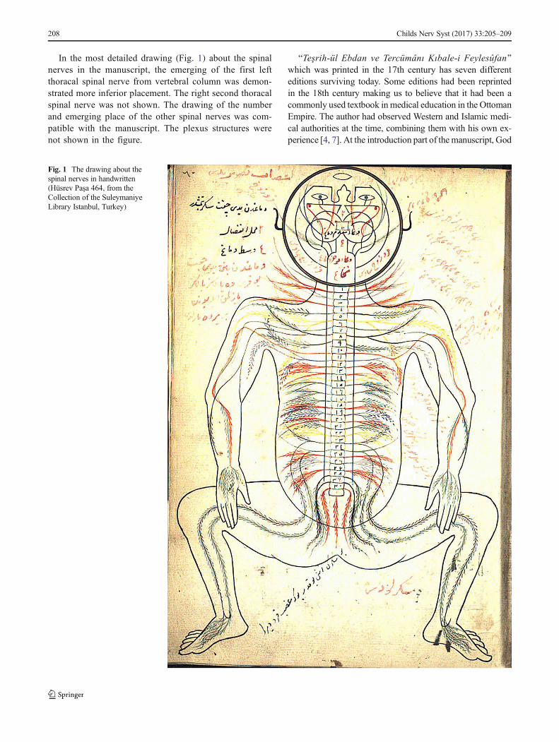
In the most detailed drawing (Fig. 1) about the spinalnerves in the manuscript, the emerging of the first leftthoracal spinal nerve from vertebral column was demon-strated more inferior placement. The right second thoracalspinal nerve was not shown. The drawing of the numberand emerging place of the other spinal nerves was com-patible with the manuscript. The plexus structures werenot shown in the figure.
BTeşrih-ül Ebdan ve Tercümânı Kıbale-i Feylesûfan^which was printed in the 17th century has seven differenteditions surviving today. Some editions had been reprintedin the 18th century making us to believe that it had been acommonly used textbook inmedical education in the OttomanEmpire. The author had observed Western and Islamic medi-cal authorities at the time, combining them with his own ex-perience [4, 7]. At the introduction part of the manuscript, God
Fig. 1 The drawing about thespinal nerves in handwritten(Hüsrev Paşa 464, from theCollection of the SuleymaniyeLibrary Istanbul, Turkey)
208 Childs Nerv Syst (2017) 33:205–209

was praised as seen in the other books printed in the era andthroughout the book some theological explanations were pro-vided as well.
İtâḳî defined some of the anatomical structures in compar-ison with the animal anatomy. Since human dissection was notcommon during the 17th century Islamic world, we believethat the author had performed dissections on animals thanapplied his knowledge to human body. Some anatomical partswere described in detail. We believe that it is a result of thehigh rates of wars helping the doctors observes many anatom-ical parts in detail.
İtâḳî correctly guessed the number of cervical, thoracic,and lumbar spinal nerves. However, he stated that sacral andcoccygeal spinal nerves consist of six couple nerves and onesingle nerve. The description of 31 spinal nerve couples isconsisted with our current knowledge. However, emergenceof a single nerve from the coccyx in addition to 31 couples ofnerve is not correct. Ulucam et al. [11] reported that the onenerve exiting the coccyx could be filum terminale. However,we do not think that this nerve could be the filum terminalelocated in the vertebral canal.
Şemseddîn-i İtâḳî’s BTeşrih-ül Ebdan ve TercümânıKıbale-i Feylesûfan^ has an important role in the history of anatomy.It had huge impact in the development of anatomy in theOttoman Empire, and it was widely used for medical educa-tion. In spite of lack and the presence of incorrect information,we believe that it played an important role in the developmentof anatomy. In addition to these, we believe that this type ofworks must be analyzed with future research in order to shinea light to the scientific history of Anatolia.
Compliance with ethical standards
Conflict of interest The authors declare that there is no conflict ofinterest.
References
1. Ferit D (2000) Osmanlıca-Türkçe Ansiklopedik Lügat. AydınKitabevi Yayınları, Ankara
2. Çıkmaz S (2006) Türkçe anatomi terimlerinin etimolojik vesemantik açıdan incelenmesi. (Doktora Tezi), Edirne: TrakyaÜniversitesi, 2006: 16
3. Adıvar A (1982) Osmanlı Türklerinde İlim. Remzi Kitabevi,İstanbul
4. Kâhya E (1996) Şemseddîn-i İt kî’nin Resimli Anatomi Kitabı.Ankara Kültür Merkezi Yayınları, Ankara
5. AkarM (2015) Cerrahi tekniklerin resimsel anlatımı. Art-Sanat: 15-45
6. Akdoğan I (2008) 17. Yüzyılda Avrupa’da ve Osmanlı DevletindeAnatomi. Türkiye Klinikleri J Med Ethics-Law Hist 16:166–170
7. Bayat AH (2010) Tıp Tarihi. Merkezefendi Geleneksel TıpDerneği, Ankara
8. Kâhya E (1988) Türkiye Diyanet Vakfı İslam Ansiklopedisi.Türkiye Diyanet Vakfı Yayın Matbaacılık, İstanbul
9. Kâhya E, Bilgen B (2014) Kitab-ı Teşrihü’l-ebdan min e’t-tıb.Atatürk Kültür Merkezi, Ankara
10. Turgut M (2008) Şemseddin-i Itaki’s contributions to neuroanato-my and embryology in the seventeenth century. Childs Nerv Syst24:1281–1282
11. Ulucam E, Mesut R, Gökçe N (2005) Neuroanatomy in Tesrih-iEbdan: a study on a book which is written in Ottoman era.Neuroanatomy 4:31–34
Childs Nerv Syst (2017) 33:205–209 209