anatomy of peritoneal spaces
TRANSCRIPT

Anatomy of
peritoneal spaces
DR VISHWANATH REDDY

PeritonumTHE PERITONEUM IS A THIN TRANSLUCENT ,SEROUS MEMBRANE LINED BY MESOTHELIAL CELL
THE PART THAT COVERS THE ABDOMINAL WALL IS CALLED THE PARIETAL
PERITONEUM
THE PART WHICH COVERS THE VISCUS IS CALLED THE VISCERAL
PERITONEUM

Definitions
LIGAMENT
-Two folds of peritoneum
-supporting structers
Mysentry
-two folds of peritoneum
-connecting to posterior abdominal wall
Omentum
- Connecting the stomach to other organs

Embryologic Characteristics
Peritoneal ligaments
Upper abdominal ligaments
-falciform ligament
-Triangular ligaments
-lesser omentum
-greater omentum

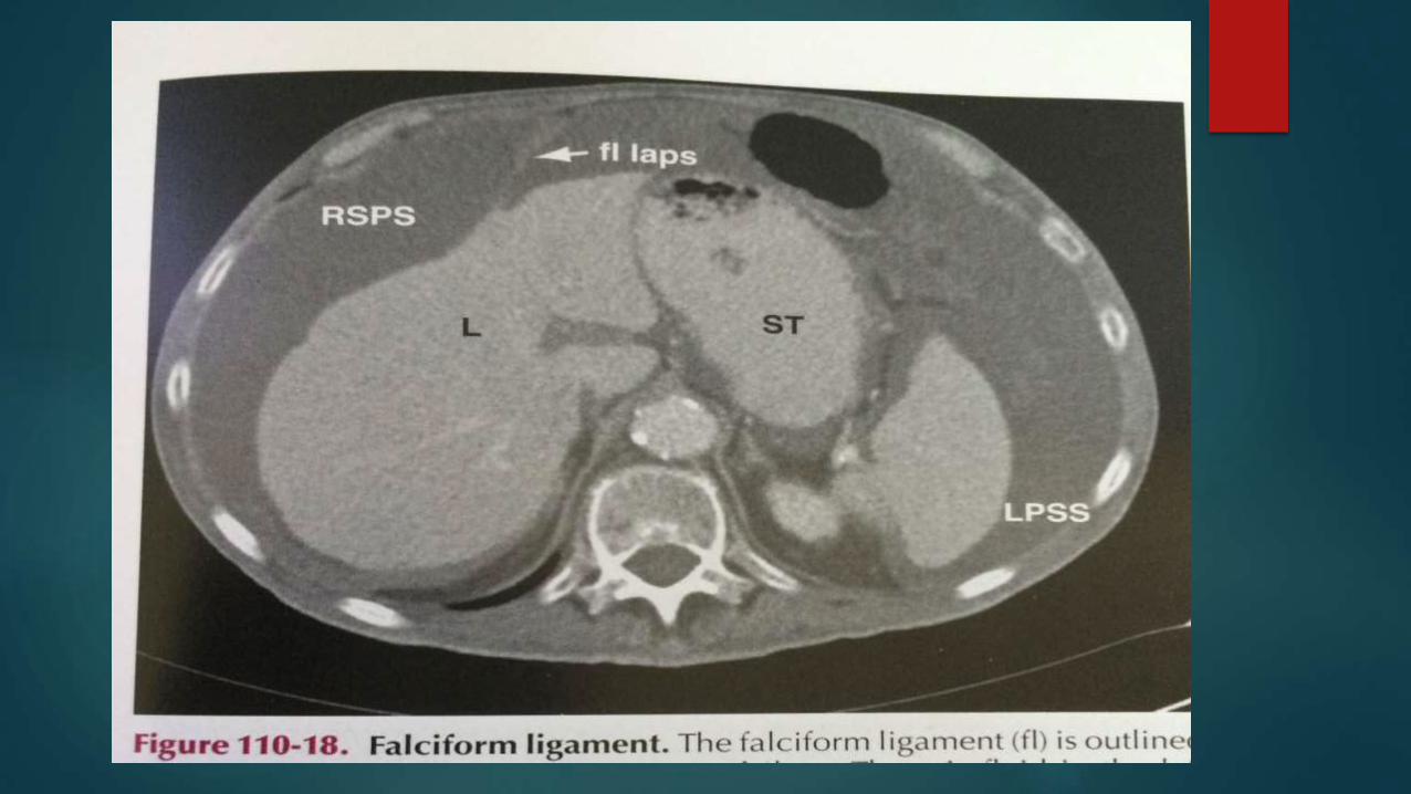
Falciform ligament
connects liver to the
posterior aspect of the
anterior abdominal wall just
to the right o the midline
.
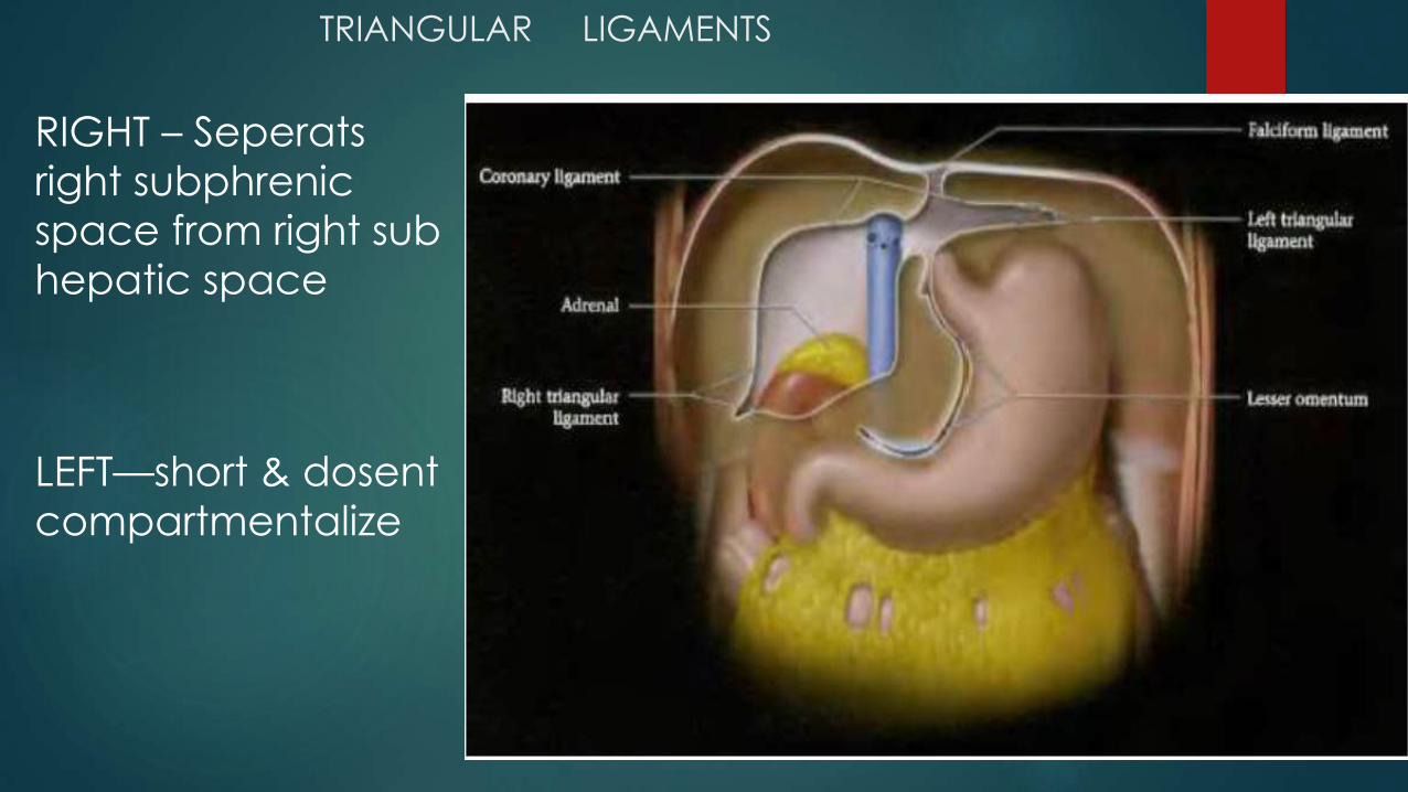
TRIANGULAR LIGAMENTS
RIGHT – Seperats
right subphrenic
space from right sub
hepatic space
LEFT—short & dosent
compartmentalize
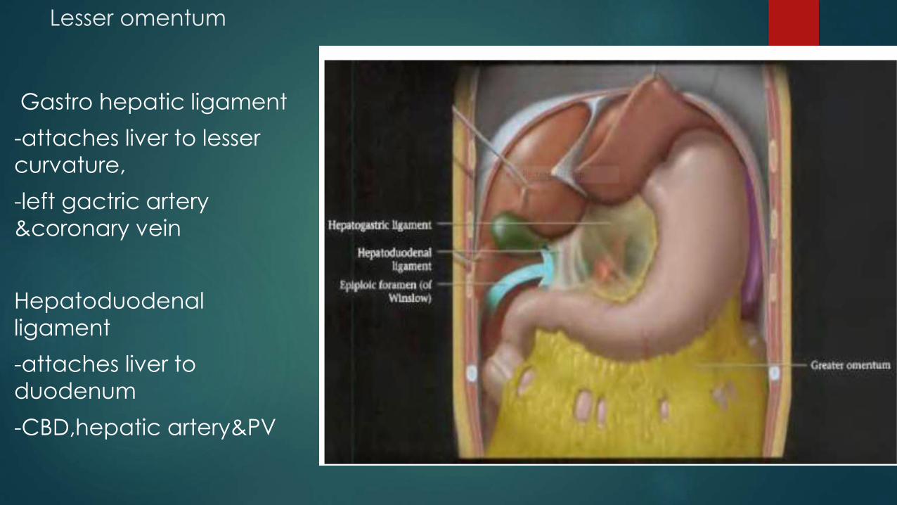
Lesser omentum
Gastro hepatic ligament
-attaches liver to lesser
curvature,
-left gactric artery
&coronary vein
Hepatoduodenal
ligament
-attaches liver to
duodenum
-CBD,hepatic artery&PV


GREATER OMENTUM
Gastrocolic ligament
Gastrosplenic
ligament
Gastrophrenic
ligament

The gastro splenic ligament
Connects the stomach to spleen
contains the short gastric vessels
It’s a major route of escape for
pancreatitis arising in the peripheral
body &tail


Spleno renal ligament
Posterior aspect of spleen to anterior pararenal space
Forms post-lat border of lesser sac
encloses tail of pancreas& distal splenic artery

ligaments of lower abdomen
Transverse mesocolon
Small bowel mesentry
Sigmoid mesocolon

Peritoneal spaces
potential space between the parietal & visceral peritoneum
contains a film that of fluid that lubricates the surface of the
peritoneum
not depicted on conventional radiologic studies or by cross
secectional imaging unless they are distended by fluid or air
-In men, the peritoneal cavity is
closed,
- in women, it communicates with
the extraperitoneal pelvis exteriorly
through the fallopian tubes, uterus
and vagina
Transverse mesocolon divides the
space in to
-supramesocolic
-inframesocolic
bilateral paracolic & pelvic spaces
are also peritoneal spaces

Peritoneal spaces

Supramesocolic space
Right -rightsubphrenic(subdiaphragmatic)
-rightsubhepatic(hepatorenal or mrrisonspouch)
Lesser sac (omental bursa)
Left
-left perihepatic space
-left subdiaphragmatic space
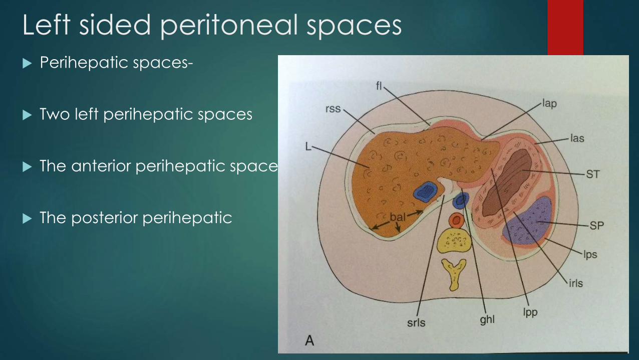
Left sided peritoneal spaces Perihepatic spaces-
Two left perihepatic spaces
The anterior perihepatic space
The posterior perihepatic

Left Subphrenic space
diaphragm ant& lat, stomach
post.
communicates with the post
subphrenic(perisplenic) space
The perisplenic space
surrounds most of the spleen
except for a portion of spleen
lying within the splenorenal
ligament

Right sided supra mesocolic spaces
The right sub
diaphragmatic space is
limited anteriorly by the
falciform ligament and
posteriorly by the hepatic
bare area .
Collections in this space
deform the surface of the
liver
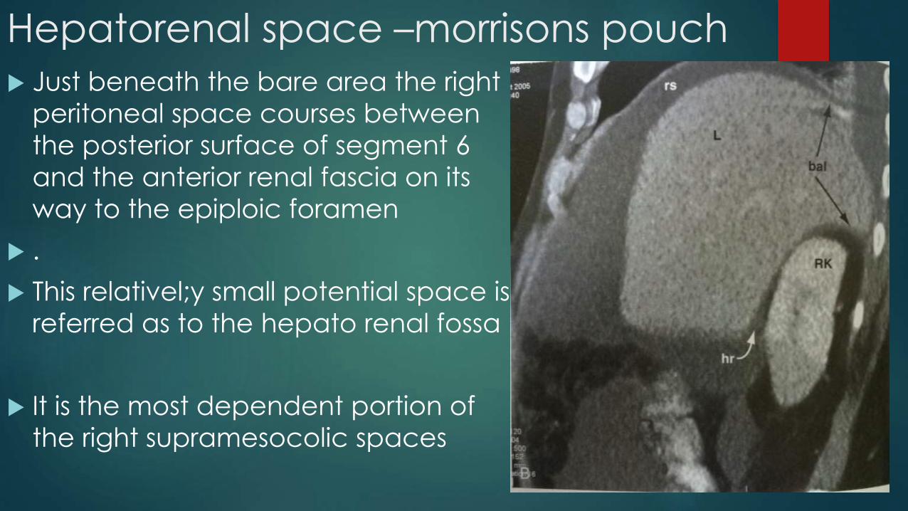
Hepatorenal space –morrisons pouch
Just beneath the bare area the right
peritoneal space courses between
the posterior surface of segment 6
and the anterior renal fascia on its
way to the epiploic foramen
.
This relativel;y small potential space is
referred as to the hepato renal fossa
It is the most dependent portion of
the right supramesocolic spaces


Lesser sacLies behind the stomach
Ant to pancreas
Left margin-gastro splenic ligament
Right margin –medial surface of coronary
ligament
Caudal boundary –gastrocolic
reflection&mesocolon

Lesser sac
Superior recess-behind
stomach,lesser omentum& left lobe
Inf recess-lies behind the
stomach,extending into the layers
of GO
The sup & inf recess are separated by
a peritoneal fold that accompanies
the left gastric artery

Lesser sac boundaries

Foramen of winslow
Communication betn the greater & lesser
sac.
Ant margin –hepatoduodenal ligament
Roof-peritoneum covering caudate lobe
Post margin-peritoneum covering IVC
Floor-peritoneum covering 1st part of
duodenum
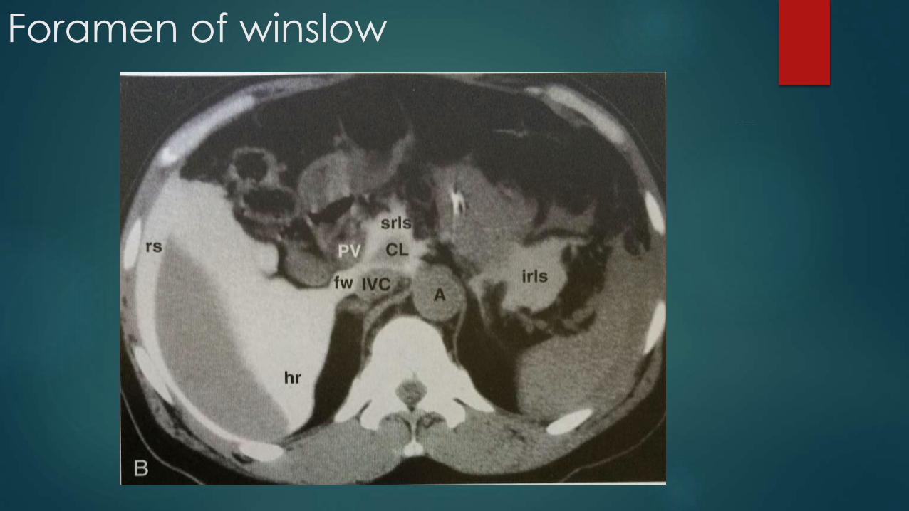
Foramen of winslow

Inframesocolic spaceslies below the transverse mesocolon and transverse colon as far as the true pelvis.
divided in two unequal spaces by the root of the mesentery of the small intestine.
It contains the right and left paracolic gutters lateral to the ascending and descending colon.

Smaller RIC is restricted inferiorly
by the junction of the distal small
bowel mesentry with the cecum
LIC opens into pelvis eccept
where it is bounded by the
sigmoid mesocolon
The paracolic gutters are located
lateral to the peritoneal
reflections of the ac&dc,the Rt
communicates freely with the
right supramesocolic spaces, Lt is
limited by the phrenicocolic
ligament
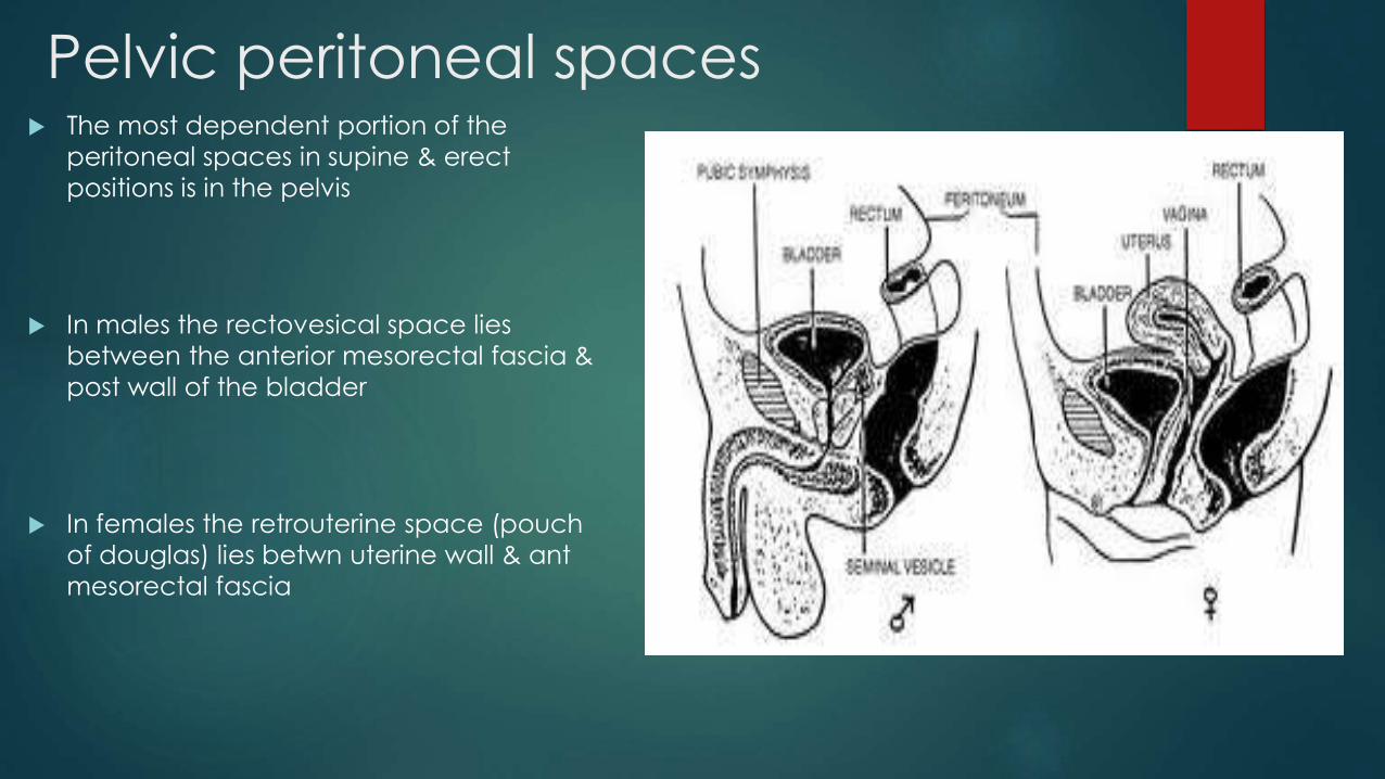
Pelvic peritoneal spaces The most dependent portion of the
peritoneal spaces in supine & erect
positions is in the pelvis
In males the rectovesical space lies
between the anterior mesorectal fascia &
post wall of the bladder
In females the retrouterine space (pouch
of douglas) lies betwn uterine wall & ant
mesorectal fascia


Peritoneal circulation
Water shed areas for fluid
collection
Ileocolic region,root of
sigmoid,pouch of douglas
Majority of the fluid is
cleared at the subphrenic
space by mesothelial
lymphatics