anatomy of cranial nerves in the first turkish illustrated ... · first turkish illustrated anatomy...
TRANSCRIPT
COVER EDITORIAL
Anatomy of cranial nerves in the first Turkish illustratedanatomy manuscript
İlhan Bahşi1 &Mustafa Orhan1&Murat Çetkin1
& Begümhan Turhan1,2& Semih Sayın3
Received: 29 July 2016 /Accepted: 1 August 2016 /Published online: 25 August 2016# Springer-Verlag Berlin Heidelberg 2016
Introduction
Teşrih-ül Ebdan ve Tercümânı Kıbale-i Feylesûfan was thefirst Turkish illustrated anatomy manuscript written byŞemseddîn-i İtâḳî in the seventeenth century. Teşrih is anArabic word which has various meanings such as dissectionof corpse, autopsy, anatomy, and skeleton [1]. This manu-script, the first illustrated anatomy book of Ottoman Period,was written during the period of Sultan Murat IV, who was thesultan of that period [2, 3]. General opinion is that this man-uscript was written in 1632 [4–7].
There are very few works which include information onlyabout anatomy in the Ottoman Empire [7]. The informationabout anatomy was usually found in the other medical booksas only a few pages in that period [3]. They are some of theimportant characteristics of the İtâḳî’s manuscript that
anatomical terms are expressed in Turkish in that manuscriptand the expression is supported by illustrations [6]. This man-uscript has led anatomical terms to be made Turkish. In themanuscript, Turkish anatomical terms were written with gen-erally Arabic and rarely Persian equivalents next to them [6, 7].
After a general part following the introduction part, theinformation was given, starting with bones, about the anatomyof internal organs, nerves, muscles, and vessels, respectively,in the manuscript. The information given about nerves followsthe information given about bones differently from the recentpoint of view. There are original definitions about central andperipheral nervous systems. Firstly, cranial nerves and laterspinal nerves were discussed and information was given aboutpoints of outlet and distribution regions in the explanationsabout the nerves [7–9].
Information presented in the manuscript was visualized byanatomical illustrations. Some parts of these illustrations re-semble the illustrations in Teşrihü’l-Ebdan min e’t-Tıb byMansur (fourteenth century) [2, 7]. Besides these illustrations,there are also some illustrations in the manuscript from vari-ous European resources including De Humani CorporisFabrica by Andreas Vesalius. Furthermore, there are someillustrations mostly about nervous system which were drawnby the author himself [6, 7].
The aims of this study are to examine cranial nerves’ anat-omy in the manuscript Teşrih-ül Ebdan ve Tercümânı Kıbale-iFeylesûfan written in the Ottoman Period and compare thisinformation in that period with today’s knowledge.
Material and methods
Şemseddîn-i İtâḳî’nin Resimli Anatomi Kitabı by Esin Kâhya,which is a translation from Old (Ottoman) Turkish text tocontemporary Turkish alphabet of Teşrih-ül Ebdan ve
* İlhan Bahş[email protected]
Mustafa [email protected]
Murat Ç[email protected]
Begümhan [email protected]
Semih Sayı[email protected]
1 Department of Anatomy, Faculty of Medicine, Gaziantep University,27310 Gaziantep, Turkey
2 Department of Physiotherapy and Rehabilitation, Faculty of HealthSciences, Hasan Kalyoncu University, 27100 Gaziantep, Turkey
3 School of Medicine, Gaziantep University, 27310 Gaziantep, Turkey
Childs Nerv Syst (2017) 33:1855–1862DOI 10.1007/s00381-016-3212-1
Tercümânı Kıbale-i Feylesûfan, was examined. As well as theparts about Anatomy of Nerves and Cerebral Nerves (CranialNerves), the other parts in which the cranial nerves were men-tioned were examined. The information in the manuscript wascompared with today’s knowledge and the correct, imperfect,or incorrect ones were determined.
Results
In the Anatomy of Nerves part of the manuscript, first-ly, general information about nervous system was given.After that, cranial and spinal nerves were described sep-arately. Description of nervous system started with theinformation that the brain was created by God. It wasstated that body organs received their strength and feel-ing from the brain and motor and sensorial features ofnerves were described [7].
The author explained the cranial nerves which match thetoday’s knowledge with the statement BGod enabled nervesfrom brain.^ İtâḳî also stated that spinal nerves came out fromthe spinal cord. He said that the brain would have to be rela-tively big in order to meet the functions of those nerves if allthe nerves came out from the brain. He stated that two prob-lems would occur if all the nerves came out from the brain.The first one, if all the nerves came out from the brain, brainwould be big, and thus, it would impose more burdens on thebody. Secondly, nerve fibers would be longer, and thus, theylose power on the way to periphery [7].
İtâḳî described the cranial nerves as seven cranial nervesand also described each nerve separately. Innervation area ofthe cranial nerves was expressed as head, neck, and internalorgans. Outlets of the cranial nerves were not described indetail; however, they were told to come out directly from thebrain [7]. Although some mistakes are seen in the descriptionsof the cranial nerves, correct information was given in general.Seven cranial nerves described by İtâḳî were examined belowand compared with today’s knowledge.
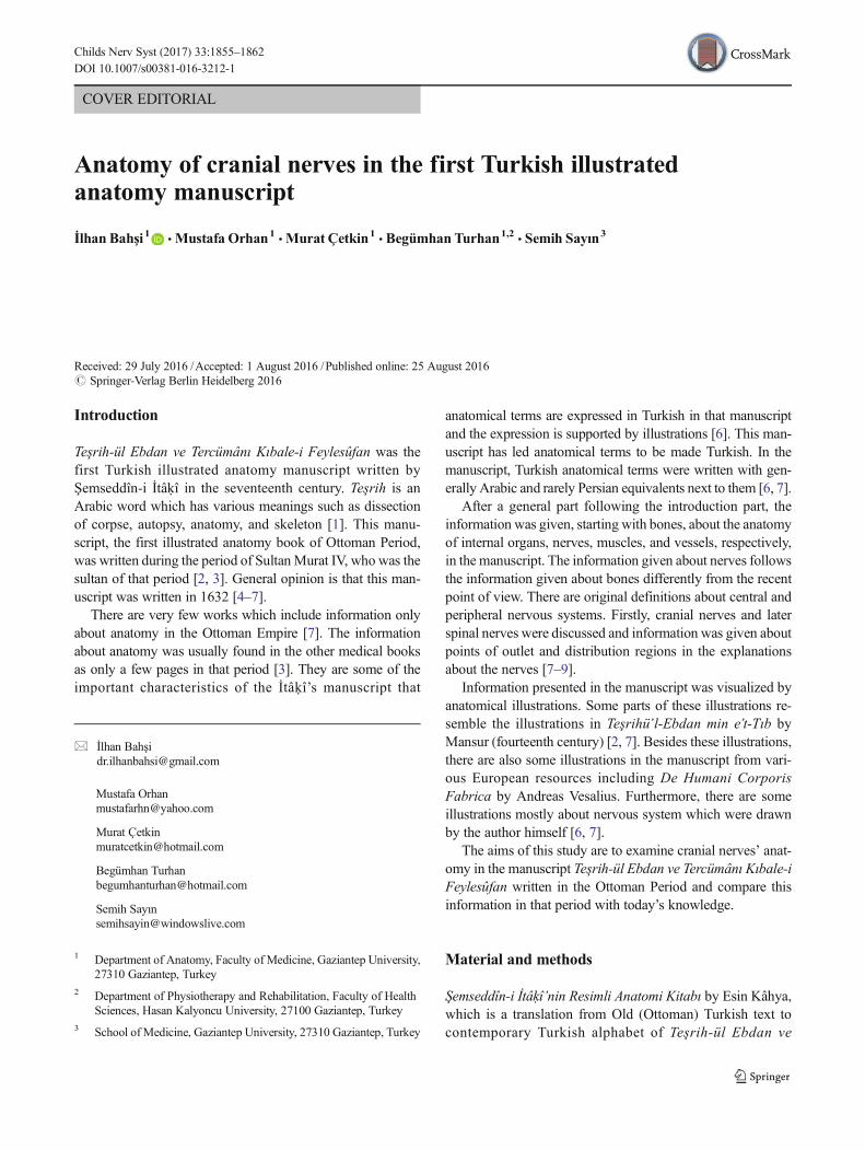
1. The first cranial nerve (Fig. 1 (cover figure)) was likenedto extensions in nipple form on the front part of the brainand calledHalemetu’s sudî in Arabic. In another part of themanuscript, these extensions were called hılmetân. In themanuscript, the ethmoid bone called miṣfât-ı müşaḳḳabeand filter-like perforated structures on that bone were men-tioned. It was stated that the inhaled air passed throughthese nipple-like holes on the upper side of the nasal cavityand went to the cranium, and smell was felt through theholes on nipple-like nerve extensions in that region [7].
Since this nerve carries the smell to the brain, it can bedescribed as olfactory nerve. Furthermore, it was expressedwrongly that the air reached the cranial cavity after passing
through the lamina cribrosa and the sense of smell was felt inthat region.
Later, it was stated that a nerve exit came out from eachextension and separated from each other in a place close toeach eye and then entered the eye. The separation place waslikened to BX^mark and calledmecma῾ en-nûr (optic chiasm)[7]. It is seen here that İt kî did not regarded the first andsecond cranial nerves as separate nerves but regarded themas a single cranial nerve.
It was stated that the nerves separated from each other onthe optic chiasm, and the nerve coming from the right sidewent to the right eye and the nerve coming from the left sidewent to the left eye. İtâḳî also said Bsome say that this forma-tion occurs in the way that the nerve coming from right sidegoes to the left eye, the nerve coming from left side goes toright eye^ in the manuscript [7]. It is seen that the functionalfeatures of fibers in the optic chiasm could not be known indetail since the structures were only examined macroscopical-ly in that period.
The nerve which entered the eye was stated to end aroundruṭubet-i celidiyye (lens) which provided vision. The opticnerve was named as aşab-ı mücevvefe and said to be the sen-sory nerve of the eye. While describing this nerve, the wordmücevvef was used which meant hollow. Thus, we can definethis nerve as the optic nerve according to the informationprovided. However, the vessels in this hollow nerve werenot mentioned. It is a wrong description that the nerve fibersended around the lens.
In the manuscript, benefits of the optic chiasm related tovisual function were emphasized. The first one of these ben-efits is that all the light goes to the healthy eye when one of theeyes is closed, and thus, the strength on that side increases.Other benefits are that the optic chiasm prevents diplopia by
Fig. 1 The first cranial nerve according to Şemseddîn-i İtâḳî (coverfigure)
1856 Childs Nerv Syst (2017) 33:1855–1862
checking the movements of the pupil and the neural transmis-sions get stronger after the two nerves join on the optic chiasm[7]. These definitions are far from today’s knowledge. In con-clusion, it is seen that anatomical information about olfactorynerve and optic nerve was given in the first cranial nerve part.
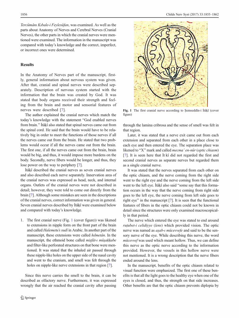
2. The second cranial nerve (Fig. 2) exited behind thefirst cranial nerve and moved forward the hole onthe eyelids, and it reached to the pupil muscles andended by being divided into six branches. It was em-phasized that the movement of the eye was providedby this nerve, and therefore, it was thick. It was ex-plained that the pupils’ movement was provided by sixmuscles, two of which were inclined [7]. We think thatthe Bpupil muscles^ term meant the muscles thatmoved eyeball. Additionally, it was said in the anato-my of the eye part of the manuscript that one of theoptic nerves was for sensation (optic nerve) and theother one was for movement. From here, we think thatthe nerve that moved the eye was the second cranialnerve. We think that the second cranial nerveconsisted of oculomotor nerve, trochlear nerve, andabducens nerve. The exit hole of this cranial nerve isprobably the superior orbital fissure.
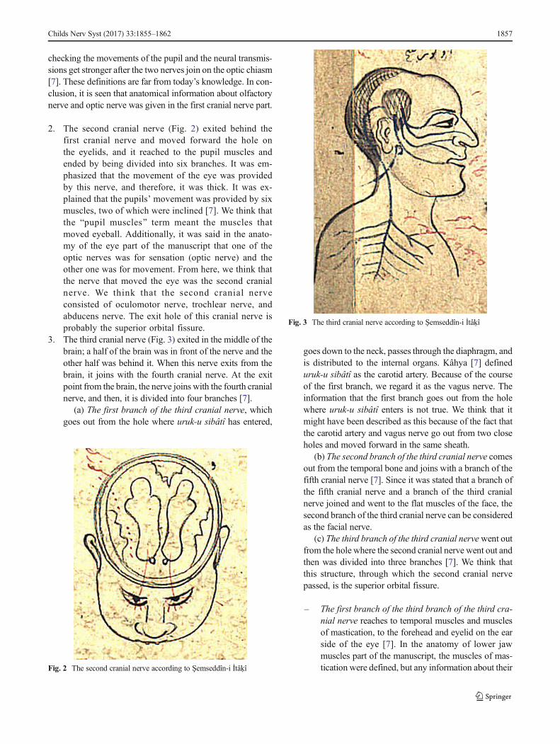
3. The third cranial nerve (Fig. 3) exited in the middle of thebrain; a half of the brain was in front of the nerve and theother half was behind it. When this nerve exits from thebrain, it joins with the fourth cranial nerve. At the exitpoint from the brain, the nerve joins with the fourth cranialnerve, and then, it is divided into four branches [7].
(a) The first branch of the third cranial nerve, whichgoes out from the hole where uruk-u sibâtî has entered,
goes down to the neck, passes through the diaphragm, andis distributed to the internal organs. Kâhya [7] defineduruk-u sibâtî as the carotid artery. Because of the courseof the first branch, we regard it as the vagus nerve. Theinformation that the first branch goes out from the holewhere uruk-u sibâtî enters is not true. We think that itmight have been described as this because of the fact thatthe carotid artery and vagus nerve go out from two closeholes and moved forward in the same sheath.
(b) The second branch of the third cranial nerve comesout from the temporal bone and joins with a branch of thefifth cranial nerve [7]. Since it was stated that a branch ofthe fifth cranial nerve and a branch of the third cranialnerve joined and went to the flat muscles of the face, thesecond branch of the third cranial nerve can be consideredas the facial nerve.
(c) The third branch of the third cranial nervewent outfrom the hole where the second cranial nerve went out andthen was divided into three branches [7]. We think thatthis structure, through which the second cranial nervepassed, is the superior orbital fissure.
– The first branch of the third branch of the third cra-nial nerve reaches to temporal muscles and musclesof mastication, to the forehead and eyelid on the earside of the eye [7]. In the anatomy of lower jawmuscles part of the manuscript, the muscles of mas-tication were defined, but any information about theirFig. 2 The second cranial nerve according to Şemseddîn-i İtâḳî
Fig. 3 The third cranial nerve according to Şemseddîn-i İtâḳî
Childs Nerv Syst (2017) 33:1855–1862 1857
innervation was not given. We think that the nervedistributed to the region of forehead and eyelid is thefrontal nerve. However, the information that thisnerve innervates temporal muscles and muscles ofmastication is not true.
– The second branch of the third branch of the thirdcranial nerve comes to the edge of the eye on the sideof the nose, where the nerve goes into to the nosethrough a hole and is distributed in the skin insidethe nose [7]. According to this definition, we assumethat this branch is the nasociliary nerve.
– The third branch of the third branch of the thirdcranial nerve enters into a hole in facial bones, andsome of its fibers are distributed to upper jaw andgums. Also, it was stated that some fibers went outand were distributed to the facial skin, the tip of thenose, and the upper lip [7]. We may regard thisbranch as the infraorbital nerve based on the infor-mation given.
(d) The fourth branch of the third cranial nervegoes down the hole in the maxilla and is distributedoutside the hole. The tongue was stated to feel bitterand sweet tastes through this nerve. Some fibers sep-arating off from the fourth branch are distributed atthe bottom of the teeth and lower lip [7]. The courseof the fourth branch resembles the course of thegreater palatine nerve. However, the information thatthe tongue receives the senses of tastes through thisnerve is not true. Any hole in the maxilla was notmentioned in the other parts of the manuscript. Inour opinion, this hole may be the greater palatineforamen. The nerve going down the maxillary boneis compatible with the greater palatine nerve, whichpasses through the greater palatine foramen on max-illa. Although innervation area of the mandibularnerve was described, any information about the ori-gin of the mandibular nerve was not given.
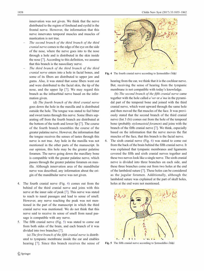
4. The fourth cranial nerve (Fig. 4) comes out from thebehind of the third cranial nerve and joins with thisnerve at the inner side of peak [7]. This nerve was statedto reach to nasal passages and lead to sense of smell.However, any nerve reaching the peak was not men-tioned in the part of the manuscript in which the thirdcranial nerve was mentioned. We do not think that thisnerve said to receive its sense of smell from nasal pas-sage is compatible with any nerve.
5. The fifth cranial nerve (Fig. 5) was stated to come outfrom both sides of the brain, and each branch of it wasdivided into two branches [7].
(a) The first branch of the fifth cranial nerve is distrib-uted to tympanic membrane inside the ear and enableshearing [7]. Since this branch receives the sense of
hearing from the ear, we think that it is the cochlear nerve.But, receiving the sense of hearing from the tympanicmembrane is not compatible with today’s knowledge.
(b) The second branch of the fifth cranial nerve cametogether with the hole called a῾ver or a῾ma in the pyrami-dal part of the temporal bone and joined with the thirdcranial nerve, which went upward through the same holeand then moved the flat muscles of the face. It was previ-ously stated that the second branch of the third cranialnerve (list 3 (b)) comes out from the hole of the temporalbone (probably stylomastoid foramen) and joins with thebranch of the fifth cranial nerve [7]. We think, especiallybased on the information that the nerve moves the flatmuscles of the face, that this branch is the facial nerve.
6. The sixth cranial nerve (Fig. 6) was stated to come outfrom the back of the brain behind the fifth cranial nerve. Itwas explained that tympanic membranes and ligamentscovered the fifth and sixth cranial nerves together andthese two nerves look like a single nerve. The sixth cranialnerve is divided into three branches on each side, andthese three branches come out from two holes at the endof the lambdoid suture [7]. These holes can be consideredas the jugular foramen. Additionally, although thelambdoid suture was explained at the part of skull holes,holes at the end were not mentioned.
Fig. 4 The fourth cranial nerve according to Şemseddîn-i İtâḳî
Fig. 5 The fifth cranial nerve according to Şemseddîn-i İtâḳî
1858 Childs Nerv Syst (2017) 33:1855–1862
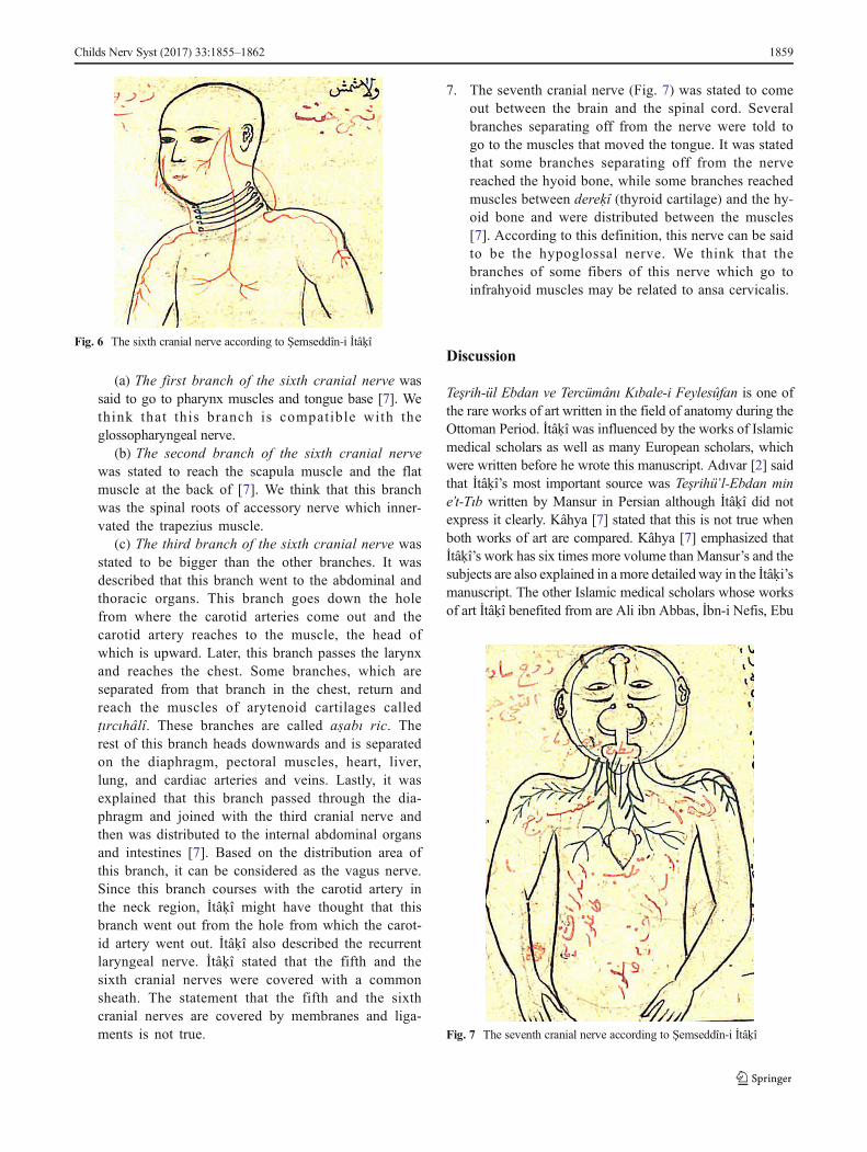
(a) The first branch of the sixth cranial nerve wassaid to go to pharynx muscles and tongue base [7]. Wethink that this branch is compatible with theglossopharyngeal nerve.
(b) The second branch of the sixth cranial nervewas stated to reach the scapula muscle and the flatmuscle at the back of [7]. We think that this branchwas the spinal roots of accessory nerve which inner-vated the trapezius muscle.
(c) The third branch of the sixth cranial nerve wasstated to be bigger than the other branches. It wasdescribed that this branch went to the abdominal andthoracic organs. This branch goes down the holefrom where the carotid arteries come out and thecarotid artery reaches to the muscle, the head ofwhich is upward. Later, this branch passes the larynxand reaches the chest. Some branches, which areseparated from that branch in the chest, return andreach the muscles of arytenoid cartilages calledṭırcıhâlî. These branches are called aṣabı ric. Therest of this branch heads downwards and is separatedon the diaphragm, pectoral muscles, heart, liver,lung, and cardiac arteries and veins. Lastly, it wasexplained that this branch passed through the dia-phragm and joined with the third cranial nerve andthen was distributed to the internal abdominal organsand intestines [7]. Based on the distribution area ofthis branch, it can be considered as the vagus nerve.Since this branch courses with the carotid artery inthe neck region, İtâḳî might have thought that thisbranch went out from the hole from which the carot-id artery went out. İtâḳî also described the recurrentlaryngeal nerve. İtâḳî stated that the fifth and thesixth cranial nerves were covered with a commonsheath. The statement that the fifth and the sixthcranial nerves are covered by membranes and liga-ments is not true.
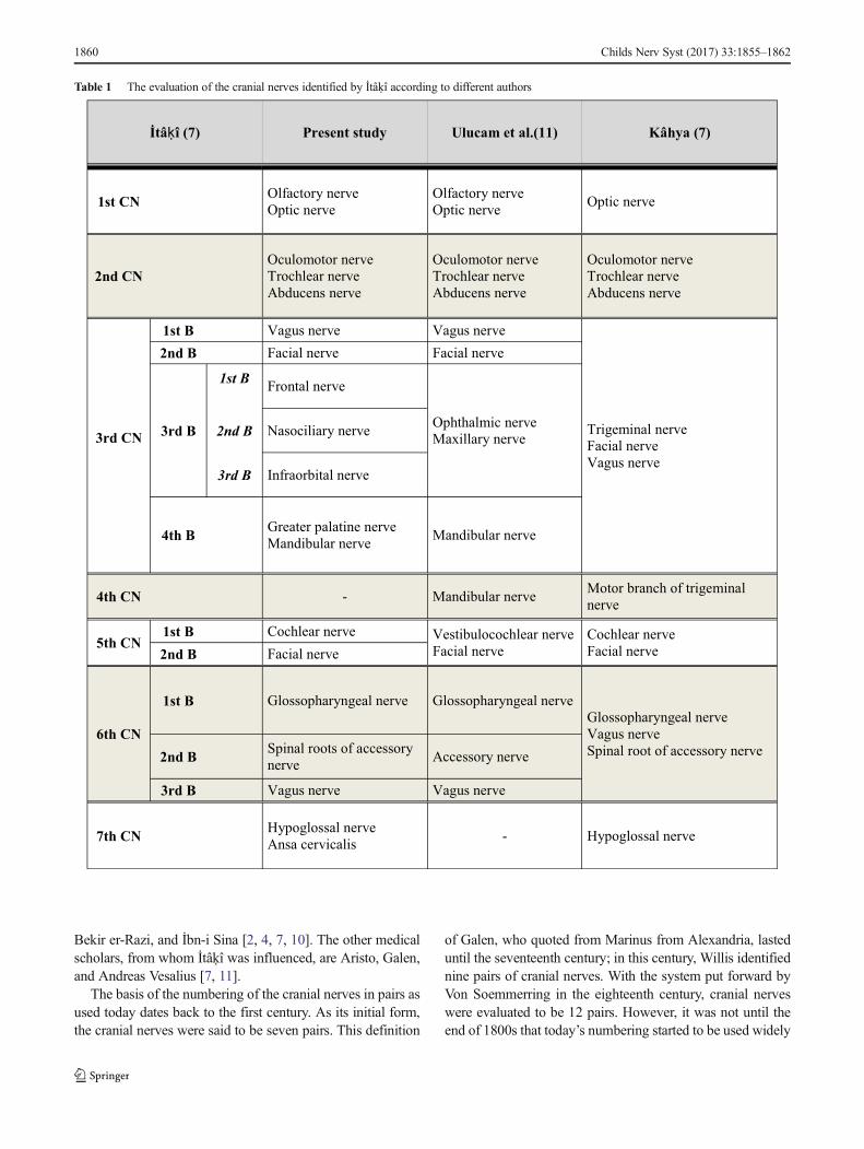
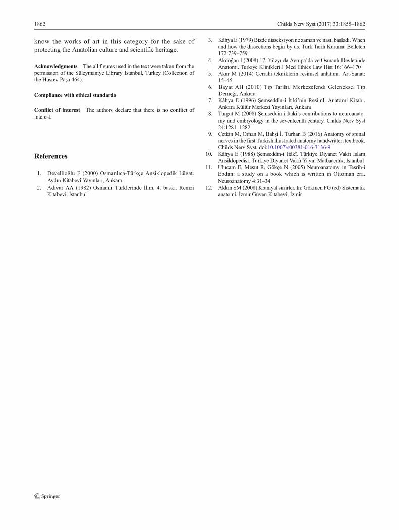
7. The seventh cranial nerve (Fig. 7) was stated to comeout between the brain and the spinal cord. Severalbranches separating off from the nerve were told togo to the muscles that moved the tongue. It was statedthat some branches separating off from the nervereached the hyoid bone, while some branches reachedmuscles between dereḳî (thyroid cartilage) and the hy-oid bone and were distributed between the muscles[7]. According to this definition, this nerve can be saidto be the hypoglossal nerve. We think that thebranches of some fibers of this nerve which go toinfrahyoid muscles may be related to ansa cervicalis.
Discussion
Teşrih-ül Ebdan ve Tercümânı Kıbale-i Feylesûfan is one ofthe rare works of art written in the field of anatomy during theOttoman Period. İtâḳî was influenced by the works of Islamicmedical scholars as well as many European scholars, whichwere written before he wrote this manuscript. Adıvar [2] saidthat İtâḳî’s most important source was Teşrihü’l-Ebdan mine’t-Tıb written by Mansur in Persian although İtâḳî did notexpress it clearly. Kâhya [7] stated that this is not true whenboth works of art are compared. Kâhya [7] emphasized thatİtâḳî’s work has six times more volume thanMansur’s and thesubjects are also explained in a more detailed way in the İtâḳi’smanuscript. The other Islamic medical scholars whose worksof art İtâḳî benefited from are Ali ibn Abbas, İbn-i Nefis, Ebu
Fig. 6 The sixth cranial nerve according to Şemseddîn-i İtâḳî
Fig. 7 The seventh cranial nerve according to Şemseddîn-i İtâḳî
Childs Nerv Syst (2017) 33:1855–1862 1859
Bekir er-Razi, and İbn-i Sina [2, 4, 7, 10]. The other medicalscholars, from whom İtâḳî was influenced, are Aristo, Galen,and Andreas Vesalius [7, 11].
The basis of the numbering of the cranial nerves in pairs asused today dates back to the first century. As its initial form,the cranial nerves were said to be seven pairs. This definition
of Galen, who quoted from Marinus from Alexandria, lasteduntil the seventeenth century; in this century, Willis identifiednine pairs of cranial nerves. With the system put forward byVon Soemmerring in the eighteenth century, cranial nerveswere evaluated to be 12 pairs. However, it was not until theend of 1800s that today’s numbering started to be used widely
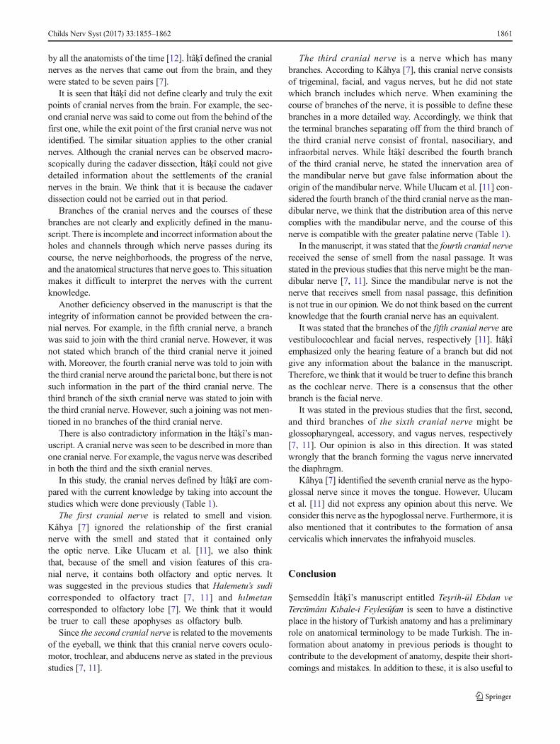
Table 1 The evaluation of the cranial nerves identified by İtâḳî according to different authors
İtâḳî (7) Present study Ulucam et al.(11) Kâhya (7)
1st CN Olfactory nerve
Optic nerve
Olfactory nerve
Optic nerveOptic nerve
2nd CNOculomotor nerve
Trochlear nerve
Abducens nerve
Oculomotor nerve
Trochlear nerve
Abducens nerve
Oculomotor nerve
Trochlear nerve
Abducens nerve
3rd CN
1st B Vagus nerve Vagus nerve
Trigeminal nerve
Facial nerve
Vagus nerve
2nd B Facial nerve Facial nerve
3rd B
1st BFrontal nerve
Ophthalmic nerve
Maxillary nerve2nd B Nasociliary nerve
3rd B Infraorbital nerve
4th B Greater palatine nerve
Mandibular nerveMandibular nerve
4th CN - Mandibular nerveMotor branch of trigeminal
nerve
5th CN1st B Cochlear nerve Vestibulocochlear nerve
Facial nerve
Cochlear nerve
Facial nerve2nd B Facial nerve
6th CN
1st B Glossopharyngeal nerve Glossopharyngeal nerve
Glossopharyngeal nerve
Vagus nerve
Spinal root of accessory nerve2nd B Spinal roots of accessory
nerveAccessory nerve
3rd B Vagus nerve Vagus nerve
7th CN Hypoglossal nerve
Ansa cervicalis- Hypoglossal nerve
1860 Childs Nerv Syst (2017) 33:1855–1862
by all the anatomists of the time [12]. İtâḳî defined the cranialnerves as the nerves that came out from the brain, and theywere stated to be seven pairs [7].
It is seen that İtâḳî did not define clearly and truly the exitpoints of cranial nerves from the brain. For example, the sec-ond cranial nerve was said to come out from the behind of thefirst one, while the exit point of the first cranial nerve was notidentified. The similar situation applies to the other cranialnerves. Although the cranial nerves can be observed macro-scopically during the cadaver dissection, İtâḳî could not givedetailed information about the settlements of the cranialnerves in the brain. We think that it is because the cadaverdissection could not be carried out in that period.
Branches of the cranial nerves and the courses of thesebranches are not clearly and explicitly defined in the manu-script. There is incomplete and incorrect information about theholes and channels through which nerve passes during itscourse, the nerve neighborhoods, the progress of the nerve,and the anatomical structures that nerve goes to. This situationmakes it difficult to interpret the nerves with the currentknowledge.
Another deficiency observed in the manuscript is that theintegrity of information cannot be provided between the cra-nial nerves. For example, in the fifth cranial nerve, a branchwas said to join with the third cranial nerve. However, it wasnot stated which branch of the third cranial nerve it joinedwith. Moreover, the fourth cranial nerve was told to join withthe third cranial nerve around the parietal bone, but there is notsuch information in the part of the third cranial nerve. Thethird branch of the sixth cranial nerve was stated to join withthe third cranial nerve. However, such a joining was not men-tioned in no branches of the third cranial nerve.
There is also contradictory information in the İtâḳî’s man-uscript. A cranial nerve was seen to be described in more thanone cranial nerve. For example, the vagus nerve was describedin both the third and the sixth cranial nerves.
In this study, the cranial nerves defined by İtâḳî are com-pared with the current knowledge by taking into account thestudies which were done previously (Table 1).
The first cranial nerve is related to smell and vision.Kâhya [7] ignored the relationship of the first cranialnerve with the smell and stated that it contained onlythe optic nerve. Like Ulucam et al. [11], we also thinkthat, because of the smell and vision features of this cra-nial nerve, it contains both olfactory and optic nerves. Itwas suggested in the previous studies that Halemetu’s sudicorresponded to olfactory tract [7, 11] and hılmetancorresponded to olfactory lobe [7]. We think that it wouldbe truer to call these apophyses as olfactory bulb.
Since the second cranial nerve is related to the movementsof the eyeball, we think that this cranial nerve covers oculo-motor, trochlear, and abducens nerve as stated in the previousstudies [7, 11].
The third cranial nerve is a nerve which has manybranches. According to Kâhya [7], this cranial nerve consistsof trigeminal, facial, and vagus nerves, but he did not statewhich branch includes which nerve. When examining thecourse of branches of the nerve, it is possible to define thesebranches in a more detailed way. Accordingly, we think thatthe terminal branches separating off from the third branch ofthe third cranial nerve consist of frontal, nasociliary, andinfraorbital nerves. While İtâḳî described the fourth branchof the third cranial nerve, he stated the innervation area ofthe mandibular nerve but gave false information about theorigin of the mandibular nerve. While Ulucam et al. [11] con-sidered the fourth branch of the third cranial nerve as the man-dibular nerve, we think that the distribution area of this nervecomplies with the mandibular nerve, and the course of thisnerve is compatible with the greater palatine nerve (Table 1).
In the manuscript, it was stated that the fourth cranial nervereceived the sense of smell from the nasal passage. It wasstated in the previous studies that this nerve might be the man-dibular nerve [7, 11]. Since the mandibular nerve is not thenerve that receives smell from nasal passage, this definitionis not true in our opinion. We do not think based on the currentknowledge that the fourth cranial nerve has an equivalent.
It was stated that the branches of the fifth cranial nerve arevestibulocochlear and facial nerves, respectively [11]. İtâḳîemphasized only the hearing feature of a branch but did notgive any information about the balance in the manuscript.Therefore, we think that it would be truer to define this branchas the cochlear nerve. There is a consensus that the otherbranch is the facial nerve.
It was stated in the previous studies that the first, second,and third branches of the sixth cranial nerve might beglossopharyngeal, accessory, and vagus nerves, respectively[7, 11]. Our opinion is also in this direction. It was statedwrongly that the branch forming the vagus nerve innervatedthe diaphragm.
Kâhya [7] identified the seventh cranial nerve as the hypo-glossal nerve since it moves the tongue. However, Ulucamet al. [11] did not express any opinion about this nerve. Weconsider this nerve as the hypoglossal nerve. Furthermore, it isalso mentioned that it contributes to the formation of ansacervicalis which innervates the infrahyoid muscles.
Conclusion
Şemseddîn İtâḳî’s manuscript entitled Teşrih-ül Ebdan veTercümânı Kıbale-i Feylesûfan is seen to have a distinctiveplace in the history of Turkish anatomy and has a preliminaryrole on anatomical terminology to be made Turkish. The in-formation about anatomy in previous periods is thought tocontribute to the development of anatomy, despite their short-comings and mistakes. In addition to these, it is also useful to
Childs Nerv Syst (2017) 33:1855–1862 1861
know the works of art in this category for the sake ofprotecting the Anatolian culture and scientific heritage.
Acknowledgments The all figures used in the text were taken from thepermission of the Süleymaniye Library Istanbul, Turkey (Collection ofthe Hüsrev Paşa 464).
Compliance with ethical standards
Conflict of interest The authors declare that there is no conflict ofinterest.
References
1. Devellioğlu F (2000) Osmanlıca-Türkçe Ansiklopedik Lügat.Aydın Kitabevi Yayınları, Ankara
2. Adıvar AA (1982) Osmanlı Türklerinde İlim, 4. baskı. RemziKitabevi, İstanbul
3. Kâhya E (1979) Bizde disseksiyon ne zaman ve nasıl başladı. Whenand how the dissections begin by us. Türk Tarih Kurumu Belleten172:739–759
4. Akdoğan I (2008) 17. Yüzyılda Avrupa’da ve Osmanlı DevletindeAnatomi. Turkiye Klinikleri J Med Ethics Law Hist 16:166–170
5. Akar M (2014) Cerrahi tekniklerin resimsel anlatımı. Art-Sanat:15–45
6. Bayat AH (2010) Tıp Tarihi. Merkezefendi Geleneksel TıpDerneği, Ankara
7. Kâhya E (1996) Şemseddîn-i İt kî’nin Resimli Anatomi Kitabı.Ankara Kültür Merkezi Yayınları, Ankara
8. Turgut M (2008) Şemseddin-i Itaki’s contributions to neuroanato-my and embryology in the seventeenth century. Childs Nerv Syst24:1281–1282
9. Çetkin M, Orhan M, Bahşi İ, Turhan B (2016) Anatomy of spinalnerves in the first Turkish illustrated anatomy handwritten textbook.Childs Nerv Syst. doi:10.1007/s00381-016-3136-9
10. Kâhya E (1988) Şemseddîn-i Itâkî. Türkiye Diyanet Vakfı İslamAnsiklopedisi. Türkiye Diyanet Vakfı Yayın Matbaacılık, İstanbul
11. Ulucam E, Mesut R, Gökçe N (2005) Neuroanatomy in Tesrih-iEbdan: a study on a book which is written in Ottoman era.Neuroanatomy 4:31–34
12. Akkın SM (2008) Kraniyal sinirler. In: Gökmen FG (ed) Sistematikanatomi. İzmir Güven Kitabevi, İzmir
1862 Childs Nerv Syst (2017) 33:1855–1862