anatomical variations of the vertebral artery and its ... · the lateral mass of the atlas and...
TRANSCRIPT

Anatomical variations of the vertebral artery and its relation to the atlas
vertebra - Radiological and dry bone study
ORIGINAL ARTICLE Eur. J. Anat. 23 (1): 49-58 (2019)
Fayza A. Abd El Gawad, Mohamed H. Shaaban, Doaa M. Shuaib, Hala M.
Shallan
Department of Anatomy and Embryology, Faculty of Medicine, Cairo University, Egypt
SUMMARY
Vertebral artery (VA) variations are important for diagnostic angiographic procedures. This study aimed to describe the anatomical variations of VA using multidetector computed tomography angi-ography (MDCTA), and to provide a quantitative and qualitative anatomy of the VA groove in dry atlas vertebrae. The study was carried out on 100 MDCTA images from adult Egyptian individuals (69 males; 31 females) and 50 dry atlas fully ossi-fied and of unknown age and sex. MDCTA films were evaluated for VA origin, level of entrance into foramen transversarium, caliber, and distance from the midline. VA grooves in dry bones were exam-ined for the presence of ponticulus posticus (PP). Inner and outer distances from the midline, width and thickness were measured using sliding Vernier caliper. The results revealed that the left VA arose directly from the aortic arch in 7% of cases and was absent in 2% of cases. Atypical entry of VA into foramen transversarium was through C5 (4.5%), followed by C7 (1.5%), then C4 (1%). The left vertebral arteries with direct aortic origin were more medially located than the left arteries with subclavian origin (p=0.005). The mean diameter was significantly greater on the left (3.67±1.07 mm), as compared to the right side (3.36±0.93 mm) (p=0.038). PP was detected in 47% of cases in radiological images and 96% of dry bones. It could be concluded that the most important varia-
tions of VA were the aortic origin of the left VA and abnormal entry through transverse foramina. PP was a common variation in atlas vertebrae. These variations should be taken into consideration dur-ing radiological and orthopedic procedures.
Key words: Vertebral artery – Variations –
MDCTA – Ponticulus posticus
INTRODUCTION
The VA is a major artery of the neck. It is unique among the cervico-cephalic vessels due to its posi-tion and relationship to the adjacent structures. It is a critical artery in perfusion of the upper spinal cord, brain stem, cerebellum and different parts of posterior cerebral hemispheres (Sonje et al., 2015). It arises from the first part of the subclavian artery and passes through the foramina transver-saria of all of the cervical vertebrae, except the seventh vertebra (C7). It curves medially behind the lateral mass of the atlas and enters the crani-um through the foramen magnum (Standring, 2016). The artery is composed of four segments. The first segment extends from the origin to the transverse foramen of the sixth cervical vertebra (C6). The second segment runs from the trans-verse process of C6 to the first cervical vertebra (C1). The third segment extends from C1 to the foramen magnum and the fourth segment runs from the dura to the vertebrobasilar junction (Shin et al., 2014).
Several anatomical variations of the VA have been reported in the literature. They include varia-tions in origin, number and of its course through
49
Submitted: 29 August, 2018. Accepted: 15 November, 2018.
Corresponding author: Doaa Mahmoud Shuaib. Department
of Anatomy and Embryology, Faculty of Medicine, Cairo Univer-
sity, Kasr Al-Ainy Street, 11562 Cairo, Egypt. Phone:
00201005859263. E-mail: [email protected]

Variations of vertebral artery
50
foramina transversaria of cervical vertebrae. It is important to search for these variations in preoper-ative workup to work in safe conditions with the lowest risk of VA injury. Unawareness of these anatomical variations exposes the artery to the risk of injury with its possible overwhelming conse-quences (George et al., 2011).
Computed tomography angiography is a popular method for the evaluation of cerebrovascular dis-eases. It is a noninvasive tool in the diagnosis of VA pathology (Sanelli et al., 2002). Computed to-mography angiography alone is sometimes consid-ered to be insufficient for evaluating complicated VA courses among the various surrounding struc-tures; therefore, post-processed images in the three dimensional workstation are used to show more details (Wakao et al., 2014 b).
The third part of the artery passes on the VA groove in the posterior arch of first cervical verte-bra. The variation in location of this groove can lead to serious complications during surgical pro-cedures, especially during posterolateral mass screw fixation (Ebraheim et al., 1998). Ponticulus posticus (PP) is a bony projection that converts the groove of the VA into a canal. It is usually asymp-tomatic; however, it may become dangerous dur-ing trauma or aggravate some troubles during spe-cific diagnostic or surgical procedures (Cushing et al., 2001). The VA is susceptible to be compressed by the presence of these ponticuli causing verte-brobasilar insufficiency (Akhtar et al., 2015).
Concerning VA, several authors examined one or two parts in the reviewed literature. However, very few studies were carried out concerning all three extracranial segments of the artery. Therefore, the objective of this study is to describe the anatomy of the VA and its variations using MDCTA images and to assess the quantitative and qualitative anat-omy of the VA groove of the atlas vertebra based on dry bones.
MATERIAL AND METHODS
This study is based on both radiological and dry bone material. For clarity, the details of each group analysis will be considered individually. Radiological study
The study was conducted on 100 angiographs of Egyptian patients (69 males and 31 females, aged between 41 and 65 years old). Multidetector com-puted tomography angiography images (MDCTA) were obtained from the Radiology Department, Faculty of Medicine, Cairo University and Nile Scan Imaging Center. Radiographs were studied retrospectively over a period of 10 months (June 2017 - March 2018). The patients underwent three dimensional carotid or cerebral CT angiographies for the evaluation of various diseases such as cer-ebral ischemia, headache, trauma and spine prob-lems. Patients with atherosclerotic diseases and VA aneurysm or stenosis were excluded from the study.
MDCTA was performed by using a 16-row
MDCTA system (Lightspeed Ultra, GE Medical Systems, Milwaukee, Wis., USA). Nonionic iodinat-ed contrast agent (Omnipaque, 300 mgI/ml) with the total amount of 1.5-2.0 ml/kg was injected through an 18-gauge cannula positioned in an an-tecubital vein at a flow rate of 4-5 ml/second by using a power jector.
After injection of contrast medium into the fore-arm vein, complete examination from the aortic arch to the circle of Willis was done using a 2.5 mm helix (pitch 0.8, 0.5-second rotation, scan trig-gered when the contrast density was 150 Houns-field Units in the ascending aorta).
For three-dimensional image, Philips workstation (MX VIEW) was used for post-processing recon-struction. The volumetric MDCTA data sets were processed on a separate workstation (Advanced Workstation 4.2, GE Healthcare, and Milwaukee, Wis.) with multiplanar reformatting, curved planar reformatting, maximum intensity projection and volume rendering.
Post-processing two dimensional and three-dimensional reformations (Extended Brilliance Workspace, Philips Medical systems) contributed significantly to accurate evaluation. Most common-ly used post-processing techniques were multipla-nar and curved planar reformations (MPR and CPR), maximum intensity projection (MIP) and volume rendering (VR).
Each radiological image was evaluated for the following:
1) Origin of the VA on both sides. 2) Absence of VA on one side or double origin. 3) Level of foramen transversarium through
which the artery passed. 4) Diameter of the second segment of the artery
at a fixed point between C4 and C5 pedicles. Ac-cording to Hong et al. (2008), the VA was labeled as dominant if its diameter was more than twice that of the other side, while the smaller one was labeled as hypoplastic.
5) Distance of VA from midline was measured at a fixed point before its entrance through the fora-men transversarium (pre-foraminal). According to Kiresi et al. (2009), determination of the midline was through a line passing through the axis and spinous processes.
6) Ponticulus posticus in the posterior arch of the atlas vertebra if present (partial or complete ring). Dry bone study
Fifty dried human atlas vertebrae (100 VA grooves) were included in the study. The bones were collected from the Anatomy and Embryology Department, Faculty of Medicine, Cairo University. The bones were fully ossified and of unknown age and sex. Vertebrae with any fracture, incomplete ossification or with any gross pathological abnor-malities were excluded from the study.
The superior surface of the posterior arch of each atlas vertebrae was examined carefully for the presence or absence of complete or partial ring for VA (ponticulus posticus) on one side or on both sides.

F.A. Abd El Gawad et al.
51
Four linear parameters were measured on both sides by the use of sliding Vernier caliper and rec-orded (Lalitha et al., 2016) (Fig. 1):
a) Inner distance of vertebral artery groove (D1): distance from mid-point on the posterior tubercle to the medial most edge of VA groove.
b) Outer distance of vertebral artery groove (D2): distance from mid-point on posterior tubercle to the lateral most edge of VA groove.
c) Width of vertebral artery groove (W): distance between the inner and outer edges at the middle of the groove.
d) Thickness of the vertebral artery groove (T): distance between the inner and outer edges at the thinnest part of the groove. Statistical study
Statistical analysis was performed using statisti-cal package for the social sciences (SPSS) ver-sion 21.0 (IBM Corporation, Somers, NY, USA) statistical software. The data were expressed as means ± standard deviation (SD). The frequency of nominal data was done.
The quantitative data were examined by Kolmo-gorov Smirnov test for normality. Independent “t” test was performed to compare the different varia-bles regarding side. Chi-Square test of independ-ence was used to determine if there was a signifi-cant relationship between different variables and sex. P value ≤ 0.05 was considered to be signifi-cant.
RESULTS Radiological study
The left VA was seen originating from the left subclavian artery in 91% of cases (61% males and 30% females). The artery arose directly from the aortic arch in 7% of cases (6% males and 1% fe-males) (Fig. 2a). The artery was absent in 2% of male patients (Fig. 2b), while it was present in all female patients. On the right side, the VA arose from the right subclavian artery in all cases (69% males and 31% females) (Fig. 3). When comparing male to female cases, the frequency of variations of VA origin showed statistically non-significant differences (p=0.2).
The left VA entered the transverse foramen of C6 in 91% of cases. Atypical level of the entrance was observed in 7% of cases (4% at the level of C5, 2% at the level of C7 and 1% at the level of C4). In the remaining two cases (2%), the artery was ab-sent. On the right side, the VA was seen entering the foramen transversarium of C6 in 93% of cases. In 7% of cases, the artery showed atypical level of entrance (5% at the level of C5, 1% at the level of C4 and 1% at the level of C7) (Figs. 4-7) (Table 1).
The mean diameter of the VA was 3.67±1.07 mm on the left side and 3.36±0.93 mm on the right side. The difference in the mean diameter of the artery between both sides was found to be statisti-cally significant (p=0.038) (Table 2). The left VA was dominant in 8% of cases, while the right artery was dominant in 3% of cases (Fig. 8). The remain-ing 89% of cases showed no dominant laterality.
The mean distance from the medial edge of the VA to the midline was 2±0.5 cm on the left side and 2±0.4 cm on the right side. The difference in mean distance between both sides was found to be statistically non-significant (p= 0.4) (Table 2).
The mean distance of the left vertebral arteries with subclavian origin from the midline was 2.05±0.27 cm and the mean distance of the left vertebral arteries with aortic arch origin from the midline was 1.77±0.42 cm. It was observed that
Fig 1. Photograph of the atlas showing the four line-ar parameters. (a): distance from mid-point on the poste-rior tubercle to the medial most edge of VA groove (D1), distance from mid-point on the posterior tubercle to the lateral most edge of VA groove (D2), width of VA groove (W). (b): thickness of VA groove (T).
Fig 2. Volume rendering images of two male patients showing variations in the origin of left vertebral artery. The left vertebral artery (arrow) originates directly from the aortic arch (AA) (a). The left vertebral artery is ab-sent (b). Brachiocephalic artery (BCA), left common carotid artery (LCA) and left subclavian artery (LSA) are illustrated.

Variations of vertebral artery
52
the left vertebral arteries with direct aortic origin were more medially located and this was found to be statistically significant (p=0.005) (Table 3).
Considering sex difference, the mean distance of the medial edge of the left VA from the midline was 1.98+0.52 cm in males and 1.94+0.24 cm in fe-male patients. This difference was found to be sta-tistically non-significant (p=0.7). On the right side, the mean distance of the artery from the midline was 2.04+0.39 cm in males and 1.93+0.24 cm in females. The difference was found to be statistical-ly non-significant (p=0.1) (Table 4).
Out of 100 atlases in CT images, PP was ob-served in 47 bones (47%) in which 26 (26%) had both complete and partial bridges; 14 (14%) had a complete bony bridge and 7 vertebrae (7%) had partial bridges. PP was absent in 53% of cases (Fig. 9).
Complete PP was unilateral in 11 vertebrae (11%) (4 on the right side and 7 on the left) and bilateral in 10 vertebrae (10%). Partial bony bridg-es were unilateral in 21 bones (21%) (14 on the right side and 7 on the left) and bilateral in 12 ver-tebrae (12%) (Figs. 10, 11). The incidence of PP was more in males (35%) as compared to females (12%) and this difference was found to be statisti-cally significant (p=0.01). Dry Bone study
Out of 50 atlas vertebrae, the PP was observed in 48 bones (96%) in which 19 (38%) had a com-
Fig 3. Bar chart of the frequency of variations of vertebral artery origin in relation to sex.
LVA RVA
Entry level
LSA origin Direct aortic
origin Total
RSA origin
n n n (%) n (%)
C3
C4 1 1 (1%) 1 (1%)
C5 4 4 (4%) 5 (5%)
C6 89 2 91 (91%) 93 (93%)
C7 2 2 (2%) 1 (1%)
Table 1. Relationship between vertebral artery origin and entry level into the foramen transversarium.
LVA: left vertebral artery, LSA: left subclavian artery, RVA: right vertebral artery, RSA: right subclavian artery
Parameter LVA
Mean + SD RVA
Mean + SD
Independent t test p-value
Diameter (mm)
3.67+1.07 3.36+ 0.93 0.038*
Distance from midline (cm)
2+0.5 2+0.4 0.4
Table 2. Mean diameter and distance of left and right vertebral arteries from midline.
p-value ≤ 0.05 was considered statistically significant
LVA distance from midline
(cm)
LVA origin
Mean + SD Independent t test p-value
DA 1.77+0.42 0.005* LSA 2.05+0.27
Table 3. Mean distance of the left vertebral artery from midline in relation to its origin
LSA: left subclavian artery, DA origin: direct aortic origin P-value ≤ 0.05 was considered statistically significant

F.A. Abd El Gawad et al.
53
plete and partial bony bridge; 15 (30%) had a com-plete bridge and 14 vertebrae (28%) had partial bridges. Partial PP was seen either in the form of bony spicule or tubercle of bone projecting over the VA groove. Ponticulus posticus was absent in two vertebrae (4%) (Fig. 12).
Complete PP was unilateral in 23 vertebrae (46%) (12 on the right side and 11 on the left side) and bilateral in 10 vertebrae (20%). Partial bony bridges were unilateral in 19 bones (38%) (10 on the right side and 9 on the left) and bilateral in 10 vertebrae (20%) (Figs. 13, 14).
The mean distance from mid-point on the posteri-or tubercle to the medial most edge of VA groove (D1) was found to be 13.8±2.7 mm on the right and 13.4±2.4 mm on the left. The difference in the
mean value of D1 between both sides was found to be statistically non-significant (p=0.2) (Table 5).
The mean distance from mid-point on the posteri-or tubercle to the lateral most edge of VA groove (D2) was found to be 27.0±2.5 mm on the right and 27.2±2.7 mm on the left side. The difference between both sides was found to be statistically non-significant (p=0.5) (Table 5).
The mean width the VA groove (W) was found to be 9.3±2.0 mm on the right and 9.9±1.8 mm on the left side. The difference in the mean width between both sides was statistically non-significant (p=0.07) (Table 5).
The thickness of VA groove (T) was 2.7±1.0 mm on the right and 2.9±2.0 mm on the left side. The difference in the mean thickness of the VA groove between both sides was statistically non-significant (p=0.3) (Table 5).
DISCUSSION
Detection of variations of VA is crucial in diag-nostic examinations and in performing interven-tional or surgical procedures (George et al., 2011; Shin et al., 2014). MDCTA imaging pro-vides important data for preventing vascular com-plications caused by iatrogenic injury of the VA (Sano et al., 2013). Knowing morphometric measurements of atlas vertebra are important for management of conditions, which may lead to al-
Fig 4. CT angiography image of a female patient (a: sagittal section, b: axial section) showing the left verte-bral artery entering the foramen transversarium at the level of C6 (arrow), while the right vertebral artery (arrow head) is still outside. It enters the foramen transversari-um at the level of fifth cervical (C5) vertebra.
Fig 5. CT angiography image of a female patient (a: sagittal section, b: axial section) showing right vertebral artery (arrow head) and left vertebral artery (arrow) en-tering the foramen transversarium at the level of seventh cervical (C7) vertebra.
Fig 6. Volume rendering image of a male patient showing the right vertebral artery (arrow) entering the foramen transversarium at the level of C4 vertebra.
Male Mean
+ SD Female Mean
+ SD Independent t test p-value
Left distance from midline
(cm) 1.98+0.52 1.94+0.24 0.7
Right distance from midline
(cm) 2.04+0.39 1.93+0.24 0.1
Table 4. Distance of right and left vertebral arteries from midline in relation to sex.
p-value ≤ 0.05 was considered statistically significant
Variations of vertebral artery
54
tanto-axial and atlanto-occipital instability (Lalitha et al., 2016).
In the radiological images of the present study, most of the vertebral arteries arose from the sub-clavian artery on both sides (91% on the left side and 100% on the right side). The most prevalent variation detected was abnormal origin of the left
VA from the aortic arch. These findings were in accordance with the study carried out by Uchino et al. (2013) in Japanese population. They reported that 94% of vertebral arteries arose from the sub-clavian artery. They further added that the most
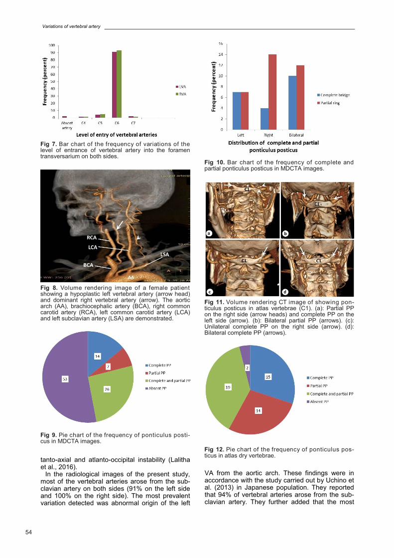
Fig 7. Bar chart of the frequency of variations of the level of entrance of vertebral artery into the foramen transversarium on both sides.
Fig 8. Volume rendering image of a female patient showing a hypoplastic left vertebral artery (arrow head) and dominant right vertebral artery (arrow). The aortic arch (AA), brachiocephalic artery (BCA), right common carotid artery (RCA), left common carotid artery (LCA) and left subclavian artery (LSA) are demonstrated.
Fig 9. Pie chart of the frequency of ponticulus posti-cus in MDCTA images.
Fig 11. Volume rendering CT image of showing pon-ticulus posticus in atlas vertebrae (C1). (a): Partial PP on the right side (arrow heads) and complete PP on the left side (arrow). (b): Bilateral partial PP (arrows). (c): Unilateral complete PP on the right side (arrow). (d): Bilateral complete PP (arrows).
Fig 10. Bar chart of the frequency of complete and partial ponticulus posticus in MDCTA images.
Fig 12. Pie chart of the frequency of ponticulus pos-ticus in atlas dry vertebrae.

F.A. Abd El Gawad et al.
55
common variation was direct origin of the left VA from the aortic arch.
The left VA was absent in two cases in the pre-sent work. Absence of the artery was also encoun-tered by Kao et al. (2003), who reported a case of absence of the right VA and contralateral extracra-nial VA aneurysm in Taiwan. Basekim et al. (2004) explained that an absent VA could be the result of a developmental factor where there are four transi-ent anastomoses between the basilar arterial sys-tem and the anterior carotid artery in early fetal life. These connections normally regress. Howev-er, sometimes they may persist into adult life. Fail-
ure of regression of one of these connections causes four different types of anomalous arteries known as persistent fetal anastomoses. The per-sistent fetal anastomoses are usually large and associated with hypoplasia or aplasia of the verte-bral arteries.
In the current study, the right VA showed no vari-ations regarding its origin. In contrast, Hsu et al. (2010) reported abnormal origin of the right VA from the aortic arch in two cases through case re-port study in the United States. The frequency of variations of VA origin in this study showed statisti-cally non-significant difference between both sexes
Fig 13. Bar chart of the frequency of complete and partial ponticulus posticus in dry bones.
Fig 14. Atlas vertebrae showing ponticulus posticus. (a): Bilateral complete PP (arrows). (b): Unilateral com-plete PP on the left side (arrow). (c): Unilateral partial PP in the form of tubercles on the right side (arrow heads). (d): Unilateral partial PP in the form of bony spicule on the left side (arrow). (e): Bilateral partial PP in the form of tubercle on the right side (arrow) and bony spicules on the left side (arrow heads). (f): Partial PP on the right side (arrow heads) and complete PP on the left side (arrow).
Right side
(Mean + SD) Left side (Mean
+ SD) Independent t test P-value
D1 (mm) 13.8 + 2.7 13.4 + 2.4 0.2
D2 (mm) 27.0 + 2.5 27.2 + 2.7 0.5
W (mm) 9.3 + 2.0 9.9 + 1.8 0.07
T (mm) 2.7 + 1.0 2.9 + 2.0 0.3
Table 5. Comparison between right and left sides in the mean inner distance, outer distance, width and thick-ness of vertebral artery groove.
D1: inner distance of vertebral artery groove, D2: outer distance of vertebral artery groove, W: width of vertebral artery groove, T: thickness of vertebral artery groove. p-value ≤ 0.05 was considered statistically significant.

Variations of vertebral artery
56
(p=0.2). Similar findings were reported by Uchino et al. (2013) in Japan.
In the current study, most of vertebral arteries entered through the foramen transversarium of C6 (91% on the left and 93% on the right). Similar findings were reported by Shin et al. (2014) in Ko-rea (91.5% on the left and 94.3% on the right). Out of 200 VA, the most common atypical entry of VA in the current study was through C5 (9 arteries, 4.5%) followed by C7 (3 arteries, 1.5%) then C4 (2 arteries, 1%). This result was consistent with Wa-kao et al. (2014a) in Japan, who reported that most of the atypical entry was at the level of C5 (3.1%), followed by C7 (0.8%) then C4 (0.5%) ver-tebrae.
In the present work, most of the left vertebral ar-teries with the direct aortic origin entered the trans-verse foramen at a higher level than C6, and were more medially located than those arising from the subclavian artery: this finding was in accordance with Tardieu et al. (2017) in Japan. Variations in the level of entry of VA through the transverse fo-ramina might be explained by Larsen (1997). At the end of the third week of development, the cer-vical intersegmental arteries form a vertical anas-tomosis with each other and then lose their in-tersegmental connections to form the vertebral arteries. The intersegmental arteries lie between the sclerotomes. However, they pass midway over vertebral bodies after resegmentation of the scle-rotomes at the fourth week of development. When the vertebral arch fuses with the vertebral body, the VA has been enclosed into foramina transver-saria of cervical vertebrae. Alteration in this devel-opment leads to the entry of the VA into different foramina transversaria.
In the present work, the mean VA diameter at a fixed point between C4 and C5 was greater on the left side (3.67±1.07 mm), as compared to the right side (3.36±0.93 mm): this difference was statisti-cally significant (p=0.038). In Japan, Sano et al. (2013) measured the diameter of VA at the pedicle of C4 and C5, and reported that the diameter was longer on the left side, as compared to the right side. The authors further added that this difference was statistically non-significant at C4 (left 3.2±0.7 mm, right 3.1±0.7 mm, p=0.25), although it was significant at C5 (left 3.2±0.7 mm, right 3.0±0.9 mm).
In the current work, most of the cases (89%) showed no dominant laterality. These data agreed with Sano et al. (2013), who reported no dominant laterality in 89% of cases. Hypoplastic vertebral arteries were more commonly seen on the left side (8%), as compared to the right side (3%) in this study. Knowing the diameter of the VA and its vari-ations is of clinical importance, as the patients with VA hypoplasia may be at increased risk of posteri-or circulation stroke (Katsanos et al., 2013).
In the present study, the mean distance of the VA to the midline was 2±0.4 cm on the right side and 2±0.5 cm on the left side. These values were greater than those reported by Güvençer et al. (2006) in Turkey (1.52±0.18 cm on the right and
1.52±0.19 cm on the left). In the current study, PP was observed in 47% of
MDCT images and 96% of dry atlas vertebrae. This difference in the incidence of PP between CT images and dry bones may be attributed to the small size of the studied samples. Sanchis-Gimeno et al. (2018) documented lower incidence than this work where they performed their study on atlas vertebrae from the 17th and 20th centu-ries and reported a percentage of 29.6% and 15.1% respectively. This discrepancy in the preva-lence rate can be attributed to geographic and eth-nic variations (Chitroda et al., 2013). It has been stated that consanguinity might have a contribution to PP formation (Sanchis-Gimeno et al., 2018). Moreover, congenital alterations in ossification of the atlas vertebra may lead to the occurrence of PP; this concept may be supported by the findings of the cartilaginous PP in fetuses and children (Cirpan et al., 2017). Ponticulus posticus is consid-ered as an epigenetic variant of the atlas vertebra, as it may result from genetically determined growth processes of other tissues (e.g. nerves, vessels and muscles) affecting bone formation. Conse-quently, it may undergo modification during ontog-eny in the presence of modifying genes or relevant environmental conditions, and show variable de-grees of expression (Hauser and De Stefano, 1989). Sinclair (1991) reported that several studies used epigenetic variants of the skull as a measure of population variation in time and space to assess the genetic similarity or divergence of populations. The author further added that atlas bridging is one of the most used and most useful traits for popula-tion analysis.
In the present study, partial PP was more com-monly seen than complete PP in MDCT images (33% and 21% respectively). These data were in accordance with Gibelli et al. (2016), who conduct-ed a study in Northern Italian patients using lateral cephalometric radiographs (9% partial and 7.7% complete PP). In contrast, the dry bones of the current work showed a slightly higher incidence of complete PP as compared to partial PP (68% and 66% respectively).
Considering sex, the result of the current work showed that PP was more commonly seen in male as compared to female patients, and this was sta-tistically significant. In contrast, Pérez et al. (2014) reported that even though there was male predi-lection, there was non-statistically significant asso-ciation between sex and presence of PP in Peruvi-an patients.
Ponticulus posticus has clinical importance as it may aggravate some troubles during specific diag-nostic or surgical procedures (Cushing et al., 2001). It can also cause compression on the VA during extreme rotation of the neck causing verte-brobasilar insufficiency (Akhtar et al., 2015).
In the current study, the mean distance from the posterior tubercle to the medial most edge of VA groove (D1) was 13.8±2.7 mm on the right side and 13.4±2.4 mm on the left side. In contrast, the values reported by Sengül and Kadioglu (2006) in

F.A. Abd El Gawad et al.
57
Turkey were less than the present work (10.3 ± 1.6 mm on the right and 10.4 ± 2.0 on the left).
In this work, the mean distance from the posteri-or tubercle to the lateral most edge of the VA groove (D2) was 27.0±2.5mm on the right and 27.2±2.7mm on the left; however, it differed from Patel and Gupta (2016) who reported less values (14.93±2.3 on the right and 15.1±2.3 mm on the left) in India.
The width of the VA groove (W) in the present study was 9.3±2.0 mm in the right side and 9.9±1.8 mm in the left side. These data disagreed with Patel and Gupta (2016), who reported less value (8.26±1.51 mm on the right side and 8.1±1.32 mm on the left side). The authors sug-gested that the difference in measurements of the VA groove between populations might be due to ethnic and geographical variations.
The thickness of the VA groove (T) in the current work was 2.7±1.0 mm on the right side and 2.9±2.0 mm on the left side. This finding was less than provided by Patel and Gupta (2016) (4.15±1.28 mm on the right and 3.99±0.98 mm on the left side). It was documented that the thickness of 5.05 mm was sufficient for some fixation tech-niques such as clamp and hook plating and atlanto-axial wiring (Sengül and Kadioglu, 2006). Our measurements would make fixation through the posterior arch as suggested by them to be unfeasi-ble. Therefore, preoperative assessment of the thickness of posterior arch of atlas using MDCTA imaging should be performed to prevent intraoper-ative damage to the VA (Hong et al., 2008).
Damage to VA can be avoided if the exposure of the posterior arch of the atlas remains medial to the midline (Ebraheim et al., 1998). Thus, accord-ing to the present results, and after taking the mini-mum distances in consideration, damage to the VA can be avoided if the surgical lateral exposure of the posterior arch did not exceed 17.3 mm from the midline, while dissection on the superior as-pect of the posterior ring should remain within 7.5 mm from the midline.
This result coincides with the conclusion provid-ed by Mukesh et al. (2014) in India, who noted that the safe zone should not exceed 17mm lateral to the midline and 8 mm within the midline when dis-section on the superior aspect is done. The stand-ard textbooks on posterior exposure suggested that a safe distance is from 15 to 20 mm lateral to the midline if no bony anomalies were present (George et al., 2011). CONCLUSION
It could be concluded that variations of the VA are common in the sample of Egyptian population studied in this work. These variations included aor-tic origin of the left VA, absence of VA, entrance of the artery through C4, C5 and C7, as well as the presence of hypoplastic vertebral arteries. Ponticu-lus posticus was a common variation in atlas ver-tebrae. Accordingly, variations in VA and its groove should be taken into consideration during
radiological, vascular and orthopaedic procedures.
REFERENCES AKHTAR MJ, FATIMA N, RITU, KUMAR V (2015) A
morphological study of ponticuli of the human atlas vertebrae and its clinical significance. Int J Anat Res, 3(4): 1597-1602.
BASEKIM CC, SILIT E, MUTLU H, PEKKAFALI MZ, OZTURK E, KIZILKAYA E (2004) Type I proatlantal artery with bilateral absence of the external carotid arteries. Am J Neuroradiol, 25: 1619-1621.
CHITRODA PK, KATTI G, BABA IA, NAJMUDIN M, GHALI SR, KALMATH B, VIJAY G (2013) Ponticu-lus posticus on the posterior arch of atlas, preva-lence analysis in symptomatic and asymptomatic pa-tients of Gulbarga population. J Clin Diagn Res, 7(12): 3044-3047.
CIPRAN S, YONGUC G, EDIZER M, MASS N, MAG-DEN AO (2017) Foramen arcuale: a rare morphologi-cal variation located in atlas vertebrae. Surg Radiol Anat, 39(8): 877-884.
CUSHING KE, RAMESH V, GARDNER-MEDWIN DT, TODD NV, GHPLKAR A, BAXTER P, GRIFFITHS PD (2001) Tethering of the vertebral artery in the congenital arcuate foramen of the atlas vertebra: a possible cause of vertebral artery dissection in chil-dren. Dev Med Child Neurol, 43: 491-496.
DE STEFANO GH, HAUSER G (1989) Epigenetic Vari-ants of the Human Skull. Schweizerbart Science Pub-lishers.
EBRAHEIM NA, XU R, AHMAD M, HECK B (1998) The quantitative anatomy of the vertebral artery groove of the atlas and its relation to the posterior atlantoaxial approach. Spine, 23(3): 320-323.
GEORGE B, BRUNEAU M, SPETZLERt R (2011) Pa-thology and surgery around the vertebral artery. Springer, Paris, pp 53-360.
GIBELLI D, CAPPELLA A, CERUTTI E, SPAGNOLI L, DOLCI C, SFORZA C (2016) Prevalence of ponticu-lus posticus in a Northern Italian orthodontic popula-tion: a lateral cephalometric study. Surg Radiol Anat, 38(3): 309-312.
GUVENCER M, MEN S, NADERI S, TOPCU I, TETIK S (2006) Evaluating the V2 segment of the vertebral artery with computed tomography to assess risks during cervical spinal surgery: an anatomic study on cadaver. World Spine Journal, 1(1): 14-20.
HONG JT, LEE SW, SON BC, SUNG JH, YANG SH, KIM IS, PARK CK (2008) Analysis of anatomi-cal variations of bone and vascular structures around the posterior atlantal arch using three-dimensional computed tomography angiography. J Neurosurg Spine, 8(3): 230-236.
HSU DP, ALEXANDER AD, GILKESON RC (2010) Anomalous vertebral artery origins: the first and second reports of two variants. J Neurointerv Surg, 2: 160-162.
KAO CL, TSAI KT, CHANG JP (2003) Large extra-cranial vertebral aneurysm with absent contralateral vertebral artery. Tex Heart Inst J, 30(2): 134-136.
KATSANOS AH, KOSMIDOU M, KYRITSIS AP, GIAN-NOPOULOS S (2013) Is vertebral artery hypoplasia a predisposing factor for posterior circulation cerebral
Variations of vertebral artery
58
ischemic events? A comprehensive review. Eur Neurol, 70(1-2): 78-83.
KIRESI D, GUMUS S, CENGIZ SL, CICEKCIBASI A (2009) The morphometric analysis of the V2 and V3 segments of the vertebral artery: Normal values on MDCT. Comput Med Imaging Graph, 33: 399-407.
LALITHA B, RIO EV, VINILA S (2016) Morphometric analyses of atlas vertebrae- A cross-sectional study. Indian J Clin Anat Physiol, 3(3): 305-308.
LARSEN WJ (1997) Larsen Human Embryology. 3rd edition. Churchill Livingstone, New York, pp 207-209.
MUKESH S, PRABHAT G, MOHD SALAHUDDIN A, KUMAR SR (2014) Distance between midline and vertebral artery groove of atlas – a real aid to the neurosurgeon. J Surg Acad, (4): 26-29.
PATEL NP, GUPTA DS (2016) A morphometric study of adult human atlas vertebrae in South Guja-rat population, India. Int J Res Med Sci, 4(10): 4380-4386.
PEREZ IE, CHAVEZ AK, PONCE D (2014) Frequency of ponticulus posticus in lateral cephalometric radiog-raphy of Peruvian patients. Int J Morphol, 32(1): 54-60.
SANCHIS-GIMENO JA, LLIDO S, MIQUEL-FEUTCH M, QUILES-GUINAU L, RIOS L, MURILLO-LLORENTE M, PEREZ-BERMEJO M, NALLA S (2018) The decreasing prevalence of the arcuate foramen. World Neurosurg, 110: 521-525.
SANELLI P, TONGS S, GONZALEZ R, ESKEY C (2002) Normal variation of vertebral artery on CT an-giography and its implications for diagnosis of ac-quired pathology. J Comput Assist Tomogr, 26(3): 462-470.
SANO A, HIRANO T, WATANABE K, IZUMI T, ENDO N, ITO T, INGWANA S (2013) Preoperative evaluation of the vertebral arteries and posterior portion of the circle of Willis for cervical spine surgery using 3-dimensional computed tomography angiography. Spine, 38(15): 960-967.
SENGUL G, KADIOGLU HH (2006) Morphometric anatomy of the atlas and axis vertebrae. Turk Neu-rosurg, 16(2): 69-76.
SHIN HY, PARK JK, PARK SK, JUNG GS, CHOI YS (2014) Variations in entrance of vertebral artery in Korean cervical spine: MDCT-based analysis. Korean J Pain, 27(3): 266-270.
SINCLAIR S (1990): Reviewed work: Epigenetic Vari-ants of the Human Skull by G. Hauser, G.F. De-Stefano. Human Biology, 63(3): 407-409.
SONJE P, AROLE V, ANAND R (2015) Study of variations in the origin and course of vertebral artery. Int J Cur Res Rev, 7(14): 85-90.
STANDRING S (2016) Neck. In: Gray's Anatomy. The anatomical basis for clinical practice. 41th edition. Elsevier Saunders, p 457.
TARDIEU GE, EDWARDS B, ALONSO F, WATANABE K, SAGA T, NAKAMURA M, MOTO-MURA M, SAMPATH R, IWANGA J, GOREN O, MONTEITH S, OSKOUIA RJ, LOUKAS M, TUBBS RS (2017) Aortic arch origin of the left vertebral ar-tery. Clin Anat, 30(6): 811-816.
UCHINO A, SAITO N, TAKAHASHI M, OKADA Y, KOZAWA E, NISHI N, MIZUKOSHI W, NAKAJIMA R,
WATANABE Y (2013) Variations in the origin of the vertebral artery and its level of entry into the trans-verse foramen diagnosed by CT angiography. Neuro-radiology, 55(5): 585-594.
WAKAO NT, TAKEUCHI M, KAMIYA M, AOYAMA M, HIRASAWA A, SATO K, TAKAYASU M (2014a) Variance of cervical vertebral artery measured by CT angiography and its influence on C7 pedicle anato-my. Spine, 39(3): 228-232.
WAKAO N, TAKEUCHI M, NISHIMURA M, RIEW KD, KAMIYA M, HIRASAWA A, KWANAMI K, IMAGAMA S, SATO K, TKAYASU M (2014b) Vertebral artery variations and osseous anomaly at the C1-2 level diagnosed by 3D CT angiography in normal subjects. Neuroradiology, 56(10): 843-849.