anatomical and functional changes in the brain after...
TRANSCRIPT

www.sciencedirect.com
c o r t e x 9 9 ( 2 0 1 8 ) 2 4 3e2 5 7
Available online at
ScienceDirect
Journal homepage: www.elsevier.com/locate/cortex
Research report
Anatomical and functional changes in the brainafter simultaneous interpreting training: Alongitudinal study
Eowyn Van de Putte a,*, Wouter De Baene a,b, Lorna Garcıa-Pent�on c,d,Evy Woumans a, Aster Dijkgraaf a and Wouter Duyck a
a Department of Experimental Psychology, Ghent University, Ghent, Belgiumb Department of Cognitive Neuropsychology, Tilburg University, Tilburg, The Netherlandsc BCBL, Basque Center on Cognition, Brain and Language, DonostiaeSan Sebasti�an, Spaind UTSC, University of Toronto Scarborough, Scarborough, Canada
a r t i c l e i n f o
Article history:
Received 6 September 2017
Reviewed 25 September 2017
Revised 28 October 2017
Accepted 30 November 2017
Action editor Sergio Della Sala
Published online 12 December 2017
Keywords:
fMRI
DTI
Simultaneous interpretation
Bilingualism
* Corresponding author. Department of ExpeE-mail address: Eowyn.vandeputte@ugen
https://doi.org/10.1016/j.cortex.2017.11.0240010-9452/© 2017 Elsevier Ltd. All rights rese
a b s t r a c t
In the recent literature on bilingualism, a lively debate has arisen about the long-term
effects of bilingualism on cognition and the brain. These studies yield inconsistent re-
sults, in part because they rely on comparisons between bilingual and monolingual control
groups that may also differ on other variables. In the present neuroimaging study, we
adopted a longitudinal design, assessing the long-term anatomical and cognitive effects of
an extreme form of bilingualism, namely simultaneous interpreting. We compared a group
of students starting interpreting training with a closely matched group of translators,
before and after nine months of training. We assessed behavioral performance and neural
activity during cognitive control tasks, as well as the structural connectivity between brain
regions that are involved in cognitive control. Despite the lack of behavioral differences
between the two groups over time, functional and structural neural differences did arise.
At the functional level, interpreters showed an increase of activation in the right angular
gyrus and the left superior temporal gyrus in two non-verbal cognitive control tasks (the
Simon task and a color-shape switch task), relative to the translators. At the structural
level, we identified a significant increment of the structural connectivity in two different
subnetworks specifically for the interpreters. The first network, the frontal-basal ganglia
subnetwork, has been related to domain-general and language-specific cognitive control.
The second subnetwork, in which the cerebellum and the supplementary motor area (SMA)
play a key role, has recently also been proposed as an important language control network.
These results suggest that interpreters undergo plastic changes in specific control-
related brain networks to handle the extreme language control that takes place during
interpreter training.
© 2017 Elsevier Ltd. All rights reserved.
rimental Psychology, Ght.be (E. Van de Putte).
rved.
ent University, Henri Dunantlaan 2, Ghent, B-9000, Belgium.
c o r t e x 9 9 ( 2 0 1 8 ) 2 4 3e2 5 7244
1. Introduction
Recently, a lively discussion originated both in the scientific
and popular literature about the broad effects of multilin-
gualism on general cognition and functioning of the brain.
Many recent studies have focused on the relationship between
the two, and found that speaking more than one language
positively affects cognitive control and problem-solving (e.g.,
Bialystok&Majumder, 1998; Bialystok,Martin,&Viswanathan,
2005; Costa, Hern�andez, & Sebasti�an-Gall�es, 2008; Woumans,
Surmont, Struys, & Duyck, 2016). This finding is typically
termed the ‘bilingual advantage’ and suggests enhanced
cognitive processing in bilinguals compared tomonolinguals. It
is believed that this enhanced processing is the result of
constantly having to juggle two or more languages. Studies on
bilingual lexical access have indeed demonstrated that a bi-
lingual's languages are simultaneously activated and interact-
ing at all times (Colom�e & Miozzo, 2010; Duyck, 2005; Van
Assche, Duyck, Hartsuiker, & Diependaele, 2009; Van Hell &
Dijkstra, 2002). A possible mechanism to handle this simulta-
neous activationwas proposed byGreen (1998) in his Inhibitory
Control (IC) model. This model for language control suggests
that bilinguals activate one language for production and inhibit
the other. This process is thought to bedomain-general andnot
language specific, implying that training the mechanism by
continually activating one language and inhibiting the other
may also improve other types of cognitive control.
However, whereas some labs have consistently replicated
bilingual advantages (e.g., Bialystok, 2006; Bialystok, Craik,
Klein, & Viswanathan, 2004; Bialystok & Feng, 2009), leading
to a significant bilingual advantage effect in the meta-analysis
of De Bruin, Treccani, and Della Sala (2015), the same meta-
analysis also showed a publication bias for positive results.
Similarly, Paap and Greenberg (2013) and Paap, Johnson, and
Sawi (2014) claimed that a large majority of studies, up to
85%, did not show a bilingual advantage. The controversy even
led to a special issue of Cortex (Paap, Johnson, & Sawi, 2015)
devoted to this particular discussion. In this issue, Woumans
and Duyck (2015) argued that the literature should move
away from the yes/no discussion, and instead focus on the
possible moderating factors that seem crucial for the bilingual
advantage to occur.
There is empirical evidence that one of those possible
moderators may be (extensive) language switching. Prior and
Gollan (2011), for example, revealed that frequent language
switchers outperformed non-frequent language switchers and
monolinguals on a non-verbal switching task. By contrast, the
non-frequent switchers did not show any task switching
advantage relative to the monolinguals. Verreyt, Woumans,
Vandelanotte, Szmalec, and Duyck (2016) confirmed these
findings in two conflict resolution tasks: the flanker task
(Eriksen & Eriksen, 1974) and the Simon task (Simon & Rudell,
1967). The authors compared two groups of highly proficient
bilinguals (frequentandnon-frequent switchers) andagroupof
lowproficient bilinguals. They only foundcognitive advantages
for the frequent language switchers and concluded that
frequent language switching, rather than mere second lan-
guage (L2) proficiency is key to developing improved cognitive
processes. In addition, Woumans, Ceuleers, Van der Linden,
Szmalec, and Duyck (2015), reported a positive correlation be-
tween an experimental measure of language switching profi-
ciency in a verbal fluency task on the one hand and conflict
resolution in theSimon task on theotherhand. Taken together,
these results suggest that notmerely being a bilingualmay lead
to better cognitive control, but rather that specific practice in
language control (among other possible factors)may be crucial.
On the other hand, such moderating effects may be quite
complex, given that other studies like those of Paap et al. (2015,
2017) and Yim and Bialystok (2012) failed to find similar effects
of language switching experience.
An interesting line of research that is closely related to this
notion of switching as the determining factor in the bilingual
advantage debate is what happens in bilinguals that need to
use extreme language control, namely simultaneous in-
terpreters. Simultaneous Interpreting (SI) requires concurrent
comprehension of a spoken message in the source language
(SL) and reformulation of the message into the target language
(TL), while at the same time producing a previously trans-
formed source message in the target language (Chernov, 1994).
Therefore it is obvious that high-level language control is
necessary tomanage this extremely challenging task. Through
all these simultaneousprocesses, requiring different languages
to a different extent, simultaneous interpretersmanage greater
levels of language control in comparison to other bilinguals,
which in turn may lead to greater cognitive gains.
At the behavioral level, different studies have investigated
the cognitive benefits of SI experience. Interpreters typically
receive special training to improve working memory, which
encouraged some researchers to focus on this aspect and
report superior working memory in interpreters compared to
other bilinguals (Christoffels, de Groot, & Kroll, 2006; K€opke &
Nespoulous, 2006; Padilla, Bajo, & Macizo, 2005).
Others have taken into account more general cognitive
processes, looking at the performance of SI on different
cognitive control tasks, following the same rationale of transfer
from language control to domain-general cognitive control.
The effect of SI on measures of inhibition is an unresolved
issue that requires further research to resolve the contradictory
findings in previous research. Some researchers have failed to
find many differences between SI and other multilinguals on
tasks relying on inhibition (Babcock&Vallesi, 2017; Dong&Xie,
2014; Morales, Padilla, G�omez-Ariza, & Bajo, 2015; Yudes,
Macizo, & Bajo, 2011), while others did report some inter-
preter advantage for inhibitory skills (Dong & Zhong, 2017;
Woumans et al., 2015).
In contrast, studiesemploying cognitiveflexibilitymeasures
have disclosed more consistent evidence for a SI advantage. In
the study by Yudes et al. (2011), SIs outperformed both mono-
linguals and bilinguals on the Wisconsin Card Sorting Test
(WCST). Both Becker, Schubert, Strobach, Gallinat, and Kuhn
(2016) and Babcock and Vallesi (2017) employed a color-shape
task shifting paradigm and compared groups of professional
SIs to othermultilinguals, one of which was actually a group of
consecutive interpreters, whowere trained to first listen to the
source text and only afterward, with the aid of notes, make a
full rendition. Still, SI seemed to outperformall other groups on
a measure called ‘mix cost’. This was calculated by comparing
performance on all trials in a blocked condition where there is
nopossibility of a task switchwithperformanceon repeat trials

c o r t e x 9 9 ( 2 0 1 8 ) 2 4 3e2 5 7 245
in a switch condition, where there is the possibility of a task
switch but it did not occur. Hence, SIs seem to have obtained a
higher level of automatic or sustained control, which com-
prises keeping multiple task sets activated and engaging
attentionalmonitoring processes to increase sensitivity to cues
that signal task changes (Funes, Lupi�a~nez,&Humphreys, 2010).
However, no differences were reported for ‘switch cost’, which
indexes transient control, by comparingperformanceonrepeat
and switch trials in a switch condition. This transient control
entails internal reconfiguration orupdatingof goals and linking
task cues to their appropriate stimuluseresponse mappings.
One issue however with these SI studies (as well as with
studies on the bilingual advantage) is that most of them
necessarily compare cognitive functioning between groups,
that differ in SI experience, but that may also differ on other
untargeted variables. To answer the question of causality and
ensure that SIs are not predisposed to cognitive superiority,
only a few studies have employed longitudinal designs, within
participants. For instance, Macnamara & Conway (2016) fol-
lowed a group of American Sign Language interpreting stu-
dents over the course of their two-year training and found that
over time, they demonstrated increased fluency in bothmental
flexibility and task shifting. There was, however, no control
group. In another longitudinal design, Dong and Liu (2016)
looked into inhibition, shifting, and updating gains in stu-
dents of consecutive interpreting, written translation, and a
general English course. After six months of training, consecu-
tive interpreters displayed progress on both shifting and
updating, whereas the translators only marginally improved
on updating and the English students showed no progress at
all. In contrast, Babcock, Capizzi, Arbula, and Vallesi (2017)
who also used a longitudinal design with a matched control
group of translators, showed no effects of SI training on the
performance of two executive control tasks: the Attention
Network Task (ANT) that taps into inhibition, and a switch
task. They only revealed an SI advantage in a verbal short
memory task.
To complement the behavioral research, neuroimaging
research has focused on neural plasticity as a consequence of
SI. Elmer, H€anggi, and J€ancke (2014) and Elmer, H€anggi, Meyer,
and J€ancke (2011a) investigated structural brain differences
associated with SI, using a cross-sectional design. Elmer et al.
(2011a) examined the structural networks with DTI in pre-
defined brain regions involved in the mapping of sounds to
articulation, the motor control of speech, and interhemi-
spheric transfer. They reported significant lower fractional
anisotropy (FA) in the networks that subserve sound to motor
mapping for the group of graduated SIs, compared to a
multilingual control group. In the same group of participants
as in the study of Elmer et al. (2011, 2014) compared the gray
matter volumes between the SI and multilingual control
subjects, uncovering a structural difference in a priori defined
brain regions that were previously shown to be involved in
language control and linguistic functions. More specifically,
this study demonstrated reduced gray matter volumes for
professional SIs, in the left middle-anterior cingulate gyrus,
bilateral pars triangularis, left pars opercularis, bilateral
middle part of the insula, and in the left supra-marginal gyrus
(SMG). Note, however that the between-group comparison
implied an age difference (age was higher in the SI group
than in the control group) that may confound anatomical
differences.
In addition to structural brain differences, Elmer, Meyer,
Marrama, and J€ancke (2011b), Hervais-Adelman, Moser-
Mercer, and Golestani (2015), and Hervais-Adelman, Moser-
Mercer, Michel, and Golestani (2015) investigated functional
brain differences associated with SI. Elmer et al. (2011b) con-
ducted a cross-sectional study on functional differences be-
tween 10 professional SIs and 16 equally educated, but
younger controls during a non-verbal auditory discrimination
task that relies on attention and categorization functions. The
results revealed functional differences in fronto-parietal re-
gions between the two groups, despite the absence of behav-
ioral differences. They concluded that intensive language
training modulates the brain activity in regions that are
involved in the top-down regulation of auditory functions.
However, due to an age difference between the two groups,
these results should again be interpreted with care.
Hervais-Adelman, Moser-Mercer, Michel, and Golestani
(2015b) compared the functional involvement of brain regions
during SI with the involvement of brain regions during
simultaneous repetition (i.e., shadowing e SH). Participants
were 50 multilinguals without previous SI experience. This
comparison revealed that the caudate nucleus, the left ante-
rior SMA, pre-SMA, the left anterior insula, the left premotor
cortex, the right cerebellum, the left inferior frontal gyrus, and
the dorsal anterior cingulate cortex (ACC) are more activated
during SI than during SH. Hervais-Adelman, Moser-Mercer,
and Golestani (2015a) opted for a longitudinal design exam-
ining brain plasticity as a result of intensive SI training.
Nineteen trainee interpreters and 16 multi-lingual control
participants were scanned at the beginning and at the end of
the fifteenmonths intensive training course. Themultilingual
controls were students in non-linguistic fieldswho reported to
have an equal degree of language proficiency in the same
number of languages. The authors reported reduced involve-
ment of the caudate nucleus during the SI task after fifteen
months of training in the interpreters. The recruitment of the
caudate nucleus in both studies, and especially the longitu-
dinally induced brain plasticity in the caudate nucleus after
intensive SI training, highlights the role of the caudate nu-
cleus in SI. This region is also known to be implicated in
domain-general cognitive control (Aron et al., 2007; Atallah,
Frank, & Reilly, 2004).
All these neural SI studies focused on linguistic tasks
rather than cognitive control tasks, which are typically used in
the behavioral literature to investigate the cognitive benefits
of SI, and in the more general literature about the bilingual
advantage. One exception is Becker et al. (2016) who investi-
gated both functional and structural brain differences related
to SI experience within a cross sectional design. Their results
revealed that SI showed less mixing costs in a (non-linguistic)
color-shape switch task, performed better in a (non-linguistic)
dual task paradigm and showed more gray matter volume in
the left frontal pole than translators.
Still, up until now, there were no studies investigating the
influence of SI experience on cognitive control tasks instead of
linguistic tasks, using a well-controlled behavioral and neural
longitudinal design that manipulated SI experience within
subjects. Hence, in the present study, we compared two

Table 1 e Language pairs that thesimultaneous interpreters and translatorsreceived during the training program.
Participant Language pairs
SI 1 German e Turkish
SI 2 German e Russian
SI 3 French e Spanish
SI 4 German e Italian
SI 5 English e Italian
SI 6 French e Italian
SI 7 French e Italian
SI 8 French e German
SI 9 German e Russian
SI 10 English e Russian
SI 11 French e German
SI 12 English e Italian
SI 13 English e Spanish
SI 14 English e Russian
SI 15 English e Spanish
SI 16 English e French
SI 17 French e German
SI 18 French e Spanish
TR 1 English e German
TR 2 English e Russian
TR 3 German e Spanish
TR 4 English e German
TR 5 English e German
TR 6 German e Spanish
TR 7 English e Spanish
TR 8 English e Spanish
TR 9 English e Russian
TR 10 French e Italian
TR 11 English e German
c o r t e x 9 9 ( 2 0 1 8 ) 2 4 3e2 5 7246
matched groups of multilinguals, one of which was about to
commence SI training and the other starting a translation
course.We opted for this very conservative comparison of two
very similar training programs (SI vs translation), organized by
the same higher education institution, in order to identify the
cognitive and neural changes specifically related to SI. We
followed the two groups that had been enrolled in the exact
same Bachelor program in Applied Linguistics until the start
of follow-up, over a period of nine months. As such, this is the
first study to examine longitudinal changes as a result of SI
training, both in behavioral performance, using non-linguistic
cognitive control tasks, as well as on a neural level, measuring
both structural connectivity and functional differences.
On the basis of previous research our expectation was that,
due to the extreme language control, SIs would outperform
translators on an inhibitory control task (Dong & Zhong, 2017;
Woumans et al., 2015) and on a switch task (Babcock& Vallesi,
2017; Becker et al., 2016), that theywould show different levels
of neural activation during these tasks, and would show
altered connectivity between brain regions that typically sub-
serve domain general cognitive control (Becker et al., 2016).
We chose an inhibition task and a switch task, because
these tasks are typically put forward to engage the core func-
tions underlying SI, namely inhibitory control and flexibility
(Christoffels & de Groot, 2005; De Groot & Christoffels, 2006;
Hiltunen et al., 2016; P€ochhacker, 2004). As specificmeasures of
these functions, we particularly chose the Simon task as the
inhibition task and the color-shape switch task as the switch
task, because these tasks are most often used in the literature
about the bilingual advantage (e.g., see the bilingual advantage
meta-analysis of De Bruin et al., 2015), next to the flanker task.
Interestingly, the Simon task and the color-shape switch task
werenot onlyused in thebilingual advantage literaturebut also
in previous research on the effect of SI experience (Babcock &
Vallesi, 2017; Becker et al., 2016; Woumans et al., 2015; Yudes
et al., 2011). For the color-shape switch task, Becker et al.
(2016) and Babcock and Vallesi (2017) found that SI's out-
performed other multi-linguals on themix cost, but not on the
switch cost. In contrast, Babcock et al. (2017) didn't find an SI
advantage on themixcost either.On theSimon task,Woumans
et al. (2015) and Yudes et al. (2011) showed that SI's didn'toutperform other bilinguals despite the idea that inhibitory
control plays a crucial role during SI (Christoffels & de Groot,
2005; De Groot & Christoffels, 2006).”
We additionally investigated the performance on, and
neural activation during, a language switch task as a linguistic
verbal control task, because language switching is proposed as
one of the possible moderators that can shape the brain re-
gions on which domain general cognitive control relies (De
Baene, Duyck, Brass, & Carreiras, 2015). This also allowed
relating the present study to the neuroimaging literature dis-
cussed above, which also focused on linguistic tasks.
TR 12 French e German
TR 13 French e German
TR 14 English e German
TR 15 English e French
TR 16 English e German
TR 17 English e German
TR 18 English e French
SI ¼ Simultaneous interpreter; TR ¼ translator.
2. Method
2.1. Participants
Eighteen right-handed trainee simultaneous interpreters (4
males, 14 females) with an average age of 21.4 years (range
21e23) and eighteen right-handed trainee translators (6
males, 12 females) with an average age of 21.9 years (range
21e26) participated in the study. All participants were scan-
ned before and after a nine-month Master course SI or
translating. Four additional participants were excluded, as
they were unable to participate in the second round of data-
collection. After each scan session, participants received a
compensation of V35. Up until the first moment of testing,
both translators and SIs had been enrolled in the same three-
year Bachelor program in Applied Linguistics. Only in this final
Master year, students had to chose between SI and translator
training. In the SI training, all courses and an internship were
aimed at developing students' interpreting skills for their two
chosen foreign languages. The same was the case for the
translating training, in which the courses and internship were
aimed at developing student's written translation skills for
two foreign languages (see Table 1 for an overview of the
distribution of the different language pairs). The two groups
were comparable on factors such as socioeconomic status
(SES), gender, second language (L2) proficiency, and age of L2
acquisition (L2 AoA). Within this design, other differences
c o r t e x 9 9 ( 2 0 1 8 ) 2 4 3e2 5 7 247
than the interpreting training itself were excluded. All par-
ticipants had Dutch as their first language (L1). They reported
a high level of proficiency in at least two other languages (see
Table 2 for an overview of the demographic data).
2.2. Materials and procedure
A longitudinal designwith both fMRI andDTIwas employed to
investigate, respectively, functional changes and structural
connectivity changes in the brain. These were administered
before and after 9 months of translator or SI training, so that
neural changes could be observed within-subjects, for both
groups. During the functional scans, participants completed
two non-verbal cognitive control tasks (a color-shape switch
task and a Simon task) and one language switching task
(verbal fluency task). First, they did the color-shape switch
task, followed by the verbal fluency task and the Simon task.
Finally, the scan session ended with the DTI-scan, during
which participants were asked to lay still and do nothing.
2.2.1. Simon taskWe used a color version of the Simon task as a non-verbal
cognitive control measure, which requires inhibition of irrel-
evant information and the response associated with it. The
Simon task is commonly used in the literature on the bilingual
advantage (e.g., Woumans et al., 2015). It primarily taps into
inhibitory S-R processes, unlike for instance Stroop tasks that
are more focused on SeS competition (Blumenfeld & Marian,
2014).
In the Simon task, a green or red dot appeared on the left or
right side of the visual field. Participants responded to the
color of the dot with the left or right index finger, while
ignoring its location on screen. Response mapping was
counterbalanced over participants. Each trial began with a
fixation screen, with a fixation cross presented in the center
for 500 msec. Then, a blank screen appeared for 500 msec
followed by a green or red dot on the left or right side of the
visual fieldwith amaximum response time of 1500msec. Only
if an incorrect response was given, a red feedback screen
appeared for 200 msec. We used a jittered blank intertrial in-
terval screen (mean ¼ 3345.27; range ¼ 2200e5320 msec;
distributed with pseudologarithmic density).
Table 2 e Means and standard deviations of theparticipant's demographic data. The self-ratings are on a5-point likert scale and are summed across listening,speaking, reading and writing.
SI Translators Test P
N 18 18
Male/female ratio 4/14 6/12 Chi2(1) ¼ .55 p > .05
Age 21.4 (.6) 21.9 (1.4) F1,34 ¼ 2.06 p > .05
L2 AOA 9.8 (1.2) 9.5 (2.0) F1,34 ¼ .23 p > .05
Amount of
languages
3.8 (.7) 3.5 (.5) F1,34 ¼ .98 p > .05
Self-Ratings L1
proficiency
19.75 (1) 20 (0) F1,34 ¼ 1 p > .05
Self-Ratings L2
proficiency
15.31 (1.54) 16.31 (1.62) F1,34 ¼ 3.21 p > .05
Self-Ratings L3
proficiency
13.88 (1.67) 14.62 (1.63) F1,34 ¼ 1.66 p > .05
In the test block, each possible combination of position and
color was presented 16 times, resulting in one run of 64 trials.
Half of the trials were congruent and the other half were
incongruent. On congruent trials, location of the stimulus on
screen corresponded to the side of the button participants had
to press as response to the color. On incongruent trials, loca-
tion of the stimulus on screen and color of the dot elicited
different responses. Before the test block, a practice block of
eight trials was applied to make sure that the participants
understood the task. An event-related approach was used for
the Simon task. More specific, we analyzed the congruency
effect as the difference in performance between incongruent
and congruent trials (Yudes et al., 2011). This was used as
measure of conflict resolution skills (i.e., the congruency or
Simon effect). The total duration of this task was approxi-
mately 15 min.
2.2.2. Color-shape switch taskA color-shape switch task was employed as a second non-
verbal cognitive control measure (Prior & Gollan, 2011). In
this task, participants judged the color and shape of blue or
yellow triangles and squares. They responded with the right
or left index and middle finger to the shape or color of the
target. Response mapping for both tasks (color and shape) to
the right or left side of the hand was counterbalanced across
participants. The task consisted of four runs. The experiment
started with two single task blocks, in which participants
judged either the color or shape of the target, followed by two
mixed-task blocks in which they had to alternate between
both tasks depending on the cue they were given (when a
rainbow is shown), they need to perform the color task, when
a geometrical figure is shown they have the perform the shape
task. The order of the single task blocks (color-shape or shape-
color) was also counterbalanced across participants.
Each trial started with a 600 msec fixation cross. In the
single task blocks, the target then directly appeared in the
center of the screen and remained on screen until the
participant responded or for a maximum duration of
2500 msec. Next, a blank interval screen was presented for
300 msec before the onset of the following trial. In the mixed-
task blocks, a task cue additionally preceded the target for
400 msec. The cue for the color task was a rainbow circle and
for the shape task, it was a geometrical octagram. The single
task blocks included eight practice trials, followed by 36
experimental trials. The twomixed-task blockswere preceded
by ten practice trials and included 47 experimental trials each.
Twenty trials were switch trials and 27 trials were repeat trials
with a maximum of four consecutive repetitions. Before each
block, an instruction screen was shown until the participant
pushed a button to continue. In previous studies an equal
amount of switch/repeat trials was often used (Prior & Gollan,
2017), however previous studies also showed that the sensi-
tivity of the switch cost increases with lower switch proba-
bilities (Duthoo, De Baene, Wuhr, & Notebaert, 2012; Mayr,
2006; Monsell & Mizon, 2006; Schneider & Logan, 2006).
Because one of our main aims was to assess a possible inter-
action between (SI vs control) groups and switching, we opted
for a color-shape switch design with less switch trials,
compared to repeat trials, in order to maximize the switch
sensitivity.

c o r t e x 9 9 ( 2 0 1 8 ) 2 4 3e2 5 7248
As in previous studies, both the mix and switch cost were
used as dependent variables (Babcock & Vallesi, 2017). The
mix cost is the difference between performance on repeat
trials in the mixed-task blocks compared to performance on
all trials in the single task blocks, whereas the switch cost is
the difference between performance on repeat trials and
performance on switch trials within the mixed-task blocks.
This event-related approach was however only used to
analyze the behavioral data. As a consequence of the temporal
resolution of fMRI, we couldn't dissociate brain activation for
switch and repeat trials that occur quickly and interchange-
ably. To compensate for this we used a blocked approach
instead of an event related approach to look both at the
transient switch cost and the mixing cost. More specific, we
chose for a contrast between mixed and single task blocks,
because this measures both the neural correlates of the
transient switch cost and the mixing cost, while at the same
time keeping the power as high as possible, despite the limited
amount of trials (Babcock & Vallesi, 2017). The total duration
of this experiment was approximately 17 min.
2.2.3. Verbal fluency taskAs a verbal switching performancemeasure, semantic fluency
was assessed. The task consisted of one run, including three
experimental blocks of 1 min each. During each block, par-
ticipants had to produce as many names of animals as
possible in 1 min while a fixation screen was shown. The first
two blocks were single language blocks, in which they had to
respond in either L1 or L2. The third block was a mixed-
language block, in which they had to alternate between both
languages. Each block was preceded by an instruction screen
with a duration of 8000 msec. A switch cost was calculated by
subtracting the number of words produced in the mixed-
language condition form those produced in the single lan-
guage conditions. The total duration of this experiment was
approximately 5 min.
2.3. Data acquisition
Functional scans were acquired using a 3T whole-body Mag-
netom Trio MRI scanner with a standard 32-channel radio-
frequency head coil (Siemens Medical Systems, Erlangen,
Germany). To avoid motion artefacts, head fixation pillows
were used and the participants were instructed not to move
their head during the whole scan session. As required for
anatomical localization, each session started with a high-
resolution 3D structural scan, using a T1-weighted 3D
MPRAGE sequence (TR ¼ 2250 msec, TE ¼ 4.18 msec,
TI ¼ 900 msec, acquisition matrix ¼ 256 � 256 � 176,
FOV¼ 256mm, flip angle¼ 9�, voxels resized to 1� 1� 1mm).
Next, whole brain functional images were acquired using a
T2*-weighted EPI sequence, sensitive to blood oxygen level-
dependent (BOLD) contrast (TR ¼ 2000 msec, TE ¼ 28 msec,
image matrix ¼ 64 � 64, FOV ¼ 224 mm, flip angle ¼ 80�, slicethickness ¼ 3 mm, distance factor ¼ 17%, voxels resized to
3 � 3 � 3 mm, 34 axial slices). A varying number of images
were acquired per run in the Simon task and the Color-shape
switch task as a consequence of the self-paced initiation of the
trial. In the verbal fluency, task a fixed number of images (119)
were acquired per run.
The experimental tasks in which the participants had to
respond to visual stimuli were projected on a screen with a
video projector. This screen was visible for the participants
through mirror glasses. Two button devices that each con-
sisted out of two buttons were given to the participants. The
required buttons depended on the specific task.
DW-MRIwas acquired using a single-shot spin echo-planar
imaging (EPI) sequence, with 64 gradient directions at b-
value ¼ 1200 sec/mm2 and 1 unweighted (b ¼ 0) image. Echo
time (ET) ¼ 83 msec, repetition time (RT) ¼ 10,800 msec,
FOV¼ 240� 240mm2,matrix size 96� 96, 60 contiguous slices
and an isotropic voxel resolution ¼ 2.5 � 2.5 � 2.5 mm were
applied as parameters. The total scan time for the DW-MRI
protocol was approximately 14 min.
2.4. fMRI analysis
2.4.1. Pre-processingThe acquired fMRI-data were processed and analyzed using
SPM8 software (Wellcome Department of Cognitive
Neurology, London, UK). The first four volumes of each run
were excluded from the analysis to reach signal equilibrium.
The functional images were corrected for slice timing and
were spatially realigned to their mean image by rigid body
transformation. To ensure an anatomically-based normali-
zation, this functional mean image was co-registered with the
high-resolution structural image and normalized to the
Montreal Neurological Institute (MNI) template. Next, the
functional images were smoothed with an 8 mm full-width
half-maximum (FWHM) Gaussian kernel. Additionally, a
high-pass filter with a cut-off of 128 sec was used to remove
low-frequency noise in the time series data at each voxel.
2.4.2. 1st level analysisStatistical analyses were performed on the data of the individ-
ual subjects by adopting the general linear model (GLM) in
SPM8. In the Simon task, the fMRI time series data were
modeled by two vectors reflecting the congruency of the trial
(incongruent vs congruent). These two vectors were convolved
with the canonical hemodynamic response function (HRF) and
entered into the regression model (the design matrix). For the
verbal fluency task and the color-shape switch task, analyses
were done within a blocked design. The predictor variables in
the design matrix were composed of epochs representing the
different conditions. Each epoch was convolved with a canon-
ical HRF.
For every task, contrast images of interest were defined and
created for every subject (contrast Simon task: incongruent
> congruent, contrast color-shape switch task: task mix > task
A& task B, contrast verbal fluency task: languagemix> L1& L2).
2.4.3. 2nd level analysisWhole brain, voxel-by-voxel second-level statistical analyses
were performed to see whether significant differences were
found between the two groups in the increase or decrease over
time of brain activation recruited by the three fMRI tasks. For
each task, the resulting first-level contrast images from the
single subject analyses were submitted to a second level
flexible factorial designwith time (Time 1 vsTime 2) and group
(SI vs translators) as factors. Group map significance was

c o r t e x 9 9 ( 2 0 1 8 ) 2 4 3e2 5 7 249
defined using a threshold of p < .005 at voxel level and an
uncorrected cluster level for the whole brain at p < .05. We
performed an interaction analysis between the group and
time to test whether SI differ from translators over time. We
opted for whole brain t-tests to better understand the direc-
tionality of the results.
2.5. Structural connectivity analysis
For this analysis, we followed the same procedure used by
Garcıa-Pent�on, P�erez Fern�andez, Iturria-Medina, Gillon-Dow-
ens, and Carreiras (2014).
2.5.1. Pre-processingThe acquired DW-MRI data was pre-processed using FMRIB'sDiffusion Toolbox (FDT; Smith et al., 2004) as part of FSL 5.0.2
software package (available at http://www.fmrib.ox.ac.uk/fsl/
). To correct for the distortions induced by the diffusion
encoding gradients and distortions induced by head motion,
Eddy currents correction was applied using affine registration
to the b ¼ 0 image (first volume in the dataset). Next, indi-
vidual diffusion parameters were estimated in each voxel by
fitting a tensor model to the raw diffusion data, resulting in
fractional anisotropy (FA) images.
For each participant, the T1-weighted images were co-
registered to the b0 images and segmented in 3 tissue proba-
bility maps: gray matter (GM), white matter (WM) and cere-
brospinal fluid (CSF) using the SPM8 software package (http://
www.fil.ion.ucl.ac.uk/spm/). Using these three tissue classes
and the matrix transformation to MNI space obtained from
the segmentation, the cerebral cortex of each participant was
automatically parcellated into 115 GM regions taken from the
AAL atlas (Tzourio-Mazoyer et al., 2002). This was done
with the IBASPM toolbox (http://www.fil.ion.ucl.ac.uk/spm/
ext/#IBASPM; Alem�an-G�omez, Melie-Garcıa, & Vald�es-
Hern�andez, 2006). Next, to create the seed points mask
needed for the tractography, the individual atlases in T1
native space were resliced to DTI space using the nearest-
neighbor interpolation in SPM8.
2.5.2. 1st level analysis: white matter tractographyFor each participant, the voxel-region connectivity was
determined with the probabilistic fiber tractography algo-
rithm implemented in the FSL software (Behrens et al., 2003).
This algorithm calculates the axonal connectivity values be-
tween each brain voxel and the surface of each of the 115 GM
regions. The connectivity values are estimated by calculating
the number of generated paths that passed through the brain
voxels from the seed region. As tracking parameters we used
5000 paths from each seed point (defaults in the FSL software
package as optimum to reach the convergence of the algo-
rithm), .5 mm as step length, 500 mm maximum trace length,
and a curvature threshold of ±80�.Next, the whole-brain undirected weighted network was
created for each participant. Each of the 115 AAL-based gray
matter regions was represented by nodes. When the connec-
tivity value between the boundary voxels of two regions i and j
was different from zero, an undirected arc aij between the
nodes i and j was established. Additionally, the arc weights
w(aij) were calculated based on the connectivity values
between regions i and j (Iturria-Medina et al., 2011). This was
done by counting the ‘effective’ number of voxels over the
surface of both regions and weighting each voxel by its voxel-
region connectivity value with the opposite zone, relative to
the total number of considered superficial voxels.
2.5.3. 2nd level analysis: network-based statistic (NBS)analysisThe NBS approach is generally used to perform a non-
parametric statistical analysis to identify components (con-
nected structures) that are formed by a set of links (connec-
tions) between regions that exceed an appropriately chosen
supra-threshold link (Zalesky, Fornito, & Bullmore, 2010a). In
the subsequent GLM analysis, a 2 � 2 way ANOVA with one
between-subject factor (Group: SIs vs Translators) and one
within-subject factor (Time: Time 1 vs Time 2) was applied to
isolate the components of the 115 � 115 undirected connec-
tivitymatrices that differ significantly between the two groups
over time. A component (sub-network) is defined as a set of
interconnected edges (i.e., links between GM regions) in the
connectivity matrix.
Within the NBS analysis, we first tested the null hypothesis
(H0) that the values of connectivity between the two pop-
ulations come from distributions with equal means. This was
done with a two sample T-test that was performed indepen-
dently at each edge of the connectivity matrix.
To identify the set of supra-threshold edges a T-value of 3
was used as threshold for the statistical values of each edge of
the connectivity matrix. All components (formed by inter-
connected supra-threshold edges atwhich theH0was rejected)
were identifiedand there sizewasestimated.Anon-parametric
permutation test, consisting out of 10,000 independent
randomly generated permutations was used to estimate the
significance of each component. The group (SIs vs translators)
to which each subject belongs was randomly exchanged and
the statistical test was then recalculated for each permutation.
Next, the same threshold (T-value¼ 3)was applied to create the
set of supra-threshold links for each permutation. Then, the
size (number of edges that the components comprise) of the
largest component in the set of supra-threshold links of each
permutation was used as an empirical estimation of the null
distribution of the maximal component size.
Finally, an FWE corrected p-value was assigned to each
connected component, based on its size. Therefore, the p-value
of each observed connected component was corrected by
calculating the proportion of permutations for which the
maximal component size was greater than the observed con-
nected component size, normalized by the number of permu-
tations (i.e., 10,000) (Formoredetails seeZaleskyetal., 2010a. For
applications and examples of the NBS approach see Bai et al.,
2012; Garcıa-Pent�on et al., 2014; Verstraete, Veldink, Mandl,
van den Berg, & van den Heuvel, 2011; Zalesky et al., 2010b).
3. Results
3.1. Behavioral data
The behavioral data was analyzed with SPSS 24 (Table 3). A
GLM 2 � 2 mixed design ANOVA (2 � 2) was used to compare

Table 3 e Mean RTs and accuracy rates for the Simon task and the color-shape switch task and mean amount of producedwords for the verbal fluency task. Standard deviations of all measures are in parentheses.
T1 T2
SI Translators SI Translators
Simon task
RT Simon effect 34 (32) 18 (44) 29 (29) 43 (29)
RT Congruent 510 (86) 529 (111) 492 (95) 439 (31)
RT Incongruent 544 (85) 547 (79) 522 (84) 482 (42)
Accuracy Simon effect .2 (.8) 0 (1.2) .1 (.4) .5 (.8)
Color-shape switch task
Mix cost 125 (93) 104 (85) 109 (89) 128 (64)
Switch cost 121 (68) 126 (74) 124 (79) 100 (48)
Accuracy all trials 96.3 (3.3) 97.4 (2.6) 95.1 (5.7) 97.5 (2.5)
Verbal fluency task
L1 block 27.3 (9.4) 25.2 (7.9) 30.5 (6.7) 26.5 (7.6)
L2 block 21.5 (5.8) 17.2 (4.9) 21.7 (5.5) 21.3 (5.9)
Switch block 20.5 (3.6) 18.2 (4.2) 19.9 (3.4) 19.5 (3.9)
c o r t e x 9 9 ( 2 0 1 8 ) 2 4 3e2 5 7250
the performance between the two groups over time, with a
within-subjects factor time (two levels: Time 1 vs Time 2)
and a between-subjects factor group (two levels: SIs
vs Translators). For the Simon task, the Simon effect (incon-
gruent e congruent) of the reaction times (RT's) and the
response accuracies were used as dependent variables. For
the color shape switch task, the switch cost (switch trials in
the mixed-task blocks e repeat trials in the mixed blocks) and
themix cost (repeat trials in themixed blockse all trials in the
single blocks) of the reaction times and the accuracies were
used as dependent variables. For the verbal fluency task, the
dependent variable was the number of produced words. Par-
ticipants with a total accuracy of less than 60% were excluded
in the color-shape switch task, so that 32 out of 36 participants
were retained. From the 4 excluded participants, 1 followed
the SI program and 3 followed the translator program. In the
Simon task, all 36 participants remained in the analysis. The
individual RTs that exceeded 2.5 SD of the mean RT across all
trials were excluded. This procedure eliminated 3.8% of all
Simon data and 7.9% of all switch data.
For the verbal fluency task, we performed our analyses on
the data of 25 participants, as the sound recordings of the
other participants were disturbed through the scanner noise.
From the 11 excluded participants, 6 followed the SI program
and 5 followed the translator program.
Simon task.1 In the RT analysis of the Simon cost (RT
incongruent e RT congruent), the main effects of Time
[F(1,34) ¼ 1.29, p > .26] and Group (F < 1) were not significant,
nor was the interaction effect of Time and Group
[F(1,34) ¼ 2.91, p ¼ .097]. In the accuracy analysis of the Simon
cost (ACC congruent e ACC congruent), the main effect of
Time (F < 1) and Group (F < 1) were not significant and also no
1 If we added congruency as a factor to our ANOVA, using plainRTs as the dependent variable, we observed a typical Simon ef-fect, with significantly faster RT's for the congruent trials thanincongruent trials [F(1,34) ¼ 68.63, p < .001]. The interaction oftime � group � congruency was however not significant[F(1,34) ¼ 2.91, p ¼ .097], which also confirms that changes in theSimon effect over time were not significantly different for bothgroups.
significant Time*Group interaction was found [F(1,34) ¼ 3.02,
p ¼ .091].
Color-shape switch task.2 In the mix cost RT analyses (RT
repeat trials in the mixed blocks e RT all trials in the single
blocks), we found nomain effect of Time (F< 1) or Group (F< 1).
The interaction between Time and Group was also not signifi-
cant [F(1,30) ¼ 1.65, p > .20]. In the switch cost RT analyses (RT
switch trials in the mixed-task blocks e RT repeat trials in the
mixed blocks), neither a main effect of Time [F(1,30) ¼ 1.14,
p > .29] nor of Group (F < 1) was found. An interaction between
the two was also absent [F(1,30) ¼ 1.97, p > .17]. The accuracy
analysis over all trials revealed no significant main effect of
Time (F < 1) or group [F(1,30)¼ 2.04, p > .16] and neither was the
interaction effect of time with group [F(1,30) ¼ 1.12, p > .30]. In
theaccuracyanalysis of theswitchcost, themaineffect ofTime
(F < 1) and Group [F(1,30) ¼ 1.29, p ¼ .723] were not significant
and also no significant Time*Group interaction was found
[F(1,30)¼ 1.91, p¼ .178]. In theaccuracy analysis of themix cost,
the main effects of Time [F(1,30) ¼ 1.99, p ¼ .169] and Group
[F(1,30) ¼ 3.97, p ¼ .056] were not significant and again no sig-
nificant Time*Group interaction was found (F < 1).
Verbal fluency task. In the L1 condition, the main effect
of Time [F(1,23) ¼ 3.00, p ¼ .097], the main effect of group
[F(1,23) ¼ 1.08, p > .30] and the interaction between the two
(F < 1) were not significant. In the L2 condition, results revealed
a significant main effect of Time [F(1,23) ¼ 4.50, p < .05]: the
number ofwordsproducedwas significantlyhigher in thepost-
test than in the pre-test. There was, however, no significant
main effect of Group [F(1,23) ¼ 1.38, p > .25] or Time*Group
interaction [F(1,23) ¼ 3.82, p ¼ .063]. In the mixed-language
condition, analyses did not yield a main effect of Group
[F(1,23) ¼ 1.30, p > .26] or Time (F < 1). Neither was there a sig-
nificant interaction effect of Time and Group [F(1,23) ¼ 1.07,
p > .31].
In the analyses of the language switch cost (amount of
produced words in the single language conditions e the
amount of produced words in the mixed-language condition),
2 When behavioral RTs are analyzed with a blocked approach,as in the neuroimaging data, no effect of time or group wasobserved either.

c o r t e x 9 9 ( 2 0 1 8 ) 2 4 3e2 5 7 251
no main effect of Group was found (F < 1), but there was a
main effect of Time [F(1,23) ¼ 6.79, p < .05]: switch cost was
significantly higher at the post-test in comparison with the
pre-test. However, this difference can be attributed to more
fluent L2 production and stable mixed language production in
the post-test and is therefore not really a reflection of less
fluent switching at Time 2. There was no Time*Group inter-
action [F(1,23) ¼ 1.18, p > .67].
3.2. Neural data
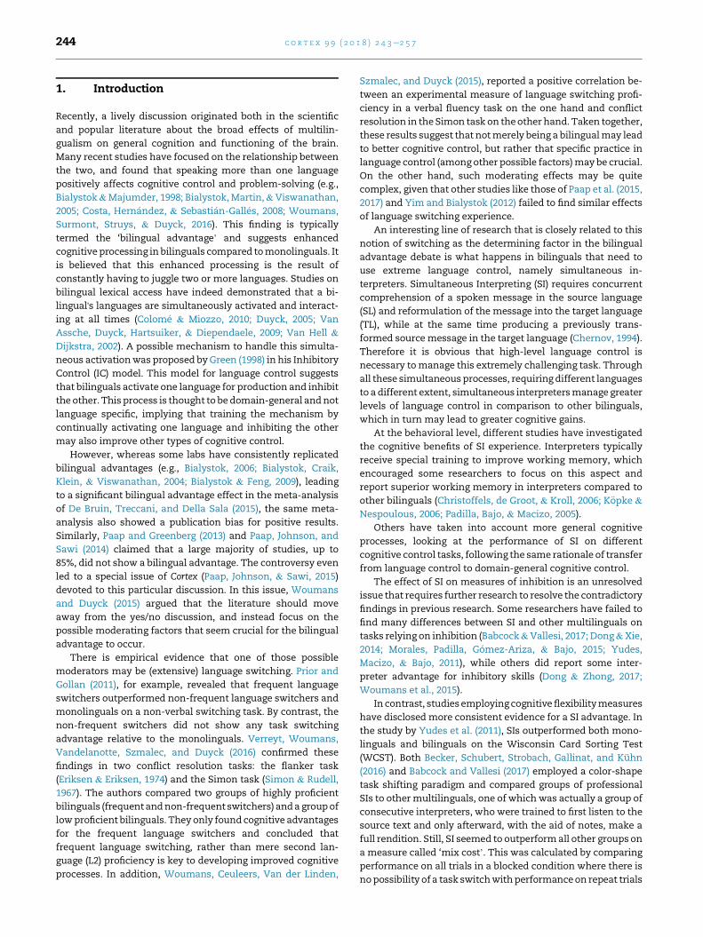
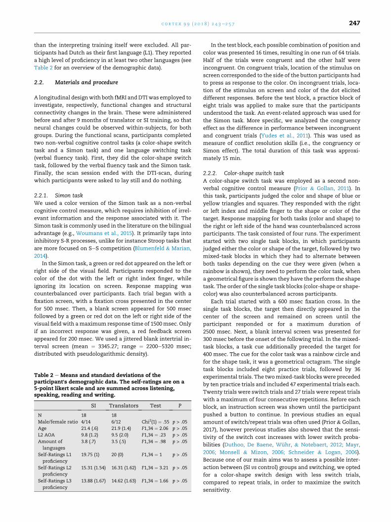
3.2.1. fMRI resultsThe whole brain fMRI analysis (Table 4) revealed a higher
involvement of the left superior temporal gyrus in the Simon
task (Fig. 1) and a higher involvement of the right angular
gyrus in the color-shape switch task (Fig. 2) after 9 months of
SI training, compared to translators, despite the absence of
differences between the two groups before their training. In
the opposite direction, the translators only showed a higher
involvement of the right cerebellum compared to SIs in the
Color-shape switch task after nine months of training (Fig. 2).
Note, however, that these brain regions were only significant
at the uncorrected cluster level. In the verbal fluency task, no
significant interactions were found.
3.2.2. Structural connectivity results (NBS analysis)Two set of regions (component/subnetworks) showed a sig-
nificant increment of the structural connectivity for SIs as
compared to translators at p < .01 FWE corrected (Fig. 3). The
first component (subnetwork I) interconnected frontal regions
with basal ganglia, comprising a total of 5 regions (Fig. 3A): left
superior frontal gyrus (SFG); left/right medial superior frontal
gyrus (SFGmed); left orbital superior frontal gyrus (SFGorb)
and the right pallidum. The second component (subnetwork
II) involved 8 nodes (Fig. 3B): left supplementary motor area
(SMA); right postcentral gyrus (PoCG); right SFG; right middle
temporal pole (TPOmid); right amygdala (AMYG), vermis 3 of
the cerebellum; left inferior parietal gyrus (IPG) and superior
parietal gyrus (SPG).
Translators did not show any set of regions with an
increment of the interconnectivity, relative to SIs. Schematic
representations of the subnetworks are depicted in Fig. 3
using BrainNet version 1.5 (Xia, Wang, & He, 2013, http://
www.nitrc.org/projects/bnv/). The anatomical name by
which each node is labeled was taken directly from the
Anatomical Automatic Labeling (AAL) atlas (Tzourio-Mazoyer
et al., 2002).
Table 4e Results of the fMRIwhole brain analysis. Significant groshape switch task and the Simon task.
Task Coordinates
Color-shape switch task
Interpreters > Translators 48 �58 37 R Angular
Translators > Interpreters 24 �58 �23 R Cerebellu
Simon task
Interpreters > Translators �45 �25 4 L superior
**p < .01.
4. Discussion
We aimed to investigate whether SI may boost cognitive
control, using a well-controlled longitudinal design in which
SI experience was manipulated over time within the same
participants. We included two highly similar groups of mul-
tilinguals with high levels of second (L2) and third (L3) lan-
guage proficiency, but different language control needs. One
group consisted of participants enrolled in a translator pro-
gram, whereas the other group was following a simultaneous
interpreting (SI) program. As SI is often associated with
extreme language control (Elmer et al., 2011; Woumans et al.,
2015), we hypothesized that these students would show both
behavioral and neural differences compared to translators
after their nine-month training course. With regard to
behavioral changes, we assumed that practicing SI would
enhance domain-general cognitive control and verbal cogni-
tive control. We also predicted that functional changes in
activation of cognitive control related brain areas would
occur, together with a modification of structural connectivity
between brain regions that are involved in cognitive control of
language.
Our expectations were, however, only confirmed at the
functional and structural neural level, not at the behavioral
level. We did not observe any cognitive behavioral advantages
in SIs compared to translators. This finding replicates the
majority of previous findings that failed to observe significant
differences between SIs and other multilinguals on tasks
relying on inhibition (Babcock & Vallesi, 2017; Dong & Xie,
2014; Morales et al., 2015; Yudes et al., 2011).
Analyses at the functional level revealed small but inter-
esting differences after 9 months of training in SI or trans-
lation. Note however, that these differences did not survive
the stringent threshold for multiple comparisons. Compared
to the translators, the SIs showed an increase of activation in
the right angular gyrus in the color-shape switch task, and an
increase in activation in the left superior temporal gyrus in the
Simon task. Increased activation in these areas in these tasks
has been interpreted as a reflection of increased capacity of
cognitive control functions (Rubia et al., 2006). Translators
only showed an increase of activation in the right cerebellum
in the color-shape switch task after nine months of training,
relative to the SIs.
Interestingly, the left superior temporal gyrus, a region that
is typically involved in a broad region of language processes,
including the auditory perception of language switches
(Abutalebi et al., 2007), appeared to show more activation in
up£ time interactions for the BOLD responses in the color-
Area Z Cluster size p
gyrus 3.51 162 .008**
m 3.82 168 .007**
temporal gyrus 4.05 161 .004**
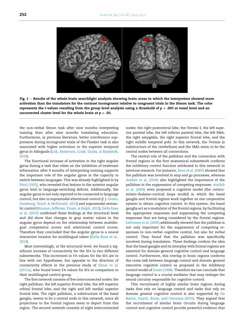
Fig. 1 e Results of the whole brain searchlight analysis showing brain areas in which the interpreters showed more
activation than the translators for the contrast incongruent relative to congruent trials in the Simon task. The color
represents the t-values resulting from the group level analysis using a threshold of p < .005 at voxel level and an
uncorrected cluster level for the whole brain at p < .05.
c o r t e x 9 9 ( 2 0 1 8 ) 2 4 3e2 5 7252
the non-verbal Simon task after nine months interpreting
training than after nine months translating education.
Furthermore, in previous literature, better interference sup-
pression during incongruent trials of the Flanker task is also
associated with higher activation in the superior temporal
gyrus in bilinguals (Luk, Anderson, Craik, Grady, & Bialystok,
2010).
The functional increase of activation in the right angular
gyrus during a task that relies on the inhibition of irrelevant
information after 9 months of interpreting training supports
the important role of the angular gyrus in the capacity to
switch between languages. This was already highlighted in by
P€otzl (1925), who revealed that lesions in the anterior angular
gyrus lead to language-switching deficits. Additionally, the
angular gyrus is not only reported to be connected to language
control, but also to supramodal attentional control (J. J. Green,
Doesburg, Ward, & McDonald, 2011) and supramodal seman-
tic control (Noonan, Jefferies, Visser,& Ralph, 2013). Della Rosa
et al. (2013) confirmed these findings at the structural level
and did show that changes in gray matter values in the
angular gyrus depend on the relationship between multilin-
gual competence scores and attentional control scores.
Therefore they concluded that the angular gyrus is a neural
interactive location for multilingual talent (Della Rosa et al.,
2013).
Most interestingly, at the structural level, we found a sig-
nificant increase of connectivity for the SI's in two different
subnetworks. This increment in FA values for the SI's are in
line with our hypotheses, but opposite to the direction of
connectivity effects in the previous study of Elmer et al.
(2011a), who found lower FA values for SI's in comparison to
their multilingual control group.
The first network consists of five interconnected nodes: the
right pallidum, the left superior frontal lobe, the left superior
orbital frontal lobe, and the right and left medial superior
frontal lobe. The right pallidum, a substructure of the basal
ganglia, seems to be a central node in this network, since all
projections to the frontal regions seem to depart from this
region. The second network consists of eight interconnected
nodes: the right postcentral lobe, the Vermis 3, the left supe-
rior parietal lobe, the left inferior parietal lobe, the left SMA,
the right amygdala, the right superior frontal lobe, and the
right middle temporal pole. In this network, the Vermis (a
substructure of the cerebellum) and the SMA seem to be the
central nodes between all connections.
The central role of the pallidum and the connection with
frontal regions in the first anatomical subnetwork confirms
the inhibitory control function attributed to this network in
previous research. For instance, Aron et al. (2007) showed that
the pallidum was involved in stop and go processes, whereas
Atallah et al. (2004) also highlighted the importance of the
pallidum in the suppression of competing responses. Atallah
et al. (2004) even proposed a cognitive model (the cortico-
striato-thalamo-cortical loops model) in which the basal
ganglia and frontal regions work together as one cooperative
system to obtain cognitive control. In this system, the basal
ganglia act as amodulator of the frontal regions, by facilitating
the appropriate responses and suppressing the competing
responses that are being considered by the frontal regions.
Lehtonen et al. (2005) additionally showed that the pallidum is
not only important for the suppression of competing re-
sponses in non-verbal cognitive control, but also for verbal
control. They found that the pallidum was specifically
involved during translation. These findings confirm the idea
that the basal ganglia and its interplaywith frontal regions are
essential for domain-general cognitive control and language
control. Furthermore, this overlap in brain regions confirms
the cross-talk between language control and domain general
executive cognitive control as proposed in the inhibitory
control model of Green (1998). Therefore we can conclude that
language control is a crucial mediator that may reshape the
neural circuitry responsible for cognitive control.
This recruitment of highly similar brain regions during
tasks that rely on language control and tasks that rely on
domain general cognitive control is also supported by De
Baene, Duyck, Brass, and Carreiras (2015). They argued that
the recruitment of similar brain circuits during language
control and cognitive control provide powerful evidence that
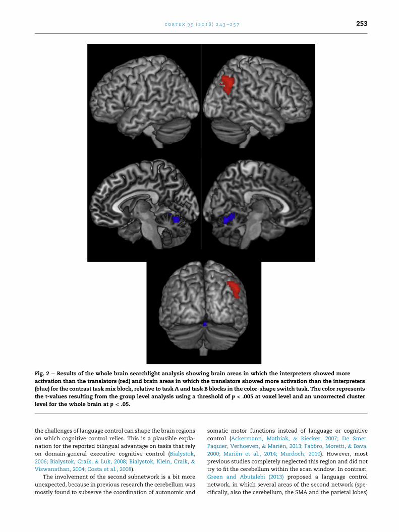
Fig. 2 e Results of the whole brain searchlight analysis showing brain areas in which the interpreters showed more
activation than the translators (red) and brain areas in which the translators showed more activation than the interpreters
(blue) for the contrast taskmix block, relative to task A and task B blocks in the color-shape switch task. The color represents
the t-values resulting from the group level analysis using a threshold of p < .005 at voxel level and an uncorrected cluster
level for the whole brain at p < .05.
c o r t e x 9 9 ( 2 0 1 8 ) 2 4 3e2 5 7 253
the challenges of language control can shape the brain regions
on which cognitive control relies. This is a plausible expla-
nation for the reported bilingual advantage on tasks that rely
on domain-general executive cognitive control (Bialystok,
2006; Bialystok, Craik, & Luk, 2008; Bialystok, Klein, Craik, &
Viswanathan, 2004; Costa et al., 2008).
The involvement of the second subnetwork is a bit more
unexpected, because in previous research the cerebellumwas
mostly found to subserve the coordination of autonomic and
somatic motor functions instead of language or cognitive
control (Ackermann, Mathiak, & Riecker, 2007; De Smet,
Paquier, Verhoeven, & Mari€en, 2013; Fabbro, Moretti, & Bava,
2000; Mari€en et al., 2014; Murdoch, 2010). However, most
previous studies completely neglected this region and did not
try to fit the cerebellum within the scan window. In contrast,
Green and Abutalebi (2013) proposed a language control
network, in which several areas of the second network (spe-
cifically, also the cerebellum, the SMA and the parietal lobes)

Fig. 3 e Results of the network-based statistical analysis over the structural brain graph. Subnetworks showing increased
structural connectivity in simultaneous interpreters as compared to translators (T-threshold ¼ 3, K ¼ 10,000 permutations,
p < .01 FWE corrected). A) Subnetwork I: regions forming an individual component with 5 nodes/regions and 4 edges/
connections. B) Subnetwork II: regions forming and individual component with 8 nodes/regions and 7 edges/connections.
Abbreviations: L, left; R, right; SMA, supplementary motor area; SFGdor, superior frontal gyrus; SFGmed, medial superior
frontal gyrus; SFGorb., orbital superior frontal gyrus, PoCG, postcentral gyrus; IPG, inferior parietal gyrus; SPG, superior
parietal gyrus; TPOmid, middle temporal pole; AMYG, amygdala.
c o r t e x 9 9 ( 2 0 1 8 ) 2 4 3e2 5 7254
also play a key role. In this model, the SMA initiates speech in
language switching and the parietal lobes are connected to the
maintenance of task representations.
Note, however that the structural and functional analysis
revealed distinct findings. The cerebellum seems to be
involved in both analyses, but unexpectedly in opposite di-
rections, with an increased involvement of the cerebellum
during the color-shape switch task for the translators and an
increased connectivity of the cerebellum for the SI's. Addi-tionally, the increased involvement of the right angular gyrus
during the color-shape switch task and the increased
involvement of the left superior temporal gyrus during the
Simon task was only apparent in the functional analysis, but
not in the structural analysis. A possible explanation could be
that translators and interpreters differ in the way they rely on
the neural network. The connectivity between the cerebellum
and other brain regions might for example become stronger
for the interpreters. Therefore a shift could occur from relying
solely on the cerebellum to relying more on the exchange
between the cerebellum and other regions.
It is important to emphasize the conservative approach
adopted here, to compare SI students with a group of closely
matched translators from the same Bachelor program, rather
than a monolingual or less L2-proficient control group. As a
result, the obtained differences between these two highly
similar groups need to be attributed to control processes that
are specific to SI. In SI, a one-time presentation of an utterance
in a source language (SL) is instantly rendered into an utter-
ance of similarmeaning in a target language (TL). According to
Christoffels and de Groot (2005) and De Groot and Christoffels
(2006), inhibitory control plays a crucial role during this
rendition. The authors describe possible inhibition accounts
of SI, assuming (functionally) distinct input and output lexi-
cons that can be separately activated and inhibited. These
accounts state that both SL and TL input lexicons should be
activated, to allow for input comprehension and output
monitoring, while the SL output lexicon should be strongly
inhibited. Other explanations for the observed differences
between the SI and the translators are the development of a
more efficient divided attention system or language switching
system. This is because besides the proposed role of inhibitory
control, an SI's attention is divided or switches rapidly be-
tween the different processes (Hiltunen, P€a€akk€onen, Gun-
Viol, & Krause, 2016; P€ochhacker, 2004). Therefore, future
studies are necessary to determine the specific processes that
distinguish translating from SI.
The lack of behavioral differences between the two groups
could similarly be explained by our conservative approach
comparing two highly similar groups of SIs and translators,
with the exact same prior education. Translating is not totally

c o r t e x 9 9 ( 2 0 1 8 ) 2 4 3e2 5 7 255
different fromSI. Translators, too, have to render a source text
into a target text, and when they are formulating this text in
the target language (TL), they need to inhibit the source lan-
guage (SL) at the output level, while keeping it activated at the
input level. Essentially the process appears the same, but the
amount of extreme language control is different between the
two groups. SIs have to perform this process in real-time, i.e.,
immediately after or simultaneous with reception of the
source text, making SI much more challenging (Babcock &
Vallesi, 2017).
Additionally, it is possible that further experience could
create behavioral differences that did not yet appear after only
nine months of SI training, especially because the amount of
SI practice was still limited during this Master course. Another
possibility is that SIs recruit brain regions in a more efficient
way, resulting in the observed functional activation differ-
ences, but that there are no behavioral differences between
SIs and translators, because both already perform close to
individual ceiling. Note that the lack of behavioral findings
within our longitudinal design may also have been influenced
by the demonstrated low test-retest reliability for the Simon
effect, and somewhat higher test-retest reliabilities for the
switch cost and mix cost (Paap & Sawi, 2016).
The lack of behavioral group differences in the inhibition
task replicates the longitudinal findings of Dong and Liu (2016)
and Babcock et al. (2017) who used a similar conservative
approach comparing SIs with translators. However, in
contrast to our results and those of Babcock et al. (2017), in the
study of Dong and Liu (2016) the SIs improved significantly
more on switching than the translators. In future research, it
may be interesting to also investigate tasks that tap into
different types of inhibitory control (ex: ANT, flanker task,
stroop task, go/no-go task) or switching-flexibility (ex: WCST)
(Miyake & Friedman, 2012).
To conclude, given this longitudinal design with closely
matched groups of SIs and translators, who received the same
previous education, the observation of neural differences over
the course of only nine months is really remarkable. Our re-
sults suggest that SIs undergo neural changes in specific
control-related brain networks to handle the extreme lan-
guage control that takes place during interpreting.
Acknowledgments
This researchwasmade possible by the Research Foundation-
Flanders (FWO-Vlaanderen; FWO grant G058914N).
r e f e r e n c e s
Abutalebi, J., Brambati, S. M., Annoni, J., Moro, A., Cappa, S. F., &Perani, D. (2007). The neural cost of the auditory perception oflanguage switches: An event-related functional magneticresonance imaging study in bilinguals. Journal of Neuroscience,27(50), 13762e13769. http://doi.org/10.1523/JNEUROSCI.3294-07.2007.
Ackermann, H., Mathiak, K., & Riecker, A. (2007). The contributionof the cerebellum to speech production and speech
perception: Clinical and functional imaging data. TheCerebellum, 6(3), 202e213. http://doi.org/10.1080/14734220701266742.
Alem�an-G�omez, Y., Melie-Garcıa, L., & Vald�es-Hern�andez, P.(2006). IBASPM: Toolbox for au- tomatic parcellation of brainstructures. In 12th annual meeting of the organization for humanbrain mapping.
Aron, A. R., Durston, S., Eagle, D. M., Logan, G. D., Stinear, C. M., &Stuphorn, V. (2007). Converging evidence for a fronto-basal-ganglia network for inhibitory control of action and cognition.Journal of Neuroscience, 27(44), 11860e11864. http://doi.org/10.1523/JNEUROSCI.3644-07.2007.
Atallah, H. E., Frank, M. J., & Reilly, R. C. O. (2004). Hippocampus,cortex, and basal ganglia: Insights from computationalmodels of complementary learning systems. Neurobiology ofLearning and Memory, 82, 253e267. http://doi.org/10.1016/j.nlm.2004.06.004.
Babcock, L., Capizzi, M., Arbula, S., & Vallesi, A. (2017). Short-termmemory improvement after simultaneous interpretationtraining. Journal of Cognitive Enhancement, 1(3), 254e267. http://doi.org/10.1007/s41465-017-0011-x.
Babcock, L., & Vallesi, A. (2017). Are simultaneous interpretersexpert bilinguals, unique bilinguals, or both? Bilingualism:Language and Cognition, 20(2), 403e417. https://doi.org/10.1017/S1366728915000735.
Bai, F., Shu, N., Yuan, Y., Shi, Y., Yu, H., Wu, D., et al. (2012).Topologically convergent and divergent structuralconnectivity patterns between patients with remitted geriatricdepression and amnestic mild cognitive impairment. TheJournal of Neuroscience: the Official Journal of the Society forNeuroscience, 32(12), 4307e4318.
Becker, M., Schubert, T., Strobach, T., Gallinat, J., & Kuhn, S.(2016). Simultaneous interpreters vs. professional multilingualcontrols: Group differences in cognitive control as well asbrain structure and function. NeuroImage, 134, 250e260. http://doi.org/10.1016/j.neuroimage.2016.03.079.
Behrens, T. E. J., Woolrich, M. W., Jenkinson, M., JohansenBerg, H.,Nunes, R. G., Clare, S., et al. (2003). Characterization andpropagation of uncertainty in diffusion weighted MR imaging.Magnetic Resonance in Medicine, 50, 1077e1088.
Bialystok, E. (2006). Effect of bilingualism and computer videogame experience on the Simon task. Canadian Journal ofExperimental Psychology, 60(1), 68e79.
Bialystok, E., Craik, F. I. M., & Luk, G. (2008). Cognitive control andlexical access in younger and older bilinguals. Journal ofExperimental Psychology: Learning, Memory, and Cognition, 34,859e873.
Bialystok, E., & Feng, X. (2009). Language proficiency andexecutive control in proactive interference: Evidence frommonolingual and bilingual children and adults. Brain andLanguage, 109, 93e100.
Bialystok, E., Klein, R., Craik, F. I. M., & Viswanathan, M. (2004).Bilingualism, aging, and cognitive control: Evidence from theSimon task. Psychology and Aging, 19, 290e303.
Bialystok, E., & Majumder, S. (1998). The relationship betweenbilingualism and the development of cognitive processes inproblem solving. Applied Psycholinguistics, 19, 69e85.
Bialystok, E., Martin, M. M., & Viswanathan, M. (2005).Bilingualism across the lifespan: The rise and fall of inhibitorycontrol. International Journal of Bilingualism, 9(1), 103e119.
Blumenfeld, H. K., & Marian, V. (2014). Cognitive control inbilinguals: Advantages in stimulus-stimulus inhibition.Bilingualism: Language and Cognition, 17(3), 610e629.
Chernov, G. V. (1994). In Lambert, & Moser-Mercer (Eds.), Messageredundancy and message anticipation in simultaneous interpretation(pp. 139e153).
Christoffels, I. K., & de Groot, A. M. B. (2005). Simultaneousinterpreting: A cognitive perspective. In J. R. Kroll, & A. M. B. de

c o r t e x 9 9 ( 2 0 1 8 ) 2 4 3e2 5 7256
Groot (Eds.), Handbook of bilingualism: Psycholinguistic approaches(pp. 454e479). Oxford: Oxford University Press.
Christoffels, I. K., de Groot, A. M. B., & Kroll, J. F. (2006). Memoryand language skills in simultaneous interpreters: The role ofexpertise and language proficiency. Journal of Memory andLanguage, 54(3), 324e345. http://doi.org/10.1016/j.jml.2005.12.004.
Colom�e, �A., & Miozzo, M. (2010). Which words are activatedduring bilingual speech production? Journal of ExperimentalPsychology: Learning, Memory, and Cognition, 36(1), 96e109.
Costa, A., Hern�andez, M., & Sebasti�an-Gall�es, N. (2008).Bilingualism aids conflict resolution: Evidence from the ANTtask. Cognition, 106, 59e86.
De Baene, W., Duyck, W., Brass, M., & Carreiras, M. (2015). Braincircuit for cognitive control is shared by task and languageswitching. Journal of Cognitive Neuroscience, 27, 1752e1765.https://doi.org/10.1162/jocn_a_00817.
De Bruin, A., Treccani, B., & Della Sala, S. (2015). Cognitiveadvantage in bilingualism: An example of publication bias?Psychological Science, 26(1), 99e107.
De Groot, A. M., & Christoffels, I. K. (2006). Language control inbilinguals: Monolingual tasks and simultaneous interpreting.Bilingualism: Language and Cognition, 9(2), 189e201.
De Smet, H. J., Paquier, P., Verhoeven, J., & Mari€en, P. (2013). Thecerebellum: Its role in language and related cognitive andaffective functions. Brain and Language, 127(3), 334e342. http://doi.org/10.1016/j.bandl.2012.11.001.
Della Rosa, P. A., Videsott, G., Borsa, V. M., Canini, M.,Weekes, B. S., Franceschini, R., et al. (2013). A neuralinteractive location for multilingual talent. Cortex; a JournalDevoted to the Study of the Nervous System and Behavior, 49(2),605e608. http://doi.org/10.1016/j.cortex.2012.12.001.
Dong, Y., & Liu, Y. (2016). Classes in translating and interpretingproduce differential gains in switching and updating. Frontiersin Psychology, 7, 1297. https://doi.org/10.3389/fpsyg.2016.01297.
Dong, Y., & Xie, Z. (2014). Contributions of second languageproficiency and interpreting experience to cognitive controldifferences among young adult bilinguals. Journal of CognitivePsychology, 26(5), 506e519.
Dong, Y., & Zhong, F. (2017). Interpreting experience enhancesearly attentional processing, conflict monitoring andinterference suppression along the time course of processing.Neuropsychologia, 95, 193e203. https://doi.org/10.1016/j.neuropsychologia.2016.12.007.
Duthoo, W., Baene, W. De, Wuhr, P., & Notebaert, W. (2012). Whenpredictions take control: The effect of task predictions on taskswitching performance. Frontiers in Psychology, 3(August), 1e9.http://doi.org/10.3389/fpsyg.2012.00282.
Duyck, W. (2005). Translation and associative priming with cross-lingual pseudohomophones: Evidence for nonselectivephonological activation in bilinguals. Journal of ExperimentalPsychology: Learning, Memory, and Cognition, 31, 1340e1359.
Elmer, S., H€anggi, J., & J€ancke, L. (2014). Processing demands uponcognitive, linguistic, and articulatory functions promote greymatter plasticity in the adult multilingual brain: Insights fromsimultaneous interpreters. Cortex; a Journal Devoted to the Studyof the Nervous System and Behavior, 54, 179e189. http://doi.org/10.1016/j.cortex.2014.02.014.
Elmer, S., H€anggi, J., Meyer, M., & J€ancke, L. (2011a). Differentiallanguage expertise related to white matter architecture inregions subserving sensory-motor coupling, articulation, andinterhemispheric transfer. Human Brain Mapping, 32(12),2064e2074. http://doi.org/10.1002/hbm.21169.
Elmer, S., Meyer, M., Marrama, L., & J€ancke, L. (2011b). Intensivelanguage training and attention modulate the involvement offronto-parietal regions during a non-verbal auditorydiscrimination task. European Journal of Neuroscience,
34(January), 165e175. http://doi.org/10.1111/j.1460-9568.2011.07728.x.
Eriksen, B. A., & Eriksen, C. W. (1974). Effects of noise letters uponthe identification of a target letter in a nonsearch task.Perception and Psychophysics, 16, 143e149.
Fabbro, F., Moretti, R., & Bava, A. (2000). Language impairmentsin patients with cerebellar lesions. Journal of Neurolinguistics,13(2e3), 173e188. http://doi.org/10.1016/S0911-6044(00)00010-5.
Funes, M. J., Lupi�a~nez, J., & Humphreys, G. (2010). Sustained vs.transient cognitive control: Evidence of a behavioraldissociation. Cognition, 114, 338e347. https://doi.org/10.1016/j.cognition.2009.10.007.
Garcıa-Pent�on, L., P�erez Fern�andez, A., Iturria-Medina, Y., Gillon-Dowens, M., & Carreiras, M. (2014). Anatomical connectivitychanges in the bilingual brain. NeuroImage, 84, 495e504.https://doi.org/10.1016/j.neuroimage.2013.08.064.
Green, D. W. (1998). Mental control of the bilingual lexico-semantic system. Bilingualism: Language and Cognition, 1, 67e82.
Green, D. W., & Abutalebi, J. (2013). Language control in bilinguals:The adaptive control hypothesis. Journal of Cognitive Psychology,25(5), 515e530. http://doi.org/10.1080/20445911.2013.796377.
Green, J. J., Doesburg, S. M., Ward, L. M., & McDonald, J. J. (2011).Electrical neuroimaging of voluntary audiospatial Attention:Evidence for a supramodal attention control network. Journalof Neuroscience, 31(10), 3560e3564. http://doi.org/10.1523/JNEUROSCI.5758-10.2011.
Hervais-Adelman, A., Moser-Mercer, B., & Golestani, N. (2015a).Brain functional plasticity associated with the emergence ofexpertise in extreme language control. NeuroImage. http://doi.org/10.1016/j.neuroimage.2015.03.072.
Hervais-Adelman, A., Moser-Mercer, B., Michel, C. M., &Golestani, N. (2015b). fMRI of simultaneous interpretationreveals the neural basis of extreme language control. CerebralCortex (New York, N.Y.: 1991). http://doi.org/10.1093/cercor/bhu158.
Hiltunen, S., P€a€akk€onen, R., Vik, G. V., & Krause, C. M. (2016). Oninterpreters’ working memory and executive control.International Journal of Bilingualism, 20(3), 297e314.
Iturria-Medina, Y., P�erez Fern�andez, A., Morris, D. M., Canales-Rodrıguez, E. J., Haroon, H. A., Garcıa Pent�on, L., et al. (2011).Brain hemispheric structural efficiency and interconnectivityright- ward asymmetry in human and nonhuman primates.Cerebral Cortex, 21, 56e67.
K€opke, B., & Nespoulous, J.-L. (2006). Working memoryperformance in expert and novice interpreters. Interpreting, 8,1e23.
Lehtonen, M. H., Laine, C. A. M., Niemi, J., Thomsen, T.,Vorobyev, V. A., & Hugdahl, K. (2005). Brain correlates ofsentence translation in FinnisheNorwegian bilinguals.NeuroReport, 16(6), 4e7.
Luk, G., Anderson, J. A., Craik, F. I., Grady, C., & Bialystok, E. (2010).Distinct neural correlates for two types of inhibition inbilinguals: Response inhibition versus interferencesuppression. Brain and Cognition, 74(3), 347e357.
Macnamara, B. N., & Conway, A. R. A. (2016). Working memorycapacity as a predictor of simultaneous language interpretingperformance. Journal of Applied Research in Memory andCognition, 5(4), 434e444. http://doi.org/10.1016/j.jarmac.2015.12.001.
Mari€en, P., Ackermann, H., Adamaszek, M., Barwood, C. H. S.,Beaton, A., Desmond, J., et al. (2014). Consensus paper:Language and the cerebellum: An ongoing enigma. TheCerebellum, 13(3), 386e410. http://doi.org/10.1007/s12311-013-0540-5.
Mayr, U. (2006).Whatmatters in the cued task-switching paradigm:Tasks or cues? Psychonomic Bulletin & Review, 5, 794e799.
c o r t e x 9 9 ( 2 0 1 8 ) 2 4 3e2 5 7 257
Miyake, A., & Friedman, N. P. (2012). The nature and organizationof individual differences in executive functions: Four generalconclusions. Current Directions in Psychological Science, 21(8),8e14. https://doi.org/10.1177/0963721411429458.
Monsell, S., & Mizon, G. A. (2006). Can the task-cuing paradigmmeasure an endogenous taskset reconfiguration process?Journal of Experimental Psychology. Human Perception andPerformance, 32, 493e516.
Morales, J., Padilla, F., G�omez-Ariza, C. J., & Bajo, M. T. (2015).Simultaneous interpretation selectively influences workingmemoryand attentional networks.Acta Psychologica, 155, 82e91.
Murdoch, B. E. (2010). The cerebellum and language: Historicalperspective and review. Cortex; a Journal Devoted to the Study ofthe Nervous System and Behavior, 46(7), 858e868. http://doi.org/10.1016/j.cortex.2009.07.018.
Noonan, K. A., Jefferies, E., Visser, M., & Ralph, M. A. L. (2013).Going beyond inferior prefrontal involvement in semanticcontrol: Evidence for the additional contribution of dorsalangular gyrus and posterior middle temporal cortex. Journal ofCognitive Neuroscience, 1824e1850. http://doi.org/10.1162/jocn.
Paap, K. R., & Greenberg, Z. I. (2013). There is no coherentevidence for a bilingual advantage in executive processing.Cognitive Psychology, 66, 232e258.
Paap, K. R., Johnson, H. A., & Sawi, O. (2014). Are bilingualadvantages dependent upon specific tasks or specific bilingualexperiences? Journal of Cognitive Psychology, 26(6), 615e639.https://doi.org/10.1080/20445911.2014.944914.
Paap, K. R., Johnson, H. A., & Sawi, O. (2015). Bilingual advantages inexecutive functioning either do not exist or are restricted to veryspecific and undetermined circumstances. Cortex; a JournalDevoted to the Study of theNervousSystemandBehavior, 69, 265e278.
Paap, K. R., Myuz, H. A., Anders, R. T., Bockelman, M. F.,Mikulinsky, R., & Sawi, O. M. (2017). No compelling evidence fora bilingual advantage in switching or that frequent languageswitching reduces switch cost. Journal of Cognitive Psychology,29(2), 89e112. http://doi.org/10.1080/20445911.2016.1248436.
Paap, K. R., & Sawi, O. (2016). The role of test-retest reliability inmeasuring individual and group differences in executivefunctioning. Journal of Neuroscience Methods, 274, 81e93.
Padilla, F., Bajo, M. T., & Macizo, P. (2005). Articulatorysuppression in language interpretation: Working memorycapacity, dual tasking and word knowledge. Bilingualism:Language and Cognition, 8, 207e213.
P€ochhacker, F. (2004). Introducing interpreting studies. London, UK:Routledge.
P€otzl, O. (1925). Uber die parietal bedingte Aphasie und ihrenEinfluß auf das Sprechen mehrerer Sprachen. Zeitschrift fur diegesamte Neurologie und Psychiatrie, 96(1), 100e124.
Prior, A., & Gollan, T. H. (2011). Good language-switchers are goodtask-switchers: Evidence from Spanish-English andMandarin-English bilinguals. Journal of the InternationalNeuropsychological Society, 17, 1e10.
Rubia, K., Smith, A. B., Woolley, J., Nosarti, C., Heyman, I.,Taylor, E., et al. (2006). Progressive increase of frontostriatalbrain activation from childhood to adulthood during event-related tasks of cognitive control. Human Brain Mapping, 27(12),973e993.
Schneider, D. W., & Logan, G. D. (2006). Priming cue encoding bymanipulating transition frequency in explicitly cued taskswitching. Psychonomic Bulletin & Review, 13, 145e151.
Simon, J. R., & Rudell, A. P. (1967). Auditory S-R compatibility: Theeffect of an irrelevant cue on information processing. Journal ofApplied Psychology, 51, 300e304.
Smith, S. M., Jenkinson, M., Woolrich, M. W., Beckmann, C. F.,Behrens, T. E. J., Johansen-Berg, H., et al. (2004). Advances infunctional and structural MR image analysis andimplementation as FSL. NeuroImage, 23(Suppl. 1), S208eS219.
Tzourio-Mazoyer, N., Landeau, B., Papathanassiou, D.,Crivello, E. O., Delcroix, N., Mazoyer, B., et al. (2002).Automated anatomical labeling of activations in SPM using amacroscopic anatomical parcellation of the MNI MRI single-subject brain. NeuroImage, 15, 273e289.
Van Assche, E., Duyck, W., Hartsuiker, R. J., & Diependaele, K.(2009). Does bilingualism change native-language reading?Cognate effects in a sentence context. Psychological Science,20(8), 923e927. https://doi.org/10.1111/j.1467-9280.2009.02389.x.
Van Hell, J. G., & Dijkstra, T. (2002). Foreign language knowledgecan influence native language performance inexclusively native contexts. Psychonomic Bulletin & Review, 9,780e789.
Verreyt, N., Woumans, E., Vandelanotte, D., Szmalec, A., &Duyck, W. (2016). The influence of language-switchingexperience on the bilingual executive control advantage.Bilingualism: Language and Cognition, 19(1), 181e190. http://doi.org/10.1017/S1366728914000352.
Verstraete, E., Veldink, J. H., Mandl, R. C. W., van den Berg, L. H., &van den Heuvel, M. P. (2011). Impaired motor connectome inamyotrophic lateral sclerosis. Plos One, 6(9), e24239.
Woumans, E., Ceuleers, E., Van der Linden, L., Szmalec, A., &Duyck, W. (2015). Verbal and non-verbal cognitive control inbilinguals and interpreters. Journal of Experimental Psychology:Learning, Memory, and Cognition, 41(5), 1579.
Woumans, E., & Duyck, W. (2015). The bilingual advantagedebate: Moving toward different methods for verifying itsexistence. Cortex; a Journal Devoted to the Study of the NervousSystem and Behavior, 73, 356e357.
Woumans, E., Surmont, J., Struys, E., & Duyck, W. (2016). Thelongitudinal effect of bilingual immersion schooling oncognitive control and intelligence. Language Learning, 66(2),76e91. https://doi.org/10.1111/lang.12171.
Xia, M., Wang, J., & He, Y. (2013). BrainNet viewer: A networkvisualization tool for human brain connectomics. Plos One, 8,e68910.
Yim, O., & Bialystok, E. (2012). Degree of conversational code-switching enhances verbal task switching in Cantonese-English bilinguals. Bilingualism: Language and Cognition, 15(4),873e883.
Yudes, C., Macizo, P., & Bajo, T. (2011). The influence of expertisein simultaneous interpreting on non-verbal executiveprocesses. Frontiers in Psychology, 2(Oct), 1e9. http://doi.org/10.3389/fpsyg.2011.00309.
Zalesky, A., Fornito, A., & Bullmore, E. T. (2010a). Network-basedstatistic: Identifying differences in brain networks.NeuroImage, 53, 1197e1207.
Zalesky, A., Fornito, A., Seal, M. L., Cocchi, L., Westin, C. F.,Bullmore, E. T., et al. (2010b). Disrupted axonal fiberconnectivity in schizophrenia. Biological Psychiatry, 69,80e89.