analysis of organic components in resin-modified pulp
TRANSCRIPT

Analysis of organic components inresin-modified pulp capping materials:critical considerations
Nilsen BW, Jensen E, €Ortengren U, Michelsen VB. Analysis of organic components inresin-modified pulp capping materials: critical considerations.Eur J Oral Sci 2017; 125: 183–194. © 2017 Eur J Oral Sci
The purpose of this study was to elucidate the organic composition and eluates ofthree resin-based pulp-capping materials in relation to their indications and safetydata sheets. Uncured samples of Theracal LC, Ultra-Blend Plus, and Calcimol LCwere investigated using gas chromatography–mass spectrometry (GC-MS) andultra-performance liquid chromatography–mass spectrometry (UPLC-MS). Identifi-cation/quantification of 7-d leachables of cured samples was performed using GC-MS for 2-hydroxyethyl methacrylate (HEMA), 2-(dimethylamino)ethyl methacrylate(DMAEMA), camphorquinone (CQ), ethylene glycol dimethacrylate (EGDMA),ethyl-4-(dimethylamino)benzoate (DMABEE), and triethylene glycol dimethacrylate(TEGDMA). A similar organic composition was found for Ultra-Blend and Calci-mol; however, only Ultra-Blend is indicated for direct pulp-capping. In contrast tothe other materials analysed, Theracal contained substances of high molecularweight. The safety data sheets of all materials were incomplete. We detectedHEMA, CQ, and TEGDMA in eluates from Ultra-Blend and Calcimol, and it wasconsidered that HEMA might have originated from decomposition of diurethanedimethacrylate (UDMA) in the GC-injector. For Theracal, additives associated withlight curing (DMABEE and CQ) were detected in higher amounts (4.11 and19.95 lg mm�2) than in the other materials. Pores were quantified in all samples bymicro-computed tomography (micro-CT) analysis, which could influence leaching.The organic substances in the investigated materials might affect their clinical suit-ability as capping agents, especially for direct capping procedures.
Bo W. Nilsen1 , Einar Jensen2,Ulf €Ortengren1,3,*, Vibeke B.Michelsen4,*1Department of Clinical Dentistry, UiT - TheArctic University of Norway, Tromsø;2Department of Pharmacy, UiT - The ArcticUniversity of Norway, Tromsø, Norway;3Department of Cariology, Institute ofOdontology/Sahlgrenska Academy, G€oteborg,Sweden; 4Department of Clinical Dentistry,University of Bergen, Bergen, Norway
*These authors contributed equally to this work.
Bo Wold Nilsen, Department of ClinicalDentistry, University of Tromsø, No-9037Tromsø, Norway
E-mail: [email protected]
Key words: dental materials; mass spectrumanalysis; micro-computed tomography;polymers; pulp capping agents
Accepted for publication March 2017
Polymer resin-based dental materials (PRMs) representa widely used group of dental materials with differentcompositions, properties, and applications. WhilePRMs in general are used for direct restoration proce-dures, new materials that utilize the photocuring abili-ties of PRMs have emerged to simplify procedures suchas pulp capping (1). However, the organic additivesand methacrylates in PRMs are, in general, associatedwith negative biological effects in vitro, in animal stud-ies, as well as clinically (2–9). Thus, this warrants acritical evaluation of the organic composition of light-curing materials indicated for pulp capping.
Pulp capping materials are used for two indications.Direct capping materials are placed in direct contactwith pulp tissue, while indirect capping materialsrequire a dentin barrier between the pulp and material.Both aim to assist in maintaining pulp vitality. Tradi-tionally, calcium hydroxide has been used for directcapping procedures. However, mineral trioxide aggre-gate (MTA), a material consisting mainly of calciumsilicates, is regarded by some as the new ‘gold standard’from results of clinical trials (10–12). Both materialsinduce hydroxylapatite formation when in contact with
physiological solutions owing to their ability to increasepH and release calcium ions; however, the biologicalresponses to these materials are not similar in vivo andin vitro (13, 14), and MTA has been shown to induce ahigher-quality dentin-bridge than calcium hydroxide(13). A beneficial interaction between the constituentsof MTA and pulp (e.g. cell adherence to the cappingmaterial) is suggested to be the reason for this response(15–18). This indicates the importance of the composi-tion of the material on its biocompatible properties andhence its effectiveness as a capping material (15).
Several light-curing resin-modified pulp-cappingmaterials are available for indirect and/or direct cap-ping. These materials consist of components associatedwith traditional pulp therapy (e.g. calcium silicate andcalcium hydroxide), in addition to methacrylates andadditives that enable light curing. Direct pulp cappingwith polymer resin-based materials are in general asso-ciated with negative clinical outcomes (8–10, 19). It istherefore of interest to investigate the organic composi-tion that will make some light-curing materials suitablefor direct capping. This can be difficult to extrapolatefrom safety data sheets, as mass spectrometry (MS)
Eur J Oral Sci 2017; 125: 183–194DOI: 10.1111/eos.12347Printed in Singapore. All rights reserved
� 2017 Eur J Oral Sci
European Journal ofOral Sciences

analyses of PRMs have shown that these data sheetsare incomplete (20–23). By analysing the organic com-position of, in addition to quantifying organic eluatesfrom, resin-modified pulp-capping materials using MS-based methods, novel insights can be obtained regard-ing the relationship between indication for use andorganic composition. This will provide clinicians andresearchers with important information about a groupof materials that may seem easy to use compared withother clinically proven materials – but may have a com-position that makes their indication for pulp cappingdubious (9–11).
The aims of this study were to: (i) analyse theorganic composition of three light-curing resin-modifiedpulp-capping materials; (ii) quantify organic leachables;and (iii) evaluate these findings in light of materialcomposition, safety data sheets, and indication for use.
Material and methods
Overview
Three resin-based pulp-capping materials, Theracal LC(TH), Ultra-Blend Plus (UB), and Calcimol LC (CA),were investigated for organic components and 7-d leach-ables in water. For identification of organic substances,uncured samples of these materials were dissolved in ethylacetate and water before analysis with gas chromatogra-phy–mass spectrometry (GC-MS) or ultra-performanceliquid chromatography–mass spectrometry (UPLC-MS).Subsequently, precured samples of each material wereimmersed in water for 7 d. The eluates were analysedusing GC-MS in full Scan mode and single ion recording(SIR) mode, for identification and quantification oforganic substances commonly found in PRMs. A flowchart of the workflow is presented in Fig. 1.
Materials and chemicals
Calcimol LC (VOCO, Cuxhaven, Germany; LOT:1533389), Theracal LC (BISCO, Schaumburg, USA; LOT:1500006301), and Ultra-Blend Plus (Ultradent Products,
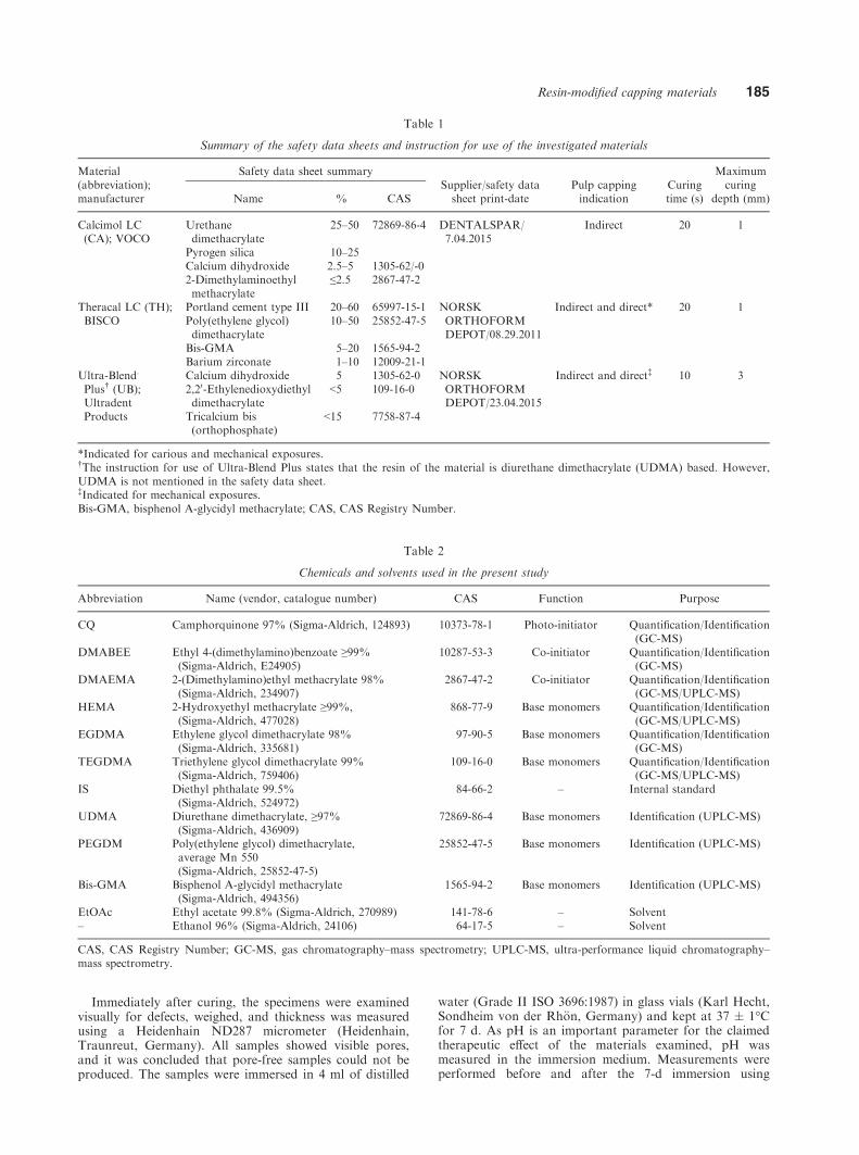
South Jordan, UT, USA; LOT: BB59V) were bought fromdental suppliers in Norway. The safety data sheet for therespective materials was requested from the suppliers. Asummary of the safety data sheets provided, and highlightsof ‘instruction for use’ are presented in Table 1. Analyticalgrade solvents and standards were obtained from Sigma–Aldrich (Oslo, Norway) (Table 2).
Preparation of uncured samples for GC-MS andUPLC-MS
A small amount (~2 mg) of TH, CA, and UB wereweighed with an analytical balance in individualpolypropylene Eppendorf tubes (Sigma-Aldrich, St. Louis,MO, USA). Two samples of each material were dissolvedin either ethyl acetate or water (ISO 3696-Grade II). Thetubes were centrifuged at 20,000 g for 5 min to separatethe filler and matrix phases. The supernatant of eachsample was transferred to a glass vial and diluted beforefurther analysis.
Preparation of cured samples, 7-d leaching in water
Pilot studies were performed to determine appropriatesample thickness as 1-mm-thick samples of TH showedincomplete cure. Accordingly, six samples of TH, CA, andUB (diameter 10 mm; thickness 0.65 � 0.05 mm) wereprepared in a Teflon mould. Uncured material wasinserted into the mould, covered with a polyethylene film,and compressed with a glass slide. After removal of theglass slide, light curing was performed using a cordedBluePhase G2 light-curing device (Ivoclar/Vivadent,Schaan, Lichtenstein), in accordance with the manufac-turer’s instructions for each capping material (Table 1).Slight movement of the light curing tip was carried outduring curing to compensate for irradiance heterogeneityand discrepancy between the optical tip area and themould diameter (24, 25). The irradiance of the curingdevice was assessed before sample preparation using a cal-ibrated laboratory-grade National Institute of Science andTechnology (NIST; Gaithersburg, MD, USA)-referencedUSB4000 Spectrometer [Managing Accurate Resin Curing(MARC) System; Bluelight Analytics, Halifax, NovaScotia, Canada] to ensure an irradiance in the range of1,350 � 100 mW cm�2.
Fig. 1. Workflow of the gas chromatography–mass spectrometry (GC-MS) and ultra-performance liquid chromatography–massspectrometry (UPLC-MS) analysis. *Theracal LC (TC), Ultra-Blend Plus (UB), and Calcimol LC (CA). SCAN, full Scan mode;SIR, single ion recording.
184 Nilsen et al.
Immediately after curing, the specimens were examinedvisually for defects, weighed, and thickness was measuredusing a Heidenhain ND287 micrometer (Heidenhain,Traunreut, Germany). All samples showed visible pores,and it was concluded that pore-free samples could not beproduced. The samples were immersed in 4 ml of distilled
water (Grade II ISO 3696:1987) in glass vials (Karl Hecht,Sondheim von der Rh€on, Germany) and kept at 37 � 1°Cfor 7 d. As pH is an important parameter for the claimedtherapeutic effect of the materials examined, pH wasmeasured in the immersion medium. Measurements wereperformed before and after the 7-d immersion using
Table 1
Summary of the safety data sheets and instruction for use of the investigated materials
Material(abbreviation);manufacturer
Safety data sheet summarySupplier/safety datasheet print-date
Pulp cappingindication
Curingtime (s)
Maximumcuring
depth (mm)Name % CAS
Calcimol LC(CA); VOCO
Urethanedimethacrylate
25–50 72869-86-4 DENTALSPAR/7.04.2015
Indirect 20 1
Pyrogen silica 10–25Calcium dihydroxide 2.5–5 1305-62/-02-Dimethylaminoethylmethacrylate
≤2.5 2867-47-2
Theracal LC (TH);BISCO
Portland cement type III 20–60 65997-15-1 NORSKORTHOFORMDEPOT/08.29.2011
Indirect and direct* 20 1Poly(ethylene glycol)dimethacrylate
10–50 25852-47-5
Bis-GMA 5–20 1565-94-2Barium zirconate 1–10 12009-21-1
Ultra-Blend.
Plus† (UB);UltradentProducts
Calcium dihydroxide 5 1305-62-0 NORSKORTHOFORMDEPOT/23.04.2015
Indirect and direct‡ 10 32,20-Ethylenedioxydiethyldimethacrylate
<5 109-16-0
Tricalcium bis(orthophosphate)
<15 7758-87-4
*Indicated for carious and mechanical exposures.†The instruction for use of Ultra-Blend Plus states that the resin of the material is diurethane dimethacrylate (UDMA) based. However,UDMA is not mentioned in the safety data sheet.‡Indicated for mechanical exposures.Bis-GMA, bisphenol A-glycidyl methacrylate; CAS, CAS Registry Number.
Table 2
Chemicals and solvents used in the present study
Abbreviation Name (vendor, catalogue number) CAS Function Purpose
CQ Camphorquinone 97% (Sigma-Aldrich, 124893) 10373-78-1 Photo-initiator Quantification/Identification(GC-MS)
DMABEE Ethyl 4-(dimethylamino)benzoate ≥99%(Sigma-Aldrich, E24905)
10287-53-3 Co-initiator Quantification/Identification(GC-MS)
DMAEMA 2-(Dimethylamino)ethyl methacrylate 98%(Sigma-Aldrich, 234907)
2867-47-2 Co-initiator Quantification/Identification(GC-MS/UPLC-MS)
HEMA 2-Hydroxyethyl methacrylate ≥99%,(Sigma-Aldrich, 477028)
868-77-9 Base monomers Quantification/Identification(GC-MS/UPLC-MS)
EGDMA Ethylene glycol dimethacrylate 98%(Sigma-Aldrich, 335681)
97-90-5 Base monomers Quantification/Identification(GC-MS)
TEGDMA Triethylene glycol dimethacrylate 99%(Sigma-Aldrich, 759406)
109-16-0 Base monomers Quantification/Identification(GC-MS/UPLC-MS)
IS Diethyl phthalate 99.5%(Sigma-Aldrich, 524972)
84-66-2 – Internal standard
UDMA Diurethane dimethacrylate, ≥97%(Sigma-Aldrich, 436909)
72869-86-4 Base monomers Identification (UPLC-MS)
PEGDM Poly(ethylene glycol) dimethacrylate,average Mn 550(Sigma-Aldrich, 25852-47-5)
25852-47-5 Base monomers Identification (UPLC-MS)
Bis-GMA Bisphenol A-glycidyl methacrylate(Sigma-Aldrich, 494356)
1565-94-2 Base monomers Identification (UPLC-MS)
EtOAc Ethyl acetate 99.8% (Sigma-Aldrich, 270989) 141-78-6 – Solvent– Ethanol 96% (Sigma-Aldrich, 24106) 64-17-5 – Solvent
CAS, CAS Registry Number; GC-MS, gas chromatography–mass spectrometry; UPLC-MS, ultra-performance liquid chromatography–mass spectrometry.
Resin-modified capping materials 185

Universal pH indicator paper (Merck Millipore, Billerica,MA, USA) and a calibrated pH meter (WTW inoLabMulti 9310P; Xylem, Rye Brook, NY, USA).
Extraction of organic substances
After 7 d of immersion, 1 ml of water from all sampleswas transferred to glass vials for extraction of organic sub-stances. In brief, 0.5 ml of ethyl acetate with 8 lg ofdiethyl phthalate (internal standard) was added to all sam-ples, and vials were vortexed. After phase separation, theethyl acetate phase was transferred to liquid chromatogra-phy–gas chromatography (LC-GC)-certified vials withglass Pasteur pipettes, before repeating the extraction pro-cess twice for all samples (without additional internal stan-dard). Then, the vials were fitted with screw-threaded capswith a Polytetrafluoroethylene/silicone septum and storedat 4 � 1°C until required for analysis.
Micro-computed tomography analysis of curedsamples
A micro-computed tomography (micro-CT) instrument(BRUKER SKYSCAN 1272; Bruker, Kontich, Belgium)was used to assess the extent of internal pores in the curedsamples after the immersion period. All samples were storedin a desiccator for 4 wk before scanning. Three randomsamples of each material were selected and scanned at a res-olution of 8.15 lm voxel�1. The projections from the scanswere reconstructed with filtered backprojection, using nRe-con (Bruker). Quantitative analysis of volume and surfacearea of voids were performed with CTAn (Bruker). A three-dimensional model of a sample from each material (withvoids) was generated using the same software.
GC-MS analysis set up
The GC-MS instrumentation consisted of a 7683B autosam-pler and a 6890N GC from Agilent (Santa Clara, CA, USA)connected to a QuattroMicro GC from Micromass (Waters,Milford, MA, USA). Instrument control, data sampling,and handling were controlled using MassLynx 4.1 (Waters,Milford, MA, USA). The gas chromatograph was equippedwith a capillary column (Restek Rxi 5MS, 30 m; internaldiameter = 0.32 mm, film thickness = 0.25 lm; Restek,Benner Circle, PA, USA). Helium (5.0 grade) was used ascarrier gas with a flow rate of 1 ml min�1. Splitless injectionwas used, and the injector temperature was 250°C. The col-umn start temperature was 50°C, which was increased at arate of 50°C min�1 up to 120°C, held at 120°C for 5 min,then increased from 120 to 280°C at a rate of 10°C min�1,then held at 280°C for 1 min.
Identification of substances in the uncured and extractedsamples was performed using the mass spectrometer in fullscan mode, scanning from 50 to 350 mass-to-charge ratio(m/z). Identification was performed by comparing theretention time and spectra obtained for the samples withreference substances (Table 3). Substances not identifiedby reference substances were compared with data from theNIST library.
Calibration curves and quantification (GC-MS)
The reference substances 2-hydroxyethyl methacrylate(HEMA), 2-(dimethylamino)ethyl methacrylate (DMAEMA),
camphorquinone (CQ), ethylene glycol dimethacrylate(EGDMA), ethyl 4-(dimethylamino)benzoate (DMABEE),and triethylene glycol dimethacrylate (TEGDMA) wereweighed out in glassware using a scientific balance, anddiluted in ethyl acetate to create a stock solution contain-ing all the reference substances. The stock solution wasserially diluted 1:2, 1:4, 1:8, 1:16; 1:32, 1:64, 1:128, and1:256. Then, 1 ml of each dilution was transferred to aGC vial (Waters) and 0.5 ml of ethyl acetate with 8 lg ofdiethyl phthalate (internal standard) was added to preparethe calibration curve by plotting the area of the analyte/in-ternal standard against the concentration of the analyte inthe eight serial dilutions (30–0.001 lg ml�1).
Quantification of substances in the extracted ethyl acet-ate was performed by SIR analysis of abundant ions char-acteristic for each analyte (Table 3). Comparison of areaunder the peak with the area of the internal standard peakwas performed for each analyte. The ratio was used inconjunction with the calibration curve to determine theconcentration of substances. The amount of eluate wasthen calculated and expressed in lg mm�2.
UPLC/Q-TOF mass spectroscopy identification
Analysis of uncured samples of TH, UB, and CA was per-formed on an Acquity UPLC I-Class System connected to aXevo G2 quadrupole time-of-flight (Q-TOF) machine (bothfrom Waters, Milfors, MA, USA). Full scan spectra [elec-tron spray ionization (ESI)+ mode] were obtained in themass range 105–1,200 Daltons with a scan time of 300 msand interscan time of 14 ms. The column used was anACQUITY UPLC BEH C18 1.7 lm. Internal diameter was2.1 mm and length was 150 mm. Mobile phase: water (A)and acetonitrile (B) with 0.1% formic acid mobile phase,gradient 95/5 (A/B) at 0 min and 5/95 at 10 min, flow rateof 0.25 ml min�1, and column temperature 65.0°C.
Identification of substances in the samples was per-formed by comparing the retention times and mass spectraobtained with the corresponding retention time and spec-tra of the reference substances of TEGDMA, HEMA,poly(ethylene glycol) dimethacrylate (PEGDM), diur-ethane dimethacrylate (UDMA), DMAEMA, and bisphe-nol A-glycidyl methacrylate (Bis-GMA).
Table 3
Molecular and characteristic ions of substances identified andquantified identified and of the internal standard
SubstanceMolecularion (m/z)
Characteristicions (GC-MS)
Characteristicions (UPLC)
HEMA 130 69*, 87, 130 130DMAEMA 193 58*, 71 193CQ 166 95*, 138, 166 166EGDMA 192 69*, 113 192DMABEE 193 148*, 164, 193 193TEGDMA 286 69*, 113 286Diethyl phthalate 222 149* 177 222
*Quantifying ion.CQ, camphorquinone; DMABEE, ethyl-4-(dimethylamino)benzo-ate; DMAEMA, 2-(dimethylamino)ethyl methacrylate; EGDMA,ethylene-glycol dimethacrylate; GC-MS, gas chromatography–mass spectrometry; HEMA, 2-hydroxyethyl methacrylate; m/z,mass-to-charge ratio; TEGDMA, triethylene-glycol dimethacry-late; UPLC, ultra-performance liquid chromatography.
186 Nilsen et al.

Validation
Blank samples of equipment (e.g. glassware, polyesterfilms, pipettes, polypropylene tubes, and rubber bulbs) andchemicals (e.g. ethyl acetate, water, and ethanol) used dur-ing sample preparation were collected and analysed usingGC-MS to identify contaminants that might interfere withthe analysis. Carry-over was assessed by analysing blanksof ethyl acetate in between samples in the GC-MS analysis.
Limit of detection and the lowest limit of quantificationwere set as ≥3 and ≥10 signal to noise, and were determinedby analysing reference substances in concentrations rangingfrom 0.001 to 30 lg ml�1. The signal to noise was deter-mined visually by inspecting the chromatograms. Precisionwas assessed by analysing the 2 and 5 lg ml�1 samplesbetween repeated measurements within and between days.
Statistical methods
The results are presented as mean � SD. The quantitativedata from the micro-CT analysis were analysed using one-way ANOVA with an alpha value of 0.05. Normality(Shapiro–Wilk) and equal variance (Brown–Forsythe) tests
were performed. Data analysis and plotting data ontographs were performed in Sigmaplot 13 (Systat, San Jose,CA, USA) and Microsoft Excel 2013 (Microsoft, Red-mond, WA, USA).
Results
GC-MS analysis of organic substances in uncuredsamples
Analysis of the calcium hydroxide-containing cappingmaterials (UB and CA) resulted in similar chro-matograms, indicating that these materials containedmany of the same substances (Fig. 2). In these materi-als, HEMA, DMAEMA, CQ, and TEGDMA wereidentified by use of reference standards. In addition,EGDMA was identified in CA. In the calcium-silicate-containing TH, only substances associated with lightcuring (CQ and DMABEE) were identified (Table 4).
The presence of TEGDMA was suggested in all mate-rials when searches in the NIST library were performed
Fig. 2. Gas chromatography–mass spectrometry (GC-MS) chromatograms of uncured samples of Theracal LC (TH), CalcimolLC (CA), and Ultra-Blend Plus (UB) (full scan mode). *As suggested by the National Institute of Science and Technology (NIST)library (Gaithersburg, MD, USA). CQ, camphorquinone; DMABEE, ethyl-4-(dimethylamino)benzoate; DMAEMA, 2-(dimethyla-mino)ethyl methacrylate; EGDMA, ethylene-glycol dimethacrylate; GC-MS, gas chromatography–mass spectrometry; HEMA, 2-hydroxyethyl methacrylate; TEGDMA, triethylene-glycol dimethacrylate.
Resin-modified capping materials 187

of the spectra obtained from the chromatography peakswith retention time 21.38–22.46 min (probability ~10%). The peak eluting at 14.15 min provided a spec-trum similar to the NIST library spectrum of butylatedhydroxytoluene (~ 30% and 65% probability in TH andCA, respectively). The peaks around 14.02 and 14.39retention time in the UB and CA chromatograms weresuggested to represent 1.3-cyclohexanedione, 2.4.6-tri-methyl, and 1.3-cyclohexanedione, 4.5-dimethyl, respec-tively (~ 30% and 8% probability, respectively).
Validation of GC-MS quantification
The coefficient of determination (r2) was calculated tobe >0.99 for all calibration curves. The limit of detec-tion and limit of quantification were determined to be0.1 and 1 lg ml�1, respectively, for all substances,except for HEMA, which had a limit of detection anda limit of quantification of 0.1–1 lg ml�1 and1 lg ml�1, respectively. A summary of precision calcu-lations is presented in Table 5.
GC-MS quantitative analysis of water eluates fromcured samples
The GC-MS analysis (SIR mode) of the ethyl acetatefractions revealed that the organic substances detectedvaried between materials. The only substance detectedin all samples was CQ (TH > UB > CA) (Table 4). Theamounts of HEMA and TEGDMA detected in the CAsamples were two-fold and 10-fold higher, respectively,compared with the amounts found in the UB samples.Co-initiators eluted only from TH samples. Of all thesubstances investigated, the highest amounts eluted andthe highest SD (19.95 � 10.08 lg mm�2) were foundfor the co-initiator DMABEE.
UPLC-MS analysis of organic substances in uncuredsamples
The chromatograms from the UPLC-MS analysis(SCAN-mode) are presented in Fig. 3. As in the GC-MSanalysis, similar chromatograms were obtained for UB
and CA. When comparing these chromatograms withanalysis of reference substances, UDMA and TEGDMAcould be identified in CA and UB. However, no HEMAwas detected. In TH, no Bis-GMA was detected despitebeing declared in the safety data sheet provided by thesupplier. The chromatogram obtained of the referencesubstance of PEGDM differed considerably from that ofthe PEGDM present in TH, yet they had the same CASnumber (Fig. 4).
pH analysis of water media
This analysis showed elevation of pH in the medium ofUB (pH 8.42 � 0.02) and CA (pH 8.41 � 0.03) com-pared with the blank (pH 7.32 � 0.06). The TH sam-ples were considerably more alkaline compared withthe other materials tested (pH 9.97 � 0.04).
Micro-CT
All samples selected for scanning had visible and inter-nal pores. The quantitative analysis of pore volume
Table 4
Summary of substances identified and quantified in the gas chromatography–mass spectrometry (GC-MS) analysis of uncured andcured samples
Resin based pulp-cappingmaterial HEMA* DMAEMA CQ EGDMA DMABEE TEGDMA
TH ND ND 4.11 � 0.41 (C) ND 19.95 � 10.08 (C) NDUB 8.65 � 3.84 (C) D 2.05 � 0.33 (C) D ND 0.13†,‡ (C)CA 14.14 � 2.02 (C) D† 0.18 � 0.02 (C) D ND 1.39 � 0.66 (C)
*HEMA was not detected in uncured samples of Ultrablend Plus and Calcimol LC using ultra-performance liquid chromatography(UPLC).†Reported in the safety data sheet supplied with the material.‡Only one sample had concentrations of TEGDMA above the lower limit of quantification.Values represent mean � SD of six samples, and are given as lg mm�2. C, detected from cured samples; CA, Calcimol LC; CQ, cam-phorquinone; D, detected from uncured samples; DMABEE, ethyl-4-(dimethylamino)benzoate; DMAEMA, 2-(dimethylamino)ethylmethacrylate; EGDMA, ethylene-glycol dimethacrylate; HEMA, 2-hydroxyethyl methacrylate; ND, not detected; TEGDMA, triethylene-glycol dimethacrylate; TH, Theracal LC; UB, Ultra-Blend Plus.
Table 5
Within- and between-day precision values for the quantified sub-stances
SubstanceConcentration
Within-day Between-day
(lg ml�1)* SD† RSD (%) SD† RSD (%)
HEMA 5.4 0.002 1.2 0.03 12.72.2 0.002 3.2 0.01 14.7
CQ 4.7 0.010 5.7 0.02 11.21.9 0.004 5.6 0.01 16.3
DMABEE 5.1 0.010 3.6 0.06 14.42.1 0.005 5.6 0.04 23.9
TEGDMA 5.0 0.003 0.9 0.07 17.22.0 0.004 3.7 0.03 26.3
*Concentration of substance in samples used for precision calcula-tions.†n = 3.CQ, camphorquinone; DMABEE, ethyl-4-(dimethylamino)benzo-ate; HEMA, 2-hydroxyethyl methacrylate; RSD, relative standarddeviation; TEGDMA, triethylene-glycol dimethacrylate.
188 Nilsen et al.

revealed that the distribution, amount, and size of poreswere heterogeneous between materials and samples(Table 6). The quantitative analysis of pore volume sug-gested that UB and CA have a higher volume of poresthan TH, although this was not statistically significantlydifferent. Yet, the statistical analysis showed that CAhad significantly higher surface area of pores compared
with UB and TH. A representative picture of the scans –in addition to three-dimensional (3D) models of thecured samples with pores – is displayed in Fig. 5.
Discussion
Analysis of the light-curing resin-modified pulp-cappingmaterials investigated in this study suggests that thesematerials contain and elute organic substances notdeclared in the safety data sheet provided by the suppli-ers. Despite apparently similar organic composition ofCA and UB, only UB was indicated for direct pulpcapping by the manufacturer. Direct pulp capping withresin-modified capping materials will increase the riskof exposing patients to high doses of organic sub-stances, usually found in PRMs, associated withadverse pulp reactions when used for this purpose (9,19, 26). The investigated materials contain substancesthat are usually not found in PRMs (i.e. calcium sili-cate cement and calcium hydroxide). This will affecttheir handling compared with other PRMs. Considera-tions are presented in the following paragraphs.
Fig. 3. Ultra-performance liquid chromatography–mass spectrometry (UPLC-MS) chromatograms of uncured samples of Thera-cal LC (TH), Calcimol LC (CA), and Ultra-Blend Plus (UB). TEGDMA, triethylene-glycol dimethacrylate; UDMA, diurethanedimethacrylate.
Table 6
Results of the quantitative micro-computed tomography(micro-CT) analysis of pores in cured samples
Variable
Resin based pulp-capping material
CA TH UB
Total volume (mm3) 42.88 � 0.08 44.61 � 2.46 43.1 � 1.61Pore volume (mm3) 0.63 � 0.10 0.08 � 0.03 0.42 � 0.48% Pore volumeof total volume
1.48 � 0.23 0.19 � 0.09 0.96 � 1.10
Pore surfacearea (mm2)
27.71 � 1.35 2.37 � 0.6 9.94 � 8.40
Values represent mean � SD of three samples.CA, Calcimol LC; TH, Theracal LC; UB, Ultra-Blend Plus.
Resin-modified capping materials 189

Pilot studies were performed to determine an appropri-ate sample preparation. In these studies, a Bluephase-Style (Ivoclar/Vivadent, Schaan, Lichtenstein) device wasused for light curing. Interestingly, it was not possible toproduce a 1-mm-thick sample of TH that was sufficientlycured with this unit (Fig. 6). As these problems partlypersisted with a Bluephase G2 device (Ivoclar/Vivadent) –a device with a more homogeneous light distribution thanthat of the Bluephase-Style device (24, 27) – it was decidedto make samples with a thickness of 0.65 mm in the finalexperiment. The curing difficulties observed could be theresult of poor light transmission in TH because of its highcontent of calcium silicate cement. In clinical use, thiscould be detrimental, as clinicians will not be able todetect if the bottom of the material is properly cured.
Additional difficulties encountered during samplepreparation were the presence of pores. All cured sam-ples had external and internal pores (Fig. 5). The shapeand size of pores varied between the materials and sam-ples. Among the parameters assessed in the statisticalanalysis of the micro-CT results, only surface area ofpores was significantly higher in one of the examinedmaterials (CA). The large surface area of pores in CAsuggests a higher number of smaller pores in compar-ison with the other materials. The origin of pores couldbe manufacturer/material related (e.g. introduced dur-ing mixing of ingredients and/or introduced duringinsertion of material into the syringes, or introducedduring sample preparation). However, great care wastaken not to introduce air into the samples. Pores will
Fig. 4. The chromatograms show the results of ultra-performance liquid chromatography–mass spectrometry (UPLC-MS) analy-sis of Theracal LC (TH) and the reference substance, polyethylene glycol dimethacrylate.
190 Nilsen et al.

decrease the sealing ability, as well as increase the inter-nal and external surfaces of the material. The lattercould increase the rate of hygroscopic/hydrolytic effects
and increase elution (28). In summary, pores will prob-ably have a negative influence on the clinical perfor-mance of these materials.
Analysis of the resin-modified calcium hydroxide-containing materials (UB and CA) demonstrated manysimilarities compared with the resin-modified calciumsilicate, TH. They had a similar effect on the pH of theincubation medium, and chromatograms and massspectra obtained from the analysis of uncured samplesof these materials suggested a similar organic composi-tion. Despite this, the manufacturers have differentindications for use of these materials. Calcimol LCshould only be used as an indirect capping material,whereas Ultra-Blend Plus can also be used directly onthe pulp (i.e. following traumatic pulp exposure). Otherdifferences observed in their instructions for use con-cerns the curing time. For example, UB is stated toneed only 10 s of light curing, in contrast to 20 s forCA. Interestingly, smaller amounts of most eluateswere detected in samples of UB than in samples of CA.This observation correlated with the higher surface areaof pores seen in CA samples. Of the eluates, CQ wasthe only one detected in smaller amounts in the CAsamples compared with the other materials analysed.The higher radiant exposure for the CA samples – withmore CQ reacting – could perhaps explain this phe-nomenon.
2-Hydroxyethyl methacrylate was detected in theGC-MS analysis of the CA and UB samples, but not
Fig. 5. The micro-computed tomography (micro-CT) analysis revealed internal pores in all samples. Left column, cross-sectionalimages of reconstructed data. Right column, three-dimensional (3D) models generated using CTAn. Sample diameter = 10 mm.CA, Calcimol LC; TH, Theracal LC; UB, Ultra-Blend Plus.
A
B
Fig. 6. The photographs show the top (A) and bottom (B)surfaces of 1 mm-thick Theracal LC (TH) samples, cured for20 s. The top surface was hard, while the bottom surface wassticky and clearly not properly cured.
Resin-modified capping materials 191

when UPLC-MS analysis was performed on the samematerials. It can therefore be questioned whetherHEMA actually was present. Fragmentation of UDMAto HEMA in the GC-injector has previously beendescribed (23). Accordingly, detection of HEMA in theGC-MS analysis could be cautiously interpreted as anindirect measurement of UDMA elution. Yet, the pres-ence of HEMA in CA and UB cannot be excluded asthe concentration of HEMA could be below the limitof detection of the UPLC-MS analysis. Regardless ofwhether or not HEMA was present in CA and UB,these materials contain and elute several organic sub-stances usually found in PRMs, such as bonding orrestorative materials (29).
Theracal LC is a resin-modified calcium silicate thathas been described as a light-cured MTA-like material(30, 31). According to its instructions for use, it can beused for direct pulp capping after mechanical and cari-ous exposures. The GC and UPLC-MS analysis ofuncured TH suggest that it is composed of someorganic substances found in PRMs (i.e. CQ and DMA-BEE) in addition to high-molecular-weight substancesthat are not widely used in PRMs (i.e. PEGDM). Inthe cured samples of TH, only substances associatedwith photopolymerization were detected. Comparedwith CA and UB, TH eluted two- and 40-fold higheramounts of CQ. In addition, the co-initiator DMABEEwas found only in TH and was found in largeramounts than any other eluate. Taking this and the dif-ficulties encountered during the light-curing procedureinto consideration, the results could suggest that thecomposition of TH is not optimal for light curing.
The UPLC-MS analysis of TH revealed that thePEGDM identified in TH did not – despite having asimilar CAS number – match the chromatogram of thereference substance. A CAS registry number is a uniquenumeric identifier that designates only one substance(32). However, a search with the CAS number ofPEGDM on Sigma Aldrich’s webpage yields five differ-ent reference substances for PEGDM [with differentnumber average molecular weight (Mn)] (33). The chro-matograms obtained from the reference substance andTH demonstrated that the CAS number of PEGDMsymbolizes a range of substances. These substanceshave shown different biological activities, as the num-ber of repeated units affects cytotoxicity (34). Thus,from a health, safety, and environment point of view,average molecular weight should be reported in thesafety data sheet.
No Bis-GMA was detected in the UPLC-MS analy-sis of TH, despite being listed in the safety data sheetprovided by the supplier (dated 2011). Upon furtherinvestigation, newer safety data sheets do not list Bis-GMA as an ingredient (35). This implies that clinicianscan be provided with outdated safety data sheets. Italso suggests that changes in composition of materialscould occur without the supplier and/or cliniciansbeing notified. Other studies on TH could, in that case,have been performed with a material of dissimilar com-position to the material tested in the present study (30,31, 36–39). The absence of transparency associated
with altering composition of materials is problematicfor clinicians and researchers.
Safety data sheets have been reported as incompletefor many products, including PRMs (21, 23, 40–42).The same was evident for the materials investigated inthe present study. In addition, manufacturers were notaligned on which substances to include in the safetydata sheet. Both CA and UB contained UDMA andTEGDMA; however, UDMA was only listed in thesafety sheet of CA. Moreover, TEGDMA was listed inthe safety data sheet for UB with the less well-knownchemical nomenclature, 2.20-ethylenedioxydiethyldimethacrylate. This implies that clinicians will not beable to evaluate the composition of the materials theyare responsible for using.
Matrix constituents can affect the availability of ther-apeutic agents, in contrast to conventional, non-matrix-associated materials. It has been shown that chemicallycured materials have a more alkaline effect comparedwith light-cured capping materials (43). In the presentstudy, all materials – and in particular TH – were ableto increase the pH of the medium. Thus, for TH it canbe speculated whether the increased pH was partly theresult of inadequate curing. The ability of a material tocause pH changes in vitro might also deviate from theperformance in vivo. Studies have shown that loweramounts of calcium (Ca) and hydroxyl (OH) ions werereleased from TH when used in a tooth model thanwhen the material was fully immersed in water (36, 44).In summary, these observations challenge the suitabilityof resin-modified capping materials as substitutes fortraditional, indirect capping-agents.
Having said this, it should be noted that the use oftraditional materials for direct capping is perhaps evenmore precarious. In the case of carious or traumaticexposure, the pulp is a non-epithelized wound surface.If TH or UB is used for direct capping, the woundsurface will be exposed to molar concentrations ofunpolymerized substances during application (45). Inaddition, the moist wound surface will interfere withpolymerization, thus continuously exposing the pulp toorganic substances (9). This can be problematic from asensitization perspective (46–50). Thermal injury as aresult of heat development during light curing may alsobe hazardous to the pulp and in vitro results indicatean average increase in pulp temperatures of 8.8°C and7.5°C for UB and TH, respectively, when used for indi-rect capping (51, 52).
Polymer-resin-based materials have been shown tocause non-symptomatic failures in the pulp–materialinterface. Although direct capping with resin adhesivescaused no clinical symptoms in patients after 30 d, allteeth were diagnosed as subclinical failures when exam-ined by microscopy after extraction (9). Concerning thematerials examined, clinicians risk interpreting ‘nosymptoms’ as evidence of clinical success. Although thelong-term effect of capping is yet to be investigated, aclinical study involving TH is in progress (53).
Direct pulp capping is a popular treatment modalityin Europe (54). In this regard, UB and TH might bevery attractive for clinicians because of their ease of
192 Nilsen et al.

handling. For example, recent research suggests thatTH is used more often than MTA for direct cappingamong dentists in Norway (55). However, based on theresults of the present study and of other in vitro studies,the use of these materials may be questioned (38, 39,56). Patient safety and evidence-based dentistry shouldgovern clinical decision-making. Several meta-analyticalstudies of randomized clinical trials have proven thesafety and efficacy of calcium hydroxide and calciumsilicate cements (10, 11). As a consequence, the use ofundocumented materials – with seemingly only one ben-efit (easier application) compared with documentedmaterials – is not recommended.
Our data suggest that the light-curing resin-modifiedpulp-capping materials investigated in the present studycontain and elute several reactive, organic substancesthat are not declared in the safety data sheets of therespective materials. These materials currently lackclinical and experimental evidence to support their usefor pulp-capping procedures over other clinically docu-mented materials.
Acknowledgement – This work was partly supported by grantsfrom the Norwegian Directorate of Health (14/1493-7).
Conflicts of interest – The authors declare no conflicts of interest.
References
1. BYOUNG S, MARK C, RUI Y, DAVID EM. Polymerizable dentalpulp healing, capping, and lining material and method foruse. International Patent A61K33/42; A61K33/42 Applicationnumber WO2008US54387 20080220; Publication numberWO2008103712 (A2). WIPO; PCT/US2008/054387, 2008.
2. SANDBERG E, DAHLGREN UI. Application of HEMA on intactmouse skin–effects on the immune system. Contact Dermatitis2006; 54: 186–191.
3. ECKHARDT A, M €ULLER P, HILLER K-A, KRIFKA S, BOLAY C,SPAGNUOLO G, SCHMALZ G, SCHWEIKL H. Influence ofTEGDMA on the mammalian cell cycle in comparison withchemotherapeutic agents. Dent Mater 2010; 26: 232–241.
4. HUANG F, KUAN Y, LEE S-S, CHANG Y. Cytotoxicity andGenotoxicity of Triethyleneglycol-Dimethacrylate in Macro-phages Involved in DNA Damage and Caspases Activation.Environ Toxicol 2013; 24: 296–303.
5. WANG Y, SPENCER P, YAO X, YE Q. Effect of coinitiator andwater on the photoreactivity and photopolymerization ofHEMA/camphoquinone-based reactant mixtures. J BiomedMater Res A 2006; 78: 721–728.
6. MASUKI K, NOMURA Y, BHAWAL UK, SAWAJIRI M, HIRATA I,NAHARA Y, OKAZAKI M. Apoptotic and necrotic influence ofdental resin polymerization initiators in human gingivalfibroblast cultures. Dent Mater J 2007; 26: 861–869.
7. SCHEDLE A, €ORTENGREN U, EIDLER N, GABAUER M, HENSTEN
A. Do adverse effects of dental materials exist? What are theconsequences, and how can they be diagnosed and treated?Clin Oral Implants Res 2007; 18: 232–256.
8. COSTA CADS, HEBLING J, HANKS CT. Current status of pulpcapping with dentin adhesive systems: a review. Dent Mater2000; 16: 188–197.
9. SILVA GAB, GAVA E, LANZA LD, ESTRELA C, ALVES JB. Sub-clinical failures of direct pulp capping of human teeth byusing a dentin bonding system. J Endod 2013; 39: 182–189.
10. SCHWENDICKE F, BROUWER F, SCHWENDICKE A, PARIS S. Dif-ferent materials for direct pulp capping: systematic reviewand meta-analysis and trial sequential analysis. Clin OralInvestig 2016; 20: 1121–1132.
11. ZHU C, JU B, NI R. Clinical outcome of direct pulp cap-ping with MTA or calcium hydroxide: a systematic reviewand meta-analysis. Int J Clin Exp Med 2015; 8: 17055–17060.
12. RASARATNAM L. Review suggests direct pulp capping withMTA more effective than calcium hydroxide. Evid Based Dent2016; 17: 94–95.
13. CHACKO V, KURIKOSE S. Human pulpal response to mineraltrioxide aggregate (MTA): a histologic study. J Clin PediatrDent 2006; 30: 203–209.
14. FARACO IM, HOLLAND R. Response of the pulp of dogs tocapping with mineral trioxide aggregate or a calcium hydrox-ide cement. Dent Traumatol 2001; 17: 163–166.
15. LI Z, CAO L, FAN M, XU Q. Direct pulp capping with cal-cium hydroxide or mineral trioxide aggregate: a meta-analy-sis. J Endod 2015; 41: 1412–1417.
16. KOH ET, MCDONALD F, PITT FORD TR, TORABINEJAD M. Cel-lular response to mineral trioxide aggregate. J Endod 1998;24: 543–547.
17. KOH ET, TORABINEJAD M, PITT FORD TR, BRADY K, MCDON-
ALD F. Mineral trioxide aggregate stimulates a biologicalresponse in human osteoblasts. J Biomed Mater Res 1997; 37:432–439.
18. PERINPANAYAGAM H. Cellular response to mineral trioxideaggregate root-end filling materials. J Can Dent Assoc 2009;75: 369–372.
19. SILVA GAB, LANZA LD, LOPES-J �UNIOR N, MOREIRA A, ALVES
JB. Direct pulp capping with a dentin bonding system inhuman teeth: a clinical and histological evaluation. Oper Dent2006; 31: 297–307.
20. KANERVA L, HENRIKS-ECKERMAN ML, JOLANKI R, ESTLANDER
T. Plastics/acrylics: material safety data sheets need to beimproved. Clin Dermatol 1997; 15: 533–546.
21. KANERVA L. Product analysis of acrylic resins compared toinformation given in material safety data sheets. ContactDermatitis 1997; 36: 164–165.
22. HENRIKS-ECKERMAN M-L, SUURONEN K, JOLANKI R, ALANKO
K. Methacrylates in dental restorative materials. Contact Der-matitis 2004; 50: 233–237.
23. SPAHL W, BUDZIKIEWICZ H, GEURTSEN W. Determination ofleachable components from four commercial dental compos-ites by gas and liquid chromatography/mass spectrometry. JDent 1998; 26: 137–145.
24. ALSHAAFI M, HARLOW J, PRICE H, RUEGGEBERG F, LABRIE D,ALQAHTANI M, PRICE R. Emission characteristics and effectof battery drain in “budget” curing lights. Oper Dent 2015; 2:14–281–L.
25. PRICE RB, FERRACANE JL, SHORTALL AC. Light-curing units:a review of what we need to know. J Dent Res 2015; 94:1179–1186.
26. VAN LANDUYT KL, SNAUWAERT J, DE MUNCK J, PEUMANS M,YOSHIDA Y, POITEVIN A, COUTINHO E, SUZUKI K, LAMBRECHTS
P, VAN MEERBEEK B. Systematic review of the chemical com-position of contemporary dental adhesives. Biomaterials 2007;28: 3757–3785.
27. PRICE RBT, LABRIE D, RUEGGEBERG FA, SULLIVAN B, KOSTY-
LEV I, FAHEY J. Correlation between the beam profile from acuring light and the microhardness of four resins. Dent Mater2014; 30: 1345–1357.
28. PELKA M, DISTLER W, PETSCHELT A. Elution parameters andHPLC-detection of single components from resin composite.Clin Oral Investig 1999; 3: 194–200.
29. VAN LANDUYT KL, NAWROT T, GEEBELEN B, DE MUNCK J,SNAUWAERT J, YOSHIHARA K, SCHEERS H, GODDERIS L, HOET
P, VAN MEERBEEK B. How much do resin-based dental mate-rials release? A meta-analytical approach. Dent Mater 2011;27: 723–747.
30. PETROLO F, COMBA A, SCANSETTI M, ALOVISI M, PASQUALINI
D, BERUTTI E, SCOTTI N. Effects of light-cured MTA likematerial on direct pulp capping. Dent Mater 2014; 30:e151.
31. GANDOLFI MG, SIBONI F, PRATI C. Chemical-physical proper-ties of TheraCal, a novel light-curable MTA-like material forpulp capping. Int Endod J 2012; 45: 571–579.
Resin-modified capping materials 193

32. CAS.org FAQ [Internet]. [cited 2016 Oct 3]. Available from:https://www.cas.org/content/chemical-substances/faqs
33. CAS Polyethylenglycol dimethacrylate and commercial stan-dards [Internet]. [cited 2016 Jun 24]. Available from: http://www.sigmaaldrich.com/catalog/search?term=25852-47-5&interface=CAS No.&N = 0&mode=match partialmax&lang=en®ion=NO&focus=product
34. TAMURA A, FUKUMOTO I, YUI N, MATSUMURA M, MIURA H.Increasing the repeating units of ethylene glycol-baseddimethacrylates directed toward reduced oxidative stress andco-stimulatory factors expression in human monocytic cells. JBiomed Mater Res A 2015; 103: 1060–1066.
35. FEDERAL REGISTER (US). TheraCal LC Safety Data Sheet.2016; 77: 1–6.
36. CAMILLERI J, LAURENT P, ABOUT I. Hydration of Biodentine,Theracal LC, and a prototype tricalcium silicate-based dentinreplacement material after pulp capping in entire tooth cul-tures. J Endod 2014; 1–9.
37. LEE B-N, LEE B-G, CHANG H-S, HWANG Y-C, HWANG I-N,OH W-M. Effects of a novel light-curable material on odonto-blastic differentiation of human dental pulp cells. Int Endod J2016; 50: 464–471.
38. HEBLING J, LESSA FCR, NOGUEIRA I, CARVALHO RM, COSTA
CAS. Cytotoxicity of resin-based light-cured liners. Am JDent 2009; 22: 137–142.
39. BORTOLUZZI EA, NIU L, PALANI CD, EL-AWADY AR, HAM-
MOND BD, PEI D, TIAN F, CUTLER CW, PASHLEY DH,TAY FR. Cytotoxicity and osteogenic potential ofsilicate calcium cements as potential protective materialsfor pulpal revascularization. Dent Mater 2015; 31: 1510–1522.
40. MICHELSEN VB, MOE G, SK�ALEVIK R, JENSEN E, LYGRE H.Quantification of organic eluates from polymerized resin-based dental restorative materials by use of GC/MS. JChromatogr B Analyt Technol Biomed Life Sci 2007; 850:83–91.
41. ROGALEWICZ R, VOELKEL A, KOWNACKI I. Application of HS-SPME in the determination of potentially toxic organic com-pounds emitted from resin-based dental materials. J EnvironMonit 2006; 8: 377–383.
42. SULEIMAN AM, SVENDSEN KVH. Are safety data sheets forcleaning products used in Norway a factor contributing tothe risk of workers exposure to chemicals. Int J Occup MedEnviron Health 2014; 27: 840–853.
43. SABRAMANIAM P, KONDE S, PRASHANTH P. An in vitro evalua-tion of pH variations in calcium hydroxide liners. J IndianSoc Pedod Prev Dent 2006; 24: 144–145.
44. CAMILLERI J. Hydration characteristics of Biodentine andTheracal used as pulp capping materials. Dent Mater 2014;30: 709–715.
45. NODA M, WATAHA JC, KAGA M, LOCKWOOD PE, VOLKMANN
KR, SANO H. Components of dentinal adhesives modulateheat shock protein 72 expression in heat-stressed THP-1human monocytes at sublethal concentrations. J Dent Res2002; 81: 265–269.
46. WALLENHAMMAR L, ORTENGREN U, ANDREASSON H, BAR-
REG�ARD L, BJ€ORKNER B, KARLSSON S, WRANGSJ€O K, MEDING
B. Contact allergy and hand eczema in Swedish. Contact Der-matitis 2000; 43: 192–199.
47. AALTO-KORTE K, ALANKO K, KUULIALA O, JOLANKI R.Methacrylate and acrylate allergy in dental personnel. ContactDermatitis 2007; 57: 324–330.
48. LAZAROV A. Sensitization to acrylates is a common adversereaction to artificial fingernails. J Eur Acad DermatologyVenereol 2007; 21: 169–174.
49. ANDERSEN SL, RASTOGI SC, ANDERSEN KE. Occupationalallergic contact dermatitis to hydroxyethyl methacrylate (2-HEMA) in a manicurist. Contact Dermatitis 2009; 61: 48–50.
50. UTER W, GEIER J. Contact allergy to acrylates and methacry-lates in consumers and nail artists - data of the InformationNetwork of Departments of Dermatology, 2004-2013. ContactDermatitis 2015; 72: 224–228.
51. SAVAS S, BOTSALI MS, KUCUKYILMAZ E, SARI T. Evaluation oftemperature changes in the pulp chamber during polymeriza-tion of light-cured pulp-capping materials by using a VALOLED light curing unit at different curing distances. DentMater J 2014; 33: 764–769.
52. MOUHAT M, MERCER J, €ORTENGREN U. Light curing unitsused in dentistry - factors associated with heat development -potential risk for patient. Clin Oral Investig 2016; doi: 10.1007/s00784-016-1962-5. [Epub ahead of print].
53. Clinicaltrails.gov - Clinical Comparison of Vital Pulp Cap-ping Restorative Protocols [Internet]. [cited 2017 Jan 19].Available from: https://clinicaltrials.gov/ct2/show/NCT02635867?term=theracal&rank=11
54. SCHWENDICKE F, STANGVALTAITE L, HOLMGREN C, MALTZ M,FINET M, ELHENNAWY K, ERIKSEN I, KUZMISZYN TC, KERO-
SUO E, DOM�EJEAN S. Dentists’ attitudes and behaviour regard-ing deep carious lesion management: a multi-national survey.Clin Oral Investig 2016; 21: 191–198.
55. STANGVALTAITE L, SCHWENDICKE F, HOLMGREN C, FINET M,MALTZ M, ELHENNAWY K, KEROSUO E, DOM�EJEAN S. Manage-ment of pulps exposed during carious tissue removal inadults: a multi-national questionnaire-based survey. Clin OralInvestig 2016; doi: 10.1007/s00784-016-2023-9. [Epub ahead ofprint].
56. POGGIO C, CECI M, DAGNA A, BELTRAMI R, COLOMBO M,CHIESA M. In vitro cytotoxicity evaluation of different pulpcapping materials: a comparative study. Arch Ind Hyg Toxicol2015; 66: 181–188.
194 Nilsen et al.