c o p y rig q u i n n effect on pulp healing of co2 laser ... · the market by kuraray medical as...
TRANSCRIPT

Copyrig
ht
by
N
otfor
Qu
in
tessence
Not
forPublication
Since the CO2 laser was developed by Patel in 1964,1
it has been broadly used as an energy efficient laser.Nowadays, the CO2 laser is applied in various fields of
dental treatment such as caries prevention, coronalrestoration, endodontic treatment, periodontal treat-ment, oral surgery, dental anesthesia, and so on.
Effect on Pulp Healing of CO2 Laser Irradiation and Direct Pulp Capping with Experimentally Developed Adhesive ResinSystems Containing Reparative Dentin-promoting Agents
Takahito Ogisua, Masaya Suzukib, Koichi Shinkaic, Yoshiroh Katohd
a Assistant Professor, Niigata Hospital, Comprehensive Dental Care, The Nippon Dental University, Niigata, Japan.
b Assistant Professor, Department of Operative Dentistry, School of Life Dentistry at Niigata, The Nippon Dental University, Niigata, Japan.
c Associate Professor, Department of Operative Dentistry, School of Life Dentistry at Niigata, The Nippon Dental University, Niigata, Japan.
d Professor, Department of Operative Dentistry, School of Life Dentistry at Niigata, The Nippon DentalUniversity, Niigata, Japan.
Purpose: The purpose of this study was to examine the reaction and hard tissue derivation of rat pulp directlycapped with experimentally developed adhesive resin systems after CO2 laser irradiation.
Materials and Methods: The experimentally developed bonding agents used for direct pulp capping containedfour kinds of calcium phosphate: hydroxyapatite, dicalcium phosphate dehydrate, β-tricalcium phosphate, and oc-tacalcium phosphate, as well as Mega Bond (MB) primer (MBP) used for adhesive treatment. The intensities of ir-radiation of the CO2 laser were set at three stages: low, medium (standard) with LLLT action, and high. The 12experimental groups were formed by combining the three laser groups with four kinds of experimentally devel-oped bonding agents. MBP was applied to the control group after direct pulp capping with Dycal without laser ir-radiation. In all groups, the cavities were filled with Clearfil AP-X and photopolymerized. Histopathological andimmunohistochemical examinations were undertaken 3, 7, and 14 days after direct pulp capping.
Results: There was no significant difference in the wound healing of exposed pulp among the laser-irradiated ex-perimental groups. However, the group irradiated with the low-intensity laser showed faster pulp healing anddentin bridge formation than the other laser groups.
Conclusion: There was no significant difference in wound healing of exposed pulp between each experimentalgroup and the control group. It was suggested that low irradiation condition of CO2 laser and direct pulp cappingwith experimentally developed bonding agents containing calcium phosphate were comparable to the preparationof calcium hydroxide DY. The thickness of irritative dentin formed on the pulpal wall showed a tendency to in-crease as the intensity of irradiation was raised.
Keywords: direct pulp capping, experimental adhesive resin system, reparative dentin-promoting agent, CO2
laser, rat pulp, dentin bridge.
J Oral Laser Application 2008; 8: 257-273. Submitted for publication: 10.09.08; accepted for publication:16.10.08.
Vol 8, No 4, 2008 257
SCIENCE

Copyrig
ht
by
N
otfor
Qu
in
tessence
Not
forPublication
Sometimes we encounter accidental dental pulp exposure in clinical practice. Several studies have re-ported that laser irradiation was effective for hemosta-sis, coagulation and sterilization of the surface ofexposed pulp, and reparative dentin formation.2-8 Onthe other hand, it was reported that the denaturationlayer yielded by laser irradiation caused a delay in pulpwound healing.9 Thus, there are conflicting views re-garding the effect of laser irradiation on the woundhealing process of exposed pulp and dentin bridge for-mation.
It has been observed that most of the energy of theCO2 laser is absorbed within a depth of 0.1 to 0.2 mmof the top layer of the soft tissue and the heat denatu-ration layer yielded by CO2 laser irradiation is less than0.5 mm thick. Therefore, the use of the CO2 laser issuitable for arresting hemorrhage from exposed pulpbecause of heat diffusion to the subsurface of pulp tis-sue. However, we speculate that the effect of CO2 laserirradiation on the exposed pulp depends upon the in-tensity of laser irradiation.
Calcium hydroxide10 and its preparations11,12 havebeen generally used for the treatment of pulp exposurefor a long time, because the strong alkalinity of calciumhydroxide is effective in encouraging the formation of adentin bridge.13-15 However, they contain a weak pointin that they tend to produce a necrosis layer 0.5 to 1.0mm thick on the surface of an exposed pulp.16 Re-cently, calcium phosphate compounds such as hydroxy-apatite (HAP),17-19 dicalcium phosphate dihydrate(DCPD, brushite),20 tricalcium phosphate (α-TCP, β-TCP, whitlockite),19,21-27 tetracalcium phosphate,28-30
and octacalcium phosphate (OCP)27,31-35 have becomeavailable for direct pulp capping. DCPD, α-TCP, β-TCPand OCP change to HAP when mixed with an aqueoussolution such as water.20,24,29-31 Several studies havereported that a dentin bridge was formed on the sur-face of exposed pulp without formation of a necroticlayer when calcium phosphate was applied to the ex-posed pulp.24,29 However, there is something left to bedesired regarding the mechanical properties of solidcalcium phosphate, but problems have to be solved re-lated to the setting manner and operation.20 When weapply the calcium phosphate powder mixed with waterto the exposed pulp, blood exudation considerably af-fects various properties of the mixture because of itsslow setting. Therefore, we have developed variouskinds of experimental bonding agents with the addi-tion of calcium phosphate, and they have been put onthe market by Kuraray Medical as direct pulp-cappingagents. This experimental bonding agent used with ad-hesive resin systems is expected to prevent bacterial in-
fection and to keep the pulp in a stable condition dueto excellent cavity sealing. Furthermore, this proceduresimplifies direct pulp-capping treatment and leads toshorter operation time.
We previously reported the efficacy of resinous ma-terials in direct pulp-capping treatment using monkeypulp.36-40 More recently, we also investigated the effectof laser irradiation on direct pulp capping.41-44 The re-sults showed that laser irradiation was effective in ar-resting hemorrhaging from the exposed pulp, butdelayed the wound healing of pulp tissue, and revealedthe problems in the intensity levels of laser irradiationand types of laser apparatus.
Therefore, the present study examined the effectsof the intensity parameters of CO2 laser irradiation ondirect pulp capping, and the effects of carbonized lay-ers and heat denaturation layers produced by laser ir-radiation on pulp wound healing. In the pilot study, weapplied various intensities of CO2 laser irradiation toexposed rat pulp, and evaluated the thickness of car-bonization layers and heat denaturation layers on theexposure surface of the rat pulp. From the results ofthe pilot study, we set three intensities of CO2 laser ir-radiation: low, medium with LLLT (low level laser ther-apy), and high to irradiate the exposed pulp.
Suzuki et al44 reported that experimental adhesivesystems formed by combining 5-NMSA and HAP wereeffective in initiating an early repair process after directpulp capping. On the basis of that study, we have devel-oped four kinds of experimental bonding agents madeby the combination of experimental monomers andvarying amounts of calcium phosphate powder. Eachamount of calcium phosphate powder was prepared bycompounding HAP with brushite, whitlockite and OCPat set ratios. In this study, the exposed rat pulp was di-rectly capped with these newly developed bondingagents after CO2 laser irradiation, and morphologicalchanges and the ability of pulp tissue to form hard tis-sue was evaluated histopathologically and immunohis-tochemically.
MATERIALS AND METHODS
Experimental Animals
Before the experiments, approval was obtained fromthe Laboratory Animal Committee of the School of LifeDentistry at Niigata, The Nippon Dental University.
Seventy-five rats (Sprague-Dawlay male, 6 weeksold and about 180 g in weight) were used. The ratswere fed solid food MF (Oriental Yeast; Tokyo, Japan)
258 The Journal of Oral Laser Applications
SCIENCE
Copyrig
ht
by
N
otfor
Qu
in
tessence
Not
forPublication
and water for 2 to 3 weeks in the cages of the breed-ing house affiliated with our university. A total of 288noncarious teeth – maxillary first and second molars –was treated by direct pulp capping when the rodentswere 8 to 9 weeks old and weighed 280 to 340 g.
CO2 Laser
A CO2 laser device (Opelaser 03S II SP, Lot #MD-036,Yoshida Dental; Tokyo, Japan) was used. The specifica-tions of this CO2 laser are as follows: wavelength, 10.6μm; power output, 0.5 to 5.0 W (adjustable per 0.1W); focus depth, 0.1 to 0.2 mm; focus beam diameter,0.4 mm; and green guide beam wavelength, 532 nm.Different power modes were available, such as continu-ous wave mode and super-pulsed modes 1 and 2. Theoptions of exposure modes included continuous irradi-ation, super-pulsed irradiation (a cycle of 0.001 to 0.9s) and repeat pulse irradiation modes. The W type(60-degree head) handpiece was used in this study.
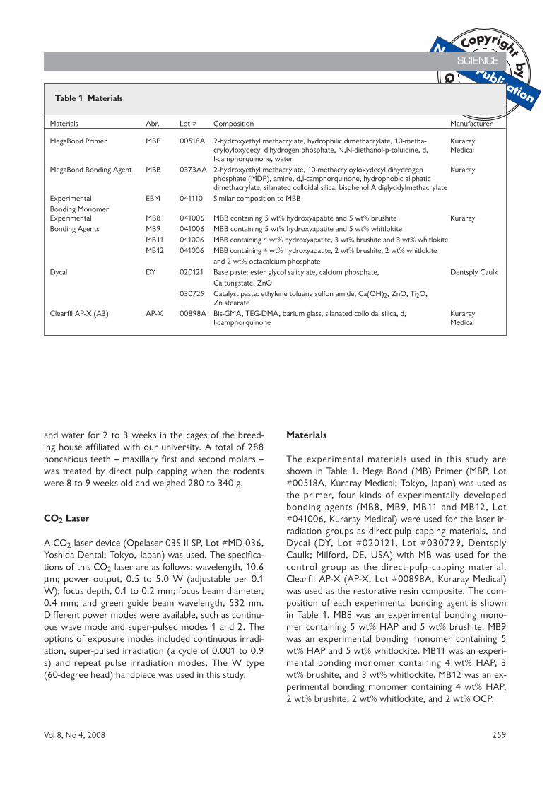
Materials
The experimental materials used in this study areshown in Table 1. Mega Bond (MB) Primer (MBP, Lot#00518A, Kuraray Medical; Tokyo, Japan) was used asthe primer, four kinds of experimentally developedbonding agents (MB8, MB9, MB11 and MB12, Lot#041006, Kuraray Medical) were used for the laser ir-radiation groups as direct-pulp capping materials, andDycal (DY, Lot #020121, Lot #030729, DentsplyCaulk; Milford, DE, USA) with MB was used for thecontrol group as the direct-pulp capping material.Clearfil AP-X (AP-X, Lot #00898A, Kuraray Medical)was used as the restorative resin composite. The com-position of each experimental bonding agent is shownin Table 1. MB8 was an experimental bonding mono-mer containing 5 wt% HAP and 5 wt% brushite. MB9was an experimental bonding monomer containing 5wt% HAP and 5 wt% whitlockite. MB11 was an experi-mental bonding monomer containing 4 wt% HAP, 3wt% brushite, and 3 wt% whitlockite. MB12 was an ex-perimental bonding monomer containing 4 wt% HAP,2 wt% brushite, 2 wt% whitlockite, and 2 wt% OCP.
Vol 8, No 4, 2008 259
SCIENCE
Table 1 Materials
Materials Abr. Lot # Composition Manufacturer
MegaBond Primer MBP 00518A 2-hydroxyethyl methacrylate, hydrophilic dimethacrylate, 10-metha- Kuraray cryloyloxydecyl dihydrogen phosphate, N,N-diethanol-p-toluidine, d, MedicalI-camphorquinone, water
MegaBond Bonding Agent MBB 0373AA 2-hydroxyethyl methacrylate, 10-methacryloyloxydecyl dihydrogen Kurarayphosphate (MDP), amine, d,I-camphorquinone, hydrophobic aliphatic dimethacrylate, silanated colloidal silica, bisphenol A diglycidylmethacrylate
Experimental EBM 041110 Similar composition to MBBBonding MonomerExperimental MB8 041006 MBB containing 5 wt% hydroxyapatite and 5 wt% brushite KurarayBonding Agents MB9 041006 MBB containing 5 wt% hydroxyapatite and 5 wt% whitlokite
MB11 041006 MBB containing 4 wt% hydroxyapatite, 3 wt% brushite and 3 wt% whitlokiteMB12 041006 MBB containing 4 wt% hydroxyapatite, 2 wt% brushite, 2 wt% whitlokite
and 2 wt% octacalcium phosphateDycal DY 020121 Base paste: ester glycol salicylate, calcium phosphate, Dentsply Caulk
Ca tungstate, ZnO030729 Catalyst paste: ethylene toluene sulfon amide, Ca(OH)2, ZnO, Ti2O,
Zn stearateClearfil AP-X (A3) AP-X 00898A Bis-GMA, TEG-DMA, barium glass, silanated colloidal silica, d, Kuraray
I-camphorquinone Medical
Copyrig
ht
by
N
otfor
Qu
in
tessence
Not
forPublication
Experimental Groups and Observation Terms
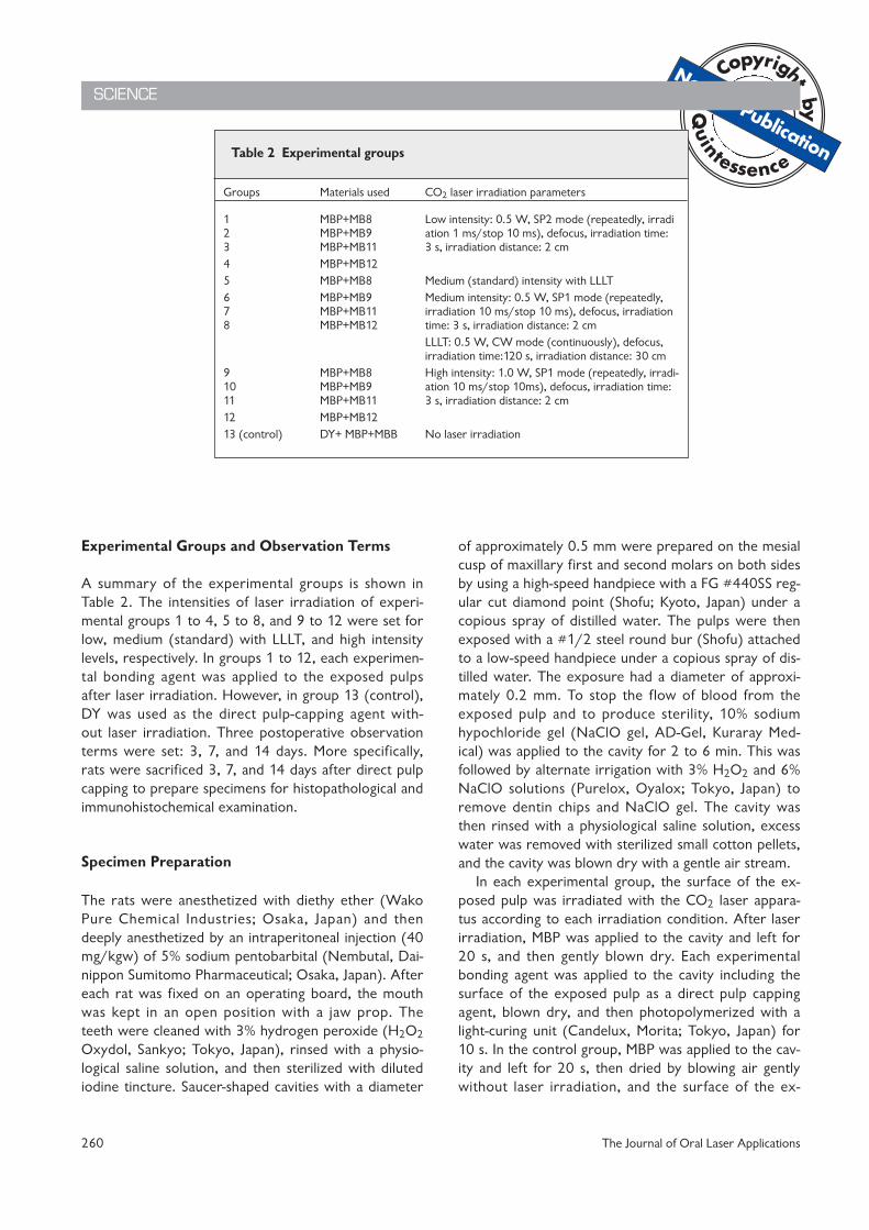
A summary of the experimental groups is shown inTable 2. The intensities of laser irradiation of experi-mental groups 1 to 4, 5 to 8, and 9 to 12 were set forlow, medium (standard) with LLLT, and high intensitylevels, respectively. In groups 1 to 12, each experimen-tal bonding agent was applied to the exposed pulpsafter laser irradiation. However, in group 13 (control),DY was used as the direct pulp-capping agent with-out laser irradiation. Three postoperative observationterms were set: 3, 7, and 14 days. More specifically,rats were sacrificed 3, 7, and 14 days after direct pulpcapping to prepare specimens for histopathological andimmunohistochemical examination.
Specimen Preparation
The rats were anesthetized with diethy ether (WakoPure Chemical Industries; Osaka, Japan) and thendeeply anesthetized by an intraperitoneal injection (40mg/kgw) of 5% sodium pentobarbital (Nembutal, Dai-nippon Sumitomo Pharmaceutical; Osaka, Japan). Aftereach rat was fixed on an operating board, the mouthwas kept in an open position with a jaw prop. Theteeth were cleaned with 3% hydrogen peroxide (H2O2Oxydol, Sankyo; Tokyo, Japan), rinsed with a physio-logical saline solution, and then sterilized with dilutediodine tincture. Saucer-shaped cavities with a diameter
of approximately 0.5 mm were prepared on the mesialcusp of maxillary first and second molars on both sidesby using a high-speed handpiece with a FG #440SS reg-ular cut diamond point (Shofu; Kyoto, Japan) under acopious spray of distilled water. The pulps were thenexposed with a #1/2 steel round bur (Shofu) attachedto a low-speed handpiece under a copious spray of dis-tilled water. The exposure had a diameter of approxi-mately 0.2 mm. To stop the flow of blood from theexposed pulp and to produce sterility, 10% sodiumhypochloride gel (NaClO gel, AD-Gel, Kuraray Med-ical) was applied to the cavity for 2 to 6 min. This wasfollowed by alternate irrigation with 3% H2O2 and 6%NaClO solutions (Purelox, Oyalox; Tokyo, Japan) toremove dentin chips and NaClO gel. The cavity wasthen rinsed with a physiological saline solution, excesswater was removed with sterilized small cotton pellets,and the cavity was blown dry with a gentle air stream.
In each experimental group, the surface of the ex-posed pulp was irradiated with the CO2 laser appara-tus according to each irradiation condition. After laserirradiation, MBP was applied to the cavity and left for20 s, and then gently blown dry. Each experimentalbonding agent was applied to the cavity including thesurface of the exposed pulp as a direct pulp cappingagent, blown dry, and then photopolymerized with alight-curing unit (Candelux, Morita; Tokyo, Japan) for10 s. In the control group, MBP was applied to the cav-ity and left for 20 s, then dried by blowing air gentlywithout laser irradiation, and the surface of the ex-
SCIENCE
260 The Journal of Oral Laser Applications
Table 2 Experimental groups
Groups Materials used CO2 laser irradiation parameters
1 MBP+MB8 Low intensity: 0.5 W, SP2 mode (repeatedly, irradi2 MBP+MB9 ation 1 ms/stop 10 ms), defocus, irradiation time: 3 MBP+MB11 3 s, irradiation distance: 2 cm4 MBP+MB125 MBP+MB8 Medium (standard) intensity with LLLT6 MBP+MB9 Medium intensity: 0.5 W, SP1 mode (repeatedly,7 MBP+MB11 irradiation 10 ms/stop 10 ms), defocus, irradiation 8 MBP+MB12 time: 3 s, irradiation distance: 2 cm
LLLT: 0.5 W, CW mode (continuously), defocus, irradiation time:120 s, irradiation distance: 30 cm
9 MBP+MB8 High intensity: 1.0 W, SP1 mode (repeatedly, irradi-10 MBP+MB9 ation 10 ms/stop 10ms), defocus, irradiation time: 11 MBP+MB11 3 s, irradiation distance: 2 cm12 MBP+MB1213 (control) DY+ MBP+MBB No laser irradiation

Copyrig
ht
by
N
otfor
Qu
in
tessence
Not
forPublication
posed pulp was covered with DY. MBB was then ap-plied to the cavity, blown dry gently, and photopoly-merized for 10 s. After direct pulp-capping and bond-ing procedures, all the cavities were restored with resincomposite, AP-X, and photopolymerized for 40 s.
Perfusion Fixation
The rats were sacrificed by an intraperitoneal injectionof 5% pentobarbital sodium after each observation pe-riod. Each pulp was fixed by transcardial perfusion witha 4% paraformaldehyde phosphate buffer solution (pH7.4). The maxillae containing experimental teeth werecarefully removed and immersed in a 4% paraformal-dehyde phosphate buffer solution at 4°C for 2 days.
Tissue Preparation and Serial Sectioning forHistopathological and Immunohistochemical Observation
We removed excess tissue from the maxillae and decal-cified them with a 10% EDTA-2Na solution (pH 7.4) atroom temperature for three to four weeks. After de-calcification, AP-X was removed from the cavity andrinsed with running water for 24 h. The specimenswere dehydrated in ascending grades of ethanol, deal-coholized by xylene, and then embedded in paraffin.Serial sections 5 to 6 μm in thickness were cut with asliding microtome in a room with the temperature at20 to 23°C and relative humidity between 50 and60%. They were alternately stained with Mayer'sHematoxylin-Eosin staining to observe the pathologicalconfiguration, van Gieson staining to observe the for-mation of collagen fibers, connective tissue and hardtissue of dentin, Hucker-Conn staining to observe bac-terial invasion, and modified NF Watanabe silver im-pregenation staining to observe reticulum fibers. Asimmunohistochemical staining, the sABC method onDMP1 staining was used to observe Dentin Matrix Protein 1 (DMP1) and the sABC method on TGFβ1staining to observe Transforming Growth Factor β1(TGFβ1).
Observation Items and Evaluation Criteria
The stained sections were observed under the light mi-croscope (Eclipse E1000, Lot #11545,Nikon; TokyoJapan) and the following items were evaluated: pulp tis-sue disorganization, inf lammatory cell inf iltration,
reparative dentin formation, and bacterial penetration.The findings were evaluated according to the followingcriteria established by Medina III-Katoh:40
A: Pulp tissue disorganization
1. Normal or almost normal t issue morphology(none).
2. Odontoblast layer disorganization, but the deeppart of the pulp was normal (mild).
3. Loss of general tissue morphology (moderate).4. Necrosis in the coronal one-third or more of the
pulp (severe).
B: Inflammatory cell infiltration
1. Absence or presence of a few scattered inflamma-tory cells in the pulp (none).
2. Mild acute/chronic cell lesions (mild).3. Moderate inf lammatory cell lesions seen as ab-
scesses or densely stained infiltrates of polymor-phonuclear leucocytes, histiocytes and lymphocytesin one-third or more of the coronal pulp and/or themid-pulp (moderate).
4. Pulp necrosis due to a severe degree of infection orlack of tissue in one half or more of the pulp (se-vere).
C: Reparative dentin formation
1. No dentin bridge formation (none).2. Initial dentin bridge formation extending to not
more than one-half of the exposure site (initial).3. Partial/incomplete dentin bridge formation extend-
ing to more than one-half of the exposure site butnot completely closing the exposure site (partial).
4. Complete dentin bridge formation (complete).
D: Bacterial penetration
1. Absence of stained bacterial profiles in any of thesections (none).
2. Presence of stained bacterial profiles along the coro-nal or apical walls of the cavity (mild).
3. Presence of stained bacterial profiles within the cutdentinal tubules or axial wall of the cavity (moder-ate).
4. Presence of stained bacterial profiles within the den-tal pulp (severe).
Immunohistochemical Staining
The sections were deparaffinized with xylene, dehy-drated in ascending grades of ethanol, and then rinsedbriefly with tap water and phosphate buffered saline
Vol 8, No 4, 2008 261
SCIENCE

Copyrig
ht
by
N
otfor
Qu
in
tessence
Not
forPublication
(pH 7.4). The sections were incubated with primaryrabbit antibodies, such as anti-DMP1 antibody workingdilution, 1:3000 for 12 h at 4°C Polyclonal (Lot#002FD, Takarabio; Siga, Japan) or anti-TGFβ1 anti-body working dilution, 1:8000 for 12 h at 4°C anti-TGFβ1(V) (Lot #F2306, Cosmobio; Tokyo, Japan).They were immunochemically stained with HistofineSAB-PO(R) Kit (Lot #H0609, Nichirei Biosciences;Tokyo, Japan) employing the Avidin-biotin HorseradishPeroxidase Complex (sABC) method. The antibody lo-calized antigen was then detected by peroxidase activa-tion of 3,3-diaminobenzidine, DAB simple stain (DABsolution, Lot #H0610, Nichirei Biosciences). The sec-tions were counterstained with Mayer's hematoxylin.
Measurement of the Diameter of the ExposedPulp Area
The diameters of the exposed areas were measuredwith a stereomicroscope (Measuring Microscope MM-40, Lot #2104048, Nikon) and the widest dimensionwas recorded as the pulp exposure size of the speci-men.
Statistical Analysis
The diameters of exposed pulp areas were statisticallyanalyzed by one-way ANOVA and the Bonferroni post-hoc test with statistical software Microsoft Excel (Mi-crosoft; Redmond, WA, USA) for differences amongthe experimental groups during each observation pe-riod at a significance level of 0.05.
The results of the histopathological evaluation werestatistically analyzed by the Kruskal-Wallis H-test usingMicrosoft Excel for differences among the experimen-tal groups during each observation period, and differ-ences among the groups according to the observationperiod under each laser irradiation condition at a signif-icance level of 0.05. Moreover, the correlation be-tween inf lammatory cell inf iltration and bacterialinvasion was investigated by the Kendall rank correla-tion using the statistical software SPSS (SPSS Japan;Tokyo, Japan) at a significance level of 0.05.
RESULTS
Diameters of Exposed Pulp Area
The mean diameters with standard deviations, and themaximum and the minimum values for the size of theexposed pulp areas of each group are shown in Table
SCIENCE
262 The Journal of Oral Laser Applications
Table 3 The diameters of exposed pulp area
Groups After 3 days After 7 days After 14 days
1 0.413 [0.141],a(0.233-0.593) 0.336 [0.069],a(0.230-0.413) 0.333 [0.091],b(0.170-0.441)2 0.296 [0.097],b(0.115-0.389) 0.235 [0.111],b(0.137-0.395) 0.355 [0.115],b(0.246-0.570)3 0.234 [0.123],b(0.074-0.450) 0.221 [0.061],b(0.142-0.312) 0.299 [0.130],b(0.125-0.493)4 0.196 [0.053],b(0.124-0.290) 0.196 [0.076],b(0.112-0.315) 0.263 [0.130],b(0.106-0.422)5 0.189 [0.047],b(0.107-0.246) 0.211 [0.070],b(0.148-0.340) 0.290 [0.071],b(0.190-0.379)6 0.279 [0.080],b(0.146-0.372) 0.144 [0.059],b(0.078-0.221) 0.357 [0.102],b(0.228-0.506)7 0.207 [0.069],b(0.129-0.322) 0.149 [0.021],b(0.129-0.185) 0.269 [0.158],b(0.046-0.459)8 0.249 [0.110],b(0.149-0.395) 0.217 [0.064],b(0.105-0.287) 0.231 [0.056],b(0.136-0.308)9 0.185 [0.042],b(0.143-0.258) 0.171 [0.033],b(0.125-0.227) 0.253 [0.071],b(0.145-0.330)10 0.204 [0.060],b(0.148-0.293) 0.158 [0.035],b(0.112-0.215) 0.339 [0.040],b(0.267-0.383)11 0.192 [0.052],b(0.123-0.280) 0.147 [0.035],b(0.107-0.200) 0.240 [0.115],b(0.111-0.393)12 0.231 [0.029],b(0.193-0.275) 0.258 [0.110],a(0.122-0.388) 0.279 [0.085],b(0.115-0.34)13 0.246 [0.014],b(0.225-0.266) 0.295 [0.058],a(0.230-0.392) 0.307 [0.068],b(0.242-0.427)
[ ]: standard deviation, (minimum-maximum), unit: mm. Same superseript letters indicate no statistically significant differences for each postoperative observation term.

Copyrig
ht
by
N
otfor
Qu
in
tessence
Not
forPublication
3. The mean diameter of the pulp exposure of all speci-mens was 0.248 ± 0.105 mm, the maximum value of allpulp exposures was 0.593 mm and the minimum valuewas 0.046 mm.
There were significant differences among the pulpexposure sizes of the groups 3 and 7 days after treat-ment (one-way ANOVA, p < 0.05), although there wasno significant difference among the pulp exposure sizesof the groups after 14 days (one-way ANOVA, p >0.05). In the groups after 3 days, the size of exposedpulp area of group 1 was significantly larger than thatof the other groups (Bonferroni test, p < 0.05). In thegroups after 7 days, the size of the exposed pulp areaof group 1 was significantly larger than that of theother groups except groups 12 and 13 (Bonferronitest, p < 0.05).
Histopathological and Immunohistochemical Find-ings
Fracture and or failing of the restorations were not ob-served in any of the specimens. A summary of the re-sults of the histopathologic evaluation is shown in Figs1 to 3. Representative histopathological and immuno-histochemical images of all the groups are shown inFigs 4 through 8.
1) Histopathological and immunohistochemicalfindings after 3 days
The results of the Kruskal-Wallis H-test for each histo-pathological evaluation after 3 days revealed no signifi-cant difference among the experimental groups (p >0.05). The eosinophilic heat denaturation layer and theprotein coagulation layer were recognized in laser-irra-diated areas under all irradiation conditions. The thick-ness of these layers had a tendency to increase as theintensity of laser irradiation increased.
One specimen in group 4 showed mild pulp tissuedisorganization. Three specimens each from groups 2,3, and 7 exhibited mild pulp tissue disorganization.Four specimens from each of the groups 1, 6, and 12exhibited mild pulp tissue disorganization. In group 5,one specimen exhibited moderate pulp tissue disorgani-zation, and the other specimens in total exhibited mildpulp tissue disorganization. In the other groups, fivespecimens exhibited mild pulp tissue disorganization.
Regardless of the irradiation condition, inflamma-tory cell infiltration was observed in most of the speci-mens. The inf lammation area was enlarged as theintensity of laser irradiation increased. In groups 8, 9,and 12, four specimens in total showed no inflamma-tory cell infiltration and one specimen exhibited mildinflammatory cell infiltration. In the other groups, twoto five specimens exhibited mild inflammatory cell infil-
Vol 8, No 4, 2008 263
SCIENCE
Legend for PTD, ICI and BP
none mild moderate severe
Irradiation Condition: low
Pulp Tissue Disorganization (PTD)
Inflammatory Cell Infiltration (ICI)
Reparative Dentin Formation (RDF)
Bacterial Penetration (BP)
Irradiation Condition: medium + LLLT
Pulp Tissue Disorganization (PTD)
Inflammatory Cell Infiltration (ICI)
Reparative Dentin (RDF)
Bacterial Penetration (BP)
Group 1 Group 3 Group 4Group 2
Irradiation Condition: high
Pulp Tissue Disorganization (PTD)
Inflammatory Cell Infiltration (ICI)
Reparative Dentin Formation (RDF)
Bacterial Penetration (BP)
Group 13
none initial partial complete
Legend for RDF
No Irradiation: control
Pulp Tissue Disorganization (PTD)
Inflammatory Cell Infiltration (ICI)
Reparative Dentin Formation (RDF)
Bacterial Penetration (BP)
Group 5 Group 7 Group 8Group 6
Group 9 Group 11 Group 12Group 10
Fig 1 Results of histopathological andimmunohistochemical evaluation (3days).
Copyrig
ht
by
N
otfor
Qu
in
tessence
Not
forPublication
tration. After 3 days in this study, a common findingwas hyperemia, and rarely a few lymphocyte infiltra-tions. TGFβ1 staining showed no positive reaction after3 days. Findings common to all the groups 3 days post-operatively were hyperemia, and no positive reaction
to TGFβ1 staining. A very few lymphocyte infiltrationswere recognized in some specimens.
None of the specimens exhibited any signs of repar-ative dentin formation or bacterial penetration.
SCIENCE
264 The Journal of Oral Laser Applications
Irradiation Condition: low
Pulp Tissue Disorganization (PTD)
Inflammatory Cell Infiltration (ICI)
Reparative Dentin Formation (RDF)
Bacterial Penetration (BP)
Irradiation Condition: medium + LLLT
Pulp Tissue Disorganization (PTD)
Inflammatory Cell Infiltration (ICI)
Reparative Dentin Formation (RDF)
Bacterial Penetration (BP)
Irradiation Condition: high
Pulp Tissue Disorganization (PTD)
Inflammatory Cell Infiltration (ICI)
Reparative Dentin Formation (RDF)
Bacterial Penetration (BP)
No Irradiation: control
Pulp Tissue Disorganization (PTD)
Inflammatory Cell Infiltration (ICI)
Reparative Dentin Formation (RDF)
Bacterial Penetration (BP)
Legend for PTD, ICI and BP
none mild moderate severe
none initial partial complete
Legend for RDF
Group 1 Group 3 Group 4Group 2
Group 13
Group 5 Group 7 Group 8Group 6
Group 9 Group 11 Group 12Group 10
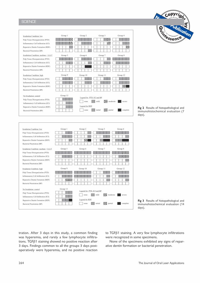
Fig 2 Results of histopathological andimmunohistochemical evaluation (7days).
Fig 3 Results of histopathological andimmunohistochemical evaluation (14days).
Irradiation Condition: medium + LLLT
Pulp Tissue Disorganization (PTD)
Inflammatory Cell Infiltration (ICI)
Reparative Dentin Formation (RDF)
Bacterial Penetration (BP)
Irradiation Condition: high
Pulp Tissue Disorganization (PTD)
Inflammatory Cell Infiltration (ICI)
Reparative Dentin Formation (RDF)
Bacterial Penetration (BP)
Irradiation Condition: low
Pulp Tissue Disorganization (PTD)
Inflammatory Cell Infiltration (ICI)
Reparative Dentin Formation (RDF)
Bacterial Penetration (BP)
No Irradiation: control
Pulp Tissue Disorganization (PTD)
Inflammatory Cell Infiltration (ICI)
Reparative Dentin Formation (RDF)
Bacterial Penetration (BP)
Legend for PTD, ICI and BP
none mild moderate severe
none initial partial complete
Legend for RDF
Group 1 Group 3 Group 4Group 2
Group 13
Group 5 Group 7 Group 8Group 6
Group 9 Group 11 Group 12Group 10
Copyrig
ht
by
N
otfor
Qu
in
tessence
Not
forPublication
2) Histopathological and immunohistochemicalfindings after 7 days
The results of the Kruskal-Wallis H-test for each histo-pathological evaluation after 7 days revealed no signifi-cant difference among the experimental groups (p >0.05).
In group 1, one specimen exhibited moderate pulptissue disorganization, and the other specimens exhib-ited mild pulp tissue disorganization. Five specimens ingroup 8 exhibited mild pulp tissue disorganization.Four specimens in group 2 and the same number ofspecimens in group 5 showed mild pulp tissue disorga-nization. Two specimens from groups 10 and as manyfrom group 11 demonstrated mild pulp tissue disorga-nization. One specimen in group 7 showed mild pulptissue disorganization. In the other groups, 3 speci-mens in total exhibited mild pulp tissue disorganization.
In group 1, one specimen exhibited moderate in-flammatory cell infiltration, and the rest exhibited mildinflammatory cell infiltration. Four specimens in group8 showed mild inflammatory cell infiltration. Threespecimens each in groups 2, 9, 11, and 12, exhibitedmild inflammatory cell infiltration. One specimen ingroup 6 showed mild inflammatory cell infiltration. As
for the rest, a total of 2 specimens exhibited mild in-flammatory cell infiltration.
Recovery from pulp tissue disorganization was ob-served in most of the specimens after 7 days. Neogen-esis of blood vessels, odontoblasts, fibroblast-like cells,and predentin occurred in the area where collagenfiber was actively yielded. The thickness of the collagenfiber increased as the intensity of laser irradiation de-creased. Recovery from pulp tissue disorganization was delayed when the intensity of laser irradiation increased. Eosinophilic heat denaturation layers and protein coagulation layers were recognized at laser-irradiated sites regardless of irradiation condition. Thethickness of these layers had a tendency to increase asthe intensity of laser irradiation increased. Calciumphosphate salt powder contained in MB8 was observednear the exposed pulp surface of one specimen. Thepulp surrounded by the area supposedly penetrated bythe laser beam was stained purplish red by modifiedNF Watanabe silver impregenation staining.
A tendency for the round type of cell infiltration todisappear was recognized under all irradiation condi-tions. Macrophages and monocytes were observedamong collagen fibers and in the area around foreignobjects. A positive reaction by TGFβ1 staining oc-
Vol 8, No 4, 2008 265
SCIENCE
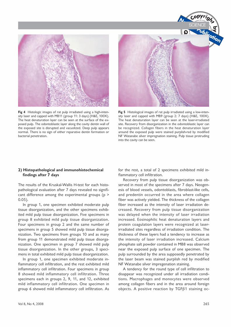
Fig 5 Histological images of rat pulp irradiated using a low-inten-sity laser and capped with MB9 (group 2: 7 days) (H&E, 100X).The heat denaturation layer can be seen at the laser-irradiatedsite. Recovery from disorganization in the odontoblastic layer canbe recognized. Collagen fibers in the heat denaturation layeraround the exposed pulp were stained purplish-red by modifiedNF Watanabe silver impregnation staining. Pulp tissue protrudinginto the cavity can be seen.
Fig 4 Histologic images of rat pulp irradiated using a high-inten-sity laser and capped with MB11 (group 11: 3 days) (H&E, 100X).The heat denaturation layer can be seen at the surface of the ex-posed pulp. The odontoblastic layer along the cavity dentin wall ofthe exposed site is disrupted and vacuolized. Deep pulp appearsnormal. There is no sign of either reparative dentin formation orbacterial penetration.

Copyrig
ht
by
N
otfor
Qu
in
tessence
Not
forPublication
curred in the area where round cell infiltration was ob-served.
Two specimens in group 6 showed thick reparativedentin formation. In group 12, moderate inflammatorycell infiltration in one specimen and severe inflamma-tory cell inf iltration in another specimen were ob-served. One specimen in group 4 showed moderatereparative dentin formation. One specimen in group 1showed mild reparative dentin formation. The othergroups exhibited no reparative dentin formation.
Mild bacterial penetration was recognized in onespecimen of group 2 (MB9) (low intensity of laser irra-
diation). However, there was no significant differencein the Kendall rank correlation between group 13 (con-trol) and the other groups (p > 0.05).
3) Histopathological and immunohistochemicalfindings after 14 days
The results of the Kruskal-Wallis H-test for each histo-pathologic evaluation after 14 days revealed no signifi-cant difference among the experimental groups (p >0.05).
SCIENCE
266 The Journal of Oral Laser Applications
Fig 6 Histological images of rat pulp irradiated using a low-intensity laser and capped with MB12 (group 4: 14 days) (a: H&E, b: NF, c: vanGieson, d: DMP1, 100X). There is a complete dentin bridge that is composed of osteo-type dentin including odontoblast-like cells. A tubu-lar-type dentin layer can be seen at the pulpal side of the dentin bridge. A positive reaction by DMP1 staining can be recognized in thedentin bridge.

Copyrig
ht
by
N
otfor
Qu
in
tessence
Not
forPublication
In group 8, two specimens exhibited moderate pulptissue disorganization, and the other specimens exhib-ited mild pulp tissue disorganization. Five specimenseach in groups 5, 6, 7, 9, and 10 exhibited mild pulptissue disorganization. Four specimens each in groups3, 4, and 12 showed mild pulp tissue disorganization.Two specimens each in groups 1, 11, and 13 exhibitedmild pulp tissue disorganization. One specimen ingroup 2 showed mild pulp tissue disorganization.
In group 8, two specimens exhibited moderate andone specimen mild inflammatory cell infiltration, andthe rest showed no inflammatory cell infiltration. Five
specimens in group 10 showed mild inflammatory cellinfiltration. Three specimens each in groups 1, 5, 9,and 12 exhibited mild inflammatory cell infiltration.Two specimens each in groups 3 and 13 showed mildinflammatory cell infiltration. One specimen each ingroups 2, 4, and 11 showed mild inflammatory cell in-filtration. The other groups exhibited no inflammatorycell infiltration.
Most of the specimens in the low-intensity irradia-tion group recovered almost normal tissue conditions.However, in the medium-intensity and high-intensity ir-radiation groups, most of the specimens had a tenden-
Vol 8, No 4, 2008 267
SCIENCE
Fig 7 Histological images of rat pulp irradiated using medium-intensity with an LLLT laser and capped with MB9 (group 6: 14 days) (a:H&E, b: van Gieson, c: DMP1, d: TGF-β1, x100). There is incomplete dentin bridge formation that is composed of very irregular osteo-type dentin and pulp tissue tunnel. A tubular-type dentin layer can be seen at the pulpal side of the dentin bridge. A positive reaction byDMP1 staining and that by TGF-β1 staining can be recognized in the dentin bridge and in the pulp tissue, respectively.
Copyrig
ht
by
N
otfor
Qu
in
tessence
Not
forPublication
cy to delay recovering, and some specimens showed al-most the same results as the specimens of the low-in-tensity irradiation group did after 7 days. In the low-intensity irradiation group, remarkable hyperplasia ofcollagen fibers and fibroblast-like cells were observedaround the wounded pulp surface where healing mech-anisms were at work. Although the medium-intensity irradiation group showed almost similarhistopathologic profiles of pulp tissue disorganizationto those shown by the low intensity irradiation group,the former presented more aggressive hyperplasia ofcollagen fibers over a wider area than the latter. In the
high-intensity irradiation group, hyperplasia of collagenfibers covered a wider area than in the other irradia-tion condition groups.
Modified NF Watanabe silver impregenation stainingrevealed that the low-intensity irradiation group showedprofiles rather similar to those of normal tissue, butthat the medium-intensity and high-intensity irradiationgroup showed argyrophilic fibers underneath the den-tin bridge.
Under all the irradiation conditions, round cell infil-tration chiefly by lymphocytes and eosinophils was observed in almost all experimental groups. The spe-
SCIENCE
268 The Journal of Oral Laser Applications
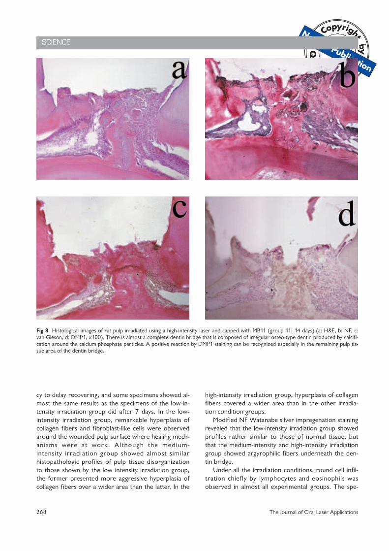
Fig 8 Histological images of rat pulp irradiated using a high-intensity laser and capped with MB11 (group 11: 14 days) (a: H&E, b: NF, c:van Gieson, d: DMP1, x100). There is almost a complete dentin bridge that is composed of irregular osteo-type dentin produced by calcifi-cation around the calcium phosphate particles. A positive reaction by DMP1 staining can be recognized especially in the remaining pulp tis-sue area of the dentin bridge.
Copyrig
ht
by
N
otfor
Qu
in
tessence
Not
forPublication
cimens of the low-intensity irradiation group showed atendency for hyperemia to disappear but for mild-round cell infiltration to remain.
On the other hand, in the specimens of the medium-intensity irradiation group, only a few cases of hy-peremia were found. A few instances of vacuolardegeneration and mature granulocytes were demon-strated in some specimens of the high-intensity irra-diation group. Macrophages were observed in the car-bonization layer. A positive reaction by TGFβ1 stainingwas recognized in the round cell infiltration area. Re-covery from inflammatory changes of the pulp weredelayed when a higher-intensity laser irradiated the ex-posed pulp.
In the low- intensity irradiation group, the speci-mens to which MB9, 11, and 12 were applied showedalmost complete dentin bridge formation with smalltunnel-shaped defects, and their results were favorablycomparable to those of the control group. Reparativedentin formation after 14 days had advanced comparedto that observed after 7 days. Replacement of theodontoblast layer was observed underneath the repar-ative osteodentin. The surface of the exposed pulp wascovered with irritative dentin protruding from the pe-riphery of the pulpal wall. Some specimens showeddentin matrix formation among the collagen fibers.This finding was similar to that in the specimens afterseven days. Pulp nodule formation was observed in afew specimens. These calcium deposits including thereparative dentin were found to be positive by vanGieson and DMP1 staining. The calcium deposits whichoccurred in the pulp tissue were a unique finding in thelaser irradiation groups compared with the control.None of the specimens exhibited bacterial penetration14 days postoperatively.
DISCUSSION
From the results of our previous studies,41-44 the pulpresponse after direct pulp capping with laser irradiationseemed to be affected by laser irradiation levels andwavelengths, and showed a tendency to delay hard tis-sue formation. Therefore, we took the following mat-ters into consideration in this study.
The Opelaser 03SII SP CO2 laser device was usedfor irradiating exposed pulps of rat molars, becausemost of the energy of the CO2 laser is absorbed withinan area of 0.1 to 0.2 mm below the tissue surface andthe thickness of the heat denaturation layer is less than0.5 mm. Furthermore, the CO2 laser has other merits,such as low caloric diffusion to the surrounding tissue,
effective control of hemorrhaging and exudation, anddisinfection of the exposed pulp surface.
The intensities of the laser irradiation used in thisstudy were decided upon from the results of our pilotstudy. The intensity of the laser irradiation used in ourprevious study51 was adopted as the medium intensity(standard) of the irradiation conditions, and the degreeof intensity was decided within the range that permit-ted the formation of a carbonization layer on the sur-face of the rat exposed pulp. Laser irradiation withLLLT was added to only the medium intensity irradia-tion after the formation of a carbonization layer. It hasbeen speculated that LLLT induces a photobioactive re-action (PAR), such as healing promotion during the in-flammation stage, increase of fibroblasts and collagenproduction, and inhibition of excess granulation forma-tion during the hyperplasia stage, regardless of lasertype.54 Consequently, we expected LLLT to controlpulp inflammation and allow for earlier dentin bridgeformation. In this study, laser irradiation with LLLT wasonly applied to the medium-intensity irradiation groupfor comparison with the other laser irradiation groups.
Takizawa et al45 reported that pulp reaction afterlaser irradiation was related not only to energy consis-tency but also to output and exposure time of laser ir-radiation. In addition, Serebro et al46 and Shoji et al47
reported that pulp reaction to the CO2 laser was moreaffected by irradiation time than output of irradiation.Therefore, the irradiation time was set as short as pos-sible in this study. However, the results of this studyshowed that the thickness of the heat denaturationlayer and protein coagulation layer tended to increasewhen the output of laser irradiation increased. Mat-sumoto et al48 reported that the main cause of pulp ir-ritation after laser irradiation was heat action, and thedegree of pulp damage was dependent upon the char-acteristic of each laser apparatus, because heat absorp-tion and reflection of the tooth surface was differentfrom one type of laser apparatus to another. Accord-ingly, the degree of pulp irritation seems to increase inproportion to the heat storage capacity of dentin whena laser was irradiated. Selzer et al49 and Zach et al50
reported that when the pulp temperature increasesmore than 5°C pulp disorder results, and more than11.1°C induces necrosis. There are other reports51-52
concerning thermal stimulus to pulp. In this study, thecooling function of the laser apparatus was used toprotect pulp from heat caused by laser irradiation.
Jukiç et al9 examined the effects of calcium hydrox-ide and CO2 laser irradiation on the coronal portion ofthe removed vital pulp of a dog. They reported thatupon histopathological examination 30 and 40 days
Vol 8, No 4, 2008 269
SCIENCE
Copyrig
ht
by
N
otfor
Qu
in
tessence
Not
forPublication
postoperatively, carbonization, necrosis, inflammatorycell infiltration, edema, and bleeding were observed,but that reparative dentin formation was hardly ob-served. The laser irradiation conditions used in thisstudy were appropriate because restoration of the pulptissue was recognized even in the tiny pulp cavity of therat.
The results of histopathological examinations afterthree days showed a decrease of odontoblasts and pulpcells, irregular alignment of odontoblasts, and disap-pearance and vacuolar degeneration of predentinunder all irradiation conditions. The thickness of heatdenaturation layers increased as the intensity of laserirradiation was raised. The heat denaturation layer andcarbonization layer served to protect the pulp. How-ever, it was reported that excessive carbonization layerformation delayed the healing process.9 At 7 days post-operatively, the healing process of pulp tissue had already started: the neogenesis of odontoblasts andpredentin and an active hyperplasia of collagen fiberwere observed in most of the specimens, althoughdelay in healing was recognized in a few specimens.Furthermore, most specimens under all irradiation con-ditions retuned almost to normal 14 days postopera-tively. Low-intensity laser irradiation was favorable forrecovery of pulp tissue compared with high-intensity ir-radiation.
In this study, an experimentally developed bondingagent containing calcium phosphate salt was used asthe direct pulp-capping agent. If the direct pulp-cappingagent could not adhere to the wound surface due touncontrolled hemorrhaging or exudation fluid, a spacemight be created between the pulp-capping agent andthe wound surface. Therefore, it was speculated that apulp tissue projection like a polyp may be produced byhyperplasia of pulp tissue into the space.19,29 Shironoet al43 reported that the use of a focus laser beam in ir-radiating the exposed pulp promoted exudation or re-bleeding because of high energy consistency on thesurface of pulp tissue. However, these actions by thefocus beam were not observed in this study, since theCO2 laser using a defocused mode was applied to theexposed pulp. Re-bleeding caused by MBP application,an exudation control deficiency after laser irradiation,and long laser irradiation time with the addition ofLLLT action are problems which should be resolved inthe future.
Inflammatory changes in the laser irradiation groupswere more severe compared with those in the control.Inflammatory change of pulp tissue was observed 3days postoperatively, and showed a decreasing ten-dency 7 days postoperatively. The experimental bond-
ing agent containing calcium phosphate showed goodbiocompatibility and might not cause irritation to thepulp. Various factors such as heat, vibration and pres-sure during cavity preparation, pulp injury at perfora-tion, and hemostasis procedures were reported as thecauses of pulp irritation after restoration by direct pulpcapping.17,19,22,24,29,30 When a laser irradiated the ex-posed pulp before direct pulp capping, the heat actionof laser irradiation was also considered responsible fordelayed wound healing and delayed reparative dentinformation.
The present study found complete dentin bridge for-mation in part of the specimens to which low-intensitylaser irradiation was applied, but incomplete dentinbridge formation in most of the specimens to which themedium- (standard) or high-intensity laser irradiationwas applied, even 14 days postoperatively. It was spec-ulated that delayed healing of pulp tissue irradiatedwith the high-intensity laser was due to damage by heatgeneration. The experimental teeth were cooled downby using the CO2 laser apparatus with a coolant in thisstudy. However, it was insufficient to remove the heataccumulation in dentin. Thus, the development of anefficient cooling system for laser irradiation is needed.
Bacterial penetration was observed on the cavitywall of only one specimen. The reason for this is pre-sumably that plaque in the occlusal pit and f issuremight mingle into the cavity during the restoration pro-cedure, because a large bacterial mass was observedoutside the cavity through the microscope. Disinfectionby laser irradiation and good marginal sealing by theexperimental bonding system used served to protectthe pulp from bacterial penetration, which was limitedto only one specimen in this study.
DMP1 and TGFβ1 were used for ensuring histo-pathological and immunohistochemical evaluation ofthe pulp. DMP1 and TGFβ1 antibodies were used as anindex of reparative dentin formation and inflammatorychange, respectively. DMP1 is used for confirming theformation of reparative dentin. This study used DMP1as the index of reparative dentin formation and thearea stained with DMP1 corresponded to the areastained with H&E and van Gieson.
TGFβ1 is a growth factor53 for fibroblasts, promo-tes production of extracellular matrices, and producesgranulation and arterialization simultaneously.53-57
However, it is said that TGFβ1 controls hyperplasia ofcells such as macrophages and lymphocytes.53 It isspeculated that discharged TGFβ1 controls pulp cell hy-perplasia and ALPase manifestation to contribute tothe formation of reparative dentin after the pulp isdamaged.58 In this study, TGFβ1 showed a positive re-
SCIENCE
270 The Journal of Oral Laser Applications

Copyrig
ht
by
N
otfor
Qu
in
tessence
Not
forPublication
action in the area of inflammation. Accordingly, it wassuggested that TGFβ1 was related to the recovery ofthe wounded part of the pulp tissue. There was a sig-nificant difference between the pulp exposure sizes ofthe specimens observed 3 days postoperatively andthose of the specimens observed 7 days postopera-tively. However, this significant difference did not affectthe histopathological results of this study.
Several studies reported that a dentin bridge wasformed when a direct pulp-capping agent containingcalcium phosphate salt was applied to the exposed pulpsurface.16,24,29 On the other hand, no dentin bridgeformation was reported in some other studies.19,22
Since the effect of laser irradiation on dentin bridgeformation seems to be stronger than that of the exper-imental bonding system, the effect of the experimentalbonding system used in this study was not clearly rec-ognized. Good biocompatibility of calcium phosphatesalt may contribute to the arrest of any inflammatoryreaction. Calcium phosphate salt has been studied as abone generative material. A mixture of calcium hydrox-ide powder and water or a physiological salt solutionwas applied to the exposed pulp as a direct pulp-cap-ping material and to root canals as an antiseptic mater-ial. Calcification occurs under the condition of alkalinityproduced by Ca and P ion from HAP and body fluid.17
Yoshida17 reported that crystalline needle-like struc-tures were generated by the deposition of Ca and Paround HAP and collagen fiber matrices. Additionalhard tissue formation made by smaller particles of cal-cium phosphate started earlier than that made by lar-ger particles of calcium phosphate, although theparticle sizes of calcium phosphate did not differ in thepulpal reaction.25 Furthermore, Katoh et al59 specu-lated that Ca, P, and Mg ions in calcium phosphate saltmight accelerate reparative dentin formation. Suzuki etal44 reported that an experimental bonding agent con-taining calcium phosphate salt used for direct-pulp cap-ping caused a less inflammatory response in the pulpand was effective in promoting reparative tubular-typedentin formation.
The mechanism of wound healing by laser irradia-tion has not been completely clarified. However, thethermal effect of laser irradiation is considered as a fac-tor for recovery and calcification of the pulp. Severalstudies reported that mild damage to odontoblasts oc-curred immediately after laser irradiation, althoughthey recovered over time, and finally reparative dentinwas formed by regenerated odontoblasts.2-8 Anotherstudy reported that the photomechanical energy of alaser beam may contribute to hard tissue formation.60
This study could not clarify any difference in thetime, speed, and thickness in reparative dentin forma-tion between the experimental and control groups be-cause the heat effect of laser irradiation seemed to betoo intense for the pulp. In MB12, a low-intensity irra-diation group, two specimens showed complete dentinbridge formation and favorable results compared withthe control. The thickness of the heat denaturationlayer increased in proportion to the intensity of irradia-tion. Therefore, delay in pulp tissue healing after laserirradiation might be related to the thickness of the heatdenaturation layer. From the results of this study, it wasspeculated that a thicker heat denaturation layer yield-ed by laser irradiation was attributable to thicker repa-rative dentin formation. However, the thick heatdenaturation layer caused a delay in pulp tissue healingand might reduce the volume of the pulp cavity due tothe formation of a considerable amount of reparativedentin. Accordingly, we recommend lower intensitylaser irradiation than that used in this study.
CONCLUSIONS
Based on the results of this study, it was concludedthat:
1. There were no significant differences in wound heal-ing of exposed pulp among the three irradiationconditions (low-, medium- and high-intensity levels)of the CO2 laser (p > 0.05), although low-intensityirradiation had a tendency to induce faster healingand better dentin bridge formation.
2. The thickness of irritative dentin formed by the sideof the pulp cavity wall increased with the irradiationintensity of CO2 laser.
3. There was no significant difference in the effect ofdirect pulp capping between the experimental bond-ing agents containing calcium phosphate and prepa-ration of calcium hydroxide as the control DY (p >0.05).
4. It was suggested that low-level irradiation with theCO2 laser and direct pulp capping with experimen-tally developed bonding agents containing calciumphosphate were comparable to the preparation ofcalcium hydroxide DY.
5. CO2 laser irradiation protected the surface of theexposed pulp through its functions of hemostasis,sterilization, and protein coagulation, and seemed toensure the favorable effects of direct pulp capping.
Vol 8, No 4, 2008 271
SCIENCE

Copyrig
ht
by
N
otfor
Qu
in
tessence
Not
forPublication
ACKNOWLEDGMENTS
The authors thank the Kuraray Medical Co. for the experimentaladhesive resins systems and other materials they generously pro-vided. This study was supported by grants-in-aid for Scientific Re-search (B) (2) in 2003-2006, Project number: 15390578 fromthe Japan Society for the Promotion of Science.
REFERENCES
1. Patel CKN. Continuous-wave laser action on vibrational-rota-tional transitions of CO2. Physical Review 1964;136:A1187-A1193.
2. Nishigawa Y. Light and Electron Microscopic Studies of the Effectsof Er:YAG Laser on Dental Pulp. Jpn J Cons Dent 2001;44:372-385.
3. Nakamura Y. Histopathological Changes of Dental Pulp of Ratsafter Irradiation by Nd:YAG Laser. J Stomatol Soc 1987;54:705-721.
4. Wang H. Histopathological Changes of Dental Pulp after Nd:YAGLaser Irradiation to Dog Tooth Cavity. Jpn J Cons Dent1990;33:1643-1658.
5. Ohkubo T, Yamamoto H. Experimental Study of Prevention ofRat Dental Caries by Laser. Tohoku Univ Dent J 1982;1:19-25.
6. Ishikawa H. Histopathological Study of Rat Dental Pulp Tissueafter Pulsed Nd:YAG Laser Irradiation. Jpn J Cons Dent 2004;47:365-377.
7. Moritz A, Schoop U, Goharkhay K, Sperr W. The CO2 laser asan aid in direct pulp capping. J Endodon 1998;24:248-251.
8. Moritz A, Schoop U, Goharkhay K, Sperr W. Advantages of apulsed CO2 laser in direct pulp capping: A long-term in vivostudy. Laser Surg Med 1998;22:288-293.
9. Jukiç S, Anic I, Koba K, Najzar-Fleger D, Matsumoto K. The effectof pulpotomy using CO2 and Nd:YAG lasers on dental pulp tis-sue. Int Endodon J 1997;30:175-180.
10. Hermann BW. Dentinobliteration der Wurzelkanäle nach Be-handlung mit Calcium. Zahnärztliche Rundschau Jahrg 1930;21:888-899.
11. Mitomo K. Clinico-pathological studies on the indirect and directpulp protective effects with calcium hydroxide preparations.Shika Gakuho/Dental Science Reports 1987;87:85-434.
12. Paterson RC, Radford JR, Watts A. The response of the ratmolar pulp to two proprietary calcium hydroxide preparations.Brit Dent J 1981;151:184-186.
13. Schröder U, Granath L. Early reaction of intact human teeth tocalcium hydroxide following experimental pulpotomy and its sig-nificance to the development of hard tissue barrier. OdontologiskRevy 1971;22:379-395.
14. Igarashi M. Vital Pulpotomy in Young Permanent Monkey Teethwith Open Apices. Jpn J Cons Dent 1984;27:769-809.
15. Ohira S, Kitajima K, Igarashi S, Kawasaki K. Light and Transmis-sion Electron Microscopic Observations of Wound Healing afterPulpotomy with Calcium Hydroxide Indicated in Permanent Teethwith Incompletely Formed Root Ends. Jpn J Cons Dent 1994;37:671-689.
16. Inoba S, Igarashi M, Kawasaki K. Histological Observation on PulpTissue Response of Monkey Teeth to CO2 Laser Beam in Pulpo-tomy with Hydroxyapatite and α-Tricalcium Phosphate Ceramics.Jpn J Cons Dent 2003;46:118-131.
17. Yoshida T. Ultrastructural Studies of Hard Tissue Formation onExposed Human Pulp Applied with Hydroxyapatite Ceramics. JpnJ Cons Dent 1990;33:498-526.
18. Asai Y, Nakagawa K, Yoshida T. Clinico-pathological observationsof direct pulp capping with hydroxyapatite. J Dent Med 1992;36:431-441.
19. Yokosuka T, Kawasaki K. A Histological Study on the Applicationof Adhesive Resinous Material, Hydroxyapatite, ·-Tricalcium Phos-phate and Dentin Chips in Exposed Monkey Tooth Pulp. Jpn JCons Dent 1996;39:807-832.
20. Shibata S. Dissolution and radio-opacity of apatite cement. J GifuDent Soc 1990;17:74-93.
21. Komoriya T, Arai H, Kota K, Iwaku M. Study on α−TCP for Di-rect Pulp Capping. Jpn J Cons Dent 1986;29:774-780.
22. Seino E. Evaluation of α-TCP Cement for Dentin-pulp Complex.Jpn J Cons Dent 1992;35:1374-1411.
23. Kota K, Iwahisa M. Application of α-tricalcium phosphate as a di-rect pulp capping agent. J Dent Med 1992;36:381-389.
24. Ayukawa Y. Pulpal Response of Human Teeth to BiocompatiblePulp-capping Agent. Jpn J Cons Dent 1993;36:1146-1159.
25. Furusawa M. Clinico-pathological Studies of Calcium PhosphateCeramics as a Basic Material in Endodontic Therapy, Shika Gaku-ho/Dent Sci Reports 1987;7:701-743.
26. Kawanishi F, Higashi T, Hagiwara K, Naruse M, Kato R, OkamotoH. Application of calcium phosphate ceramics in vital pulp ampu-tation/III. The effect of beta-tricalcium phosphate ceramics on theformation of a hard tissue barrier. J Hiroshima Univ Dent Soc1984;16:338-344.
27. Jean A, Kerebel B, Kerebel LM, Legeroos RZ, Hamel H. Effects ofVarious Calcium Phosphate Biomaterials on Reparative DentinBridge Formation. J Endodon 1988;14:83-87.
28. Yoshida K, Usami H, Aoki S, Ishikawa T. A histopathological in-vestigation of pulpal response to a tetracalcium phosphate coagu-lant/direct pulp capping. J Dent Med 1992;36:411-418.
29. Morigami M. Study on an Experimental Tetracalcium PhosphateCement as a Direct Pulp Capping Agent. Jpn J Cons Dent1994;37:467-497.
30. Usami H. Histopathological and Ultrastructural Studies on PulpResponse and Dentin Bridge Formation by the Treatment for Ex-posed Pulp with a Tetracalcium Phosphate Solid. Jpn J Cons Dent1994;37:498-525.
31. Ookubo S, Yagishita H, Aoba T. Studies on RemineralizationMechanisms Taking Place in an Oral Fluid Environment/Octacal-cium Phosphate and Its Hydrolyzates. Jpn J Cons Dent 2001;44:521-536.
32. Ban S, Hasegawa J, Maruno S. Synthesis of Octacalcium Phos-phate and its Transformation to Apatite. J Jpn Soc Dent MaterDevices 1996;15:210-217.
33. Arimoto N. Conversion of Mixtures of Tetracalcium-and Octacal-cium-Phosphates to Apatite and Their Application to Bone FillingMaterials. Aichi-Gakuin J Dent Sci 1996;34:63-80.
34. Kamakura S, Sasano Y, Ohki H, Suzuki O, Kagayama M, MotegiK. Experimental study of alveolar augmentation with a mixture ofoctacalcium phosphate and hydroxyapatite. Jpn J Oral Surg1996;42:93-97.
35. Ohki H. Enhancement of bone formation in bone marrow causedby implantation of octacalcium phosphate (OCP). Jpn J Oral Surg1995;41:945-956.
36. Ebihara T, Katoh Y. Histopathological Study on Development ofAdhesive Resinous Material Containing Calcium Hydroxide as Di-rect Pulp Capping Agent. Jpn J Cons Dent 1996;39:1288-1315.
SCIENCE
272 The Journal of Oral Laser Applications
Copyrig
ht
by
N
otfor
Qu
in
tessence
Not
forPublication
37. Suzaki T, Katoh Y. Histopathological Study on 4-META/MMA-TBB Resin Containing Calcium Hydroxide as a Direct Pulp Cap-ping Agent. Jpn J Cons Dent 1997;40:49-77.
38. Jin C, Shinkai K, Kimura T, Ebihara T, Suzaki T, Ohara A, Kita-mura Y, Tanaka N, Katoh Y. Pulp Response to Adhesive Com-posite Resin Systems/Histopathological Evaluation in Macacafascicularis. Jpn J Cons Dent 1998;41:643-654.
39. Ohara A, Katoh Y. Histopathological Study on Healing Propertiesof Exposed Pulp Alternately Treated by Different ChemicalCleansing and Direct Capping Agents. Jpn J Cons Dent 1999;42:435-458.
40. Medina III VO, Shinkai K, Shirono M, Tanaka N, Katoh Y.Histopathologic study on pulp response to single-bottle and self-etching adhesive systems. Oper Dent 2002;27:330-342.
41. Kitamura Y, Katoh Y. Histopathological Study on Healing Proper-ties of Exposed Pulp Irradiated by Laser and Capped Directlywith Adhesive Resin. Jpn J Cons Dent 1999;42:461-477.
42. Tanaka N, Katoh Y. Histopathological Study on Pulpal ResponseRestored with an Adhesive Composite Resin System after Cuttingby Er: YAG Laser in Macaca fascicularis. Jpn J Cons Dent 2001;44:214-233.
43. Shirono M, Ebihara T, Katoh Y. Effect of CO2 Laser Irradiationand Direct Capping with Two-step Bonding System on ExposedPulp of Macaca fascicularis. Jpn J Cons Dent 2003;46:761-781.
44. Suzuki M, Katsumi A, Watanabe R, Shirono M, Katoh Y. Effectsof an Experimentally Developed Adhesive Resin System and CO2Laser Irradiation on Direct Pulp Capping. Oper Dent 2005;30:702-718.
45. Takizawa M, Seino E, Aoki S, Takase Y, Hirai Y, Takahashi K,Ishikawa T. Histopathological Study of Pulp Response to CO2Laser Irradiation in Adult Dogs. Jpn J Cons Dent 1996;39:24-37.
46. Serebro L, Segal T, Nordenberg D, Gorfil C, Bar-Lev M. Examina-tion of Tooth Pulp Following Laser Beam Irradiation. Laser SurgMed 1987;7:236-239.
47. Shoji S, Nakamura M, Horiuchi H. Histopathological Changes inDental Pulps Irradiated by CO2 Laser/A Preliminary Report onLaser Pulpotomy. J Endodon 1983;11:379-384.
48. Matsumoto K, Wakabayashi H, Funato A, Shirasuga T, NishihamaR, Onodera A. Pathohistologic Findings of Dental Pulp Irradiatedby He-Ne Gass Laser. Jpn J Cons Dent 1988;31:947-952.
49. Selzer S, Bender I. Chemical Irritants. In: Selzer S, Bender I (eds).The dental pulp, ed 3. Philadelphia: J.B. Lippincott, 1973:201.
50. Zach L, Cohen G. Pulp response to externally applided heat. OralSurg Oral Med Oral Pathol 1965;19:515-530.
51. Kawada H, Tani Y, Sato T. Effect of laser irradiation on dentin/1.Regarding the degree of temperture rise in the pulp by Nd-YAGand CO2 laser irradiation. J Jpn Soc Laser Surg Med 1987;7:203-204.
52. Ishikawa H, Izumi T, Eida T, Kawanami Y, Hirano H, Inoue H.Temperature Change of Pulp Cavity during CO2 Laser Irradia-tion. Jpn J Cons Dent 2004;47:127.
53. Li MO, Wan YY, Sanjabi S, Robertson AK, Flavell RA. Transform-ing Growth Factor-β Regulation of Immune Responses. AnnualRev Immunol 2006;24:99-146.
54. Ishikawa O, Yamakage A, LeRoy EC, Trojanowska M. Persistenteffect of TGF-β1 on extracellular matrix gene expression inhuman dermal fibroblasts. Biochem Biophys Res Commun 1990;169:232-238.
55. Varga J, Rosenbloom J, Jimenez SA. Transforming growth factor ‚(TGF-β) causes a persistent increase in steady-state amounts oftype I and type III collagen and fibronectin mRNAs in normalhuman dermal fibroblasts. Biochem J 1987;247:597-604.
56. Hoshino J, Ohshima M, Yoshida K, Takeichi O, Ogiso B, Akashi T,Otsuka K. Autocrine Transforming Growth Factor-β (TGF-β)Regulates Fibronectin and Type I Collagen Production by HumanDental Pulp-derived Fibroblast-like Cells. Jpn J Cons Dent 2002;45:184-192.
57. Hoshino J, Ohshima M, Yamaguchi Y, Takeichi O, Ogiso B, Ot-suka K. Effect of Transforming Growth Factor-‚ (TGF-‚) on VEGFand HGF Production by Human Dental Pulp-derived Fibroblast-like Cells. Jpn J Cons Dent 2002;45:1131-1139.
58. Shiba H. A Study on Calcification Ability of Human Cultured Pul-pal Cells. J Hiroshima Univ Dent Soc 1993;25:58-71.
59. Katoh Y, Suzuki M, Katoh C, Ogisu T, Shinkai K, Ogawa M, Ya-mauchi J. Shape Composition Analysis of Calcium PhosphatesMixed to Monomer Liquid Developed Experimentally for DirectPulp Capping. Jpn J Cons Dent 2006;49:239.
60. Matsui S, Sakamoto M, Kimura M, Tsujimoto Y, Ikemi T, Mat-sushima K. Effects of temperature elevation on mineralizationability on human dental pulp cells after irradiated by Ga-Al-Aslaser. J Jpn Endodon Assoc 2006;27:17-22.
Vol 8, No 4, 2008 273
SCIENCE
Contact address: T. Ogisu, Niigata Hospital, Comprehen-sive Dental Care, The Nippon Dental University, 1-8Hamaura-cho, chuo-ku, Niigata 951-8580, Japan. Tel: +81-25-267-1500 Ext.(774), Fax: +81-25-267-1661. e-mail:[email protected]