analysis of biosurfaces by neutron reflectometry: from...
TRANSCRIPT

Analysis of biosurfaces by neutron reflectometry: From simple to complex interfacesAnn Junghans, Erik B. Watkins, Robert D. Barker, Saurabh Singh, Mary Jo Waltman, Hillary L. Smith, LukaPocivavsek, and Jaroslaw Majewski Citation: Biointerphases 10, 019014 (2015); doi: 10.1116/1.4914948 View online: http://dx.doi.org/10.1116/1.4914948 View Table of Contents: http://scitation.aip.org/content/avs/journal/bip/10/1?ver=pdfcov Published by the AVS: Science & Technology of Materials, Interfaces, and Processing Articles you may be interested in One directional polarized neutron reflectometry with optimized reference layer method J. Appl. Phys. 112, 054301 (2012); 10.1063/1.4747913 Electrochemical cell for neutron reflectometry studies of the structure of ionic liquids at electrified interface Rev. Sci. Instrum. 81, 074101 (2010); 10.1063/1.3455178 Variable temperature, relative humidity (0%–100%), and liquid neutron reflectometry sample cell suitable forpolymeric and biomimetic materials Rev. Sci. Instrum. 76, 065101 (2005); 10.1063/1.1921550 Neutron confinement cell for investigating complex fluids Rev. Sci. Instrum. 72, 1715 (2001); 10.1063/1.1347981 Characterization of thin SiO 2 on Si by spectroscopic ellipsometry, neutron reflectometry, and x-ray reflectometry AIP Conf. Proc. 449, 185 (1998); 10.1063/1.56796

Analysis of biosurfaces by neutron reflectometry: From simple to complexinterfaces
Ann JunghansMPA-CINT/Lujan Neutron Scattering Center, Los Alamos Neutron Science Center, Los Alamos NationalLaboratory, Los Alamos, New Mexico 87545
Erik B. WatkinsMPA-11/Lujan Neutron Scattering Center, Los Alamos Neutron Science Center, Los Alamos NationalLaboratory, Los Alamos, New Mexico 87545 and Institut Laue Langevin, 71 Avenue des Martyrs, CS 20156,38042 Grenoble Cedex 9, France
Robert D. BarkerInstitut Laue Langevin, 71 Avenue des Martyrs, CS 20156, 38042 Grenoble Cedex 9, France
Saurabh SinghMPA-CINT/Lujan Neutron Scattering Center, Los Alamos Neutron Science Center, Los Alamos NationalLaboratory, Los Alamos, New Mexico 87545
Mary Jo WaltmanBiosciences Division, Bioenergy and Biome Sciences, Los Alamos National Laboratory, Los Alamos,New Mexico 87545
Hillary L. SmithDepartment of Applied Physics and Materials Science, California Institute of Technology, Pasadena,California 91125
Luka PocivavsekDepartment of Surgery, University of Pittsburgh Medical Center, Pittsburgh, Pennsylvania 15213
Jaroslaw Majewskia)
MPA-CINT/Lujan Neutron Scattering Center, Los Alamos Neutron Science Center, Los Alamos NationalLaboratory, Los Alamos, New Mexico 87545
(Received 15 December 2014; accepted 5 February 2015; published 16 March 2015)
Because of its high sensitivity for light elements and the scattering contrast manipulation via isotopic
substitutions, neutron reflectometry (NR) is an excellent tool for studying the structure of soft-
condensed material. These materials include model biophysical systems as well as in situ living tissue
at the solid–liquid interface. The penetrability of neutrons makes NR suitable for probing thin films
with thicknesses of 5–5000 A at various buried, for example, solid–liquid, interfaces [J. Daillant and
A. Gibaud, Lect. Notes Phys. 770, 133 (2009); G. Fragneto-Cusani, J. Phys.: Condens. Matter 13,
4973 (2001); J. Penfold, Curr. Opin. Colloid Interface Sci. 7, 139 (2002)]. Over the past two decades,
NR has evolved to become a key tool in the characterization of biological and biomimetic thin films.
In the current report, the authors would like to highlight some of our recent accomplishments in utiliz-
ing NR to study highly complex systems, including in-situ experiments. Such studies will result in a
much better understanding of complex biological problems, have significant medical impact by sug-
gesting innovative treatment, and advance the development of highly functionalized biomimetic
materials. VC 2015 American Vacuum Society. [http://dx.doi.org/10.1116/1.4914948]
I. INTRODUCTION
Understanding biological systems on a nanometer-
resolution scale1–3 is important for solving complex biologi-
cal problems, develop innovative treatment, and advance the
development of highly functionalized biomimetic materials.
A multitude of techniques exist for studying biorelated sys-
tems of various complexity, such as interference reflection
microscopy,4–8 fluorescence interference contrast micros-
copy,9–11 total internal reflection fluorescence micros-
copy,12–16 surface plasmon resonance microscopy,17,18
transendothelial electrical resistance,19–22 atomic force
microscopy (AFM),23–26 electrical impedance spectros-
copy,27–29 and numerous more.
Over the past decades, neutron reflectometry (NR) has
become a key tool in the characterization of biological and
biomimetic thin films.2,30 Typically, NR measurements are
performed on homogeneous model systems, such as phos-
pholipid monolayers at the air–liquid interface,31 pure and
hybrid phospholipid bilayers on silicon and quartz sub-
strates,32–35 and supported phospholipid bilayers mimicking
biological membranes.36–38 For more information, we refer
the interested reader to several recent reviews and publica-
tions.3,30,39–42 Small angle neutron scattering is also fre-
quently employed to study living matter. One of the many
recent examples are studies of suspensions of activelya)Electronic mail: [email protected]
019014-1 Biointerphases 10(1), March 2015 1934-8630/2015/10(1)/019014/12/$30.00 VC 2015 American Vacuum Society 019014-1

metabolizing human erythrocytes and thermal fluctuations of
hemoglobin.43,44 In the current paper, we would like to high-
light recent advances in utilizing neutron scattering to study
biorelated structure in dynamic conditions (under the shear
flow) including in-situ investigations of the interfacial prop-
erties of living cells. NR can be applied in the study of living
tissue to better understand biological mechanisms such as
cellular adhesion within the vascular system and the differ-
ences that may be specifically associated with brain tumor
invasion.45–47
II. METHOD
NR is an excellent tool for studying the structure of bio-
logical molecules and living tissue at the solid–liquid inter-
face in situ. Due to the penetrability of neutrons, NR is
commonly used to probe thin films with thicknesses of
5–5000 A at buried solid–liquid interfaces. Neutrons are
highly sensitive to the scattering contrast between hydrogen-
ated and deuterated materials. An example is neutron scatter-
ing from hydrogenated alkyl tails of phospholipid bilayers
(which are not capable of an isotopic exchange with the sol-
vent) measured in a deuterated subphase (D2O). During a
NR experiment, neutrons penetrate through the support sub-
strate (e.g., quartz wafer), which is in contact with the liquid
subphase. The roughness and nature of this substrate is cru-
cial for the measurements.48–51 The neutrons are scattered
from the structure of interest, for example, living cells de-
posited on the substrate, at a small angle, h [Fig. 1(b)]. In
NR, the ratio of elastically and specularly scattered neutrons
to incident neutrons is measured. This ratio is defined as the
reflectivity, R, and is measured as a function of the momen-
tum transfer vector, Qz, where Qz¼ 4p sin[h]k�1 and k is the
neutron wavelength. For monochromatic neutron beams, fre-
quently employed at nuclear reactor source, the variation in
the magnitude of Qz vector is achieved via changes in the
angle of incidence h. In experiments performed at neutron
spallation facilities, changes in the magnitude of the Qz vec-
tor are obtained by both k and h variation. In these cases, kis measured by the time-of-flight method (ToF). If not other-
wise noted, NR measurements presented in this paper were
performed at the Surface Profile Analysis Reflectometer
beam line [Fig. 1(a)] at the Los Alamos Lujan Neutron
Scattering Center.52 The neutron beam is produced from a
spallation source and moderated by liquid H2. The different
k are discriminated by a ToF position-sensitive detector. The
range of k utilized in this work was from 4.5 to 16 A. The
reflectivity data are plotted as R/RFresnel versus the perpen-
dicular scattering vector Qz, thus accounting for the Qz�4
decrease of the reflectivity due to Fresnel’s law.53
Unlike x-rays, neutron scattering lengths (amplitudes
indicating the strength of interactions of neutron with the
atomic nucleus) can vary for different isotopes of the same
element. Frequently the so called isotopic substitution is
used to increase or decrease the neutron scattering contrast
of the measured system and its components. For that reason
hydrogenous materials are frequently measured in D2O rich
subphases or vice-versa.
Analysis of the specular reflectivity versus Qz enables de-
velopment of the scattering length density (SLD) distribution
normal to the sample surface.54 SLD is a function of chemi-
cal composition and density of the material. The SLD profile
obtained from NR measurements provides structural
information at a very high spatial resolution (<10 A).
Specifications of NR measurements and modeling of the
data have been detailed elsewhere.52,54 If not noted other-
wise, modeling of the SLD distributions was performed
using an open-source reflectivity package, MOTOFIT, which
runs in the IGOR Pro environment.55 MOTOFIT approxi-
mates the continuous SLD function by a number of layers
with constant SLDs. An error function centered between two
adjacent interfaces is used to address the interfacial rough-
ness. A theoretical reflectometry curve can be calculated
using Abeles matrix formalism. Both Genetic optimization
and Levenberg–Marquardt nonlinear least-square methods
were employed to obtain the best fits with the lowest v2 val-
ues and structurally meaningful parameters for the NR data.
To model the NR data, the SLD value of the quartz substrate
was fixed at 4.18� 10�6 A�2 in all cases.
To illustrate the changes detectable by an NR experiment,
simulated SLD distributions and corresponding calculated
NR spectra are shown in Fig. 2. If any component of the sys-
tem changes, e.g., the packing density of a lipid bilayer, the
scattering length density distribution will change and will be
immediately apparent in the neutron scattering. For example,
FIG. 1. Schematic of NR and solid–liquid interface flow cell. (a) Diagram of specular NR. The momentum transfer perpendicular to the interface, Qz, given by
the change in the incoming wave vector (ki) and outgoing wavevector (kf) on reflection at the boundary (kf). (b) Side view of the flow cell used for the experi-
ments. (c) Schematic of a lipid bilayer on a quartz wafer. (Not drawn to scale.) After deposition of the lipid bilayer, the quartz substrate was clamped against a
Macor disk with a 0.1–0.3 mm thick, subphase-filled gap created by an O-ring. The neutron beam penetrated the lateral face of the quartz substrate and was
scattered from the solid–liquid interface.
019014-2 Junghans et al.: Analysis of biosurfaces by neutron reflectometry 019014-2
Biointerphases, Vol. 10, No. 1, March 2015

a rearrangement in the density or chemical composition of
the bilayer, e.g., upon shear, can be observed as a change in
the reflectivity signal. A modification of the thickness of the
bilayer would result in altered fringe spacings. This is illus-
trated in Figs. 2(a) and 2(b) where the density, hydration,
and interfacial roughness of all components were kept con-
stant while the thickness of the lipid bilayer was varied.
Figures 2(c) and 2(d) represent another case where the thick-
ness and interfacial roughness of all the scattering compo-
nents was kept constant but the amount of lipids in the
bilayer, and hence the surface coverage, was decreased. This
case would be visible as an increase of the SLD due to
replacement of the regions not covered by the bilayer by the
D2O subphase.
We will discuss several cases of the NR experiments
starting from the most simple and ending with the most com-
plex systems.
III. RESULTS
A. Simple systems
NR is frequently executed to evaluate film thickness,
roughness, packing, and chemical composition, but can also
reveal the response of a (multi-)layer to external stimuli such
as UV light,56 shear,46,57,58 electric fields,59–61 change in
temperature,38,62,63 hydration,38,64,65 or pH.66–68
In the following text, we will briefly discuss a few recent
exemplary studies of our group, illustrating the potential and
broadness of neutron reflectivity measurements for biore-
lated surfaces. The provided examples are by no means com-
prehensive and we invite the interested reader to study the
above mentioned literature to gain a deeper understanding of
the history and future potential of neutron reflectivity for the
investigation of biorelated surfaces and interfaces.
1. Shear (stability of lipid bilayer)
Mechanical forces play a vital role in all living systems.
For example, shear rates in the human body are in the order
of �100 s�1 in large veins to �1500 s�1 in capillaries and
arterioles.69 In contrast, ocular shear rates range from 5000
to 28 000 s�1 based on the thickness of tear film of either
40 lm (Ref. 70) or 3 lm (Ref. 71) and a blink velocity of
�20 cm/s.72 Hence, understanding the response of compo-
nent properties of biological systems from lipid membranes
to cell colonies and tissue to mechanical stress is of great im-
portance in a variety of medical application.
In the human body, most interfaces are covered by lipid
membranes and are exposed to flow, such as blood, air, bile,
etc., and subsequent shear stress. Understanding how such
thin films withstand these stimuli, and how their behavior
deviates from tissue, has huge implications in many biomed-
ical applications and problems.
To further our comprehension of such systems, we con-
ducted several shear experiments in a custom-built neutron
scattering solid–liquid interface flow cell (Fig. 3), similar to
the ones that were presented by Hamiliton et al.103 and
Baker et al.104 The measurement cell consists of two main
parts: a base made of Macor ceramic [Fig. 3(a)] and a mono-
crystalline quartz wafer. The studied system (thin film,
bilayer, living cells, etc.) is deposited on the bottom of the
quartz disk [shown schematically in Fig. 3(b)]. The Macor
part is equipped with a fluid inlet, outlet, and an O-ring
FIG. 2. Simulated SLD distributions and corresponding calculated NR spectra of a lipid bilayer in D2O. Left panel (a)—three SLD distributions corresponding
to the same (100% surface occupancy) density of the scattering components but with the thickness of the bilayer varied: 50, 100, and 25 A. Right panel (b)—
simulated Fresnel divided NR spectra corresponding to the SLD profiles shown in (a). (c) SLD distributions corresponding to the same thickness of the scatter-
ing components but with the SLD of the bilayer varied, simulating its different surface coverages: 100%, 75%, and 50%. (d) Calculated NR spectra corre-
sponding to the SLD profiles shown in (c). (e) SLD distributions corresponding to the same thickness and SLD of the scattering components but with the
roughness of the bilayer varied, simulating an undulating layer. (f) Calculated NR spectra corresponding to the SLD profiles shown in (e). (g) SLD distribu-
tions corresponding to the same thickness and SLD of the scattering components but with the solvation of the bilayer varied, simulating the effect of solvent
penetration of 0%, 25%, and 50%. This has the same impact as changing the surface coverage shown in (c) and (d). (h) Calculated NR spectra corresponding
to the SLD profiles shown in (g). For simplicity, in all cases the scattering contribution of the bilayer headgroups has been neglected and except for (e) and (d)
the roughness parameter for all interfaces was set to 5 A.
019014-3 Junghans et al.: Analysis of biosurfaces by neutron reflectometry 019014-3
Biointerphases, Vol. 10, No. 1, March 2015
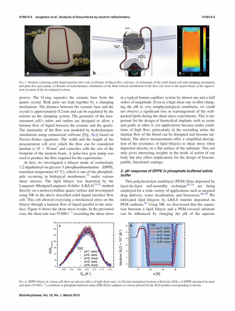
groove. The O-ring separates the ceramic base from the
quartz crystal. Both parts are kept together by a clamping
mechanism. The distance between the ceramic base and the
crystal is approximately 0.2 mm and can be regulated by the
tension on the clamping system. The geometry of the mea-
surement cell’s inlets and outlets are designed to allow a
laminar flow of liquid between the ceramic and the quartz.
The laminarity of the flow was modeled by hydrodynamic
simulations using commercial software [Fig. 3(c)] based on
Navier–Stokes equations. The width and the length of the
measurement cell over which the flow can be considered
laminar is 35 � 50 mm2 and coincides with the size of the
footprint of the neutron beam. A pulse-less gear pump was
used to produce the flow required for the experiments.
At first, we investigated a bilayer made of zwitterionic
1,2-dipalmitoyl-sn-glycero-3-phosphoethanolamine (DPPE,
transition temperature 63 �C), which is one of the phospholi-
pids occurring in biological membranes,73 under various
shear stresses. The lipid bilayer was deposited by the
Langmuir–Blodgett/Langmuir–Sch€afer (LB/LS)74,75 method
directly on a monocrystalline quartz surface and investigated
using NR in the above described solid–liquid interface flow
cell. This cell allowed exercising a mechanical stress on the
bilayer through a laminar flow of liquid parallel to the inter-
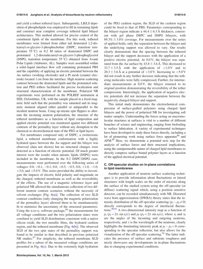
face. Figure 4 shows the shear stress results. In the presented
case, the shear rate was 55 000 s�1 exceeding the shear stress
in a typical human capillary system by almost one and a half
orders of magnitude. Even at a high shear rate or after chang-
ing the pH to very nonphysiological conditions, we could
not observe a significant loss or rearrangement of the well-
packed lipids during the shear stress experiments. This is im-
portant for the design of biomedical implants such as stents
and grafts or other in situ applications because under condi-
tions of high flow, particularly in the ascending aorta, the
laminar flow of the blood can be disrupted and become tur-
bulent. The above measurements offer a simplified descrip-
tion of the resistance of lipid bilayers to shear stress when
deposited directly on a flat surface of the substrate. This not
only gives interesting insights in the mode of action of our
body but also offers implications for the design of biocom-
patible, functional coatings.
2. pH response of DPPE in phosphate buffered salinebuffer
Thin polyelectrolyte multilayer (PEM) films deposited by
layer-by-layer self-assembly technique76–79 are being
employed for a wide variety of applications such as targeted
drug delivery, water desalination, and biosensors.80–82 We
fabricated lipid bilayers by LB/LS transfer deposited on
PEM cushions.83 Using NR, we discovered that the separa-
tion between a lipid bilayer and a PEM-covered substrate
can be influenced by changing the pH of the aqueous
FIG. 3. Neutron scattering solid–liquid interface flow cell. (a) Picture of Macor flow cell base. (b) Schematic of the solid–liquid cell with clamping mechanism
and pulse-less gear pump. (c) Results of hydrodynamic simulations of the fluid velocity distribution in the flow cell close to the quartz block, at the approxi-
mate location of the investigated systems.
FIG. 4. DPPE bilayer at various pH show no adverse effect of high shear rates. (a) Fresnel normalized neutron reflectivity (R/RF) of DPPE measured in static
and shear (55 000 s�1) conditions in phosphate buffered saline (PBS-D2O) subphase at various pH and (b) the SLD profiles corresponding to the fits.
019014-4 Junghans et al.: Analysis of biosurfaces by neutron reflectometry 019014-4
Biointerphases, Vol. 10, No. 1, March 2015

environment. Decreasing the pH of the surrounding pure
water subphase, and therefore changing the electrical proper-
ties of the system, significantly increases the distance
between the lipid bilayer and the polyelectrolyte cushion due
to electrostatic repulsion. Returning to the original pH recon-
stitutes the initial separation, with little or no change in the
lipid bilayer structure. Hence, we are able to create a free
floating lipid bilayer that is easy to attach and release to a
substrate in a reversible manner. We hypothesize that this
behavior is due to electrostatic repulsion between the lipid
head groups and the polymer that increases when the pH is
decreased.67
Below we present a case where the pure water subphase
is replaced by PBS and a bilayer composed of 1,2-dipalmi-
toyl-sn-glycero-3-phosphocholine (DPPC, transition temper-
ature 41 �C) molecules is used. Unlike the case of the pure
water subphase, the use of PBS partially screens the charges
on the lipids and polymers. As can be seen in Fig. 5, where
the pH is decreased to pH 2, and the charge of the PEM
changes from a net positive charge (pH 7) to a neutral PEM
charge (pH 2), the bilayer interactions with the polymeric
cushion weakens and the membrane starts to undulate.
However, due to diminished electrostatic repulsion, no com-
plete bilayer detachment was observed. The ability of the
bilayer to decouple from the supporting layer, to undulate,
and to induce curvature can be very beneficial for incorporat-
ing proteins or channels in the system, as the local curvature
parameter is known to be crucial for interactions between a
lipid bilayer and membrane proteins.84 Moreover, such a
free-floating bilayer, even if only partially removed from the
underlying substrate, would be a more accurate platform for
in situ studies of a variety of phenomena. Examples include
flip-flop of membrane components, incorporation of trans-
membrane channels or interactions between a lipid bilayer
and soluble proteins, interaction between membrane recep-
tors and proteins, etc. Because most studied supported lipid
bilayers tend to restrict the curvatures and motion of model
membranes, this could result in decreased functionalities and
lead to denaturation of the transmembrane proteins embed-
ded in their structure. It needs to be stated that most
biological systems only function well within a narrow pH
range and changing it drastically might affect their viability.
The authors are currently exploring the possibility of induc-
ing the same repulsion by applying a small electric field,
which would circumvent the drawbacks of an acidic
environment.
B. Investigation of tethered lipid membranesunder applied electric fields
In this example, polarized NR and a magnetic reference
layer were employed to study structural changes in tethered
lipid membranes induced by an electric potential applied
across the bilayer. A tethered lipid membrane typically con-
sists of hydrophilic polymers with functionalized ends such
that one end binds to a substrate and the other incorporates
within a lipid bilayer thereby “tethering” it to the surface.85
Advantages of such architectures include (1) a planar mem-
brane geometry enabling the application of surface sensitive
techniques and use in biomimetic devices and (2) providing
a highly hydrated cushion between membrane and support
enabling fluctuations and facilitating the incorporation of
transmembrane proteins into the bilayer.
Here, tethered lipid membranes were prepared on large
surface area supports suitable for NR experiments using
bifunctionalized polyethylene glycol (PEG) tethering mole-
cules and primarily tail deuterated phospholipids. Sputter de-
posited Au thin films were coated on single crystal Si blocks
(5.0 � 8.0 � 1.5 cm) to provide an electrode surface. A soft
magnetic alloy, permalloy (Ni:Fe 4:1), was used as a binding
layer between the gold and silicon. The permalloy also acted
as a magnetic contrast layer, with a different magnetic SLD
component depending on the spin direction of the atoms in
the layer relative to the spin eigenstate of the incoming neu-
trons. Bifunctionalized PEG molecules (n¼ 114) with thiol
and 1,2-distearoyl-sn-glycero-3-phosphoethanolamine (tran-
sition temperature 74 �C) terminal ends were obtained from
Nanocs, Inc. (New York, NY) and used as tethering mole-
cules on these solid supports. Dissolved in ethanol at a con-
centration of 1 mg/ml, the tethering molecule solution was
incubated with the substrate for 5 h to form Au-thiol bonds
FIG. 5. Lipid bilayers deposited on PEM exhibited increased undulation in acidic environment. (a) Fresnel-divided NR measurements and corresponding SLD
profiles, obtained from the fits, (b) of DPPC bilayer deposited on PEM ([PEI-PSS]3-PEI). Measurements were performed in PBS at pH 7.0, pH 2.0, and pH
again restored to 7.0. The error bars denote the standard deviation (SD) for each measurement.
019014-5 Junghans et al.: Analysis of biosurfaces by neutron reflectometry 019014-5
Biointerphases, Vol. 10, No. 1, March 2015
and yield a robust tethered layer. Subsequently, LB/LS depo-
sition of phospholipids was employed to fill in remaining lipids
and construct near complete coverage tethered lipid bilayer
architectures. This method allowed for precise control of the
constituent lipids of the membrane. For this work, tethered
membranes were formed from either tail deuterated 1,2-dis-
tearoyl-sn-glycero-3-phosphocholine (DSPC, transition tem-
perature 55 �C) or 8:2 M ratios of deuterated DSPC and
protonated 1,2-dioctadecanoyl-sn-glycero-3-phosphoglycerol
(DSPG, transition temperature 55 �C) obtained from Avanti
Polar Lipids (Alabaster, AL). Samples were assembled within
a solid–liquid interface flow cell containing protonated PBS
and the capability of applying an electric potential between the
Au surface (working electrode) and a Pt mesh (counter elec-
trode) located 1 cm from the interface. High neutron scattering
contrast between the deuterated lipids and the protonated solu-
tion and PEG tethers facilitated the precise localization and
structural characterization of the membrane. Polarized NR
experiments were performed on the POLREF instrument at
ISIS, Oxfordshire, UK, with the sample placed in a fixed mag-
netic field such that the permalloy was saturated and its mag-
netic moment aligned either parallel or antiparallel to the
incident neutron beam. Using up-stream spin flippers to alter-
nate the incoming neutron polarization, the structure of the
tethered membranes as a function of lipid composition and
applied electric potential was monitored against two different
contrasts of the permalloy layer without any disruption to the
chemical or electrochemical state of the PEG or lipid layers.
For membranes composed only of DSPC, a zwitterionic
lipid, a tethered membrane conformation with a highly
hydrated space between the Au support and the bilayer was
observed (data not shown) but no structural changes were
detected as a function of electric potential. Of much greater
interest were the results when anionic lipids (DSPG) were
included in the membrane. In the 8:2 DSPC:DSPG case,
measurements were performed over the following series of
voltages: 0.0, þ0.1, �0.1, 0.0, þ0.5, �0.5, 0.0, þ1.0, �1.0,
þ3.0, and þ5.0 V. This series provided the ability to investi-
gate the impacts of electric field polarity and magnitude on
the charged, tethered membrane as well as the reversibility
of the effects. The use of a magnetic reference layer and
polarized NR allowed the simultaneous collection of two dif-
ferent neutron contrast scenarios without the necessity of
solvent exchanges [Fig. 6(a)]. The collection of two SLD
contrast conditions (only changing the magnetic polarization
of the permalloy layer) allowed them to be simultaneously
fit to minimize the uncertainty of the structural parameters.
Using the RASCAL software package,86 the measurements for
all voltage conditions and the two polarization states were
corefined to yield SLD distributions consistent with a native
silicon oxide, the two metallic films, the hydrated cushion
region, and the tethered membrane [Fig. 6(b)]. The observed
SLD of the two spin states of the permalloy support was
found to be similar to that described in previous polarized
NR studies.87 For simplicity, the tethered membrane SLD
profiles for a subset of the measured voltage conditions are
presented in Fig. 6(c). Due to the extremely high hydration
of the PEG cushion region, the SLD of the cushion region
could be fixed to that of PBS. Parameters corresponding to
the bilayer region indicate a 44.4 6 6.3 A thickness, consist-
ent with gel phase DSPC and DSPG bilayers, with
89.3% 6 3.8% coverage. For measurements over the series
of applied fields, only the separation between the bilayer and
the underlying support was allowed to vary. Our results
clearly demonstrate that the spacing between the tethered
bilayer and the support decreases with the application of a
positive electric potential. At 0.0 V, the bilayer was sepa-
rated from the Au surface by 43.8 6 3.6 A. This decreased to
33.7 6 3.5 A with the application of þ1.0 V and to
20.3 6 3.6 A at a potential of þ3.0 V. Increasing to þ5.0 V
did not result in any further decrease indicating that the teth-
ering molecules were fully compressed. Further, for interme-
diate measurements at 0.0 V, the bilayer returned to its
original position demonstrating the reversibility of the tether
compression. Interestingly, the application of negative elec-
tric potentials did not increase the separation between the
negatively charged bilayer and support.
This initial study demonstrates the electrochemical com-
pression of surface-grafted polymers using charged lipid
bilayers and the power of polarized NR for investigating soft
matter samples. Understanding the forces acting on macromo-
lecular structures at surfaces is vital to a number of different
branches of science and engineering, ranging from biofouling
to surface lubrication. A variety of experimental techniques
have been developed to study these forces directly, including a
lot of pioneering work using surface force apparatus88 and
AFM.89 Here, we demonstrate a potential next step for the
analysis of surface forces and their structural implications,
using the semipermeable nature of charged lipid membranes to
directly compress surface bound polymer layers as a function
of the applied electrical potential.
C. Off-specular studies on in-plane correlationsin lipid membranes
Another application of neutron surface scattering techni-
ques is to provide information about fluctuations or lateral
structures with length scales on the order of microns along
the surface of the studied system using the off-specular (or
diffuse) scattering signal which, using a position sensitive
detector, can be recorded simultaneously with NR. Distorted
wave born approximation (DWBA) theory states that the in-
tensity distribution of the off-specular scattering (pi� pf 6¼ 0)
directly corresponds to the degree of interfacial fluctua-
tions.90,91 A two-dimensional intensity map as a function of
pi (pi¼ 2p sin ai/k) and pf (pf¼ 2p sin af/k), where ai and af
are the angles of the incoming and outgoing neutrons,
respectively, and k is the wavelength of the neutrons, clearly
highlights the dominating intensity peak at pi� pf¼ 0 corre-
sponding to the specular reflection, but also allows for the
visualization of the off-specular scattering.92 The latter indi-
cates the presence of surface and substrate roughness and
nicely showcases any developments in in-plane fluctuations
due to changing experimental conditions.
019014-6 Junghans et al.: Analysis of biosurfaces by neutron reflectometry 019014-6
Biointerphases, Vol. 10, No. 1, March 2015

As one example, we studied poly(N-isopropylacrylamide)
(pNIPAAm)-supported single lipid bilayers,90 which can
serve as models of cell membranes. By permitting the struc-
tural freedom of the membrane, they more accurately repro-
duce cellular membrane morphology compared to directly
supported bilayers. Moreover, they also allow for the incor-
poration of membrane-bound proteins while potentially min-
imizing their denaturation.
By modeling the off-specular scattering, we were able to
quantify in-plane height-height correlations of interfacial
fluctuations of a DPPC bilayer supported on pNIPAAm as a
function of temperature. As the temperature decreased from
37 to 25 �C, the polymer swelled and the supported lipid
membrane deviated from its initially nearly planar structure.
Further analysis of the data revealed that correlation lengths
characteristic of capillary waves changed from 30 lm at
37 �C to 11 lm at 25 �C, while the membrane bending rigid-
ity remains roughly constant in this temperature range.
Analysis of the off-specular scattering can provide a
powerful tool for understanding the complex behavior of cel-
lular membranes, protein transport and docking, lipid segre-
gation into ordered domains, and modification of bulk
membrane elastic properties due to membrane constituents
and external stimuli. By successfully mimicking the com-
plexity of cellular membrane morphology, the presented
polymer-membrane system opens the door for numerous
other investigative opportunities in the future, e.g., how bio-
logical agents such as toxins, viruses, and other pathogens
affect in- and out-of-plane membrane structures.
D. Study of adhesion of living cells to the solidsubstrate
In a significant advancement toward employing neutron
reflectometry to measure complex biological samples, we
studied live cells adhered to a quartz substrate.47 Neutrons
are the perfect probe for the investigation of living tissue
because they offer unparalleled resolution on the 1–1000 A
level where even the best microscopy techniques fail.
Moreover, NR provides global information about the cellular
monolayer averaged over the footprint size. Lastly, neutrons
are ideal for working with living systems such as cells
because, unlike x-rays, they have a nonperturbative nature.
In a series of preliminary experiments, we showed that NR is
capable of probing the composition and thickness of the
interfacial area between the lipid plasma membrane and the
rigid substrate.
Measurements were conducted on live cell monolayers
grown on a flat surface of monocrystalline quartz. Cell
monolayers were perfused with culture medium at controlled
conditions to reach a laminar shear stress level of 1.5 Pa or
left static, as previously described.47
In all experimental data presented below, D2O based buf-
fers were used as the subphase. From our experience, we
know that the cells can survive up to 6 h in such conditions
without visible changes in the cells’ viability. Using D2O
based subphases was necessary to obtain sufficient scattering
contrast. The main source of such contrast is due to the neu-
tron scattering differences between the hydrogenated lipid
membrane (low SLD) facing the quartz substrate and the
D2O environment (high SLDs).
In a series of experiments, we observed a clear signal at-
tributable to live HK-03 mouse fibroblast cells, as confirmed
by comparison with samples of pure medium. We believe
these experiments represent the first successful visualization
and quantization of the interface between live cells and a
substrate using NR. Due to the pronounced neutron scatter-
ing from the hydrogenated alkyl tails of the phospholipid
bilayers (which are not capable of an isotopic exchange), we
were able to clearly distinguish the lipid membrane of the
FIG. 6. Polarized NR measurements of the structure of a tethered bilayer composed of 8:2 DSPC:DSPG exposed to applied electric fields. Panel (a) displays
representative NR data and fits for the 0.0 V (top) and þ5.0 V (bottom) cases. Spin up data (þ) are shown in blue and spin down (–) in red. Data are shifted
vertically for clarity. The full SLD profiles of the 0.0 V case in panel (b) show the different magnetic scattering lengths of the two spin states in the permalloy
magnetic contrast layer. Zooming in on the tethered bilayer region [panel (c)] demonstrates that the application of positive electric potentials compresses the
tethering polymer and decreases the spacing between the substrate and bilayer.
019014-7 Junghans et al.: Analysis of biosurfaces by neutron reflectometry 019014-7
Biointerphases, Vol. 10, No. 1, March 2015

live cells from the deuterated environment. With an approx.
80 A thickness (Fig. 7), the membrane region was measured
to be twice as thick as the length of two hydrophobic tails,
indicating that the adhering cells were not organized as a ho-
mogeneous plane uniformly spaced on the quartz substrate.
This suggests that the membrane is either undulating, nonho-
mogeneously distributed due to the surface topography of
the underlying media and adherence proteins, or that the
scattered neutron, averaged over several neighboring cells,
varied in their distance from the solid substrate.
Those very simplistic experiments visualizing the adher-
ence layer of mouse fibroblasts to a quartz substrate show
how powerful NR is, compared to other surface techniques
such as AFM and various microscopies, for unraveling the
information about buried subphases. In the last paragraph,
we would like to go one step further and present in detail a
much more complex and medically relevant system.
E. Investigation of off-specular scatteringof endothelial adhesion at varying temperaturesand fluid shear stress
As mentioned above, mechanical forces play an intricate
role in biological systems from the tissue and organ level
down to individual cells and proteins.93–95 Certain tissues
are inherently reliant on solid mechanical and fluid mechani-
cal stresses for normal healthy function. The foremost
among these is the vascular system encompassing all blood
vessels.95 The innermost layer of blood vessels, which
makes contact with blood, is composed of a single mono-
layer of endothelial cells. Endothelial cells are the main gate-
keepers of a key function of blood vessels known as vascular
permeability. Blood vessels must strike a delicate balance in
allowing fluid and cellular flux across their wall. In healthy
people, vascular permeability (degree of transblood vessel
flux) is tightly controlled, however in certain diseases it
becomes deranged with devastating consequences. Among
these diseases is acute respiratory distress syndrome
(ARDS). In ARDS, the blood vessels located in our lungs
become extremely leaky leading to pulmonary edema, i.e.,
accumulation of fluid in the lungs that severely curtails the
lung’s oxygen diffusion capacity. It is believed that mechani-
cal stress in the form of both cyclic elastic stress and fluid
shear stress from blood flow are key components in regulat-
ing endothelial permeability and play roles in diseases such
as ARDS.96,97
Adhesion to the underlying extracellular matrix (ECM) is
a crucial component of how endothelial cells sense and
transmit mechanical signals. NR was used for the first time
to measure the nanoscopic structure of the adhesion layer of
confluent endothelial cells.46 As in the case discussed above,
neutron scattering is probing only the interfacial space
between the lipid plasma membrane and the rigid substrate.
Endothelial cells generate their own extracellular matrix (ba-
sal lamina) making it possible to grow monolayers on inert
surfaces like quartz without prior exogenous deposition of
protein layers.98,99 We grew confluent monolayers of human
pulmonary arterial endothelial cells on single quartz blocks.
They were then subjected to NR under shear stress of
s¼ 1.5 Pa (equivalent to arterial conditions).
We observed that the basal lamina thickness l is highly
sensitive to changes in temperature or shear stress.
Under static conditions [Figs. 8(b) and 8(c)], we observed
a near tripling of l as the temperature was raised from 25 to
37 �C (lst25 �C� 200 A, lst
37 �C� 600 A). Interestingly, shear
stress has the opposite effect at the two temperatures [Figs.
8(d) and 8(e)], causing cell separation at 25 �C while induc-
ing movement closer to the substrate at physiologic tempera-
ture (lss25 �C� 700 A, lss
37 �C� 200 A).
We also analyzed the off-specular scattering since the
scattered neutron intensity as a function of the in-plane com-
ponents of the momentum transfer vector provides valuable
information about fluctuations along the surface of the cell
monolayer. Figure 9 shows the off-specular neutron reflec-
tivity data at 25 �C. The high intensity peak at pi� pf¼ 0
corresponds to the specular reflection. DWBA states that the
intensity distribution of the off-specular scattering
(pi� pf 6¼ 0) directly corresponds to the degree of interfacial
FIG. 7. Schematic of solid–liquid interface flow cell used for all live cell experiments þ NR profiles and corresponding SLD distributions of mouse fibroblasts
(a). Schematic representation of the neutron measurement cell used. Confluent layer of cells is grown on the quartz substrate that is inverted into the measure-
ment cell. A neutron beam enters through the quartz at a glancing angle and scatters from the adhesion layer and cell membrane interfaces. Fresnel divided
NR profiles (b) and corresponding SLD profiles (c) for high (black) and low (gray) cell surface densities. NR data are shown by closed squares, and error bars
indicate 1 SD. The lower surface cell density is evident from the decreased scattering intensity (b) and the increased SLD in the membrane region
(360–440 A) and interior of the cell (440–600 A) (b).
019014-8 Junghans et al.: Analysis of biosurfaces by neutron reflectometry 019014-8
Biointerphases, Vol. 10, No. 1, March 2015

FIG. 8. Differences in response to shear flow of healthy endothelial cells at ambient and physiological temperatures. Left (a) Fresnel-divided NR measurements
(circles/squares with error bars) and corresponding best-fit models (solid lines) at the conditions studied. Black (open circles): 25� (static), black (closed
circles): 25� (shear), gray (open squares): 37� (static), gray (closed squares): 37� (shear). The NR spectra are offset along y-axis for clarity. Right [(b)–(e)]:
SLD profiles obtained by fitting the data sets using a 3-box model (extracellular matrix—cell membrane—partial cell interior).
FIG. 9. Off-specular data depicts changes in surface roughness at 25 �C: Data is shown as two-dimensional intensity maps as a function of pi and pf. (a)
pi¼ 2p sin ai/k and pf¼ 2p sin af/k are the components perpendicular to the sample surface of the incoming and outgoing neutron wave vectors, respectively
(Ref. 92). The dominating intensity peak at pi� pf¼ 0 corresponds to the specular reflection. The off-specular is visible to the right from the specular line
along picture’s diagonal indicating the presence of surface and substrate roughness. (b) Comparison of the intensity distribution along the peaks vs pi � pf of
endothelial cells under static (black) and shear (red) conditions show decrease in scattering under shear stress. (c) and (d) show experimental two-
dimensional intensity maps of endothelial cells at 37 �C at static and shear conditions, respectively. The extension of the off-specular signal (right top corner)
in (c) compared to (d) indicates more in-plane fluctuations at static conditions.
019014-9 Junghans et al.: Analysis of biosurfaces by neutron reflectometry 019014-9
Biointerphases, Vol. 10, No. 1, March 2015
fluctuations.90 Our data show that the off-specular peak
enlarges with increasing temperature under static conditions
(data for 37 �C not shown) while it substantially decreases
with shear flow for both temperatures. Temperature, as
expected, increases interfacial thermal fluctuations. Shear
stress acts in a reverse manner by placing the cells (and
therefore their lipid membranes) under tension, thereby sup-
pressing fluctuations and decreasing the off-specular
intensity.
From individual cells and proteins up to complete tissue
and organs, mechanical forces play an intricate role in bio-
logical systems.93–95 Due to the limitation of most techni-
ques, the existing work in mechanobiology has focused on
cells, single molecules, and protein complexes, leaving the
collective behavior of cell monolayers less well explored.
The strength of neutron reflectometry is its nonperturba-
tive nature, the ability to probe large surface areas of buried
interfaces with nanometer resolution, and the possibility of
scattering contrast manipulation via the isotopic substitu-
tions. That allows us to probe details of the cell–substrate
interface that are not accessible with any other standard tech-
niques. Such capabilities can also be employed to obtain bet-
ter insight into the mechanisms of cell adhesion and
cell–surface properties of clinically relevant systems. For
example, the above results demonstrate that neutron reflec-
tometry experiments can provide valuable insight into com-
plex biomedical systems, such as living human cells under
fluid mechanical shear stress. Specifically, the above studies
could show the major redistribution of proteins involved in
cell–cell and cell–substrate adhesion shear stress is causing,
even on relatively short biological time scales (4 h) relevant
to the time scale of our neutron experiments. The localiza-
tion of adhesion proteins is postulated to promote linkage
between the actin skeleton and extracellular matrix, forming
enhanced adhesion zones.96 Shear stress at 37 �C caused
localization of specific proteins, which may alter the adhe-
sion potential in favor of overall increased adhesion as meas-
ured with neutrons. This biological response competes
against the purely physical effects of shear flow observed at
25 �C and the increased repulsive potential of the basal lam-
ina at higher temperatures. Those findings may lead to
advances in the treatment of atherosclerosis and other disor-
ders associated with the cardiovascular system, but also
open the door to a complete new way of investigating and
understanding living tissue in-situ.In this spirit, we have started to expand those studies to
glioblastoma (GBM),45 one of the most common and aggres-
sive type of brain tumors.100 Under static conditions, we
have observed profound differences in noninvasive U251
cells, compared to the invasive CNS1 and GL261 cells.
Namely, the human U251 cells showed a much thicker gly-
cocalyx or “adhesion” layer than the cells derived from rat
and mouse. We hypothesized that U251 cells may have a
higher production of hyaluronic acid (HA) and associated
proteoglycans101 than the rodent GBM cells. This could con-
tribute both to the nonfibroblastic morphology and limited
invasive ability since a thick HA coating can promote cell
adhesion, but limit GBM invasion in vivo if not degraded by
tumor-produced hyaluronidases.102 In summary, the pre-
sented results reveal differences in the thickness and compo-
sition of adhesion layers of different GBM cells in the static
conditions as well as changes to this layer when the cells
were subjected to shear stress. These differences may be spe-
cifically associated with mechanisms of brain tumor inva-
sion. Further studies of these cells by neutron reflectometry
under a variety of conditions (surface coatings, flow, and
drug treatments) will allow to determine conditions trigger-
ing changes in the composition, density, and thickness of the
biomaterial at the cell surface (in our case, the ECM layer).
This, in turn, can help to identify changes that correlate with
increased or decreased tumor invasiveness. Pursuit of those
studies can have significant medical impact for the develop-
ment of targeted anti-invasive therapies for GBM. Also, by
revealing differences that may be specifically associated
with mechanisms of brain tumor invasion, NR shows the
power it holds for understanding biomedical questions.
IV. CONCLUSION AND OUTLOOK
The discussed experiments illustrate the potential of NR
in addressing the structural properties of soft materials from
simple supported lipid membranes in static and dynamic
conditions to the adherence of complex endothelial cells
under liquid shear stress.
We hope that an inclined reader will utilize some of these
concepts and experimental approaches to conduct new stud-
ies that will provide better understanding of biomedical
questions, suggest innovative treatment, or advance the de-
velopment of novel biomimetic materials.
ACKNOWLEDGMENTS
This work benefited from the use of the Lujan Neutron
Scattering Center at Los Alamos Neutron Science Center
funded by the DOE Office of Basic Energy Sciences and Los
Alamos National Laboratory under DOE Contract No. DE-
AC52-06NA25396. The authors thank the ISIS Neutron
Source (STFC, UK) for access to neutron facilities on
POLREF through Proposal No. RB1120119.
1J. Daillant and A. Gibaud, Lect. Notes Phys. 770, 133 (2009).2G. Fragneto-Cusani, J. Phys.: Condens. Matter 13, 4973 (2001).3J. Penfold, Curr. Opin. Colloid Interface Sci. 7, 139 (2002).4A. S. Curtis, J. Cell Biol. 20, 199 (1964).5D. Gingell and I. Todd, Biophys. J. 26, 507 (1979).6C. S. Izzard and L. R. Lochner, J. Cell Sci. 21, 129 (1976).7M. Schindl, E. Wallraff, B. Deubzer, W. Witke, G. Gerisch, and E.
Sackmann, Biophys. J. 68, 1177 (1995).8H. Verschueren, J. Cell Sci. 75, 279 (1985).9D. Braun and P. Fromherz, Appl. Phys. A 65, 341 (1997).
10D. Braun and P. Fromherz, Phys. Rev. Lett. 81, 5241 (1998).11R. Parthasarathy and J. T. Groves, Cell Biochem. Biophys. 41, 391
(2004).12D. Axelrod, J. Cell Biol. 89, 141 (1981).13J. S. Burmeister, L. A. Olivier, W. M. Reichert, and G. A. Truskey,
Biomaterials 19, 307 (1998).14J. S. Burmeister, G. A. Truskey, and W. M. Reichert, J Microsc. 173, 39
(1994).15D. Gingell, I. Todd, and J. Bailey, J. Cell Biol. 100, 1334 (1985).
019014-10 Junghans et al.: Analysis of biosurfaces by neutron reflectometry 019014-10
Biointerphases, Vol. 10, No. 1, March 2015

16D. K. Hoover, E.-J. Lee, and M. N. Yousaf, Langmuir: ACS J. Surf.
Colloids 25, 2563 (2009).17K. Giebel, C. Bechinger, S. Herminghaus, M. Riedel, P. Leiderer, U.
Weiland, and M. Bastmeyer, Biophys. J. 76, 509 (1999).18A. W. Peterson, M. Halter, A. Tona, K. Bhadriraju, and A. L. Plant, BMC
Cell Biol. 10, 16 (2009).19A. M. Butt, H. C. Jones, and N. J. Abbott, J. Physiol. 429, 47 (1990).20M. J. Rutten, R. L. Hoover, and M. J. Karnovsky, Brain Res. 425, 301
(1987).21P. A. Vogel, S. T. Halpin, R. S. Martin, and D. M. Spence, Anal. Chem.
83, 4296 (2011).22J. Wegener, M. Sieber, and H.-J. Galla, J. Biochem. Biophys. Methods
32, 151 (1996).23D. P. Allison, N. P. Mortensen, C. J. Sullivan, and M. J. Doktycz, Wiley
Interdiscip. Rev.: Nanomed. Nanobiotechnol. 2, 618 (2010).24R. Lal and S. A. John, Am. J. Physiol.: Cell Physiol. 35, 1 (1994).25D. J. M€uller and Y. F. Dufrene, Trends Cell Biol. 21, 461 (2011).26A. Vinckier and G. Semenza, FEBS Lett. 430, 12 (1998).27K. Cheung, S. Gawad, and P. Renaud, Cytometry, Part A 65A, 124 (2005).28J. E. da Silva, J. P. M. de S�a, and J. Jossinet, Med. Biol. Eng. Comput.
38, 26 (2000).29D. A. Dean, T. Ramanathan, D. Machado, and R. Sundararajan,
J. Electrost. 66, 165 (2008).30S. Krueger, Curr. Opin. Colloid Interface Sci. 6, 111 (2001).31J. Majewski, T. L. Kuhl, M. C. Gerstenberg, J. N. Israelachvili, and G. S.
Smith, J. Phys. Chem. B 101, 3122 (1997).32I. Burgess, M. Li, S. L. Horswell, G. Szymanski, J. Lipkowski, J.
Majewski, and S. Satija, Biophys. J. 86, 1763 (2004).33G. Fragneto, F. Graner, T. Charitat, P. Dubos, and E. Bellet-Amalric,
Langmuir 16, 4581 (2000).34B. W. Koenig, S. Krueger, W. J. Orts, C. F. Majkrzak, N. F. Berk, J. V.
Silverton, and K. Gawrisch, Langmuir 12, 1343 (1996).35S. Krueger, J. F. Ankner, S. K. Satija, C. F. Majkrzak, D. Gurley, and M.
Colombini, Langmuir 11, 3218 (1995).36D. A. Doshi, A. M. Dattelbaum, E. B. Watkins, C. J. Brinker, B. I.
Swanson, A. P. Shreve, A. N. Parikh, and J. Majewski, Langmuir: ACS J.
Surf. Colloids 21, 2865 (2005).37A. Junghans and I. K€oper, Langmuir 26, 11035 (2010).38H. L. Smith, M. S. Jablin, A. Vidyasagar, J. Saiz, E. Watkins, R.
Toomey, A. J. Hurd, and J. Majewski, Phys. Rev. Lett. 102, 228102
(2009).39J. Fitter, T. Gutberlet, and J. Katsaras, Neutron Scattering in Biology:
Techniques and Applications (Springer-Verlag, Berlin Heidelberg, 2006).40G. Pabst, N. Kucerka, M. P. Nieh, M. C. Rheinst€adter, and J. Katsaras,
Chem. Phys. Lipids 163, 460 (2010).41J. Penfold and R. K. Thomas, Curr. Opin. Colloid Interface Sci. 19, 198
(2014).42H. P. Wacklin, Curr. Opin. Colloid Interface Sci. 15, 445 (2010).43C. J. Garvey, R. B. Knott, E. Drabarek, and P. W. Kuchel, Eur. Biophys.
J. 33, 589 (2004).44A. M. Stadler, C. J. Garvey, A. Bocahut, S. Sacquin-Mora, I. Digel, G. J.
Schneider, F. Natali, G. M. Artmann, and G. Zaccai, J. R. Soc. Interface
9, 2845 (2012).45A. Junghans, M. J. Waltman, H. L. Smith, L. Pocivavsek, N. Zebda, K.
Birukov, M. Viapiano, and J. Majewski, Mod. Phys. Lett. B 28, 1430015
(2014).46L. Pocivavsek, A. Junghans, N. Zebda, K. Birukov, and J. Majewski, Am.
J. Physiol.: Lung Cell. Mol. Physiol. 306, L1 (2014).47H. L. Smith, J. Hickey, M. S. Jablin, A. Trujillo, J. P. Freyer, and J.
Majewski, Biophys. J. 98, 793 (2010).48P. S. Swain and D. Andelman, Langmuir 15, 8902 (1999).49J. Raedler, H. Strey, and E. Sackmann, Langmuir 11, 4539 (1995).50E. T. Castellana and P. S. Cremer, Surf. Sci. Rep. 61, 429 (2006).51E. Sackmann, Science 271, 43 (1996).52M. Dubey, M. S. Jablin, P. Wang, M. Mocko, and J. Majewski, Eur.
Phys. J. Plus 126, 110 (2011).53J. Als-Nielsen, Phys. A: Stat. Mech. Appl. 140, 376 (1986).54J. Penfold and R. K. Thomas, J. Phys.: Condens. Matter 2, 1369 (1990).55A. Nelson, J. Appl. Crystallogr. 39, 273 (2006).56H. L. Smith, M. C. Howland, A. W. Szmodis, Q. Li, L. L. Daemen, A. N.
Parikh, and J. Majewski, J. Am. Chem. Soc. 131, 3631 (2009).
57S. M. Baker, G. S. Smith, D. L. Anastassopoulos, C. Toprakcioglu, A. A.
Vradis, and D. G. Bucknall, Macromolecules 33, 1120 (2000).58J. Penfold, E. Staples, I. Tucker, and G. Fragnetto, Phys. B: Condens.
Matter 221, 325 (1996).59I. Burgess, M. Li, S. L. Horswell, G. Szymanski, J. Lipkowski, S. Satija,
and J. Majewski, Colloids Surf. B – Biointerfaces 40, 117 (2005).60S. Lecuyer, G. Fragneto, and T. Charitat, Eur. Phys. J. E: Soft Matter
Biol. Phys. 21, 153 (2006).61P. Mansky, J. DeRouchey, T. P. Russell, J. Mays, M. Pitsikalis, T.
Morkved, and H. Jaeger, Macromolecules 31, 4399 (1998).62W. J. Orts, J. H. van Zanten, W.-L. Wu, and S. K. Satija, Phys. Rev. Lett.
71, 867 (1993).63R. Steitz, V. Leiner, K. Tauer, V. Khrenov, and R. v. Klitzing, Appl.
Phys. A 74, s519 (2002).64J. Generosi, C. Castellano, D. Pozzi, A. C. Castellano, R. Felici, F.
Natali, and G. Fragneto, J. Appl. Phys. 96, 6839 (2004).65M. L€osche, J. Schmitt, G. Decher, W. G. Bouwman, and K. Kjaer,
Macromolecules 31, 8893 (1998).66S. W. An, R. K. Thomas, F. L. Baines, N. C. Billingham, S. P. Armes,
and J. Penfold, J. Phys. Chem. B 102, 5120 (1998).67S. Singh, A. Junghans, J. Tian, M. Dubey, S. Gnanakaran, J. Chlistunoff,
and J. Majewski, Soft Matter 9, 8938 (2013).68T. J. Su, J. R. Lu, R. K. Thomas, Z. F. Cui, and J. Penfold, Langmuir 14,
438 (1998).69T. G. Papaioannou and C. Stefanadis, Hell. J. Cardiol. 46, 9 (2005).70J. I. Prydal, P. Artal, H. Woon, and F. W. Campbell, Invest. Ophthalmol.
Visual Sci. 33, 2006 (1992).71P. E. King-Smith, B. A. Fink, N. Fogt, K. K. Nichols, R. M. Hill, and G.
S. Wilson, Invest. Ophthalmol. Visual Sci. 41, 3348 (2000).72A. Heryudono, R. J. Braun, T. A. Driscoll, K. L. Maki, L. P. Cook, and P.
E. King-Smith, Math. Med. Biol. 24, 347 (2007).73H. Lodish, A. Berk, S. L. Zipursky, P. Matsudaira, D. Baltimore, and J.
Darnell, Molecular Cell Biology, 4th ed. (W. H. Freeman, San Francisco,
2000).74I. Langmuir, J. Franklin Inst. 218, 143 (1934).75I. Langmuir and V. J. Schaefer, Chem. Rev. 24, 181 (1939).76R. K. Iler, J. Colloid Interface Sci. 21, 569 (1966).77G. Decher, Science 277, 1232 (1997).78J. Schmitt, T. Gruenewald, G. Decher, P. S. Pershan, K. Kjaer, and M.
Loesche, Macromolecules 26, 7058 (1993).79H. Ai, S. Jones, and Y. Lvov, Cell Biochem. Biophys. 39, 23 (2003).80X. Liu and M. L. Bruening, Chem. Mater. 16, 351 (2004).81S. Singh and M. McShane, Biosens. Bioelctron. 25, 1075 (2010).82E. W. Stein, S. Singh, and M. J. McShane, Anal. Chem. 80, 1408
(2008).83S. Singh, A. Junghans, M. J. Waltman, A. Nagy, R. Iyer, and J.
Majewski, Soft Matter 8, 11484 (2012).84H. T. McMahon and J. L. Gallop, Nature 438, 590 (2005).85E. B. Watkins, R. J. El-khouri, C. E. Miller, B. G. Seaby, J. Majewski, C.
M. Marques, and T. L. Kuhl, Langmuir 27, 13618 (2011).86A. Hughes, RasCAL (2014), see http://sourceforge.net/projects/rscl/.87S. A. Holt, A. P. Le Brun, C. F. Majkrzak, D. J. McGillivray, F. Heinrich,
M. L€osche, and J. H. Lakey, Soft Matter 5, 2576 (2009).88J. N. Israelachvili, Intermolecular and Surface Forces (Academic, New
York, 2011).89J. F. Joanny, Interface Sci. 11, 157 (2003).90M. S. Jablin, M. Zhernenkov, B. P. Toperverg, M. Dubey, H. L. Smith,
A. Vidyasagar, R. Toomey, A. J. Hurd, and J. Majewski, Phys. Rev. Lett.
106, 138101 (2011).91L. G€osta and W. L. Stephen, Phys. Scr. 53, 734 (1996).92V. Lauter-Pasyuk, H. J. Lauter, G. P. Gordeev, P. M€uller-
Buschbaum, B. P. Toperverg, M. Jernenkov, and W. Petry, Langmuir
19, 7783 (2003).93B. Alberts, D. Bray, J. Lewis, M. Ra, K. Roberts, and J. D. Watson,
Molecular Biology of the Cell (Garland Publishing, New York, 1994).94D. Boal, Mechanics of the Cell (Cambridge University, Cambridge,
2002).95Y.-T. Shiu, “Mechanical forces on cells,” in The Biomedical
Engineering Handbook: Tissue Engineering and Artificial Organs,
3rd ed. (CRC Press Taylor & Francis Group, LLC, Boca Raton, FL,
2006), pp. 33.31–33.18.
019014-11 Junghans et al.: Analysis of biosurfaces by neutron reflectometry 019014-11
Biointerphases, Vol. 10, No. 1, March 2015

96K. G. Birukov, A. A. Birukova, S. M. Dudek, A. D. Verin, M. T. Crow,
X. Zhan, N. DePaola, and J. G. N. Garcia, Am. J. Respir. Cell Mol. Biol.
26, 453 (2002).97A. A. Birukova, S. Chatchavalvanich, A. Rios, K. Kawkitinarong, J. G.
N. Garcia, and K. G. Birukov, Am. J. Pathol. 168, 1749 (2006).98K. G. Birukov, N. Leitinger, V. N. Bochkov, and J. G. N. Garcia,
Microvas. Res. 67, 18 (2004).99B. V. Howard, E. J. Macarak, D. Gunson, and N. A. Kefalides, Proc.
Natl. Acad. Sci. U. S. A. 73, 2361 (1976).
100D. N. Louis, Annu. Rev. Pathol.: Mech. Dis. 1, 97 (2006).101M. S. Viapiano and S. E. Lawler, CNS Cancer: Models, Prognostic Factors
and Targets, edited by E. Van Meir (Humana, NJ, 2009), pp. 1219–1252.102B. Enegd, J. A. King, S. Stylli, L. Paradiso, A. H. Kaye, and U. Novak,
Neurosurgery 50, 1311 (2002).103W. A. Hamilton, P. D. Butler, S. M. Baker, G. S. Smith, J. B. Hayter, L.
J. Magid, and R. Pynn, Phys. Rev. Lett. 72, 2219 (1994).104S. M. Baker, P. D. Butler, W. A. Hamilton, J. B. Hayter, and G. S. Smith,
Rev. Sci. Instrum. 65, 412 (1994).
019014-12 Junghans et al.: Analysis of biosurfaces by neutron reflectometry 019014-12
Biointerphases, Vol. 10, No. 1, March 2015