anal. bioanal. electrochem., vol. 9, no. 7, 2017, 874-887 .... 7-2017/2017, 9(7), 874-887.pdf ·...
TRANSCRIPT
Anal. Bioanal. Electrochem., Vol. 9, No. 7, 2017, 874-887
Full Paper
Simultaneous Determination of Dopamine, Ascorbic Acid and Uric Acid at Poly (cinnamic acid) Modified Carbon Paste Electrode: a Voltammetric Investigation Vipparla Prabhakara Rao, Yenugu Veera Manohara Reddy, Kalluri Gangadhar Reddy, Chougoni Madhuri, Duddukuru Saritha and Gajulapalle Madhavi*
Electrochemical Research Laboratory, Department of Chemistry, Sri Venkateswara University, Tirupathi-517502, Andhra Pradesh, India
*Corresponding Author, Tel.: 0877-2228303; Fax: +91-9440096500 E-Mail: [email protected]
Received: 1 December 2016 / Received in revised form: 17 August 2017 / Accepted: 24 September 2017 / Published online: 15 November 2017
Abstract- A Polymer film of cinnamic acid was formed on the surface of a bare carbon paste electrode (BCPE) by electropolymerisation of cinnamic acid in pH 7 Phosphate buffer solution (PBS) by using cyclic voltammetry (CV). This lead to the formation of polycinnamic acid modified carbon paste electrode (PCA/CPE). CV was used to study the electrochemical properties of the polymer film on the PCA/CPE. The modified electrode was used for the simultaneous electrochemical detection of dopamine (DA), ascorbic acid (AA) and uric acid (UA). The PCA/CPE showed a very good electrocatalytic activity for the oxidation of DA, AA and UA and exhibited an excellent ability to separate DA from AA by means of CV. The anodic peak potentials of DA and AA were separated by 0.259 V in pH 7 PBS containing 0.1 mM of DA and 2 mM of AA at the scan rate of 50 m V/S. The detection limit (LOD) and quantification limit (LOQ) of the present technique were found to be 3.52×10⁻⁶ M and 1.16×10⁻⁵ M respectively for DA. Compared to BCPE, the PCA/CPE exhibited the enhanced peak currents for DA, AA and UA. Also, the PCA/CPE showed good recoveries for DA in real sample analysis i.e. for both pharmaceutical formulations and blood serum. Hence, this modified electrode can be used for the selective determination of DA in the presence of higher concentrations of AA and UA in pharmaceuticals and medicine without any problem with greater sensitivity, selectivity and reproducibility.
Keywords- Cinnamic acid modified carbon paste electrode, Dopamine, Ascorbic acid, Uric acid, Cyclic voltammetry, Differential pulse voltammetry
Analytical & Bioanalytical Electrochemistry
2017 by CEE
www.abechem.com

Anal. Bioanal. Electrochem., Vol. 9, No. 7, 2017, 874-887 875
1. INTRODUCTION
Design, fabrication and application of novel electrochemical sensors to detect the biologically important neurotransmitters such as DA, AA and UA have received a great attention in recent years [1,2] because of their better performance. One among such electro chemical sensors is polymer modified carbon paste electrodes (MCPEs) prepared by electro polymerization using cyclic voltammetry (CV). These electrodes have better sensitivity, selectivity compared to bare carbon paste electrodes (BCPEs). It was also found that the polymer film has good stability, more active sites, strong adherence to the electrode surface and gives good reproducibility [3-5]. MCPEs were also found to show low residual currents over a wide range and have feeble chances of electrode fouling compared to BCPEs which otherwise could lead to poor sensitivity, selectivity and reproducibility [6- 8].
DA, AA and UA coexist in the extracellular fluids of mammalian brain. Dopamine (DA) is a very significant catechol amine functioning as a neurotransmitter in the central nervous system [9]. It is used as medicine for the treatment of drug addiction, Parkinson [10] and Alzheimer’s diseases [11]. Ascorbic acid (AA) (Vitamin-C) prevents scurvy and takes part in several biological reactions. It is believed to be primary antioxidant in human blood plasma. It is significant as it is used in the treatment of common cold, mental illness, AIDS and cancer [12]. In humans and higher primates uric acid (UA) is the final oxidation product of purine metabolism [13] and is excreted in urine. Uric acid like ascorbic acid is a strong reducing agent and potent antioxidant. In humans over half of the antioxidant capacity of blood plasma comes from uric acid. High blood concentrations of uric acid called Hyperuricemia can lead to Gout, a kind of arthritis [14]. The substance is also associated with other medical conditions including diabetes and formation of ammonium acid urate called kidney stones. So, it is essential to develop effective techniques to determine DA, AA and UA in the field of biochemistry and medicine. A major problem in detection however is the interference of AA and UA in the determination of DA as AA and UA are present at much higher concentrations than DA in Physiological fluids [15] and also their oxidation potentials are very close to that of DA. It is known that the direct electro oxidation of DA, AA and UA at BCPE is irreversible and requires higher potentials. Moreover, DA and AA are oxidized at nearly the same potentials at BCPES [16,17] which leads to the overlapping of voltammetric responses. Another problem is the fouling of the BCPE surface by the adsorption of oxidation products of DA and AA which result in rather poor selectivity and reproducibility. Thus, it is difficult to detect DA in the presence of higher levels of AA and UA in real biological samples using BCPEs. Hence, it is essential to construct suitable electrodes and to establish a sensitive, selective and promising detection method for the detection and determination of DA in biological samples.
Various types of MCPEs were tried to detect DA in past years. Some of them used expensive substrates such as gold [18] or glassy carbon electrodes [19], which involve complicated and time consuming methods of preparation. Carbon ionic liquid electrodes [20,21], nano materials modified electrodes [22,23] and self-assembled monolayers [24,25] have also been tried to solve the problems. In recent years, polymer MCPEs prepared by electropolymerisation are being used widely for the said purpose. This is because by

Anal. Bioanal. Electrochem., Vol. 9, No. 7, 2017, 874-887 876
adjusting the electrical parameters in this technique, the thickness, permeation and charge transport characteristics could be tailored for a specific purpose.
In the present study, carbon paste electrode (CPE) is modified with cinnamic acid by electropolymerisation using cyclic voltammetry (CV) in 0.1 M PBS of pH 7. The prepared PCA/CPE showed an excellent electrocatalytic activity for the oxidation of DA, AA and UA. The results indicate that the PCA/CPE prepared could be used to detect DA in the presence of excess quantities of AA and UA in routine analysis in pharmaceuticals and medicine without any problem with greater sensitivity, selectivity and reproducibility.
2. EXPERIMENTAL
2.1. Chemicals and Reagents
Dopamine hydrochloride and Ascorbic acid were obtained from Merck((Darmstadt, Germany), Uric acid and cinnamic acid were purchased from Himedia chemical company Mumbai, India). All other chemicals used in this investigation were of analytical grade. The 0.1 M pH 7 phosphate buffer solution was prepared by mixing 0.2 M sodium dihydrogen phosphate and disodium hydrogen phosphate stock solutions in appropriate quantities. The aqueous solutions were prepared with double distilled water. 2.2. Apparatus
Cyclic Voltammetric and differential pulse voltammetric measurements were made with CH instrument CHI 610 D electrochemical analyzer controlled by a personal computer. A conventional three electrode cell was used for the investigation. Standard calomel electrode (SCE) was used as reference electrode, a bare and polycinnamic acid MCPE as working electrodes and a platinum wire as a counter electrode. pH values of the solutions were measured by Systronics made digital pH meter MKVI. The surface morphology of BCPE and PCA/CPE was studied by using scanning Electron Microscope (SEM). 2.3. Preparation of bare carbon paste electrode
To prepare bare carbon paste electrode, graphite powder and silicon oil were hand mixed in the ratio of 70:30 (w/w) using an agate mortar to get a homogenous paste. The paste so obtained was used to fill tightly a PVC tube of 3 mm internal diameter and the electrical contact to the paste was provided with a copper wire and the surface of the electrode was smoothened by rubbing on a paper. 2.4. Preparation of Poly (cinnamic acid) modified carbon paste electrode
The bare carbon paste electrode prepared as described in 2.3 was immersed in 1 mM cinnamic acid solution in 0.1 M PBS of pH 7 for electropolymerisation. The electropolymerisation was carried out in the potential window of -0.8 V to 1.2 V at the scan rate of 50 mV/S for 20 cycles using cyclic voltammetry. The electropolymerization was successfully achieved with the formation of a thin polymer layer on the surface of the carbon paste electrode. The polymer layer formation was confirmed with scanning electron

Anal. Bioanal. Electrochem., Vol. 9, No. 7, 2017, 874-887 877
microscopic analysis (SEM). After polymerization the modified electrode was thoroughly rinsed with distilled water to remove any traces of unreacted cinnamic acid and used for the simultaneous determination of DA, AA and UA.
3. RESULTS AND DISCUSSION
3.1. Electropolymerisation of cinnamic acid at the surface of carbon paste electrode
The electropolymerisation of cinnamic acid with 1 mM cinnamic acid solution in pH 7 PBS on BCPE was done by running 20 cycles in the potential window of -0.8 V to 1.2 V. The resulting CV is shown in figure 1.
Fig. 1. Cyclic voltammogram for the electropolymerisation of cinnamic acid on bare carbon paste electrode for 20 cycles at the scan rate of 50 mV/S
Scheme 1. Polymerisation of cinnamic acid
The development of progressively increasing redox waves and increase of current with each potential cycle indicated the formation of an electro conductive polymerfilm on the surface of the carbon paste electrode. Increase in the number of cycles lead to the formation of thickened polymer layer [26]. The mechanism of formation of polymerfilm of cinnamic acid is shown in scheme 1. Cinnamic acid is oxidized to free radical at C=C on the surface of

Anal. Bioanal. Electrochem., Vol. 9, No. 7, 2017, 874-887 878
BCPE and the free radical so formed gets attached to the surface of BCPE. This acts as a templet for further polymerization. Other cinnamic acid molecules get attached to this templet one after another resulting in a polymer film. Figure 2(a) and 2(b) reveal the surface morphology of BCPE and PCA/CPE obtained using SEM. From the figures it is clear that the surface of BCPE is uneven with flakes of graphite, where as the surface of PCA/CPE is quite even. This confirms that the BCPE surface is coated with polycinnamic acid by electropolymerisation.
Fig. 2. Scanning electron microscopic images of A) Bare carbon paste electrode; B) Poly (cinnamic acid) modified carbon paste electrode
The effect of number of cycles of polymerization on the sensitivity of the PCA/CPE was investigated. The optimum sensitivity for the determination of 1 mM DA in pH 7 PBS was obtained at 20 cycles which is shown in figure 3(a) and the corresponding CV is shown in figure 3(b).
Fig. 3. a) Anodic peak current versus the number of cycles of polymerisation for 1 mM dopamine detection; b) Voltammograms for the detection of dopamine for different number of cycles of polymerization (a-g :5,10,15,20,25,30 and 35 cycles)
Up to 20 cycles the anodic peak current was increased, at 20 cycles the current response was maximum and after that the current response was decreased. The increase of current up to 20 cycles may be due to gradual covering of the surface of BCPE with poly cinnamicacid

Anal. Bioanal. Electrochem., Vol. 9, No. 7, 2017, 874-887 879
and thus leading to gradual increase in the number of carboxylate anion (COO-) active sites. At 20 cycles the entire surface of the BCPE is covered with poly cinnamic acid. This corresponds to the maximum number of active sites and so there is a maximum current response. However, after 20 cycles the current was decreased as there is no further increase of number of active sites and also the thickening of polymer layer might have decreased the permeability. 3.2. Electrochemical investigation of potassium Ferrocyanide at PCA/CPE
The electro catalytic activity of the modified electrode was investigated with CV by using 1 mM potassium ferrocyanide solution in 1 M KCl at the scan rate of 50 mV/S in the potential range of – 0.2 V to 0.6 V. Figure 4 represents the cyclic voltammograms obtained using BCPE (dotted line curve) and PCA/CPE (normal line curve). Well defined oxidation and reduction peaks due to the Fe+2/Fe+3 redox couple were observed. The peak to peak potential separation (∆Ep) at PCA/CPE is less (0.175 V) than at BCPE (0.225 V). This is due to the higher electro catalytic activity of the PCA/CPE. The anodic peak current (IPa) at PCA/CPE is higher (1.7 folds) than at BCPE. All these reveal that the modified electrode possesses the better electro catalytic activity than BCPE.
Fig. 4. Cyclic voltammograms of potassium ferrocyanide in 1 M KCl containing 1 mM potassium ferrocyanide at BCPE (dotted line curve) and PCA/CPE (normal line curve) at the scan rate of 50 mV/S 3.3. Electro catalytic oxidation of DA at PCA/CPE
Cyclic voltammograms of DA at BCPE (dotted line curve) and PCA/CPE (normal line curve) are shown in figure 5(a). It is evident that the peak to peak potential separation (∆Ep) at PCA/CPE is less (0.108 V) compared to BCPE (0.263 V). The anodic peak current (IPa) at PCA/CPE is larger (2.6 folds) than at BCPE. It indicates the better electrocatalytic activity of PCA/CPE than BCPE. Further, the ratio of redox peak currents (IPa/IPc) was 2 at BCPE indicating that the electrode reaction is a quasi-reversible, while the ratio (IPa/IPc) was 1.3 at PCA/CPE, which indicates the electrode reaction as a reversible one. Enhancement in anodic

Anal. Bioanal. Electrochem., Vol. 9, No. 7, 2017, 874-887 880
peak current for DA at PCA/CPE could be explained based on hydrogen bond formation at PCA/CPE as shown in scheme 2. Hydrogen bond formation takes place between the hydrogen in hydroxyl group on the benzene ring of DA and O- in COO- in polycinnamic acid film leading to weakening of H-O bond on DA and hence it is prone for rupture. So, oxidation process of DA is accelerated leading to the enhancement in anodic peak current of DA.
The stability and reproducibility of the PCA/CPE was tested by running 15 cycles on 1 mM DA solution. As shown in figure 5(b), the CV curves for almost all the cycles were overlapped indicating that the electrode is fairly stable.
Fig. 5. a) Cyclic voltammograms of 1 mM dopamine in 0.1 M phosphate buffer solution of pH 7 at BCPE (dotted line curve) and PCA/CPE (normal line curve) at 50 mV/S scan rate; b) Cyclic voltammograms of 1 mM dopamine for 15 multiple cycles in 0.1 M phosphate buffer solution of pH 7 at 50 mV/S scan rate to test the stability of the electrode 3.4. Effect of scan rate on peak current and peak potential at PCA/CPE
Effect of scan rate was studied to investigate the kinetics of the electrode reaction. The cyclic voltammograms of 1 mM DA using PCA/CPE was studied at different scan rates from 50 mV/S to 350 mV/S in the potential range of -0.1 V to 0.6 V. Figure 6(a) shows the cyclic voltammograms at various scan rates. From the figure it is known that the increase in scan rate increased the redox peak current indicating that there is a direct electron transfer between DA and PCA/CPE. Scan rate also shifted the oxidation peak potential (EPa) to more positive values. The plots of IPa Vs scan rate (ν) (Ipa=8.984×10⁻⁵+1.251×10⁻³×ν) figure 6(b) (inset of figure 6(a)) and IPa Vs square root of scan rate ( )ν (Ipa=1.05×10⁻³×ν1/2-1.098×10-4) figure 6(c) are linear with the correlation coefficients of 0.9774 and 0.9983 respectively. It indicates that the electrode reaction is both adsorption and diffusion controlled [27,28].

Anal. Bioanal. Electrochem., Vol. 9, No. 7, 2017, 874-887 881
Fig. 6. a) Cyclic voltammograms of 1 mM dopamine on PCA/CPE at different scan rates (a-g : 50,100,150,200,250,300 and 350 mV/S ) in 0.1 M phosphate buffer solution of pH 7; b) The plot of anodic peak current versus the scan rate (ν), c) The plot of anodic peak current versus the square root of scan rate(√ν) 3.5. Effect of pH on peak current and peak potential
To find the optimum pH of the phosphate buffer solution (PBS) for which the best electro chemical response could be obtained using the modified electrode for the oxidation of DA, studies were carried out at various pH values from 5.5 to 8. Figure 7 is plotted between anodic peak current (IPa), peak potential (EPa) and pH. This figure shows that the maximum peak current response was obtained at pH 7. Also, the EPa value was decreased linearly with pH at a slope of 54 mV/pH. This behavior nearly obeyed the Nernest equation for two electron and two proton transfer reaction [19,29].
Fig. 7. The plot of anodic peak current, peak potential versus pH

Anal. Bioanal. Electrochem., Vol. 9, No. 7, 2017, 874-887 882
Scheme 2. Interaction of DA and AA with the MCPE
Fig. 8. a) Cyclic voltammograms for the simultaneous determination of 0.05 mM dopamine (DA), 1 mM ascorbic acid (AA) and 0.1 mM uric acid (UA) mixture at BCPE (normal line curve) and at PCA/CPE ( dotted line curve ) in 0.1 M phosphate buffer solution of pH 7 at the scan rate of 50 mV/S; b) Differential pulse voltammogram of 0.05 mM DA, 1 mM AA and 0.1 mM UA mixture at PCA/CPE; c) Differential pulse voltammograms of DA, AA and UA mixture by varying the concentration of DA (a-g : 0.05 mM to 0.35 mM) keeping AA and UA concentrations fixed at 1 mM and 0.1 mM respectivel; d) Differential pulse voltammograms of DA, AA and UA mixture by varying the concentrations of all the components .a-g : DA(0.05 to 0.27 mM), AA(1 to 5.5 mM) and UA (0.1 to 0.54 mM)

Anal. Bioanal. Electrochem., Vol. 9, No. 7, 2017, 874-887 883
3.6. Simultaneous determination of DA in the presence of AA and UA at PCA/CPE
In order to examine the sensitivity and selectivity of PCA/CPE, the electrochemical behaviour of a mixture of 0.05 mM DA, 1 mM AA and 0.1 mM UA was investigated using CV. Figure 8(a) shows the cyclic voltammograms at both BCPE (normal line curve) and PCA/CPE (dotted line curve) for DA, AA and UA mixture. The figure infers that the BCPE could not detect DA and AA separately. Only one broad voltammetric signal for DA and AA was observed at 0.351 V indicating the co-oxidation of DA and UA. The fouling of the BCPE surface by the oxidation products of AA and DA results in a single voltammetric peak for DA and AA [30].
On the other hand, with PCA/CPE, the two compounds DA and AA gave two distinct voltammetric peaks at 0.315 V and 0.056 V respectively. With enhancement in peak current compared to BCPE. So, PCA/CPE could detect the two components distinctly. The resulting negative shift in the anodic peak potential from 0.351 V to 0.056 V of AA is related to the electrostatic repulsion effect between carboxylate (COO-) anionic species of polycinnamic acid and the oxidized form of AA (ascorbate anion) as shown in scheme 2, The results showed that the sensitivity of the method for the detection of DA was more than AA. A reason for this sensitivity may be the more reversibility, large surface area and faster reaction kinetics of the electron transfer for the anodic oxidation of DA relative to AA.
Simultaneous determination of DA, AA and UA were also carried out by differential pulse voltammetry (DPV) for their oxidation at PCA/CPE in 0.1 M PBS. The oxidation peaks were well separated as shown in figure 8(b) indicating the excellent selectivity of the modified electrode.
Interestingly, the peak current for DA is increased linearly with the increase in its concentration from 0.05 mM to 0.35 mM at 0.232 V in the mixture while the concentrations of AA and UA are unchanged. This result is shown in figure 8(c) and it indicates that there is absolutely no interference of any sort by varying the concentration of one of the components of the mixture keeping remaining unchanged.
Studies were also done by varying the concentrations of all the three components DA, AA and UA in the mixture. Concentration of DA, AA and UA were varied from 0.05 mM to 0.27 mM, 1.0 mM to 5.5 mM and 0.1 mM to 0.54 mM respectively, the results are shown in figure 8(d). It indicates that the current increases with increase in the concentration of each of them simultaneously and independently.
3.7. Effect of DA concentration on modified electrode
The electro catalytic oxidation of DA was carried out by varying the concentration of DA from 0.02 mM to 0.38 mM at PCA/CPE. Figure 9(a) is the resultant DPV from which it is clear that the peak current is linearly increasing with the concentration of DA. The plot of Peak current vs. concentration of DA in figure 9(b) is linear indicating that the peak current is proportional to the concentration of DA with a correlation coefficient of 0.9949. Also, the detection limit (LOQ) and the quantification limit (LOQ) [29,31,32] of the PCA/CPE for DA were found to be 3.52×10⁻⁷ M and 1.17×10⁻⁶ M respectively. The following formulae were used to calculate the values.

Anal. Bioanal. Electrochem., Vol. 9, No. 7, 2017, 874-887 884
LOD=3SD/M (1) LOQ=10SD/M (2)
Where SD is the standard deviation and M is the slope of the calibration curve. Comparison of LOD of PCA/CPE with other electrodes for the detection of DA is shown in the table 1.
Fig. 9. a) Differential pulse voltammograms for various concentrations of DA (0.02 to 0.38 mM) at PCA/CPE in 0.1 M phosphate buffer solution of pH 7 at the scan rate of 50 mV/S; b) The plot of anodic peak current versus concentration. Table 1. Comparison of detection limit of PCA/CPE with other modified electrodes for the detection of DA
S.No Modified electrode LOD (mol L-1) Ref. 1. Poly(L-Arginine)/CPE 5.0×10⁻⁷ [33] 2. P-XO-MCPE 2.3×10⁻⁸ [34] 3. Poly (L-Serine)/CPE 5.0×10⁻⁸ [35] 5. Poly (Gabapentin)/CPE 3.5×10⁻⁷ [36] 6. Poly (PR)/CPE 1.6×10⁻⁸ [37] 7. Poly (Glycine)/CPE 1.0×10⁻⁷ [38] 8. PCA/CPE 3.52×10⁻⁷ Present work
3.8. Real sample analysis
The validity of the PCA/CPE was demonstrated by quantitative determination of DA in the pharmaceutical formulations and human blood serum. A drug injection containing 200 mg of dopamine hydrochloride in 5 ml of sterilized water (Neon laboratories private ltd., India) was suitably diluted to provide different known standard concentrations of DA which were analysed by CV using the modified electrode. Each experiment was carried out 5 times and the results are shown in table 2. The concentrations detected were well in agreement with
Anal. Bioanal. Electrochem., Vol. 9, No. 7, 2017, 874-887 885
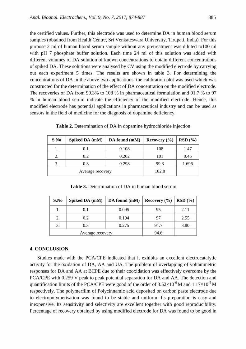
the certified values. Further, this electrode was used to determine DA in human blood serum samples (obtained from Health Centre, Sri Venkateswara University, Tirupati, India). For this purpose 2 ml of human blood serum sample without any pretreatment was diluted to100 ml with pH 7 phosphate buffer solution. Each time 24 ml of this solution was added with different volumes of DA solution of known concentrations to obtain different concentrations of spiked DA. These solutions were analysed by CV using the modified electrode by carrying out each experiment 5 times. The results are shown in table 3. For determining the concentrations of DA in the above two applications, the calibration plot was used which was constructed for the determination of the effect of DA concentration on the modified electrode. The recoveries of DA from 99.3% to 108 % in pharmaceutical formulation and 91.7 % to 97 % in human blood serum indicate the efficiency of the modified electrode. Hence, this modified electrode has potential applications in pharmaceutical industry and can be used as sensors in the field of medicine for the diagnosis of dopamine deficiency.
Table 2. Determination of DA in dopamine hydrochloride injection
S.No Spiked DA (mM) DA found (mM) Recovery (%) RSD (%)
1. 0.1 0.108 108 1.47 2. 0.2 0.202 101 0.45 3. 0.3 0.298 99.3 1.696
Average recovery 102.8
Table 3. Determination of DA in human blood serum
S.No Spiked DA (mM) DA found (mM) Recovery (%) RSD (%)
1. 0.1 0.095 95 2.11
2. 0.2 0.194 97 2.55 3. 0.3 0.275 91.7 3.80
Average recovery 94.6
4. CONCLUSION
Studies made with the PCA/CPE indicated that it exhibits an excellent electrocatalytic activity for the oxidation of DA, AA and UA. The problem of overlapping of voltammetric responses for DA and AA at BCPE due to their cooxidation was effectively overcome by the PCA/CPE with 0.259 V peak to peak potential separation for DA and AA. The detection and quantification limits of the PCA/CPE were good of the order of 3.52×10⁻⁶ M and 1.17×10⁻⁵ M respectively. The polymerfilm of Polycinnamic acid deposited on carbon paste electrode due to electropolymerisation was found to be stable and uniform. Its preparation is easy and inexpensive. Its sensitivity and selectivity are excellent together with good reproducibility. Percentage of recovery obtained by using modified electrode for DA was found to be good in

Anal. Bioanal. Electrochem., Vol. 9, No. 7, 2017, 874-887 886
real sample analysis i.e. in both pharmaceutical formulations and blood serum. With all these virtues, the PCA/CPE could be used as an effective tool for the detection of DA, AA and UA in biological samples.
REFERENCES
[1] G. P. Jin, and X. Q. Lin, Electrochem. Comm. 6 (2004) 454. [2] P. Jeevan Jyoti, K. Gangadhara Reddy, V. Prabhakara Rao, M. Lavanya, and
G. Madhavi, Chem Sci Trans. 4 (2015) 541. [3] Y. Ohnuki, T. Ohsaka, H. Matsuda, and N. Oyama, J. Electroanal. Chem. 158 (1983)
55. [4] Volkov, G. Tourillon, P. C. Lacaze, and J. E. Dubois, J. Electroanal. Chem. 115 (1980)
279. [5] O. Gilbert, U. Chandra, B. E. Kumara Swamy, M. Panduranga Char, C. Nagaraj, and B.
S. Sherigara, Int. J. Electrochem. Sci. 3 (2008) 1186. [6] S. Jahani, and H. Beitollahi, Anal. Bioanal. Electrochem. 8 (2016) 158. [7] G. Ewing, M. A. Dayton, and R. M. Wightman, Anal. Chem. 53 (1981) 1842. [8] R. F. Rane, and C. D. Blaha, Langmuir 6 (1990) 56. [9] M. Lavanya, Y. V. Manohara Reddy, S. Kiranmai, M. Venu, and G. Madhavi, Anal.
Bioanal. Electrochem. 7 (2015) 555. [10] S. Marceglia, G. Foffani, A. Bianchi, G. Baselli, F. Tamma, M. Egidi, and A. Priori,
J. Phys. 571 (2006) 579. [11] N. Kemppainen, P. Marjamaki, M. Roytta, and J. O. Rinne, J. Neural Transm. 108
(2001) 827. [12] V. Prabhakara Rao, Y. Veera Manohara Reddy, M. Lavanya, M. Venu, and G.
Madhavi, Asian J. Chem. 28 (2016) 1828. [13] Rekha, B. E. Kumara Swamy, and P. Siddappa Ganesh, Anal. Bioanal. Electrochem. 8
(2016) 184. [14] Grabowska, M. Chudy, A. Dybko, and Z. Brzoz, Sens. Actuators B 130 (2008) 508. [15] R. D. O’Neill, Analyst 119 (1994) 767. [16] M. R. Deakin, P. M. Kovach, K. J. Stutts, and R. M. Wightman, Anal. Chem. 58 (1986)
1474. [17] L. Zhang, and X. Lin, Anal. Bioanal. Chem. 382 (2005) 1669. [18] M. Chen, and H. Li, Electroanalysis 10 (1998) 477. [19] G. P. Jin, X. Q. Lin, and J. M. Gong, J. Electroanal. Chem. 569 (2004) 135. [20] M. Lavanya, Y. Veera manohara Reddy, V. Prabakara Rao, and G. Madhavi, Chem Sci
Trans. 3 (2014) 1404. [21] W. Sun, M. Yang, and K. Jiao, Anal. Bioanal. chem. 389 (2007) 1283. [22] X. Lin, Q. Zhuang, J. Chen, S. Zang, and Y. Zheng, Sens. Actuator B 125 (2007) 240.

Anal. Bioanal. Electrochem., Vol. 9, No. 7, 2017, 874-887 887
[23] L. Zang, and X. E. Jiang, J. Electroanal. Chem. 583 (2005) 292. [24] P. F. Huang, L. Wang, J. Y. Bai, H. J. Wang, Y. Q. Zhao, and S. D. Fan, Microchim.
Acta 157 (2007) 41. [25] S. A. Kumar, C. F. Tang, and S. M. Chen, Talanta 74 (2008) 860. [26] M. Ates, J. Castillo, A. S. Sarac, and W. Schuhmann, Microchim. Acta 160 (2008) 247. [27] S. Reddy, B. E. K. Swamy, S. Aruna, M. Kumar, R. Shashanka, and H. Jayadevappa,
ChemicalSensors 2 (2012) 1. [28] M. Lavanya, Y. Veera Manohara Reddy, M. Venu, and G. Madhavi, Anal. Bioanal.
Electrochem. 7 (2015) 22. [29] V. Prabhakara Rao, Y. Veera Manohara Reddy, P. Jeevan Jyothi, S. Kiranmai, and
G. Madhavi, Chem. Sci. Trans. 5 (2016) 567. [30] C. R. Raj, K. Tokuda, and T. Ohsaka, Bioelectrochemistry 53 (2001) 183. [31] Y. Veera Manohara Reddy, V. Prabhakara Rao, A. Vijaya Bhaskar Reddy, M. Lavanya,
M. Venu, and G. Madhavi, Mater. Sci. Eng. C 57 (2015) 378. [32] G. Madhavi, J. Damodar, S. K. Mohan, and S. J. Reddy, Bull Electrochem.15 (1999)
535. [33] B. N. Chandrashekar, B. E. Kumara Swamy, M. Pandurangachar, T. V. Satisha, and B.
S. Sherigara, Coll. Surfaces B 88 (2011) 413. [34] S. Sharath Shankar, B. E. Kumara Swamy, B. N. Chandrashekar, and K. J. Gururaj,
Chemical Sensors 2 (2012) 1. [35] S. Chitravathi, B. E. Kumara Swamy, G. P. Mamatha, and B. S. Sherigara, J Mole Liq.
160 (2011) 193. [36] M. T. Shreenivas, B. E. Kumara Swamy, Umesh Chandra, and B. S. Sherigara, Int. J.
Electrochem. 2011 (2011) 1. [37] M. Pandorangachar, B. E. Kumara Swamy, U. Chandra, O. Gilbert, and B. S. Sherigara,
Int. J. Electrochem. Sci. 4 (2009) 672. [38] O. Gilbert, B. E. Kumara Swamy, U. Chandra, and B. S. Sherigara, J. Electroanal.
Chem. 636 (2009) 80.
Copyright © 2017 by CEE (Center of Excellence in Electrochemistry)
ANALYTICAL & BIOANALYTICAL ELECTROCHEMISTRY (http://www.abechem.com)
Reproduction is permitted for noncommercial purposes.