an nqo1 substrate with potent anti-tumor ... - cancer...
TRANSCRIPT
1
An NQO1 substrate with potent anti-tumor activity that selectively kills by PARP1-induced programmed necrosis
Xiumei Huang1,2*, Ying Dong1,2*, Erik A. Bey1,2, Jessica A. Kilgore3, Joseph S.
Bair4, Long-Shan Li1,2, Malina Patel1,2, Elizabeth Parkinson4, Yiguang Wang1,
Noelle S. Williams3, Jinming Gao1, Paul J. Hergenrother4**, David A.
Boothman1,2**
Departments of 1Pharmacology, 2Radiation Oncology, 3Biochemistry Simmons Cancer Center,
UT Southwestern Medical Center, Dallas TX, 75390-8807; and 4Department of Chemistry, Roger Adams Laboratory,
University of Illinois at Urbana-Champaign, Urbana, Il, 61801
Running Title: PARP1 hyperactivation mediates tumor-specific DNQ killing
*Equal contribution
Key Words: Deoxynyboquinone; NQO1; PARP1 hyperactivation; Programmed
necrosis; Non-small-cell Lung Cancer
**To whom correspondence should be addressed:
Paul Hergenrother, Ph.D.
e-mail: [email protected]
Tel: (217) 333-0363; �Fax: (217) 244-8024
Or
David A. Boothman, Ph.D.
e-mail: [email protected]
Tel: (214) 645-6371; Fax: (214) 645-6347
Conflicts of Interest: Drs. Hergenrother, Gao and Boothman are consultants for StemPAR Sciences, Inc., which licensed patents from UT Southwestern and U. Illinois at Urbana-Champaign. The other authors have no conflicts of interest.
on July 6, 2018. © 2012 American Association for Cancer Research. cancerres.aacrjournals.org Downloaded from
Author manuscripts have been peer reviewed and accepted for publication but have not yet been edited. Author Manuscript Published OnlineFirst on April 24, 2012; DOI: 10.1158/0008-5472.CAN-11-3135
2
Abstract
Agents, such as beta-lapachone, that target the redox enzyme,
NAD(P)H:quinone oxidoreductase 1 (NQO1) to induce programmed necrosis in
solid tumors have shown great promise, but more potent tumor-selective
compounds are needed. Here, we report that deoxynyboquinone (DNQ) kills a
wide spectrum of cancer cells in an NQO1-dependent manner with greater
potency than beta-lapachone. DNQ lethality relies on NQO1-dependent futile
redox cycling that consumes oxygen and generates extensive reactive oxygen
species (ROS). Elevated ROS levels cause extensive DNA lesions, PARP1
hyperactivation and severe NAD+/ATP depletion that stimulates Ca2+-dependent
programmed necrosis, unique to this new class of NQO1 'bioactivated' drugs.
Short-term exposure of NQO1+ cells to DNQ was sufficient to trigger cell death,
while genetically matched NQO1- cells were unaffected. Moreover, siRNA-
mediated NQO1 or PARP1 knockdown spared NQO1+ cells from short-term
lethality. Pretreatment of cells with BAPTA-AM (a cytosolic Ca2+ chelator) or
Catalase (enzymatic H2O2 scavenger) was sufficient to rescue DNQ-induced
lethality, as noted with beta-lapachone. Investigations in vivo demonstrated
equivalent antitumor efficacy of DNQ to beta-lapachone, but at a 6-fold greater
potency. PARP1 hyperactivation and dramatic ATP loss was note in tumor, but
not in associated normal lung tissue. Our findings offer preclinical proof of
concept for DNQ as a potent chemotherapeutic agent for treatment of a wide
spectrum of therapeutically challenging solid tumors, such as pancreatic and lung
adenocarcinomas.
Introduction
Lack of selectivity of most cancer chemotherapeutics remains a major limiting
factor (1). We have focused on exploiting elevated NAD(P)H:quinone
oxidoreductase-1 (NQO1, EC 1.6.99.2) levels in most solid tumors, particularly in
non-small cell lung (NSCLC), prostate, pancreatic and breast (2-4), for drug
development. NQO1 is an inducible Phase II detoxifying two-electron
on July 6, 2018. © 2012 American Association for Cancer Research. cancerres.aacrjournals.org Downloaded from
Author manuscripts have been peer reviewed and accepted for publication but have not yet been edited. Author Manuscript Published OnlineFirst on April 24, 2012; DOI: 10.1158/0008-5472.CAN-11-3135
3
oxidoreductase capable of reducing most quinones, forming stable
hydroquinones. Glutathione S-transferase then detoxifies hydroquinones,
conjugating them with glutathione for secretion (5).
Certain rare compounds, however, may undergo NQO1-mediated
bioreduction for antitumor activity. Rather than detoxifying, NQO1 converts
specific quinones into highly cytotoxic species. Most antitumor quinones
dependent on NQO1 are DNA alkylators: (a) mitomycin C (MMC) (6, 7); (b) RH1
(8); (c) E09 (7); and (d) AZQ (9). Use of these agents is limited because they are:
(i) subject to detoxification pathways; (ii) efficient substrates for ubiquitously
expressed one-electron oxidoreductases that cause normal tissue toxicity; and
(iii) subject to emergent drug resistance from various DNA repair pathways.
�-Lapachone (β-lap, Fig. 1A), kills cancer cells and murine xenograft and
orthotopic human or mouse tumor models in vivo in an NQO1-dependent manner
(3, 10). β-Lap induces cell death by NQO1-dependent ROS formation and
oxidative stress (2-4). NQO1 metabolizes �-lap into an unstable hydroquinone
that spontaneously oxidizes, by two dioxygen equivalents, generating superoxide
(10, 11). Elevated long-lived hydrogen peroxide (H2O2) causes extensive DNA
base and single strand break (SSB) lesions that are normally easily and rapidly
repaired. However, extensive NQO1-dependent SSBs cause poly(ADP-
ribose)polymerase-1 (PARP1) hyperactivation, inhibiting the enzyme’s essential
base and SSB repair function. PARP1 hyperactivation causes dramatic
NAD+/ATP pool losses due to ADP-ribosylation, resulting in extensive energy
depletion and cell death (2-4). Thus, ß-lap kills NQO1+ cancer cells by
programmed necrosis that is: (a) independent of caspase activation or p53
status; (b) independent of bcl-2 levels; (c) not affected by BAX/BAK deficiencies;
(d) independent of EGFR, Ras or other constitutive signal transduction activation;
and/or (e) not dependent on proliferation. β-Lap is, therefore, an attractive
experimental drug, and various β-lap formulations have been, or are in, phase I/II
clinical trials.
However, β-lap has a modest potency (LD50: 2-10 μmol/L) in vitro, with limited
aqueous solubility that complicates formulation and delivery. Although
on July 6, 2018. © 2012 American Association for Cancer Research. cancerres.aacrjournals.org Downloaded from
Author manuscripts have been peer reviewed and accepted for publication but have not yet been edited. Author Manuscript Published OnlineFirst on April 24, 2012; DOI: 10.1158/0008-5472.CAN-11-3135

4
nanoparticle strategies for �-lap delivery solved formulation issues, resulting in
dramatic antitumor efficacy (12), there is a clear need for higher potency drugs.
Deoxynyboquinone (DNQ, Fig. 1A) is a promising anti-neoplastic agent whose
mechanism of action has not been elucidated. DNQ killed cancer cells through
oxidative stress and ROS formation (13,14), and N-acetyl cysteine (NAC), a
global free radical scavenger (13), partially blocked lethality. Here, we show that
DNQ undergoes an NQO1-dependent futile cycle similar to ß-lap, where O2 is
consumed, ROS formed and extensive DNA damage triggers PARP1
hyperactivation. Dramatic NAD+/ATP pool decreases caused programmed
necrosis. DNQ is 20- to 100-fold more potent than �-lap, with a significantly
enhanced therapeutic window in human breast, prostate, and pancreatic cancer
models in vitro. NQO1 processes DNQ in vitro more efficiently than β-lap,
suggesting that increased utilization accounts for its enhanced potency.
Significant efficacy of DNQ at 6-fold greater potency versus ß-lap against
orthotopic Lewis Lung Carcinoma (LLC) is shown.
Materials and Methods Chemicals, Reagents, Antibodies. DNQ and ß-lap were synthesized as
described (10, 13). Streptonigrin, mitomycin C, menadione, Hoechst 33258,
hydrogen peroxide (H2O2), cytochrome c, propidium iodide (PI) and dicoumarol
were purchased from Sigma-Aldrich (St. Louis, MO). All quinones and BAPTA-
AM (Calbiochem (La Jolla, CA) were dissolved in DMSO. Dihydroethidium (DHE,
5 mM in DMSO) was purchased from Invitrogen Life Technologies (Eugene, OR).
RH1 and α-human NQO1 antibody were provided by Dr. David Ross (University
of Colorado Health Science Center, Denver, CO) and used at a 1:5000 dilution
overnight, 4 ºC. α-PAR (BD Pharmingen, San Jose, CA), which detects poly
(ADP-ribosyl)ated (PAR) proteins (typically ADP-ribosylated PARP1), and α-
PARP1 (sc-8007, Santa Cruz Biotechnology) antibodies were used at 1:4000
and 1:2000 dilutions, respectively. α-Tubulin (α-tub) was monitored for loading
(15).
on July 6, 2018. © 2012 American Association for Cancer Research. cancerres.aacrjournals.org Downloaded from
Author manuscripts have been peer reviewed and accepted for publication but have not yet been edited. Author Manuscript Published OnlineFirst on April 24, 2012; DOI: 10.1158/0008-5472.CAN-11-3135

5
NQO1 enzyme Assays. DNQ, β-lap, or other quinones (see Supplemental Fig. 1
for structures) were monitored as NQO1 substrates using an NADH (400 μmol/L)
recycling assay and recombinant NQO1 (Sigma) (10), where NADH oxidation to
NAD+ was monitored by absorbance (A340 nm) and data recorded at 2 s intervals
for 5 mins. NADH oxidation rates were compared to reactions lacking β-lap or
DNQ, or containing dicoumarol (10 μmol/L). Initial velocities were calculated and
data expressed as dicoumarol-inhibited relative units (μmol NADH
oxidized/min/gm protein) (10).
O2 Consumption Rates. Assays were performed using the Seahorse 24-well
dish in conjunction with an XF24 sensor cartridge and a XF24 Extracellular Flux
Analyzer (Seahorse Biosciences, Billerica, MA) as per the manufacturer’s
instructions. Briefly, 30,000 cells/well were seeded using a two-step process, and
cells grown as above with unseeded background correction wells. O2
consumption rates (OCR) and proton production rates (PPR) were measured
using the XF24 Analyzer and Assay Wizard software. Data represent means,
%treated/control (T/C, %) ±SE from quadruplet assessments.
Nucleotide Analyses. Changes in intracellular NAD+ pools were measured
using Fluorescent NAD/NADH Detection Kits (Cell Technology, Inc., Mountain
View, CA) (2). NAD+/NADH levels were graphed as means, ±SE from at least
three independent experiments performed in sextuplets each. Changes in ATP in
vivo were measured as described (2-4), and using a colorimetric/fluorometric
assay (BioVision, Milpitas, CA).
Immunoblotting, ROS Formation. Westerns were performed as described (3).
ROS (superoxide) formation was monitored by DHE staining and microscopy.
Quantitative data were analyzed using NIH Image J software, where data are
means, ±SE of 100 cells and duplicate experiments performed in triplicate.
on July 6, 2018. © 2012 American Association for Cancer Research. cancerres.aacrjournals.org Downloaded from
Author manuscripts have been peer reviewed and accepted for publication but have not yet been edited. Author Manuscript Published OnlineFirst on April 24, 2012; DOI: 10.1158/0008-5472.CAN-11-3135

6
PAR formation. PAR formation in vivo was assessed by Western blotting
controlled for α-tub (2-4). Chemiluminescence ELISA assays, to quantify PAR
formation, were performed by HT PARP in vivo Pharmacodynamic II Assays
(Trevigen, Inc., Gaithersburg, MD). Untreated (UT) or treated cells were
incubated with α−PAR antibody, then goat α−Rabbit IgG-HRP.
Chemiluminescence by PeroxyGlowTM assays were expressed as means, ±SE
from three independent experiments.
TUNEL Assays. Terminal deoxynucleotidyl transferase dUTP nick end-labeling
(TUNEL) assays were performed by FC-500 flow cytometry (Beckman Coulter
Electronics, Brea, CA) (10), and data were means, ±SE from three independent
experiments, performed in triplicate.
Cell Lines and Culture. Endogenous NQO1 over-expressing human A549
NSCLC, MCF-7 breast, MIA PaCa-2 pancreatic, PC-3 prostate, and HT1080
sarcoma cancer cells were obtained (3, 4, 16), MAP tested and DNA
fingerprinted (ATC, Charles River). NQO1- *2 polymorphic human H596 NSCLC
or MDA-MB-231 triple-negative breast, and genetically matched NQO1+, cancer
cells were generated by us (2, 4). NQO1+ human PC-3, and genetically matched
stable lentiviral shRNA-NQO1 knockdown, prostate cancer cells were generated
by us (16). Cancer cells were grown in DMEM with 5% FBS. MDA-MB-231 cells
were grown in RPMI 1640. Cells were cultured at 37 °C in a 5% CO2-95% air
humidified atmosphere and were mycoplasma free.
PARP1 siRNA knock down. siRNA specific to the open reading frame (orf) of
PARP1, 5’-CCAAAGGAATTCCGAGAAA-3’ (Thermo Fisher Scientific, Lafayette,
CO) was transiently transfected into cancer cells. PARP1 knockdown was
confirmed using Western assays. Results were confirmed using the ON-
TARGETplus PARP1 SMARTpool.
on July 6, 2018. © 2012 American Association for Cancer Research. cancerres.aacrjournals.org Downloaded from
Author manuscripts have been peer reviewed and accepted for publication but have not yet been edited. Author Manuscript Published OnlineFirst on April 24, 2012; DOI: 10.1158/0008-5472.CAN-11-3135

7
Survival. Relative survival assays were assessed as described (3) and
correlated well with colony forming assays (15). Results were reported as means,
±SE from sextuplate repeats. Experiments were independently repeated three
times.
Hepatocyte Metabolic Stability and Pharmacokinetic (PK) Assessments. During initial maximum tolerated dose (MTD) evaluations for DNQ (10 mg/kg),
PK for DNQ was assessed and compared to prior ß-lap (30 mg/kg) PK data (12)
as described in Supplemental Material and Methods. The metabolic stability of
DNQ versus ß-lap was examined in a standard in vitro hepatocyte assay
(Supplemental Materials and Methods).
DNQ Efficacy against Orthotopic LLC. Antitumor efficacy. Lewis lung carcinoma (LLC) cells (0.5 X 10⁶) were
intravenously (iv) injected into the tail veins of female (~22 gm) athymic nude
mice. Randomized groups of mice (5/group) were treated iv with hydroxypropyl-
ß-cyclodextrin (HPßCD) vehicle alone, or ß-lapachone (30 mg/kg) or DNQ (2.5-
10 mg/kg) dissolved in HPßCD (12) every other day for five injections; mice
treated with HPßCD (600 or 1000 mg/kg for ß-lap or DNQ, respectively) did not
influence tumor growth or survival. Later (18 days post-tumor inoculation), mice
were euthanized, lungs removed, and average lung wet-weights of tumor-bearing
minus control non-tumor-bearing animals calculated. Lungs were visually
examined to confirm LLC nodules. Tumors and associated normal lung tissue
were also assessed for PARP1 hyperactivation (PAR formation) and ATP loss.
Experiments were performed twice and average lung wet-weights
graphed/group.
Survival. LLC-bearing mice were treated with HPßCD alone, ß-lap (30 mg/kg) or
DNQ (5 mg/kg) as above and monitored for changes in weight and survival, and
Kaplan Meier curves generated. p values were reported with asterisks. All animal
protocols have IACUC approval (#2008-1080, UT Southwestern).
on July 6, 2018. © 2012 American Association for Cancer Research. cancerres.aacrjournals.org Downloaded from
Author manuscripts have been peer reviewed and accepted for publication but have not yet been edited. Author Manuscript Published OnlineFirst on April 24, 2012; DOI: 10.1158/0008-5472.CAN-11-3135

8
Statistical Analyses. Student’s t-tests were used to determine statistical
significance from experiments repeated at least three independent times. p
values were reported by asterisks as indicated.
Results DNQ kills cancer cells in an NQO1-dependent manner. We examined the
lethality of DNQ with or without dicoumarol, an NQO1 inhibitor, using two cancer
cells, MCF-7 (breast) and A549 (NSCLC), that endogenously express elevated
NQO1 levels (Fig. 1). DNQ-induced lethality was compared to �-lap and
menadione (Fig. 1A); dicoumarol prevented �-lap, but potentiated menadione
lethality in NQO1+ cells (10, 17). Dicoumarol spared DNQ lethality in both cells
(Fig. 1B). However, DNQ was ~20-fold more potent than �-lap (compare Figs. 1B
to 1C). As with �-lap (5 μmol/L), a minimum 2 h exposure of A549 cells to DNQ
(0.25 μmol/L) was required for complete lethality (Supplemental Fig. 2A) and
extensive DNA base damage and SSBs (Supplemental Fig. 2B). This dose was
used in all subsequent studies.
DNQ was processed in vitro at a significantly increased rate relative to β-
lap, or other quinones (streptonigrin, menadione, RH1, mitomycin C). At 1
μmol/L, DNQ was reduced by NQO1 ~13-fold more efficiently than other
quinones (Supplemental Table 1, Supplemental Fig. 1). Importantly, at LD100
levels (0.25 μmol/L for DNQ, 5 μmol/L for �-lap) in A549 cells, the NQO1 NADH
recycling activities in vitro using DNQ or �-lap were essentially identical
(Supplemental Table 2), suggesting that the catalytic efficiency of NQO1 for DNQ
was at least 20-fold greater than for �-lap. Menadione was a significantly (>20-
fold) less efficient substrate in vitro for NQO1 at its LD90, as reported (10).
Dicoumarol potentiated menadione lethality in MCF-7 cells, but not in A549 cells
(Fig. 1D).
on July 6, 2018. © 2012 American Association for Cancer Research. cancerres.aacrjournals.org Downloaded from
Author manuscripts have been peer reviewed and accepted for publication but have not yet been edited. Author Manuscript Published OnlineFirst on April 24, 2012; DOI: 10.1158/0008-5472.CAN-11-3135

9
DNQ efficiently kills a wide spectrum of cancer cells in an NQO1-dependent manner. DNQ efficiently killed human MIA PaCa-2 pancreatic (Fig. 2A) and
HT1080 sarcoma cancer cells (Fig. 2B) with LD50 values of 48 and 178 nM,
respectively; DNQ showed greater potency in pancreatic cells, in general.
Dicoumarol protected both cells. We then treated genetically matched NQO1+
vs NQO1- human H596 NSCLC or MDA-MB-231 breast cancer cells with DNQ;
*2 NQO1- polymorphic H596 and MDA-MB-231 cells were separately corrected
for NQO1 expression as clones or pooled populations (4, 10). DNQ killed all
NQO1+ clones and pooled populations, but not genetically matched NQO1- cells
(Figs. 2C-D). Similarly, human PC-3 cells stably knocked down for NQO1
expression (shNQ) were spared vs NQO1+ PC-3 cells expressing non-silencing
shRNA (Fig. 2E). DNQ-induced cytotoxicity in nonsilenced (Ns) NQO1+ PC-3
cells was prevented by dicoumarol (Fig. 2E), as in parental PC-3 cells. PC-3 cells
treated with dicoumarol were similarly spared from DNQ lethality as stable
NQO1-knockdown shNQ PC-3 cells (Fig. 2E). Colony forming assays confirmed
the NQO1-dependency of DNQ (Fig. 2F). LD50 values for DNQ vs �-lap
cytotoxicities in various cancers are listed in Supplemental Table 3, highlighting
the NQO1-dependent potency of DNQ.
DNQ NQO1-dependent lethality involves a futile cycle generating ROS. �-
Lap-induced lethality involves a futile cycle mediated by NQO1, where ~60 moles
of NAD(P)H are used, forming ~120 moles superoxide (O2.) per mole �-lap in 5
mins (10). We compared OCR in A549 NSCLC cells after DNQ versus �-lap
treatments. As with �-lap (Fig. 3A), DNQ-treated A549 cells (Fig. 3B) showed
dramatic OCR increases that were dose-dependent and reached peak levels in
30 mins at LD50 levels (Fig. 3C-D). Minimal differences between �-lap and DNQ
were noted, except that significantly lower DNQ doses were needed (Fig. 3B).
Dicoumarol blocked OCR from either DNQ- or �-lap-treated cells, strongly
suggesting that O2 consumption was NQO1-mediated. Similar OCR increases
were noted in NQO1+, but not in NQO1-, H596 cells (Supplemental Figs. 3A, B).
Detectable O2 consumption in NQO1+ A549 cells after co-treatment of �-lap and
on July 6, 2018. © 2012 American Association for Cancer Research. cancerres.aacrjournals.org Downloaded from
Author manuscripts have been peer reviewed and accepted for publication but have not yet been edited. Author Manuscript Published OnlineFirst on April 24, 2012; DOI: 10.1158/0008-5472.CAN-11-3135

10
dicoumarol (Figs. 3A, C) may be due to one-electron oxidoreductases (b5R and
p450R) metabolizing �-lap. Interestingly, such OCRs with dicoumarol were not
noted with DNQ, suggesting that DNQ was not a good substrate for one-electron
oxidoreductases (Fig. 3B, D).
We then monitored superoxide (O2.-) formation using DHE staining. DNQ
(0.25 μmol/L) induced dramatic ROS formation in 60 min in A549 cells that was
prevented by dicoumarol (Fig. 3E). Exogenous catalase (CAT, 1000 U) partially
rescued DNQ cytotoxicity (Fig. 3F). Thus, NQO1-dependent DNQ metabolism
resulted in extensive OCR, O2.- formation, and long-lived H2O2 production similar
to �-lap (2), but at 20-fold lower concentrations.
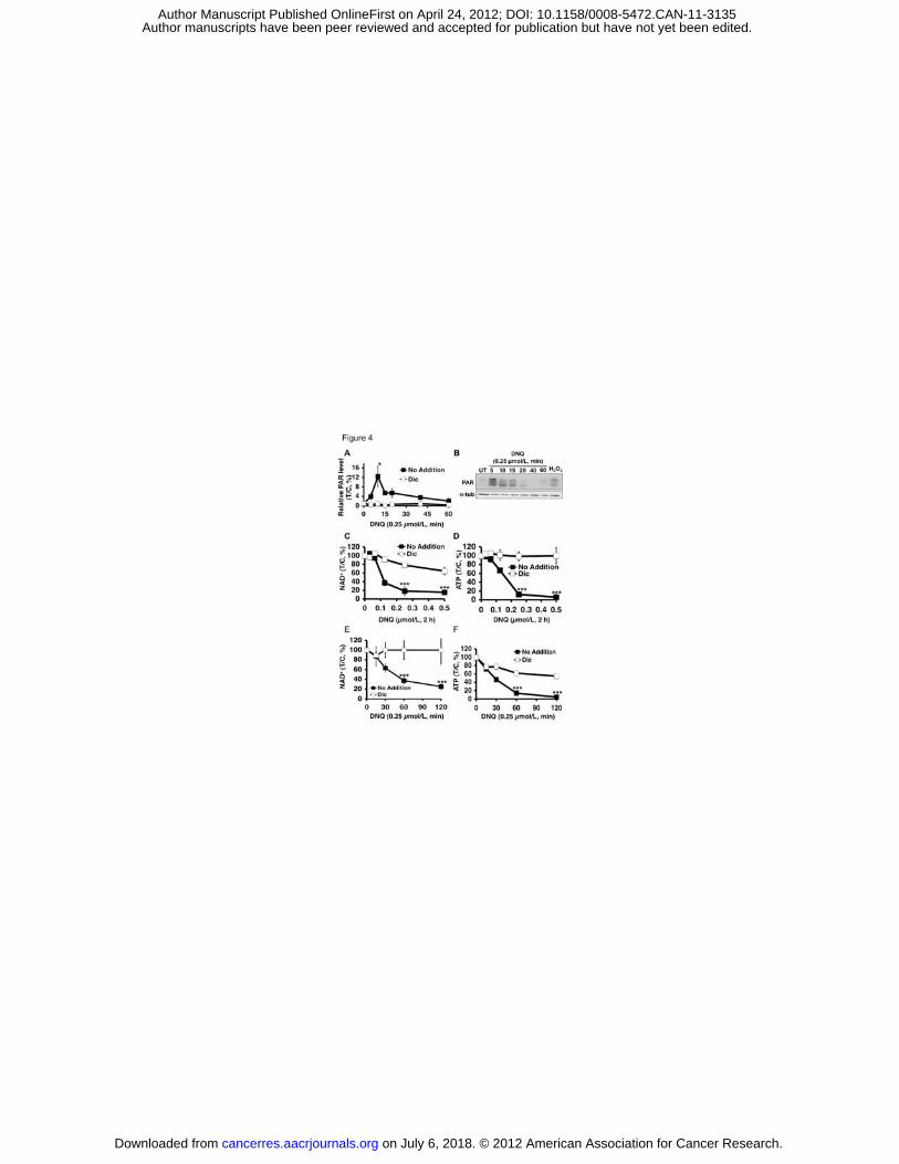
DNQ induces PARP1 hyperactivation, loss of essential nucleotides. DNQ-
treated A549 cells were examined for poly(ADP-ribosyl)ated protein (PAR)
formation using an ELISA method, where peak levels were noted at ~10 min (Fig.
4A). PAR-PARP1 (PAR) formation was confirmed by Western analyses with
similar kinetics, lasting ~20 mins (Fig. 4B). Dicoumarol effectively suppressed
PAR formation and blocked PARP1 hyperactivation in DNQ-treated A549 cells
(Fig. 4A). PARP1 hyperactivation induced by DNQ in A549 cells was
accompanied by dramatic NAD+ and ATP losses. Both dose-response and time-
course studies showed that NAD+ and ATP were rapidly and extensively
depleted (Figs. 4C-F), and a 2 h exposure was sufficient for near complete NAD+
loss in DNQ-exposed cells (0.25 μmol/L, 2 h, Figs. 4C, E). Dramatic ATP loss
followed NAD+ depletion in both dose- and time-dependent manners (Fig. 4D, F)
that were prevented by dicoumarol. DNQ-induced ATP losses occurred in an
NQO1-dependent manner in NQO1+, but not NQO1-, H596 cells (Supplemental
Figs. 4A, B).
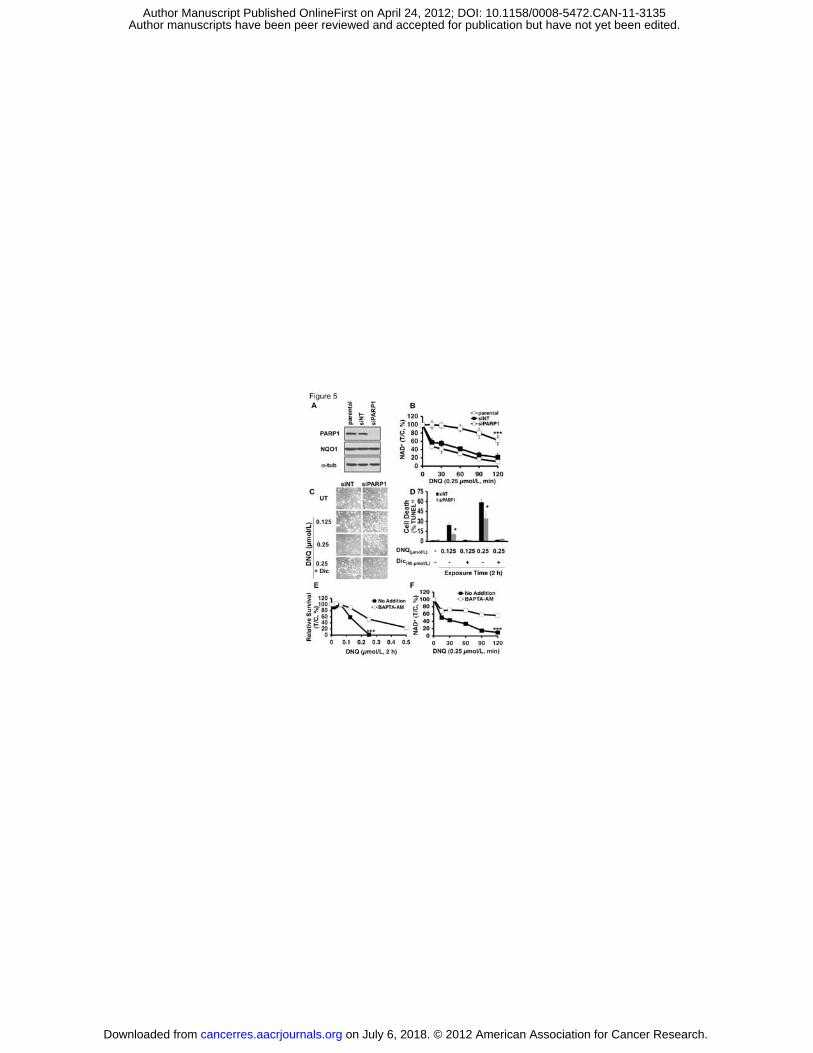
Both PARP1 and Ca2+ play pivotal roles in DNQ-induced cell death. We then
transiently knocked down PARP1 using specific siRNA, that did not affect NQO1
expression (Fig. 5A). PARP1 knockdown significantly prevented NAD+ (Fig. 5B)
and ATP losses (Supplemental Fig. 5) in DNQ-exposed A549 cells versus
on July 6, 2018. © 2012 American Association for Cancer Research. cancerres.aacrjournals.org Downloaded from
Author manuscripts have been peer reviewed and accepted for publication but have not yet been edited. Author Manuscript Published OnlineFirst on April 24, 2012; DOI: 10.1158/0008-5472.CAN-11-3135
11
parental or non-targeting siRNA (siNT)-treated cells. Transient PARP1
knockdown significant decreased programmed necrosis (TUNEL+), 24 h after
treatment with either LD50 (0.125 μmol/L) or lethal (0.25 μmol/L) DNQ doses (Fig.
5C-D), similar to effects noted with �-lap (2). Dicoumarol spared parental, and
PARP1 knockdown, cells (Fig. 5D).
PARP1 hyperactivation required endoplasmic reticulum (ER) Ca2+ release,
since BAPTA-AM, an endogenous cytosolic Ca2+ chelator, spared �-lap-induced
lethality (2). BAPTA-AM pretreatment significantly spared DNQ-induced lethality
in NQO1+ A549 or H596 NSCLC cells using long-term survival assays (Fig. 5E,
Supplemental Fig. 6), and significantly suppressed PARP1 hyperactivation, noted
by abrogation of NAD+ depletion (no addition vs BAPTA-AM, Fig. 5F).
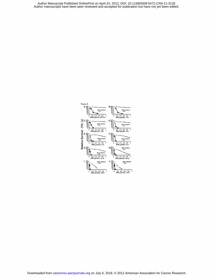
Potential NQO1-dependent therapeutic window of DNQ. Differential NQO1
expression in various solid tumors vs normal tissue can be effectively exploited
using �-lap, resulting in a wide therapeutic window in vitro (4) that predicted its
antitumor efficacy in vivo (3, 16). A549 cells were exposed to various DNQ
doses, with or without dicoumarol (40 μmol/L), at various times to assess the
long-term survival ‘potential therapeutic window assessments’, as performed with
ß-lap in NSCLC, pancreatic and prostate cancers (3, 12, 16). In A549 cells, DNQ
exhibited a much broader therapeutic window than �-lap (compare Figs. 6A-J to
those in ref. (4)). DNQ (0.25 μmol/L)-treated A549 cells were rescued by
dicoumarol (Fig. 6A-H). However, as the length of DNQ exposure was increased
to >24 h, only ~70% cells were spared by dicoumarol (Fig. 6I). Lethality was
further increased after >48 h DNQ exposure in cells exposed to dicoumarol (Fig.
6J), with limited lethality increases in parental NQO1+ cells (compare solid
square lines at 2 vs 24 h, Figs. 6C and 6H, respectively). Similar responses were
noted in H596 cells (Supplemental Figs. 7A-C).
DNQ antitumor efficacy. Using an orthotopic LLC model in athymic mice, we
examined the antitumor efficacy of DNQ (Fig. 7); murine LLC tumors have ~80 U
of NQO1 activity versus <10 U in mouse normal lungs. Mice were treated every
on July 6, 2018. © 2012 American Association for Cancer Research. cancerres.aacrjournals.org Downloaded from
Author manuscripts have been peer reviewed and accepted for publication but have not yet been edited. Author Manuscript Published OnlineFirst on April 24, 2012; DOI: 10.1158/0008-5472.CAN-11-3135
12
other day for 5 injections with HPßCD alone, DNQ (2.5, 5, and 10 mg/kg) or ß-lap
(30 mg/kg). Later (18 days), lungs were removed, weighed and visually scored
for tumor nodules (Figs. 7A, B). DNQ (5 and 10 mg/kg)- or ß-lap-treated mice
showed significant tumor growth reductions, confirmed by decreases in tumor
nodule formation and histology (Figs. 7A, B). No long-term pathological affects
(monitor 18 days after one regimen) of DNQ or ß-lap were noted in liver (Fig.
7C), lung, bone marrow, spleen, or thymus; unlike humans, mice have relatively
high liver NQO1 levels. Overall survival confirmed significant antitumor efficacy of
DNQ (at 5 mg/kg) (p<0.04), at a 6-fold lower dose than ß-lap (30 mg/kg) (Fig.
7D). Finally, PAR formation and energy (ATP) losses in LLC tumors after DNQ or
ß-lap exposures confirmed their PARP1 hyperactivation-mediated programmed
necrotic mechanism (Figs. 7E, F); tumor-specific ATP losses were confirmed by
LC/MS/MS analyses. In contrast, associated normal lung tissue was unaffected,
showing no PARP1 hyperactivation or ATP loss (Figs. 7E, F). Metabolic stability measurements using murine hepatocytes and plasma PK
analyses, revealed greater metabolic stability and prolonged plasma half-life of
DNQ versus ß-lap (Supplemental Figures 8A, B and Supplemental Table 4).
Discussion Identifying exploitable differences between cancerous and healthy tissue
is critical for development of next-generation cancer treatments. NQO1 is over-
expressed in a majority of solid tumors, in angiogenic and bone marrow
endothelial cells (18, 19), and is induced by exposure to certain quinones (20) or
ionizing radiation (21, 22). NQO1 over-expression in solid tumor versus
associated normal tissue was reported in: (a) ~80% NSCLC tumors at 20- to 40-
fold (4); (b) ~70% breast tumors at 10- to 20-fold (23); (c) ~90% pancreatic
tumors at 20- to 30-fold (24); (d) ~60% prostate tumors at 10- to 20-fold (3); and
(e) ~60% colon tumors at 5- to 10-fold (25). Thus, compounds ‘bioactivated’ by
NQO1 have potential for ‘personalized’ anticancer therapy.
�-Lap and DNQ kill cancer cells through the same NQO1-dependent
programmed necrotic (necroptosis) pathway. At each agent’s minimally lethal
on July 6, 2018. © 2012 American Association for Cancer Research. cancerres.aacrjournals.org Downloaded from
Author manuscripts have been peer reviewed and accepted for publication but have not yet been edited. Author Manuscript Published OnlineFirst on April 24, 2012; DOI: 10.1158/0008-5472.CAN-11-3135

13
dose (0.25 μmol/L for DNQ, 5 μmol/L for �-lap): (a) a minimum exposure time (2
h) was noted for 100% lethality in vitro; (b) H2O2 was the apparent obligatory
ROS intermediate required for PARP1 hyperactivation and lethality, since
catalase prevented both; (c) Ca2+ release (2, 26) for PARP1 hyperactivation and
lethality was required, as BAPTA-AM prevented both; (d) PARP1 hyperactivation
caused dramatic ATP and NAD+ losses; and (e) cells were killed in the same
time-frame and with unique proteolysis (e.g., atypical PARP1 and p53 cleavage).
Understanding this mechanism will lead to further improvement in their efficacy.
The potency and NQO1-dependent therapeutic window (Fig. 6) of DNQ,
and its apparent reduced metabolism by one-electron oxidoreductases, make
this drug (or derivatives) very promising. We elucidated its mechanism of action
in various cancer cells, showing that it induced programmed necrosis identical to
�-lap, but with ~20-fold increased potency. NQO1+ cells were selectively
hypersensitive to DNQ, whereas genetically matched NQO1- cells were resistant.
NQO1+ cells were rescued from DNQ-induced cytotoxicity by NQO1 knockdown
or by chemical inhibition (via DIC). Exposure of NQO1+ cancer cells to DNQ
elicited elevated superoxide levels, with concomitant and significant OCR.
Downstream, DNQ stimulated PARP1 hyperactivation in a mechanism
essentially identical to �-lap, which correlated well with NAD+/ATP losses, and
NADH recycling assays confirmed futile cycling of DNQ by NQO1.
DNQ has an apparently broader NQO1-dependent therapeutic window in
vitro (Fig. 6) than ß-lap (4) using A549 cells as a surrogate. Similar data were
obtained using NQO1+ versus NQO1- H596 cells (Supplemental Fig. 7). Thus,
DNQ may not be as good a substrate as �-lap for p450R and b5R, since β-lap-
treated cells consumed O2 even with DIC, whereas OCR was blocked in DNQ-
treated cells (Figs. 3C-D). Antitumor (Fig. 7), PK and metabolic stability
(Supplemental Material, Table 4 and Supplemental Figs. 8A, B) data support a
strong antitumor efficacy potential for DNQ, with increased metabolic stability in
intact hepatocytes in vitro, and a longer half-life than ß-lap. The increased
metabolic stability of DNQ is consistent with a lower affinity for one-electron
oxidoreductases (i.e., b5R and P450R), since these enzymes are elevated in
on July 6, 2018. © 2012 American Association for Cancer Research. cancerres.aacrjournals.org Downloaded from
Author manuscripts have been peer reviewed and accepted for publication but have not yet been edited. Author Manuscript Published OnlineFirst on April 24, 2012; DOI: 10.1158/0008-5472.CAN-11-3135

14
mouse liver. DNQ showed equivalent antitumor activity, suppressed tumor
growth and increased survival of tumor-bearing mice as ß-lap, but at 6-fold lower
doses (Fig. 7), consistent with its therapeutic window in vitro (Fig. 6). However,
our studies in vivo are in no way an optimization of DNQ delivery. Significant
dose-limiting methemaglobinemia (MH) was noted in DNQ-treated mice, and
much less with ß-lap (12). Such blood-borne toxicity can be easily resolved,
however, since MH was completely prevented using ß-lap ‘stealth’ micellar
delivery (12). In addition, novel DNQ analogs that we believe will avoid such
limiting toxicities, are currently being tested using orthotopic breast, lung,
prostate and pancreatic tumor models.
Our comparison of DNQ to various quinones as NQO1 substrates in vitro
using NADH reutilization assays (10) may also explain its increased potency. At
1 μM, NQO1 processed DNQ ~13-fold more efficiently than β-lap (1400 ±80 vs
110 ±20 Relative Units, Supplemental Table 1). At equitoxic (LD100) levels,
similar NQO1 enzymatic activities (680 ±110 vs 530 ±60 Relative�Units for 5
μmol/L β-lap vs 0.25 μmol/L DNQ, respectively, Supplemental Table 2) were
noted. The potential therapeutic window of DNQ appears superior to �-lap,
making DNQ a potentially safer candidate for use as a chemotherapeutic drug,
although increased potency may translate into greater normal tissue toxicity.
Thus, our studies strongly suggest that DNQ, and future more soluble derivatives
and/or stealth polymeric nanoparticles, will be very promising anticancer
therapeutics for the treatment of NSCLC, pancreatic, and breast cancers.
Acknowledgements This work was supported by NIH/NCI grant CA102792 to DAB and the
University of Illinois. We thank Dr. William G. Bornmann (MD Anderson) for
supplying �-lap and the Imaging and Chemistry and Cancer Shared Resources,
SCC. This is CSCN 065.
on July 6, 2018. © 2012 American Association for Cancer Research. cancerres.aacrjournals.org Downloaded from
Author manuscripts have been peer reviewed and accepted for publication but have not yet been edited. Author Manuscript Published OnlineFirst on April 24, 2012; DOI: 10.1158/0008-5472.CAN-11-3135

15
References
1. Pavet V, Portal MM, Moulin JC, Herbrecht R, Gronemeyer H. Towards novel
paradigms for cancer therapy. Oncogene. 2011; 30:1-20.
2. Bentle MS, Reinicke KE, Bey EA, Spitz DR, Boothman DA. Calcium-dependent
modulation of poly(ADP-ribose) polymerase-1 alters cellular metabolism and DNA
repair. J Biol Chem 2006;281: 33684-33696.
3. Dong Y, Bey EA, Li LS, Kabbani W, Yan J, Xie XJ, Hsieh JT, Gao J, Boothman
DA. Prostate cancer radiosensitization through poly(ADP-Ribose) polymerase-1
hyperactivation. Cancer Res 2010; 70: 8088-8096.
4. Bey EA, Bentle MS, Reinicke KE, Dong Y, Yang CR, Girard L, et al., An NQO1-
and PARP1-mediated cell death pathway induced in non-small-cell lung cancer
cells by beta-lapachone. Proc Natl Acad Sci., USA 2007; 104:11832-11837.
5. Ross D, Siegel D. NAD(P)H:quinone oxidoreductase 1 (NQO1, DT-diaphorase),
functions and pharmacogenetics. Methods Enzymol 2004; 382: 115-144.
6. Danson S, Ward TH, Butler J, Ranson M. DT-diaphorase: a target for new
anticancer drugs. Cancer Treat Rev 2004; 30: 437-449.
7. McKeown SR, Cowen RL, Williams KJ. Bioreductive drugs: from concept to clinic.
Clin Oncol (R Coll Radiol) 2007; 19: 427-442.
8. Winski SL, Hargreaves RH, Butler J, Ross D. A new screening system for
NAD(P)H:quinone oxidoreductase (NQO1)-directed antitumor quinones:
identification of a new aziridinylbenzoquinone, RH1, as a NQO1-directed antitumor
agent. Clin Cancer Res 1998; 4: 3083-3088.
9. Curt GA, Kelley JA, Kufta CV, Smith BH, Kornblith PL, Young RC, et al., Phase II
and pharmacokinetic study of aziridinylbenzoquinone [2,5-diaziridinyl-3,6-
bis(carboethoxyamino)-1,4-benzoquinone, diaziquone, NSC 182986] in high-grade
gliomas. Cancer Res 1983; 43: 6102-6105.
on July 6, 2018. © 2012 American Association for Cancer Research. cancerres.aacrjournals.org Downloaded from
Author manuscripts have been peer reviewed and accepted for publication but have not yet been edited. Author Manuscript Published OnlineFirst on April 24, 2012; DOI: 10.1158/0008-5472.CAN-11-3135
16
10. Pink JJ, Planchon SM, Tagliarino C, Varnes ME, Siegel D, Boothman DA.
NAD(P)H:Quinone oxidoreductase activity is the principal determinant of beta-
lapachone cytotoxicity. J Biol Chem 2000; 275: 5416-5424.
11. Reinicke KE, Bey EA, Bentle MS, Pink JJ, Ingalls ST, Hoppel CL, et al.,
Development of beta-lapachone prodrugs for therapy against human cancer cells
with elevated NAD(P)H:quinone oxidoreductase 1 levels. Clin Cancer Res 2005;
11: 3055-3064.
12. Blanco E, Bey EA, Khemtong C, Yang SG, Setti-Guthi J, Chen H, et al., Beta-
lapachone micellar nanotherapeutics for non-small cell lung cancer therapy.
Cancer Res 2010; 70: 3896-3904.
13. Bair JS, Palchaudhuri R, Hergenrother PJ. Chemistry and biology of
deoxynyboquinone, a potent inducer of cancer cell death. J Am Chem Soc 2010;
132:5469-5478.
14. Tudor G, Gutierrez P, Aguilera-Gutierrez A, Sausville EA. Cytotoxicity and
apoptosis of benzoquinones: redox cycling, cytochrome c release, and BAD
protein expression. Biochem Pharmacol 2003; 65: 1061-1075.
15. Wuerzberger SM, Pink JJ, Planchon SM, Byers KL, Bornmann WG, Boothman DA.
Induction of apoptosis in MCF-7:WS8 breast cancer cells by beta-lapachone.
Cancer Res 1998; 58:1876-1885.
16. Li LS, Bey EA, Dong Y, Meng J, Patra B, Yan J, et al., Modulating endogenous
NQO1 levels identifies key regulatory mechanisms of action of beta-lapachone for
pancreatic cancer therapy. Clin Cancer Res 2011; 17: 275-285.
17. Criddle DN, Gillies S, Baumgartner-Wilson HK, Jaffar M, Chinje EC, Passmore S,
et al., Menadione-induced reactive oxygen species generation via redox cycling
promotes apoptosis of murine pancreatic acinar cells. J Biol Chem 2006; 281:
40485-40492.
18. Siegel D, Franklin WA, Ross D. Immunohistochemical detection of
NAD(P)H:quinone oxidoreductase in human lung and lung tumors. Clin Cancer
Res 1998; 4:2065-2070.
19. Siegel D, Ryder J, Ross D. NAD(P)H: quinone oxidoreductase 1 expression in
human bone marrow endothelial cells. Toxicol Lett 2001;125: 93-98.
on July 6, 2018. © 2012 American Association for Cancer Research. cancerres.aacrjournals.org Downloaded from
Author manuscripts have been peer reviewed and accepted for publication but have not yet been edited. Author Manuscript Published OnlineFirst on April 24, 2012; DOI: 10.1158/0008-5472.CAN-11-3135
17
20. Begleiter A, Fourie J. Induction of NQO1 in cancer cells. Methods Enzymol 2004;
382:320-351.
21. Boothman DA, Meyers M, Fukunaga N, Lee SW. Isolation of x-ray-inducible
transcripts from radioresistant human melanoma cells. Proc Natl Acad Sci, USA
1993;90: 7200-7204.
22. Choi EK, Terai K, Ji IM, Kook YH, Park KH, Oh ET, et al., Upregulation of
NAD(P)H:quinone oxidoreductase by radiation potentiates the effect of
bioreductive beta-lapachone on cancer cells. Neoplasia 2007; 9: 634-642.
23. Marin A, Lopez de Cerain A, Hamilton E, Lewis AD, Martinez-Penuela JM, Idoate
MA, et al., DT-diaphorase and cytochrome B5 reductase in human lung and breast
tumours. Br J Cancer 1997; 76: 923-929.
24. Lewis AM, Ough M, Hinkhouse MM, Tsao MS, Oberley LW, Cullen JJ. Targeting
NAD(P)H:quinone oxidoreductase (NQO1) in pancreatic cancer. Mol
Carcinogenesis 2005; 43: 215-224.
25. Mikami K, Naito M, Ishiguro T, Yano H, Tomida A, Yamada T, et al.,
Immunological quantitation of DT-diaphorase in carcinoma cell lines and clinical
colon cancers: advanced tumors express greater levels of DT-diaphorase. Jpn J
Cancer Res 1998; 89: 910-915.
26. Tagliarino C, Pink JJ, Dubyak GR, Nieminen AL, Boothman DA. Calcium is a key
signaling molecule in beta-lapachone-mediated cell death. J Biol Chem 2001; 276:
19150-19159.
on July 6, 2018. © 2012 American Association for Cancer Research. cancerres.aacrjournals.org Downloaded from
Author manuscripts have been peer reviewed and accepted for publication but have not yet been edited. Author Manuscript Published OnlineFirst on April 24, 2012; DOI: 10.1158/0008-5472.CAN-11-3135
18
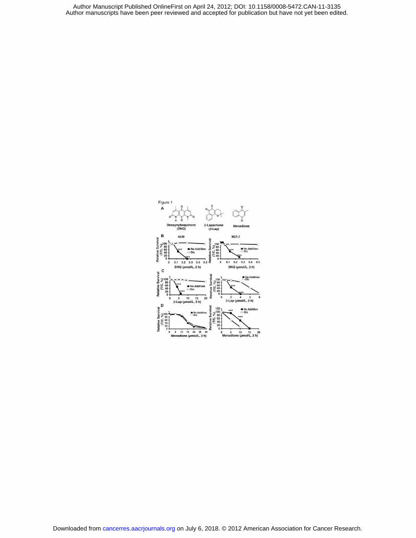
Figure Legends Figure 1. DNQ-induced lethality in endogenous NQ01+ cells. In (A),
Structures of deoxynyboquinone (DNQ), ß-lapachone (ß-lap) and menadione. In
(B), Endogenous, NQO1 over-expressing A549 or MCF-7 cells were treated with
or without various DNQ doses (μmol/L, 2 h), + dicoumarol (Dic, 40 μmol/L, 2 h).
Lethality was monitored by relative survival. Data are means, ±SE for sextuplets
performed thrice. In (C), Cells from (B) were treated with or without various ß-lap
doses (μmol/L, 2 h), + dicoumarol. In (D), Cells were treated with or without
menadione (μmol/L, 2 h), + dicoumarol as in (B). Dicoumarol potentiated
menadione toxicity in MCF-7, but not in A549, cells. Control cells in ‘(B-D)’ were
treated with identical DMSO concentrations (<0.05%). ***, p < 0.001.
Figure 2. NQO1-dependent, DNQ-induced lethality in various cancer cells. DNQ killed human MIA PaCa-2 pancreatic (A) and HT1080 sarcoma (B) cells
with lethal doses of 6 and 300 nM, respectively and dicoumoral rescued lethality.
In (C,D), NQO1- H596 NSCLC (C) or MDA-MB-231 breast cancer (D) cells were
resistant to DNQ. Exogenous over-expression of NQO1 enhanced DNQ lethality.
In (E), Human PC-3 prostate cancer cells knocked down for NQO1 expression
using shRNA-NQO1 (shNQ) were resistant to DNQ (μmol/L, 2h). PC-3 cells
containing non-silencing shRNA (no addition) were sensitive to DNQ, while
dicoumarol (Dic) suppressed lethality similar to shNQ PC-3 cells. In (F),
Clonogenic assays confirmed DNQ lethality in NQO1+ shRNA-nonsilenced Ns
PC-3 cells, whereas resistance was noted in shNQ PC-3 cells, or in Ns PC-3
cells + dicoumarol. All treatments were as in (E). No addition, DMSO alone as in
Fig. 1. Inset, Western blot shows NQO1 knockdown, with 26 +10 units
(Supplemental Table 1). ***, p < 0.001.
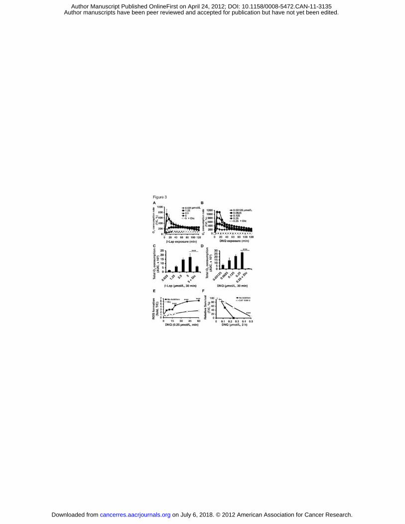
Figure 3. Elevated ROS in DNQ-treated endogenous NQO1+ cancer cells. In
(A), Oxygen (O2) consumption rates (OCR) in A549 cells treated with or without
�-lap +dicoumarol (40 μmol/L) were monitored at eight min intervals for 2 h. Data
were means, ±SE normalized to untreated controls. Experiments were performed
on July 6, 2018. © 2012 American Association for Cancer Research. cancerres.aacrjournals.org Downloaded from
Author manuscripts have been peer reviewed and accepted for publication but have not yet been edited. Author Manuscript Published OnlineFirst on April 24, 2012; DOI: 10.1158/0008-5472.CAN-11-3135
19
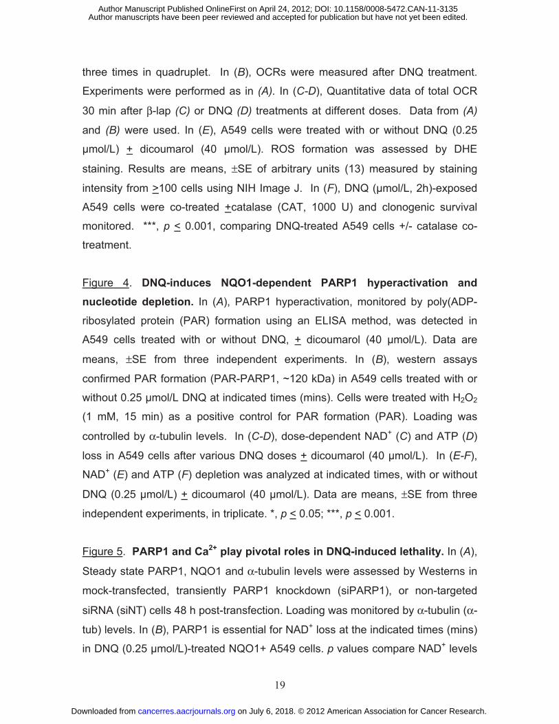
three times in quadruplet. In (B), OCRs were measured after DNQ treatment.
Experiments were performed as in (A). In (C-D), Quantitative data of total OCR
30 min after β-lap (C) or DNQ (D) treatments at different doses. Data from (A)
and (B) were used. In (E), A549 cells were treated with or without DNQ (0.25
μmol/L) + dicoumarol (40 μmol/L). ROS formation was assessed by DHE
staining. Results are means, ±SE of arbitrary units (13) measured by staining
intensity from >100 cells using NIH Image J. In (F), DNQ (μmol/L, 2h)-exposed
A549 cells were co-treated +catalase (CAT, 1000 U) and clonogenic survival
monitored. ***, p < 0.001, comparing DNQ-treated A549 cells +/- catalase co-
treatment.
Figure 4. DNQ-induces NQO1-dependent PARP1 hyperactivation and nucleotide depletion. In (A), PARP1 hyperactivation, monitored by poly(ADP-
ribosylated protein (PAR) formation using an ELISA method, was detected in
A549 cells treated with or without DNQ, + dicoumarol (40 μmol/L). Data are
means, ±SE from three independent experiments. In (B), western assays
confirmed PAR formation (PAR-PARP1, ~120 kDa) in A549 cells treated with or
without 0.25 μmol/L DNQ at indicated times (mins). Cells were treated with H2O2
(1 mM, 15 min) as a positive control for PAR formation (PAR). Loading was
controlled by α-tubulin levels. In (C-D), dose-dependent NAD+ (C) and ATP (D)
loss in A549 cells after various DNQ doses + dicoumarol (40 μmol/L). In (E-F),
NAD+ (E) and ATP (F) depletion was analyzed at indicated times, with or without
DNQ (0.25 μmol/L) + dicoumarol (40 μmol/L). Data are means, ±SE from three
independent experiments, in triplicate. *, p < 0.05; ***, p < 0.001.
Figure 5. PARP1 and Ca2+ play pivotal roles in DNQ-induced lethality. In (A),
Steady state PARP1, NQO1 and α-tubulin levels were assessed by Westerns in
mock-transfected, transiently PARP1 knockdown (siPARP1), or non-targeted
siRNA (siNT) cells 48 h post-transfection. Loading was monitored by α-tubulin (α-
tub) levels. In (B), PARP1 is essential for NAD+ loss at the indicated times (mins)
in DNQ (0.25 μmol/L)-treated NQO1+ A549 cells. p values compare NAD+ levels
on July 6, 2018. © 2012 American Association for Cancer Research. cancerres.aacrjournals.org Downloaded from
Author manuscripts have been peer reviewed and accepted for publication but have not yet been edited. Author Manuscript Published OnlineFirst on April 24, 2012; DOI: 10.1158/0008-5472.CAN-11-3135
20
in PARP1 knockdown vs parental or siNT A549 cells. In (C), Micrographs of
DNQ-treated non-targeted (siNT) or PARP1-specific (siPARP1) siRNA
knockdown A549 cells at 24 h post-treatment with LD50 or lethal doses, +
dicoumarol (40 μmol/L). In (D), PARP1 knockdown significantly protected A549
cells from DNQ-induced programmed necrosis (%TUNEL+ cells). Treatment
conditions were as in (C). Data are means, ±SE from three independent
experiments. In (E), Long-term survival of A549 cells pretreated or not with
BAPTA-AM (5 μmol/L, 60 mins), with or without a 2-h DNQ pulse. In (F), BAPTA-
AM pretreatment of DNQ-exposed A549 cells prevented NAD+ loss. *, p < 0.05;
***, p < 0.001.
Figure 6. DNQ shows a broad NQO1-dependent therapeutic window. Relative survival was monitored in A549 cells treated with various DNQ
concentrations (μmol/L) + dicoumarol (40 μmol/L) for 30 min (A), 1 h (B), 2 h (C),
4 h (D), 6 h (E), 8 h (F), 12 h (G), 24 h (H), 48 h (I) and 72 h (J). Graphed are
means, ±SE of duplicate experiments, repeated in sextuplets.
Figure 7. DNQ shows equivalent efficacy to ß-lap at a 6-fold lower dose. In
(A-C), Mice were treated as in the text, and average wet-weight tumor volumes
assessed (A,B). Tumor nodules were confirmed visually (A) and histologically
(not shown), and average wet-weights calculated (B). DNQ (5 or 10 mg/kg)- or ß-
lap (30 mg/kg)-induced tumor weight decreases were equivalent (p>0.6). Normal
tissues were assessed with no long-term pathological injury noted; see H&E
stained livers (C). In (D), Kaplan Meier survival curves showed significant
(p<0.04) survival advantages of DNQ (5 mg/kg)- or ß-lap (30 mg/kg)-treated
groups. Groups were: HPßCD alone (1000 mg/kg), DNQ (2.5, 5.0 mg/kg) or ß-
lap (30 mg/kg) in HPßCD. *, p<0.04; **, p<0.01. In (E,F), PAR formation and ATP
loss confirmed programmed necrotic mechanism of DNQ- or ß-lap-treated LLC
tumors where animals were treated as in (A-C) with three doses and 24 h later
lungs were removed. Note the lack of response of associated normal lung tissue.
**, p<0.01; ***, p<0.001; α-tub, α-tubulin; and PAR, PAR-PARP1.
on July 6, 2018. © 2012 American Association for Cancer Research. cancerres.aacrjournals.org Downloaded from
Author manuscripts have been peer reviewed and accepted for publication but have not yet been edited. Author Manuscript Published OnlineFirst on April 24, 2012; DOI: 10.1158/0008-5472.CAN-11-3135

on July 6, 2018. © 2012 A
merican A
ssociation for Cancer R
esearch. cancerres.aacrjournals.org
Dow
nloaded from
Author m
anuscripts have been peer reviewed and accepted for publication but have not yet been edited.
Author M
anuscript Published O
nlineFirst on A
pril 24, 2012; DO
I: 10.1158/0008-5472.CA
N-11-3135

Published OnlineFirst April 24, 2012.Cancer Res Xiumei Huang, Ying Dong, Erik A Bey, et al. selectively kills by PARP1-induced programmed necrosisAn NQO1 substrate with potent anti-tumor activity that
Updated version
10.1158/0008-5472.CAN-11-3135doi:
Access the most recent version of this article at:
Material
Supplementary
http://cancerres.aacrjournals.org/content/suppl/2012/04/24/0008-5472.CAN-11-3135.DC1
Access the most recent supplemental material at:
Manuscript
Authoredited. Author manuscripts have been peer reviewed and accepted for publication but have not yet been
E-mail alerts related to this article or journal.Sign up to receive free email-alerts
Subscriptions
Reprints and
To order reprints of this article or to subscribe to the journal, contact the AACR Publications
Permissions
Rightslink site. Click on "Request Permissions" which will take you to the Copyright Clearance Center's (CCC)
.http://cancerres.aacrjournals.org/content/early/2012/04/24/0008-5472.CAN-11-3135To request permission to re-use all or part of this article, use this link
on July 6, 2018. © 2012 American Association for Cancer Research. cancerres.aacrjournals.org Downloaded from
Author manuscripts have been peer reviewed and accepted for publication but have not yet been edited. Author Manuscript Published OnlineFirst on April 24, 2012; DOI: 10.1158/0008-5472.CAN-11-3135