an introduction to breast cancer: biology, pathology, and the latest
TRANSCRIPT
An Introduction to BreastAn Introduction to BreastCancer: Biology, Pathology,Cancer: Biology, Pathology,and the Latest in Screeningand the Latest in Screening
and Diagnostic Toolsand Diagnostic Tools
Katherine S. Tzou, M.D.Katherine S. Tzou, M.D.Mayo Clinic JacksonvilleMayo Clinic Jacksonville
Department of Radiation OncologyDepartment of Radiation Oncology

DisclosuresDisclosures
• I have no relevant relationships to disclose
ObjectivesObjectives
• To understand the epidemiology of breast cancer• To understand the risk factors, natural history, and
histopathology of breast cancer• To learn the current guidelines for breast cancer screening• To understand mammographic findings significant for
breast cancer• To understand when a Breast MRI is indicated

Epidemiology of Breast CancerEpidemiology of Breast Cancer
• Most common cancer in women
• 2nd leading cause of cancerdeath in women
• In 2008, in the United States– 182,460 new cases of invasive
breast cancer in women– 1,990 new cases in men
American Cancer Society. Cancer Facts & Figures 2008. Atlanta: American Cancer Society: 2008.
Epidemiology of Breast CancerEpidemiology of Breast Cancer• Incidence rates had been increasing from 1980-2000
– Increased use of screening mammography– Detecting breast cancers in earlier stages of disease
• Incidence rates began decreasing by 3.5% per yearbetween 2001-2004– Reduced use of hormone replacement therapy (HRT)
• Publication of results from the Women’s Health Initiative in 2002• HRT linked with increased r/o heart disease and breast cancer
– Slight drop in mammography rates in women 40+ y/o• 70.1% to 66.4% between 2000-2005
• Death rates from breast cancer have steadily decreasedsince the 1990’s.– 2.3% per year– Early detection– Improved treatment
American Cancer Society. Cancer Facts & Figures 2008. Atlanta: American Cancer Society: 2008.
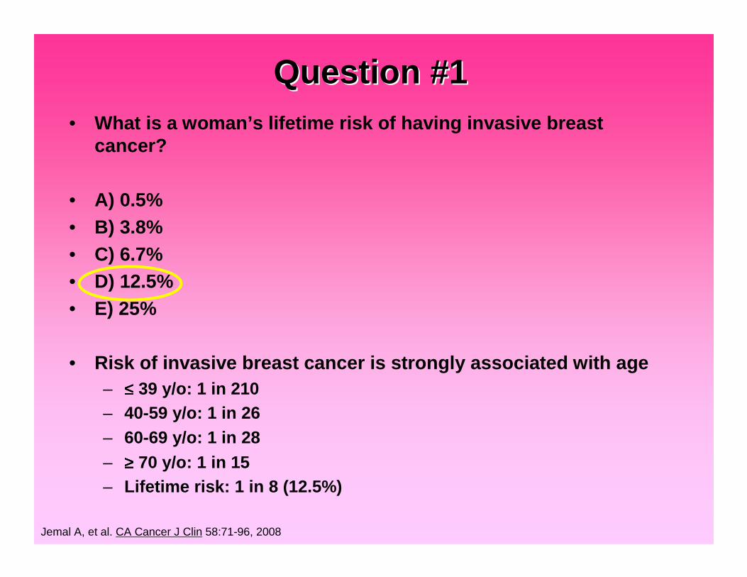
Question #1Question #1• What is a woman’s lifetime risk of having invasive breast
cancer?
• A) 0.5%• B) 3.8%• C) 6.7%• D) 12.5%• E) 25%
Question #1Question #1• What is a woman’s lifetime risk of having invasive breast
cancer?
• A) 0.5%• B) 3.8%• C) 6.7%• D) 12.5%• E) 25%
• Risk of invasive breast cancer is strongly associated with age– ≤≤≤≤ 39 y/o: 1 in 210– 40-59 y/o: 1 in 26– 60-69 y/o: 1 in 28– ≥≥≥≥ 70 y/o: 1 in 15– Lifetime risk: 1 in 8 (12.5%)
Jemal A, et al. CA Cancer J Clin 58:71-96, 2008
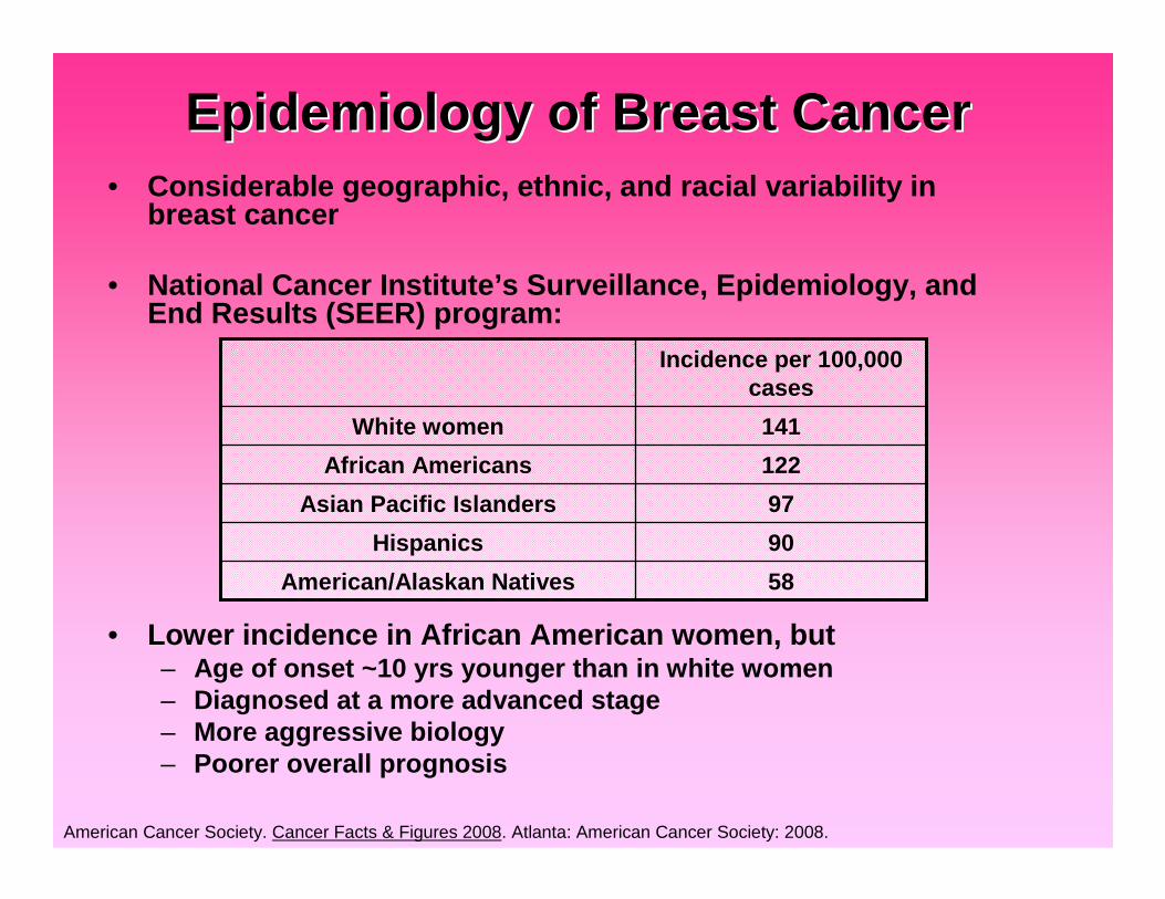
Epidemiology of Breast CancerEpidemiology of Breast Cancer• Considerable geographic, ethnic, and racial variability in
breast cancer
• National Cancer Institute’s Surveillance, Epidemiology, andEnd Results (SEER) program:
• Lower incidence in African American women, but– Age of onset ~10 yrs younger than in white women– Diagnosed at a more advanced stage– More aggressive biology– Poorer overall prognosis
American Cancer Society. Cancer Facts & Figures 2008. Atlanta: American Cancer Society: 2008.
58American/Alaskan Natives
90Hispanics
97Asian Pacific Islanders
122African Americans
141White women
Incidence per 100,000cases

Breast AnatomyBreast Anatomy• Anterior chest wall• Superficial to pectoralis major muscle• Borders
– Medial: Midline chest wall– Lateral: mid-axillary line– Superior: 2nd anterior rib– Inferior: 6th anterior rib
Halperin E, Perez C, Brady L, Ed. Perez and Brady’s Principles and Practice of Radiation Oncology: Fifth edition. Lippincott Williams& Wilkins, Philadelphia: 2008. 1162-1317.
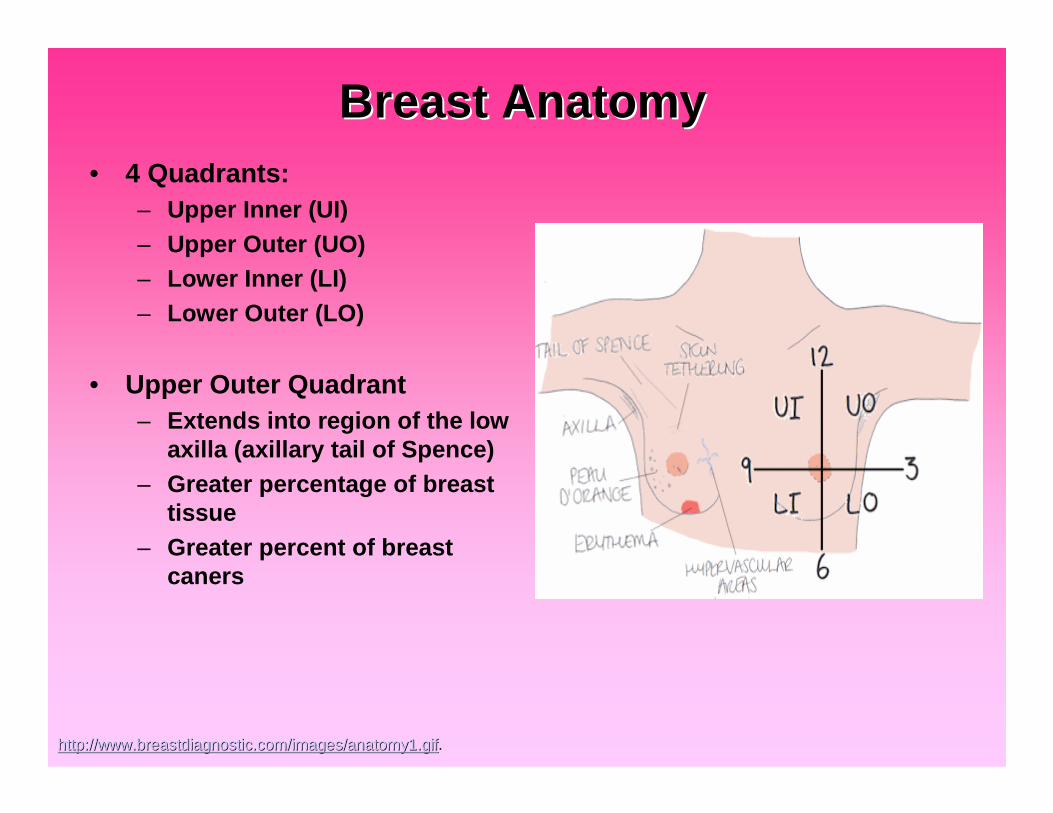
Breast AnatomyBreast Anatomy• 4 Quadrants:
– Upper Inner (UI)– Upper Outer (UO)– Lower Inner (LI)– Lower Outer (LO)
• Upper Outer Quadrant– Extends into region of the low
axilla (axillary tail of Spence)– Greater percentage of breast
tissue– Greater percent of breast
caners
http://www.breastdiagnostic.com/images/anatomy1.gifhttp://www.breastdiagnostic.com/images/anatomy1.gif.
Breast AnatomyBreast Anatomy
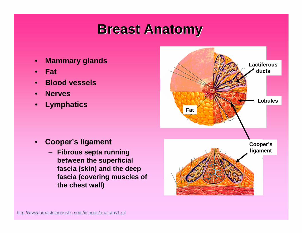
• Mammary glands• Fat• Blood vessels• Nerves• Lymphatics
• Cooper’s ligament– Fibrous septa running
between the superficialfascia (skin) and the deepfascia (covering muscles ofthe chest wall)
http://www.breastdiagnostic.com/images/anatomy1.gifhttp://www.breastdiagnostic.com/images/anatomy1.gif
Cooper’sligament
Fat
Lactiferousducts
Lobules
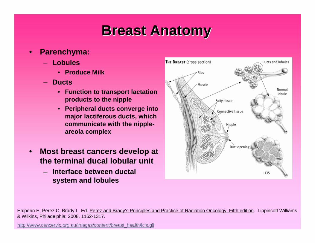
Breast AnatomyBreast Anatomy• Parenchyma:
– Lobules• Produce Milk
– Ducts• Function to transport lactation
products to the nipple• Peripheral ducts converge into
major lactiferous ducts, whichcommunicate with the nipple-areola complex
• Most breast cancers develop atthe terminal ducal lobular unit– Interface between ductal
system and lobules
Halperin E, Perez C, Brady L, Ed. Perez and Brady’s Principles and Practice of Radiation Oncology: Fifth edition. Lippincott Williams& Wilkins, Philadelphia: 2008. 1162-1317.
http://www.cancervic.org.au/images/content/breast_health/lcis.gihttp://www.cancervic.org.au/images/content/breast_health/lcis.giff
Breast AnatomyBreast Anatomy
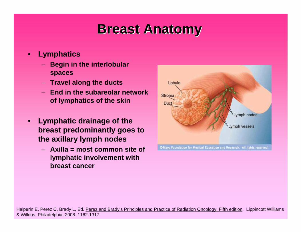
• Lymphatics– Begin in the interlobular
spaces– Travel along the ducts– End in the subareolar network
of lymphatics of the skin
• Lymphatic drainage of thebreast predominantly goes tothe axillary lymph nodes– Axilla = most common site of
lymphatic involvement withbreast cancer
Halperin E, Perez C, Brady L, Ed. Perez and Brady’s Principles and Practice of Radiation Oncology: Fifth edition. Lippincott Williams& Wilkins, Philadelphia: 2008. 1162-1317.
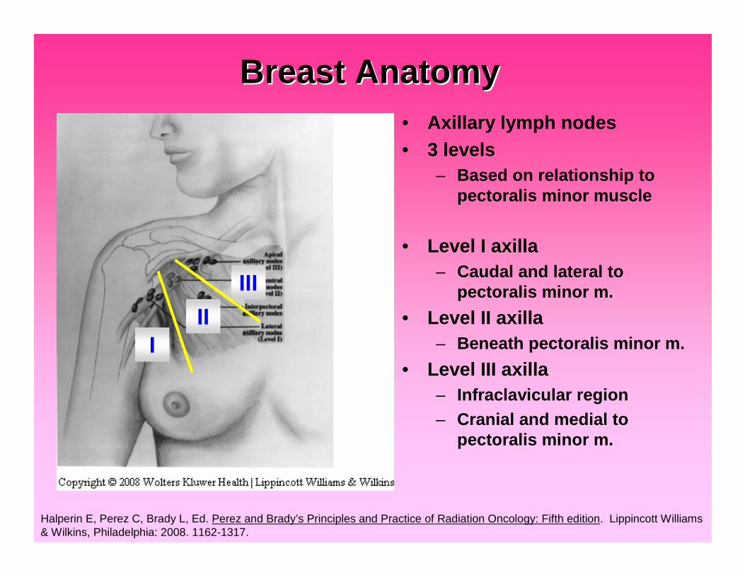
Breast AnatomyBreast Anatomy• Axillary lymph nodes• 3 levels
– Based on relationship topectoralis minor muscle
• Level I axilla– Caudal and lateral to
pectoralis minor m.
• Level II axilla– Beneath pectoralis minor m.
• Level III axilla– Infraclavicular region– Cranial and medial to
pectoralis minor m.
Halperin E, Perez C, Brady L, Ed. Perez and Brady’s Principles and Practice of Radiation Oncology: Fifth edition. Lippincott Williams& Wilkins, Philadelphia: 2008. 1162-1317.
III
III

Breast AnatomyBreast Anatomy• Supraclavicular (SCV) LNs
– Axillary lymph nodes continueunderneath the clavicle tobecome the SCV lymph nodes.
– Locally advanced cancers
• Internal mammary chain (IMC)lymph nodes– Intrathoracic in the
parasternal space– 3-4 cm lateral to midline– 1st 3 interspaces– More commonly seen with
medial, central, or lowerbreast cancers
Halperin E, Perez C, Brady L, Ed. Perez and Brady’s Principles and Practice of Radiation Oncology: Fifth edition. Lippincott Williams& Wilkins, Philadelphia: 2008. 1162-1317.
www.intrawww.intra--medical.com/lymphatic.htmlmedical.com/lymphatic.html
IMC
SCV
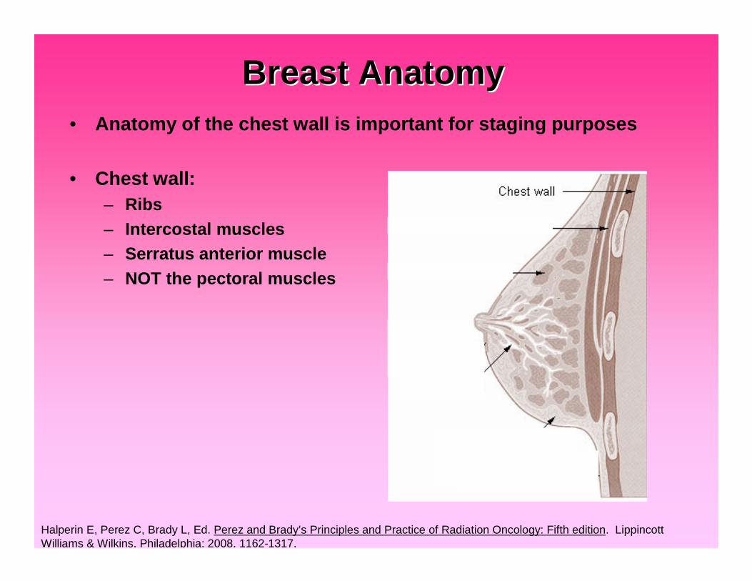
Breast AnatomyBreast Anatomy• Anatomy of the chest wall is important for staging purposes
• Chest wall:– Ribs– Intercostal muscles– Serratus anterior muscle– NOT the pectoral muscles
Halperin E, Perez C, Brady L, Ed. Perez and Brady’s Principles and Practice of Radiation Oncology: Fifth edition. LippincottWilliams & Wilkins, Philadelphia: 2008. 1162-1317.

Question #2Question #2• What is the most significant risk factor for developing breast
cancer (other than gender)?
• A) Prior history of breast cancer• B) Alcohol consumption• C) Obesity• D) Use of hormone replacement therapy• E) Increasing age
Question #2Question #2• What is the most significant risk factor for developing breast
cancer (other than gender)?
• A) Prior history of breast cancer• B) Alcohol consumption• C) Obesity• D) Use of hormone replacement therapy• E) Increasing age
Jemal A, et al. “Cancer statistics, 2006.” CA Cancer J Clin 2006;56:106-130
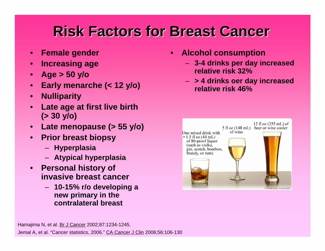
Risk Factors for Breast CancerRisk Factors for Breast Cancer• Female gender• Increasing age• Age > 50 y/o• Early menarche (< 12 y/o)• Nulliparity• Late age at first live birth
(> 30 y/o)• Late menopause (> 55 y/o)• Prior breast biopsy
– Hyperplasia– Atypical hyperplasia
• Personal history ofinvasive breast cancer– 10-15% r/o developing a
new primary in thecontralateral breast
Hamajima N, et al. Br J Cancer 2002;87:1234-1245.
Jemal A, et al. “Cancer statistics, 2006.” CA Cancer J Clin 2006;56:106-130
• Alcohol consumption– 3-4 drinks per day increased
relative risk 32%– > 4 drinks oer day increased
relative risk 46%
Risk Factors for Breast CancerRisk Factors for Breast Cancer
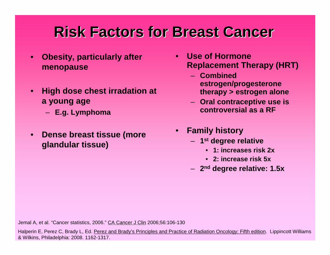
• Obesity, particularly aftermenopause
• High dose chest irradation ata young age– E.g. Lymphoma
• Dense breast tissue (moreglandular tissue)
• Use of HormoneReplacement Therapy (HRT)– Combined
estrogen/progesteronetherapy > estrogen alone
– Oral contraceptive use iscontroversial as a RF
• Family history– 1st degree relative
• 1: increases risk 2x• 2: increase risk 5x
– 2nd degree relative: 1.5x
Jemal A, et al. “Cancer statistics, 2006.” CA Cancer J Clin 2006;56:106-130
Halperin E, Perez C, Brady L, Ed. Perez and Brady’s Principles and Practice of Radiation Oncology: Fifth edition. Lippincott Williams& Wilkins, Philadelphia: 2008. 1162-1317.
Genetic and Familial FactorsGenetic and Familial Factors• Familial breast cancer
– 10% of patients– Germline mutations in tumor suppressor genes
• p53, BRCA1, BRCA2
Halperin E, Perez C, Brady L, Ed. Perez and Brady’s Principles and Practice of Radiation Oncology: Fifth edition. Lippincott Williams& Wilkins, Philadelphia: 2008. 1162-1317.

Genetic and Familial FactorsGenetic and Familial Factors• BRCA1 and BRCA2
– < 1% of the population– < 7% of breast cancer patients– Mediate effects of cell response to DNA damage– BRCA1
• Lifetime risk of breast cancer 65-85%• Lifetime risk of ovarian cancer 50%• Increased risk of colon and prostate cancer
– BRCA2• Lifetime risk of breast cancer 65-85%• Increased lifetime risk of ovarian cancer, but less than that for BRCA1• Associated with pancreatic cancer and male breast cancer

Signs and Symptoms of Breast CancerSigns and Symptoms of Breast Cancer• Often found as an abnormality on mammogram
• Painless firm mass
• Persistent changes to the breast– Thickening– Swelling– Dimpling
• Cooper’s ligament affected– Distortion– Tenderness– Skin irritation– Redness– Scaling– Prominent superficial veins
Giordano, SH. Update on locally advanced breast cancer. Oncologist 2003; 8:526. Copyright © 2003 AlphaMed Press.
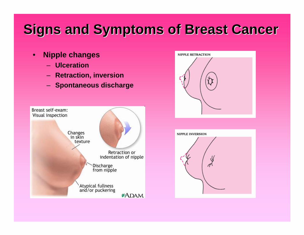
Signs and Symptoms of Breast CancerSigns and Symptoms of Breast Cancer
• Nipple changes– Ulceration– Retraction, inversion– Spontaneous discharge
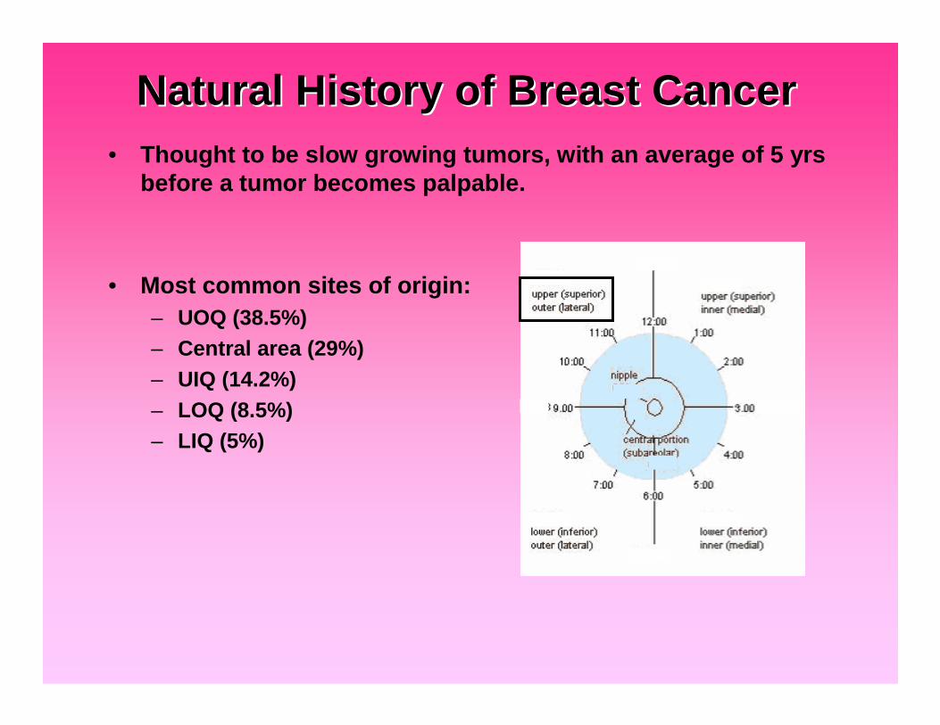
Natural History of Breast CancerNatural History of Breast Cancer• Thought to be slow growing tumors, with an average of 5 yrs
before a tumor becomes palpable.
• Most common sites of origin:– UOQ (38.5%)– Central area (29%)– UIQ (14.2%)– LOQ (8.5%)– LIQ (5%)
Natural History of Breast CancerNatural History of Breast Cancer• Spread of disease
– Travels along the ducts (carcinoma in situ)
– Eventually breaking through the basement membrane (invasivecarcinoma)
• Invades adjacent lobules, ducts, fascial strands, & mammary fat• Spreads through breast lymphatics into peripheral lymphatics
– Can grow through blood vessel walls and spread into the deeplymphatics of the dermis (skin)
• Edema and dimpling of the skin (peau d’orange)
– Ulceration and infiltration of the overlying skin
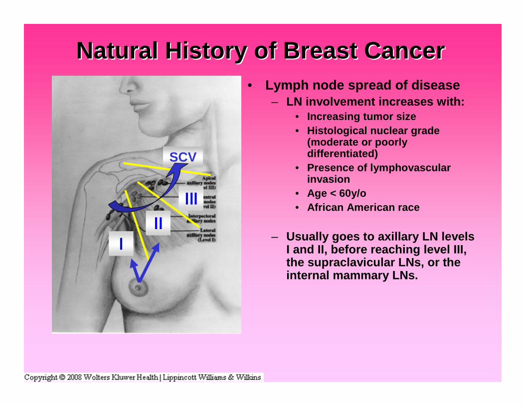
Natural History of Breast CancerNatural History of Breast Cancer• Lymph node spread of disease
– LN involvement increases with:• Increasing tumor size• Histological nuclear grade
(moderate or poorlydifferentiated)
• Presence of lymphovascularinvasion
• Age < 60y/o• African American race
– Usually goes to axillary LN levelsI and II, before reaching level III,the supraclavicular LNs, or theinternal mammary LNs.
SCV
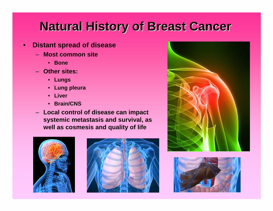
Natural History of Breast CancerNatural History of Breast Cancer• Distant spread of disease
– Most common site• Bone
– Other sites:• Lungs• Lung pleura• Liver• Brain/CNS
– Local control of disease can impactsystemic metastasis and survival, aswell as cosmesis and quality of life
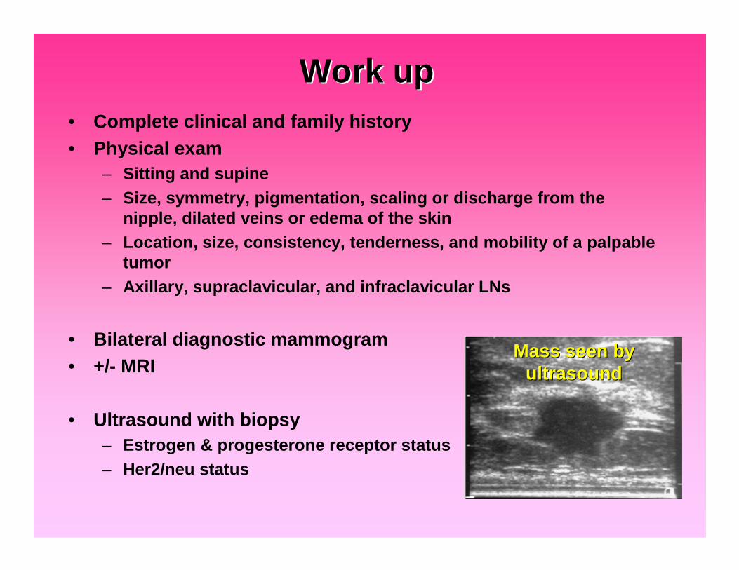
Work upWork up• Complete clinical and family history• Physical exam
– Sitting and supine– Size, symmetry, pigmentation, scaling or discharge from the
nipple, dilated veins or edema of the skin– Location, size, consistency, tenderness, and mobility of a palpable
tumor– Axillary, supraclavicular, and infraclavicular LNs
• Bilateral diagnostic mammogram• +/- MRI
• Ultrasound with biopsy– Estrogen & progesterone receptor status– Her2/neu status
Mass seen byMass seen byultrasoundultrasound
Work upWork up
• CXR• Labs
– Complete blood cell count– General chemistries– LFTs
• AST, ALT, Lactatedehydrogenase (LD),Bilirubin
• +/- Bone scan– Clinical indication, stage II or III
disease, or elevated alkalinephosphatase level
• +/- CT chest/abdomen/pelvis– Stage II or III disease, or elevated
liver function tests

Breast Cancer HistologyBreast Cancer Histology• In situ carcinomas
– Confinement of malignant cellswithin the basement membrane
– Ductal carcinoma in situ (DCIS)• 15-20% of all breast cancer• Prognostic variables:
– Large tumor size– Close or positive surgical
margins– High nuclear grade (how
abnormal cancer cells appear)– Presence of necrosis
– Lobular carcinoma in situ (LCIS)• Marker for bilateral breast cancer
LakhaniLakhani Breast Cancer ResBreast Cancer Res 19991999 11:31:31 doi:10.1186/bcr10doi:10.1186/bcr10
www.gastricbreastcancer.com/figures/figure3.htmwww.gastricbreastcancer.com/figures/figure3.htm
Lobular carcinoma in situ
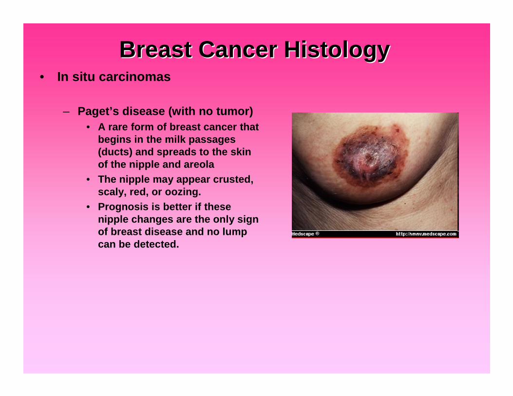
Breast Cancer HistologyBreast Cancer Histology• In situ carcinomas
– Paget’s disease (with no tumor)• A rare form of breast cancer that
begins in the milk passages(ducts) and spreads to the skinof the nipple and areola
• The nipple may appear crusted,scaly, red, or oozing.
• Prognosis is better if thesenipple changes are the only signof breast disease and no lumpcan be detected.
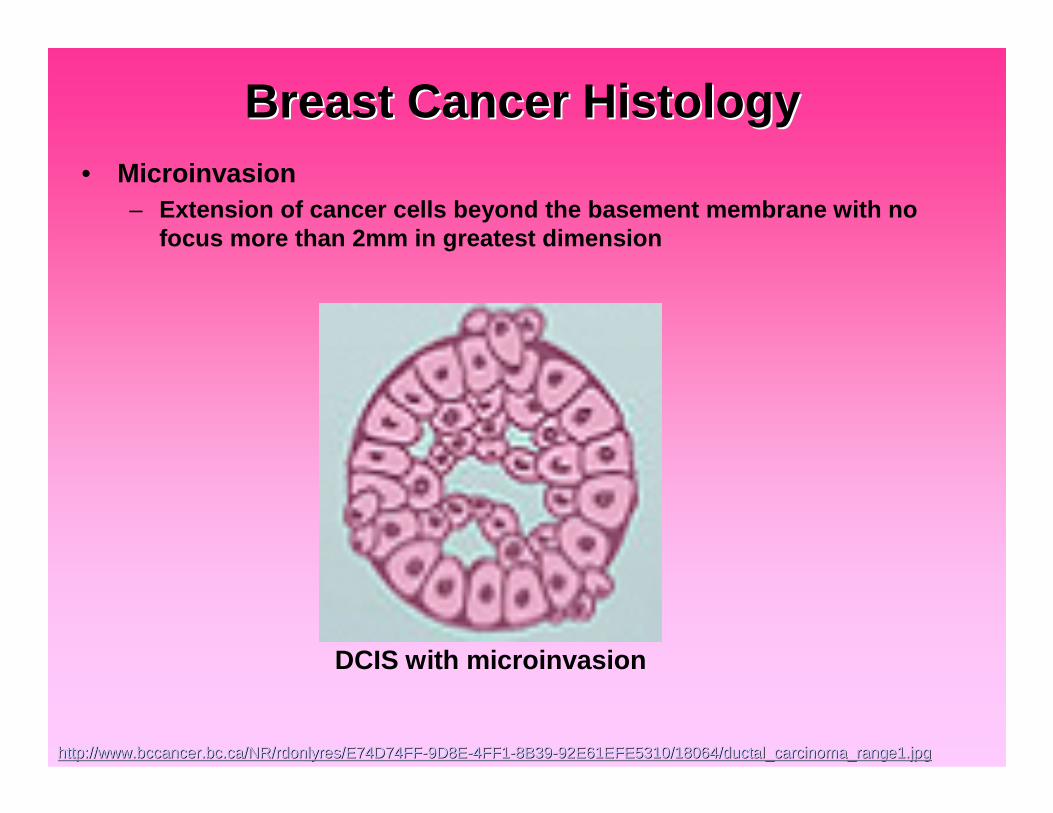
Breast Cancer HistologyBreast Cancer Histology• Microinvasion
– Extension of cancer cells beyond the basement membrane with nofocus more than 2mm in greatest dimension
http://www.bccancer.bc.ca/NR/rdonlyres/E74D74FFhttp://www.bccancer.bc.ca/NR/rdonlyres/E74D74FF--9D8E9D8E--4FF14FF1--8B398B39--92E61EFE5310/18064/ductal_carcinoma_range1.jpg92E61EFE5310/18064/ductal_carcinoma_range1.jpg
DCIS with microinvasion
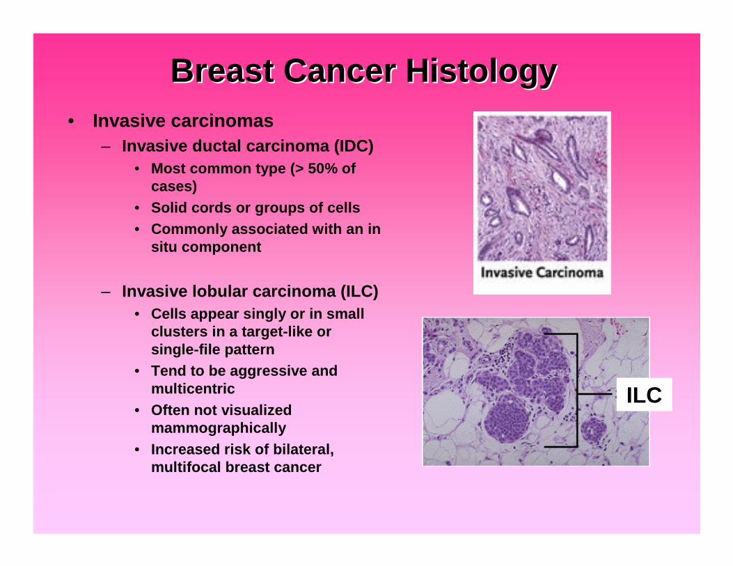
Breast Cancer HistologyBreast Cancer Histology• Invasive carcinomas
– Invasive ductal carcinoma (IDC)• Most common type (> 50% of
cases)• Solid cords or groups of cells• Commonly associated with an in
situ component
– Invasive lobular carcinoma (ILC)• Cells appear singly or in small
clusters in a target-like orsingle-file pattern
• Tend to be aggressive andmulticentric
• Often not visualizedmammographically
• Increased risk of bilateral,multifocal breast cancer
ILC
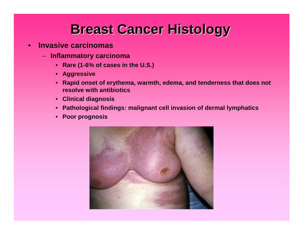
Breast Cancer HistologyBreast Cancer Histology• Invasive carcinomas
– Inflammatory carcinoma• Rare (1-6% of cases in the U.S.)• Aggressive• Rapid onset of erythema, warmth, edema, and tenderness that does not
resolve with antibiotics• Clinical diagnosis• Pathological findings: malignant cell invasion of dermal lymphatics• Poor prognosis

Breast Cancer HistologyBreast Cancer Histology• Other invasive carcinomas
– Medullary carcinoma– Mucinous carcinoma– Tubular carcinoma
– Papillary or micropapillary carcinoma– Undifferentiated carcinoma– Squamous cell carcinoma– Adenoid cystic carcinoma– Secretory carcinoma– Cribiform carcinoma
Better prognosis
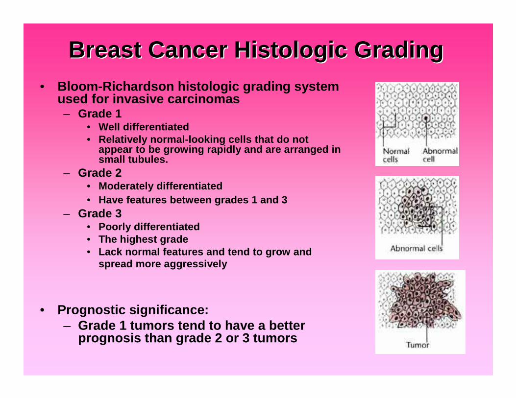
Breast Cancer Histologic GradingBreast Cancer Histologic Grading• Bloom-Richardson histologic grading system
used for invasive carcinomas– Grade 1
• Well differentiated• Relatively normal-looking cells that do not
appear to be growing rapidly and are arranged insmall tubules.
– Grade 2• Moderately differentiated• Have features between grades 1 and 3
– Grade 3• Poorly differentiated• The highest grade• Lack normal features and tend to grow and
spread more aggressively
• Prognostic significance:– Grade 1 tumors tend to have a better
prognosis than grade 2 or 3 tumors

Hormone Receptor Status TestingHormone Receptor Status Testing• Receptors are proteins on the surface of cells that can attach to
certain substances circulating in the blood stream, likehormones.
• Normal breast cells and some breast cancer cells havereceptors for the hormones estrogen and progesterone– Cancer cells positive for estrogen receptors = ER+– Cancer cells positive for progesterone receptors = PR+– In hormone receptor positive cells, the presence of estrogen or
progesterone can fuel the growth of breast cancer cells
Hormone Receptor StatusHormone Receptor Status• Implications: Systemic therapy can be directed at blocking
these hormone receptors (hormone therapy)– Selective Estrogen-Receptor Modulators (SERM)
• Example: Tamoxifen• Blocks estrogen from attaching to receptors
– Aromatase inhibitors• Examples: Arimidex and Femara• Blocks the effect of an enzyme that aids in production of estrogen
– Aromatase inactivator• Example: Aromasin• Inactivates or destroys the enzyme that aids in estrogen production
• Women with hormone receptor-positive cancers tend to have abetter prognosis and are much more likely to respond tohormone therapy than women with cancers without thesereceptors.

HERHER--2/neu Testing2/neu Testing• HER-2/neu gene
– Proto-oncogene (also called c-erbB-2)– Codes for a transmembrane glycoprotein
(p185)• Tyrosine kinase activity• Is a growth-promoting protein
– Amplified and over-expressed in up to30% of breast cancers
– Over-expression is associated with• Tumor aggressiveness• Decreased disease-free survival in node-
positive patients• Variable significance in node-negative
patients
HER2-positive cancers are much more likely to benefit fromtreatment with drugs that target the HER2/neu protein, such astrastuzumab (Herceptin) and lapatinib (Tykerb)
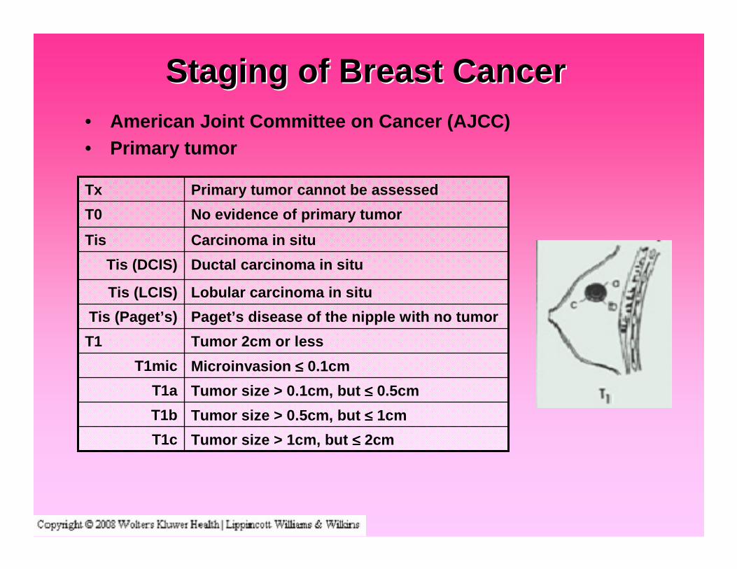
Staging of Breast CancerStaging of Breast Cancer• American Joint Committee on Cancer (AJCC)• Primary tumor
Ductal carcinoma in situTis (DCIS)
Lobular carcinoma in situTis (LCIS)
Paget’s disease of the nipple with no tumorTis (Paget’s)
No evidence of primary tumorT0
Tumor size > 1cm, but ≤≤≤≤ 2cmT1c
Tumor size > 0.5cm, but ≤≤≤≤ 1cmT1b
Tumor size > 0.1cm, but ≤≤≤≤ 0.5cmT1a
Microinvasion ≤≤≤≤ 0.1cmT1mic
Tumor 2cm or lessT1
Carcinoma in situTis
Primary tumor cannot be assessedTx
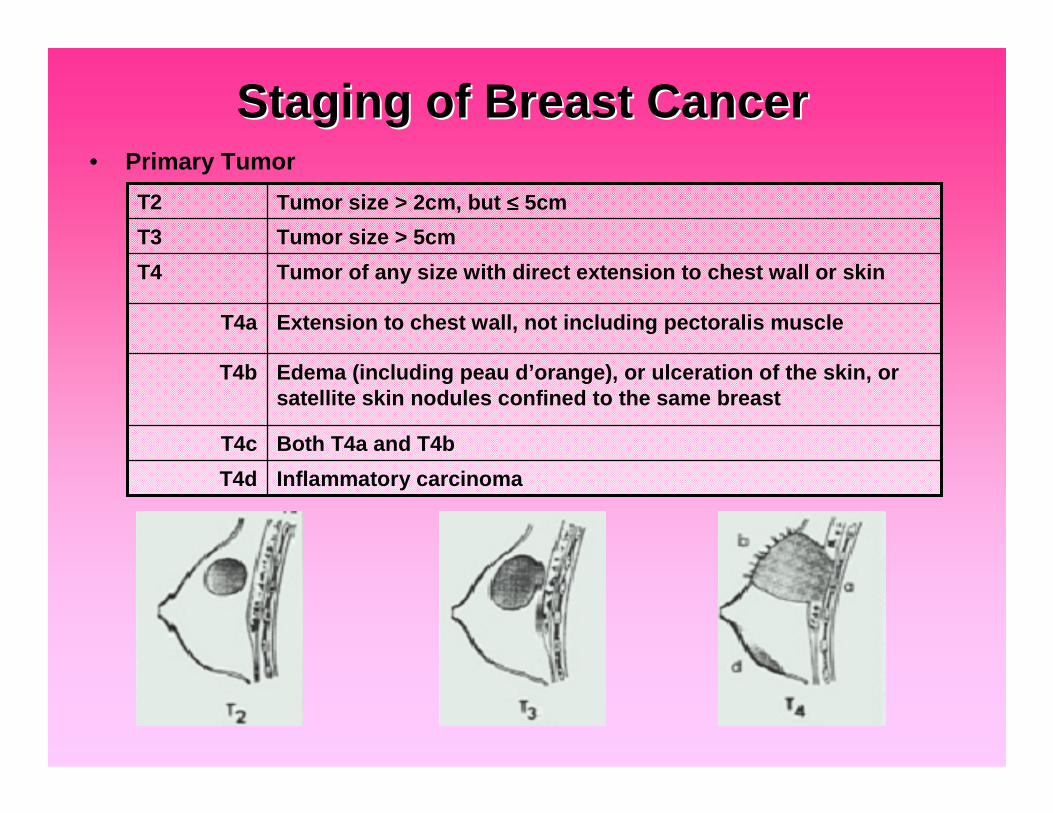
Staging of Breast CancerStaging of Breast Cancer• Primary Tumor
Inflammatory carcinomaT4d
Both T4a and T4bT4c
Edema (including peau d’orange), or ulceration of the skin, orsatellite skin nodules confined to the same breast
T4b
Extension to chest wall, not including pectoralis muscleT4a
Tumor of any size with direct extension to chest wall or skinT4
Tumor size > 5cmT3
Tumor size > 2cm, but ≤≤≤≤ 5cmT2
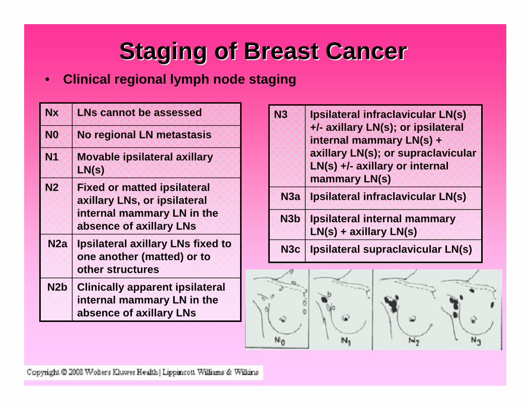
Staging of Breast CancerStaging of Breast Cancer• Clinical regional lymph node staging
Clinically apparent ipsilateralinternal mammary LN in theabsence of axillary LNs
N2b
Ipsilateral axillary LNs fixed toone another (matted) or toother structures
N2a
Fixed or matted ipsilateralaxillary LNs, or ipsilateralinternal mammary LN in theabsence of axillary LNs
N2
Movable ipsilateral axillaryLN(s)
N1
No regional LN metastasisN0
LNs cannot be assessedNx
Ipsilateral supraclavicular LN(s)N3c
Ipsilateral internal mammaryLN(s) + axillary LN(s)
N3b
Ipsilateral infraclavicular LN(s)N3a
Ipsilateral infraclavicular LN(s)+/- axillary LN(s); or ipsilateralinternal mammary LN(s) +axillary LN(s); or supraclavicularLN(s) +/- axillary or internalmammary LN(s)
N3
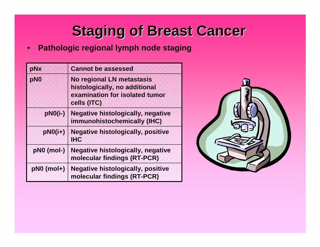
Staging of Breast CancerStaging of Breast Cancer• Pathologic regional lymph node staging
Negative histologically, positivemolecular findings (RT-PCR)
pN0 (mol+)
Negative histologically, negativemolecular findings (RT-PCR)
pN0 (mol-)
Negative histologically, positiveIHC
pN0(i+)
Negative histologically, negativeimmunohistochemically (IHC)
pN0(i-)
No regional LN metastasishistologically, no additionalexamination for isolated tumorcells (ITC)
pN0
Cannot be assessedpNx
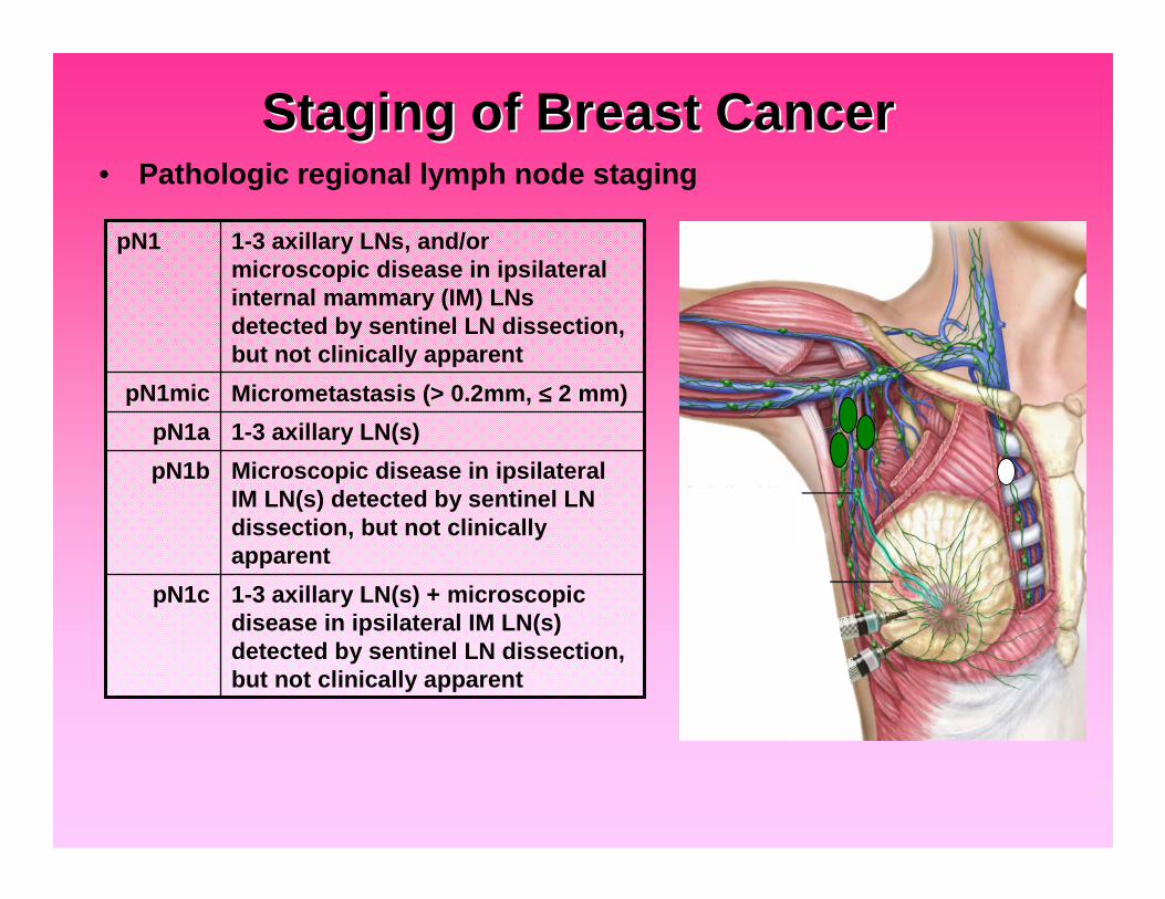
Staging of Breast CancerStaging of Breast Cancer• Pathologic regional lymph node staging
Microscopic disease in ipsilateralIM LN(s) detected by sentinel LNdissection, but not clinicallyapparent
pN1b
1-3 axillary LN(s) + microscopicdisease in ipsilateral IM LN(s)detected by sentinel LN dissection,but not clinically apparent
pN1c
1-3 axillary LN(s)pN1a
Micrometastasis (> 0.2mm, ≤≤≤≤ 2 mm)pN1mic
1-3 axillary LNs, and/ormicroscopic disease in ipsilateralinternal mammary (IM) LNsdetected by sentinel LN dissection,but not clinically apparent
pN1
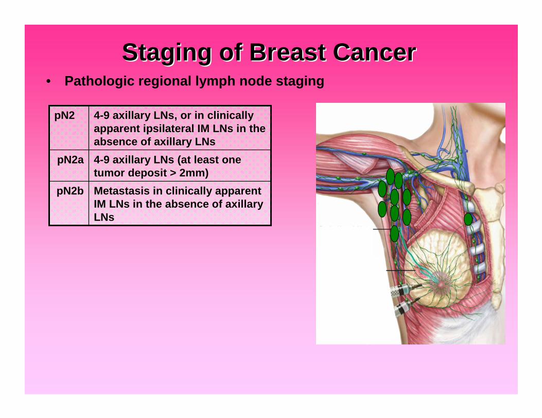
Staging of Breast CancerStaging of Breast Cancer• Pathologic regional lymph node staging
Metastasis in clinically apparentIM LNs in the absence of axillaryLNs
pN2b
4-9 axillary LNs (at least onetumor deposit > 2mm)
pN2a
4-9 axillary LNs, or in clinicallyapparent ipsilateral IM LNs in theabsence of axillary LNs
pN2

Staging of Breast CancerStaging of Breast Cancer• Pathologic regional lymph node staging
Ipsilateral supraclavicular LNspN3c
Clinically apparent ipsilateral IM LNs +≥≥≥≥ 1 axillary LN(s), orin > 3 axillary LNs with microscopicdisease in ipsilateral IM LNs detectedby sentinel LN dissection but notclinically apparent
pN3b
≥≥≥≥ 10 axillary LNs (at least one tumordeposit > 2mm), orin infraclavicular LNs
pN3a
≥≥≥≥ 10 axillary LNs, or in infraclavicularLNs, or in clinically apparentipsilateral IM LNs, or in ipsilateralsupraclavicular LNs
pN3
Distant metastasisM1
No distant metastasisM0
Distant metastasis cannot be assessedMx
• Distant metastatic disease staging

Staging of Breast CancerStaging of Breast Cancer• Stage grouping
M0N3any TIIIC
M1any Nany TIV
M0N0-2T4IIIB
M0N2T0-3
M0N1T3IIIA
M0N1T2
M0N0T3IIB
M0N1T1-0
M0N0T2IIA
M0N0T1I
M0N0Tis0
DistantMetastasis
Nodalinvolvement
Tumorsize
Stage
Pictures courtesy of http://www.faslodex.com/

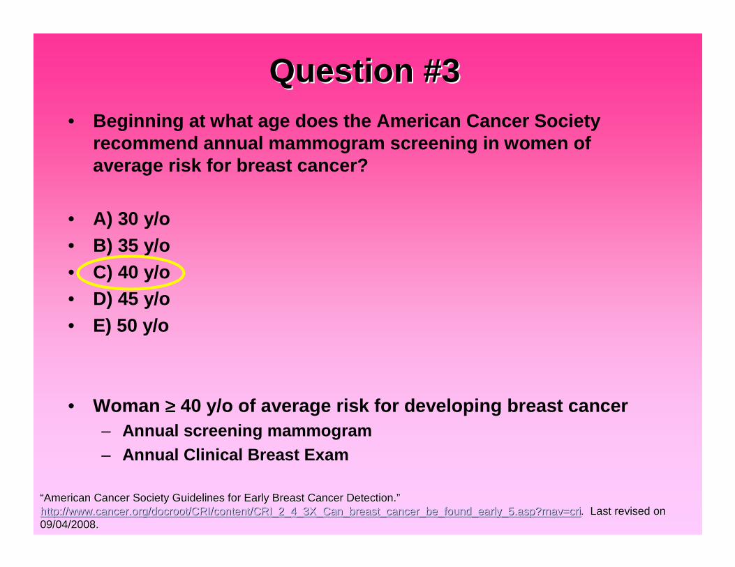
Question #3Question #3• Beginning at what age does the American Cancer Society
recommend annual mammogram screening in women ofaverage risk for breast cancer?
• A) 30 y/o• B) 35 y/o• C) 40 y/o• D) 45 y/o• E) 50 y/o
Question #3Question #3• Beginning at what age does the American Cancer Society
recommend annual mammogram screening in women ofaverage risk for breast cancer?
• A) 30 y/o• B) 35 y/o• C) 40 y/o• D) 45 y/o• E) 50 y/o
• Woman ≥≥≥≥ 40 y/o of average risk for developing breast cancer– Annual screening mammogram– Annual Clinical Breast Exam
““American Cancer Society Guidelines for Early Breast Cancer DetecAmerican Cancer Society Guidelines for Early Breast Cancer Detection.tion.””http://www.cancer.org/docroot/CRI/content/CRI_2_4_3X_Can_breast_http://www.cancer.org/docroot/CRI/content/CRI_2_4_3X_Can_breast_cancer_be_found_early_5.asp?rnav=cricancer_be_found_early_5.asp?rnav=cri. Last revised on. Last revised on09/04/2008.09/04/2008.
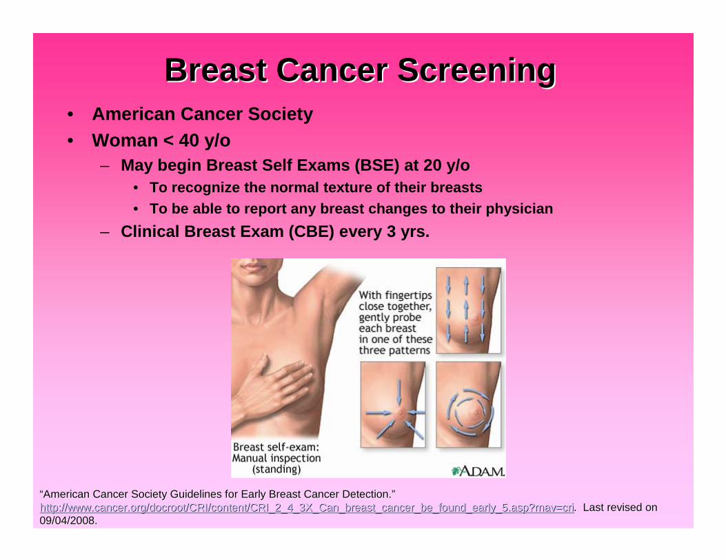
Breast Cancer ScreeningBreast Cancer Screening• American Cancer Society• Woman < 40 y/o
– May begin Breast Self Exams (BSE) at 20 y/o• To recognize the normal texture of their breasts• To be able to report any breast changes to their physician
– Clinical Breast Exam (CBE) every 3 yrs.
““American Cancer Society Guidelines for Early Breast Cancer DetecAmerican Cancer Society Guidelines for Early Breast Cancer Detection.tion.””http://www.cancer.org/docroot/CRI/content/CRI_2_4_3X_Can_breast_http://www.cancer.org/docroot/CRI/content/CRI_2_4_3X_Can_breast_cancer_be_found_early_5.asp?rnav=cricancer_be_found_early_5.asp?rnav=cri. Last revised on. Last revised on09/04/2008.09/04/2008.
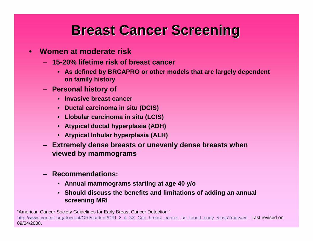
Breast Cancer ScreeningBreast Cancer Screening• Women at moderate risk
– 15-20% lifetime risk of breast cancer• As defined by BRCAPRO or other models that are largely dependent
on family history
– Personal history of• Invasive breast cancer• Ductal carcinoma in situ (DCIS)• Llobular carcinoma in situ (LCIS)• Atypical ductal hyperplasia (ADH)• Atypical lobular hyperplasia (ALH)
– Extremely dense breasts or unevenly dense breasts whenviewed by mammograms
– Recommendations:• Annual mammograms starting at age 40 y/o• Should discuss the benefits and limitations of adding an annual
screening MRI
““American Cancer Society Guidelines for Early Breast Cancer DetecAmerican Cancer Society Guidelines for Early Breast Cancer Detection.tion.””http://www.cancer.org/docroot/CRI/content/CRI_2_4_3X_Can_breast_http://www.cancer.org/docroot/CRI/content/CRI_2_4_3X_Can_breast_cancer_be_found_early_5.asp?rnav=cricancer_be_found_early_5.asp?rnav=cri. Last revised on. Last revised on09/04/2008.09/04/2008.
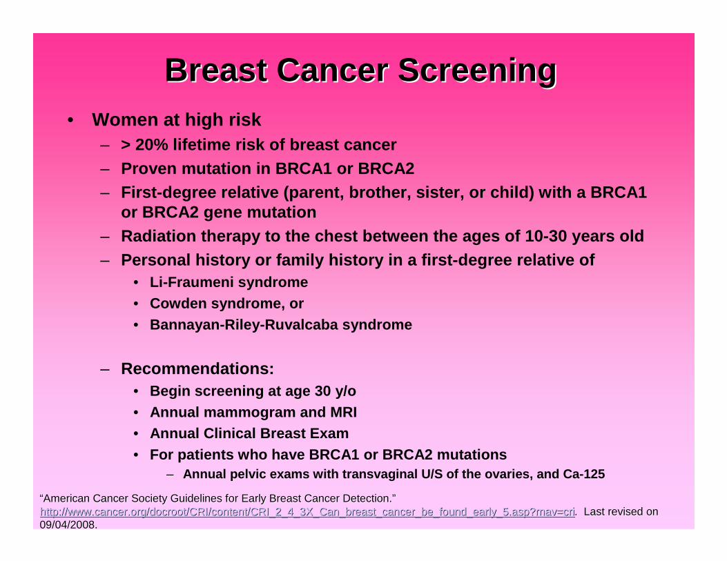
Breast Cancer ScreeningBreast Cancer Screening• Women at high risk
– > 20% lifetime risk of breast cancer– Proven mutation in BRCA1 or BRCA2– First-degree relative (parent, brother, sister, or child) with a BRCA1
or BRCA2 gene mutation– Radiation therapy to the chest between the ages of 10-30 years old– Personal history or family history in a first-degree relative of
• Li-Fraumeni syndrome• Cowden syndrome, or• Bannayan-Riley-Ruvalcaba syndrome
– Recommendations:• Begin screening at age 30 y/o• Annual mammogram and MRI• Annual Clinical Breast Exam• For patients who have BRCA1 or BRCA2 mutations
– Annual pelvic exams with transvaginal U/S of the ovaries, and Ca-125
““American Cancer Society Guidelines for Early Breast Cancer DetecAmerican Cancer Society Guidelines for Early Breast Cancer Detection.tion.””http://www.cancer.org/docroot/CRI/content/CRI_2_4_3X_Can_breast_http://www.cancer.org/docroot/CRI/content/CRI_2_4_3X_Can_breast_cancer_be_found_early_5.asp?rnav=cricancer_be_found_early_5.asp?rnav=cri. Last revised on. Last revised on09/04/2008.09/04/2008.
Question #4Question #4• On average, screening mammograms detect what percentage of
malignancies?
• A) < 1%• B) 4%• C) 10%• D) 20%• E) 35%
Question #4Question #4• On average, screening mammograms detect what percentage of
malignancies?
• A) < 1%• B) 4%• C) 10%• D) 20%• E) 35%
• For every 1000 screening mammograms:– 80 (8%) patients will be recalled for additional imaging– 10 (1%) patients will require tissue diagnosis (biopsy)– 3 (0.003%) patients will have a malignancy
Harris J, et al. Diseases of the breast. Philadelphia: Lippincott Williams & Wilkins, 2004.

MammogramMammogram• Mammography has been associated with:
– Detection of earlier stage breast cancers– Reduction in breast cancer mortality rates
• Mammography for all woman:– Sensitivity: ~90% (60-95%)– Specificity: ~94% (50-98%)– Positive Predictive Value: 8-14% for all screened patients, higher
for symptomatic patients
Harris J, et al. Diseases of the breast. Philadelphia: Lippincott Williams & Wilkins, 2004.
http://www.med.yale.edu/intmed/cardio/imaging/anatomy/breast_anahttp://www.med.yale.edu/intmed/cardio/imaging/anatomy/breast_anatomy/graphics/breast_anatomy.giftomy/graphics/breast_anatomy.gif
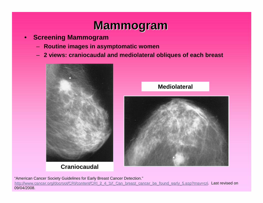
MammogramMammogram• Screening Mammogram
– Routine images in asymptomatic women– 2 views: craniocaudal and mediolateral obliques of each breast
““American Cancer Society Guidelines for Early Breast Cancer DetecAmerican Cancer Society Guidelines for Early Breast Cancer Detection.tion.””http://www.cancer.org/docroot/CRI/content/CRI_2_4_3X_Can_breast_http://www.cancer.org/docroot/CRI/content/CRI_2_4_3X_Can_breast_cancer_be_found_early_5.asp?rnav=cricancer_be_found_early_5.asp?rnav=cri. Last revised on. Last revised on09/04/2008.09/04/2008.
Craniocaudal
Mediolateral
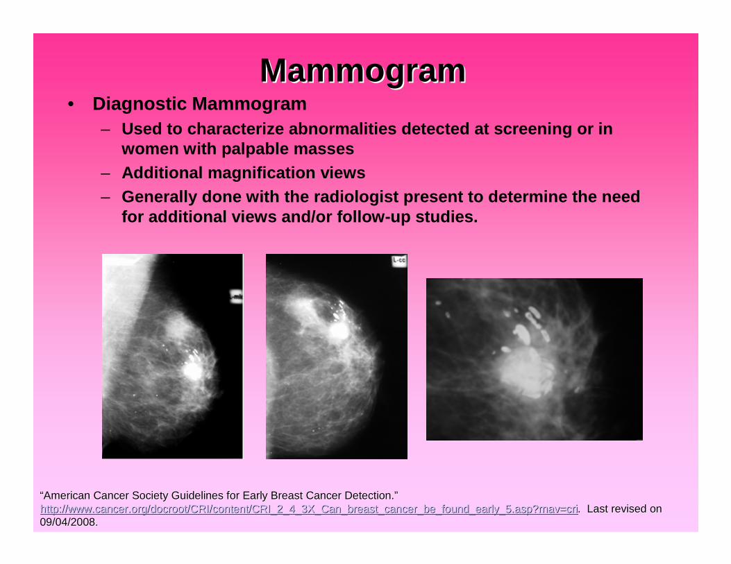
MammogramMammogram• Diagnostic Mammogram
– Used to characterize abnormalities detected at screening or inwomen with palpable masses
– Additional magnification views– Generally done with the radiologist present to determine the need
for additional views and/or follow-up studies.
““American Cancer Society Guidelines for Early Breast Cancer DetecAmerican Cancer Society Guidelines for Early Breast Cancer Detection.tion.””http://www.cancer.org/docroot/CRI/content/CRI_2_4_3X_Can_breast_http://www.cancer.org/docroot/CRI/content/CRI_2_4_3X_Can_breast_cancer_be_found_early_5.asp?rnav=cricancer_be_found_early_5.asp?rnav=cri. Last revised on. Last revised on09/04/2008.09/04/2008.

Mammogram Classification SystemMammogram Classification System• Breast Imaging Reporting and Data System (BI-RADS)
Characteristic of breast cancer.Highly suggestive ofmalignancy — appropriateaction should be taken
Category 5
Do not have the characteristicmorphologies of breast CA, but havedefinite probability of being malignant
Suspicious abnormality –biopsy should beconsidered
Category 4
Not expected to change over thefollow-up interval, but the radiologistwould prefer to establish its stability.
Probable benign finding —short interval follow-upsuggested
Category 3
Calcified fibroadenomas, secretorycalcifications, cysts, lipomas,hamartomas, etc.
Benign findingCategory 2
No findings to comment on.NegativeCategory 1
Screening situation. Need additionalmagnification views, spotcompression, U/S, etc.
Need additional imagingevaluation
Category 0
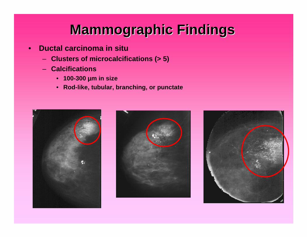
Mammographic FindingsMammographic Findings• Ductal carcinoma in situ
– Clusters of microcalcifications (> 5)– Calcifications
• 100-300 µµµµm in size• Rod-like, tubular, branching, or punctate
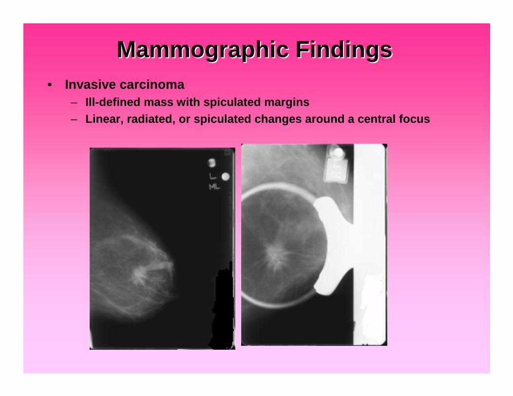
Mammographic FindingsMammographic Findings• Invasive carcinoma
– Ill-defined mass with spiculated margins– Linear, radiated, or spiculated changes around a central focus
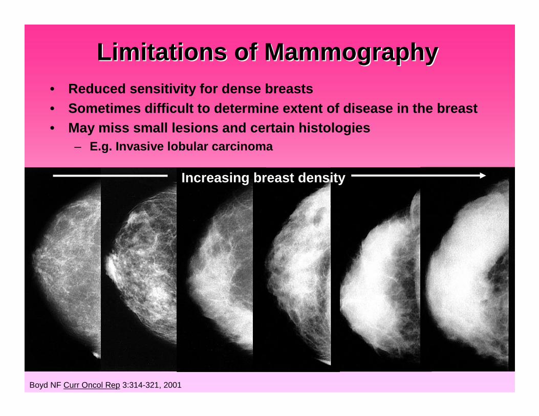
Limitations of MammographyLimitations of Mammography• Reduced sensitivity for dense breasts• Sometimes difficult to determine extent of disease in the breast• May miss small lesions and certain histologies
– E.g. Invasive lobular carcinoma
Boyd NF Curr Oncol Rep 3:314-321, 2001
Increasing breast density
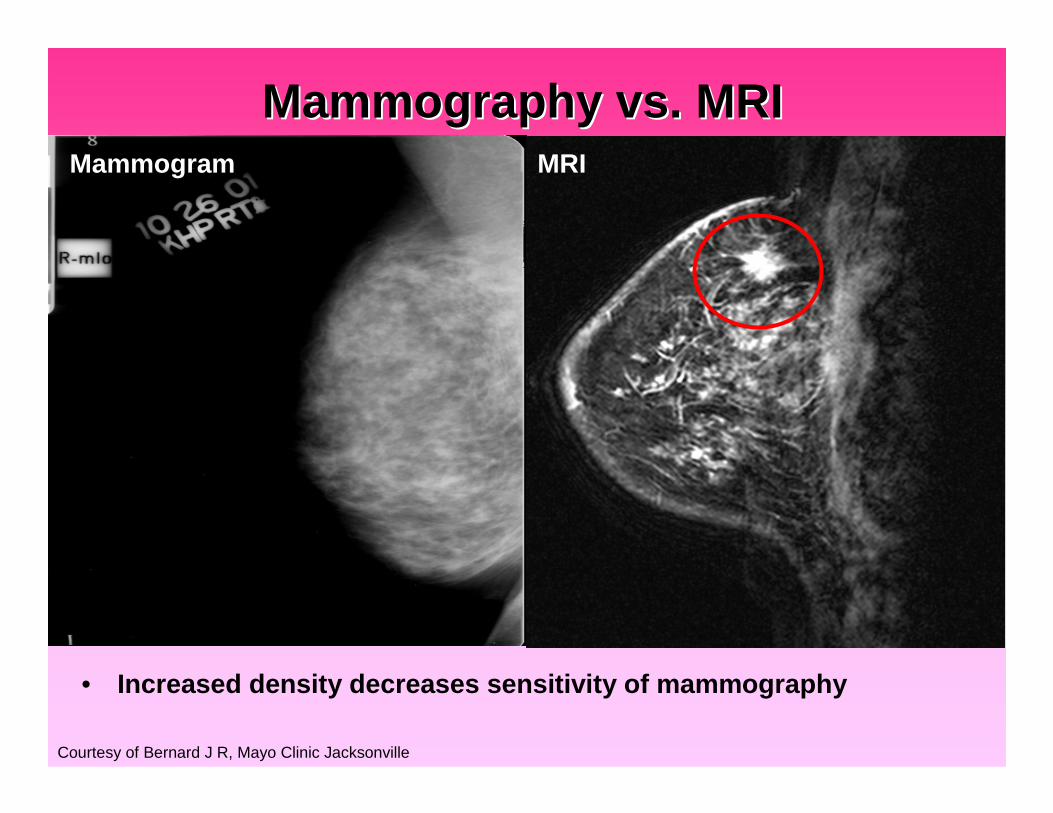
Mammography vs. MRIMammography vs. MRI
Courtesy of Bernard J R, Mayo Clinic Jacksonville
• Increased density decreases sensitivity of mammography
Mammogram MRI
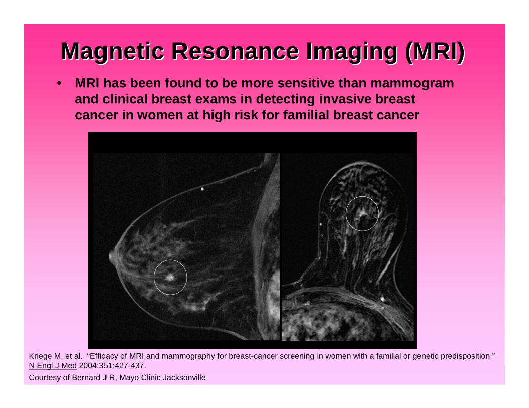
Magnetic Resonance Imaging (MRI)Magnetic Resonance Imaging (MRI)• MRI has been found to be more sensitive than mammogram
and clinical breast exams in detecting invasive breastcancer in women at high risk for familial breast cancer
Kriege M, et al. “Efficacy of MRI and mammography for breast-cancer screening in women with a familial or genetic predisposition.”N Engl J Med 2004;351:427-437.
Courtesy of Bernard J R, Mayo Clinic Jacksonville
Question #5Question #5• Magnetic Resonance Imaging (MRI) detects what percentage of
contralateral otherwise occult malignancies?
• A) < 1%• B) 3-4%• C) 10-15%• D) 20-25%• E) 35%
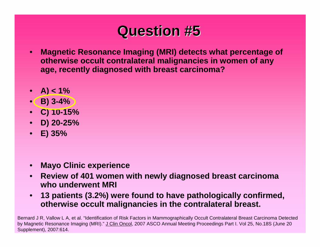
Question #5Question #5• Magnetic Resonance Imaging (MRI) detects what percentage of
otherwise occult contralateral malignancies in women of anyage, recently diagnosed with breast carcinoma?
• A) < 1%• B) 3-4%• C) 10-15%• D) 20-25%• E) 35%
• Mayo Clinic experience• Review of 401 women with newly diagnosed breast carcinoma
who underwent MRI• 13 patients (3.2%) were found to have pathologically confirmed,
otherwise occult malignancies in the contralateral breast.
Bernard J R, Vallow L A, et al. “Identification of Risk Factors in Mammographically Occult Contralateral Breast Carcinoma Detectedby Magnetic Resonance Imaging (MRI).” J Clin Oncol, 2007 ASCO Annual Meeting Proceedings Part I. Vol 25, No.18S (June 20Supplement), 2007:614.

Magnetic Resonance Imaging (MRI)Magnetic Resonance Imaging (MRI)• Mayo Clinic Jacksonville• Increased age is an independent risk factor for breast cancer• Further evaluation of the value of MRI in detecting occult
malignancies in the contralateral breast in women > 70 y/o– Retrospective review– 159 women > 70 y/o, with newly diagnosed breast cancer– 9 (5.7%) women were found to have synchronous,
pathologically confirmed, otherwise occult malignances in thecontralateral breast.
Bernard J R, Vallow L A, et al. “Mammographically Occult Contralateral Breast Carcinoma Detected by Magnetic Resonance Imagingin the Elderly.” J Clin Oncol 26:2008 (May 20 supplement; abstr 500).
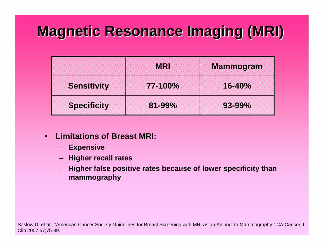
Magnetic Resonance Imaging (MRI)Magnetic Resonance Imaging (MRI)
• Limitations of Breast MRI:– Expensive– Higher recall rates– Higher false positive rates because of lower specificity than
mammography
Saslow D, et al. “American Cancer Society Guidelines for Breast Screening with MRI as an Adjunct to Mammography.” CA Cancer JClin 2007;57;75-89.
93-99%81-99%Specificity
16-40%77-100%Sensitivity
MammogramMRI
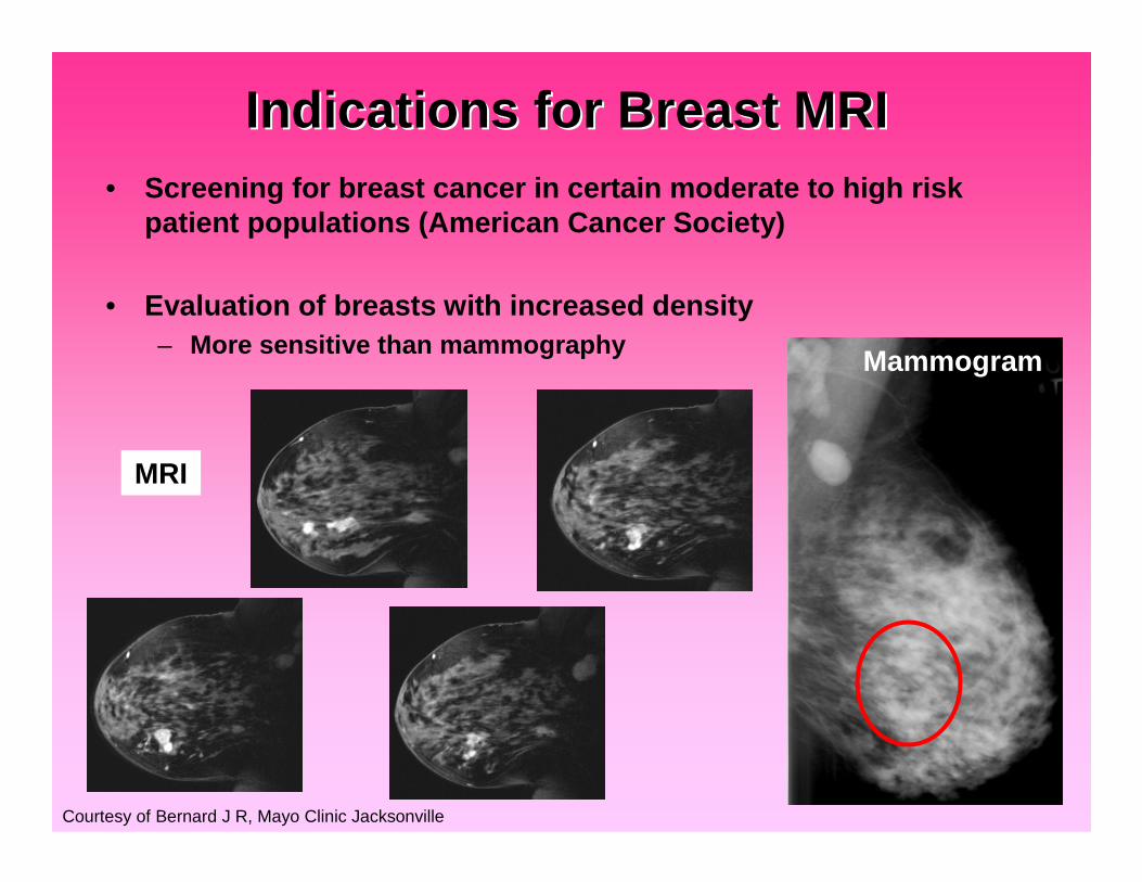
Indications for Breast MRIIndications for Breast MRI• Screening for breast cancer in certain moderate to high risk
patient populations (American Cancer Society)
• Evaluation of breasts with increased density– More sensitive than mammography
Courtesy of Bernard J R, Mayo Clinic Jacksonville
Mammogram
MRI
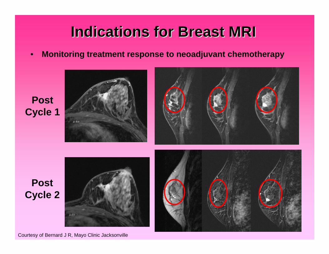
Indications for Breast MRIIndications for Breast MRI• Monitoring treatment response to neoadjuvant chemotherapy
PostCycle 1
PostCycle 2
Courtesy of Bernard J R, Mayo Clinic Jacksonville

Indications for Breast MRIIndications for Breast MRI
• Monitoring women with a personal history of breast cancer– Evaluate the extent of disease in the ipsilateral breast– Evaluate the presence of disease in the contralateral breast

Indications for Breast MRIIndications for Breast MRI
• Aid in surgical planning forbreast cancer treatment– Evaluate for the presence
of disease in multiplequadrants of the samebreast (multicentric)
– Breast conserving therapy(BCT) vs. mastectomy
1
23
4 5
Courtesy of Bernard J R, Mayo Clinic Jacksonville

SummarySummary• Breast cancer is a highly prevalent disease
• Improvements in screening have lead to an increase indetection of early stage breast cancers
• Detection of earlier stage breast cancers has allowed patientsmore treatment options and has decreased mortality rates
• Breast cancer is generally thought to be a slow growing diseasewith a propensity to spread lymphatically to the axilla anddistantly to bones.
• The greatest risk factor for breast cancer development isincreasing age
SummarySummary• American Cancer Society recommends
– Women ≥≥≥≥ 40 y/o:• Annual mammogram• Annual clinical breast exam
– Women with a moderate risk profile for developing breast cancer:• Should discuss the benefits and limitations of adding an annual
screening MRI
– Women with a high risk profile for developing breast cancer:• Begin screening at age 30 y/o• Annual mammogram and MRI• Annual Clinical Breast Exam
• MRI does not reliably detect calcifications, so it should not beused to replace mammography screening
SummarySummary• Breast MRI should be used for
– Screening in select groups of moderate to high risk women– Evaluation of breasts with increased density– Monitory neoadjuvant treatment response– Evaluation of extent of disease in women with a personal history of
breast cancer– Surgical planning for breast cancer treatment
• Breast conservation vs. mastectomy

Thank YouThank You
Questions?