an integrated raman and petrographic characterization of
TRANSCRIPT

Research article
Received: 2 August 2013 Revised: 21 October 2013 Accepted: 21 October 2013 Published online in Wiley Online Library: 18 December 2013
(wileyonlinelibrary.com) DOI 10.1002/jrs.4414
114
An integrated Raman and petrographiccharacterization of Italian mediaeval artifactsin pietra ollare (soapstone)Chiara Baita,a Pier Paolo Lottici,a Emma Salvioli-Mariani,a
Peter Vandenabeele,b Mauro Librenti,c Fabrizio Antonellid
and Danilo Bersania*
Ten fragments of pots made by pietra ollare (basic and ultrabasic metamorphic rocks belonging to greenschist facies) found inthe archaeological site of Crocetta di Sant’Agata Bolognese (Bologna, Italy) have been studied to characterize them and todefine their provenance. The fragments, dated between the 9th and 10th century AD, show traces of blackening from fire,indicating the use of these findings as pots by fire. The extensive use of a non-destructive technique, Raman spectroscopy,in both laboratory and portable forms, for the provenance analysis of pietra ollare artifacts has been evaluated. Micro-Raman spectroscopy was used for a detailed study of the main components, to identify the secondary minerals and to studythe distribution of the mineral phases in the samples. The samples show schistose texture, are fine grained and consist mainlyof talc and chlorite, and subordinate carbonates, oxides and serpentine. Olivine is rare. Based on the mineralogical composi-tion, the material can be defined as chlorite and carbonate talc-schist. Our samples of pietra ollare may be assigned to chloriteand magnesite-bearing talc-schists. The alpine areas of origin of this lithotype are Valtellina, Valchiavenna and Val Bregaglia.In particular, we can hypothesize that the origin of these findings is Valchiavenna, which had great commercial importance inthe Middle Ages. Further measurements, obtained with a portable Raman spectrometer, directly on a quarry near Chiavenna,support our hypothesis. Copyright © 2013 John Wiley & Sons, Ltd.
Keywords: micro-Raman spectroscopy; mobile Raman spectroscopy; in situ analysis; pietra ollare; archaeometry
* Correspondence to: Danilo Bersani, Physics and Earth Sciences, University ofParma, Parco Area delle Scienze 7/A, 43124 Parma, Italy. E-mail: [email protected]
a Dipartimento di Fisica e Scienze della Terra, Università di Parma, Parco Areadelle Scienze 7/A, 43124 Parma, Italy
b Ghent University, Department of Archaeology, Archaeometry Research Group,Sint-Pietesnieuwstraat 35, B-9000 Ghent, Belgium
c Dipartimento di Scienze dell’ Antichità e del Vicino Oriente, Università Ca’Foscari, Dorsoduro 3484/d, 30123 Venezia, Italy
d Dipartimento di Architettura Costruzione Conservazione, Università IUAV diVenezia, Badoer, San Polo 2468B, 30125 Venezia
Introduction
The Italian term pietra ollare (sometimes translated in English as‘soapstone’) hasn’t a specific petrological meaning. Usually itrefers to a metamorphic rock constituted prevalently by talc,magnesite and dolomite including chlorite, amphibole, mica,epidote and albite as accessory minerals. In the archaeologicalfield, this term is used to denote different lithotypes with distinctcomposition, colour and aspect, but having many physical andchemical characteristics in common. They are mostly composedof silicates with high chemical resistance to weathering and alsoto food cooking. They have good thermal stability up to very hightemperature (generally>1000 °C) before sintering and they areresistant to fast temperature changes assuring a regular linearexpansion.[1] Pietra ollare shows very low porosity despite itsschistose texture (total porosity is generally ≤1–3%, as a functionof the more or less marked schistosity[1]). Their principal mineralshave very low hardness (up to 4 in the Mohs scale);[2] they couldtherefore be easily processed with metallic instruments both byhand and lathe. Common lithotypes of pietra ollare are representedby grey to pale-green, medium- to fine-grained rocks containingpredominantly talc, chlorite and carbonates, but they can be morecorrectly classified in four main groups: chlorite-schist, talc-schist(sometimes amphibole-bearing talc-schists±carbonates), serpentine-schists and ultrabasites.In all cases, they are rocks that have been transformed in the
frame of the Alpine polyphasic metamorphic evolution and so
J. Raman Spectrosc. 2014, 45, 114–122
they are located along the metamorphic axes of the Alps; theserocks outcrop from the Sestri-Ponente-Voltaggio line near Genoato the Stiria. Due to its interesting physical–thermal features, thepietra ollare lithotypes were largely used between the LateRoman Empire and the Middle Ages, when the trade networkswiftly expanded becoming clearly extra-regional.
The present investigation is carried out on ten fragments ofpots made from pietra ollare, dated between 9th and 10th centuryAD, found in the archaeological site of Crocetta di Sant’AgataBolognese (Bologna, Italy).[3] The main scope of this study is todefine their geological–geographic provenance mainly byRaman measurements.
Copyright © 2013 John Wiley & Sons, Ltd.
Raman and petrographic characterization of soapstone
Examples of provenance studies on pietra ollare pots andmillstones are already present in literature.[4–8] They are usuallybased on standard petrological techniques as thin sections, X-raydiffraction (XRD) and scanning electron microscopy coupled withenergy-dispersive X-ray spectroscopy (SEM-EDX). Here, we wantto evaluate the potential of Raman spectroscopy in provenancestudies, as a first step to establish a completely non-destructiveway to characterize this kind of artifact. In particular, we test thecombination between the laboratory study of the archaeologicalfindings and the in situ study, with portable Raman equipment,directly on rocks in a Valchiavenna quarry. In recent years, the useofmobile and portable Raman systems is increasing for the analysisof archaeological and artistic objects and in situ identification ofminerals.[9–13] Nevertheless, to the authors’ knowledge, no workshave been published on the Raman study of archaeologicalfindings in pietra ollare, despite its very large diffusion.
Experimental
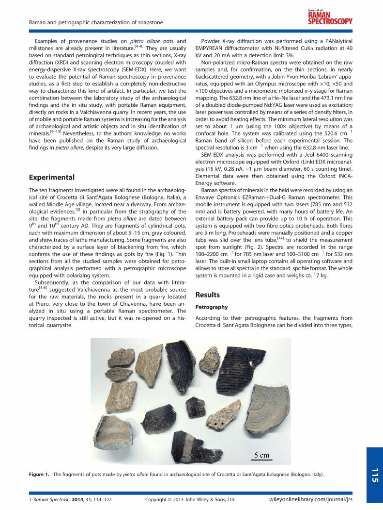
The ten fragments investigated were all found in the archaeolog-ical site of Crocetta di Sant’Agata Bolognese (Bologna, Italia), awalled Middle Age village, located near a riverway. From archae-ological evidences,[3] in particular from the stratigraphy of thesite, the fragments made from pietra ollare are dated between9th and 10th century AD. They are fragments of cylindrical pots,each with maximum dimension of about 5–15 cm, gray coloured,and show traces of lathe manufacturing. Some fragments are alsocharacterized by a surface layer of blackening from fire, whichconfirms the use of these findings as pots by fire (Fig. 1). Thinsections from all the studied samples were obtained for petro-graphical analysis performed with a petrographic microscopeequipped with polarizing system.
Subsequently, as the comparison of our data with litera-ture[5,6] suggested Valchiavenna as the most probable sourcefor the raw materials, the rocks present in a quarry locatedat Piuro, very close to the town of Chiavenna, have been an-alyzed in situ using a portable Raman spectrometer. Thequarry inspected is still active, but it was re-opened on a his-torical quarrysite.
Figure 1. The fragments of pots made by pietra ollare found in archaeologi
J. Raman Spectrosc. 2014, 45, 114–122 Copyright © 2013 John
Powder X-ray diffraction was performed using a PANalyticalEMPYREAN diffractometer with Ni-filtered CuKα radiation at 40kV and 20 mA with a detection limit 3%.
Non-polarized micro-Raman spectra were obtained on the rawsamples and, for confirmation, on the thin sections, in nearlybackscattered geometry, with a Jobin-Yvon Horiba ‘Labram’ appa-ratus, equipped with an Olympus microscope with ×10, ×50 and×100 objectives and a micrometric motorized x–y stage for Ramanmapping. The 632.8 nm line of a He–Ne laser and the 473.1 nm lineof a doubled diode-pumped Nd:YAG laser were used as excitation;laser power was controlled by means of a series of density filters, inorder to avoid heating effects. The minimum lateral resolution wasset to about 1 μm (using the 100× objective) by means of aconfocal hole. The system was calibrated using the 520.6 cm�1
Raman band of silicon before each experimental session. Thespectral resolution is 3 cm�1 when using the 632.8 nm laser line.
SEM-EDX analysis was performed with a Jeol 6400 scanningelectron microscope equipped with Oxford (Link) EDX microanal-ysis (15 kV, 0.28 nA, ~1 μm beam diameter, 60 s counting time).Elemental data were then obtained using the Oxford INCA-Energy software.
Raman spectra ofminerals in the field were recorded by using anEnwave Optronics EZRaman-I-Dual-G Raman spectrometer. Thismobile instrument is equipped with two lasers (785 nm and 532nm) and is battery powered, with many hours of battery life. Anexternal battery pack can provide up to 10 h of operation. Thissystem is equipped with two fibre-optics probeheads. Both fibresare 5 m long. Probeheads were manually positioned and a coppertube was slid over the lens tube,[14] to shield the measurementspot from sunlight (Fig. 2). Spectra are recorded in the range100–2200 cm�1 for 785 nm laser and 100–3100 cm�1 for 532 nmlaser. The built-in small laptop contains all operating software andallows to store all spectra in the standard .spc file format. The wholesystem is mounted in a rigid case and weighs ca. 17 kg.
Results
Petrography
According to their petrographic features, the fragments fromCrocetta di Sant’Agata Bolognese can be divided into three types,
cal site of Crocetta di Sant’Agata Bolognese (Bologna, Italy).
Wiley & Sons, Ltd. wileyonlinelibrary.com/journal/jrs
115

Figure 2. The portable Raman apparatus used in the Piuro quarry in Valchiavenna (Italy). In the inset, a detail of the probehead with the copper tubeacting as a spacer and a light shield.
C. Baita et al.
116
with small variations within each one: (1) carbonate (magnesite)-bearing talc-schists, (2) pure talc-schists, (3) talc-carbonate (mag-nesite and dolomite)-bearing chlorite-schists. Sample #9 can beconsidered an isolated case.The talc-schists of the first group have a texture which varies
from schistosity with more or less well-developed microfolds(#1, #2, #3) (Fig. 3A, B) to a very deformed texture without anypreferred orientation (#4, #5) (Fig. 3C). Talc and magnesiumcarbonate (magnesite, rare dolomite only in #4) are the mainminerals, with variable relative abundance among the samples.Talc occurs as slightly coloured or colourless flakes, with variablesize. The flakes are sometimes arranged to define a good foliationeven if some small deformation as microfolds occurs (Fig. 3B),sometimes without any preferential orientation giving to the rocka highly disordered texture (Fig. 3C). Larger lamellas cut theschistosity and thus they are subsequent to the foliation-formingdeformation. Small crystals of magnesite are often idiomorphic,with rhombohedral shape (Fig.3D); they are grown up along thefoliation and help to describe the microfolds (Fig. 3B), suggestingrecrystallization contemporaneously to the deformation. Thelarger crystals are pecilitic and contain small laminas of talc whichare inflected in continuity with the rock foliation suggestingcrystal growth during the formation of the foliation (Fig. 3E).Chlorite is rare or absent, it is slightly coloured and forms smallfibrous-radial aggregates; rarely flakes of chlorite are associatedwith talc. In the sample #3, rarely serpentine occurs, associatedwith talc.The talc-schists of the second group have an extremely
regular schistose texture (Fig. 3F), without any successivedeformation (#6, #7). Talc is the main mineral, as colourlessor slightly coloured flakes. Abundant opaque minerals occur,above all in the sample #6; they are placed in some areasassociated with rare chlorite, colourless larger laminas of talc,discordant with respect to the foliation, and serpentine. These aggre-gates constituted by talc+opaque minerals + chlorite± serpentine(#6) or by serpentine+ talc +opaque minerals (#7) are probably theproduct of the transformation of olivine (Fig. 3F).
wileyonlinelibrary.com/journal/jrs Copyright © 2013 John
The fragments belonging to the third group are characterized bya schistose texture withmicrofolds defined by flakes of chlorite (#8).Colourless chlorite is the main mineral; thus, the rock can be de-fined as chlorite-schist (Fig. 3G). Small laminas of talc also occurand they are associated to chlorite to form the bulk foliation,whereas larger laminas cut the foliation (Fig. 3H). Small crystals ofmagnesite, very rarely dolomite, form aggregates often discordantwith the foliation. Some of them contain small laminas of talc whichshow an orientation completely different from bulk foliation; thissuggests crystal growth after formation of schistosity (Fig. 3I).
The isolated sample (#9) is formed by talc in large laths associ-ated to form a cluster, without any preferential orientation(Fig. 3L). Chlorite is subordinate but closely associated with talcand with the same features. Opaque minerals are abundant andare associated with red-orange coloured material, difficult toidentify. In these areas, rare calcite and serpentine also occurand they are probably the product of transformation of olivineand pyroxene. The results are summarized in Table 1
Micro-Raman spectroscopy
Raman analysis shows that the main component of the fragmentsis talc Mg3Si4O10(OH)2, identified by themain band at 676 cm�1, at-tributed to the Si–Ob–Si vibration (Ob=bridging oxygen).[15] OtherRaman bands of talc are at 1052 cm�1, due to the Si–Onb stretching(Onb=non-bridging oxygen), at 195 cm�1 and 363 cm�1 (Fig. 4).The Raman band positions of themineral phases found in our sam-ples are summarized in Table 2.
The second phase, in terms of abundance, in some fragments, is amember of the chlorite group (Fig. 4). It is distinguishable from talcthanks to the band at 547 cm�1, typical of Fe-bearingphyllosilicates.[15] The other bands are at 199 cm�1, typical of manytrioctahedral phyllosilicates, at 354 cm�1, common formostMgbear-ing phyllosilicates and a band at 679 cm�1 (Si–Ob–Si vibration). Fromthe Raman spectra, it is difficult to determine the exact member ofthe chlorite family present in our samples. We can make use ofmicro-Raman mapping[16] to study the chlorite lamellae orientation.
Wiley & Sons, Ltd. J. Raman Spectrosc. 2014, 45, 114–122
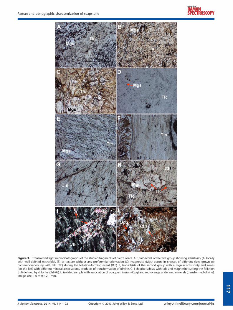
Figure 3. Transmitted light microphotographs of the studied fragments of pietra ollare. A-E, talc-schist of the first group showing schistosity (A) locallywith well-defined microfolds (B) or texture without any preferential orientation (C); magnesite (Mgs) occurs in crystals of different sizes grown upcontemporaneously with talc (Tlc) during the foliation-forming event (D,E). F, talc-schists of the second group with a regular schistosity and zones(on the left) with different mineral associations, products of transformation of olivine. G–I chlorite-schists with talc and magnesite cutting the foliation(H,I) defined by chlorite (Chl) (G). L, isolated sample with association of opaque minerals (Opq) and red–orange undefined minerals (transformed olivine).Image size: 1.6 mm×2.1 mm.
Raman and petrographic characterization of soapstone
J. Raman Spectrosc. 2014, 45, 114–122 Copyright © 2013 John Wiley & Sons, Ltd. wileyonlinelibrary.com/journal/jrs
117
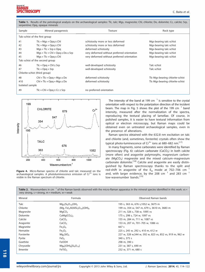
Table 1. Results of the petrological analysis on the archaeological samples: Tlc, talc; Mgs, magnesite; Chl, chlorite; Do, dolomite; Cc, calcite; Srp,serpentine; Opq, opaque minerals
Sample Mineral paragenesis Texture Rock type
Talc-schist of the first group
#1 Tlc +Mgs +Opq±Chl schistosity more or less deformed Mgs-bearing talc-schist
#2 Tlc +Mgs +Opq±Chl schistosity more or less deformed Mgs-bearing talc-schist
#3 Mgs+ Tlc + Srp ±Opq deformed schistosity Mgs-bearing talc-schist
#4 Mgs+ Tlc + Chl +Opq±Do± Srp very deformed without preferred orientation Mgs-bearing talc-schist
#5 Mgs+ Tlc +Opq±Chl very deformed without preferred orientation Mgs-bearing talc-schist
Talc-schist of the second group
#6 Tlc +Opq+Chl ± Srp well-developed schistosity Talc-schist
#7 Tlc +Opq+ Srp well-developed schistosity Talc-schist
Chlorite-schist (third group)
#8 Chl + Tlc +Opq+Mgs±Do deformed schistosity Tlc-Mgs-bearing chlorite-schist
#10 Chl + Tlc +Opq+Mgs±Do deformed schistosity Tlc-Mgs-bearing chlorite-schist
Isolated sample
#9 Tlc + Chl +Opq±Cc± Srp no preferred orientation
Figure 4. Micro-Raman spectra of chlorite and talc measured on thearchaeological samples. A photoluminescence emission of Cr3+ ions isvisible in the Raman spectrum of chlorite.
Table 2. Wavenumbers in cm�1 of the Raman bands observed with the mvery strong, s = strong, m=medium, w=weak
Mineral Formula
Talc Mg3(Si4O10)OH2
Chlorite (Mg, Fe)5Al(AlSi3O10)(OH)8Magnesite MgCO3
Dolomite CaMg(CO3)2Calcite CaCO3
Aragonite CaCO3
Magnetite Fe3O4
Hematite Fe2O3
Forsterite Mg2SiO4
Pyrite FeS2Goethite FeOOH
Chrysotile Mg6[(OH)8|Si4O10]
Ilmenite FeTiO3
C. Baita et al.
wileyonlinelibrary.com/journal/jrs Copyright © 2013 John
118
The intensity of the band at 199 cm�1 is sensitive to the crystalorientation with respect to the polarization direction of the incidentbeam. The map in Fig. 5 shows the plot of the 199 cm�1 bandintensity, measured after the normalization of the spectra,reproducing the textural placing of lamellae. Of course, inpolished samples, it is easier to have textural information fromoptical or electron microscopy, but Raman maps could beobtained even on untreated archaeological samples, even inthe presence of alterations.
Raman spectra obtained with the 632.8 nm excitation on talc
and chlorite (and, sometimes, forsterite) crystals often show the
typical photo-luminescence of Cr3+ ions at 680–682 nm.[17]
In many fragments, some carbonates were identified by Ramanspectroscopy (Fig. 6): calcium carbonate (CaCO3) in both calcite(more often) and aragonite polymorphs, magnesium carbon-ate (MgCO3) magnesite and the mixed calcium–magnesiumcarbonate dolomite.[18] Calcite and aragonite are easily distin-guished by Raman spectroscopy thanks to the split andred-shift in aragonite of the Eg mode at 702–706 cm�1
and, with larger evidence, by the 208 cm�1 and 283 cm�1
low-wavenumber bands.[19]
icro-Raman apparatus in the mineral species identified in this work. vs =
Observed Raman bands
195 s, 363 m, 676 s,1052 w, 3675 m
199 vs, 354 w, 547 m, 679 s, 3610 m, 3683 m
211 m, 326 s, 738 w, 1095 vs
175 s, 298 s, 724 w, 1097 vs
155 m, 284 m, 711 w, 1087 vs
153 m, 207 m, 701–705 w, 1086 vs
667 s
225 s, 245 w, 292 s, 410 m, 612 w
227 w, 328 w,544 w, 592 w, 823 vs, 852 vs, 919 m, 962 w
340 s, 375 s
298 m, 390 s
231 w, 387 s, 690 m
232 w, 371 m, 680 s
Wiley & Sons, Ltd. J. Raman Spectrosc. 2014, 45, 114–122
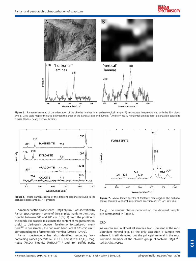
Figure 5. Raman micro-map of the orientation of the chlorite laminas in an archaeological sample. A) microscope image obtained with the 50× objec-tive. B) Grey-scale map of the ratio between the areas of the bands at 681 and 200 cm�1. White = nearly horizontal laminas (laser polarization parallel toc axis). Black = nearly vertical laminas.
Figure 6. Micro-Raman spectra of the different carbonates found in thearchaeological samples. * = gypsum.
Figure 7. Micro-Raman spectra of forsterite measured on the archaeo-logical samples. A photoluminescence emission of Cr3+ ions is visible.
Raman and petrographic characterization of soapstone
119
Amember of the olivine series – (Mg,Fe)2SiO4 –was identified byRaman spectroscopy in some of the samples, thanks to the strongdoublet between 800 and 900 cm�1 (Fig. 7): from the position ofthe bands, it is possible to estimate the content ofmagnesium/iron,useful to distinguish between fayalite- or forsterite-rich mem-bers.[20] In our samples, the two main bands are at 823–855 cm�1,corresponding to a forsterite-rich member (90%Fo–10%Fa).
Raman spectroscopy has also identified secondary iron-containing oxides goethite (α-FeOOH), hematite (α-Fe2O3), mag-netite (Fe3O4), ilmenite (FeTiO3)
[21,22] and iron sulfide pyrite
J. Raman Spectrosc. 2014, 45, 114–122 Copyright © 2013 John
(FeS2). The various phases detected on the different samplesare summarized in Table 3.
XRD
As we can see, in almost all samples, talc is present as the mostabundant mineral (Fig. 8); the only exception is sample #10,where it is still detected but the principal mineral is the mostcommon member of the chlorite group: clinochlore (Mg,Fe2+)
5Al(Si3Al)O10(OH)8.
Wiley & Sons, Ltd. wileyonlinelibrary.com/journal/jrs
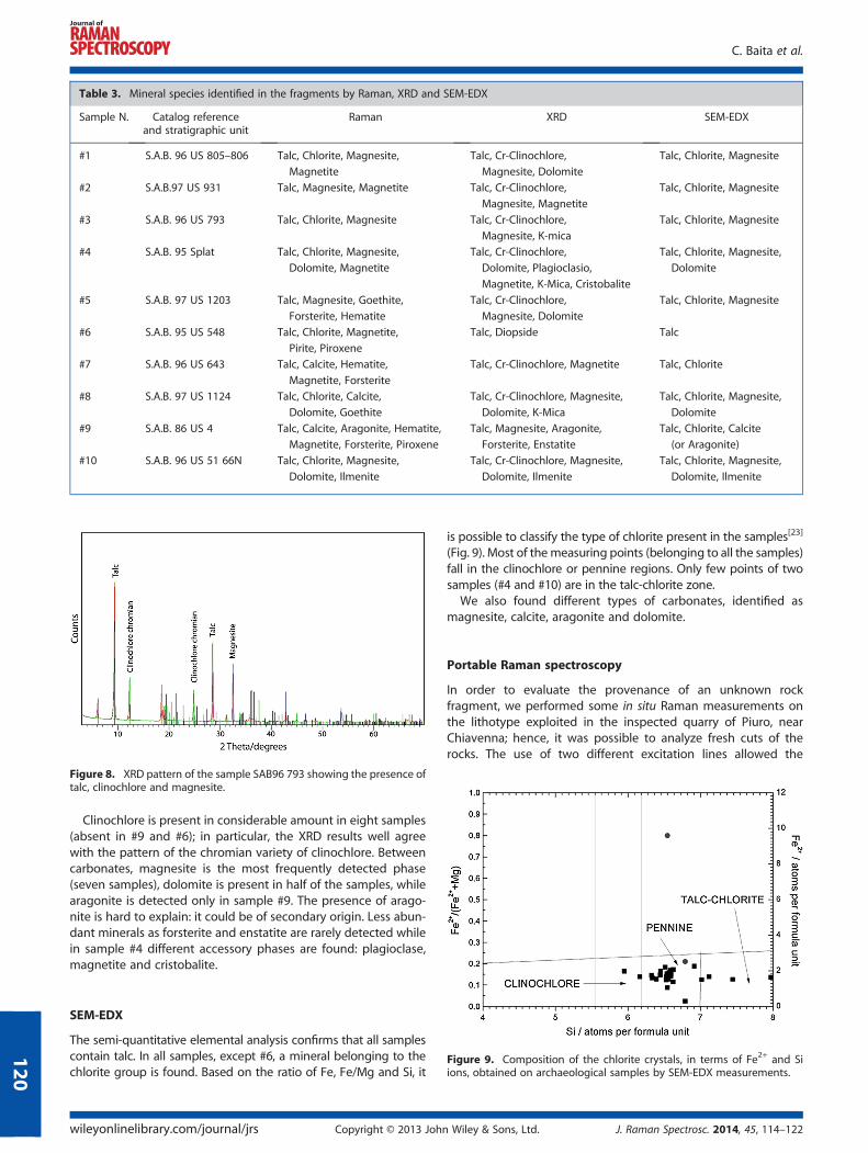
Figure 8. XRD pattern of the sample SAB96 793 showing the presence oftalc, clinochlore and magnesite.
Table 3. Mineral species identified in the fragments by Raman, XRD and SEM-EDX
Sample N. Catalog referenceand stratigraphic unit
Raman XRD SEM-EDX
#1 S.A.B. 96 US 805–806 Talc, Chlorite, Magnesite,
Magnetite
Talc, Cr-Clinochlore,
Magnesite, Dolomite
Talc, Chlorite, Magnesite
#2 S.A.B.97 US 931 Talc, Magnesite, Magnetite Talc, Cr-Clinochlore,
Magnesite, Magnetite
Talc, Chlorite, Magnesite
#3 S.A.B. 96 US 793 Talc, Chlorite, Magnesite Talc, Cr-Clinochlore,
Magnesite, K-mica
Talc, Chlorite, Magnesite
#4 S.A.B. 95 Splat Talc, Chlorite, Magnesite,
Dolomite, Magnetite
Talc, Cr-Clinochlore,
Dolomite, Plagioclasio,
Magnetite, K-Mica, Cristobalite
Talc, Chlorite, Magnesite,
Dolomite
#5 S.A.B. 97 US 1203 Talc, Magnesite, Goethite,
Forsterite, Hematite
Talc, Cr-Clinochlore,
Magnesite, Dolomite
Talc, Chlorite, Magnesite
#6 S.A.B. 95 US 548 Talc, Chlorite, Magnetite,
Pirite, Piroxene
Talc, Diopside Talc
#7 S.A.B. 96 US 643 Talc, Calcite, Hematite,
Magnetite, Forsterite
Talc, Cr-Clinochlore, Magnetite Talc, Chlorite
#8 S.A.B. 97 US 1124 Talc, Chlorite, Calcite,
Dolomite, Goethite
Talc, Cr-Clinochlore, Magnesite,
Dolomite, K-Mica
Talc, Chlorite, Magnesite,
Dolomite
#9 S.A.B. 86 US 4 Talc, Calcite, Aragonite, Hematite,
Magnetite, Forsterite, Piroxene
Talc, Magnesite, Aragonite,
Forsterite, Enstatite
Talc, Chlorite, Calcite
(or Aragonite)
#10 S.A.B. 96 US 51 66N Talc, Chlorite, Magnesite,
Dolomite, Ilmenite
Talc, Cr-Clinochlore, Magnesite,
Dolomite, Ilmenite
Talc, Chlorite, Magnesite,
Dolomite, Ilmenite
C. Baita et al.
120
Clinochlore is present in considerable amount in eight samples(absent in #9 and #6); in particular, the XRD results well agreewith the pattern of the chromian variety of clinochlore. Betweencarbonates, magnesite is the most frequently detected phase(seven samples), dolomite is present in half of the samples, whilearagonite is detected only in sample #9. The presence of arago-nite is hard to explain: it could be of secondary origin. Less abun-dant minerals as forsterite and enstatite are rarely detected whilein sample #4 different accessory phases are found: plagioclase,magnetite and cristobalite.
Figure 9. Composition of the chlorite crystals, in terms of Fe2+ and Siions, obtained on archaeological samples by SEM-EDX measurements.
SEM-EDX
The semi-quantitative elemental analysis confirms that all samplescontain talc. In all samples, except #6, a mineral belonging to thechlorite group is found. Based on the ratio of Fe, Fe/Mg and Si, it
wileyonlinelibrary.com/journal/jrs Copyright © 2013 John
is possible to classify the type of chlorite present in the samples[23]
(Fig. 9). Most of the measuring points (belonging to all the samples)fall in the clinochlore or pennine regions. Only few points of twosamples (#4 and #10) are in the talc-chlorite zone.
We also found different types of carbonates, identified asmagnesite, calcite, aragonite and dolomite.
Portable Raman spectroscopy
In order to evaluate the provenance of an unknown rockfragment, we performed some in situ Raman measurements onthe lithotype exploited in the inspected quarry of Piuro, nearChiavenna; hence, it was possible to analyze fresh cuts of therocks. The use of two different excitation lines allowed the
Wiley & Sons, Ltd. J. Raman Spectrosc. 2014, 45, 114–122
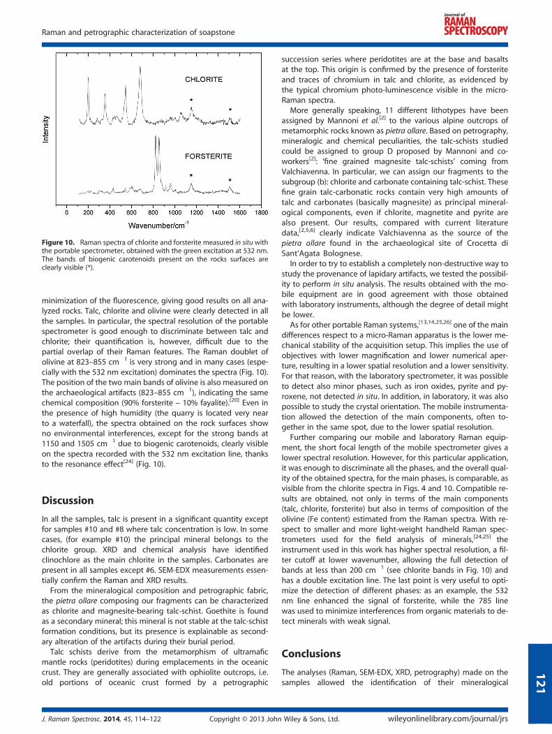
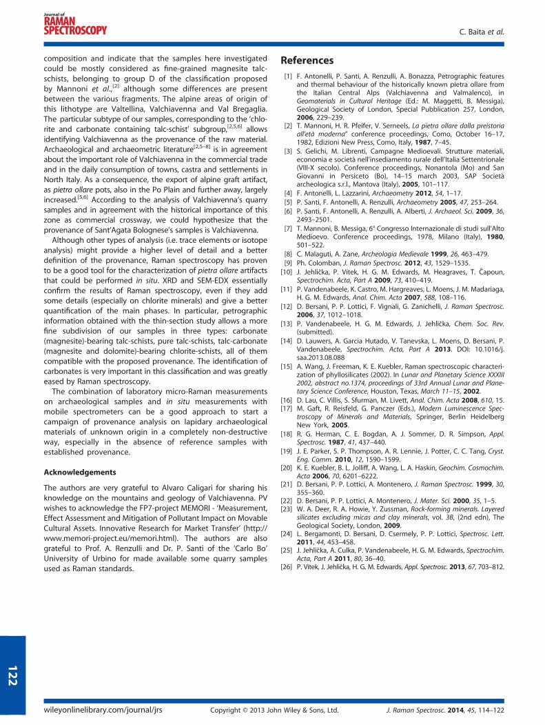
Figure 10. Raman spectra of chlorite and forsterite measured in situ withthe portable spectrometer, obtained with the green excitation at 532 nm.The bands of biogenic carotenoids present on the rocks surfaces areclearly visible (*).
Raman and petrographic characterization of soapstone
minimization of the fluorescence, giving good results on all ana-lyzed rocks. Talc, chlorite and olivine were clearly detected in allthe samples. In particular, the spectral resolution of the portablespectrometer is good enough to discriminate between talc andchlorite; their quantification is, however, difficult due to thepartial overlap of their Raman features. The Raman doublet ofolivine at 823–855 cm�1 is very strong and in many cases (espe-cially with the 532 nm excitation) dominates the spectra (Fig. 10).The position of the two main bands of olivine is also measured onthe archaeological artifacts (823–855 cm�1), indicating the samechemical composition (90% forsterite – 10% fayalite).[20] Even inthe presence of high humidity (the quarry is located very nearto a waterfall), the spectra obtained on the rock surfaces showno environmental interferences, except for the strong bands at1150 and 1505 cm�1 due to biogenic carotenoids, clearly visibleon the spectra recorded with the 532 nm excitation line, thanksto the resonance effect[24] (Fig. 10).
121
Discussion
In all the samples, talc is present in a significant quantity exceptfor samples #10 and #8 where talc concentration is low. In somecases, (for example #10) the principal mineral belongs to thechlorite group. XRD and chemical analysis have identifiedclinochlore as the main chlorite in the samples. Carbonates arepresent in all samples except #6. SEM-EDX measurements essen-tially confirm the Raman and XRD results.
From the mineralogical composition and petrographic fabric,the pietra ollare composing our fragments can be characterizedas chlorite and magnesite-bearing talc-schist. Goethite is foundas a secondary mineral; this mineral is not stable at the talc-schistformation conditions, but its presence is explainable as second-ary alteration of the artifacts during their burial period.
Talc schists derive from the metamorphism of ultramaficmantle rocks (peridotites) during emplacements in the oceaniccrust. They are generally associated with ophiolite outcrops, i.e.old portions of oceanic crust formed by a petrographic
J. Raman Spectrosc. 2014, 45, 114–122 Copyright © 2013 John
succession series where peridotites are at the base and basaltsat the top. This origin is confirmed by the presence of forsteriteand traces of chromium in talc and chlorite, as evidenced bythe typical chromium photo-luminescence visible in the micro-Raman spectra.
More generally speaking, 11 different lithotypes have beenassigned by Mannoni et al.[2] to the various alpine outcrops ofmetamorphic rocks known as pietra ollare. Based on petrography,mineralogic and chemical peculiarities, the talc-schists studiedcould be assigned to group D proposed by Mannoni and co-workers[2]: ‘fine grained magnesite talc-schists’ coming fromValchiavenna. In particular, we can assign our fragments to thesubgroup (b): chlorite and carbonate containing talc-schist. Thesefine grain talc-carbonatic rocks contain very high amounts oftalc and carbonates (basically magnesite) as principal mineral-ogical components, even if chlorite, magnetite and pyrite arealso present. Our results, compared with current literaturedata,[2,5,6] clearly indicate Valchiavenna as the source of thepietra ollare found in the archaeological site of Crocetta diSant’Agata Bolognese.
In order to try to establish a completely non-destructive way tostudy the provenance of lapidary artifacts, we tested the possibil-ity to perform in situ analysis. The results obtained with the mo-bile equipment are in good agreement with those obtainedwith laboratory instruments, although the degree of detail mightbe lower.
As for other portable Raman systems,[13,14,25,26] one of the maindifferences respect to a micro-Raman apparatus is the lower me-chanical stability of the acquisition setup. This implies the use ofobjectives with lower magnification and lower numerical aper-ture, resulting in a lower spatial resolution and a lower sensitivity.For that reason, with the laboratory spectrometer, it was possibleto detect also minor phases, such as iron oxides, pyrite and py-roxene, not detected in situ. In addition, in laboratory, it was alsopossible to study the crystal orientation. The mobile instrumenta-tion allowed the detection of the main components, often to-gether in the same spot, due to the lower spatial resolution.
Further comparing our mobile and laboratory Raman equip-ment, the short focal length of the mobile spectrometer gives alower spectral resolution. However, for this particular application,it was enough to discriminate all the phases, and the overall qual-ity of the obtained spectra, for the main phases, is comparable, asvisible from the chlorite spectra in Figs. 4 and 10. Compatible re-sults are obtained, not only in terms of the main components(talc, chlorite, forsterite) but also in terms of composition of theolivine (Fe content) estimated from the Raman spectra. With re-spect to smaller and more light-weight handheld Raman spec-trometers used for the field analysis of minerals,[24,25] theinstrument used in this work has higher spectral resolution, a fil-ter cutoff at lower wavenumber, allowing the full detection ofbands at less than 200 cm�1 (see chlorite bands in Fig. 10) andhas a double excitation line. The last point is very useful to opti-mize the detection of different phases: as an example, the 532nm line enhanced the signal of forsterite, while the 785 linewas used to minimize interferences from organic materials to de-tect minerals with weak signal.
Conclusions
The analyses (Raman, SEM-EDX, XRD, petrography) made on thesamples allowed the identification of their mineralogical
Wiley & Sons, Ltd. wileyonlinelibrary.com/journal/jrs
C. Baita et al.
122
composition and indicate that the samples here investigatedcould be mostly considered as fine-grained magnesite talc-schists, belonging to group D of the classification proposedby Mannoni et al.,[2] although some differences are presentbetween the various fragments. The alpine areas of origin ofthis lithotype are Valtellina, Valchiavenna and Val Bregaglia.The particular subtype of our samples, corresponding to the ‘chlo-rite and carbonate containing talc-schist’ subgroup,[2,5,6] allowsidentifying Valchiavenna as the provenance of the raw material.Archaeological and archaeometric literature[2,5–8] is in agreementabout the important role of Valchiavenna in the commercial tradeand in the daily consumption of towns, castra and settlements inNorth Italy. As a consequence, the export of alpine graft artifact,as pietra ollare pots, also in the Po Plain and further away, largelyincreased.[5,6] According to the analysis of Valchiavenna’s quarrysamples and in agreement with the historical importance of thiszone as commercial crossway, we could hypothesize that theprovenance of Sant’Agata Bolognese’s samples is Valchiavenna.Although other types of analysis (i.e. trace elements or isotope
analysis) might provide a higher level of detail and a betterdefinition of the provenance, Raman spectroscopy has provento be a good tool for the characterization of pietra ollare artifactsthat could be performed in situ. XRD and SEM-EDX essentiallyconfirm the results of Raman spectroscopy, even if they addsome details (especially on chlorite minerals) and give a betterquantification of the main phases. In particular, petrographicinformation obtained with the thin-section study allows a morefine subdivision of our samples in three types: carbonate(magnesite)-bearing talc-schists, pure talc-schists, talc-carbonate(magnesite and dolomite)-bearing chlorite-schists, all of themcompatible with the proposed provenance. The identification ofcarbonates is very important in this classification and was greatlyeased by Raman spectroscopy.The combination of laboratory micro-Raman measurements
on archaeological samples and in situ measurements withmobile spectrometers can be a good approach to start acampaign of provenance analysis on lapidary archaeologicalmaterials of unknown origin in a completely non-destructiveway, especially in the absence of reference samples withestablished provenance.
Acknowledgements
The authors are very grateful to Alvaro Caligari for sharing hisknowledge on the mountains and geology of Valchiavenna. PVwishes to acknowledge the FP7-project MEMORI - ‘Measurement,Effect Assessment and Mitigation of Pollutant Impact on MovableCultural Assets. Innovative Research for Market Transfer’ (http://www.memori-project.eu/memori.html). The authors are alsograteful to Prof. A. Renzulli and Dr. P. Santi of the ‘Carlo Bo’University of Urbino for made available some quarry samplesused as Raman standards.
wileyonlinelibrary.com/journal/jrs Copyright © 2013 John
References[1] F. Antonelli, P. Santi, A. Renzulli, A. Bonazza, Petrographic features
and thermal behaviour of the historically known pietra ollare fromthe Italian Central Alps (Valchiavenna and Valmalenco), inGeomaterials in Cultural Heritage (Ed.: M. Maggetti, B. Messiga),Geological Society of London, Special Pubblication 257, London,2006, 229–239.
[2] T. Mannoni, H. R. Pfeifer, V. Serneels, La pietra ollare dalla preistoriaall’età moderna” conference proceedings, Como, October 16–17,1982, Edizioni New Press, Como, Italy, 1987, 7–45.
[3] S. Gelichi, M. Librenti, Campagne Medioevali. Strutture materiali,economia e società nell’insediamento rurale dell’Italia Settentrionale(VIII-X secolo). Conference proceedings, Nonantola (Mo) and SanGiovanni in Persiceto (Bo), 14–15 march 2003, SAP Societàarcheologica s.r.l., Mantova (Italy), 2005, 101–117.
[4] F. Antonelli, L. Lazzarini, Archaeometry 2012, 54, 1–17.[5] P. Santi, F. Antonelli, A. Renzulli, Archaeometry 2005, 47, 253–264.[6] P. Santi, F. Antonelli, A. Renzulli, A. Alberti, J. Archaeol. Sci. 2009, 36,
2493–2501.[7] T. Mannoni, B. Messiga, 6° Congresso Internazionale di studi sull’Alto
Medioevo. Conference proceedings, 1978, Milano (Italy), 1980,501–522.
[8] C. Malaguti, A. Zane, Archeologia Medievale 1999, 26, 463–479.[9] Ph. Colomban, J. Raman Spectrosc. 2012, 43, 1529–1535.
[10] J. Jehlička, P. Vítek, H. G. M. Edwards, M. Heagraves, T. Čapoun,Spectrochim. Acta, Part A 2009, 73, 410–419.
[11] P. Vandenabeele, K. Castro, M. Hargreaves, L. Moens, J. M. Madariaga,H. G. M. Edwards, Anal. Chim. Acta 2007, 588, 108–116.
[12] D. Bersani, P. P. Lottici, F. Vignali, G. Zanichelli, J. Raman Spectrosc.2006, 37, 1012–1018.
[13] P. Vandenabeele, H. G. M. Edwards, J. Jehlička, Chem. Soc. Rev.(submitted).
[14] D. Lauwers, A. Garcia Hutado, V. Tanevska, L. Moens, D. Bersani, P.Vandenabeele, Spectrochim. Acta, Part A 2013. DOI: 10.1016/j.saa.2013.08.088
[15] A. Wang, J. Freeman, K. E. Kuebler, Raman spectroscopic characteri-zation of phyllosilicates (2002). In Lunar and Planetary Science XXXIII2002, abstract no.1374, proceedings of 33rd Annual Lunar and Plane-tary Science Conference, Houston, Texas, March 11–15, 2002.
[16] D. Lau, C. Villis, S. Sfurman, M. Livett, Anal. Chim. Acta 2008, 610, 15.[17] M. Gaft, R. Reisfeld, G. Panczer (Eds.), Modern Luminescence Spec-
troscopy of Minerals and Materials, Springer, Berlin HeidelbergNew York, 2005.
[18] R. G. Herman, C. E. Bogdan, A. J. Sommer, D. R. Simpson, Appl.Spectrosc. 1987, 41, 437–440.
[19] J. E. Parker, S. P. Thompson, A. R. Lennie, J. Potter, C. C. Tang, Cryst.Eng. Comm. 2010, 12, 1590–1599.
[20] K. E. Kuebler, B. L. Jolliff, A. Wang, L. A. Haskin, Geochim. Cosmochim.Acta 2006, 70, 6201–6222.
[21] D. Bersani, P. P. Lottici, A. Montenero, J. Raman Spectrosc. 1999, 30,355–360.
[22] D. Bersani, P. P. Lottici, A. Montenero, J. Mater. Sci. 2000, 35, 1–5.[23] W. A. Deer, R. A. Howie, Y. Zussman, Rock-forming minerals. Layered
silicates excluding micas and clay minerals, vol. 3B, (2nd edn), TheGeological Society, London, 2009.
[24] L. Bergamonti, D. Bersani, D. Csermely, P. P. Lottici, Spectrosc. Lett.2011, 44, 453–458.
[25] J. Jehlička, A. Culka, P. Vandenabeele, H. G. M. Edwards, Spectrochim.Acta, Part A 2011, 80, 36–40.
[26] P. Vítek, J. Jehlička, H. G. M. Edwards, Appl. Spectrosc. 2013, 67, 703–812.
Wiley & Sons, Ltd. J. Raman Spectrosc. 2014, 45, 114–122