an in-vitro evaluation of the effectiveness of …...an in-vitro evaluation of the effectiveness of...
TRANSCRIPT

An in-vitro Evaluation of the Effectiveness
of Endodontic Irrigants, with and without
Sonic and Laser Activation, in the
Eradication of Enterococcus faecalis
Biofilm
A report submitted to the University of Adelaide in partial
fulfilment of the requirements of the Degree of Doctor of
Clinical Dentistry (Endodontics)
Dr Aaron Nicholas Seet
BSc (Hons) (S’pore), BDS (Adel)
Contents Declaration ......................................................................................................... i
Acknowledgements ........................................................................................... ii
Abstract ............................................................................................................. 1
Introduction ................................................................................................................... 1
Aim ............................................................................................................................. 1
Methodology .................................................................................................................. 1
Results ............................................................................................................................ 2
Conclusion ..................................................................................................................... 3
Chapter 1. Literature Review ............................................................................ 4
1.1 Introduction ............................................................................................................. 4
1.2 The microbiological basis of endodontic therapy ................................................ 4
1.3 The single versus multiple visit endodontic therapy debate ............................... 7
1.4 Microbial root canal sampling ............................................................................. 12
1.5 Post treatment disease .......................................................................................... 13
1.5.1 The prevalence of post treatment disease ..................................................... 13
1.5.2 The causes of post treatment disease ............................................................ 14
1.5.3 The microbiology of post-treatment disease ................................................ 16
1.6 Enterococcus faecalis ............................................................................................ 19
1.6.1 An introduction to Enterococcus faecalis .................................................... 19
1.6.2 The significance of Enterococcus faecalis in infections .............................. 20
1.6.3 Enterococcus faecalis and endodontic infections ......................................... 21
1.6.4 The virulence of Enterococcus faecalis ........................................................ 21
1.6.4.1 Aggregation substance ........................................................................ 22
1.6.4.2 Cytolysin ............................................................................................. 23
1.6.4.3 Gelatinase ........................................................................................... 24
1.6.4.4 Surface adhesins ................................................................................. 24
1.6.4.5 Extracellular superoxide production ................................................... 25
1.6.4.6 Hyaluronidase ..................................................................................... 25
1.6.4.7 AS-48 .................................................................................................. 26
1.6.5 The survival of Enterococcus faecalis within the root canal ........................ 27
1.7 Endodontic therapy .............................................................................................. 33
1.7.1 The importance of chemomechanical debridement ...................................... 33
1.7.2 The complex anatomy - Preventing disinfection of the root canal system ... 35
1.7.3 The solution – Sodium Hypochlorite ............................................................ 36
1.7.4 Irrigation - The limitations of current techniques ......................................... 42
1.8 Acoustic energy – Sonic and ultrasonic .............................................................. 42
1.8.1 The early years .............................................................................................. 42
1.8.2 Activation of irrigants – The new wave ....................................................... 44
1.8.3 Acoustic streaming ....................................................................................... 45
1.8.5 The effectiveness of sonic and ultrasonic activated irrigation ..................... 49
1.9 Lasers ..................................................................................................................... 52
1.9.1 Lasers in endodontics ................................................................................... 53
1.9.2 Disinfection of the root canal system with lasers ......................................... 54
1.9.2.1 Photo activated disinfection................................................................ 54
1.9.2.2 Direct irradiation of root canals .......................................................... 57
1.9.2.3 Laser activation of irrigants ................................................................ 61
1.9.3 The mechanism underlying laser activation of irrigants - Cavitation .......... 62
1.9.4 Potential clinical applications of laser-induced cavitation ........................... 64
1.10 Aim ....................................................................................................................... 65
Chapter 2. Materials and Methods .................................................................. 67
2.1 Flow cell ................................................................................................................. 67
2.2 The test organism .................................................................................................. 68
2.3 Preparation of teeth .............................................................................................. 68
2.4 Sterilisation of the flow cell .................................................................................. 69
2.5 The flow cell apparatus ........................................................................................ 69
2.6 Sampling of root canals ........................................................................................ 70
2.7 Determination of cellular viability and protein concentration ......................... 73
2.8 Treatment groups ................................................................................................. 74
2.9 Scanning electron microscopy (SEM) ................................................................. 75
Chapter 3. Results ........................................................................................... 77
3.1 Biofilm growth ...................................................................................................... 77
3.2 Treatment groups ................................................................................................. 79
3.2.1 Syringe irrigation with saline ....................................................................... 79
3.2.2 Syringe irrigation with sodium hypochlorite ................................................ 80
3.2.3 Sonic activation of saline .............................................................................. 84
3.2.4 Sonic activation of sodium hypochlorite ...................................................... 85
3.2.5 Laser activation of saline .............................................................................. 87
3.2.6 Laser activation of sodium hypochlorite ...................................................... 91
Chapter 4. Discussion ...................................................................................... 95
4.1 Comparing syringe irrigation with 0.9% saline & 4% sodium hypochlorite . 97
4.2 Comparing sonic activation of 0.9% saline & 4% sodium hypochlorite ......... 99
4.3 Comparing laser activation of 0.9% saline & 4% sodium hypochlorite ......... 99
4.4 Clinical considerations ....................................................................................... 101
4.5 Limitations of the study ...................................................................................... 102
Chapter 5. Conclusion ................................................................................... 104
Chapter 6. References ................................................................................... 106
Appendix 1 .................................................................................................... 125
Appendix 2 .................................................................................................... 126

i
Declaration
I, Aaron Seet, declare that this work to the best of my knowledge and belief contains
no material previously published or written by another person, except where due reference
has been made in the text. It contains no material which has been accepted for the award of
any other degree of diploma in any university or tertiary institution.
I give consent to this copy of my thesis to be made available to the University
Library, for loan or photocopying, subject to the provisions of the Copyright Act 1968, as
well for the digital version of my thesis to be made available on the web, via the
University‟s digital research repository, the Library catalogue, the Australasian Digital
Theses Program (ADTP) and also through web search engines, unless permission has been
granted by the University to restrict access for a period of time.
Declared by:______________________ Witnessed by: _________________
Aaron Seet
Date: _________ Date: __________
ii
Acknowledgements
Firstly, I would like to express my greatest gratitude to A/Prof Peter Cathro for his
guidance and support, both on a professional and personal basis. His boundless patience
and encouragement, as well as the constant reminders to keep things in perspective will
never be forgotten. Thank you for seeing me through some of the darkest days.
Deepest thanks to Dr Peter Zilm, for sharing his wealth of research experience. His
expertise and uncanny ability to devise ingenious solutions were invaluable in seeing the
research project through. I am always indebted for his assistance so that I could see the
birth of my daughter while the project was running.
I would also like to extend my thanks to Prof Geoffrey Heithersay for his insights
and pearls of wisdom. He has been most generous with his wealth of knowledge and I am
always grateful and privileged to have been able to partake of it. His friendship and
willingness to listen will always be treasured.
Many thanks also to Dr Neville Gully for his assistance and advice with the research
project. I appreciate the time taken to read and correct my thesis.
I would also like to acknowledge the assistance of all the staff at the Adelaide
Microscopy Centre, especially Lyn Waterhouse, who was always willing to lend a hand.
The postgraduate program would not have been the same without the friendship of
Drs Barbara Plutzer, Mark Stenhouse and Jonathan Christo. It has been an honour to be in
the company of such fine friends.
Thank you, Dad, Mum and Lyn for your love and always believing in me.
San, thank you for your unconditional love, patience and sacrifice so that I can fulfil
my aspirations. Thank you for our beautiful daughter, Shevonne. The completion of this
degree would have been empty if not for the both of you.
Finally, I would like to acknowledge the support of Dentsply and Biolase for the loan
of equipment necessary for the conduct of the research, as well as financial support from
the Australian Dental Research Foundation and the Australian Society of Endodontology.
1
Abstract
Introduction
It is well established that the causative agent of endodontic disease is the presence
and growth of bacteria (Kakehashi et al., 1965; Möller et al., 1981). Therefore, eradication
of bacteria is essential to prevent or eliminate apical periodontitis. Studies have shown that
elimination of bacteria prior to obturation has resulted in a more favourable outcome for
endodontic therapy (Sjögren et al., 1997). When endodontic treatment fails, bacteria is
often isolated from the root canals of these teeth. One of the most commonly isolated
bacteria is Enterococcus faecalis (Molander et al., 1998; Sundqvist et al, 1998). As such,
endodontic therapy is founded upon three principles: mechanical instrumentation;
irrigation with antimicrobial agents and placement of an intracanal medicament (Haapasalo
et al., 2005). However, the complex anatomy of the root canal system often prevents the
penetration of irrigants and medicaments into recesses that cannot be accessed by
mechanical instrumentation. The advent of sonic, ultrasonic and laser instruments has led
to many investigations looking at their potential for the activation of irrigants (Lee et al.,
2004; de Gregorio et al., 2009; De Moor et al., 2009). However, most of these studies have
concentrated on the removal of dentinal debris and smear layer (Lee et al., 2004).
Aim
The aim of this study was to evaluate and compare the effectiveness of three modes
of irrigation: syringe irrigation; sonic activation and laser activation of the irrigant in
eradicating E. faecalis that had been cultivated within the root canals of extracted single
rooted teeth.
Methodology
A flow cell was designed and constructed. The extracted teeth were decoronated, and
prepared with rotary instruments to #40 to 1 mm beyond the apex of the tooth. This was to
allow nutrient media to flow through the root canals. The flow cell was connected to a
nutrient reservoir containing Todd Hewitt Broth, which was pumped into the flow cell via
a peristaltic pump.

2
The flow cell was inoculated with E. faecalis (ATCC 700802) and cultivated for a
period of four weeks. The flow cell was then dismantled and the teeth were assigned to 6
treatment groups:
1. syringe irrigation with saline
2. syringe irrigation with 4% sodium hypochlorite
3. sonic activation of saline (EndoActivator, Dentsply)
4. sonic activation of 4% sodium hypochlorite
5. laser activation of saline (Er,Cr:YSGG Waterlase, Biolase Technology)
6. laser activation of 4% sodium hypochlorite
Teeth were irrigated with 5 ml of either saline or 4% sodium hypochlorite for 1
minute. The 4% sodium hypochlorite solution was inactivated with 5% sodium
thiosulphate. Teeth that received sonic activation were irrigated by hand for 5 seconds,
followed by 10 seconds of sonic activation, and this was repeated four times over 1 minute.
Laser activation of the irrigants was also performed. Irrigant was introduced into the canal
for 10 seconds, followed by 5 seconds of laser activation, (0.25W, 20 Hz) this cycle was
repeated four times.
Teeth were then crushed and serial dilutions were performed to determine the
number of viable bacteria (CFU/ml) remaining in the root canals. Protein assays were
conducted to quantitate the amount of biofilm obtained. Samples were also taken from
each treatment group and the radicular dentinal surfaces of the root canals were viewed
under scanning electron microscopy (SEM).
Results
The root canals that were syringe irrigated with saline were the positive controls.
Activation of the irrigants with either the sonic or laser instruments resulted in reduced
cellular viability of E. faecalis. The most dramatic reduction in viability of E. faecalis was
seen when the Er,Cr:YSGG laser was used to activate 4% sodium hypochlorite, resulting
in 99.93% ± 0.14% percentage kill.
SEM analysis showed that sonic activation with saline only caused minimal
disruption to the biofilm. Teeth irrigated with sodium hypochlorite showed fewer bacterial
cells on the radicular dentine but was not effective in eliminating E. faecalis that had
invaded the dentinal tubules. Laser activation of sodium hypochlorite resulted in clean
dentine walls and minimal bacteria within the dentinal tubules.
3
Conclusion
Sonic or laser activation of an antimicrobial irrigant resulted in more effective
bacterial elimination compared to hand irrigation. Compared to syringe irrigation and sonic
activation of sodium hypochlorite, laser activation of sodium hypochlorite was able to
effectively disinfect the root canal.
4
Chapter 1. Literature Review
1.1 Introduction
Pulpal necrosis and apical periodontitis are the result of invasion by bacteria and /or
their by-products into the pulp and periapical tissues respectively (Dahlén et al., 1981;
Sundqvist, 1992). Apical periodontitis is defined as an “inflammatory process in the
periradicular tissues caused by microorganisms in the necrotic root canal” (Haapasalo et
al., 2005). Thus, the primary objective of endodontic therapy is to eliminate the causative
agent of disease so as to provide an environment conducive to healing. This is achieved
through a series of procedures that have become part of the established protocol of root
canal therapy. The steps involved include the mechanical debridement of the root canal
(Byström and Sundqvist, 1981; Dalton et al., 1998), irrigation of the root canal with
suitable antimicrobial agents (Byström and Sundqvist, 1983), placement of an appropriate
inter-appointment medicament (Byström and Sundqvist, 1985; Shuping et al., 2000), and
finally obturation of the root canal filling with a material that elicits minimal inflammatory
response and entomb the bacteria not previously eliminated (Gutmann and Witherspoon,
2002). This subsequently sets the stage for post-operative healing (Byström et al., 1987).
Successful root canal therapy depends critically on the management and control of pulpal
space infection. While complete removal of bacteria from an infected root canal system on
a consistent basis is at present still an elusive goal, the reduction in bacterial numbers is a
critical step in attaining a successful treatment outcome (Shuping et al., 2000).
1.2 The microbiological basis of endodontic therapy
The first suggestion of an association between bacteria and the development of apical
periodontitis dates back to 1894, when Miller first made the discovery of microorganisms
within the root canal system. However, this novel finding failed to establish bacteria as the
causative agent in apical periodontitis. In fact, it took another 70 years until Kakehashi et
al (1965) published the findings of their landmark work, firmly establishing the role of
bacteria in the aetiology of pulpal and periapical pathology. The methodology of this
simple yet elegant study involved the use of gnotobiotic and conventional rats. The pulps
of both groups of animals were exposed and left open to the oral environment. The latter
group of animals (conventional rats) developed an inflammatory response characterised by
5
pulpal necrosis, abscess and apical periodontitis, while the germ-free rats experienced
minimal pulpal inflammation and exhibited substantial healing of the exposure site. As
such, it can be clearly seen that the absence of bacteria permitted predictable healing, while
the presence of bacteria resulted in a cascade of inflammatory responses that ultimately led
to the development of periapical pathology.
Subsequent studies by Sundqvist (1976) and Möller et al. (1981) corroborated the
pivotal role that bacteria play in the development of apical periodontitis. Using monkey
incisors, Möller et al. (1981) devitalised a total of 78 teeth under aseptic conditions. In 26
of these incisors, the access cavities were sealed immediately, while the remaining 52 teeth
were left exposed to the oral environment for a few days before they were sealed. After an
observation period lasting 6 months, the results showed an absence of apical pathology in
the 26 teeth that were sealed immediately, while 90% of the teeth which had the access
cavities left open developed apical periodontitis. In the Sundqvist (1976) study, human
incisors that had experienced trauma were evaluated. All 32 incisors were intact after the
trauma, and it was established that the pulps in all 32 teeth were necrotic.
Radiographically, 19 of these incisors exhibited signs of apical periodontitis. Employing
anaerobic culturing techniques, it was found that 95% of teeth with apical periodontitis
returned a positive culture. All of this provided compelling evidence that necrotic pulpal
tissue in itself does not cause apical periodontitis, but the concomitant presence of bacteria
is necessary for the development of apical pathology.
Having established the causative agent of endodontic disease, root canal therapy is
thus aimed at the prevention or elimination of pulp space infections and the associated
inflammatory sequelae (Trope and Bergenholtz, 2002). Prevention refers to the
„prophylactic‟ endodontic treatment of teeth with irreversibly inflamed pulps, since the
infection is relatively superficial and the radicular pulp is often vital and free of bacteria
(Haapasalo et al., 2005). The aim of treatment is specifically targeted at averting the spread
of infection into the periradicular tissues (Haapasalo et al., 2005). However, once infection
extends to the entire root canal system, inflammation of the periradicular tissues and apical
periodontitis results. When this occurs, elimination or at least reduction of bacteria within
the root canal is necessary so as to ensure healing of periapical tissues. The importance of
bacterial elimination was highlighted by Sjögren et al. (1997), whose study showed that
this played a critical role in determining success of root canal treatment. A sample of 55
single rooted teeth with necrotic pulps and radiographic evidence of apical periodontitis
were treated. Bacteriological samples of the canals were taken twice, once before and
another following instrumentation of the root canal and irrigation with 0.5% sodium

6
hypochlorite. The teeth were then obturated at that same visit and followed for 5 years.
Success was judged according Strindberg‟s (1956) criteria, which stipulates that the
contours, width and structure of the periodontal ligament margin had to be normal or the
periodontal contours could be widened around an excess of filling material for treatment to
be deemed successful. The success rate of teeth which had positive cultures prior to
obturation was 68%, compared with 94% for teeth that yielded a negative culture, a
difference that was statistically significant. Based on these findings, the authors concurred
that despite the significant reduction in bacterial load that could be achieved through
instrumentation and antimicrobial irrigation, chemomechanical preparation of the root
canal in itself is insufficient to completely eradicate bacteria from the root canal. As such,
they recommended the placement of an antimicrobial dressing between visits so as to
achieve “thorough elimination of bacteria” (Sjögren et al., 1997). However, it should be
noted that healing of the periapical lesion did occur amongst the teeth that yielded positive
cultures, and the authors postulated that the remaining bacteria that survived
chemomechanical preparation may have been killed following entombment within the
obturated root canal, or due to the inability to access nutrition (Sjögren et al., 1997).
Another possibility is that the bacteria may have survived but were not able to reach the
periapical tissues to cause infection (Sjögren et al., 1997), or that the numbers of bacteria
were sufficiently small such the immune response was able to manage.
It is therefore possible that endodontic therapy can be successful even with bacteria
still present within the root canal after chemomechanical preparation, albeit in lower
numbers according to the results of the study by Sjögren et al. (1997). However, other
studies have shown that no significant differences exist whether positive or negative
cultures are obtained prior to obturation (Peters and Wesselink, 2002). Separating their
sample size of 39 cases into 2 groups; 21 teeth would be treated in a single visit, while the
remaining teeth were treated in two visits with placement of calcium hydroxide as an
intracanal medicament for 4 weeks (Peters and Wesselink, 2002). The canals were sampled
several times during the root canal procedure, and the teeth were followed up for a period
of 4.5 years. Prior to obturation, 8 teeth returned a positive culture, 7 teeth were from the
single visit group while the remaining tooth with a positive culture had been treated in two
visits. Of these 8 cases, it was found that 87.5% of these teeth that had a positive culture
had healed, compared with 74% of cases that healed after yielding a negative culture prior
to obturation (Peters and Wesselink, 2002). Statistical analysis revealed that there was no
significant difference in terms of healing. Accordingly, Peters and Wesselink (2002) stated
that there was no correlation between the “healing of endodontic lesions and the presence
7
or absence of a positive canal culture after proper cleaning and shaping”. When the
treatment outcome of the two treatment regimens was examined, a higher percentage of
teeth (81%) that had been treated in a single visit had healed compared with healing in
71% of teeth which received an intracanal calcium hydroxide medicament between the two
visits (Peters and Wesselink, 2002). This led the authors to question the effectiveness of
placing calcium hydroxide within the canals between visits, as other authors have also
found that calcium hydroxide failed to effectively render root canals sterile (Reit and
Dahlén, 1988; Ørstavik et al., 1991). Despite dressing the root canals with calcium
hydroxide for a period of 14 days, 23% of the canals still yielded positive cultures (Reit
and Dahlén, 1988), while Ørstavik et al. (1991) found 35% of the canals had residual
infection following a 7 day application of calcium hydroxide. Nonetheless, Reit and
Dahlén (1988) and Ørstavik et al. (1991) still recommended the use of an interappointment
intracanal medicament, a view not shared by other authors (Peters et al., 2002; Weiger et
al., 2000). It should be noted, however, that saline was used as the irrigant in the study by
Ørstavik et al. (1991). It is thus no wonder that the results could not replicate that of
Byström et al. (1985), who employed the use of 0.5% sodium hypochlorite.
1.3 The single versus multiple visit endodontic therapy debate
There have been issues that have been raised by proponents of both camps
supporting a single visit approach and those who favour the placement of an inter-
appointment antimicrobial medicament. The advantages of single visit endodontic therapy
include less time spent in the surgery, reduced cost for the patient, and reports have
indicated that the amount of post-operative pain is minimal (Mulhern et al., 1982; Trope,
1991). Endodontic therapy is concerned with the control of microbial infection, and hence
the question of whether elimination of bacteria can be achieved in a single visit is
paramount.
It should be recognised that endodontic disease possesses different clinical
presentations. In primary endodontic disease, the pulp may be vital, or the pulp may be
completely necrotic and infected, and these states represent different stages of a disease
process that is dynamic and distinct (Spångberg, 2001). The diseased vital pulp occurs as a
result of a carious exposure and is characterised by a locally inflamed pulp with superficial
infection (Spångberg, 2001). Since the microorganisms are generally limited to the pulpal
wound surface, the more apical aspect of the root canal system is generally free of bacterial
infection and hence can be considered sterile (Trope and Bergenholtz, 2002). The focus is

8
thus on the maintenance of strict asepsis to prevent microbial contamination of the
radicular root canal space, so as to avert the occurrence of apical periodontitis (Trope and
Bergenholtz, 2002).
The case for single visit endodontic therapy for a pulp that is irreversibly inflamed
can be made due to the relative „sterility‟ of the radicular portion. In a study by Pekruhn
(1981), 102 patients with irreversible pulpitis were randomly assigned into either single
visit or multiple visit treatment groups. Amongst the 604 cases where pulpectomy and
obturation of the root canal were performed in a single visit, a remarkable 98% success rate
was achieved. A subsequent study was conducted by the same author in 1986 involving
1140 teeth in 918 patients (Pekruhn, 1986). Teeth were grouped in a dental problem coding
system, and it could be inferred that teeth in the groups defined as “disorders of the dental
pulp of carious and noncarious origin” and “intentional pulp removal for periodontal or
prosthetic reasons” most likely had vital pulps or some vital pulpal tissue in the radicular
portion prior to root canal therapy (Pekruhn, 1986). A total of 604 teeth were classified in
both groups, the former group containing 504 teeth, while the latter group consisted of 100
teeth. The success rates were 97.8% and 99% respectively (Pekruhn, 1986).
However, when the pulp is completely necrotic and infected, the microorganisms
exist both within the pulp space and in the radicular dentinal wall (Spångberg, 2001) and
may survive on the external root surface. In the latter stages of the disease process,
development of an apical inflammatory lesion may occur (Trope and Bergenholtz, 2002).
Root canal treatment in this scenario is targeted at removal of the necrotic tissue debris and
elimination of the microorganisms that have colonised the root canal system, infecting the
necrotic pulp tissue remnants as well as having invaded the dentinal tubules. Controversy
still surrounds the issue of single visit endodontic treatment in the management of teeth
that have necrotic pulps and associated with a periapical lesion. This is due to differing
opinions as to whether or not adequate microbial control can be obtained in one
appointment, without the use of an intracanal medicament.
The proponents of the single visit protocol, however, argue that the placement of an
intracanal medicament does not significantly improve the outcome of endodontic therapy
(Penesis et al., 2008; Peters et al., 2002; Peters and Wesselink et al., 2002; Weiger et al.,
2000). In fact, microbiological sampling in the study by Peters et al. (2002) showed that a
4 week dressing with calcium hydroxide not only failed to eliminate the bacteria, but the
number of bacteria actually increased significantly at the second visit in comparison to the
sample taken at the end of the first treatment appointment. The study evaluated 42 canals
which were divided into equal numbers into two treatment groups, one group would

9
receive endodontic treatment in a single visit, while the other would be treated in two
visits, with an interappointment medicament placed (Peters et al., 2002). Bacteriological
samples of the canals were taken at baseline and at the end of preparation on the first visit,
while the two visit treatment group were sampled again at the start of the second visit and
before obturation. As mentioned, the number of colony forming units increased
significantly between samples taken at the end of the first visit and those taken at the start
of the second appointment. While only 3 root canals from the two visit treatment group
yielded a positive sample at the end of the first visit, this number had increased to 15
canals at the start of the second visit (71%). This is in contrast to other published studies
which have shown a decrease in bacterial numbers following the placement of calcium
hydroxide (Byström et al., 1985; Shuping et al., 2000). One potential reason why this
occurred in the Peters et al. (2000) study might be the way in which calcium hydroxide
was placed in the canal. Despite the increase in the number of canals that yielded a positive
culture, the number of colony forming units in these canals was only 0.93% of that taken at
baseline. By the end of the second visit, only 2 canals harboured bacteria that could be
cultured, and the number of colony forming units had become reduced significantly. The
authors thus concluded that calcium hydroxide could only limit, but was not capable of
preventing regrowth of bacteria within the root canal system (Peters et al., 2002).
However, it should be noted that both the Peters et al. (2002) and Peters and Wesselink
(2002) studies only utilised roots with a singular canal, despite having mandibular molars
in the study, as only the distal roots of these teeth were evaluated. Single canals have
simpler anatomy compared with those of multi-rooted teeth, which have cul-de-sacs and
other recesses which may preclude mechanical instrumentation and access to irrigants. As
such, the lack of any significant differences between the two treatment protocols may have
been due to the ability of chemomechanical debridement to disinfect these single canals
more effectively.
Other studies have also compared the effectiveness of calcium hydroxide, but in
relation to how it could potentially affect the outcome of endodontic treatment when
compared against a single visit treatment protocol (Weiger et al., 2000; Peters and
Wesselink, 2002). In the study by Weiger et al. (2000), the inclusion criteria stated that
teeth had to have a demonstrable radiographic lesion, and both single and multi-rooted
teeth were used. Thirty-six teeth were subjected to a single visit protocol, while 31 teeth
were treated in two visits, with an interappointment dressing with calcium hydroxide. The
teeth were reviewed at 6 months, and then annually for up to 5 years. Radiographic
examination was the means to evaluate the outcome of endodontic therapy. Overall, 52
10
teeth were classified as having shown evidence of „complete healing‟, while 11 teeth
showed signs of „incomplete healing‟ and root canal treatment had failed in 4 teeth. Out of
the 52 teeth that had healed completely, 30 teeth (30/36) belonged to the single visit
treatment group, while 22 teeth (22/31) had been treated in two visits. Three out of the 4
cases that failed received the single visit treatment protocol, while the remaining 1 was
from the two visit treatment group. Based on the results, it was shown that the probability
of treatment success for the one visit and two visit treatment were 87% and 84%
respectively (Weiger et al., 2000). The authors thus concluded that the single visit
treatment protocol provided healing comparable to the two visit treatment from a
microbiological perspective (Weiger et al., 2000).
More recently, a randomised controlled clinical trial was conducted to compare
single visit and two visit endodontic therapy by evaluating radiographic evidence of
periapical healing after 1 year (Penesis et al., 2008). In contrast to the studies by Peters et
al. (2002) and Peters and Wesselink (2002), this study included multi-rooted teeth in the
sample. The medicament of choice in the two visit treatment protocol was calcium
hydroxide mixed with 2% chlorhexidine. Among the 63 patients that were evaluated at the
12 month follow up, 33 patients received endodontic treatment in a single visit, while 30
patients were subject to the two visit protocol. The types of teeth (i.e. anterior, premolar
and molar) were distributed evenly amongst the two treatment groups. Using the periapical
index (PAI) to evaluate radiographic healing (Ørstavik et al., 1986), both treatment groups
exhibited statistically significant differences in the PAI score at the 12 month evaluation,
but there was no significant differences between the groups (Penesis et al., 2008). The
single visit group had 67% of the teeth show complete healing, while 70% of the teeth in
two visit treatment group healed completely at 12 months. Again, there was no significant
difference between the two treatment groups. Radiographic evaluation of healing using the
PAI was also employed in the study by Trope et al. (1999). At the end of the 52 week
evaluation period, the results of the study showed 64% and 74% of patients showed
improvements in the PAI score when they had received single visit and two visit
endodontic therapy respectively (Trope et al., 1999). Despite the 10% difference in
complete healing between the two treatment groups, statistical analysis revealed no
significant difference.
A two part study by Kvist et al. (2004) and Molander et al. (2007) sought to compare
the outcome of single and two visit endodontic treatment from both a microbiological and
radiographic perspective. Teeth treated in a single visit received a 10 minute dressing with
iodine potassium iodide, while the two treatment protocol involved placement of calcium

11
hydroxide within the canal between appointments. In the first part of the study, a total of
96 teeth were evaluated microbiologically through sampling of the root canal (Kvist et al.,
2004). Samples were taken at the start, after instrumentation of the root canal, and after
medication of the root canal. All teeth types were examined in this study. Initial sampling
from the root canals demonstrated the presence of bacteria in 98% of all the root canals,
and following instrumentation, this number had been reduced to 62% and 64% in the
single visit and two visit treatment groups, respectively. After medication with 5% iodine
potassium iodide, the root canals in the single visit group had 29% of teeth with a positive
culture, while 36% of the teeth that had received calcium hydroxide in the two visit
treatment protocol had residual bacteria within the root canals. The differences between the
groups were not statistically significant, which led the authors to conclude that the
effectiveness of placing 5% iodine potassium iodide for 10 minutes in a single visit
treatment protocol was similar to a two visit treatment strategy involving the placement of
calcium hydroxide between appointments (Kvist et al., 2004). Curiously, care was taken to
ensure that the smear layer root canals in the one-visit treatment group was removed, but
this was not performed for root canals that were treated in two visits. This might have
potentially affected the effectiveness of calcium hydroxide as an intracanal medicament
(Haapasalo et al., 2000), and may have influenced the outcome in favour of the one visit
treatment protocol. The second part of the study was aimed at following up on the teeth
two years later to determine the clinical and radiographic outcome (Molander et al., 2007).
The authors also sought to determine if the results from the microbiological sampling had
an influence on the outcome of endodontic treatment. After the loss of some teeth during
the follow-up period, a total of 89 teeth were evaluated at the end of 2 years. Based on
radiographic evaluation, 65% of teeth that had been treated in one visit had healed, while
27% of these teeth were classified as uncertain. The teeth that had been dressed with
calcium hydroxide experienced a greater proportion of healed lesions at 75%, while 13%
of these teeth deemed uncertain (Molander et al., 2007). Statistical analysis revealed no
significant differences in healing between the two treatment protocols. When the results of
microbial sampling were compared against the radiographic results, it was found that 80%
of teeth that been obturated with a negative culture had healed, while only 44% of teeth
that still had cultivable bacteria within the root canal system prior to obturation were
classified as healed. Despite this fairly large difference, the statistical analysis led the
authors to conclude that the “presence or absence of detectable microbes just before
obturation did not influence the healing results at a statistically significant level”
(Molander et al., 2007). While these conclusions contradicted those of Sjögren et al.

12
(1997), these studies have demonstrated the lack of significant difference that calcium
hydroxide has on the improvement in success rates of endodontic treatment, albeit the
debatable statistical data (Trope et al., 1999; Molander et al., 2007). However, if there was
a means by which the number of bacteria can be reduced to the level which is achieved
following placement of calcium hydroxide as an intracanal medicament, then this may
provide further evidence to support single visit endodontic therapy.
1.4 Microbial root canal sampling
In light of the current available evidence, it seems that a favourable outcome with
endodontic therapy can be achieved despite the detection of bacteria within the root canal
system through microbial sampling prior to obturation (Peters and Wesselink, 2002;
Molander et al., 2007). The concept of microbial root canal sampling and culturing is not
new, and was first suggested by Onderdonk (1901) to be implemented in a clinical
endodontic strategy. However, due to the complexity of the technique, it fell out of favour
with many clinicans, who then proposed alternative methods, such as the use of clinical
indicators such as swelling, pain, draining sinus tracts, wet canals and foul odours, to
detect the presence of intracanal microorganisms (Grossman, 1950). The publication of
Möller‟s work in 1966 led to the resurgence of microbial sampling. His comprehensive
review of the methodology provided for a systematic protocol, starting from sterilisation of
the operating field to the sampling techniques with charcoal impregnated paper points and
anaerobically prepared and stored sampling fluid (VMGA). The advent of state-of-the-art
anaerobic culturing techniques led to the ability to identify the microorganisms that were
sampled up to species level (Möller, 1966).
This method of microbial sampling, as difficult as it is technically demanding, is
currently the most effective way of establishing the presence of microbes within the root
canal system. However, it is not without its failings. The sample that is obtained may or
may not be a true representation of the variety of microbes that are involved in the
instigation and persistence of apical periodontitis, as the bacteria that have been obtained
from the paper point may be those which are least tenacious and hence most easily
displaced from the root canal. Moreover, there are instances when false positive or false
negative samples may be obtained. The former usually occurs when a canal is sterile, but
contamination of the paper point with microorganisms from the other sources, such as a
non-sterile working field, occurs; while the latter arises when no microorganisms are
cultivable from an infected canal. This can be due to the inability of the paper point to
13
sample bacteria that are residing in areas of the root canal beyond the reach of the paper
point, as a result of the complex anatomy of the root, which include various accessory
canals and isthmuses and various other cul de sacs (Vertucci, 1984). Bacteria that have
invaded the dentinal tubules would not be disclosed through conventional sampling with
paper points (Haapasalo and Ørstavik, 1987). Another reason could be the fastidious nature
of some bacteria, which makes them very difficult, if not possible, to culture, or that these
microorganisms may be recovered but in poor condition, thereby resulting in the absence
of growth when cultured (Reit et al., 1999). Although speculative, the presence of viable
but non culturable bacteria are also thought to exist within the root canal (Lleo et al.,
1998).
Nonetheless, paper point sampling is arguably the most reliable technique available
currently. There have even been suggestions that sampling of the root canal after
chemomechanical debridement may replace long term studies using radiographs to
evaluate success of treatment, thereby acting as a “surrogate endpoint” to evaluate
treatment outcome (Molander et al., 2007). While this approach may potentially be
„convenient‟ for the patient, it should not be embarked upon without caution. In cases
where a negative culture is obtained, the root canal system may not be „sterile‟, but rather
bacteria may be in inaccessible regions of the root canal that cannot be sampled or in low
numbers that are not cultivable. The root canals that have positive cultures should be
approached with more prudence, since there is potential for such bacteria remaining within
the root canal system to perpetuate apical periodontitis, leading to disease that persists after
endodontic treatment has been completed.
1.5 Post treatment disease
1.5.1 The prevalence of post treatment disease
Epidemiological studies have shown that the prevalence of post treatment disease to
be fairly high across the population, and the trend seems to be similar in different countries
and on different continents. In a survey of the Lithuanian population, the incidence of
apical periodontitis in endodontically treated teeth was 35% (Sidaravicius et al., 1999). De
Moor et al. (2000) investigated the prevalence of post treatment disease in a Belgian
population, and found that 40.4% of root-filled teeth had radiographic evidence of apical
periodontitis. The highest incidence of apical periodontitis in root-filled teeth was that of a
Danish population, where the periapical status of almost 600 endodontically treated teeth
14
were compared in the periods from 1974 to 1975 and from 1997 to 1998 (Kirkevang et al.,
2001). In both samples of the population, approximately 50% of root-filled teeth showed
evidence of apical periodontitis, and the prevalence was as high as 65% when root canal
treated molars were examined (Kirkevang et al., 2001). Across the Atlantic, two selected
Canadian populations were evaluated and the incidence of apical periodontitis after
endodontic treatment had been completed was 44% and 51% (Dugas et al., 2003).
1.5.2 The causes of post treatment disease
There are a variety of reasons for the failure of endodontic treatment leading to
persistence of disease. One of the most logical reasons would be root canal treatment that
has not been performed to a high standard and a poor coronal seal (Ray and Trope, 1995).
In this often cited study, the authors evaluated radiographs of 1010 endodontically treated
teeth. They classified endodontic therapy to be „good‟ when all canals were obturated to
within 0 to 2 mm from the radiographic apex and no voids were present. A „good‟
restoration was a permanent one that “radiographically appeared sealed” (Ray and Trope,
1995). The results showed that when both the root canal treatment and restoration were
„good‟, 91.4% of the teeth had no radiographic evidence of periradicular inflammation.
However, if the endodontic treatment and restoration were „poor‟, then the number of teeth
that showed absence of apical periodontitis fell to 18.1%. This meant that a significant
number of teeth with poor endodontic treatment and coronal restorations had persistent
endodontic disease.
The persistence of apical periodontitis following root canal treatment can also be
attributed to the fact that the lesion is a radicular true cyst, which is independent of the
presence of bacteria and its by-products within the root canal (Simon, 1980). As such, a
true cyst is self-sustaining and is generally less likely to resolve following non-surgical
root canal treatment (Nair et al., 1993; Nair 1998). A foreign body reaction may be another
cause of a persisting lesion following endodontic therapy. Endogenous cholesterol crystals
deposited in periapical tissues can perpetuate apical periodontitis due to a foreign body
reaction at the periapical tissues (Nair et al., 1993). Exogenous materials extruded out
through the apex into the periapical tissues can also result in the failure of resolution of a
periapical lesion. Examples of such exogenous materials include endodontic clinical
materials, such as gutta percha, (Nair et al., 1990) and certain food particles (Simon et al.,
1982). The presence of infected dentine debris in the periapical lesion as a result of apical
extrusion has also been implicated in the persistence of apical periodontitis (Yusuf, 1982).

15
Extraradicular infection is another reason for failure of root canal treatment. A study
employing the use of cultivation techniques showed the presence of extraradicular bacteria
when bacteriological samples were taken from periapical tissues and scrapings of the root
tip or entire root tip (Tronstad et al., 1987). Gram-negative and Gram-positive obligate
anaerobes and Gram-positive facultative anaerobes were cultured (Tronstad et al., 1987).
Scanning electron microscopy (SEM) has also been used and images have revealed root
tips denuded of cementum and the presence of a smooth, amorphous coating adjacent to
the apical foramen (Tronstad et al., 1990). Present also were “scattered bacteria of different
morphological types” and these were found in “aggregates or colonies” (Tronstad et al.,
1990). The authors postulated that the origin of bacteria on the external root surfaces were
from the root canal, and a recent study has shown, with the aid of histological sections, that
almost all of the samples which had extraradicular bacteria were associated with
intraradicular bacterial biofilm (Ricucci and Siqueira, 2010). Their findings suggest that
the extraradicular bacteria are sustained by the bacteria within the root canal (Ricucci and
Siqueira, 2010).
As such, the final and possibly most common reason for failure of root canal
treatment and persistence of apical periodontitis is the presence of intraradicular bacteria
that was not eradicated during the various procedures carried out in endodontic therapy
(Siqueira, 2001) or due to secondary infection associated with defective coronal
restorations. This can occur even in well treated teeth, as many studies have demonstrated
that a significant portion of the root canal remains untouched during chemomechanical
instrumentation (Peters et al., 2001; Peters et al., 2003). This is due to the complex
morphology of the root canal system, with many areas that are inaccessible to the current
armamentarium (Vertucci, 2005). Even in single-canal teeth with seemingly „simple‟
anatomy, many are long, oval shaped, and a study has shown that up to 65% of such canals
remain uninstrumented (Wu and Wesselink, 2001). Many of these uninstrumented recesses
also remain unfilled following obturation of the root canal (Wu and Wesselink, 2001). A
recent study by Nair et al. (2005) examined the apical 3 mm of 16 mandibular molars that
had the mesial root apices resected. The endodontic treatment was performed in a single
visit and the apicectomy was performed on the mesial roots at the same visit. Histological
examination and transmission electron microscopy (TEM) found that microorganisms were
still present in the apical root canal system of 14 of the 16 root apices, constituting 88% of
all specimens. Ten out of 11 root tips that had an isthmus showed presence of
microorganisms within. In some of the sections, there were “multilayered microbial
condensation that resembled biofilms” attached to the radicular dentinal walls. It can be

16
argued that since there was no sampling of the root canals and culturing of the
microorganisms, the bacteria visualised on the histological sections and on TEM may not
be vital. However, TEM images showed the presence of “numerous dividing forms of
cocci and rods”, indicative of vital microorganisms. Also, the use of paper points to sample
the root canal system may not be able to gain access to areas such as isthmuses and
accessory canals. The presence of these microorganisms after the root canal treatment has
the potential to perpetuate apical periodontitis, leading to post-treatment disease. As such,
the question of whether single visit endodontic therapy can effectively disinfect the root
canal system is still debated, despite evidence indicating success rates equalling multi visit
treatment (Weiger et al., 2000; Peters and Wesselink, 2002; Peters et al., 2002; Penesis et
al., 2008).
1.5.3 The microbiology of post-treatment disease
The fact that endodontic treatment can succeed despite the presence of viable
bacteria following chemomechanical preparation and medicament placement is because the
vast majority of the bacteria have been eliminated and the complex interactions between
different bacterial species and its environment have been disturbed (Siqueira and Rôças,
2008). However, the bacteria that remain within the root canal system, and continue to
survive within this nutrient-depleted, environmentally-altered condition are those that have
been able to resist or escape the disinfection procedures and adapt to these new parameters.
These „survivors‟ are then able to perpetuate the disease process of apical periodontitis.
While these bacteria may have survived the initial root canal treatment, others may have
gained access into the obturated root canal system through coronal leakage, also referred to
as secondary infection (Siqueira and Rôças, 2008). Nonetheless, it is recognised that the
incidence of post treatment disease is significantly higher in teeth diagnosed with
preoperative apical periodontitis (Sjögren et al., 1990; Marquis et al., 2006), and some
authors have thus inferred that “persistent infections instead of secondary infections are the
major cause” of post-treatment disease (Siqueira and Rôças, 2008).
The profile of the microbiology within the root canal of a tooth with post-treatment
disease differs markedly from a tooth with primary infection. In primary apical
periodontitis, the microorganisms that are found within the root canal system are those that
are able to thrive in the ecological conditions of the necrotic root canal. There is a
dominance of obligate anaerobes, along with the presence of some facultative anaerobes,
and the genus frequently isolated include Bacteroides, Prevotella, Porphyromonas,

17
Campylobacter, Eubacterium, Propionibacterium, Lactobacillus, Actinomyces,
Peptostreptococcus and Streptococcus (Sundqvist, 1992). The number of strains of bacteria
found in the root canals with primary infection generally range between 1 and 9 (Gomes et
al., 1996; Gomes et al., 2004), with one study finding an average of 5.4 strains in the root
canals (Sundqvist, 1992).
In contrast, the microbiota in teeth with post-treatment disease usually consists of
either one or only a few species (Molander et al., 1998; Sundqvist et al, 1998). As
mentioned, within a root canal that has undergone endodontic therapy, there exist selective
pressures upon the microorganisms. Currently, it is not known if this process of selection is
dependent upon a specific resistance of the microorganisms to the antimicrobial agents
employed or on a particular ability of some bacteria to survive in the environment of a
treated root canal where nutrition and cooperative interactions with other bacteria is
minimal. However, it is thought that bacteria involved in post-treatment disease have
developed various strategies to persist within the root canal. Firstly, the bacteria within the
root canal are organised in biofilms, which offer resistance to many of the antimicrobial
agents (Distel et al., 2002). The ability of some of these bacteria to evade
chemomechanical debridement contributes to their survival, by residing in anatomical
irregularities and invading dentinal tubules (Haapasalo and Ørstavik, 1987; Nair et al.,
2005). Moreover, some bacteria, such as Enterococcus faecalis, have the ability to resist
the antimicrobial effects of medicaments such as calcium hydroxide (Byström et al., 1985).
When faced with scarcity or absence of nutrients, E. faecalis is also potentially capable of
entering a viable but non-cultivable state (Lleo et al., 2001), and may subsequently flourish
when a nutrient source is restored (Figdor et al., 2003).
Generally, there is a predominance of Gram-positive bacteria in root canals with
post-treatment disease. There are approximately equal proportions of facultative and
obligate anaerobes (Molander et al., 1998; Sundqvist et al, 1998). One study found that
58% of bacterial species isolated from root canals with failed endodontic treatment were
facultative anaerobes, while obligate anaerobes constituted the other 42%, and Gram-
positive species comprised 80% of all bacteria cultivated (Pinheiro et al., 2003a). The
continued survival and persistence of Gram-positive microorganisms may imply that these
bacteria are more resistant and have superior adaptive capabilities to withstand the
inhospitable environment wrought by the antimicrobial measures of endodontic therapy.
Some of the more commonly isolated bacterial species include Enterococci, Actinomyces,
Peptostreptococcus, Streptococcus, Lactobacilli, and the most commonly isolated fungi is
Candida albicans (Sundqvist et al., 1998; Molander et al., 1998; Pinheiro et al., 2003b;
18
Gomes et al., 2004; Siqueira et al., 2004; Schirmeister et al., 2007; Gomes et al., 2008).
The most frequently recovered bacterial genus is Enterococcus faecalis, and a significant
number of studies have shown E. faecalis in teeth with post-treatment disease to be the
dominant species or even present as a monoinfection (Sundqvist et al., 1998; Peciulene et
al., 2000).
In a study of 100 teeth exhibiting radiographic signs of post-treatment apical
periodontitis that received non-surgical retreatment, the presence of bacteria was detected
in 68 teeth (Molander et al., 1998). Gram-positive facultative anaerobes dominated the
spectrum of bacteria isolated, and amongst the Gram-positive bacteria, Enterococcus was
the most frequently isolated genus, which was cultured from 32 teeth and represented
approximately 47% of the teeth which were culture positive (Molander et al., 1998).
Amongst these teeth, it was noted that 20 teeth had „heavy‟ growth of Enterococcus
(Molander et al., 1998). This trend is supported by the work of Sundqvist et al. (1998).
Bacteria were recovered from 24 canals from a total of 54, and of these, E. faecalis was
isolated in 9 cases. This constituted 38% of teeth which had cultivable bacteria (Sundqvist
et al., 1998). It was not only the most commonly recovered microorganism, it was also
present within the canal as the only species (Sundqvist et al., 1998). The teeth were also
followed up for a period of 5 years and the authors noted that the success rate for retreated
teeth which had E. faecalis isolated from the root canals had a lower success rate of 66%
compared with the overall average (75%) of teeth which had cultivable bacteria but which
healed completely.
Another study by Peciulene et al. (2000) investigated the occurrence of E. faecalis in
root canals of teeth undergoing root canal retreatment in a population in Lithuania. Within
a sample of 25 teeth, bacteria were isolated from 20 teeth. Of the 20 teeth which had
cultivable bacteria, E. faecalis was isolated from 14 (70%). Also, E. faecalis was the
dominant isolate in 12 of the 14 teeth, and in 5 of these teeth, E. faecalis was present as a
monoinfection (Peciulene et al., 2000). A more recent study by Pinheiro et al. (2003a)
sought to investigate the microorganisms that were commonly isolated from root canals
that had failed endodontic treatment. The sample size consisted of 30 teeth, and like all the
studies previously mentioned, the teeth had radiographic evidence of apical periodontitis.
A total of 24 teeth had cultivable bacteria, and E. faecalis was isolated from 11 canals,
representing 46% of teeth with bacteria present within the canals. In 6 of the teeth, E.
faecalis was the only species recovered. A further 60 teeth with post-treatment disease
were examined in another study by Pinheiro et al. (2003b). Gram-positive facultative
anaerobes and obligate anaerobes dominated, accounting for 83% of all species recovered
19
(Pinheiro et al., 2003b). Out of these 60 teeth, only 9 teeth had no cultivable bacteria,
while 28 of the teeth had a monoinfection, representing 47%. E. faecalis was isolated as
the single microorganism in 18 of the 28 teeth and was the most commonly encountered
bacterial species recovered (Pinheiro et al., 2003b). In the last few years, molecular
biological techniques have been employed to aid in the identification of bacteria that are
particularly fastidious or are present in such low numbers as to render them almost
uncultivable. Schirrmeister et al (2008) used both culture and polymerase chain reaction
(PCR) to detect the presence of bacteria in endodontically treated teeth with persistent
apical periodontitis; the results showed that 60% and 65% of teeth yielded
microorganisms. Of the teeth that tested positive, DNA from E. faecalis was present in
31% of teeth (Schirrmeister et al., 2007).
1.6 Enterococcus faecalis
1.6.1 An introduction to Enterococcus faecalis
The frequent recovery of E. faecalis from previously treated root canals of teeth with
persistent apical periodontitis is testimony to its involvement in the pathogenesis of post-
treatment disease. Its‟ ability to withstand the antimicrobial measures employed in
endodontic therapy allows it to survive where other bacterial species succumb, which is
why it is sometimes isolated as a monoculture (Sundqvist et al., 1998; Peciulene et al.,
2000). However, it is possible also that E. faecalis is the only organism isolated due to the
limitations of current sampling techniques.
Bacteria belonging to the genus enterococci are common inhabitants of the human
gastrointestinal and genitourinary tracts (Murray, 1990). While they do not form part of the
normal oral commensal microbiota of the mouth (Aas et al., 2005), Enterococci can gain
access to the oral cavity since they are present in foods that are consumed raw, such as
cheese and fermented sausages (Franz et al., 2003). As such they are transient oral
microorganisms. They are Gram positive cocci that can occur singly, in pairs, or as short
chains, and are frequently elongated in the direction of the chain. Most strains are non-
haemolytic and non-motile. Being facultative anaerobes, they possess the ability to grow in
the presence or absence of oxygen (Rôças et al., 2004). Enterococcus species live in vast
quantities (105-108 colony-forming units (cfu) per gram of faeces) in the human intestinal
lumen and generally do not pose any problems to their hosts. They are also present in the
human female genital tract and the oral cavity, albeit in lesser numbers (Koch et al., 2004).
20
They can rely on a multitude of substrates for energy, and these include carbohydrates,
glycerol, lactate, malate, citrate, arginine, agmatine, and many α-keto acids (Koch et al.,
2004).
The capacity of the enterococci for tolerating harsh environments ensures their
ability to survive where other bacteria would not thrive. They are capable of growing
despite extreme alkaline conditions (pH 9.6) and high salt concentrations, and are able to
resist bile salts, detergents, heavy metals, ethanol, azide and desiccation (Tendolkar et al.,
2003). Their resilience has also endowed upon them the potential to grow within the range
of 10°C to 45°C, and even survive at temperatures of 60°C for 30 min (Tendolkar et al.,
2003). When faced with extreme stress, they are capable of potentially entering a viable
but non-culturable (VBNC) state, as mentioned previously (Figdor et al., 2003). There are
currently 23 enterococci species that have been identified, and these are divided into five
groups based on their interaction with mannitol, sorbose, and arginine. E. faecalis belongs
to the same group as E. faecium, E.casseliflavus, E. mundtii, and E. gallinarum (Portenier
et al., 2003).
1.6.2 The significance of Enterococcus faecalis in infections
The significance of the enterococci species in the medical community lies in their
capacity to cause nosocomial infection. The medical importance of the enterococci far
outweighs the relatively insignificant proportion (less than 1%) of the total adult human
intestinal microbiota they represent. They were first recognized in the 1970s as major
nosocomial pathogens causing bacteraemia, endocarditis, bacterial meningitis, infection of
intra-abdominal wounds, urinary tract infections, and various other infections (Jett et al.,
1994). There are the approximately 800,000 cases of enterococcal infections each year in
the United States alone and estimates have placed the cost of curing these infections at
around $0.5 billion. The vast majority of infection-derived clinical isolates belong to the
species E. faecalis (more than 80%). While vancomycin is usually the drug of choice when
dealing with such infections, especially with enterococcal urinary tract and soft tissue
infections, there is an increasing occurrence of vancomycin-resistant enterococci (Edmond
et al., 1996). This poses a significant problem, as standard recommendations for treatment
of enterococcal infections can no longer provide optimal results. Moreover, there is also
the increased risk of horizontal transfer of this resistant determinant to other vancomycin-
susceptible species (Pearson, 2002).
21
1.6.3 Enterococcus faecalis and endodontic infections
Enterococcus faecalis is a normal inhabitant of the oral cavity. A study by Sedgley
(2004) showed the presence of E. faecalis in oral rinse samples from patients with no
history of endodontic treatment, but the prevalence of the bacteria was increased
significantly when the samples were taken from patients who were receiving initial
endodontic treatment, midway through treatment, and patients receiving endodontic
retreatment.
Clinical studies have shown that E. faecalis is implicated in different forms of
periradicular disease. With respects to primary endodontic infections, the prevalence of
this species is higher in chronic asymptomatic periradicular lesions as compared with acute
periradicular periodontitis or acute periradicular abscesses. However, they generally make
up a small proportion of the initial flora, which is dominated by Gram-negative species
(Sundqvist, 1992). While E. faecalis was detected in 18% of cases of primary endodontic
infections, 67% of failed cases of endodontic treatment were found to harbour the bacteria
(Rôças et al., 2004). This suggests that the presence of other bacteria in a mixed
consortium may have an inhibitory effect on E. faecalis, and harsh environmental
conditions that may impair other species may actually allow for the proliferation of E.
faecalis and not impact its survival (Rôças et al., 2004). This accounts for the involvement
of E. faecalis in persistent infections within failed root canal cases. However, the
possibility that E. faecalis may gain entry into the root canal during treatment, between
appointments or even after the conclusion of treatment, thereby resulting in secondary
infection, should not be discounted. As Rôças et al. (2004) pointed out, the origin of E.
faecalis infecting root-filled teeth should be investigated, so as to determine whether E.
faecalis is a major pathogen involved with the aetiology of endodontic failures or whether
it is an opportunistic coloniser that takes advantage of the environmental conditions within
obturated root canals. Whichever the case may be, the common recovery of E. faecalis in
root canals of teeth where endodontic treatment has failed is testimony to the fact that this
species is intimately involved in the pathogenesis and maintenance of persistent apical
periodontitis.
1.6.4 The virulence of Enterococcus faecalis
The virulence of E. faecalis is attributable to certain factors involved in disease
causation, and these include lytic enzymes such as gelatinase and hyaluronidase (involved
22
in tissue damage), cytolysin (which is lytic towards selected mammalian cells),
aggregation substance (involved in the binding to leukocytes and connective extracellular
matrix), pheromones (which are small linear peptides involved in conjugative transfer of
plasmids and chemoattractant for neutrophils), and lipoteichoic acid (involved in adhesion
to host surfaces and stimulates cytokine production by monocytes) (Rôças et al., 2004). All
these factors allow E. faecalis to compete with other bacterial cells and alter host
responses, which causes suppression of the action of lymphocytes, potentially contributing
to endodontic failure. Jett et al. (1994) has indicated that apart from possessing these
virulence factors, E. faecalis is also able to share these virulence traits among species,
further contributing to its ability to cause disease.
The main virulence factors are:
aggregation substance (AS),
cytolysin,
gelatinase,
enterococcal surface proteins,
extracellular superoxide production,
hyaluronidase,
AS-48.
1.6.4.1 Aggregation substance
The aggregation substance is a 37 kDa protein anchored onto the enterococci cell
membrane (Muscholl et al., 1993). It is an adhesion encoded by pheromone-responsive
plasmid, facilitating the exchange of genetic material between the recipient and the donor
bacteria by allowing the two bacterial cells to come into contact (Dunny, 1990; Clewell et
al, 1993). It is through this mechanism that certain characteristics such as antibiotic
resistance and plasmid-encoded virulence factors (e.g. enterococcal cytolysin) can be
transferred between E. faecalis strains and to other species of bacteria (Clewell, 1981). The
relationship between cytolysin and aggregation substance has been shown by Chow et al.
(1993) to be synergistic, through activation of the quorum-sensing mode of cytolysin
regulation. This results in deep tissue invasion and subsequent tissue damage (Chow et al.,
1993). Apart from adhesion to other bacterial cells, aggregation substance has also been
shown to facilitate attachment of the bacteria to eukaryotic cells, such as renal tubular cells
(Kreft et al., 1992).

23
Aggregation substance has also been shown to provide protection against
polymorphonuclear leukocyte (PMN) or macrophage-mediated killing (Rakita et al., 1999;
Süßmuth et al., 2000). Using deconvolution fluorescence microscopy, it was shown that
phagosomes within PMN cells that contained bacteria expressing aggregation substance
were distinctly larger compared to those phagosomes that contained bacteria that had been
opsonised (Rakita et al., 1999). It was suggested that maturation of the phagosome had
been affected, leading to the ability of bacteria bearing aggregation substance to resist
killing by PMN (Rakita et al., 1999). As such, Rakita et al. (1999) indicated that this
ability to allow for intracellular survival of E. faecalis inside PMNs strongly suggests the
role of aggregation substance as a virulence factor. In another study involving
macrophages, enterococci that expressed aggregation substance were significantly more
resistant to killing by macrophages (Süßmuth et al., 2000). It is postulated that aggregation
substance may potentially inhibit the respiratory burst that is normally involved in
macrophage phagosome killing of microorganisms (Süßmuth et al., 2000)
1.6.4.2 Cytolysin
Cytolysin is a plasmid-encoded toxin, which is produced by beta-hemolytic E.
faecalis isolates (Ike and Clewell, 1992). Previously called haemolysin, this virulence
factor causes the lysis of erythrocytes, polymorphonuclear neutrophils and macrophages
(Miyazaki et al., 1993). There is also evidence to suggest that cytolysin isolated from E.
faecalis belongs to a new branch of the lantibiotic family. This is a group of small secreted
proteins that have been shown to possess bactericidal activity against Gram-positive
bacteria such as Staphylococci, Streptococci and Propionibacteria (Schnell et al., 1988; Jett
and Gilmore, 1990). On the other hand, the E. faecalis producing cytolysin is protected
from lysis by the cylI gene product. However, the mechanism through which this occurs is
unknown (Coburn et al., 1999).
Other investigations have shown that environmental factors play a role in the
expression of genes that express cytolysin. A study by Haas et al. (2002) elucidated a
quorum-sensing mechanism for the production of cytolysin. The way in which quorum-
sensing systems function is through the production of an inducing substance that
“accumulates in the environment until a threshold is reached, at which point there is a
change in cellular behaviour” (Hass et al., 2002). In the case of cytolysin in Enterococcus,
the products of two regulatory genes, cylR1 and cylR2, repress the transcription of the
cytolysin structural gene. However, once the level of one of the cytolysin subunit, CylLS”

24
reaches a certain threshold, de-repression occurs and expression of cytolysin thus results.
There is also the possibility that environmental oxygen levels may also regulate the
production of cytolysin (Day et al., 2003). The genes cylLL and cylLS encode the structural
subunits of cytolysin, and have been shown to be controlled by changing oxygen
conditions, with increased transcription occurring under anaerobic conditions (Day et al.,
2003). This has particular relevance to endodontics considering the anaerobic conditions of
the root canal system (Kayaoglu and Ørstavik, 2004).
1.6.4.3 Gelatinase
Gelatinase is an extracellular zinc-containing metalloproteinase which is capable of
hydrolysing gelatine, collagen, fibrinogen, casein, hemoglobin, insulin, certain E. faecalis
sex-pheromone-related peptides, and some other bioactive peptides (Mäkinen et al., 1989).
Gelatinase is also produced by a variety of different mammalian cells, including
inflammatory cells, epithelial cells, fibroblasts, and osteoclasts. The main function of
bacterial proteases is to provide peptide nutrients to the organism. However, it is possible
that proteases cause direct or indirect damage to the host tissues and are therefore classified
as virulence factors. Epidemiologic studies have shown that gelatinase production by E.
faecalis was detected in 45-68% of clinical isolates obtained from hospitalised patients
with various infections (Coque et al., 1995; Elsner et al., 2000). This was much higher
compared to 27% of faecal isolates from healthy volunteers, suggesting that gelatinase
plays a role in virulence.
1.6.4.4 Surface adhesins
The Enterococcal gene esp, is responsible for encoding the large, high-molecular-
weight surface protein Esp, with a protein structure that consists of multiple repeat motifs.
Esp has been shown to facilitate the primary attachment and biofilm formation of E.
faecalis on abiotic surfaces (Toledo-Arana et al., 2001). Strains of E. faecalis that had
defective esp gene transcription were unable to form a biofilm, and as such, a genetic
association between the presence of esp and ability to express surface adhesins may exist
(Toleda-Arana et al., 2001). While its role in the virulence of enterococci bacteria is
unclear, there is speculation that the central repeat region may serve to retract the protein
from the bacterial surface to evade immune surveillance of the host (Portenier et al., 2003).
25
Another protein, EfaA, is also expressed by E. faecalis, and is encoded by the efaA
gene. Studies involving animal models have shown that mutants that possess the efaA gene
exhibited prolonged survival, while strains of E. faecalis that were lacking the efaA gene
had reduced survival (Singh et al., 1998). This could potentially implicate EfaA as a
virulence factor (Singh et al., 1998).
1.6.4.5 Extracellular superoxide production
Superoxide anion is a “highly reactive oxygen radical involved in cell and tissue
damage”, and can cause damage to a variety of biological compounds, including lipds,
proteins and nucleic acids (Cross et al., 1987; Kayaoglu and Ørstavik, 2004). While
production of superoxide by neutrophils and macrophages form part of the immune
defence, bacteria have also been shown to produce superoxide anions. Huycke et al. (1996)
reported that production of superoxide was common occurrence in strains of E. faecalis,
especially if the bacteria were associated with bacteraemia or endocarditis. Commensal
isolates found in the stool of healthy subjects had significantly lower levels of extracellular
superoxide (Huycke et al., 1996).
1.6.4.6 Hyaluronidase
The enzyme hyaluronidase degrades hyaluronic acid, thereby causing tissue damage
as the consequence of its activity. Occurring commonly in nature, it is produced by a wide
variety of animals, ranging from snake venom to spermatozoa (Kayaoglu and Ørstavik,
2004). Hyaluronidase causes the depolymerisation of the mucopolysaccharide moiety of
connective tissues, and in doing so facilitates bacterial invasion of tissues. It is also
believed that one of the functions of hyaluronidase is to provide nutrition for the bacteria,
as the end products of enzyme break down are disaccharides which are transported within
and subsequently metabolised within the bacterial cell (Hynes and Walton, 2000).
In a study involving 28 teeth with apical periodontitis, Hashioka et al. (1994) found
that bacterial production of hyaluronidase was related to “subacute clinical symptoms
involving percussion pain”. It is postulated that the presence of extra-radicular infection,
including the presence of E. faecalis in periapical lesions (Sunde et al., 2002), may be
related to the action of hyaluronidase (Kayaoglu and Ørstavik, 2004). Enzymatic
breakdown of the periapical tissues may be crucial in the migration of bacteria from within
the root canal into the periapical lesion (Kayaoglu and Ørstavik, 2004).
26
1.6.4.7 AS-48
AS-48 is a 7.4 kDa peptide antibiotic that was originally isolated from E. faecalis S-
48 (Martinez-Bueno et al., 1990). It is capable of causing lysis to a wide spectrum of
Gram-negative and Gram-positive bacteria (Galvez et al., 1989). Its lytic action on target
cells is through the generation of pores in the cytoplasmic membrane, leading to increased
permeability to ions and subsequent depolarisation (Galvez et al., 1991). Other studies
have now demonstrated that the mechanism by which AS-48 exerts its effects on the
cytoplasmic membrane of susceptible bacteria is more complex than initially thought, and
that certain regions of the AS-48 peptide must be present to amplify the degree of
accumulation of AS-48, thereby allowing permeation of the membrane (Jiménez et al.,
2005). This results in the collapse of membrane potential of the susceptible bacteria
(Maqueda et al., 2007).
AS-48, similar to cytolysin, is encoded by a transmissible plasmid (Martinez-Bueno
et al., 1990), and it has been shown that the synthesis of AS-48 is controlled at the post-
transcriptional level (Fernández et al., 2008). The prevalence of bacteria strains that
produce AS-48 has yet to be determined, and as such, its significance as a virulence factor
is still unclear, especially since there has yet to be any reports of AS-48 activity against
eukaryotic cells (Jett et al., 1994).
However, these factors may or may not contribute to the innate characteristics of E.
faecalis to cause disease. For example, there is currently a dearth of studies concerning the
role of hyaluronidase in enterococcal virulence, and as such, the involvement of this
virulence factor in the disease process of apical periodontitis remains largely speculative
(Kayaoglu and Ørstavik, 2004). Moreover, E. faecalis relies more upon its ability to
survive and persist as a pathogen in the root canals of teeth, rather than on its virulence
factors (Rôças et al., 2004). This is supported by the finding that E. faecalis is usually
involved in asymptomatic periradicular diseases, and their pathogenic nature is related
more to their resistance to antimicrobial agents, rather than their high virulence. However,
these virulence factors may be crucial to the survival of E. faecalis within the nutrient-poor
environment of the endodontically-treated root canal. Cytolysin and AS-48 may be
instrumental in suppressing the growth of other bacteria so as to minimise competition, and
the effectiveness of these virulence factors against other Gram-positive and Gram-negative
bacteria may be the reason why the number of species in a tooth with post-treatment
disease is low (Kayaoglu and Ørstavik, 2004). The ability of E. faecalis to produce
hyaluronidase may allow for enzymatic degradation of hyaluronan, which is present in

27
dentine, thereby obtaining nutrition from the scarce resources within the root canal
(Kayaoglu and Ørstavik, 2004).
1.6.5 The survival of Enterococcus faecalis within the root canal
The ability of E. faecalis to invade the dentinal tubules enhances its ability to evade
the antimicrobial agents employed in endodontic therapy and thus improve its chances of
survival. In 1964, Shovelton conducted histological examination of 97 extracted teeth that
had been diagnosed clinically as non-vital. It was evident that bacteria had invaded the
dentinal tubules in 61 of these teeth. Chronic infection generally yielded a greater degree
of bacterial penetration of dentinal tubules, and the invasion of the tubules did not occur
immediately after bacterial invasion of the root canal system (Shovelton, 1964). Also, the
bacterial penetration seemed to be fairly random in that the number of tubules that
contained bacteria was highly variable between different teeth and between different
sections of the same tooth (Shovelton, 1964). Also variable was the depth of penetration,
the presence of sclerotic tubules physically hinders the penetration of bacteria into the
tubules, and variation in the depth of bacterial invasion is due to the differences in tubule
diameters (Love, 1996). The presence of an intact cementum layer also determines the
degree of penetration, since its absence permits a greater degree of bacterial invasion from
the pulpal side (Haapasalo and Ørstavik, 1987). In- vitro studies have demonstrated the
ability of E. faecalis to penetrate dentinal tubules at distances of 300-400 μm, with the
front of the infection reaching up to 800-1000 μm, and the depth of penetration was shown
to increase with time of incubation (Haapasalo and Ørstavik, 1987). The capacity for
facultative anaerobes to invade dentinal tubules had been shown earlier by Akpata and
Blechman (1982), who used Streptococcus sanguis and E. faecalis. However, the obligate
anerobes used in the study, Bacteroides melaninogenicus ss. Melaninogenicus and
Peptococcus asaccharolyticus, both failed to show any evidence of dentinal tubule
penetration (Akpata and Blechman, 1982). The extent of penetration of the dentinal tubules
was related to the period of incubation, with E. faecalis exhibiting a greater degree of
invasion after 3 weeks compared to S. sanguis; nonetheless, both “thick masses” of these
bacteria could be visualised in histological section “abutting” the radicular dentinal walls
on the pulpal side (Akpata and Blechman, 1982). It was also found that the cervical aspect
of the tooth root showed a greater amount of dentinal tubule penetration compared with the
apical third (Akpata and Blechman, 1982), which is likely to be due to the differences in
tubule diameters and distribution of dentinal tubules in different parts of the tooth (Love,

28
1996). In an in-vitro model similar to that of Haapasalo and Ørstavik (1987), Siqueira et al.
(1996) used scanning electron microscopy to investigate the ability of certain commonly
isolated microorganisms to invade dentinal tubules. Dentine cylinders were infected with
Porphyromonas endodontalis, Fusobacterium nucleatum, Actinomyces israelii,
Porphyromonas gingivalis, Propionibacterium acnes and E. faecalis. The former 5 strains
of bacteria used were obligate anaerobes, and E. faecalis being the only facultative aerobe.
Only E. faecalis, P. acnes, and A. israelii invaded the dentinal tubules, while P. gingivalis,
P. endodontalis and F. nucleatum showed limited success in penetrating the dentinal
tubules, although when P. gingivalis invaded dentinal tubules, they travelled to great
depths (Siqueira et al., 1996). This supports other studies showing that E. faecalis is able to
penetrate dentinal tubules (Akpata and Blechman, 1982; Haapasalo and Ørstavik, 1987;
Ørstavik and Haapasalo, 1990; Siqueira et al., 1996). However, it should be noted that
apart from the Akpata and Blechman (1982) study which used human teeth, Haapasalo and
Ørstavik (1987), Ørstavik and Haapasalo (1990) and Siqueira et al. (1996) used bovine
dentine cylinders, the justification being that dentinal tubules in bovine teeth were similar
in size, morphology, and density to human teeth. Nonetheless, the ability to penetrate the
dentinal tubules is an effective strategy in evading the antimicrobial measures taken during
root canal therapy. As shown in the Haapasalo and Ørstavik (1987) and Ørstavik and
Haapasalo (1990) studies, calcium hydroxide was unable to effectively eradicate E.
faecalis despite the placement of the medicament within the lumen of the dentine cylinder
for a period of 10 days. Live bacteria could be cultivated from the circumferential dentine
within the first 100 μm, indicating the relative ineffectiveness of calcium hydroxide to
exert its bactericidal effects once the E. faecalis had penetrated within the dentinal tubules
(Haapasalo and Ørstavik, 1987). In addition, the inherent pH buffering ability of dentine
and the poor diffusion of hydroxyl ions into the dentine significantly reduces the
antibacterial properties of calcium hydroxide (Haapasalo et al., 2000). A pH decrease from
12.5 to 11.5 has been shown to allow for an increase in survival of E. faecalis (Byström et
al., 1985). Camphorated paramonochlorophenol (CMCP), in both liquid and gaseous
forms, was shown to be more effective at eradicating E. faecalis from the depths of
dentinal tubules (Haapasalo and Ørstavik, 1987; Ørstavik and Haapasalo, 1990). However,
Messer and Chen (1984) had previously shown that CMCP was extremely volatile, and up
to 95% of it was lost within the first 24 hours which would negatively impact its
antibacterial capabilities. Moreover, the dentine cylinder model employed in the studies by
Haapasalo and Ørstavik (1987) and Ørstavik and Haapasalo (1990) allowed for maximum
contact of the radicular dentine with the medicament. In an in-vivo situation, the
29
anatomical complexity of the root canal system would result in the inability of the
medicament to reach some areas.
The capacity of E. faecalis to endure prolonged periods of starvation until an
adequate nutritional supply becomes available accounts for their persistence in obturated
root canals where nutritive sources are scarce. E. faecalis cells have been shown to be able
to reside within dentinal tubules and remain viable within obturated canals for up to 12
months (Sedgley et al., 2005). The starved cells are able to recover by using serum as a
nutritional source when it becomes available, and is derived from the surrounding tissues
including bone and periodontal ligament (Love, 2001). The availability of serum results in
the “recovery and resumption of growth of E. faecalis”, as the bacteria are capable of
growth in the presence of 50% serum for more than 4 months (Figdor et al., 2003). Even
when the concentration of serum was reduced to 1%, sustained growth of a small number
of E. faecalis occurred (Figdor et al., 2003). Serum has also been shown to aid in the
binding of E. faecalis to type I collagen, and there may be a synergistic effect between
serum and E. faecalis, such that the invasion of the dentinal tubules by E. faecalis is
promoted by the presence of serum (Love, 2001).
When challenged with starvation within the root canal environment, E. faecalis may
not be able to survive if it did not enter a state of dormancy. It is believed that E. faecalis is
capable of entering a viable but nonculturable state (VBNC), which is a survival strategy
when faced with environmental stress (Lleo et al., 1998). The VBNC state is characterised
by failure of the bacteria to grow on routine bacteriological media, but the cells are still
alive and demonstrate active metabolism, albeit at very low levels (Lleo, 1998; Oliver,
2005). As such, these bacterial cells escape detection when conventional microbiological
tests are applied. Once resuscitated, cells are capable of returning to a culturable state
(Oliver, 2005). In a study by Lleo et al. (2001) monitoring the transcription of pbp5
mRNA, E. faecalis was shown to be capable of persisting in the VBNC state for up to 3
months.
However, the subject of the VBNC state is controversial, as several investigators
have argued against its existence (Bogosian et al., 1998). Bogosian et al. (1998) used a
mixed culture recovery method which involves mixing two easily distinguishable strains of
culturable and non culturable cells together, and then applying various resuscitation
techniques to determine whether the culturable and non culturable cells respond. This way,
the cells that are resuscitated from a nonculturable state can be distinguished from those
that were merely residual culturable cells (Bogosian et al., 1998). Their results showed the
nonculturable cells were dead, while only the culturable strain were recovered, such that

30
the resuscitation that was meant to occur from the VBNC state was due to the growth of
the remaining culturable cells (Bogosian et al., 1998). A more recent study has recently
investigated the protein expression of E. faecalis cells growing under severe nutrient
limitation, to distinguish them from that of those cells that are in the VBNC state (Heim et
al., 2002). It was found that the protein profile of VBNC cells differed from those that
were starved, and it was concluded that the VBNC state is a “distinct physiological phase
within the life cycle of E. faecalis” (Heim et al., 2002).
Apart from the ability to invade dentinal tubules and endure starvation, E. faecalis is
also able to form biofilms. A biofilm is defined as microbial communities that are attached
to a surface substrate, whether it is organic or inorganic (Svensäter and Bergenholtz, 2004).
Extracellular proteins and polysaccharides are produced to facilitate the initial attachment
of the microorganisms and to hold the various species of bacteria together within the
biofilm, constituting up to 85% of the volume of the biofilm (Svensäter and Bergenholtz,
2004; Portenier et al., 2003). There are different stages in the formation of the biofilm. In
summary, it begins with the adsorption of macromolecules that are present in bacteria that
are in the planktonic phase to the substrate surface. This results in the formation of the
conditioning film, consisting of secreted microbial products (Bushner et al., 2000). This
conditioning film is responsible for the selection of various planktonic phase
microorganisms which attach, thereby influencing the composition of bacteria within the
biofilm (Bushner et al., 2000). This sets the stage for the next phase, which involves the
adhesion and co-adhesion of selected microorganisms. The early microbial colonisers are
responsible for exerting selective pressures on subsequent co-adhesion of other
microorganisms (Kolenbrander et al., 2002). The third and final stage involves the
maturation of the biofilm through the multiplication of attached microorganisms, leading to
a “structurally organised mixed microbial community”. The innate attributes of the
microorganisms as well as the architecture and nature of the environment within the
biofilm determine the growth and succession of microorganisms that reside within the
biofilm (Svensäter and Bergenholtz, 2004). While the biofilm structure has long been
established in dental plaque research, it was first described in the infected root canal by
Nair (1987). His study involved the use of transmission electron microscopy (TEM) to
examine the root canals of 31 teeth. He noted dense aggregates adhering to the radicular
dentinal walls, with bacteria embedded within an extracellular matrix of amorphous
material (Nair, 1987).
There are several advantages to being a part of a biofilm. One of the benefits of being
a part of a large microbial community is the ability break down large macromolecules,

31
such as glycoproteins, for nutrition through the combination of various enzymes secreted
by the different bacteria (Marsh and Bowden, 2000). However, it is currently not known if
such a cooperative relationship exists for bacteria within the biofilm in the root canal
system (Svensäter and Bergenholtz, 2004). It has also been shown that different bacteria
within the biofilm are capable of cell-to-cell communication, a mechanism known as
quorem sensing. While research into this area first began with the expression of
bioluminescence in marine symbiotic bacteria (Engebrecht et al., 1983), it is now known
that bacteria within a biofilm are also capable of being involved within a signalling
network. Communication between bacteria is mediated by diffusible molecules known as
autoinducers that accumulate in the external environment. Within the biofilm, acylated-
homoserine lactone (HSL) molecules act as the autoinducer and HSL have been shown to
play a critical role in the development of bacterial biofilms (Davies et al., 1998). Mutant
bacteria that were not capable of autoinducer production produced thinner biofilms
compared to bacteria that had normal HSL production (Davies et al., 1998). Apart from
biofilm formation, quorum sensing is also important in the function and survival of
bacteria within the biofilm.
Bacteria within a biofilm have been shown to be up to 1000 fold more resistant to
antimicrobial agents (Gilbert et al., 1997). In the case of E. faecalis, it has been shown to
be resistant to vancomycin at a concentration 4 times that of the minimum inhibitory
concentration (MIC) when grown as a biofilm on cellulose filters compared to the
planktonic state (Foley and Gilbert, 1997). The increased resistance to vancomycin as a
result of being in a biofilm was confirmed when cells derived from the biofilm were
regrown in planktonic cultures and exhibited similar susceptibility to vancomycin as the
original cells. The authors thus suggested that the increased resistance of E. faecalis to
vancomycin was due to the expression of a different “biofilm” phenotype compared to the
planktonic phenotype (Foley and Gilbert, 1997). It is also thought that the environment
within the biofilm may account for the decreased effectiveness of antimicrobial agents.
The anaerobic conditions within the biofilm reduce the efficacy of antibacterial agents such
as aminoglycosides, which function optimally under aerobic circumstances (Tack and
Sabath, 1985). The lowered pH due to the accumulation of acidic metabolic waste products
may also dull the effectiveness of certain antibiotics (Portenier et al., 2003). Another
reason for the reduced effectiveness of antibiotics in a lower pH environment may be
because the bacteria enter a non-growing state as a result of the accumulation of metabolic
waste. Since antibiotics such as penicillin affect the cell wall synthesis of bacteria, non-
32
growing cells would not be affected by the antibiotics, thus rendering the antibiotic
ineffective (Tuomanen et al., 1986).
Another reason for the increased resistance to antimicrobial agents is the reduced
penetration through the biofilm, as shown by Suci et al. (1994) who found that even a
relatively thin biofilm (15 to 30 μm) could significantly impede transport of ciprofloxacin
through a biofilm composed of Pseudomonas aeruginosa cells. However, calculations
made by Stewart (1996) suggest that while “transport limitations may impinge on the
efficacy of selected antibiotics when used against biofilm infections”, other mechanisms
may result in the reduced efficacy of antimicrobial agents against biofilm infections, such
as the possibility that phenotypically altered or slow growing bacteria within the biofilm
are less susceptible to the effects of the antimicrobial agents. Deactivation of the
antimicrobial agent may also occur, for example, enzymes such as β-lactamase can
inactivate ampicillin (Anderl et al., 2000).
The ability of E. faecalis to survive and subsequently persist in the root canal stems
from its resistance to various medicaments, such as calcium hydroxide. In a study by Distel
et al., 2002), E. faecalis managed to grow within the root canals of extracted single-canal
teeth despite the presence of calcium hydroxide within the canal, both as a paste and as a
medicated point. The latter group required only 2 days before all the root canal samples
were colonised by E. faecalis, while prolonged exposure of the root canal sample to E.
faecalis in the former group resulted in the development of a biofilm (Distel et al., 2002).
Scanning confocal laser microscopy revealed E. faecalis colonies on the dentine surface
and the surface of the calcium hydroxide dressing after an incubation period of 86 days,
while another root canal that had been inoculated for 160 days showed the presence of
mushroom shaped colonies characteristic of a well-developed biofilm (Distel et al., 2002).
Previously, it was thought that production of certain proteins in response to
environmental stress might aid in the resistance of E. faecalis to the effects of calcium
hydroxide. However, the use of chloramphenicol to block protein synthesis in E. faecalis
revealed that this has no impact on cell survival, indicating that stress-induced protein
production is not important for the survival of E. faecalis at high pH (Flahaut et al., 1997).
The induction of tolerance to high pH was independent of protein synthesis because pre-
treatment in the presence of chloramphenicol did not alter cell survival.
A study by Evans et al. (2002) sought to investigate the resistance mechanisms
employed by E. faecalis to resist the effects of calcium hydroxide and revealed that E.
faecalis is resistant to killing by calcium hydroxide at or below pH 11.1. There are several
mechanisms that were found to be at play. Firstly, E. faecalis is able to maintain pH

33
homeostasis, in which the internal pH is kept within a narrow range so that enzymes and
proteins can maintain normal function. This is achieved through the buffering ability of the
cytoplasm and the low cell membrane permeability to ions (Booth, 1985). Secondly, the
proton pump that the E. faecalis possesses is capable of driving protons into the cell to
acidify the cytoplasm, thereby lowering the internal pH in the presence of an external
environment that has an elevated pH due to calcium hydroxide. When the proton pump
inhibitor carbonyl cyanide m-chlorophenylhydrazone (CCCP) was used to inhibit its
function, a 20 fold reduction in E. faecalis survival resulted after 30 minutes of exposure to
high pH (Evans et al., 2002). The effectiveness of this stress response, however, is not
without limits. When overwhelmed, it is unable to maintain cellular viability when the pH
reaches 11.5 and above (McHugh et al., 2004). However, root dentine has the tendency to
buffer the high pH of calcium hydroxide, and thus, it is unlikely that a pH of 11.5 can be
maintained within the dentinal tubules (Tronstad et al., 1981). Nerwich et al. (1993) had
shown that the environment within the radicular dentine dressed with calcium hydroxide
may reach a pH of only about 10.3, well within the tolerance of E. faecalis. In addition,
Haapasalo et al. (2000) illustrated the inhibitory effect of dentine on various root canal
medicaments including calcium hydroxide, sodium hypochlorite, chlorhexidine, and iodine
potassium iodide. The inactivation of the antibacterial effects of these medicaments may be
attributed to the diverse components of dentin including dentin matrix, type-I collagen,
hydroxyapatite, and serum (Portenier et al., 2001).
1.7 Endodontic therapy
1.7.1 The importance of chemomechanical debridement
The absence of a functioning circulatory system in the root canal system following
bacterial infection and pulpal necrosis dictates that conventional measures for the
elimination of infection, i.e. the host defence system and systemic antibiotic therapy, will
not be effective. As such, predictable eradication of the bacterial infection requires
physical disruption and removal of the microorganisms. Mechanical instrumentation is one
of the primary means of bacterial reduction in endodontic treatment, provided it achieves
the following goals:
1. Removal of vital and necrotic tissue from the main root canal(s);
2. Creation of sufficient space for irrigation and medication;
3. Preservation of the integrity and location of the apical root canal anatomy; and
34
4. Establishment of a convenience form to facilitate root canal filling (Hulsmann et al,
2005).
The effectiveness of mechanical instrumentation alone in the elimination of bacteria
was studied by Byström and Sundqvist in 1981. Using then state-of-the-art anaerobic
bacteriological techniques, they were able to demonstrate that mechanical debridement of
the canal, while using only sterile saline as an irrigant, was capable of reducing the number
of microorganisms by 100-1000 fold. However, despite the multiple appointments, none of
the teeth were rendered bacteria-free. Subsequent studies using contemporary endodontic
techniques and rotary nickel-titanium (Ni-Ti) instruments confirmed these findings. Dalton
et al. (1998) assessed the difference between traditional stainless steel files and 0.04 taper
engine-driven Ni-Ti files, while following Byström and Sundqvist‟s (1981) protocol of
using only sterile saline as an irrigant. Again, it was not possible to ensure complete
eradication of the bacteria predictably, as only 28% of the canals were rendered bacteria-
free. However, the number of bacteria was reduced significantly when the canal was
instrumented to larger sizes. These results supported the results of earlier studies, such as
that by Ørstavik et al. (1991), who were able to demonstrate that instrumentation of the
apical canal up to a size #45 file was capable of diminishing the bacterial growth by 10-
fold. Other investigators published results which corroborated these findings, but despite
the significant reduction in bacterial numbers with increasing apical enlargement, complete
elimination remained elusive (Siqueira et al., 1997; Wu and Wesselink, 1995). Card et al.
(2002) did, however, record 100% elimination of bacteria in single canal canines and
premolars following a single instrumentation visit, with the apical aspect instrumented to
approximately size #60 (#8 size Profile series 29 instrument). Root canals of multi-rooted
teeth, however, did not register similar success. About 89% to 93% of the canals of these
molars were bacteria-free following enlargement with bigger file sizes, a difference that
was significant from those that were not prepared to such large apical sizes (Card et al.,
2002).
The recognition that mechanical debridement suffered severe limitations in the
complete elimination of bacteria led to the use of irrigants in conjunction to facilitate
removal of tissue remnants, dentine debris and bacteria. Coupled with irrigants possessing
anti-bacterial properties, bacteria killing could be enhanced, especially in areas
inaccessible to mechanical debridement (Trope and Bergenholtz, 2002). This was
illustrated by Cvek et al. (1976), who compared the antibacterial effect of
chemomechanical preparation of root canals in permanent non-vital maxillary incisors with
immature apices with those with mature roots. The experimental sample comprised of

35
three groups of teeth, which were mechanically debrided and irrigated with either sterile
saline, 0.5% or 5.0% sodium hypochlorite solutions. Sampling of the root canals were
conducted at two points during the experiment: initially after removal of necrotic tissue;
and later, after completion of chemomechanical preparation. The results clearly showcased
the limited effectiveness of an irrigant without anti-bacterial properties; the group of teeth
irrigated with sterile saline still had significant numbers of bacteria, especially those with
immature apices. The groups of teeth that were irrigated with either 0.5% or 5% sodium
hypochlorite exhibited a suppression of bacterial numbers. The classic study by Byström
and Sundqvist (1983) compared the antibacterial properties of 0.5% sodium hypochlorite
and sterile saline irrigants in infected root canals, and the results of their study
substantiated the findings of Cvek et al. (1976). The use of sodium hypochlorite irrigation
coupled with mechanical instrumentation rendered 80% of the canals bacteria-free after the
fifth and final appointment, while saline irrigation was only capable of only 53%. Again,
despite the combination of mechanical debridement and irrigation with anti-bacterial
irrigants, attaining complete eradication of these endodontic microorganisms was
unachievable. Even studies that utilised contemporary rotary endodontic files, in
conjunction with sodium hypochlorite as an irrigant, failed in the quest for complete
eradication. Shuping et al. (2000) reported on the ability to decrease numbers of intra-canal
bacteria with increased apical instrumentation sizes. The supplementary use of 1.25%
sodium hypochlorite resulted in 61.9% of the canals being bacteria-free canals. However,
the use of calcium hydroxide as an intra-canal medicament increased this figure to 92.5%
of the canals (Shuping et al., 2000). Clearly, these studies illustrate the inability to achieve
perfectly bacteria-free canals with chemomechanical preparation. Nonetheless, their
capacity to reduce bacterial numbers is significant in the quest to eradicate infection.
1.7.2 The complex anatomy - Preventing disinfection of the root canal system
The hard tissue repository of the dental pulp takes on a multitude of configurations
and shapes. The root canal system comprises of 2 portions, the pulp chamber, situated in
the anatomic crown of tooth, and the root canal, located in the anatomic root. The root
canal begins with funnel-shaped canal orifices, which end at the apical foramen, which
open onto the root surface between 0.5 to 3 mm from the centre of the root apex (Kuttler,
1955; Vertucci 1984). Factors such as physiologic aging, pathology and occlusion can alter
the configuration of the root canal system through the production of secondary and tertiary
dentine and cementum. The complexity of its anatomy is further enhanced by the presence
36
of lateral, accessory and furcation canals, intercanal connections, apical deltas and apical
foramina (Vertucci, 2005).
Apart from varying canal configurations, canals often also have different shapes in
cross section, and difficulties have been noted in the cleaning and obturation of these
canals (Wu and Wesselink, 2001). An investigation into the preparation and obturation of
oval canals by Wu and Wesselink (2001) found that 40% of such canals could not be
adequately instrumented. This finding was echoed in the study by Peters et al. (2001).
Using micro-computed tomography (micro-CT), they compared the effects of four
preparation techniques on root canal volume and surface area using three-dimensionally
reconstructed root canals in extracted teeth. Their results showed that at least 35% of the
surface area of root canals was left untouched regardless of the instrumentation technique
(Peters et al., 2001).
Accessory and lateral canals are extensions from the pulp to the periodontium, the
former being any branch of the main pulp chamber or canal that communicates with the
external surface of the root, while the latter is an horizontally extending accessory canal
that is located within the root (American Association of Endodontists, 2003). These lateral
canals occur mainly in the apical third of the root canal, accounting for 73.5%, while the
incidence in the middle and cervical third of the root is 11.4% and 6.3% respectively
(Vertucci, 1984). Developmentally, these accessory and lateral canals are formed as a
result of the entrapment of periodontal vessels in Hertwig‟s epithelial root sheath during
calcification (Cutright and Bhaskar 1969). These canals are often difficult, if not
completely impossible, to instrument. As such, predictable obturation of these canals is
similarly difficult.
As mentioned, mechanical instrumentation has its limitations. Hence, there is a need
to ensure that areas within the root canal anatomy that cannot be accessed by files be
rendered clean and disinfected. Irrigation with an antimicrobial solution is thus important
in the elimination of microorganisms from the infected root canal. It is thus no wonder that
much of contemporary research credits shaping and cleaning of the root canal as the most
significant factor in the prevention and treatment of endodontic disease (Haapasalo et al.,
2005).
1.7.3 The solution – Sodium Hypochlorite
Through the history of endodontics, there have been a myriad of solutions used as
irrigants. Ideally, an irrigant should have a broad antimicrobial spectrum, efficacious
37
against microorganisms that are have become organised in a biofilm. It should also be able
to prevent formation of the smear layer, and remove it following instrumentation. Also, in
a perfect world, the irrigant should be able to dissolve necrotic pulp tissue but be non-toxic
to periradicular tissues and non-allergenic (Zehnder, 2006). Unfortunately, such an irrigant
is not available to us. To date, sodium hypochlorite is the most widely used endodontic
irrigant in contemporary endodontic practice, and is arguably still the best irrigating
solution, since it fulfils most of the criteria stipulated for an ideal irrigant. However, the
use of sodium hypochlorite is associated with potential dangers due to its cytoxicity, as it
has the ability to cause necrosis, haemolysis and skin ulceration (Pashley et al., 1985).
Swelling, extraoral ecchymosis and ulceration of the oral soft tissues have been reported
following extrusion of sodium hypochlorite (Linn and Messer, 1993, Hulsmann and Hahn,
2000). Severe pain and paresthesia of the involved area are some of the symptoms
experienced by patients (Witton et al., 2005).
Sodium hypochlorite is non-specific and is capable of killing a wide spectrum of
microorganisms, ranging from bacteria to fungi and viruses (Zehnder, 2006). The effects of
sodium hypochlorite on necrotic tissue are due to the hypertonicity of the solution. The
ability to oxidise, hydrolyse and osmotically draw fluids out of tissues makes it a potent
proteolytic solvent (Pashley et al., 1985). The mechanism through which sodium
hypochlorite exerts its bactericidal effects has never been shown experimentally, and is
believed to be brought about by the oxidative action of the undissociated hypochlorous
acid on the sulphydryl groups of bacterial enzymes. The inhibition of these enzymes result
in interference with metabolic functions, which then presumably result in the death of these
bacteria (Dychdala, 1991).
One of the most compelling reasons to use sodium hypochlorite is its strong
antibacterial properties. In a study by Spratt et al. (2001), the anti-bacterial capabilities of
different irrigants were tested against single-species biofilms of Prevotella intermedia,
Peptostreptococcus micros, Streptococcus intermedius, Fusobacterium nucleatum and
Enterococcus faecalis cultivated on membrane filter discs. Amongst the field of irrigants,
which included 5 ppm colloidal silver, 2.25% sodium hypochlorite, 0.2% chlorhexidine
and 10% iodine, it was found that sodium hypochlorite was found to be the most effective
irrigant, especially against E. faecalis, P. intermedia and P. micros, capable of achieving
100% kills at 15 min and 60 min exposure (Spratt et al., 2001). Sodium hypochlorite
proved to be somewhat less effective with S. intermedius and F. nucleatum after 15 min of
exposure, but was still able to achieve 100% kill at the longer time interval. It should be
noted, however, that the biofilm had been incubated for only 48 hrs and were grown on
38
cellulose nitrate membrane filters placed on agar plates, which thus allowed for maximal
exposure to the irrigant. Gomes et al. (2001) tested the effectiveness of several
concentrations of sodium hypochlorite in the elimination of E. faecalis grown up as
colonies on agar plates. The results showed that sodium hypochlorite was able to kill E.
faecalis, but the time taken to kill the bacteria was inversely proportional to the
concentration of sodium hypochlorite. Full strength sodium hypochlorite (5.25%) was able
to kill E. faecalis in less than 30 secs, while a concentration of 0.5% required 30 min for
complete eradication of E. faecalis. A more recent study by Vianna et al. (2007),
employing the same study design as that Gomes et al. (2001), investigated the
effectiveness of sodium hypochlorite at concentrations, ranging from 0.5% to 5.25%,
against black-pigmented gram negative anaerobic bacteria and facultative anaerobic
microorganisms. Sodium hypochlorite at all concentrations, even the lowest, was capable
of eliminating the obligate anaerobes (consisting of Porphyromonas endodontalis,
Porphyromonas gingivalis and Prevotella intermedia) in 15 secs. The facultative anaerobic
microorganisms, comprising Staphylococcus aureus, Candida albicans and E. faecalis,
required longer periods of time, which were again inversely related to the concentration of
sodium hypochlorite used. This shows the greater resistance of facultative anaerobes to
antimicrobial agents. Nonetheless, the maximum concentration of 5.25% of sodium
hypochlorite was able to kill the facultative anaerobes in 15 secs. Another in vitro study by
Oliveira et al. (2007), unlike those mentioned above, attempted to replicate the in vivo
scenario by using extracted premolar root canals. The canals were inoculated with E.
faecalis for 7 days, and paper points were used to sample the root canals. Three
microbiological samples were taken from each root canal, before chemomechanical
preparation, immediately after chemomechanical preparation and a final one 7 days after
completion of chemomechanical preparation. Two concentrations of sodium hypochlorite
were tested, 1.5% and 5.25%. Both these concentrations reduced the number of colony
forming units (CFU) significantly immediately after chemomechanical preparation, with
the higher concentration resulting in 100% eradication of E. faecalis. However, while the
5.25% sodium hypochlorite was able to maintain the lower number of CFU after 7 days,
there was a greater increase in CFU in the group irrigated with 1.5% sodium hypochlorite,
although the difference was not statistically significant (Oliveira et al., 2007). Nonetheless,
the common vein amongst these studies is that the effectiveness of the antimicrobial action
of sodium hypochlorite is directly proportional to its concentration.
While this seems to be the trend for in vitro studies, it has not been shown to be true
when extrapolated to in vivo studies. In fact, an in vitro study by Siqueira et al. (2000)

39
which investigated the effectiveness of three different concentrations of sodium
hypochlorite of 1%, 2.5% and 5.25% found increasing concentrations of sodium
hypochlorite resulted in greater reduction of E. faecalis from the root canals of extracted
premolars, but the difference was not statistically significant. The authors suggested that
the organic material consumed available chlorine, thus reducing the antibacterial activity of
sodium hypochlorite. While higher concentrations of sodium hypochlorite may have a
higher level of reserve of chlorine, irrigation with copious amounts of a weaker sodium
hypochlorite solution with frequent replenishing may maintain a high enough level of
available chlorine to eradicate bacteria from the root canal (Siqueira et al., 2000). The
classic clinical study by Byström and Sundqvist (1983) showed the relative efficacy of
sodium hypochlorite at the low concentration of 0.5% compared to saline. No bacteria
could be recovered from 12 out of 15 teeth despite the absence of an intracanal
medicament between appointments, only 8 of the 15 teeth had no recoverable bacteria
when irrigated with saline (Byström and Sundqvist, 1983). It was the opinion of the
authors that 0.5% sodium hypochlorite was the “most desirable concentration” as it
provided maximal antimicrobial effect while the toxicity to the periradicular tissues was
minimal (Byström and Sundqvist, 1983). However, it should be noted that the teeth
received treatment over 5 appointments, and hence the results may not be completely
applicable especially if the root canal was completed in a single visit. Results from a
subsequent clinical study by Byström and Sundqvist in 1985 validated their earlier opinion
when their study found no significant differences between 0.5% and 5% concentrations of
sodium hypochlorite in terms of the antibacterial action. The work by Siqueira et al. (2000)
seems to corroborate the evidence provided by the two Byström and Sundqvist studies
(1983, 1985). However, it should be noted that E. faecalis was the bacteria used in the
study by Siqueira et al. (2000), while the bacterial population in the Byström and
Sundqvist studies (1983, 1985) are characteristic of a primary infection, which generally
are more susceptible to the antibacterial effects of sodium hypochlorite. The Gram-positive
facultative anaerobe, E. faecalis, is often isolated in cases of persistent infection, and is
generally more resistant. Another point of note is that E. faecalis had been inoculated
within the root canal and incubated for only 24 hrs in the Siqueira et al. (2000) study. As
such, an established biofilm may not be present, rendering the E. faecalis susceptible to
even the lower concentrations of sodium hypochlorite. This point was subsequently
addressed in a more recent study by Berber et al. (2006), which examined the effects of
three different concentrations of sodium hypochlorite on extracted premolars, similar to the
samples used by Siqueira et al. (2000). However, the teeth were inoculated with E. faecalis

40
and incubated for 21 days to allow establishment of a biofilm. Bacterial penetration of the
dentinal tubules was also confirmed with scanning electron microscopy (SEM). Their
results showed that the highest concentration of sodium hypochlorite at 5.25% was most
effective at eradicating E. faecalis from the root canal. Apart from microbiological
sampling with paper points, Berber et al. (2006) also used burs of increasing diameter sizes
to obtain dentine samples of various cross sectional depths of radicular dentine. Again,
sodium hypochlorite at a concentration of 5.25% was most effective at eliminating E.
faecalis that had penetrated into the dentinal tubules. The next most effective concentration
was 2.5%, followed by 0.5%. However, despite the greater effectiveness of the higher
concentrations, the differences between the three were not statistically significant (Berber
et al., 2006).
The ability of sodium hypochlorite to eliminate bacteria from the dentinal tubules of
radicular dentine has been the subject of several investigations. Zou et al. (2010) evaluated
the effect of concentration, time of exposure and temperature on the penetration of sodium
hypochlorite into dentinal tubules using extracted human anterior teeth. The crowns and
apical thirds of the roots were removed, and the remaining root section stained with crystal
violet. The root sections were treated with varying concentrations of sodium hypochlorite
(1%, 2%, 4% and 6%) for 2 min, 5 min and 20 min at different temperatures. The results
revealed that the greatest penetration into dentinal tubules (300 μm) was achieved with the
highest concentration of sodium hypochlorite, incubated for 20 min at the highest
temperature of 45ºC (Zou et al., 2010). Despite this result, it should be noted that the use of
dye does not mimic the true in vivo situation, and bacteria such as E. faecalis have been
shown to be capable of penetrating up to 800-1000 μm along the length of the dentinal
tubules (Haapasalo and Ørstavik, 1987). Another recent in vitro study sought to determine
the concentration of sodium hypochlorite and irrigation time required to disinfect bovine
dentine cylinders inoculated with E. faecalis for a period of 3 weeks (Retamozo et al.,
2010). Again, different concentrations of sodium hypochlorite were tested and applied for
different time intervals. The results showed once again that the highest concentration of
sodium hypochlorite at 5.25% applied for a period of 40 min resulted in the most effective
disinfection of the dentine cylinder. Treatment with the lower concentrations of 1.3% and
2.5% sodium hypochlorite was not able to effectively eradicate the infection. While the
results of the study indicate that E. faecalis was not completely eradicated with the use of
the lower concentrations, it did not quantify the number of colony forming units of E.
faecalis that had remained following treatment with the lower concentrations of 1.3% and
2.5% sodium hypochlorite since the dentine cylinders were placed into a tube of broth and

41
the presence of absence of turbidity used as the only means to determine the presence of
remaining viable bacteria. Even if a small number of bacteria were remaining, placing it in
a nutrient-rich broth would cause it to multiply and grow. Moreover, irrigating with
sodium hypochlorite for a period of 40 min may not be completely practical in a clinical
setting. To date, the optimal time for any given concentration of sodium hypochlorite to
exert its antimicrobial effects has yet to be determined in randomised controlled in vivo
studies. Consideration into this area should be given since the advent of rotary endodontic
instruments has significantly reduced treatment time (Peters, 2004), and subsequently
reduced the amount of time that sodium hypochlorite is within the root canal system.
Nonetheless, this study clearly illustrates the ability of E. faecalis to withstand the
antimicrobial effects of sodium hypochlorite.
The resistance of E. faecalis is further emphasised in a clinical study by Peciuliene et
al. (2001), which sought to determine the effect of instrumentation and the antimicrobial
effect of irrigation with sodium hypochlorite in previously endodontically treated teeth
with persistent apical periodontitis. To prevent chloroform from having a negative effect
on the viability of the microorganisms, all root filling material was removed with hand
instruments. Two microbiological samples were taken, the first one prior to
instrumentation and irrigation with 2.5% sodium hypochlorite, the second was taken
immediately after the completion of chemomechanical preparation, which was completed
at the same appointment. Microbiological samples taken prior to chemomechanical
preparation revealed the presence of bacteria in 33 of the 40 root canals; amongst them E.
faecalis was detected in 21 canals. Following chemomechanical preparation, E. faecalis
was still isolated from 6 root canals, a testament of the persistence and resistance of these
bacteria to sodium hypochlorite. Unfortunately, the study only used a single concentration
of sodium hypochlorite, which would otherwise have aided in determining if higher
concentrations of sodium hypochlorite would result in more effective eradication of E.
faecalis.
From the aforementioned studies, it is clear that there are a multitude of factors that
determine the effectiveness of sodium hypochlorite as an antimicrobial irrigant, namely
concentration, time of exposure, the volume used and the rate of exchange, and possibly
temperature. However, it is clear that the eradication of bacteria involved in post-treatment
endodontic infections, such as E. faecalis, are extremely persistent and may require more
extreme measures to ensure their eradication.

42
1.7.4 Irrigation - The limitations of current techniques
The traditional use of the syringe and needle irrigation technique has stood the test of
time, but the limitations are immediately apparent. The physical flushing action from this
technique is weak, and the complex anatomy severely limits the ability of the irrigant to
penetrate within the confines of the root canal system. Much of the literature has
consistently shown that syringe irrigation, when used in combination with mechanical
instrumentation, fails to thoroughly eliminate the microbial population within infected root
canals.
There are a number of variables, such as the depth of placement of the needle, the
diameter of the needle, that determine the effectiveness of the flushing action (Chow,
1983; Teplitsky et al., 1987). Ram‟s study (1977) showed that when syringe irrigation is
used, the irrigant is unable to reach more than 1 mm beyond the tip of the needle. To
counter the limited penetration of irrigant, some believed that the solution lay in increasing
the volume of irrigant. However, studies have shown that an increase in volume does little
to improve the flushing action (van der Sluis et al., 2006). The efficacy of removal of
dentinal debris is not significantly improved either; however, the improvement is
somewhat more pronounced in canals where the apical third have been instrumented to
larger apical sizes (Abou-Rass and Piccinino, 1982). This view is substantiated by more
contemporary studies. An in-vitro study showed that instrumentation of the mesiobuccal
canals of mandibular molars to an apical size #50 could significantly reduce the amount of
intracanal bacteria compared with size #35 (Rollison et al, 2002). Plotino et al (2007)
recommended the use of needles with smaller diameters (30 gauge) to facilitate penetration
of the irrigant into the apical depths of the root canal. Studies detailing the effectiveness of
the use of such needles are still lacking, and as such, no firm conclusions can be made as
yet.
1.8 Acoustic energy – Sonic and ultrasonic
1.8.1 The early years
Ultrasound refers to sound energy that possesses a frequency beyond the range of
human hearing, which is 20 kHz. Ultrasonic instruments were introduced into the field of
endodontics by Richman in 1957, but it took almost two decades before Martin (1976)
experimented with the utilisation of acoustic energy from an ultrasonic instrument to
43
activate irrigants. The device employed in this study was essentially a direct adaptation of
the ultrasonic scaler utilising the phenomenon of magnetostriction, which involves the
conversion of electromagnetic energy to mechanical energy, to produce the characteristic
oscillatory action of a file. His study involved the inoculation of molar root canals with
four different strains of bacteria, organisms that were frequently cultured from root canals
during endodontic therapy. The aim was to test the quantitative bactericidal effect of the
ultrasonically generated acoustic energy, as well as the potential additive effect when anti-
bacterial irrigants were used. The results showed that the ultrasonic activation of a neutral
buffered solution, in itself, possessed bactericidal effects as a consequence of its “physical
biologic acoustic action”, but this had an “effective plateau after 4 to 5 minutes” (Martin,
1976). When the acoustic energy was used in conjunction with anti-bacterial agents, there
was a synergistic effect which led to more “effective destruction of the test organisms”
(Martin, 1976). The results led Martin to believe that “ultrasonic lavage” would aid in
enabling irrigants to penetrate into various complexities of the root canal system. The other
advantage of combining ultrasonic energy with antibacterial agents was the chemical
aspect, which involved bonding, dissociation effects, activation of radicals and oxidation of
the irrigant used. As seemingly devastating as the physical effects of ultrasonics were,
which was postulated to lead to the rupturing of bacterial cell walls and a “scrubbing and
cleaning” mechanism, Martin prudently added that ultrasonics alone would be “insufficient
for good bacterial reduction”, and strongly recommended the use of irrigants with
bactericidal properties.
A few years later, K-type files were attached to an ultrasonic instrument to facilitate
the instrumentation of the root canal (Martin et al., 1980). When this occurred, the file is
converted into a velocity transformer, which is defined as “a mechanical device designed
to match the impedance of the transducer to that of the load” (Martin et al., 1980). The way
in which this is achieved is by funnelling the vibrations through a tapered structure, which
was in the form of a K-type file with a conical linear taper (Martin et al., 1980). When
compared against hand instrumentation of root canal dentine, the ultrasonically powered
K-type file was found to be superior in terms of total dentine removed (Martin et al.,
1980). It was believed that the ultrasound behaved in accordance with Huygen‟s principle,
which stated that “every vibrating point on a wave front is regarded as the centre of a new
disturbance”, and that collective effect constitutes a new wave form, and as such, this
propagating oscillating wave caused the file to be drawn through the canal (Martin et al.,
1980). As a result of their work, Martin and Cunningham (1984, 1985) subsequently

44
coined the term „endosonics‟, and this was defined as the “ultrasonic synergistic system” of
root canal instrumentation and disinfection.
The popularity of this instrumentation technique soon soared, with a surge of
publications in this field. The clinical effectiveness of endosonics was gauged through
histological (Cunningham et al., 1982a; Langeland et al., 1985), bacteriological (Martin,
1976; Cunningham et al., 1982b) and microscopic techniques (Martin et al., 1980;
Cunningham and Martin, 1982; Cameron, 1983). When the endosonic method was
compared against hand instrumentation, the majority of publications highlighted the
superiority of the former technique, in terms of its efficiency in the mechanical preparation
of the root canal (Martin et al., 1980; Nehammer and Stock, 1985). However, there were
other studies that found no significant differences in the efficacy of both endosonic and
hand techniques (Weller et al., 1980). Some investigators even showed that conventional
hand instrumentation was superior to the ultrasonic device in the elimination of bacteria
from the root canal (Barnett et al., 1985).
Evolution of these „endosonic‟ devices soon followed, with the development of low-
frequency ultrasonic units that operated between 1 to 8 kHz (von Arx et al., 1998; von Arx
and Kurt, 1999). The advantage of these units in terms of instrumentation was that the
shear stresses produced were lower, and hence caused lesser alteration to the tooth surface
(Layton et al., 1996). However, results of studies investigating the effectiveness of
ultrasonic and sonic activation of files to mechanically prepare the root canal system are
generally divided and contradictory. The inability of these instruments to live up to
expectations was due to its relative inefficiency, and this is due to the restrictive
environment of the narrow, uninstrumented root canal. This constraint severely limits the
oscillatory motion of the file, thereby limiting its effectiveness (Walmsley et al., 1989)
Much of the early work focused on studying the effectiveness of the „endosonic‟
technique at that time, and there was initially a paucity of literature on the biophysical
aspects of ultrasonic employed in endodontics (Walmsley, 1987). Subsequently, studies
which investigated the underlying mechanisms of the ultrasonic phenomenon in
endodontics started to emerge.
1.8.2 Activation of irrigants – The new wave
Since Martin‟s (1976) early study, there is now renewed interest in the role of
ultrasonics in enhancing the flushing action of irrigants (Druttman and Stock, 1989;
Cheung and Stock, 1992). It has been shown to be a useful adjunct in improving the
45
efficacy of irrigation solutions in the removal of organic and inorganic debris from the root
canal system (Weller et al., 1980). Ultrasonic activated irrigation, or passive ultrasonic
irrigation (as van der Sluis et al. (2007) prefers), was first described by Weller et al.
(1980), in reference to the fact that unlike earlier uses of the ultrasonic instrument in
endodontics, the file attached to the ultrasonic device does not actively engage the root
canal dentine, and hence is not involved in mechanical instrumentation of the root canal.
Rather, ultrasonic activated irrigation is dependent on the acoustic energy transmitted from
the oscillating file, driven by an ultrasonic device, to an irrigant introduced into the root
canal (van der Sluis, 2007). This results in the irrigant travelling at higher velocity and an
increased volume of flow and exchange of irrigant (Lee et al., 2004a). The energy derived
from the ultrasonic device is capable of inducing acoustic streaming and cavitation of the
irrigant (Ahmad et al., 1987a; Ahmad et al., 1987b; Ahmad et al., 1988; Lumley et al.,
1991; Ahmad et al., 1992; Roy et al., 1994). This would facilitate the movement of
irrigants into anatomical areas of the root canal not normally accessible with syringe
irrigation, such as in webs, fins and anastomoses, since the active streaming of the irrigant
enhances its potential to contact a greater surface area of the canal wall (Baumgartner and
Cuenin, 1992). Other researchers have also shown that ultrasonic activated irrigation may
enhance the tissue-dissolving capability of solutions with a good wetting ability, especially
if the pulp tissue remnants or smear layer are wetted completely by the appropriate irrigant
solution (Moorer and Wesselink, 1982).
1.8.3 Acoustic streaming
Acoustic streaming is defined as the “the rapid movement of fluid in a circular or
vortex-like motion around a vibrating file” (Walmsley, 1987). Within the root canal
system, the acoustic streaming that occurs is known as acoustic microstreaming, which
refers to the streaming which arises from the frictional forces between a boundary and a
medium carrying vibrations of circular frequency (Leighton, 1994). The boundary is in
reference to the obstacles that are within the sound field of the ultrasonic instrument, and
this leads to complex steady-state or non-oscillatory streaming patterns formed close to the
wire. This streaming pattern is not random; it corresponds to the characteristic pattern of
nodes and antinodes along the length of the oscillating file. The nodes refer to the area of
the file where the oscillation is minimal, while the greatest oscillation occurs at the
antinodes (Walmsley, 1987). This is illustrated in figure 1, where N denotes the nodes,
and A refers to the antinodes.
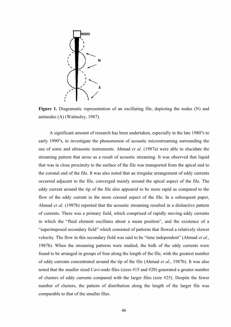
46
Figure 1. Diagramatic representation of an oscillating file, depicting the nodes (N) and
antinodes (A) (Walmsley, 1987).
A significant amount of research has been undertaken, especially in the late 1980‟s to
early 1990‟s, to investigate the phenomenon of acoustic microstreaming surrounding the
use of sonic and ultrasonic instruments. Ahmad et al. (1987a) were able to elucidate the
streaming pattern that arose as a result of acoustic streaming. It was observed that liquid
that was in close proximity to the surface of the file was transported from the apical end to
the coronal end of the file. It was also noted that an irregular arrangement of eddy currents
occurred adjacent to the file, converged mainly around the apical aspect of the file. The
eddy current around the tip of the file also appeared to be more rapid as compared to the
flow of the eddy current in the more coronal aspect of the file. In a subsequent paper,
Ahmad et al. (1987b) reported that the acoustic streaming resulted in a distinctive pattern
of currents. There was a primary field, which comprised of rapidly moving eddy currents
in which the “fluid element oscillates about a mean position”, and the existence of a
“superimposed secondary field” which consisted of patterns that flowed a relatively slower
velocity. The flow in this secondary field was said to be “time independent” (Ahmad et al.,
1987b). When the streaming patterns were studied, the bulk of the eddy currents were
found to be arranged in groups of four along the length of the file, with the greatest number
of eddy currents concentrated around the tip of the file (Ahmad et al., 1987b). It was also
noted that the smaller sized Cavi-endo files (sizes #15 and #20) generated a greater number
of clusters of eddy currents compared with the larger files (size #25). Despite the fewer
number of clusters, the pattern of distribution along the length of the larger file was
comparable to that of the smaller files.

47
Apart from the above features, closer examination of the individual eddy currents
within the primary field revealed that the direction of rotation of the fluid within each eddy
current was opposite to that of the adjacent eddy current (Ahmad et al., 1997). In the
secondary field, the flow pattern was longitudinal and symmetrical along both sides of the
activated file, and it was evident that movement of fluid was from the apical end of the file
towards the coronal aspect. It was also noted that there were flow patterns near the tip of
the file that were directed apically, and the streaming velocities of the eddy currents at the
tip of the file were much more rapid compared with those located more coronally (Ahmad
et al., 1987). Interestingly, the velocity of the eddy currents are related inversely to the size
of the file used; the streaming was slower when the larger size #25 file was used as
compared to the smaller sizes #15 and #20 (Ahmad et al., 1987). When power settings
were increased, there were no increases in the number of eddy currents observed, although
the size of the eddy currents and the velocity of those currents were noted to be increased.
Figure 2 shows the acoustic streaming of a file immersed in water, while figure 3 is an
illustration of the primary and secondary fields, with the directions of the eddy currents
indicated.
Figure 2. Acoustic streaming of a file oscillating in water (van der Sluis et al., 2007).
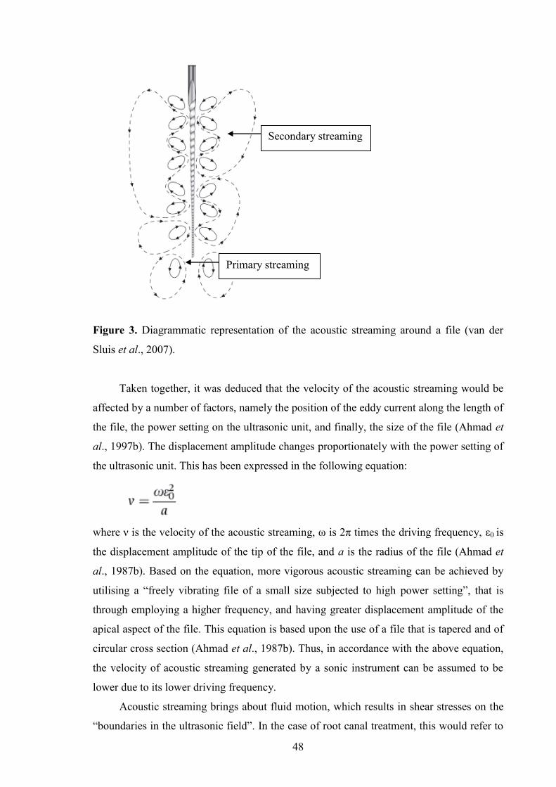
48
Figure 3. Diagrammatic representation of the acoustic streaming around a file (van der
Sluis et al., 2007).
Taken together, it was deduced that the velocity of the acoustic streaming would be
affected by a number of factors, namely the position of the eddy current along the length of
the file, the power setting on the ultrasonic unit, and finally, the size of the file (Ahmad et
al., 1997b). The displacement amplitude changes proportionately with the power setting of
the ultrasonic unit. This has been expressed in the following equation:
where ν is the velocity of the acoustic streaming, ω is 2π times the driving frequency, ε0 is
the displacement amplitude of the tip of the file, and a is the radius of the file (Ahmad et
al., 1987b). Based on the equation, more vigorous acoustic streaming can be achieved by
utilising a “freely vibrating file of a small size subjected to high power setting”, that is
through employing a higher frequency, and having greater displacement amplitude of the
apical aspect of the file. This equation is based upon the use of a file that is tapered and of
circular cross section (Ahmad et al., 1987b). Thus, in accordance with the above equation,
the velocity of acoustic streaming generated by a sonic instrument can be assumed to be
lower due to its lower driving frequency.
Acoustic streaming brings about fluid motion, which results in shear stresses on the
“boundaries in the ultrasonic field”. In the case of root canal treatment, this would refer to
Primary streaming
Secondary streaming
49
the radicular walls of the root canal. As such, the generation of hydrodynamic shear stress
would be the dominant physical mechanism by which the surface of the root canal walls
are cleaned when ultrasonic activated irrigation is applied (Roy et al., 1994). The amount
of hydrodynamic shear stress is proportional to the streaming velocity, which occurs at its
maximum at the tip of the ultrasonically activated instrument. As such, it is reasonable to
assume that the hydrodynamic shear stress would be concentrated in the vicinity of the tip,
which most likely would be positioned closer to the apical aspect of the root canal. The
progressive taper of an instrumented canal could also be a potential aid, since the smaller
surface area of the apical aspect of the root canal would be subjected to increased energy
from the acoustic streaming. This would result in the concentration of hydrodynamic
stresses at the apical end of the canal, thereby facilitating the removal of debris from the
root canal walls (Ahmad et al., 1987b). The explanation for the increased effectiveness in
more confined areas is the stronger forces generated by the primary streaming currents,
which are located in closer proximity to the surface of the vibrating instrument. The
increased speed of the primary streaming, as compared to the secondary streaming
currents, accounts for the greater hydrodynamic stresses (Ahmad et al., 1987b). The caveat
is that the tip must be freely vibrating at this part of the root canal, not dampened by
contact with the root canal wall, and as such, the file is not used as a device to
mechanically debride the canal (Ahmad et al., 1987b).
There are studies outside of the field of dentistry that have discovered the physical
and biological effects of hydrodynamic shear stress. Williams (1972) found that acoustic
streaming was capable of generating hydrodynamic shear stresses capable of disrupting
biological cells. Other researchers have found that these shear stresses are of sufficient
intensity to remove the mucopolysaccharide surface coatings from mammalian cells
(Martinez Palomo, 1970).
1.8.5 The effectiveness of sonic and ultrasonic activated irrigation
Investigations into the effectiveness of ultrasonic activation of irrigants date back to
the 1980‟s. Many of the studies have attempted to assess the ability of this irrigation
technique in the removal of pulpal remnants and dentinal debris (Cameron, 1987; Lee et
al., 2004a; Gutarts et al., 2005), smear layer (Cameron, 1983) and planktonic bacteria
(Sjögren and Sundqvist, 1987; Huque et al., 1998; Weber et al., 2003). Amongst the
earliest studies was that by Ahmad et al. (1987b), which compared the cleanliness of root
canals following instrumentation with ultrasonically activated files with those that

50
underwent ultrasonic activation of irrigants. All the teeth in both experimental groups were
mechanically debrided in a similar manner, i.e. with ultrasonically activated size #15 files.
However, one group was additionally subject to ultrasonic irrigation for 5 minutes, where
an ultrasonic activated file was inserted to within 1 mm of the apex and allowed to oscillate
freely. With the aid of scanning electron microscopy, the results revealed that the canals
which had the ultrasonically activated file oscillating freely within the canal were
consistently cleaner. There was virtually no smear layer in the apical third of canals that
underwent ultrasonic irrigation, an unsurprising result considering the finding that the
greatest velocity of acoustic microstreaming, and hence hydrodynamic shear stresses, are
generated at the most apical aspect of an ultrasonically activated file (Ahmad et al.,
1987a). Their results place the effectiveness of ultrasonic irrigation in favourable light,
given that ethylenediamine tetracetic acid (EDTA) was not used as an irrigant, and a lower
concentration of sodium hypochlorite was employed in the group which had the irrigant
(1% sodium hypochlorite) ultrasonically activated, compared to the 2.5% sodium
hypochlorite used for the other group. In a recent study by Lee et al. (2004), grooves and
depressions were cut into instrumented canine root canals to simulate uninstrumented canal
irregularities. These grooves and depressions were filled with dentinal debris and subject to
ultrasonic irrigation with 2% sodium hypochlorite for 3 min. This was compared against
conventional syringe irrigation, and the results showed that ultrasonic irrigation was
superior in the removal of debris from the simulated canal irregularities. In some of the
specimens, there was complete removal of the debris from the grooves and depressions.
While the creation of such „irregularities‟ may be somewhat artificial, the advantage is that
the amount of debris at baseline prior to irrigation is known, and as such, comparison of
the amount of debris removal by each irrigation protocol can potentially be more accurate.
Given the relatively simple anatomy of the teeth examined in the aforementioned
studies (Ahmad et al., 1987b, Lee et al., 2004), other investigators have utilised curved
molar canals (Jensen et al., 1999). Generally, the greater complexity of such canal anatomy
would hamper the complete debridement of the root canal system. In this study, the curved
molar root canals, numbering 60 in total, were subjected to hand instrumentation up to an
apical size of #35. Comparing syringe irrigation against sonic and ultrasonic activated
irrigation, it was found that the latter two methods were significantly superior to syringe
irrigation when 5.25% sodium hypochlorite was used as the irrigant. It was noted that there
was no difference between the sonic and ultrasonic activation, with the ultrasonic method
scoring slightly higher in terms of remaining debris. While this is surprising, since the
acoustic microstreaming generated by the sonic unit is of lower intensity compared with
51
that of ultrasonic, the investigators did not employ the use of a scanning electron
microscope (SEM). Instead, photomicrographs with a transparent grid were used to assess
the amount of debris remaining in the root canal. Despite the relatively rudimentary
assessment technique, the effectiveness of irrigant activation in producing cleaner canals is
evident.
Another study which compared sonic and ultrasonic activation of irrigants found no
significant difference between the two instruments (de Gregorio et al., 2009). The sonic
instrument used in the study is the EndoActivator®, and had recently been introduced into
the market (Ruddle, 2007). It is claimed that the “vigorous intracanal fluid agitation”
occurs though both “acoustic streaming and cavitation” (Ruddle, 2007). The phenomenon
of acoustic microstreaming and the resultant hydrodynamic shear stresses is based on the
assumption that flow of the fluid occurs around the file without becoming „detached‟ from
the surface of the file (Roy et al., 1994). However, when the displacement amplitude
reaches a certain threshold and the negative acoustic pressure amplitude attains a critical
value, the flow can separate from the surface of the file, “forming a wake which results in
bubble formation”. This process is known as hydrodynamic cavitation (Ahmad et al.,
1988; Roy et al., 1994). Given that the EndoActivator® operates at a maximum of 10,000
cycles per minute only (166 Hz), it is doubtful that the phenomenon of cavitation is at
work here. The low frequency generated by the sonic instrument is too low, and below the
threshold necessary for cavitation to occur (Ahmad et al., 1987a). The claim that cavitation
occurs with the EndoActivator® is also disputed by Jiang et al. (2009), whose group used a
high speed camera to record the oscillation of the polymer tip within a glass root canal
model at 4000 frames per second. Not only was there no cavitation observed, but their
results also contradicted those of de Gregorio et al. (2009). Using a similar model to that in
the Lee et al. (2004a) study, they found that the ultrasonic instrument, which was capable
of generating a driving frequency of 30 kHz, caused greater removal of dentinal debris
from grooves made in the root canal wall compared with the EndoActivator®.
All of the aforementioned studies occurred under in vitro conditions. Also, ultrasonic
irrigation occurred for 3 min in both the Jensen et al. (1999) and Lee et al. (2004) studies,
while in the Ahmad et al. (1987b) study, ultrasonic irrigation was implemented for 5 min
within each canal. While this would ensure that there would be sufficient time for the
activation of irrigants to cause more thorough debridement, it would also translate into a
significant amount of clinical time added to the preparation of a molar. Moreover, not all
studies have found ultrasonic activation of irrigants to be more effective. Mayer et al.
(2002) found that while ultrasonic activation of the irrigants used in their study, namely
52
sodium hypochlorite and EDTA, did produce slightly cleaner canals as compared with the
non-activated controls, the difference was not statistically significant. It should be noted
that in their study, the irrigating time was 1 min, compared with the longer irrigation times
mentioned in the above studies. This timing, the authors felt, was more “clinically
practical”. The results in the study by Abbott et al. (1991) also revealed no significant
difference between canals that were subject to ultrasonic activation of the irrigants, and
other groups which had the irrigant introduced with a syringe. The study examined a
variety of irrigants, with a total of 6 treatment groups, and used SEM to evaluate the
results. The volume of irrigants used was 18 ml per group, which is more than the volume
used in the previously mentioned studies. It has been shown that the volume of irrigant that
is used to flush the root canal system can influence the quality of root canal debridement
(Yamada, 1983). It was shown that when higher volumes of sodium hypochlorite and
EDTA were used to irrigate the root canal system, significantly cleaner canals were
obtained as compared with smaller volumes (Yamada, 1983). The high volumes used in
the Abbott et al. (1991) study may explain why there were no significant differences in
canal cleanliness between those canals irrigated with the syringe and those where
ultrasonic activation was applied.
As such, a combination of an adequate volume of irrigant, type of irrigant and the
application of acoustic energy can bring about effective cleaning and disinfection of the
root canal system. Despite the fact that the use of acoustic energy to activate irrigants is not
a recent introduction to endodontics, it has yet to be established as an essential part of the
protocol for root canal treatment. The potential of this „old school‟ technology, employed
in a new fashion, is immense and should be harnessed to enhance the predictability of root
canal disinfection and improving the success rate of endodontic therapy.
1.9 Lasers
In 1960, Maiman developed the world‟s first laser (Kimura et al., 2000). A laser is
defined as “a device which transforms light of various frequencies into a chromatic
radiation in the visible, infrared, and ultraviolet regions with all the waves in phase capable
of mobilizing immense heat and power when focused at close range” (Kimura et al., 2000).
Traditionally, lasers are classified according to the physical construction of the laser, i.e.
whether the laser is gas, liquid, solid state, or semiconductor diode, and the type of
medium which undergoes lasing (e.g. erbium, chromium:yttrium-scandium-gallium-garnet
(Er,Cr:YSGG) laser) (Walsh, 2003). Lasers have also been classified according to the
53
potential hazards, when in contact with skin or exposure to the eye (Walsh, 2003). An
exhaustive list of the different types of lasers is beyond the scope of this review. Suffice to
say, the potential that the power of laser possessed led to the exploration of its use in the
field of dentistry (Goldman et al., 1964; Stern and Sognnaes, 1964). Early studies were
conducted on the use of the ruby laser, which Maiman (1960) had developed, on caries by
studying its effects on subsurface demineralisation. Since then, the list of applications has
expanded, encompassing the various aspects of dentistry.
1.9.1 Lasers in endodontics
The first reported use of lasers in endodontics was published by Weichman and
Johnson (1971). In their in vitro study, their attempt to seal the apical foramen with an
infrared (CO2) laser was met with failure. Despite the less than desired result, tremendous
interest was generated and has led to a body of research into the potential applications of
lasers in the field of endodontics.
The promise and potential of lasers in removal of dental hard tissue have also led to
the exploration of its use in the instrumentation of the root canal. A recent in vitro study
sought to compare the efficacy of the Er,Cr:YSGG laser to that of „traditional‟ rotary
nickel titanium files (Radatti et al., 2006). The manner in which the laser would instrument
the canal is known as hydrokinetic debridement, which is described as a process of
“removing biologic material through the use of high speed water spray” (Eversole et al.,
1997). The results of the study showed that the canals that were treated with the
Er,Cr:YSGG laser resulted in significantly more debris than the canals that had been
instrumented with rotary nickel-titanium files (Radatti et al., 2006). Moreover, the rotary
instruments were more efficient, as the laser powered hydrokinetic debridement required
twice as much time to perform (Radatti et al., 2006). Other problems included the ledging
and other irregularities of the canals with the laser that resulted in blockage of the canal,
while apical patency was maintained for all samples instrumented with the rotary nickel-
titanium files (Radatti et al., 2006). The authors thus suggested that the laser be used as an
“adjunct to rotary instrumentation”, potentially in conjunction with an antimicrobial
irrigant such as sodium hypochlorite (Radatti et al., 2006).
54
1.9.2 Disinfection of the root canal system with lasers
One of the areas of greatest interest in endodontic research remains the elimination of
bacteria within the root canal system. It would almost seem natural that the lasers also be
employed in the disinfection of the root canal.
1.9.2.1 Photo activated disinfection
Amongst the multitude of methods to employ this technology to aid in eradicating
microorganisms within the root canal was in the form of photo-activated disinfection.
Photo-activated disinfection is defined as “a method of disinfecting or sterilising a hard
tissue or soft tissue site by topically applying a photosensitising compound to the site, and
then irradiating this with laser light at a wavelength absorbed by the photosensitising
compound, so as to destroy microbes at the site” (Wilson and Wilson, 1997). Compared
with direct killing with laser, low power lasers are employed to drive the photochemical
reactions, and thus reduced the tendency of direct laser energy to cause cracking, cratering
and carbonisation of the dentinal walls of the root canal (Anic et al., 1998). The laser light
that is used in conjunction with the photosensitiser usually emits light from the middle red
portion of the visible spectrum, since this wavelength penetrates dentine to the greatest
extent, as well as any blood products that are present (Odor et al., 1996). While the low
power laser energy used in this technique is generally not sufficient to kill bacteria per se,
certain Gram-negative anaerobic bacteria may be particularly susceptible to laser light
emitted in the middle red wavelengths (Lee et al., 2004b). The photochemical reactions
resulting from interaction between the absorption of the laser light by the photosensitising
compound would then lead to the production of singlet oxygen, free radicals, and other
reactive oxygen species (Walsh, 1997). The photosensitiser molecule is elevated to a high-
energy state when it is irradiated by light of a specific wavelength, and this then leads to
transference of energy to an oxygen molecule. This generates singlet oxygen and other
reactive oxygen species (Walsh, 1997), which are highly lethal to practically all species of
bacteria that are present in the oral cavity (Donson and Wilson, 1992; Wilson et al., 1992).
There are generally two pathways by which “photodynamic microbial damage”
occurs (Ochsner, 1997). In the Type I pathway, the photosensitiser triplet state causes the
transfer of electrons to molecules. The free radicals which result from these electron
transfer reactions then combine with oxygen to produce cytotoxic species of oxygen. These
cytotoxic species, such as superoxide, hydroxyl and lipid-derived radicals, are thereby

55
capable of causing damage to the bacterial cell membrane, acting against the membrane
phospholipids, enzymes and receptors (Oschner, 1997). The alternate action, known as the
Type II pathway, occurs when the photosensitiser triplet state transfers electrons to
molecular oxygen to produce singlet oxygen. This highly excited and reactive state is
capable of oxidation of proteins, nucleic acids and lipids, and is hence cytotoxic to bacteria
(Oschner, 1997). The absence of resistance to these extremely reactive molecules, and the
short half-life of these molecules, ensure that not only is the cytoxic effect highly effective
against microorganisms, but their actions are also localised (Lee et al., 2004b).
Photo-activated disinfection has the potential to be highly effective in eradicating the
bacteria in primary root canal infections. The dominance of obligate anaerobic species,
such as Fusobacterium, Prevotella, and Prophyromonas, in primary infection of the root
canal system translates into susceptibility to killing by either Type I or Type II pathways,
since Gram-negative anerobes, both obligate and facultative, are extremely sensitive to the
toxic effects of oxygen compounds. This is due to the absence of catalase and other
protective enzyme systems in these bacteria (Lee et al., 2004b). The incorporation of
porphyrin-like molecules within the cellular structure of Gram-negative bacteria also
renders them vulnerable to photo-activated disinfection, as these molecules function as
endogenous photosensitisers, which are activated by particular wavelengths of visible red
light (Konig et al., 2000). While effective against Gram-negative bacteria, Gram-positive
bacteria may not be as vulnerable to photo-activated disinfection, since most species lack
endogenous photosensitising compounds, making them quite resistant to killing by visible
red light alone. The use of an appropriate photosensitiser dye, such as tolonium chloride,
would allow killing of most, if not all, species of bacteria, provided binding to the cell
membranes of the bacteria occurs (Silbert et al., 2000).
The effectiveness of photo-activated disinfection may thus be limited against bacteria
that persist within root canals following root canal treatment, leading to failure of the
endodontic therapy. The microbiota found in such cases of secondary or persistent root
canal infection generally tends to be Gram-positive facultative bacteria (Sundqvist et al.,
1998). While studies have shown that the addition of an appropriate photosensitiser dye
may render them susceptible to killing by photo-activated disinfection, these bacteria must
be in direct contact with the dye so that binding of the dye to their membranes can occur
(Lee et al., 2004b). The presence of bubbles within the photosensitiser dye solution may
also interfere with the binding to the bacterial cell membranes (Lee et al., 2004b).
Experiments have thus been conducted using planktonic cultures of Streptococcus mutans
and Enterococcus faecalis, and the results showed that a 120 second exposure to photo-

56
activated disinfection, employing such dyes as methylene blue or tolonuim chloride, is
capable of killing 97 - 99.9% of the bacteria present in the culture (Silbert et al., 2000).
Moshonov et al. (1995) investigated the use of neodymium:yttrium, aluminium, garnet
(Nd:YAG) laser in combination with a photosensitiser dye, nigrosin, on the eradication of
E. faecalis. Irradiation of the canals was conducted for 2 minutes at 4.5W and results were
compared against positive and negative controls, as well as exposure to 1% sodium
hypochlorite for 2 minutes. While the Nd:YAG laser, when used in conjunction with the
photosensitiser dye, eliminated more than 99% of the bacteria culture, complete
disinfection of the root canals did not occur. In contrast, 1% sodium hypochlorite was able
to eliminate all of the bacteria, and no bacteria could be detected on their cultures and
scanning electron microscope images (Moshonov et al., 1995). The results of this study
reflect poorly upon the effectiveness of photo-activated disinfection as a means of root
canal disinfection, compared with the traditional and less costly method of irrigating with
sodium hypochlorite. However, it should be noted that the culture of E. faecalis was grown
up overnight, and was predominantly a planktonic culture. As such, elimination of the
bacteria was achieved much more easily, even with 1% sodium hypochlorite. The way in
which the canal was irradiated was conducted with the fibre-optic tip placed in the apical
and coronal aspects of the canal for two 15 second cycles in a “circumferential painting
movement”. While the concept of activating an irrigant with laser energy had not been
conceived at that time, the act of placing the laser tip within the root canal with the
nigrosin dye in distilled water may have inadvertently activated it. It should also be noted
that the Nd:YAG laser at 4.5W is at a fairly high power setting and may potentially result
in apical extrusion of the irrigant in an in vivo situation.
As is the case in many in vitro studies, the bacteria culture is often an overnight
culture. This is in contrast to the bacteria present in secondary or persistent root canal
infection cases, such as E. faecalis, which have the propensity to invade dentinal tubules
(Love, 2001). This would severely limit the ability of the photosensitiser dye to come into
direct contact with the bacteria. Moreover, some of the studies that have examined the
efficacy of photo-activated disinfection have utilised planktonic cultures of bacteria
(Moshonov et al., 1995; Silbert, 2000). These results potentially may not be extrapolated to
in vivo situations, where the bacteria, such as E. faecalis forms a biofilm, which provide
increased protection against much of the anti-microbial agents in the form of irrigants and
medicaments (Love, 2001).

57
1.9.2.2 Direct irradiation of root canals
Other investigators have examined the effect of directly irradiating the root canal
with the laser on the eradication of bacteria. Much of this work has been conducted with
the use of the Nd:YAG laser. Prior to the approval for its use in dentistry, Nd:YAG lasers
had been used extensively in dermatology, ophthalmology, gynaecology and general
surgery (Hardee et al., 1994). This laser was, at that time, arguably the most widely used
laser in endodontics. It emits at a wavelength of 1,064 nm, which is in the near infra-red
range (Rooney et al., 1994). Gutknecht et al. (1996) investigated the effect of this laser
when irradiation was carried on the root canals of extracted teeth. The teeth consisted of
both anterior and posterior teeth, and the root canals were infected with E. faecalis and
cultured for a period of 24 hours. The results garnered for the experiment were impressive;
when the canal was irradiated with the laser at 1.5W, a 99.91% reduction in bacteria was
observed. However, it should be noted that the E. faecalis culture had been grown up for
24 hours only. As such, the bacteria might have only been a planktonic culture. The
sampling technique employed in the study consisted of rinsing the canals of all the teeth
with 1ml of 0.9% saline solution, and then collecting the eluate in a test tube. Serial
dilutions were subsequently conducted using this eluate. This method may have missed
some of the bacteria as the saline rinse may not have reached some parts of the canal,
especially within the complex anatomy of multi-rooted posterior teeth, where isthmuses
and joining canals exist.
A more recent study by Berkiten et al. (2000) also used a pulsed Nd:YAG laser and
examined its effects on the disinfection of root canals infected with Streptococcus sanguis
and Prevotella intermedia for 20 days. Given that the bacteria had been cultured within the
root canals for 20 days, it would have been likely that the bacteria would have established
a biofilm (Plutzer, 2009). Employing two power settings of 1.8W and 2.4W, they found
that the former was able to eliminate 86.3% of S. sanguis with a mean penetration of 607.6
μm into dentinal tubules, while the higher power setting of 2.4W was capable of achieving
98.5% sterility. The laser at the setting of 2.4W was also capable of disinfecting up to a
mean of 666.6 μm. The laser had also completely eliminated P. intermedia from the root
canal at both settings (Berkiten et al., 2000). Results from light microscopy showed that a
significant number of S. sanguis had penetrated the dentinal tubules. As such, this study
provided evidence of the effectiveness of laser irradiation in the elimination of bacteria,
and that the laser also had an effect on the bacteria that had penetrated dentinal tubules
within the root canal. However, scanning electron microscopy employed in the study
58
revealed that the laser had caused alterations carbonisation of the dentinal walls, and
closure of some dentinal tubules due to melting and recrystallisation of the radicular
dentine. In addition, root canals lased at 2.4W also showed evidence of crumbling and
fissures on the dentine layer (Berkiten et al., 2000). As such, the surface morphology of the
radicular dentine had been altered irreversibly, and this may have ramifications on the
penetration of root canal sealer into the dentinal tubules.
Another problem with the use of direct irradiation is the heat generated by the laser.
A study by Hardee et al. (1994) to evaluate the effectiveness of Nd:YAG laser in sterilising
root canals of single-rooted teeth infected with Bacillus stearothermophilus spores. These
spores are extremely heat resistant, and were cultivated by incubating at 55ºC in the study.
The canals which were irradiated for 1 min at 3W had a 98% reduction in colony forming
units (CFU), while those that had been exposed to 3 minutes of 0.5% sodium hypochlorite
registered an 88% reduction in CFU. However, the teeth group that had been irradiated for
1 minute became extremely hot and could not be handled by the end of the period of
irradiation. The authors also noted charring of the dentine and production of smoke. In
another 2 groups, laser irradiation was conducted for 1 and 2 minutes, with 0.5% sodium
hypochlorite within the canal. This resulted in 98% and 99% reduction in CFU
respectively, but the temperature of the teeth did not rise as rapidly. However, once the
sodium hypochlorite had vaporised completely, these teeth also became too hot to touch.
In an in vivo situation, this would have adverse effects on the surrounding
periodontium. In a histological study in dogs, Bahcall et al. (1992) reported periradicular
osteocyte necrosis 24 hours after root canal debridement was performed with Nd:YAG
laser irradiation for 30 seconds. These same teeth exhibited external resorption and
ankylosis after 30 days, and it was postulated that the damage to the periradicular
structures may have been the result of heat transfer from the tooth structure, or absorption
of the laser light by the periradicular tissues. As such, it may be necessary to reduce either
the time of irradiation, or the power output of the laser. However, this may compromise the
effective disinfection of the root canal, because even at the settings used in the Hardee et
al. (1994) study, complete sterilisation of the root canal was not achieved.
Ramsköld et al. (1997) sought to determine the thermal changes following irradiation
with Nd:YAG laser in an in vitro setting. At 3W and irradiation for a period of 15 seconds,
there was an increase of 6 ºC registered. As they irradiated the canal intermittently, there
was a fall in temperature, followed by a subsequent increase during lasing of the canal.
Temperatures as high as 42.6 ºC were recorded, while the lowest temperature reached as
36.7 ºC. It should be noted that the canals were dry during irradiation of the root canals. As
59
such, the amount of energy necessary to raise the temperature is lower, as compared to a
canal which is wet. Water on the other hand, requires nearly four times the amount of
energy as compared to dentine to raise the temperature to the same degree (Ramsköld et
al., 1997). This is because the properties of water and dentine differ for both absorption
and conduction of heat (Ramsköld et al., 1997). As such, a wet canal will subsequently be
able to insulate against rapid increases in temperature. The higher density of dentine
compared to water also translates into greater thermal diffusivity of dentine (Ramsköld et
al., 1997). All these factors must be taken into consideration when deciding on the power
output and timing for irradiating a canal during debridement, so as to prevent any potential
damage to the periodontium.
More recently, other investigators have used other lasers, such as the erbium:yttrium-
aluminum-garnet (Er:YAG) laser (Perin et al., 2004), and the erbium, chromium:yttrium-
scandium-gallium-garnet (Er,Cr:YSGG) laser (Gordon et al., 2007; Schoop et al., 2007;
Schoop et al., 2009), and some others have compared its effectiveness against other
irrigation regimes in the disinfection of the root canal (Wang et al., 2007). These lasers
emit at wavelengths of 2,940 nm and 2,780 nm respectively (Schoop et al., 2007). Their
action is carried out through photoablation, as the wavelengths correlate closely with the
absorption maximum of hydroxyapatite (Hibst et al., 1989). This has been found to be
ideal for the removal of dental hard tissues, as water that is present within dentine
evaporates instantaneously when irradiated, resulting in ablation of the tissue with minimal
increases in temperature (Hibst et al., 1989). Due to this ability, much of the research
conducted using these lasers have predominantly been on caries removal and cavity
preparation (Hadley et al., 2000). However, in recent times, the effect of the Er,Cr:YSGG
laser on the morphology of the root canal dentine following irradiation has been examined
(Yamazaki et al., 2001; Altundasar et al., 2006). In one of the studies, irradiation with the
Er,Cr:YSGG laser occurred at the power output of 3W, and scanning electron microscopy
revealed several sites that had partial melting and occlusion of the dentinal tubules
(Altundasar et al., 2006). Other areas showed carbonisation, despite the use of water
coolant during irradiation (Altundasar et al., 2006).
Subsequent to these investigations, the use of the Er,Cr:YSGG laser in the
disinfection of root canals was evaluated. Results from one of the studies showed that root
canals infected with Escherichia coli and E. faecalis could not be completely sterilised
(Schoop et al., 2007). This was despite the culture having been grown up within the root
canals for 4 hours only, and was thus unlikely to have been a biofilm (Schoop et al., 2007).
Using power output settings of 1W and 1.5W, increases of 2.7ºC and 3.2 ºC respectively
60
were recorded (Schoop et al., 2007). The authors believed that this range of increase was
within safe limits. Scanning electron microscopy of the root canal dentinal walls showed
minimal disruption to the natural topography, with patent dentinal tubules present after
irradiation at 1W. Root canals that were irradiated with 1.5W, however, exhibited partial
melting and recrystallisation of dentine, thereby occluding some of the dentinal tubules
(Schoop et al., 2007).
Wang and co-workers (2007) set out to compare the bactericidal effect of irradiation
of the root canal with the Er,Cr:YSGG and Nd:YAG lasers, against irrigation with 2.5%
sodium hypochlorite. Using single rooted teeth with a single root canal, these teeth were
infected with E. faecalis and allowed to incubate for 3 weeks prior to treatment. Irradiation
of the root canals with the Er,Cr:YSGG and Nd:YAG lasers were at power outputs of 1W
and 1.5W for a period of 40 seconds, and this would be compared against a 2 minute
irrigation with 2.5% sodium hypochlorite. The Nd:YAG laser proved to be more effective
than the Er,Cr:YSGG laser at reducing the number of CFU, as the former resulted in 97%
(1W) and 98% (1.5W) reduction, while the latter was not as effective at the power setting
of 1W, with 77% reduction in E. faecalis CFU. Irradiation with the Er,Cr:YSGG laser at
1.5W was more effective, resulting in 96% killing of the bacteria, but the traditional
method of irrigation with sodium hypochlorite resulted in 100% reduction in bacteria. This
study highlighted the effectiveness of sodium hypochlorite as an irrigant and why it has
stood the test of time in the face of technological advancements in root canal debridement
and disinfection methods. One of the strengths of the study was the cultivation of E.
faecalis for 3 weeks, which meant that the different treatment modalities were working to
eradicate a biofilm. It should be noted that sampling of the root canals occurred with paper
points, and thus, there may still be E. faecalis present in the dentinal tubules or not
detected with paper point sampling. As such, the 100% kill rate registered by sodium
hypochlorite may not be entirely accurate, but this sampling method was standardised
across all treatment groups.
Up to this time, all of the research with lasers had employed the use of end firing
tips. The introduction of radial firing tips, which are conical in shape, was aimed at
providing better coverage of the root canal (Gordon et al., 2007). Compared with the
forward emission of laser energy that occurs with end firing tips, the radial firing tips were
also thought to result in the irradiation of the dentinal tubules given the more favourable
direction of laser emission, thereby potentially eliminating bacteria that penetrate the
dentinal tubules (Gordon et al., 2007). This was tested with the use of dentine cylinders
which were infected with E. faecalis (Gordon et al., 2007). Following the model described

61
by Haapasalo and Orstavik (1987), cementum from the root section were removed to
facilitate invasion of the dentinal tubules. After incubating the bacteria for 1 week, the
dentine cylinders were divided into different treatment groups which received either
sodium hypochlorite irrigation or irradiation with the Er,Cr:YSGG laser at different power
settings, timings, and with or without the water spray. The results showed that irradiation
at 0.35W without water spray resulted in the greatest reduction in E. faecalis CFU. The
authors believed that dry laser treatment meant that the laser had a direct effect on the
water-containing bacteria, since the wavelength of the Er,Cr:YSGG laser is absorbed by
the water (Gordon et al., 2007). When the water spray was used, the laser energy was
diminished consequently. The penetration of the laser energy into dentine also contributed
to the significant reduction in CFU, since the wavelength of the Er,Cr:YSGG is readily
absorbed by dentine as a result of the hydroxyapatite and interstitial water present within.
The pulse of this laser is composed of approximately 150 micropulses, and each
micropulse causes penetration of energy of about 3 μm into interstitial water. As such, it is
the opinion of the authors that expansion of intratubular water and collapse of the water
vapour can occur up to 1000 μm into dentine, a phenomenon they termed “micropulse-
induced sequential absorption”. This expansion and collapse was thought to be capable of
generating acoustic waves which could disrupt bacteria that had penetrated into the
dentinal tubules (Gordon et al., 2007).
The use of the radial firing tip was also evaluated by Schoop et al. (2009), assessing
the bactericidal, morphological and thermal effects of the Er,Cr:YSGG laser in conjunction
with the radial firing tip. Employing the laser at two power output settings of 0.6W and
0.9W, it was found that neither of these was capable of completely sterilising root canals
that were infected with E. faecalis in their in vitro study. The advantage of using the laser
at such low power settings meant minimal morphological alterations to the root dentine
surface, and negligible increases in temperature (1.3 – 1.6 ºC) (Schoop et al., 2009). Given
the results, it would seem that the effectiveness of the Er,Cr:YSGG laser in disinfection of
the root canal is severely limited.
1.9.2.3 Laser activation of irrigants
When considering the use of the Er,Cr:YSGG laser in disinfection of the root canal
system, a balance must be achieved between sufficient power output for effective
sterilisation and avoidance of excessive morphological alterations to the root canal dentine.
Due to the ablative nature of the Er,Cr:YSGG laser, the creation of ledges, especially
62
within curved canals, is a clear and present danger. The in vitro studies that have been
carried have generally shown that at lower power outputs, complete sterilisation of the root
canal with the Er,Cr:YSGG laser is generally unattainable (Gordon et al., 2007; Schoop et
al., 2007; Schoop et al., 2009; Wang et al., 2007). It should be noted also that these studies
were conducted on single rooted teeth with relatively simple anatomy. The ability of direct
irradiation with the Er,Cr:YSGG to sterilise root canals with various anatomical variations,
such as fins, anastomoses and isthmuses, is thus doubtful.
1.9.3 The mechanism underlying laser activation of irrigants - Cavitation
The immense of energy generated by laser irradiation could however be harnessed in
a different manner. Similar to ultrasound, cavitation could potentially be generated by laser
activation of a fluid, and the subsequent explosion and implosions could result in pressure
waves (Blanken et al., 2009). These waves would subsequently create shear stresses along
the dentinal walls of the root canal. The fluid has been shown to travel at up to 20
m/second (72 km/hour), and the speed has been attributed to vapour bubble expansion and
implosion with secondary cavitation effects, thereby inducing such high speed fluid
motions (Blanken and Verdaasdonk, 2007).
Using high speed imaging techniques and glass cylinders which simulate the root
canal, images were captured and combined to show the dynamics of the generation and
collapse of vapour bubbles in a time range from 1 to 300 – 3,000 microseconds (Blanken et
al., 2009). When the laser pulse was fired, the energy was absorbed in a 2 mm-thick layer
of fluid that was instantly heated to boiling temperature (100 ºC) at high pressure and
converted into vapour. The high pressure of this vapour caused it to expand at high speed
and provided an opening in front of the fibre for the laser light. At the subsequent pulse,
the light emitted from the laser passed through the bubble and the water surface in front of
the bubble is evaporated. Blanken et al. (2009) described it as the laser light having
“drilled a channel through the liquid until the pulse ends after 140 microseconds”. This
phenomenon has been described previously as the “Moses effect in the microsecond
region” by van Leeuwen et al. (1991). On cessation of irradiation, cooling of the vapour
occurred and condensation began. The momentum of the previous expansion, however,
caused the creation of a region of lower pressure within the vapour bubble. Liquid that
surrounded the bubble then accelerated to fill in the gap. Implosion of the vapour bubble
then occurred near the tip of the fibre, and resulted in the separation of the bubble from the
fibre. Fluid then rushed into the bubble, causing it to implode. The imploding bubble was
63
noticed to be shaped like a sickle. The process of implosion is completed after 260
microseconds, and the vapour bubble was no longer present. This is an occurrence which is
reproducible in an open environment. However, within the confines of the root canal, the
bubble cannot expand freely. Fluid is thus pushed forwards and backwards, leading to
compression of the bubble into a flattened disk. The further need to increase in size is
prevented by fluid that is apical to the expanding bubble, and thus, the bubble then grew in
a coronal direction along the length of the laser fibre. The pressure within the bubble
remained high for an extended period of time, given the resistance of fluid displacement
within the space restrictions of the root canal. This thus delayed the dynamics of expansion
and implosion, and it has been shown that it may take three times as long for cavitation to
occur because lateral and forward expansion of the bubble would be prevented by the root
canal wall, and backward expansion restricted by the fibre tip (Blanken and Verdaasdonk,
2007). However, the restricted space creates shear stresses along the wall of the canal, and
potentially generates secondary cavitation bubbles as a result of the high speed fluid
motion. The implosion of the primary and secondary bubbles creates microjets in the fluid
aimed at the wall with very high forces locally (Blanken et al., 2009).
Other researchers have recorded similar findings utilising similar experimental set-
ups (de Groot et al., 2009). They noted that a higher energy pulse would result in the
longer growth time of the bubble, and that collapse of the bubble occurred with a velocity
of 1 m/s. It was also noticed that when sodium hypochlorite was used as the irrigant, the
bubble grew to a larger size, and consequently took a longer time before collapsing. A
higher number of smaller bubbles were also present after laser activation with sodium
hypochlorite as compared to water (de Groot et al., 2009). Hmud and co-workers (2010)
examined the potential for two diode lasers, which operate at the wavelengths of 940 nm
and 980 nm, to generate cavitation in a fluid medium. These smaller laser units are usually
employed in soft tissue surgery, and the cost of each unit is generally less prohibitive
compared with the solid state lasers. Because these lasers are within the near infrared
spectrum, these wavelengths are “close to harmonics for water absorption”, and tend to be
more strongly absorbed compared with other near infrared wavelengths of 810 nm, 830 nm
and 1064 nm (Gutknecht et al., 2004). The results showed that the two laser systems were
capable of inducing cavitation within a variety of fluid media, and that an increase in
power output generally resulted in a decrease in the time taken to induce cavitation (Hmud
et al., 2010). The presence of dissolved air within the fluid, however, failed to speed up the
induction of cavitation, which leads to the conclusion that the bubbles that are formed
within the fluid is indeed water vapour (Hmud et al., 2010).

64
1.9.4 Potential clinical applications of laser-induced cavitation
The potential of laser activation of irrigants has since been explored in the removal
of smear layer. George et al. (2008) assessed the ability of two different lasers, the
Er:YAG and the Er,Cr:YSGG, to activate different irrigants (water, hydrogen peroxide and
EDTAC) and subsequent effectiveness at removing the smear layer from the apical third of
root canals in an in vitro study. They also sought to determine if the two different tips, i.e.
the end firing and the radial firing tips, would have any influence on the outcome. The
results revealed that laser activation of the irrigants, in particular EDTAC, had a significant
influence on removal of the smear layer. Water and hydrogen peroxide was found to be
inferior to EDTAC in terms of smear layer removal. The authors did not find any
difference between the two laser systems. However, the radial firing tips were shown to
perform better than the end firing tips, when the same laser and same irrigant were used.
The authors postulated that “hydraulic stresses created in the EDTAC solution by lasing
might agitate it”, leading to an increase in its temperature and subsequent increase in
penetration into the smear layer (George et al., 2008).
De Moor et al. (2009) have also evaluated the ability of laser activated irrigation in
the removal of dentine debris in an in vitro model. They compared the efficacy of different
modes of irrigation, namely, active hand irrigation, passive ultrasonic irrigation and laser
activated irrigation. Using extracted single rooted teeth, the teeth were split and a groove
made in the root canal to simulate uninstrumented canal extensions (De Moor et al., 2009).
Dentine debris was placed into the grooves and the two halves were then reassembled.
Active hand irrigation involved moving the needle up and down within the root canal,
while ultrasonic activation of the irrigant was achieved with an Irrisafe tip (Satelec). The
Er,Cr:YSGG laser was used to activate the irrigant at a setting of 75 mJ at 20 Hz. The
irrigant used in the experiment was 2.5% sodium hypochlorite. The results showed that the
laser activated irrigation was the most effective means, with 75% of canals having no
debris left within the groove compared with 30% and 0% for ultrasonic activated irrigation
and active hand irrigation respectively (De Moor et al., 2009).
As promising as the results reveal, there are considerations with regards to safety to
the patient. A recent in vitro study by George and Walsh (2008) examined the extrusion of
fluid through the apex following laser activation of irrigant with two types of lasers, the
Er:YAG and Er,Cr,:YSGG lasers, and two different types of tips, end-firing and radial
firing tips. The lasers were set at a power setting of 4W and 1.25W respectively and placed
at either 5 mm or 10 mm from the apex. This was compared against conventional hand
65
irrigation with two different types of needles, one a non-bevelled needle and the other a
side venting needle. The teeth were mounted horizontally, the root canal was then filled
with a dye solution and paper placed under the teeth samples to record the extruded dye. It
was found that while neither laser type nor tip design were significant variables, the
amount of apical extrusion from teeth that received laser activation of the dye was
significantly higher than syringe irrigation (George and Walsh, 2008). There was no
significant difference whether the laser tip was placed 5 mm or 10 mm from the apex,
although teeth with the tip placed 5 mm from the apex had a greater amount of extrusion
(George and Walsh, 2008). Results from the study clearly show that one of the risks of
laser activated irrigation is apical extrusion of the irrigant and this may have deleterious
effects on the periradicular tissues and to the patient. However, the authors did note that
the in vitro study did not take the presence of an intact periodontium into consideration
(George and Walsh, 2008). Nonetheless, the promising results as seen in the study by De
Moor et al. (2009) must be tempered with caution, as the risk of irrigant extrusion must
always be considered, especially if the irrigant used is sodium hypochlorite.
To date, most of the literature that has pertained to the use of the laser to eliminate
infection from the root canal system through direct irradiation (Wang et al., 2007, Schoop
et al., 2009), or to activate irrigants to examine the effects on removing dentinal debris (De
Moor et al., 2009). As such, no study has examined the effects of laser energy induced
cavitation on an antimicrobial irrigant to eliminate a bacterial biofilm.
1.10 Aim
At the moment, there is a paucity of studies that have examined the effectiveness of
sonic or laser activation against a mature E. faecalis biofilm and the capacity to eradicate
cells that have invaded the dentinal tubules. Given the highly promising results of many of
the in vitro studies employing the use of the Er,Cr:YSGG laser activated irrigation in the
removal of dentinal debris and the smear layer, it would of interest to see if the outcomes
achieved by this device could translate into more efficacious eradication of bacteria. Also,
to date, there are no studies that have investigated the effectiveness of single visit
retreatment against a multiple visit protocol. Perhaps the power of laser activated irrigation
may provide the predictability in bacterial eradication that endodontics has been seeking,
and thus allow single visit retreatment to be a much more viable option. Moreover, this
study also hopes aid clinicians in comparing between a simple device that is relatively
inexpensive and compact, with one that is much more expensive and elaborate.

66
The aim is to compare activation of irrigants achieved through acoustic streaming via
the EndoActivator® sonic device, and cavitation resulting from the Er,Cr:YSGG laser
against an E. faecalis biofilm cultivated within root canals of extracted anterior teeth.

67
Chapter 2. Materials and Methods
2.1 Flow cell
The study employed the use of a continuous flow cell which was designed to
establish a biofilm within decoronated tooth roots. The body of the flow cell was
constructed from Delrin poly-acetyl resin (Dupont, Wilmington, DE, USA), while the
viewing window of the flow cell and the plate on which tooth roots were mounted was
made from Perspex. The flow cell dimensions were 200 mm (W) × 200 mm (L) × 60 mm
(H). A growth medium inlet and outlet were secured to the top Perspex plate, while another
outlet was situated on the lower half of the flow cell. The inlet allowed the flow of growth
medium into the flow cell, while the upper outlet allowed air to escape (Figure 4).
Figure 4. Diagram of flow cell
The flow cell is divided into two chambers, separated by the Perspex plate on which
the tooth roots are mounted. Holes were drilled into this Perspex plate to accommodate 20
tooth roots, and the plate was secured onto the body of the flow cell with stainless steel
screws. The depth of the upper chamber was approximately 4.0 mm and acted as a
reservoir for growth medium flowing into the flow cell. The growth medium flowed into
the lower chamber through the root canals of the tooth roots. The second outlet located in
the lower chamber allowed the growth medium to be removed from the flow cell. Two
neoprene gaskets were used to ensure that the flow cell only allowed passage of the growth
medium through the tooth root canals.
Medium inlet
Medium outlet
Tooth root

68
2.2 The test organism
The Enterococcus faecalis (V583, ATCC 700802) was purchased from Cryosite
(New South Wales, Australia). The organism was maintained on Todd Hewitt Broth agar
(Oxoid, Victoria, Australia), and the purity of the cultures was periodically checked by
Gram Staining. Frozen stock cultures were kept in 40% v/v glycerol at -80 ºC.
For flow cell inoculation, the organism was cultured overnight in 50 ml Todd Hewitt
Broth (Oxoid, Victoria, Australia).
2.3 Preparation of teeth
Teeth used in this study were obtained from the Oral Surgery Department of the
Adelaide Dental Hospital. Ethics approval had been obtained for the use of de-identified,
extracted teeth from the University of Adelaide Human Research Ethics Committee.
Teeth selected for the study were single rooted teeth with completed apices. They
were stored in 0.02% thymol solution at 25 ºC until ready for use. The teeth were closely
examined for caries and resorptive lesions and teeth that had extensive root caries, where
the cementum was deemed to have been severely compromised, were discarded. All
remnants of soft tissue were removed mechanically, and calculus was removed with an
ultrasonic scaler (EMS, Nyon, Switzerland) set at a power setting of 70%, prior to root
canal preparation.
The teeth were decoronated with the use of a diamond coated disc (3M, Minnesota,
USA) in a laminar flow hood, and the length of each root was then adjusted to 15.0 mm. A
glide path was first established with the use of size #10 and #15 stainless steel K-files
(SybronEndo, California, USA). Mechanical instrumentation of the root canal was
subsequently performed with K3 rotary NiTi files (SybronEndo, California, USA). The
canals were prepared with file sizes #25/0.10, #25/0.08, #25/0.06, #35/0.04, #35/0.06 and
#40/0.06, using a crown down technique. Final instrumentation with the #40/0.06 was done
so that the file would penetrate 1.0 mm beyond the apex of the root. The rationale for this
would be to facilitate the flow of media through the tooth root for establishment of a
biofilm within the root canal.
During mechanical preparation, a total of 20.0 ml of 17% ethylenediamine tetra-
acetic acid (EDTA) was used to irrigate the root canal. EDTA was left within the root
canal for 1 minute prior to completion of debridement, so as to ensure thorough removal of
the smear layer produced during instrumentation. Irrigation was performed with a 3.0 ml

69
syringe with a 27-gauge needle. Paper points were used to dry the canals, and the external
surfaces of the teeth were then allowed to air dry. Handling of the teeth was facilitated by
placing two size #40 paper points into the root canal, and two coats of nail varnish were
applied over the entire root surface, as well as the coronal aspect. One of the paper points
was pushed through the apex so as to prevent the nail varnish from occluding the apex of
the tooth root. The presence of the paper points also prevented nail varnish from covering
the orifice of the root canal, or flowing into the root canal.
When the nail varnish had dried, the paper points were removed and the roots were
mounted onto the Perspex plate by syringing medium body polyvinyl siloxane impression
material (3M ESPE, Seefeld, Germany) around the root, up to 5.0 mm in thickness, which
was the thickness of the Perspex plate. The roots were mounted such that the coronal
aspect of the tooth root and the orifice of the root canal were flushed with the superior
aspect of Perspex plate, while the rest of the root would be projecting through the inferior
aspect of the Perspex plate (see Figure 1). The Perspex plate containing the 20 root
samples was then secured onto the flow cell with stainless steel screws.
A total of 100 teeth were initially planned for use in the treatment groups. However,
there was a scarcity of supply of single canal, single rooted teeth and six of those teeth
were deemed to have root caries that was too extensive for consideration in the study.
92 teeth were used. Four of these teeth were used in a pilot study to determine if an E.
faecalis biofilm could be established in the root canals. These four teeth were analysed
using scanning electron microscope (SEM) imaging to visualise the biofilm.
2.4 Sterilisation of the flow cell
The flow cell was set up with the tooth roots in place, then closed and secured with
screws. After attaching the silicone and peristaltic pump tubing, the entire apparatus was
placed into a sterilisation bag. The apparatus was sterilised by gamma irradiation at 2 kGy
(Steritech Pty Ltd, Dandedong, Victoria, Australia). This method is an extremely effective
sterilisation method and has been shown to cause minimal disturbances to the dentine
(White et al., 1994).
2.5 The flow cell apparatus
The continuous flow system consisted of a nutrient reservoir, the flow cell, a
peristaltic pump, and a waste vessel (Figure 5). The nutrient reservoir, containing Todd

70
Hewitt Broth (Oxoid, Victoria, Australia) and waste vessel were sterilised by autoclaving
at 121 ºC for 20 min and the system was then attached aseptically to the flow cell. The
flow cell was placed on a heating bed, and the temperature maintained at 37 ºC.
Figure 5. Flow cell apparatus consisting of a nutrient reservoir (Todd Hewitt Broth)
pumped into the flow cell via the peristaltic pump and out into the waste receptacle.
Todd Hewitt Broth (THB) (Oxoid, Victoria, Australia) was pumped into the flow cell
using a peristaltic pump. When the flow cell was filled the peristaltic pump was stopped
and air bubbles were cleared from the flow cell through the upper outlet. 50 ml of E.
faecalis culture grown overnight in THB (Oxoid, Aust.) was introduced into the flow cell
through a syringe injection port located upstream of the flow cell. After 24 hrs, laminar
flow was established at a rate of speed of 23 ml/hr, giving a dilution rate of 0.038 hr-1.
The continuous flow cell was allowed to run for 4 weeks, as a previous pilot study
using 4 tooth root samples in our laboratory had shown that growth over 4 weeks produced
a confluent biofilm over dentine disks.
2.6 Sampling of root canals
At the end of four weeks, the silicone tubes connected to the flow cell via the inlet
and two outlets were clamped with artery forceps and cut to isolate the flow cell. The flow
cell was opened and the THB (Oxoid, Victoria, Australia) removed. Using sterile,
Peristaltic pump
Waste receptacle
Nutrient reservoir
Flow cell
71
autoclaved instruments Cavit (3M ESPE, Seefeld, Germany) was then placed to seal the
orifice and the apex of the roots. Two coats of nail varnish were painted over the Cavit
(3M ESPE, Seefeld, Germany) and a hair dryer was used to hasten the drying process.
When the nail varnish had set, the flow cell was then filled with 4% sodium
hypochlorite (Endosure, Hypochlor, Dentalife, Croydon, Australia) and allowed to soak for
1 hour. This was to eliminate any bacteria that may be on the root surfaces, as it has
previously been shown that exposure of an E. faecalis biofilm to 4% sodium hypochlorite
for 60 minutes was sufficient to eliminate 99.99% of the bacteria (Plutzer et al, 2009).
Placing Cavit (3M ESPE, Seefeld, Germany) and nail varnish over the orifices and apices
prevented penetration of Sodium hypochlorite into the root canal. After 1 hour, the 4%
sodium hypochlorite was removed and the flow cell rinsed with 5% sodium thiosulphate to
neutralise the effects of sodium hypochlorite and prevent any residual anti-microbial action
from influencing the results.
Cavit (3M ESPE, Seefeld, Germany) and nail varnish from the orifice was then
removed with a sterile instrument. The Cavit (3M ESPE, Seefeld, Germany) and nail
varnish placed at the apices were left intact, simulating the periapical tissues in vivo.
Irrigation of the root canal by syringe or activation with a sonic instrument or laser, was
then performed according to the allocation of the tooth root to the respective treatment
group. There were a total of 6 treatment groups. During irrigation and due to the proximity
of the teeth, sterile gauze was placed around the orifice of the root canal to prevent the
irrigant from flowing onto adjacent teeth. This additional measure was taken despite the
presence of Cavit (3M ESPE, Seefeld, Germany) and nail varnish occluding the orifices of
the adjacent teeth.
When irrigation or activated irrigation was complete, two size #40 paper points were
used to soak up the canal contents. These were then streaked onto a Todd Hewitt agar plate
to detect growth of E. faecalis. One tooth from each group was always selected randomly
for scanning electron microscopy (SEM) and immersed in fixative (4%
paraformaldehyde/1.25% glutaraldehyde in phosphate buffered saline (PBS) + 4% sucrose)
for at least 24 hours. All other tooth root samples were removed from their mounting on
the Perspex plate with sterile forceps and placed into a sterile custom made tooth-crusher.
This device was constructed from brass, and consisted of a piston which fitted closely into
a cylinder, into which the root was placed (Figures 6 to 8). The piston was inserted into the
cylinder and a hammer used to hit the piston. On removal of the piston, 2 ml of 0.9%
sterile saline was added to the cylinder containing the crushed tooth fragments. The
contents were then poured out into a sterile tube and labelled. Between samples, the piston

72
and cylinder were rinsed with 10 ml of 70% ethanol to eliminate bacteria from the previous
sample, and the effect of the ethanol was then neutralised by a subsequent rinse with 40 ml
0.9% sterile saline.
Figure 6. Instrument for crushing teeth consisting of a brass piston (left) and cylinder
(right).
Figure 7. Brass container with tooth sample placed within.

73
Figure 8. Instrument assembled together. The piston was then hit with a hammer to crush
the tooth sample.
2.7 Determination of cellular viability and protein concentration
The sterile tubes containing the crushed tooth root samples in 0.9% sterile saline
were then vortexed for 30 seconds. Serial dilutions from 10-1 to 10-4 were made for each
sample in 0.9% sterile saline, and 100 μl aliquots of each dilution were plated in duplicate
onto THB agar (Oxoid, Victoria, Australia) plates. The plates were then incubated
aerobically at 37ºC for 24 hours, and the number of colony forming units (CFU) per ml
were counted.
Cellular protein levels of the crushed root suspension were then determined to allow
normalisation of the amount of harvested bacterial biofilm within the root canal. For the
protein assay, 100 μl of 1M sodium hydroxide (NaOH) was added to 900 μl of suspension
and placed in a boiling water bath for 30 minutes. 150 μl of each sample was then pipetted
into microtitre plate wells in triplicate before the addition of 150 μl of Coomassie Plus
protein assay reagent (Pierce biotechnology, Rockford, Illinois, USA). The microtitre plate
was then shaken for 5 minutes, and the absorbance read at 595 nm using the microplate
reader (Bio-Tek Instruments, Winooski, Vermont, USA). The concentration of the samples

74
were standardised against known dilutions of bovine serum albumin (BSA), which were
assayed in parallel with the samples. The CFU/ml/mg of protein was then calculated by
dividing the CFU/ml by the cellular protein levels per ml of suspension.
2.8 Treatment groups
A total of 58 teeth were subject to 6 different irrigation protocols.
Saline (n=6)
Root canals were irrigated with 5 ml of 0.9% sterile saline, for a period of 60
seconds with a syringe and 27-gauge needle. The tip of the needle was placed
within 2 mm of the apex.
Saline + sonic activation (n=11)
Using a 27-gauge needle, the root canals were irrigated with 5 ml of 0.9%
sterile saline for 5 seconds, followed by 10 seconds of sonic activation with the
EndoActivator (Dentsply, Maillefer, Ballaigues, Switzerland). This cycle was
repeated 4 times, for a total of 60 seconds.
The size of polymer tip used in the EndoActivator was in accordance with the
manufacturer‟s recommendation which was to select the tip which would be
accommodated comfortably within 2 mm of the apex. As such, a #30/0.04 tip
was selected.
The EndoActivator was then set at its maximum power setting, which was
10,000 cycles per minute, which equates to about 166 Hz.
Saline + laser activation (n=12)
Using a 27-gauge needle, the root canals were irrigated with 5 ml of 0.9%
sterile saline for 10 seconds, followed by 5 seconds of laser activation with the
erbium, chromium:yttrium-scandium-gallium garnet (Er,Cr:YSGG) laser
(Waterlase, Biolase Technology, San Clemente, USA). This cycle was also
repeated for 4 times, for a total of 60 seconds.
The tip used was a radial firing tip (17 mm, 52º), and 4 mm of the firing tip
was inserted into the canal. The tip was withdrawn coronally during activation
of the irrigant.
Laser activation of the irrigant was performed at a power output of 0.25W,
with a pulse rate of 20 Hz, in the presence of air spray set at 10%. No water
spray was activated in this study.
75
Sodium hypochlorite (n=7)
The root canals were irrigated with 5 ml of 4% sodium hypochlorite for a
period of 60 seconds with a syringe and 27-gauge needle. The tip of the needle
was placed within 2 mm of the apex.
At the end of the 60 second period, the sodium hypochlorite was neutralised
with 5ml of 5% sodium thiosulphate for 60 seconds to prevent any residual
anti-microbial activity of sodium hypochlorite.
Sodium hypochlorite + sonic activation (n=11)
This treatment protocol was the same as that described for saline + sonic
activation, except that 4% sodium hypochlorite was used instead of saline.
Again, after the irrigation protocol was complete, 5 ml of 5% sodium
thiosulphate was used to neutralise the effect of sodium hypochlorite.
Sodium hypochlorite + laser activation (n=11)
The sequence of irrigation protocol was the same to that described for saline +
laser activation, except that 4% sodium hypochlorite was used instead of
saline, and at the end of treatment, 5 ml of 5% sodium thiosulphate was
employed to neutralise any residual anti-microbial effect of sodium
hypochlorite.
2.9 Scanning electron microscopy (SEM)
Following storage in fixative for a period of at least 24 hours, the roots were split
into 2 halves for scanning electron microscopy (SEM). A cotton pellet was first placed in
the orifice of the root canal to prevent any dentine debris from entering the root canal. The
root surface was then notched with a diamond coated disc, and then the tooth root was split
with a sterile plastic instrument. The entire procedure was conducted in a laminar flow
hood.
Processing of the samples occurred in the following manner:
Wash in PBS + 4% sucrose
Dehydration of the samples in ascending concentrations of ethanol
70% ethanol – 15 minutes
90% ethanol – 15 minutes
95% ethanol – 15 minutes
100 % ethanol – 3 changes of 10 minutes each
76
After the samples had undergone critical point of drying, they were coated with
carbon and gold and analysed under a scanning electron microscope (Philips XL 30, field
emission scanning electron microscope). The confluence of the bacterial biofilm, the
density of bacterial cells and the penetration into dentinal tubules were all assessed and
images captured. A total of 30 root samples were used for imaging.
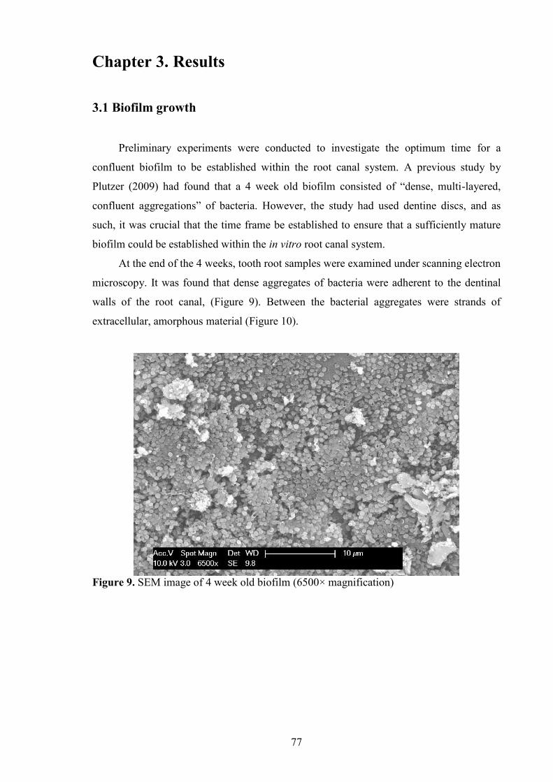
77
Chapter 3. Results
3.1 Biofilm growth
Preliminary experiments were conducted to investigate the optimum time for a
confluent biofilm to be established within the root canal system. A previous study by
Plutzer (2009) had found that a 4 week old biofilm consisted of “dense, multi-layered,
confluent aggregations” of bacteria. However, the study had used dentine discs, and as
such, it was crucial that the time frame be established to ensure that a sufficiently mature
biofilm could be established within the in vitro root canal system.
At the end of the 4 weeks, tooth root samples were examined under scanning electron
microscopy. It was found that dense aggregates of bacteria were adherent to the dentinal
walls of the root canal, (Figure 9). Between the bacterial aggregates were strands of
extracellular, amorphous material (Figure 10).
Figure 9. SEM image of 4 week old biofilm (6500× magnification)
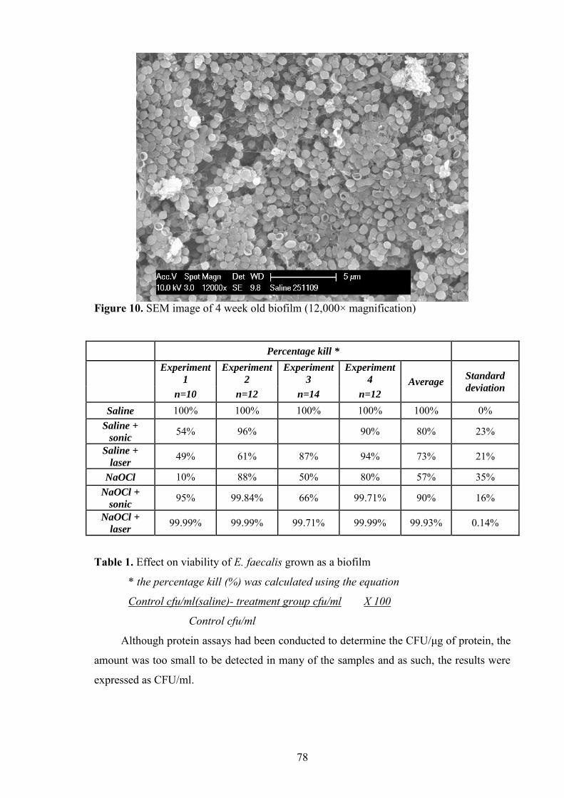
78
Figure 10. SEM image of 4 week old biofilm (12,000× magnification)
Percentage kill *
Experiment
1
Experiment
2
Experiment
3
Experiment
4 Average Standard
deviation n=10 n=12 n=14 n=12
Saline 100% 100% 100% 100% 100% 0% Saline +
sonic 54% 96% 90% 80% 23%
Saline +
laser 49% 61% 87% 94% 73% 21%
NaOCl 10% 88% 50% 80% 57% 35% NaOCl +
sonic 95% 99.84% 66% 99.71% 90% 16%
NaOCl +
laser 99.99% 99.99% 99.71% 99.99% 99.93% 0.14%
Table 1. Effect on viability of E. faecalis grown as a biofilm
* the percentage kill (%) was calculated using the equation
Control cfu/ml(saline)- treatment group cfu/ml X 100
Control cfu/ml
Although protein assays had been conducted to determine the CFU/μg of protein, the
amount was too small to be detected in many of the samples and as such, the results were
expressed as CFU/ml.
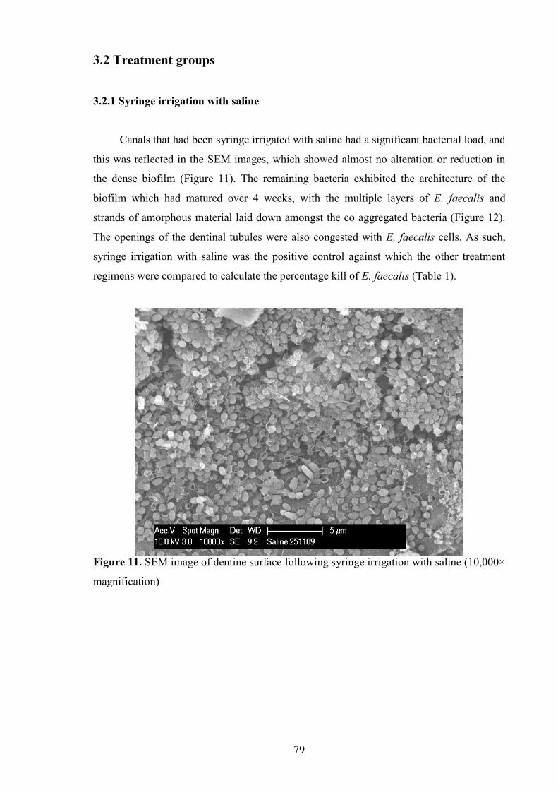
79
3.2 Treatment groups
3.2.1 Syringe irrigation with saline
Canals that had been syringe irrigated with saline had a significant bacterial load, and
this was reflected in the SEM images, which showed almost no alteration or reduction in
the dense biofilm (Figure 11). The remaining bacteria exhibited the architecture of the
biofilm which had matured over 4 weeks, with the multiple layers of E. faecalis and
strands of amorphous material laid down amongst the co aggregated bacteria (Figure 12).
The openings of the dentinal tubules were also congested with E. faecalis cells. As such,
syringe irrigation with saline was the positive control against which the other treatment
regimens were compared to calculate the percentage kill of E. faecalis (Table 1).
Figure 11. SEM image of dentine surface following syringe irrigation with saline (10,000×
magnification)

80
Figure 12. SEM image of dentine surface following syringe irrigation with saline (25,000×
magnification)
3.2.2 Syringe irrigation with sodium hypochlorite
Generally, the root canals that had been had been irrigated with sodium hypochlorite
showed a decrease in the number of E. faecalis cells compared to those that had been
irrigated with saline, given that it was able to achieve a mean percentage kill of 57% ±
35% (Table 1).
Evidence of surface bacterial killing was clearly present on the SEM images, as
exposure of the biofilm to sodium hypochlorite caused the removal of much of the
bacterial biofilm (Figures 13 and 14). The confluent, multi-layered architecture was no
longer present, and only pockets of cell aggregates remained. In some areas of the radicular
dentine, only single or pairs of cells were present (Figure 13).
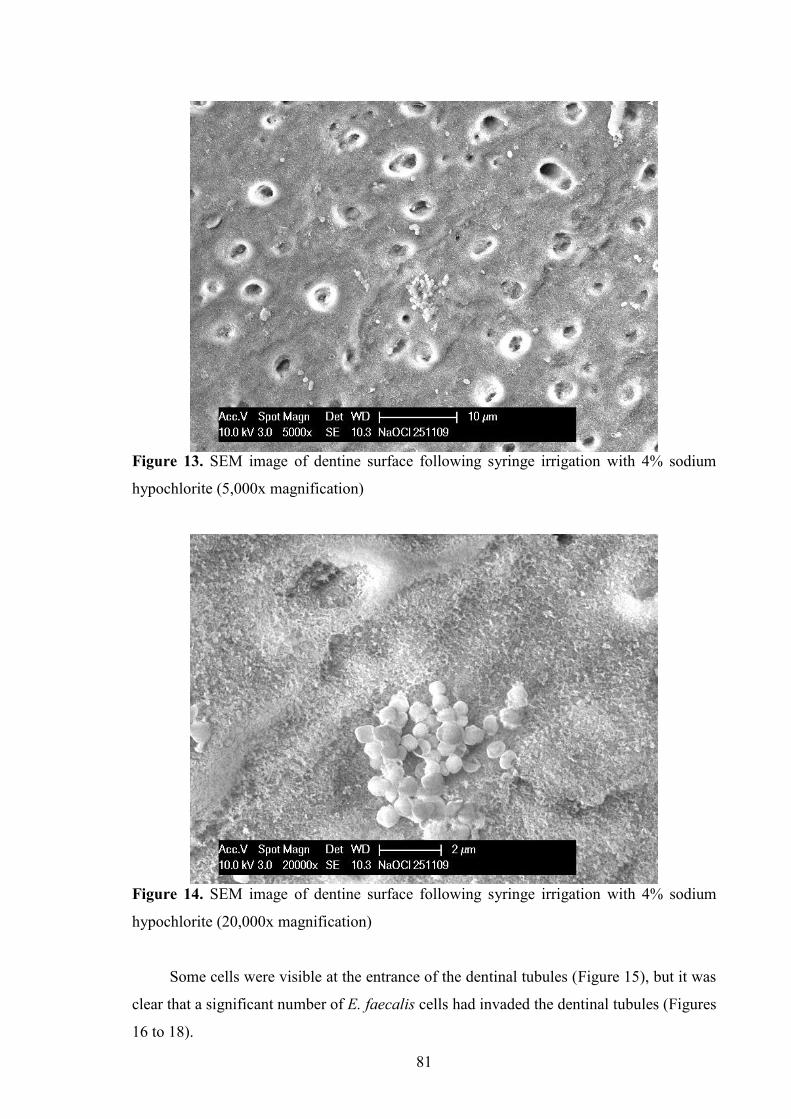
81
Figure 13. SEM image of dentine surface following syringe irrigation with 4% sodium
hypochlorite (5,000x magnification)
Figure 14. SEM image of dentine surface following syringe irrigation with 4% sodium
hypochlorite (20,000x magnification)
Some cells were visible at the entrance of the dentinal tubules (Figure 15), but it was
clear that a significant number of E. faecalis cells had invaded the dentinal tubules (Figures
16 to 18).

82
Figure 15. SEM image of E. faecalis at the entrance of the dentinal tubule following
syringe irrigation with 4% sodium hypochlorite (15,000x magnification)
Figure 16. SEM image of E. faecalis within the dentinal tubules following syringe
irrigation with 4% sodium hypochlorite (10,000x magnification)

83
Figure 17. SEM image of E. faecalis within the dentinal tubules following syringe
irrigation with 4% sodium hypochlorite (20,000x magnification)
Figure 18. SEM image of E. faecalis within the dentinal tubules following syringe
irrigation with 4% sodium hypochlorite (35,000x magnification)
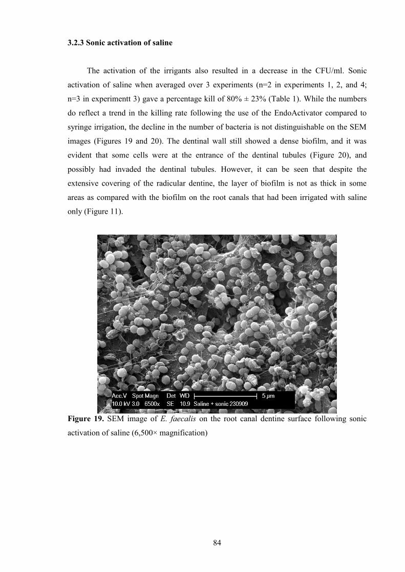
84
3.2.3 Sonic activation of saline
The activation of the irrigants also resulted in a decrease in the CFU/ml. Sonic
activation of saline when averaged over 3 experiments (n=2 in experiments 1, 2, and 4;
n=3 in experimentt 3) gave a percentage kill of 80% ± 23% (Table 1). While the numbers
do reflect a trend in the killing rate following the use of the EndoActivator compared to
syringe irrigation, the decline in the number of bacteria is not distinguishable on the SEM
images (Figures 19 and 20). The dentinal wall still showed a dense biofilm, and it was
evident that some cells were at the entrance of the dentinal tubules (Figure 20), and
possibly had invaded the dentinal tubules. However, it can be seen that despite the
extensive covering of the radicular dentine, the layer of biofilm is not as thick in some
areas as compared with the biofilm on the root canals that had been irrigated with saline
only (Figure 11).
Figure 19. SEM image of E. faecalis on the root canal dentine surface following sonic
activation of saline (6,500× magnification)
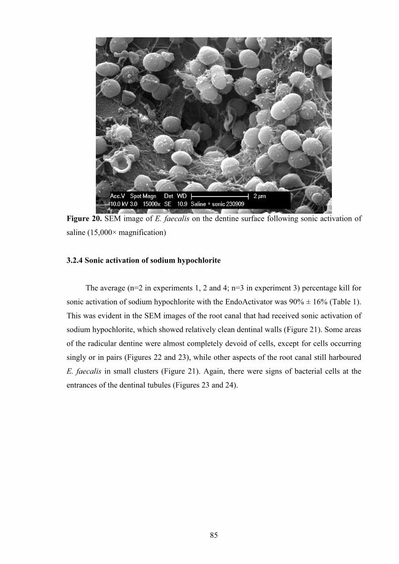
85
Figure 20. SEM image of E. faecalis on the dentine surface following sonic activation of
saline (15,000× magnification)
3.2.4 Sonic activation of sodium hypochlorite
The average (n=2 in experiments 1, 2 and 4; n=3 in experiment 3) percentage kill for
sonic activation of sodium hypochlorite with the EndoActivator was 90% ± 16% (Table 1).
This was evident in the SEM images of the root canal that had received sonic activation of
sodium hypochlorite, which showed relatively clean dentinal walls (Figure 21). Some areas
of the radicular dentine were almost completely devoid of cells, except for cells occurring
singly or in pairs (Figures 22 and 23), while other aspects of the root canal still harboured
E. faecalis in small clusters (Figure 21). Again, there were signs of bacterial cells at the
entrances of the dentinal tubules (Figures 23 and 24).

86
Figure 21. SEM image of E. faecalis on the dentine surface following sonic activation of
4% sodium hypochlorite (2,000× magnification)
Figure 22. SEM image of E. faecalis on the dentine surface following sonic activation of
4% sodium hypochlorite (6,500× magnification)

87
Figure 23. SEM image of E. faecalis at the entrance of a dentinal tubule following sonic
activation of 4% sodium hypochlorite (12,000× magnification)
Figure 24. SEM image of E. faecalis within a dentinal tubule following sonic activation of
4% sodium hypochlorite (12,000× magnification)
3.2.5 Laser activation of saline
The average (n=2 in experiments 1 and 4; n=3 in experiments 2 and 3) percentage
kill for laser activation was 73% ± 21% (table 1). The use of the Er,Cr:YSGG laser to

88
activate saline resulted in no visible damage to the dentinal walls that could be discerned
on the SEM images although it was evident that the biofilm has been disrupted. Exposed
areas of the root canal dentine can be seen, but areas of dense aggregates of E. faecalis
were present (Figure 25). The fibrillar, extracellular matrix is still evident, but the overall
architecture of the mature four-week old biofilm has been destroyed (Figure 26).
However, it is evident from the dentinal surface that the dentinal tubules harboured
E. faecalis (Figure 27). Imaging of the longitudinal sections of the dentinal tubules
revealed dense aggregates of E. faecalis cells in some of the tubules (Figures 28 to 30).
They were arranged mainly in pairs and short chains, although some of the tubules had
single cells. Clearly, during the formation and maturation of the biofilm, some of the E.
faecalis cells had also invaded the dentinal tubules.
Figure 25. SEM image of E. faecalis on the root canal dentine surface following laser
activation of saline (3,500× magnification)

89
Figure 26. SEM image of E. faecalis on the dentine surface following laser activation of
saline (8,000× magnification)
Figure 27. SEM image of E. faecalis at the entrances of dentinal tubules following laser
activation of saline (12,000× magnification)

90
Figure 28. SEM image of E. faecalis within the dentinal tubules following laser activation
of saline (8,000× magnification)
Figure 29. SEM image of E. faecalis within the dentinal tubules following laser activation
of saline (12,000× magnification)
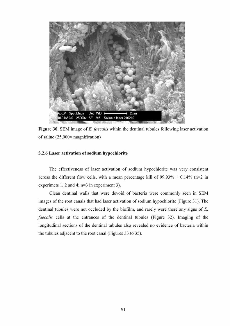
91
Figure 30. SEM image of E. faecalis within the dentinal tubules following laser activation
of saline (25,000× magnification)
3.2.6 Laser activation of sodium hypochlorite
The effectiveness of laser activation of sodium hypochlorite was very consistent
across the different flow cells, with a mean percentage kill of 99.93% ± 0.14% (n=2 in
experimets 1, 2 and 4; n=3 in experiment 3).
Clean dentinal walls that were devoid of bacteria were commonly seen in SEM
images of the root canals that had laser activation of sodium hypochlorite (Figure 31). The
dentinal tubules were not occluded by the biofilm, and rarely were there any signs of E.
faecalis cells at the entrances of the dentinal tubules (Figure 32). Imaging of the
longitudinal sections of the dentinal tubules also revealed no evidence of bacteria within
the tubules adjacent to the root canal (Figures 33 to 35).

92
Figure 31. SEM image of the dentine surface following laser activation of 4% sodium
hypochlorite (2,500× magnification)
Figure 32. SEM image of the dentine surface following laser activation of 4% sodium
hypochlorite (8,000× magnification)

93
Figure 33. SEM image of the dentinal tubules following laser activation of saline (12,000×
magnification)
Figure 34. SEM image of the dentinal tubules following laser activation of saline (20,000×
magnification)

94
Figure 35. SEM image of the dentinal tubules following laser activation of saline (50,000×
magnification)

95
Chapter 4. Discussion
The aim of the present study was to compare the effectiveness of sonic and laser
activation of sodium hypochlorite, against traditional syringe irrigation, in the eradication
of an Enterococcus faecalis biofilm. There are a number of studies that have shown the
effectiveness of various antimicrobial agents against bacteria. However, much of the work
has been conducted with the use of planktonic cultures (Schoop et al., 2007) which do not
accurately reflect the manner in which bacteria grow within the root canal system. Nair
(1987) had shown, with the aid of light and electron microscopy, that bacteria grow as a
biofilm within the radicular dentinal walls, although the term „biofilm‟ was not used at that
time. Since then, in–vitro studies have attempted to replicate in-vivo conditions by
cultivating bacteria as a biofilm (Dunavant et al., 2006; Gutknecht et al., 1996; Spratt et
al., 2001). However, the „biofilm‟ had been developed over periods ranging between 24 to
48 hours. Other studies have cultivated biofilms for longer periods of time, e.g. up to 3
weeks (Berber et al., 2006). In the present study, a biofilm model has been designed which
is based loosely on the flow cell first described in the Dunavant et al. (2006). However,
instead of using ceramic coupons (Dunavant et al., 2006) or dentine discs (Plutzer, 2009),
root canals of extracted single canal teeth were used so as to recreate a more clinically
relevant environment for growth of the biofilm. The use of ceramic coupons or dentine
discs allowed the various antimicrobial agents to have maximum contact against the
bacterial biofilm. Bacteria often invade the dentinal tubules (Love and Jenkinson, 2002),
which limits exposure to antimicrobials and this could not be easily replicated with the use
of flat discs.
The selection of E. faecalis as the bacteria for the present study was due to its
frequent association with persistent endodontic infections (Molander et al, 1998; Sundqvist
et al., 1998; Peciuliene et al., 2000; Pinheiro et al., 2003). In many of these studies, it was
found that not only was E. faecalis the most commonly recovered microorganism from
root canals of teeth with persistent endodontic disease, it was also often present within the
canal as a mono-infection (Sundqvist et al., 1998; Peciulene et al., 2000; Pinheiro et al.,
2003b). The results of these studies clearly demonstrate the dominance of E. faecalis in
retreatment cases. The significant proportion of root canals of teeth exhibiting signs of
persistent infection from which E. faecalis is recovered as a pure culture justifies the use of
this microorganism for this study. The high incidence of recovery of E. faecalis from root
canals of failed endodontic treatment also reflects the tenaciousness of the organism (Love,
2001; Evans et al, 2002) and highlights the importance of seeking an efficacious and

96
effective means to eradicate it, so as to eliminate disease and facilitate healing of the
periapical tissues.
The choice of 4% sodium hypochlorite for the present study was due to its
widespread use in endodontic practice, its broad spectrum of antimicrobial action and its
ability to hydrolyse necrotic pulp tissue (Pashley et al., 1985; Zehnder, 2006). Due to short
period of time for irrigation (1 min) and the small volume of irrigant employed in this
study, it was deemed that a concentration of sodium hypochlorite that was sufficiently high
to exert its antimicrobial effect was necessary. Moreover, no instrumentation was
performed in this study, so the eradication of the E. faecalis biofilm was dependent solely
on the antimicrobial effect of sodium hypochlorite.
This study is the first to investigate the effects of Er,Cr:YSGG laser activation of an
antimicrobial irrigant against a bacterial biofilm. Also, to our knowledge, no in-vitro
studies have been conducted whereby a bacterial biofilm has been cultivated within the
root canal of extracted teeth. Little is also known of the effect of activating sodium
hypochlorite solution with the Er,Cr:YSGG laser against the E. faecalis biofilm. Compared
to previous studies, which investigated the use of the laser activated irrigation in the
removal of dentinal debris and smear layer (George et al, 2008; de Groot et al 2009; de
Moor et al., 2009), the present study sought to determine the effectiveness of the
Er,Cr:YSGG laser activation of sodium hypochlorite in the removal of a mature four-week
old E. faecalis biofilm. Most studies that had examined the use of the Er,Cr:YSGG laser in
the eradication of E. faecalis employed the laser without an irrigant present within the
canal and directly irradiated the root canal (Gordon et al 2007; Schoop et al, 2007; Wang
et al, 2007). With the exception of the Wang et al. (2007) study, most of these
investigations had cultivated E. faecalis for relatively short periods, ranging from 4 hours
to 7 days (Schoop et al., 2007; Gordon et al., 2007). Based on a previous investigation,
cultivation of the E. faecalis biofilm for a period of 4 weeks yielded a dense, multi-layered
aggregation of cells within an amorphous extracellular matrix (Plutzer, 2009). It is evident
that in our model that a 4 week period permitted the growth of a dense, mature biofilm,
with cells that clearly had penetrated the dentinal tubules. When studying the effectiveness
of an irrigation protocol, there are vast differences between the conditions of in vivo and in
vitro studies. In some in vitro studies, a high volume of the irrigant is often available to
directly contact bacteria which are often grown as a planktonic culture, resulting in optimal
conditions for the eradication of the microoganisms (Gomes et al., 2001; Vianna et al.,
2004; Dunavant et al., 2006). In the present study, although there was an attempt to
97
simulate in vivo conditions as much as possible, it is acknowledged that nutrient supply,
redox and pH may not truly reflect the “in vivo lifestyle”.
The power setting of lasers in different in-vitro studies have ranged from 1.25W to
1.5W (George and Walsh, 2008; De Moor et al., 2009). However, these settings were
deemed to be too high for use within the root canal, especially in conjunction with sodium
hypochlorite. Preliminary visual investigations showed that the Er,Cr:YSGG laser at these
settings caused a significant amount of agitation when the tip was placed in a beaker of
water. Placing the laser tip into a plastic block simulating the root canal also caused a
significant amount of extrusion through the „apex‟ of the block at power settings of 1W to
1.5W. The lowest power setting on the Waterlase unit was 0.25W, and it was found that
when only 4 mm of the laser tip was immersed in the irrigant, the amount of extrusion was
minimised.
The selection of the EndoActivator® sonic instrument for comparison stemmed from
its recent release, and it was our intention to test these novel devices to activate irrigants.
The EndoActivator® comprises of a handpiece with detachable, single-use polymer tips of
various sizes. Its ease of use, coupled with its cordless, lightweight design and relatively
low cost, makes it a viable option for the endodontic practice. This provided an interesting
contrast against the solid-state Er,Cr:YSGG laser which is significantly more expensive.
The use of these two instruments permitted examination and comparison of the
effectiveness of laser energy induced cavitation against the acoustic streaming achieved by
the sonic activation of the irrigant.
4.1 Comparing syringe irrigation with 0.9% saline and 4% sodium
hypochlorite
The results of this study illustrate clearly that the use of an antimicrobial irrigant
such as sodium hypochlorite provides more effective eradication of bacteria as compared
to saline. Using scanning electron microscopy (SEM), canals that had saline irrigation with
a syringe showed minimal disruption of the E. faecalis biofilm. Syringe irrigation with 4%
sodium hypochlorite was capable of achieving a percentage kill of 57% ± 35%. This
finding was confirmed with the scanning electron microscopy (SEM) images. Following
syringe irrigation with saline, the E. faecalis biofilm was thick and composed of multiple
layers (Figure 11) and had barely been disrupted (Figure 12). The bacterial cells could be
seen occluding the dentinal tubules (Figure 12), and was similar in appearance to the 4
week old biofilm which had been cultivated in our pilot study to evaluate the optimal time

98
for the biofilm development (Figures 9 and 10). SEM images of the radicular dentinal wall
from samples that received sodium hypochlorite as an irrigant applied with a syringe
showed that irrigant had eradicated most of the bacteria, leaving small groups or pairs of
bacterial cells (Figures 13 and 15). Much of the canal wall appeared to be free from E.
faecalis, although many had invaded the dentinal tubules, and had not been completely
eradicated following irrigation with sodium hypochlorite (Figures 16 to 18). Under lower
magnification, it is evident that most of the dentinal tubules contained E. faecalis (Figure
16).
The results are consistent with the findings of Byström and Sundqvist (1983), whose
results showed that in 8 out of 15 canals, bacteria could not be recovered with paper points
sampling after irrigation with saline. This was compared to 12 out of 15 canals that were
irrigated with 0.5% sodium hypochlorite. However, unlike the study by Byström and
Sundqvist (1983) which examined the efficacy of mechanical instrumentation combined
with an antimicrobial irrigant, the present study only investigated the effects of irrigation.
Despite the consistency of the findings with available literature, the percentage kill
following syringe irrigation with sodium hypochlorite was somewhat less consistent. The
percentage kill ranged from 10% (Experiment 1) to 88% (Experiment 2). One of the
reasons for this may be due to the variability of the root canals. Despite attempts to
standardise the root canal lengths (15 mm) and preparation of the root canal (#40, 0.06),
the volume of the root canal was not equal in all teeth samples, and may have had an effect
on the percentage kill. Increasing the volume of irrigant and placement of the needle to
approximately 1 mm from the apex has been shown to significantly increase the
effectiveness of bacterial elimination (Sedgley et al., 2005b). Similar to our study, no
mechanical instrumentation was conducted in the study by Sedgley et al. (2005b) and
interestingly, irrigation was performed with a non-antimicrobial irrigant. The rational of
the authors was to assess the “mechanical efficacy of irrigants when delivered at different
distances from the apex in the removal of bacteria”. However, other studies have shown
that an increase in volume does little to improve the flushing action (van der Sluis et al.,
2006). In the present study, the tip of the irrigating needle was placed within 2 mm from
the apex and 5 ml of irrigant was introduced into the root canal. Even with the use of an
antimicrobial irrigant in the form of 4% sodium hypochlorite, the canals could not be
rendered bacteria-free, and this is most likely because the bacteria were exposed to sodium
hypochlorite for only 1 min and only a small volume of 5 ml was used.
99
4.2 Comparing sonic activation of 0.9% saline and 4% sodium
hypochlorite
When using the EndoActivator® to induce sonic activation of the irrigants, the group
irrigated with sodium hypochlorite experienced a higher percentage kill of 90% ± 16%
compared with 80% ± 23% for saline. In this study, the EndoActivator® was set at
maximum power, which was 10,000 cycles per minute (166 Hz). It can be deduced from
the SEM images of sonic activation of saline that the hydrodynamic shear forces resulting
from acoustic streaming are relatively small (Figures 19 and 20). The biofilm is not thick
and composed of multiple layers as before, but it is still covers a significant portion of the
radicular dentine. The combination of sonic activation with sodium hypochlorite resulted
in much more disruption of the dense biofilm, with only a few bacterial cells evident on the
radicular dentine surface (Figures 21 to 24).
While none of the SEM images showed the presence of E. faecalis within the
dentinal tubules, the number of CFU‟s reflects the presence of bacteria despite the
relatively clean radicular dentinal walls. Due to the imprecise nature of the way the teeth
were notched and split into halves for visualisation of the root canal, it was impossible to
obtain images of the longitudinal sections of all the different treatment groups. However,
some of the sections yielded areas which could be imaged under SEM, albeit they were
mainly of E. faecalis that had invaded the tubules superficially. Since the diamond disc had
been used to notch the teeth from the cementum side, most of the canals proximal to the
cementum side were covered with dentinal debris. As such, this rendered it impossible to
determine if any E. faecalis cells penetrated this far into the dentinal tubules. Nevertheless,
based on the percentage kill rate (Table 1), it can be deduced that acoustic streaming, even
with an antimicrobial irrigant, is insufficient in eradicating bacteria that have invaded the
dentinal tubules. It can be postulated that the acoustic energy generated by the sonic
instrument results in limited penetration of the irrigant into the dentinal tubules. However,
research into this is still in its infancy, and more investigation into this area is warranted.
4.3 Comparing laser activation of 0.9% saline and 4% sodium
hypochlorite
This difference between the two irrigants was more marked when the CFU counts for
teeth that received laser activation of saline and sodium hypochlorite were observed; the

100
group that had been irrigated with sodium hypochlorite had achieved a mean percentage
kill of 99.93% ± 0.14% compared with the saline group, 73% ± 21%. Although much of
the biofilm had been disrupted following activation of the saline irrigant (Figures 25 and
26), a significant number of E. faecalis cells were still residing within the dentinal tubules
(Figures 27 to 30). It is thus evident that despite the ability of laser mediated cavitation to
eradicate bacteria on the surface, unless coupled with an antimicrobial agent, it was not
possible to eliminate the bacteria that had invaded the dentinal tubules. Laser activation of
sodium hypochlorite, on the other hand, was able to eliminate almost all of the bacteria,
and the SEM images showed not only clean radicular dentinal walls (Figures 31 and 32),
but also dentinal tubules that were devoid of bacteria (Figures 33 to 35). It is evident that
the higher level of energy imparted by the Er,Cr:YSGG laser was able to permit greater
penetration of the sodium hypochlorite into the dentinal tubules to cause more effective
eradication of the E. faecalis bacterial cells. The consistency of the results, whereby laser
activated irrigation was able to achieve percentage kills of almost 100% provides more
evidence of the efficacy of this method of irrigation.
The use of extracted teeth and the root canals instead of cellulose nitrate membrane
filters or dentine blocks in this study was to replicate the in-vivo environment, albeit a
simplified model. Nonetheless, the teeth used in this study had relatively simple anatomy.
Future directions may include the use of root canals with more complex anatomy, such as
the mesial roots of lower molars. The presence of isthmuses and fins will test the ability of
laser activated irrigation to clean these normally inaccessible areas, and may provide
greater insight into the effectiveness of such an irrigation protocol.
Studies that investigated the mechanism of laser energy mediated cavitation effects
have shown that the phenomenon is brought about by the rapid implosion of a vapour
bubble (Blanken et al., 2009; de Groot et al., 2009). Visualisation of the generation and
subsequent implosion of vapour cavities have provided a comprehensive understanding of
the ways in which laser activation of anti-bacterial irrigants can aid in debridement of the
root canal system. However, most of these studies have visualised these mechanisms at
work in plastic containers or glass capillary tubes that simulate a root canal (Blanken et al.,
2009; de Groot et al., 2009). Containers or tubes do not share similar dimensions to the
small and confined space of the root canal system. It is thus assumed that the pattern of
vapour cavity generation and implosion would be duplicated when the irrigant is activated
in a similar manner within the root canal. However, we cannot be sure if the presence of a
non-uniformly shaped canal, with various fins and isthmuses and the apical foramen, may
cause a shift in fluid dynamics, thereby dampening the effectiveness of these mechanisms.
101
A total of 58 teeth were used in this study. However, the percentage kill results for
10 root canal samples from one of the flow cells could not be calculated (Appendix 1 –
Flow cell 5). This was because no detectable bacteria could be cultivated from the root
canal of the tooth treated with syringe irrigation of saline. This may have been due to an
operator error during serial dilution or culturing of the bacteria. Nonetheless, the results of
the viable count showed consistency of the number of colony forming units/ml with the
other flow cells. There was a detectable trend, whereby the use of sodium hypochlorite
resulted in lower bacterial numbers, and that the use of laser activated irrigation provided
the greatest reduction in bacterial cells.
4.4 Clinical considerations
The concentration of sodium hypochlorite used in this study was relatively high
(4%), and may have contributed quite significantly to the elimination of E. faecalis. The
cytotoxic nature of sodium hypochlorite is a concern, and can elicit a severe reaction when
extruded out through the apex of the root canal (Hulsmann and Hahn, 2000). Undoubtedly,
the potential for extrusion of sodium hypochlorite is increased with the use of the
Er,Cr:YSGG laser (George and Walsh, 2008). It is important also to note that their results
revealed that most significant factor that determined the amount of extrusion was the size
of the apical foramen. It thus reinforces the importance of maintaining instrumentation
within the root canal system, the need to respect and preserve the apical constriction as
much as possible (Ricucci, 1998), and that the laser be used judiciously to prevent the
occurrence of a hypochlorite incident.
In this study, the apices of the root canals were blocked with Cavit and nail varnish
prior to irrigation and activation of the irrigant. This creates an apical air lock, which has
been shown to reduce the efficacy of hand irrigation. The occlusion of the apices also
limited the forward expansion of the vapour bubble generated by the laser, and prevented
the expulsion of the irrigant out of the root canal, especially as these canals had been
prepared with a size #40 (0.06 taper) instrument to 1 mm beyond the apex. Blanken et al.
(2009) have shown that within an artificial root canal within a glass block, the limited
lateral and forward expansion within the confines of the root canal resulted in the
generation of primary and secondary cavitation bubbles. The cavitation effect subsequently
translates into the generation of shear forces against the canal walls which produced the
results shown in the present study, i.e. disruption of the biofilm and eradication of the E.
faecalis cells when used in conjunction with sodium hypochlorite.
102
It should be noted that the power setting in the George and Walsh (2008) study was
1.25W at 20 Hz; the Er,Cr:YSGG laser in our study was set at one-fifth of that employed
in their investigation. The Er,Cr:YSGG laser was set at 0.25W at 20 Hz. Also, to mimic a
clinical situation where minimisation of extrusion would be ideal, only 4 mm of the tip was
inserted into the canal and withdrawn slowly while the laser was being activated. The
power setting of 0.25W used in the present study was the lowest possible setting for the
Waterlase unit. New software upgrades and the new Waterlase MD models now allow for
a lower power setting of 0.10W. Thus, a protocol which ensures efficacious debridement of
the root canal will require investigation into different permutations of irrigant
concentration and power setting to achieve the maximum disinfection with minimum risk
of morbidity to the patient. Other factors such as the length of time in which the irrigant
should be activated, the type(s) and/or combination of irrigant(s), and the volume of
irrigant used should also be established. Ideally, a large volume of irrigant should be
activated for as long as possible. However, there are practical clinical considerations such
as treatment time, as well as safety issues. Further research into the potential use of lower
concentrations of sodium hypochlorite, in combination with different power settings of the
Er,Cr:YSGG laser are currently underway.
4.5 Limitations of the study
The ability of E. faecalis to persist in an endodontically treated root canal is
testimony to its survivability. It has been shown to be capable of withstanding protracted
periods of time within the root canal, maintaining their viability for up to 12 months
without nutrition (Sedgley et al., 2005a). Figdor et al. (2003) have shown it to be able to
survive in water for more than 4 months, with the viable cell population being maintained
at about 103 CFU/ml. These studies reflect the conditions within an instrumented and
obturated root canal. In the present study however, the biofilm was constantly bathed in
Todd Hewitt Broth and thus received a constant supply of nutrients. Many studies have
shown that the capacity for antimicrobial agents in endodontic treatment to exert its effects
is dependent upon the physiological state of the bacteria. Portenier et al. (2005) examined
the susceptibility of E. faecalis to 0.0001% sodium hypochlorite when it was in
exponential growth phase, stationary phase and starvation phase. The results showed that
the resistance of E. faecalis was highest when the cells were in the starvation phase, with
more than 4% of the bacterial cells surviving after 10 min of exposure to sodium
hypochlorite. When compared against E. faecalis cells that were in the exponential growth
103
phase or the stationary phase, the starved cells were able to survive in numbers 1,000 to
10,000 times higher following exposure to sodium hypochlorite. Cells that were growing
were the most sensitive to the antimicrobial agent (Portenier et al., 2005).
Liu et al. (2010) also investigated the ability of E. faecalis cells to form biofilms
when in starvation phase, and compared against cells in the exponential growth phase and
stationary phase. The results showed that starved E. faecalis cells were capable of biofilm
formation, albeit slower as compared to cells in the other two phases. When the biofilm
was subject to sodium hypochlorite exposure, cells in the starvation phase were more
resistant to killing by 5.25% sodium hypochlorite. The resistance of E. faecalis biofilm to
sodium hypochlorite in the starved state also increased with maturation of the biofilm, as
the 48 hour-old biofilm showed significant lower reduction of viable cells compared to
those of the 6 hour and 24 hour-old biofilm (Liu et al., 2010). Interestingly, their results
also showed that E. faecalis cells in the exponential growth phase had lower susceptibility
to sodium hypochlorite when compared against cells in the stationary phase. This is in
contrast to the findings of Portenier et al. (2005). Despite the conflicting evidence, the
elevated resistance of E. faecalis cells in the starvation phase must account for its ability to
persist within the root canal following therapy. Thus, the use of cells in the starvation
phase within our current experimental protocol is an avenue for further research.
The method with which the teeth were sampled was highly effective in recovering
most, if not all, of the bacteria within the root canal, including those that were residing
within the dentinal tubules. This method would also allow for sampling of root canals with
anatomy more complex than those used in this study, where fins, anastomoses and
isthmuses exist. The shortcoming of paper point sampling is the inability to access these
sites (Sundqvist et al., 1998). Molander et al. (1998) also noted that in retreatment cases,
particles of remnant gutta percha may preclude sampling fluid from areas where bacteria
were present, thereby leading to a false negative result. However, this technique of
sampling is destructive, and cannot be applied in in vivo studies. Care was also taken to
ensure that the bacteria sampled would be from within the root canal, as the bacteria
biofilm that may have developed on the surface of the root would have been eliminated by
soaking the entire tooth in 4% sodium hypochlorite for 1 hour. A previous study had
shown that this regime would result in 99.99% kill of E. faecalis (Plutzer, 2009).

104
Chapter 5. Conclusion
Sonic and laser activated irrigation, in conjunction with the use of an antimicrobial
irrigant, resulted in reduction of E. faecalis cells from within the root canal. The results
were particularly marked when the Er,Cr:YSGG laser was used to activate a 4% sodium
hypochlorite solution. This superior ability to disinfect the root canal system predictably
may potentially remove the need for the placement of an inter-appointment intra-canal
medicament. It would be of interest to determine if the application of sonic and laser
activated irrigation would be capable of reducing bacterial numbers to levels where
calcium hydroxide had been placed as an intra-canal medicament. Well-controlled
prospective clinical studies are necessary to determine if the microbial load following
ultrasonic activation within a single visit endodontic treatment would be comparable to that
after medicament placement. Long term evaluation of the outcome would also be essential
to determine the implications of sonic and laser activated irrigation in the healing of post-
treatment apical periodontitis.
There is mounting evidence to support the application of single visit endodontics.
Apart from the similar success rate to two visit endodontic therapy, and the lack of “flare
ups” and post operative pain (Oliet, 1983; Trope, 1991), there are the practical concerns
that need to be addressed. To date, there are no studies which have compared the outcomes
of single visit against multiple visit root canal retreatment. In a vast country like Australia,
many patients who require endodontic therapy often live outside of the metropolitan areas
and the commute to receive treatment can involve hours of travel. This may not be feasible
for many patients. There are also patients who, due to physical impediments, may find it
difficult to access treatment should it require several appointments. This should, however,
not be a reason to deprive such patients of the right to receive endodontic therapy, and the
right to preserve their dentition. These reasons reinforce the convenience of single visit
endodontic therapy, and from a practicality standpoint, a more viable option for some of
our patients. The persistence of bacteria such as E. faecalis, which have evolved to
maintain their viability in spite of the antimicrobial measures employed in endodontic
therapy, presents a significant problem in ensuring the eradication of these bacteria and
thus eliminating the post-treatment disease.
It is without doubt that the present study will require much refinement and further
research into laser activated irrigation to maximise the potential of this technology.
However, it is hoped that this will pave the way for the possible institution of a single visit

105
treatment protocol which can predictably eliminate E. faecalis and other bacteria so as to
ensure the most favourable treatment outcome.
106
Chapter 6. References
Abbott PV, Heijkoop PS, Cardaci SC, Hume WR, Heithersay GS. An SEM study of
the effects of different irrigation sequences and ultrasonics. Int Endod J 1991; 24:308-316.
Abou-Rass M, Piccinino MV. The effectiveness of four clinical irrigation methods
on the removal of root canal debris. Oral Surg Oral Med Oral Pathol 1982; 54:323– 8.
Ahmad M, Pitt Ford ThR, Crum LA. Ultrasonic debridement of root canals: an
insight into the mechanism involved. J Endod 1987a; 13:93–100.
Ahmad M, Pitt Ford TR, Crum LA, Walton AJ. Ultrasonic debridement of root
canals: acoustic cavitation and its relevance. J Endod 1988; 14:486–493.
Ahmad M, Pitt Ford TR, Crum LA. Ultrasonic debridement of root canals: acoustic
streaming and its possible role. J Endod 1987b; 14:490–499.
Ahmad M, Roy RA, Kamarudin AG. Observations of acoustic streaming fields
around an oscillating ultrasonic file. Endod Dent Traumatol 1992; 8:189–94.
Akpata ES, Blechman H. Bacterial invasion of pulpal dentine wall in vitro. J Dent
Res 1982; 61:435-438.
Altundasar E, Ozecelik B., Cehreli ZC, Matsumoto K. Ultramorphological and
histochemical changes after Er,Cr:YSGG laser irradiation and two different irrigation
regimes. J Endod 2006; 32:465-468.
American Association of Endodontists. Glossary of Endodontic Terms, 7th edn.
American Association of Endodontists, Chicago, IL. 2003.
Anderl JN, Franklin MJ, Stewart PS. Role of antibiotic penetration limitation in
Klebsiella pneumoniae biofilm resistance to ampicillin and ciprofloxacin. Antimicrob
Agents Chemother 2000; 44: 1818–1824.
Anic I, Segovic S, Katanec D, Prskalo K, Najzar-Fleger D. Scanning electron
microscopic study of dentin lased with argon, CO2, and Nd:YAG laser. J Endod 1998;
24:77-81.
Bahcall J, Howard P, Miserendino L, Walia H. Preliminary investigation of the
histological effects of laser endodontic treatment on the periradicular tissues in dogs. J
Endod 1992; 18:47-51.
Barnett F, Trope M, Khoja M, Tronstad L. Bacteriologic status of the root canal after
sonic, ultrasonic and hand instrumentation. Endod Dent Traumatol 1985; 1:228-231.
Baumgartner JC, Cuenin PR. Efficacy of several concentrations of sodium
hypochlorite for root canal irrigation. J Endod 1992; 18:605–612.

107
Berber VB, Gomes BPFA, Sena NT, Vianna ME, Ferraz CCR, Zaia AA, Souza-
Filho FJ. Efficacy of various concentrations of Sodium hypochlorite and instrumentation
techniques in reducing Enterococcus faecalis within root canals and dentinal tubules. Int
Endod J 2006; 39:10–17.
Berkiten M, Berkiten R, Okar I. Comparative evaluation of antibacterial effects of
Nd:YAG laser irradiation in root canals and dentinal tubules. J Endod 2000; 26:268-270.
Blanken JW, De Moor RJG, Meire M, Verdaasdonk RM. Laser induced explosive
vapour and cavitation resulting in effective irrigation of the root canal. Part 1: A
visualisation study. Lasers Surg Med 2009; 41:514-519.
Blanken JW, Verdaasdonk RM. Cavitation as a working mechanism of the
Er,Cr:YSGG laser in endodontics: A visualisation study. J Oral Laser Appl 2007; 7:97–
106.
Bogosian G, Morris PJ, O‟Neil JP. A mixed culture recovery method indicates that
enteric bacteria do not enter the viable but nonculturable state. Appl Environ Microbiol
1998; 64:1736–1742.
Buschner JH, van der Mei HC. Initial microbial adhesion events: mechanisms and
implications. In: Allison DG, Gilbert P, Lappin-Scott HM, Wilson M, eds. Community
Structure and Co-Operation in Biofilms. Cambridge: Cambridge University Press, 2000:
25–36.
Byström A, Claesson R, Sundqvist G. The antibacterial effect of camphorated
paramonochlorophenol, camphorated phenol and calcium hydroxide in the treatment of
infected root canals. Endod Dent Traumatol 1985; 1:170–175.
Byström A, Happonen R-P, Sjögren U, Sundqvist G. Healing of periapical lesions of
pulpless teeth after endodontic treatment with controlled asepsis. Endod Dent Traumatol
1987; 3:58–63.
Byström A, Sundqvist G. Bacteriologic evaluation of the effect of 0.5% sodium
hypochlorite in endodontic therapy. Oral Surg Oral Med Oral Pathol 1983: 55:307–312.
Byström A, Sundqvist G. Bacteriologic evaluation of the efficacy of mechanical root
canal instrumentation in endodontic therapy. Scand J Dent Res 1981: 89:321– 328.
Byström A, Sundqvist G. The antibacterial action of sodium hypochlorite and EDTA
in 60 cases of endodontic therapy. Int Endod J 1985: 18:35–40.
Cameron JA. The use of 4 per cent sodium hypochlorite, with or without ultrasound,
in cleansing of uninstrumented immature root canals: SEM study. Aust Dent J 1987;
32:204–213.
108
Cameron JA. The use of ultrasonics in the removal of the smear layer: a scanning
electron microscope study. J Endod 1983; 9:289-292.
Card S, Sigurdsson A, Orstavik D, Trope M. The effectiveness of increased apical
enlargement in reducing intracanal bacteria. J Endod 2002; 28:779–783.
Chen RS, Messer HH. The duration of effectiveness of root canal medicaments. J
Endod 1984; 10:240-245.
Chow JW, Thal LA, Perri MB, Vazquez JA, Donabedian SM, Clewell DB, Zervos
MJ. Plasmid-associated hemolysin and aggregation substance production contribute to
virulence in experimental enterococcal endocarditis. Antimicrob Agents Chemother 1993;
37:2474–2477.
Chow TW. Mechanical effectiveness of root canal irrigation. J Endod 1983; 11:475–
479.
Clewell DB. Bacterial sex pheromone-induced plasmid transfer. Cell 1993; 73:9–12.
Clewell DB. Plasmids, drug resistance, and gene transfer in the genus Streptococcus.
Microbiol Rev 1981; 45:409–436.
Coque TM, Patterson ME, Steckelberg JM, Murray BE. Incidence of hemolysin,
gelatinase, and aggregation substance among enterococci isolated from patients with
endocarditis and other infections and from feces of hospitalized and community based
persons. J Infect Dis 1995; 171:1223-1229.
Cross CE, Halliwell B, Borish ET, Pryor WA, Ames BN, Saul RL. Oxygen radicals
and human disease. Ann Intern Med 1987; 107:526-545.
Cunnigham WT, Martin H, Forrest WR. Evaluation of root canal debridement by the
endosonic ultrasonic synergistic system. Oral Surg Oral Med Oral Pathol 1982a; 53:401-
404.
Cunnigham WT, Martin H, Peller GB, Stoops DE. A comparison of antimicrobial
effectiveness of endosonic and hand root canal therapy. Oral Surg Oral Med Oral Pathol
1982b; 54:238-241.
Cunnigham WT, Martin H. A scanning electron microscope evaluation of root canal
debridement with the endosonic ultrasonic synergistic system. Oral Surg Oral Med Oral
Pathol 1982; 53:527-531.
Cutright DE, Bhaskar SN. Pulpal vasculature as demonstrated by a new method. Oral
Surg Oral Med Oral Pathol Oral Radiol Endod 1969: 27:678–683.
Cvek M, Nord CE, Hollender L. Antimicrobial effect of root canal debridement in
teeth with immature root. A clinical and microbiologic study. Odontol Revy 1976; 27:1–
10.

109
Dahlén G, Magnusson BC, Möller Å. Histological and histochemical study of the
influence of lipopolysaccharide extracted from Fusobacterium nucleatum on the periapical
tissues in the monkey Macaca fascicularis. Arch Oral Biol 1981; 26:591–598.
Dalton BC, Orstavik D, Phillips C, Pettiette M, Trope M. Bacterial reduction with
nickel-titanium rotary instrumentation. J Endod 1998; 24:763–7.
Davies DG, Parsek MR, Pearson JP, Iglewski BH, Costerton JW, Greenberg EP: The
involvement of cell-to-cell signals in the development of a bacterial biofilm. Science 1998;
280:295-299.
Day AM, Cove JH, Phillips-Jones MK. Cytolysin gene expression in Enterococcus
faecalis is regulated in response to aerobiosis conditions. Mol Genet Genomics 2003;
269:31-39.
de Gregorio C, EstevezR, Cisneros R, Heilborn C, Cohenca N. Effect of EDTA,
sonic, and ultrasonic activation on the penetration of sodium hypochlorite into simulated
lateral canals: An in vitro study. J Endod 2009; 35:891-895.
de Groot SD, Verhaagen B, Versluis M, Wu M.-K, Wesselink PR, van der Sluis
LWM. Laser-activated irrigation within root canals: cleaning efficacy and flow
visualization. Int Endod J 2009; 42:1077–1083.
De Moor RJ, Blanken J, Meire M, Verdaasdonk R. Laser induced explosive vapour
and cavitation resulting in effective irrigation of the root canal. Part 2: Evaluation of the
efficacy. Lasers Surg Med 2009; 41:520-523.
De Moor RJ, Hommez GM, De Boever JG, Delme KI, Martens GE. Periapical health
related to the quality of root canal treatment in a Belgian population. Int Endod J 2000: 33:
113–120.
Distel JW, Hatton JF, Gillespie MJ. Biofilm formation in medicated root canals. J
Endod 2002; 28:689–693.
Donson J, Wilson M. Sensitisation of oral bacteria in biofilrns to killing by light
from a low-power laser. Arch Oral Biol 1992: 37:883-887.
Druttman ACS, Stock CJR. An in vitro comparison of ultrasonic and conventional
methods of irrigant replacement. Int Endod J 1989; 22:174–178.
Dugas NN, Lawrence HP, Teplitsky PE, Pharoah MJ, Friedman S. Periapical health
and treatment quality assessment of root-filled teeth in two Canadian populations. Int
Endod J 2003: 36: 181–192.
Dunavant TR, Regan JD, Glickman GN, Solomon ES, Homeyman AL. Comparative
evaluation of endodontic irrigants against Enterococcus faecalis biofilms. J Endod 2006;
32:527–531.

110
Dunny GM. Genetic functions and cell–cell interactions in the pheromone-inducible
plasmid transfer system of Enterococcus faecalis. Mol Microbiol 1990: 4: 689–696.
Dychdala GR. Chlorine and chlorine compounds. In: Block SS, ed. Disinfection,
Sterilization, and Preservation, 4th edn. Philadelphia, USA: Lea & Febiger, 1991:133–135.
Edmond MB, Ober JF, Dawson JD, Weinbaum DL, Wenzel RP. Vancomycin-
resistant enterococcal bacteremia: natural history and attributable mortality. Clin Infect Dis
1996; 23:1234 –1239.
Elsner HA, Sobottka I, Mack D, Claussen M, Laufs R, Wirth R. Virulence factors of
Enterococcus faecalis and Enterococcus faecium blood culture isolates. Eur J Clin
Microbiol Infect Dis 2000; 19:39-42.
Engebrecht J, Nealson K, Silverman M: Bacterial bioluminescence: isolation and
genetic analysis of functions from Vibrio fischeri. Cell 1983; 32:773-781.
Evans M, Davies JK, Sundqvist G, Figdor D. Mechanisms involved in the resistance
of Enterococcus faecalis to calcium hydroxide. Int Endod J 2002; 35:221–228.
Eversole L, Rizoiu I, Kimmel A. Pulpal response to cavity preparation by an erbium,
chromium:YSGG laser-powered hydrokinetic system. J Am Dent Assoc 1997; 128:1099-
106.
Fernández M, Sánchez-Hidalgo M, García-Quintáns N, Martínez-Bueno M, Valdivia
E, López P, Maqueda M. Processing of as-48ABC RNA enterocin production by
Enterococcus faecalis. J Bacteriol 2008; 190:240-250.
Figdor D, Davies JK, Sundqvist G. Starvation survival, growth and recovery of
Enterococcus faecalis in human serum. Oral Microbiol Immunol 2003; 18:234 –239.
Flahaut S., Hartke A., Giard J-C, Auffray Y. Alkaline stress response in
Enterococcus faecalis: Adaptation, cross-protection, and changes in protein synthesis.
Appl Environ Microbiol 1997; 63:812–814.
Foley I, Gilbert P. In vitro studies of the activity of glycopeptide combinations
against Enterococcus faecalis biofilms. J Antimicrob Chemother 1997; 40: 667–672.
Franz CM, Stiles ME, Schleifer KH, Holzapfel WH. Enterococci in foods – a
conundrum for food safety. Int J Food Microbiol 2003; 88:105–22.
Galvez A, Maqueda M, Martinez-Bueno M, Valdivia E. Bactericidal and
bacteriolytic action of peptide antibiotic AS-48 against Gram-positive and Gram-negative
bacteria and other organisms. Res Microbiol 1989; 140:57-68.
Galvez A, Maqueda M, Martinez-Bueno M, Valdivia E. Permeation of bacterial
cells, permeation of cytoplasmic and artificial membrane vesicles, and channel formation
on lipid bilayers of peptide antibiotic AS-48. J Bacteriol 1991; 173:886-892.

111
George R, Ian A. Meyers IA, Walsh LJ. Laser activation of endodontic irrigants with
improved conical laser fiber tips for removing smear layer in the apical third of the root
canal. J Endod 2008; 34:1524-1527.
George R, Walsh. Apical extrusion of root canal irrigants when using Er:YAG and
Er,Cr:YSGG lasers with optical fibers: An in vitro dye study. J Endod 2008; 34:706-708.
Gilbert P, Das J, Foley I. Biofilm susceptibility to antimicrobials. Adv Dent Res
1997: 11: 160–167.
Goldman L, Hornby P, Meyer R, Goldman B. Impact of the laser on dental caries.
Nature 1964; 203:417.
Gomes BP, Pinheiro ET, Gade-Neto CR, Sousa ELR, Ferraz CCR, Zaia AA,
Teixeira FB, Souza-Filho FJ. Microbiological examination of infected dental root canals.
Oral Microbiol Immunol 2004; 19:71– 76.
Gomes BP, Pinheiro ET, Jacinto RC, Zaia AA, Ferraz CC, Souza-Filho FJ. Microbial
analysis of canals of root-filled teeth with periapical lesions using polymerase chain
reaction. J Endod 2008; 34:537– 540.
Gomes BPFA, Ferraz CCR, Vianna ME, Berber VB, Teixeira FB, Souza-Filho FJ.In
vitro antimicrobial activity of several concentrations of sodium hypochlorite and
chlorhexidine gluconate in the elimination of Enterococcus faecalis. Int Endod J 2001;
34:424–428.
Gomes BPFA, Lilley JD, Drucker DB. Variations in the susceptibilities of
components of the endodontic microflora to biomechanical procedures. Int Endod J 1996;
29:235-241.
Gordon W, Atabakhsh VA, Meza F, Doms A, Nissan R, Rizoiu I, Stevens RH. The
antimicrobial efficacy of the erbium, chromium:yttrium-scandium-gallium-garnet laser
with radial emitting tipps on root canal dentin walls infected with Entercoccus faecalis. J
Am Dent Assoc 2007; 138:992-1002.
Grossman LI. Bacteriologic examination. In Root canal therapy Ed Grossman Lea &
Febiger Philadelphia USA, 1950
Gutarts R, Nusstein J, Reader A, Beck M. In vivo debridement efficacy of ultrasonic
irrigation following hand/rotary instrumentation in human mandibular molars. J Endod
2005; 31:166–170.
Gutknecht N, Franzen R, Schippers M, Lampert F. Bactericidal effect of a 980 nm
diode laser in the root canal wall dentin of bovine teeth. J Clin Laser Med Surg 2004; 22:9-
13.
112
Gutknecht N, Moritz A, Conrads G, Sievert T, Lampert F. Bactericidal effect of the
Nd:YAG laser in in vitro root canals. J Clin Laser Med Surg 1996; 14:77-80.
Haapasalo HK, Siren EK, Waltimo TMT, Orstavik D, Haapasalo MPP. Inactivation
of local root canal medicaments by dentine: an in vitro study. Int Endo J 2000: 33:126-131.
Haapasalo M, Endal U, Zandi H, Coil JM. Eradication of endodontic infection by
instrumentation and irrigation solutions. Endod Topics 2005: 10:77–102.
Haapasalo M, Orstavik D. In vitro infection and disinfection of dentinal tubules. J
Dent Res 1987; 66:1375-1379
Haas W, Shepard BD, Gilmore MS. Two-component regulator of Enterococcus
faecalis cytolysin responds to quorum-sensing autoinduction. Nature 2002; 415:84-87.
Hadley J, Young DA, Eversole LR, et al. A laser-powered hydrokinetic system for
caries removal and cavity preparation. J Am Dent Assoc 2000; 131:777–785
Hardee MW, Miserendino LJ, Kos W, Walia H. Evaluation of the antibacterial
effects of intracanal Nd:YAG laser irradiation. J Endod 1994; 20:377-380.
Hashioka K, Suzuki K, Yoshida T, Nakane A, Horiba N, Nakamura H. Relationship
between clinical symptoms and enzyme producing bacteria isolated from infected root
canals. J Endod 1994; 20:75-77.
Heim S, Lleo MDM, Bonato B, Guzman, CA, Canepari P. The viable but
nonculturable state and starvation are different stress responses of Enterococcus faecalis,
as determined by proteome analysis. J Bacteriol 2002; 184:6739-6745.
Hibst R, Keller U. Experimental studies of the application of the Er:YAG laser on
dental hard substances: I. Measurement of the ablation rate. Lasers Surg Med 1989; 9:338-
344.
Hmud R, Kahler WA, George R, Walsh LJ. Cavitational effects in aqueous
endodontic irrigants generated by near-infrared lasers J Endod 2010; 36:275–278.
Hulsmann M, Hahn W. Complications during root canal irrigation – literature review
and case reports. Int Endod J 2000; 33:186–193.
Hulsmann M, Peters OA, Dummer PMH. Mechanical preparation of root canals:
shaping goals, techniques and means. Endod Topics 2005: 10:30-76.
Huque J, Kota K, Yamaga M, Iwaku MI, Hoshino E. Bacterial eradication from root
dentine by ultrasonic irrigation with sodium hypochlorite. Int Endod J 1998; 31:242-250.
Huycke MM, Joyce W, Wack MF. Augmented production of extracellular
superoxide by blood isolates of Enterococcus faecalis. J Infect Dis 1996; 173:743-746.
Hynes WL, Walton SL (2000). Hyaluronidases of Gram-positive bacteria. FEMS
Microbiol Lett 183:201-207.
113
Ike Y, Clewell DB. Evidence that the hemolysin/bacteriocin phenotype of
Enterococcus faecalis subsp. zymogenes can be determined by plasmids in different
incompatibility groups as well as by the chromosome. J Bacteriol 1992: 174: 8172–8177.
Jensen SA, Walker TL, Hutter JW, Nicoll BK. Comparison of the cleaning efficacy
of passive sonic activation and passive ultrasonic activation after hand instrumentation in
molar root canals. J Endod 1999; 25:735-738.
Jett BD, Gilmore MS. The growth-inhibitory effect of the Enterococcus faecalis
bacteriocin encoded by pAD1 extends to the oral streptococci. J Dent Res 1990; 69:1640-
1645.
Jett BD, Huycke MM, Gilmore MS. Virulence of Enterococci. Clin Microbiol Rev
1994; 7:462-478.
Jiang LM, Verhaagen B, Versluis M, van der Sluis LWM. Evaluation of a sonic
device designed to activate irrigant in the root canal. J Endod 2009; 36:1-4.
Jiménez MA, Barrachi-Saccilott AC, Valdivia E, Maqueda M, Rico M. Design,
NMR characterization and activity of a 21-residue peptide fragment of bacteriocin AS-48
containing its putative membrane interacting region. J Pept Sci 11:29-36.
Kakehashi S, Stanley H, Fitzgerald R. The effect of surgical exposures of dental
pulps in germ-free and conventional laboratory rats. Oral Surg Oral Med Oral Pathol Oral
Radiol Endod 1965: 20:340 –349.
Kayaoglu G, Ørstavik D. Virulence factors of Enterococcus faecalis: Relationship to
endodontic disease. Crit Rev Oral Biol Med 2004; 15:308-320.
Kimura Y, Wilder-Smith P, Matsumoto K. Lasers in endodontics: a review. Int
Endod J 2000; 33:173–185.
Kirkevang LL, Horsted-Bindslev P, Orstavik D,Wenzel A. A comparison of the
quality of root canal treatment in two Danish subpopulations examined 1974–75 and 1997–
98. Int Endod J 2001: 34: 607–612.
Koch S, Hufnagel M, Theilacker C, Huebner J. Enterococcal infections: host
response, therapeutic, and prophylactic possibilities. Vaccine 2004; 22:822–830.
Kolenbrander PE, Andersen RN, Blehert DS, Egland PG, Foster JS, Palmer RJ Jr.
Communication among oral bacteria. Microbiol Mol Biol Rev 2002: 66: 486–505.
Konig K, Teschke M, Sigusch B, Glockmann E, Eick S, Pfister W. Red light kills
bacteria via photodynamic acton. Cell Mol Bioi 2000; 46:1297-1303.
Kreft B, Marre R, Schramm U, Wirth R. Aggregation substance of Enterococcus
faecalis mediates adhesion to cultured renal tubular cells. Infect Immun 1992: 60: 25–30.

114
Kuttler Y. Microscopic investigation of root apexes. J Am Dent Assoc 1955; 50:544–
552.
Kvist T, Molander A, Dahlén G, Reit C. Microbiological evaluation of one- and two-
visit endodontic treatment of teeth with apical periodontitis: a randomized, clinical trial. J
Endod 2004;30:572– 6.
Lee MT, Bird PS, Walsh LJ. Photo-activated disinfection of the root canal: A new
role for lasers in endodontics. Aust Endod J 2004b; 30:93-98.
Lee SJ, Wu MK, Wesselink PR. The effectiveness of syringe irrigation and
ultrasonics to remove debris from simulated irregularities within prepared root canal walls.
Int Endod J 2004a; 37:672– 678.
Linn JL, Messer HH. Hypochlorite injury to the lip following injection via a labial
perforation. Case report. Aust Dent J 1993; 38:280–282.
Liu H, Ling J, Wang W, Huang X. Biofilm formation capability of Enterococcus
faecalis cells in starvation phase and its susceptibility to sodium hypochlorite. J Endod
2010; 36:630-635.
Lleo M, Tafi MC, Canepari P. Nonculturable Enterococcus faecalis cells are
metabolically active and capable of resuming active growth. System Appl Microbiol 1998;
21:333–339.
Lleo MM, Bonato B, Tafi MC, Signoretto C, Boaretti M, Canepari P. Resuscitation
rate in different enterococcal species in the viable but nonculturable state. J Appl Microbiol
2001; 91:1095–1102.
Love RM, Jenkinson HF. Invasion of dentinal tubules by oral bacteria. Crit Rev Oral
Biol Med 2002; 13:171-183.
Love RM. Regional variation in root dentinal tubule infection by Streptococcus
gordonii. J Endod 1996; 22:290-293.
Love RM. Enterococcus faecalis – a mechanism for its role in endodontic failure. Int
Endod J 2001: 34:399–405.
Lumley PJ, Walmsley AD, Laird WRE. Streaming patterns produced around
endosonic files. Int Endod J 1991; 24:290–297.
Mäkinen PL, Clewell DB, An F, Mäkinen KK. Purification and substrate specificity
of a strongly hydrophobic extracellular metalloendopeptidase ('gelatinase') from
Streptococcus faecalis (strain OG1-10). J Biol Chem 1989; 264:3325-3334.
Maqueda M, Sánchez-Hidalgo M, Fernández M, Montalbán-López, Valdivia E
Martínez-Bueno M. Genetic features of circular bacteriocins produced by Gram-positive
bacteria. FEMS Micriobiol Rev 2008; 32:2-22.
115
Marquis VL, Dao T, Farzaneh M, Abitbol S, Friedman S. Treatment Outcome in
Endodontics: The Toronto Study. Phase III: initial Treatment. J Endod 2006;32:299 –306.
Marsh PD, Bowden GHW. Microbial community interaction in biofilms. In: Allison
DG, Gilbert P, Lappin-Scott HM, Wilson M, eds. Community Structure and Co-Operation
in Biofilms. Cambridge: Cambridge University Press, 2000: 167–198.
Martin H, Cunningham W. Endosonic endodontics: the ultrasonic synergistic system.
Int Dent J 1984; 34:198 –203.
Martin H, Cunningham W. Endosonics: the ultrasonic synergistic system of
endodontics. Endod Dent Traumatol 1985; 1:201–206.
Martin H, Cunningham WT, Norris JP, Cotton WR. Ultrasonic versus hand filing of
dentin: a quantitative study. Oral Surg Oral Med Oral Pathol 1980; 49:79–81.
Martin H. Ultrasonic disinfection of the root canal. Oral Surg Oral Med Oral Pathol
1976; 42:92–99.
Martinez Palomo A. The surface coats of animals cells. Int Rev Cytol 1970; 29:29-
75.
Martinez-Bueno M, Galvez A, Valdivia E, Maqueda M. A transferable plasmid
associated with AS-48 production in Enterococcus faecalis. J Bacteriol 1990; 172:2817-
2818.
Mayer BE, Peters OA, Barbakow F. Effects of rotary instruments and ultrasonic
irrigation on debris and smear layer scores: a scanning electron microscopic study. Int
Endod J 2002; 35:582–589.
Messer HH, Chen RS. The duration and effectiveness of root canal medicaments. J
Endod 1984: 10:240-245.
Miyazaki S, Ohno A, Kobayashi I, Uji T, Yamaguchi K, Goto S. Cytotoxic effect of
hemolytic culture supernatant from Enterococcus faecalis on mouse polymorphonuclear
neutrophils and macrophages. Microbiol Immunol 1993: 37: 265–270.
Molander A, Reit C, Dahlén G, Kvist T. Microbiological status of root-filled teeth
with apical periodontitis. Int Endod J 1998; 31:1-7.
Molander A, Warfvinge J, Reit C, Kvist T. Clinical and radiographic evaluation of
one- and two-visit endodontic treatment of asymptomatic necrotic teeth with apical
periodontitis: A randomized clinical trial. J Endod 2007; 33:1145–1148
116
Möller AJ, Fabricius L, Dahlen G, Heyden G. Influence on periradicular tissues of
indigenous oral bacteria and necrotic pulp tissue in monkeys. Scand J Dent Res 1981:
89:475–484.
Moorer WR, Wesselink PR. Factors promoting the tissue dissolving capability of
sodium hypochlorite. Int Endod J 1982; 15:187–96.
Moshonov J, Orstavik D, Yamauchi S, Pettiette M, Trope M. Nd:YAG laser
irradiation in root canal disinfection. Endod Dent Traumatol 1995; 11:220-224.
Mulhern J, Patterson S, Newton C, Ringel A. Incidence of postoperative pain after
one appointment endodontic treatment of asymptomatic pulpal necrosis in single rooted
teeth. J Endod 1982; 8:370–375.
Murray BE. The life and times of the Enterococcus. Clin Microbiol Rev 1990; 3:46–
65.
Muscholl, A, Galli D, Wanner G, Wirth R. Sex pheromone plasmid pADl-encoded
aggregation substance of Enterococcusfaecalis is positively regulated in trans by traEl. Eur
J Biochem 1993; 214:333-338.
Nair PNR, Henry S, Cano V, Vera J. Microbial status of apical root canal system of
human mandibular first molars with primary apical periodontitis after one visit endodontic
treatment. Oral Surg Oral Med Oral Pathol Oral Radiol Endod 2005; 99:231–252.
Nair PNR, Sjögren U, Krey G, Sundqvist G. Therapy-resistant foreign-body giant
cell granuloma at the periapex of a root filled human tooth. J Endod 1990; 16:589-595.
Nair PNR, Sjögren U, Schumacher E, Sundqvist G. Radicular cyst affecting a root-
filled human tooth: a long-term post-treatment follow-up. Int Endod J 1993; 26:225-233.
Nair PNR, Sjögren U, Sundqvist G. Cholesterol crystals as an etiological factor in
non-resolving chronic inflammation: an experimental study in guinea pigs. Eur J Oral Sci
1998; 106:644-650.
Nair PNR. Light and electron microscopic studies on root canal flora and periapical
lesions. J Endod 1987; 13: 29–39.
Nerwich A, Figdor D, Messer H. pH changes in root dentin over a 4-week period
following root canal dressing with calcium hydroxide. J Endod 1993; 19:302–306.
Ochsner M. Photophysical and photobiological processes in the photodynamic
therapy in tumours, J Photochem Photobiol B 1997; 39:1-18.
Odor TM, Watson TO, Pitt Ford TR, McDonald F. Pattern of transmission of laser
light in teeth. Int Endod J 1996; 29:228-234.
117
Oliveira DP, Barbizam JVB, Trope M, Teixeira FB. In vitro antibacterial efficacy of
endodontic irrigants against Enterococcus faecalis. Oral Surg Oral Med Oral Pathol Oral
Radiol Endod 2007; 103:702-706
Oliver JD. The viable but nonculturable state in bacteria. J Microbiol 2005: 43:93-
100.
Onderdonk TW. The treatment of unfilled root canals. Int Dent J 1901; 22:20-22
Ørstavik D, Haapasalo M. Disinfection by endodontic irrigants and dressings of
experimentally infected dentinal tubules. Endod Dent Traumatol 1990: 6:142-149.
Ørstavik D, Kerekes K, Molven O. Effects of extensive apical reaming and calcium
hydroxide dressing on bacterial infection during treatment of apical periodontitis: a pilot
study. Int Endod J 1991: 24 –27.
Pashley EL, Birdson NL, Bowman K, Pashley DH. Cytotoxic effects of Sodium
hypochlorite on vital tissue. J Endod 1985; 11:525-528.
Pearson H. „Superbug‟ hurdles key drug barrier. Nature 2002; 418:469.
Peciuliene V, Balciuniene I, Eriksen HM, Haapasalo M. Isolation of Enterococcus
faecalis in previously root-filled canals in a Lithuanian population. J Endod 2000; 26:593-
595.
Peciuliene V, Reynaud AH, Balciuniene I, Haapasalo M. Isolation of yeasts and
enteric bactetria in root filled teeth with chronic apical periodontitis. Int Endod J 2001;
34:429-434.
Pekruhn R. Single visit endodontic therapy: a preliminary clinical study. J Am Dent
Assoc 1981: 103:875-877.
Pekruhn RB. The incidence of failure following single visit endodontic therapy. J
Endod 1986: 12:68-72.
Penesis VA, Fitzgerald PI, Fayad MI, Wenckus CS, BeGole EA, Johnson BR.
Outcome of one-visit and two-visit endodontic treatment of necrotic teeth with apical
periodontitis: A randomised controlled trial with one year evaluation. J Endod 2008;
34:251-257.
Perin RM, Franca SC, Silva-Sousa YTC, Alfredo E, Saquy PC, Estrela C, Sousa-
Neto MD. Evaluation of the antimicrobial effect of Er:YAG laser irradiation versus 1%
sodium hypochlorite irrigation for root canal disfection. Aust Endod J 2004; 30:20-22.
Peters LB, van Winkelhoff AJ, Buijs JF, Wesselink PR. Effects of instrumentation,
irrigation and dressing with calcium hydroxide on infection in pulpless teeth with
periapical bone lesions. Int Endod J 2002; 35:13-26.

118
Peters LB, Wesselink PR. Periapical healing of endodontically treated teeth in one
and two visits obturated in the presence or absence of detectable microorganisms. Int
Endod J 2002; 35:660-667.
Peters OA, Schonenberger K, Laib A. Effects of four Ni–Ti preparation techniques
on root canal geometry assessed by micro computed tomography. Int Endod J 2001:
34:221–230.
Peters OA. Current challenges and concepts in the preparation of root canal systems:
a review. J Endod 2004; 30:559–567.
Pinheiro ET, Gomes BPFA, Ferraz CC, Sousa EL, Teixeira FB, Souza-Filho FJ.
Microorganisms from canals of root-filled teeth with periapical lesions. Int Endod J
2003b;36:1–11.
Pinheiro ET, Gomes BPFA, Ferraz CCR, Teixeira FB, Zaia AA, Souza-Filho FJ.
Evaluation of root canal microorganisms isolated from teeth with endodontic failure and
their antimicrobial susceptibility. Oral Microbiol Immunol 2003a; 18:100-103.
Plotino G, Pameijer CH, Grande NM, Somma F. Ultrasonics in Endodontics: A
Review of the Literature. J Endod 2007; 33:81–95.
Plutzer B. Comparative efficacy of endodontic medicaments against Enterococcus
faecalis biofilms. D.Clin.Dent thesis. University of Adelaide, Australia 2009.
Porteiner I, Haapasalo H, Rye A, Waltimo T, Ørstavik D, Haapasalo M. Inactivation
of root canal medicaments by dentine, hydroxyapatite and bovine serum albumin. Int
Endod J 2001; 34:184-188.
Portenier I, Waltimo T, Haapasalo M. Enterococcus faecalis – the root canal survivor
and „star‟ in posttreatment disease. Endod Topics 2003; 6:135–159.
Portenier I, Waltimo T, Ørstavik D, Haapasalo M. The susceptibility of starved,
stationary phase and frowing cells of Enterococcus faecalis to endodontic medicaments. J
Endod 2005; 31:380-386.
Radatti DA, Baumgartner JC, Marshall JG. A comparison of the efficacy of
Er,Cr:YSGG laser and rotary instrumentation in root canal debridement. J Am Dent Assoc
2006; 137:1261-1266.
Rakita RM, Vanek NN, Jacques-Palaz K, Mee M, Mariscalco MM, Dunny GM.
Enterococcus faecalis bearing aggregation substance is resistant to killing by human
neutrophils despite phagocytosis and neutrophil activation. Infect Immun 1999; 67:6067-
6075.

119
Ramsköld LO, Fong CD, Strömberg T. Thermal effects and antibacterial properties
of energy levels required to sterilise stained root canals with an Nd:YAG laser. J Endod
1997; 23:96-99.
Reit C, Dahle.n G (1988) Decision making analysis of endodontic treatment
strategies in teeth with apical periodontitis. Int Endod J 1998; 21, 291-299.
Reit C, Molander A, Dahlen G. The diagnostic accuracy of microbiolgic root canal
sampling and the influence of antimicrobial dressings. Endod Dent Traumatol 1999:
15:278–283.
Retamozo B, Shabahang S, Johnson N, Aprecio RM, Torabinejad M. Minimum
contact time and concentration of sodium hypochlorite required to eliminate Enterococcus
faecalis. J Endod 2010; 36:520-523.
Richman RJ. The use of ultrasonics in root canal therapy and root resection. Med
Dent J 1957; 12:12– 8.
Ricucci D, Siqueira JF. Biofilms and apical periodontitis: Study of prevalence and
association with clinical and histopathologic findings. J Endod 2010;36:1277–1288
Rôças IN, Siqueira JF, Santos KRN. Association of Enterococcus faecalis with
different forms of periradicular diseases. J Endod 2004; 30:315–320.
Rollison S, Barnett F, Stevens R. Efficacy of bacterial removal from instrumented
root canals in vitro related to instrumentation technique and size. Oral Surg Oral Med Oral
Pathol Oral Radiol Endod 2002; 94:366 –371.
Rooney J, Midda M, Leeming J. A laboratory investigation of the bactericidal effect
of a Nd:YAG laser. Br Dent J 1994; 176:61-64.
Roy RA, Ahmad M, Crum LA. Physical mechanisms governing the hydrodynamic
response of an oscillating ultrasonic file. Int Endod J 1994; 27:197–207.
Ruddle CJ. Hydrodynamic disinfection. Tsunami Endodontics. Dentistry Today.
2007; May:1-9.
Schirmeister JF, Liebenow AL, Braun G, Wittmer A, Hellwig E, Al-Ahmad A.
Detection and eradication of microorganisms in root-filled teeth associated with
periradicular lesions: an in vivo study. J Endod 2007;33:536–40.
Schnell N, Entian KD, Schneider U, Gotz F, Zahner H, Kellner R, Jung G.
Prepeptide sequence of epidermin, a ribosomally synthesized antibiotic with four sulphide-
rings. Nature 1988; 333:276-279.
Schoop U, Barylak A, Goharkhay K, Beer F, Wernisch J, Georgopoulos A, Sperr W,
Moritz A. The impact of an erbium, chromium:yttrium-scandium-gallium-garnet laser with
radial-firing tips on endodontic treatment. Lasers Med Sci 2009; 24:59-65.

120
Schoop U, Goharkay K, Klimscha J, Zagler M, Wernisch J, Georgopoulos A, Sperr
W, Moritz A. The use of the erbium, chromium:yttrium-scandium-gallium-garnet laser in
endodontic treatment: The results of an in vitro study. J Am Dent Assoc 2007; 138:949-
955.
Sedgley CM, Lennan SL, Appelbe OK. Survival of Entercoccus faecalis in root
canals ex vivo. Int Endod J 2005a; 38:735-742.
Sedgley CM, Lennan SL, Clewell DB. Prevalence, phenotype, and genotype of oral
enterococci. Oral Microbiol Immunol 2004; 19:95–101.
Sedgley CM, Nagel AC, Hall D, Applegate B. Influence of irrigant needle depth in
removing bioluminescent bacteria inoculated into instrumented root canals using real-time
imaging in vitro. Int Endod J 2005b; 38:97–104.
Shovelton DH. The presence and distribution of microorganisms within non-vital
teeth. Br Dent J 1964; 117:101–107.
Shuping G, Orstavik D, Sigurdsson A, Trope M. Reduction of intracanal bacteria
using Nickel-titanium rotary instrumentation and various medications. J Endod 2000;
26:751–755.
Sidaravicius B, Aleksejuniene J, Eriksen HM. Endodontic treatment and prevalence
of apical periodontitis in an adult population of Vilnius, Lithuania. Endod Dent Traumatol
1999: 15: 210–215.
Silbert T, Bird PS, Milbum GJ, Walsh LJ. Disinfection of root canals by laser dye
photosensitisation. J Dent Res 2000: 79:569.
Simon JHS, Chimenti Z, Mintz G. Clinical significance of the pulse granuloma. J
Endod 1982; 8:116-119.
Simon JHS. Incidence of periapical cysts in relation to the root canal. J Endod 1980;
6:845-848.
Singh KV, Coque TM, Weinstock GM, Murray BE. In vivo testing of an
Enterococcus faecalis efaA mutant and use of efaA homologs for species identification.
FEMS Immunol Med Microbiol 1998; 21:323-331.
Siqueira JF, Araujo M, Garcia P, Fraga R, Saboia Dantas C. Histological evaluation
of the effectiveness of five instrumentation techniques for cleaning the apical third of root
canals. J Endod 1997: 23:499–502.
Siqueira JF, Rôças IN, Favieri A, Lima KC. Chemomechanical reduction of the
bacterial population in the root canal after instrumentation and irrigation with 1%, 2.5%
and 5.25% sodium hypochlorite. J Endod 2000; 26:331-334.

121
Siqueira JF, Rôças IN. Clinical implications and microbiology of bacterial
persistence after treatment procedures. J Endod 2008; 34:1291-1301.
Siqueira JF, Rôças IN. Polymerase chain reaction-based analysis of microorganisms
associated with failed endodontic treatment. Oral Surg Oral Med Oral Pathol Oral Radiol
Endod 2004;97:85–94.
Siqueira JF, Uzeda M, Fonseca MEL. Scanning electron microscopic evaluation of in
vitro dentinal tubules penetration by selected anaerobic bacteria. J Endod 1996; 22: 308–
310.
Siqueira JF. Aetiology of root canal treatment failure: Why well-treated teeth can
fail. Int Endod J 2001; 34:1–10.
Sjögren U, Figdor D, Persson S, Sundqvist G. Influence of infection at the time of
root filling on the outcome of endodontic treatment of teeth with apical periodontitis. Int
Endod J 1997: 30:297–306.
Sjögren U, Hagglund B, Sundqvist G, Wing K. Factors affecting the long-term
results of endodontic treatment. J Endod 1990: 16:498–504.
Sjögren U, Sundqvist G. Bacteriologic evaluation of ultrasonic root canal
instrumentation. Oral Surg Oral Med Oral Pathol 1987; 63:366–370.
Spångberg LSW. Evidence-based endodontics: The one-visit treatment idea. Oral
Surg Oral Med Oral Pathol Oral Radiol Endod 2001: 91:617-618.
Spratt DA, Pratten J, Wilson M, Gulabivala K. An in vitro evaluation of the
antimicrobial efficacy of irrigants on biofilms of root canal isolates. Int Endod J 2001;
34:300-307.
Stewart PS. Theoretical aspects of antibiotic diffusion into microbial biofilms.
Antimicrob Agents Chemother 1996: 40: 2517–2522.
Strindberg LZ. The dependence of the results of pulp therapy on certain factors. An
analytic study based on radiographic and clinical follow-up examination. Acta Odontol
Scand 1956; 14:1-175.
Suci PA, Mittelman MW, Yu FP, Geesey GG. Investigation of ciprofloxacin
penetration into Pseudomonas aeruginosa biofilms. Antimicrob Agents Chemother 1994;
38:2125–2133.
Sunde PT, Olsen I, Debelian GJ, Tronstad L. Microbiota of periapical lesions
refractory to endodontic therapy. J Endod 2002; 28:304-310.
Sundqvist G, Figdor D, Persson S, Sjögren U. Microbiologic analysis of teeth with
failed endodontic treatment and the outcome of conservative re-treatment. Oral Surg Oral
Med Oral Pathol Oral Radiol Endod 1998;85:86-93.
122
Sundqvist G, Figdor D. Endodontic treatment of apical periodontitis. In: Orstavik D,
Pitt Ford TR, eds. Essential endodontology: prevention and treatment of apical
periodontitis, Oxford: Blackwell Science Ltd, 1998.
Sundqvist G. Associations between microbial species in dental root canal infections.
Oral Microbiol Immunol 1992; 7:257-262.
Süßmuth SD, Muscholl-Silberhorn A, Wirth R, Susa M, Marre R, Rodzinski E.
Aggregation substance promotes adherence, phagocytosis, and intracellular survival of
Enterococcus faecalis within human macrophages and suppresses respiratory burst. Infect
Immun 2000; 68:4900-4906.
Svensäter G, Bergenholtz G. Biofilms in endodontic infections. Endod Topics 2004;
9:27-36.
Tack KJ, Sabath LD. Increased minimum inhibitory concentrations with anaerobiasis
for tobramycin, gentamicin, and amikacin, compared to latamoxef, piperacillin,
chloramphenicol, and clindamycin. Chemotherapy 1985: 31: 204–210.
Tendolkar PM, Baghdayan AS, Shankar N. Pathogenic enterococci: new
developments in the 21st century. Cell Mol Life Sci 2003; 60:2622–2636.
Teplitsky PE, Chenail BL, Mack B, Machnee CH. Endodontic irrigation: a
comparison of endosonic and syringe delivery systems. Int Endod J 1987; 20:233– 241.
Toledo-Arana A, Valle J, Solano C, Arrizubieta MJ, Cucarella C, Lamata M,
Amorena B, Leiva J, Penades JR, Lasa I. The enterococcal surface protein, Esp, is involved
in Enterococcus faecalis biofilm formation. Appl Environ Microbiol 2001; 67: 4538–4545.
Tronstad L, Barnett F, Cervone F. Periapical bacterial plaque in teeth refractory to
endodontic treatment. Endod Dent Traumatol 1990; 6: 73-77.
Tronstad L, Barnett F, Riso K, Slots J. Extraradicular endodontic infections. Endod
Dent Traumatol 1987; 3: 86-90.
Trope M, Bergenholtz G. Microbiological basis for endodontic treatment: can a
maximal outcome be achieved in one visit? Endod Topics 2002; 1:40-53.
Trope M. Flare-up rate of single-visit endodontics. Int Endod J 1991; 24: 24–27.
Tuomanen E, Cozens R, Tosch W, Zak O, Tomasz A. The rate of killing of
Escherichia coli by beta-lactam antibiotics is strictly proportional to the rate of bacterial
growth. J Gen Microbiol 1986; 132: 1297–1304.
van der Sluis LW, Gambarini G, Wu MK, Wesselink PR. The influence of volume,
type of irrigant and flushing method on removing artificially placed dentine debris from
the apical root canal during passive ultrasonic irrigation. Int Endod J 2006; 39:472– 476.

123
van der Sluis LWM, Versluis M, Wu MK, Wesselink PR. Passive ultrasonic
irrigation of the root canal: a review of the literature. Int Endod J 2007; 40:415–426.
van Leeuwen TG, van de Veen MJ, Verdaasdonk RM, Borst C. Non contact tissue
ablation by Holmium:YSGG laser pulses in blood. Lasers Surg Med 1991; 11:26–34.
Vertucci FJ. Root canal morphology and its relationship to endodontic procedures.
Endod Topics 2005; 10:3-29.
Vianna ME, Gomes BPFA, Berber VB, Zaia AA, Ferraz CCR, de Souza-Filho FJ. In
vitro evaluation of the antimicrobial activity of chlorhexidine and sodium hypochlorite.
Oral Surg Oral Med Oral Pathol Oral Radiol Endod 2007; 97:79-84.
von Arx T, Kurt B, Ilgenstein B, Hardt N. Preliminary results and analysis of a new
set of sonic instruments for root-end cavity preparation. Int Endod J 1998; 31:32– 38.
von Arx T, Kurt B. Root-end cavity preparation after apicoectomy using a new type
of sonic and diamond-surfaced retrotip: a 1-year follow-up study. J Oral Maxillofac Surg
1999; 57:656–661.
Walmsley AD, Lumley PJ, Laird WR. Oscillatory pattern of sonically powered
endodontic files. Int Endod J 1989; 22:125–32.
Walmsley AD. Ultrasound and root canal treatment: the need for scientific
evaluation. Int Endod J 1987; 20:105–111.
Walsh LJ. The current status of laser applications in dentistry. Aust Dent J 2003;
48:146-155.
Walsh LJ. The current status of low-level laser therapy in dentistry. Part 2. hard
tissue applications. Aust Dent J 1997; 42:302-06.
Wang Q, Zhang C, Yin X. Evaluation of the bactericidal effect of Er,Cr:YSGG, and
Nd:YAG lasers in experimentally infected root canals. J Endod 2007; 33:830-832.
Weber CD, McClanahan SB, Miller GA, Diener-West M, Johnson J. The effect of
passive ultrasonic activation of 2% chlorhexidine or 5.25% sodium hypochlorite irrigant on
residual antimicrobial activity in root canals. J Endod 2003; 29:562–564.
Weichman JA, Johnson FM. Laser use in endodontics. A preliminary investigation.
Oral Surg 1971; 31:416–20.
Weiger R, Rosendahl R, Lost C. Influence of calcium hydroxide intracanal dressings
on the prognosis of teeth with endodontically induced periapical lesions. Int Endod J 2000;
33:219-226.
Weller RN, Brady JM, Beknier WE. Efficacy of ultrasonic cleaning. J Endod 1980;
6:740-743.
124
Wilson M, Dobson J, Harvey W. Sensitisation of oral bacteria to killing by low-
power laser radiation. Curr Microbiol 1992; 25:77-81.
Wilson M, Wilson H. Laser treatment. US Patent 5,611,793, 1997.
Witton R, Henthorn K, Ethunandan M, Harmer S, Brennan PA. Neurological
complications following extrusion of sodium hypochlorite solution during root canal
treatment. Int Endod J 2005; 38:843–848.
Wu MK, Wesselink PR. A primary observation on the preparation and obturation of
oval canals. Int Endod J 2001: 34:137–141.
Wu MK, Wesselink PR. Efficacy of three techniques in cleaning the apical portion of
the curved root canals. Oral Surg Oral Med Oral Path 1995; 79:492–496.
Yamada RS. A scanning electron microscopic comparison of high volume final flush
with several irrigating solutions: Part 3. J Endod 1983; 9:137–142.
Yamazaki R, Goya C, Da-Guang Y, Kimura Y, Matsumoto K. Effects of
Er:Cr:YSGG laser irradiation on root canal walls: a scanning electron microscopic and
thermographic study. J Endod 2001; 27:9 –12.
Yusuf H. The significance of the presence of foreign material periapically as a cause
of failure of root treatment. Oral Surg Oral Med Oral Pathol Oral Radiol Endod 1982:
54:566–574.
Zehnder M. Root canal irrigants. J Endod 2006; 32:389-398.
Zou L, Shen Y, Li W, Haapasalo M. Penetration of sodium hypochlorite into dentin.
J Endod 2010; 36:793-796.
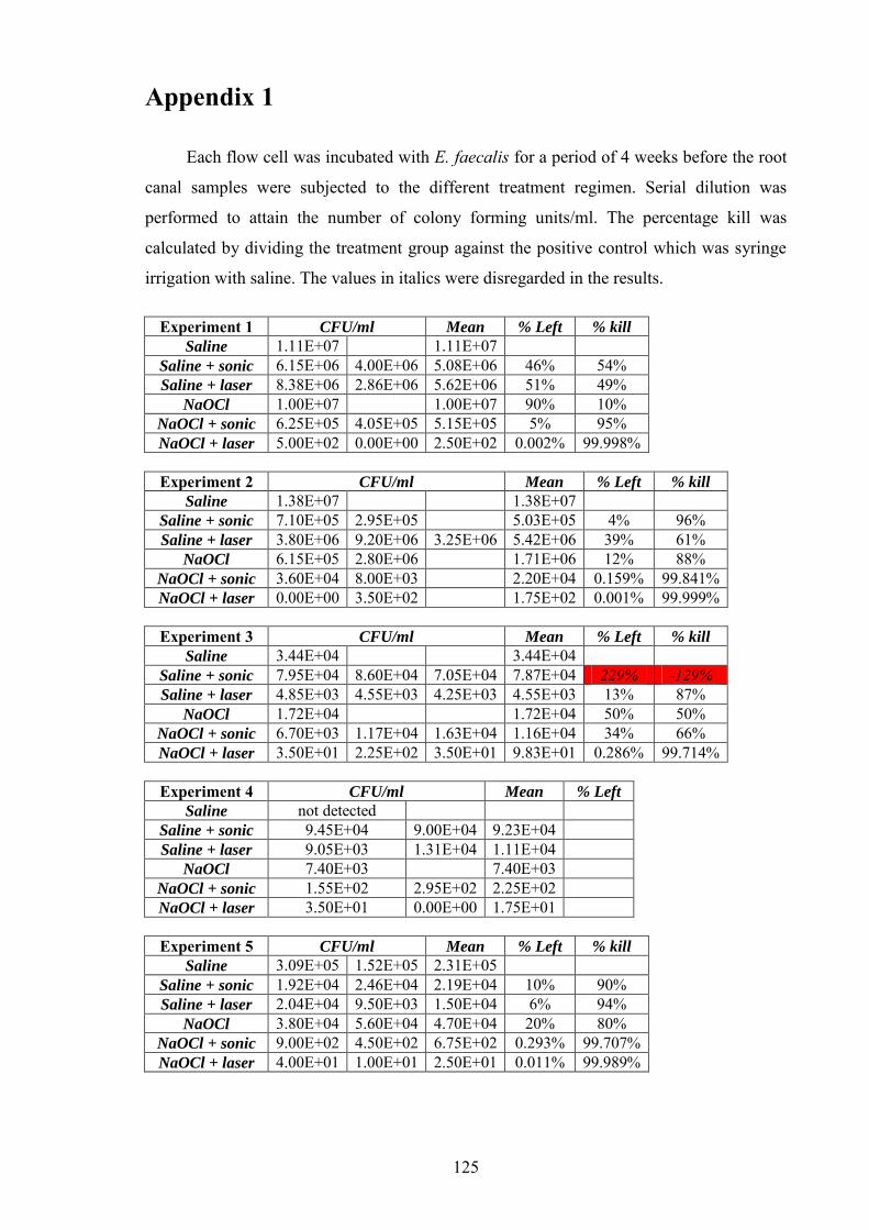
125
Appendix 1
Each flow cell was incubated with E. faecalis for a period of 4 weeks before the root
canal samples were subjected to the different treatment regimen. Serial dilution was
performed to attain the number of colony forming units/ml. The percentage kill was
calculated by dividing the treatment group against the positive control which was syringe
irrigation with saline. The values in italics were disregarded in the results.
Experiment 1 CFU/ml Mean % Left % kill
Saline 1.11E+07 1.11E+07 Saline + sonic 6.15E+06 4.00E+06 5.08E+06 46% 54% Saline + laser 8.38E+06 2.86E+06 5.62E+06 51% 49%
NaOCl 1.00E+07 1.00E+07 90% 10% NaOCl + sonic 6.25E+05 4.05E+05 5.15E+05 5% 95% NaOCl + laser 5.00E+02 0.00E+00 2.50E+02 0.002% 99.998%
Experiment 2 CFU/ml Mean % Left % kill
Saline 1.38E+07 1.38E+07 Saline + sonic 7.10E+05 2.95E+05 5.03E+05 4% 96% Saline + laser 3.80E+06 9.20E+06 3.25E+06 5.42E+06 39% 61%
NaOCl 6.15E+05 2.80E+06 1.71E+06 12% 88% NaOCl + sonic 3.60E+04 8.00E+03 2.20E+04 0.159% 99.841% NaOCl + laser 0.00E+00 3.50E+02 1.75E+02 0.001% 99.999%
Experiment 3 CFU/ml Mean % Left % kill
Saline 3.44E+04 3.44E+04 Saline + sonic 7.95E+04 8.60E+04 7.05E+04 7.87E+04 229% -129% Saline + laser 4.85E+03 4.55E+03 4.25E+03 4.55E+03 13% 87%
NaOCl 1.72E+04 1.72E+04 50% 50% NaOCl + sonic 6.70E+03 1.17E+04 1.63E+04 1.16E+04 34% 66% NaOCl + laser 3.50E+01 2.25E+02 3.50E+01 9.83E+01 0.286% 99.714%
Experiment 4 CFU/ml Mean % Left
Saline not detected Saline + sonic 9.45E+04 9.00E+04 9.23E+04 Saline + laser 9.05E+03 1.31E+04 1.11E+04
NaOCl 7.40E+03 7.40E+03 NaOCl + sonic 1.55E+02 2.95E+02 2.25E+02 NaOCl + laser 3.50E+01 0.00E+00 1.75E+01
Experiment 5 CFU/ml Mean % Left % kill
Saline 3.09E+05 1.52E+05 2.31E+05 Saline + sonic 1.92E+04 2.46E+04 2.19E+04 10% 90% Saline + laser 2.04E+04 9.50E+03 1.50E+04 6% 94%
NaOCl 3.80E+04 5.60E+04 4.70E+04 20% 80% NaOCl + sonic 9.00E+02 4.50E+02 6.75E+02 0.293% 99.707% NaOCl + laser 4.00E+01 1.00E+01 2.50E+01 0.011% 99.989%
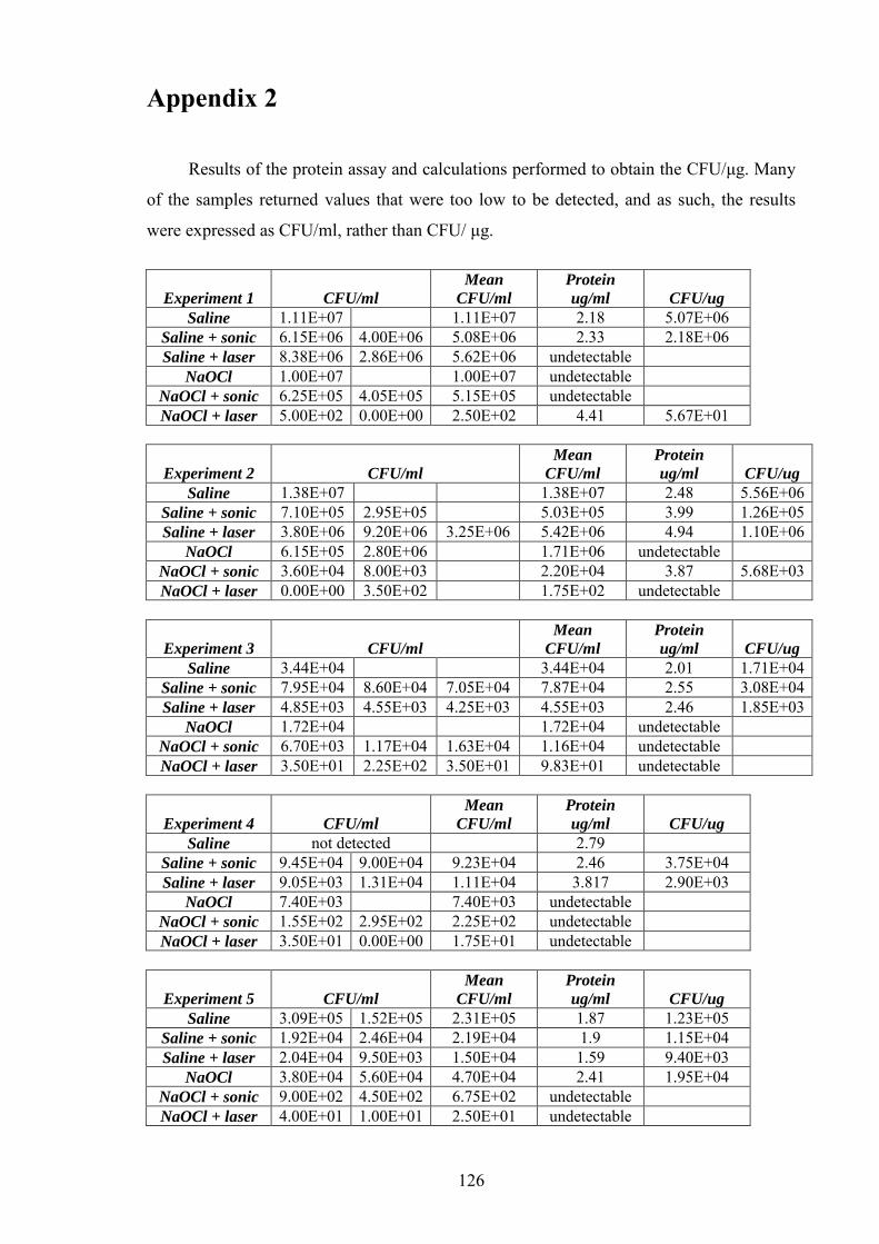
126
Appendix 2
Results of the protein assay and calculations performed to obtain the CFU/μg. Many
of the samples returned values that were too low to be detected, and as such, the results
were expressed as CFU/ml, rather than CFU/ μg.
Experiment 1 CFU/ml
Mean
CFU/ml
Protein
ug/ml CFU/ug
Saline 1.11E+07 1.11E+07 2.18 5.07E+06 Saline + sonic 6.15E+06 4.00E+06 5.08E+06 2.33 2.18E+06 Saline + laser 8.38E+06 2.86E+06 5.62E+06 undetectable
NaOCl 1.00E+07 1.00E+07 undetectable NaOCl + sonic 6.25E+05 4.05E+05 5.15E+05 undetectable NaOCl + laser 5.00E+02 0.00E+00 2.50E+02 4.41 5.67E+01
Experiment 2 CFU/ml
Mean
CFU/ml
Protein
ug/ml CFU/ug
Saline 1.38E+07 1.38E+07 2.48 5.56E+06 Saline + sonic 7.10E+05 2.95E+05 5.03E+05 3.99 1.26E+05 Saline + laser 3.80E+06 9.20E+06 3.25E+06 5.42E+06 4.94 1.10E+06
NaOCl 6.15E+05 2.80E+06 1.71E+06 undetectable NaOCl + sonic 3.60E+04 8.00E+03 2.20E+04 3.87 5.68E+03 NaOCl + laser 0.00E+00 3.50E+02 1.75E+02 undetectable
Experiment 3 CFU/ml
Mean
CFU/ml
Protein
ug/ml CFU/ug
Saline 3.44E+04 3.44E+04 2.01 1.71E+04 Saline + sonic 7.95E+04 8.60E+04 7.05E+04 7.87E+04 2.55 3.08E+04 Saline + laser 4.85E+03 4.55E+03 4.25E+03 4.55E+03 2.46 1.85E+03
NaOCl 1.72E+04 1.72E+04 undetectable NaOCl + sonic 6.70E+03 1.17E+04 1.63E+04 1.16E+04 undetectable NaOCl + laser 3.50E+01 2.25E+02 3.50E+01 9.83E+01 undetectable
Experiment 4 CFU/ml
Mean
CFU/ml
Protein
ug/ml CFU/ug
Saline not detected 2.79 Saline + sonic 9.45E+04 9.00E+04 9.23E+04 2.46 3.75E+04 Saline + laser 9.05E+03 1.31E+04 1.11E+04 3.817 2.90E+03
NaOCl 7.40E+03 7.40E+03 undetectable NaOCl + sonic 1.55E+02 2.95E+02 2.25E+02 undetectable NaOCl + laser 3.50E+01 0.00E+00 1.75E+01 undetectable
Experiment 5 CFU/ml
Mean
CFU/ml
Protein
ug/ml CFU/ug
Saline 3.09E+05 1.52E+05 2.31E+05 1.87 1.23E+05 Saline + sonic 1.92E+04 2.46E+04 2.19E+04 1.9 1.15E+04 Saline + laser 2.04E+04 9.50E+03 1.50E+04 1.59 9.40E+03
NaOCl 3.80E+04 5.60E+04 4.70E+04 2.41 1.95E+04 NaOCl + sonic 9.00E+02 4.50E+02 6.75E+02 undetectable NaOCl + laser 4.00E+01 1.00E+01 2.50E+01 undetectable