an essential staphylococcus aureus cell division protein
TRANSCRIPT

University of South Florida University of South Florida
Scholar Commons Scholar Commons
Cell Biology, Microbiology, and Molecular Biology Faculty Publications
Cell Biology, Microbiology, and Molecular Biology
10-2018
An Essential An Essential Staphylococcus aureus Cell Division Protein directly Cell Division Protein directly
Regulates FtsZ Dynamics Regulates FtsZ Dynamics
Prahathees J. Eswara University of South Florida, [email protected]
Robert S. Brzozowski University of South Florida, [email protected]
Marissa G. Viola University of Rhode Island
Gianni Graham University of South Florida
Catherine Spanoudis University of South Florida
See next page for additional authors
Follow this and additional works at: https://scholarcommons.usf.edu/bcm_facpub
Scholar Commons Citation Scholar Commons Citation Eswara, Prahathees J.; Brzozowski, Robert S.; Viola, Marissa G.; Graham, Gianni; Spanoudis, Catherine; Trebino, Catherine; Jha, Jyoti; Aubee, Joseph I.; Thompson, Karl M.; Camberg, Jodi L.; and Ramamurthi, Kumaran S., "An Essential Staphylococcus aureus Cell Division Protein directly Regulates FtsZ Dynamics" (2018). Cell Biology, Microbiology, and Molecular Biology Faculty Publications. 39. https://scholarcommons.usf.edu/bcm_facpub/39
This Article is brought to you for free and open access by the Cell Biology, Microbiology, and Molecular Biology at Scholar Commons. It has been accepted for inclusion in Cell Biology, Microbiology, and Molecular Biology Faculty Publications by an authorized administrator of Scholar Commons. For more information, please contact [email protected].

Authors Authors Prahathees J. Eswara, Robert S. Brzozowski, Marissa G. Viola, Gianni Graham, Catherine Spanoudis, Catherine Trebino, Jyoti Jha, Joseph I. Aubee, Karl M. Thompson, Jodi L. Camberg, and Kumaran S. Ramamurthi
This article is available at Scholar Commons: https://scholarcommons.usf.edu/bcm_facpub/39

*For correspondence:
[email protected] (PJE);
[email protected] (KSR)
Competing interests: The
authors declare that no
competing interests exist.
Funding: See page 20
Received: 01 June 2018
Accepted: 22 August 2018
Published: 02 October 2018
Reviewing editor: Tam Mignot,
Aix Marseille University-CNRS
UMR7283, France
This is an open-access article,
free of all copyright, and may be
freely reproduced, distributed,
transmitted, modified, built
upon, or otherwise used by
anyone for any lawful purpose.
The work is made available under
the Creative Commons CC0
public domain dedication.
An essential Staphylococcus aureus celldivision protein directly regulates FtsZdynamicsPrahathees J Eswara1,2*, Robert S Brzozowski2, Marissa G Viola3,Gianni Graham1,2, Catherine Spanoudis2, Catherine Trebino3, Jyoti Jha4,Joseph I Aubee5, Karl M Thompson5, Jodi L Camberg3,6,Kumaran S Ramamurthi1*
1Laboratory of Molecular Biology, National Cancer Institute, National Institutes ofHealth, Bethesda, United States; 2Department of Cell Biology, Microbiology andMolecular Biology, University of South Florida, Tampa, United States; 3Departmentof Cell and Molecular Biology, University of Rhode Island, Kingston, United States;4Laboratory of Biochemistry and Molecular Biology, National Cancer Institute,National Institutes of Health, Bethesda, United States; 5Department ofMicrobiology, College of Medicine, Howard University, Washington, United States;6Department of Nutrition and Food Sciences, University of Rhode Island, Kingston,United States
Abstract Binary fission has been well studied in rod-shaped bacteria, but the mechanisms
underlying cell division in spherical bacteria are poorly understood. Rod-shaped bacteria harbor
regulatory proteins that place and remodel the division machinery during cytokinesis. In the
spherical human pathogen Staphylococcus aureus, we found that the essential protein GpsB
localizes to mid-cell during cell division and co-constricts with the division machinery. Depletion of
GpsB arrested cell division and led to cell lysis, whereas overproduction of GpsB inhibited cell
division and led to the formation of enlarged cells. We report that S. aureus GpsB, unlike other
Firmicutes GpsB orthologs, directly interacts with the core divisome component FtsZ. GpsB
bundles and organizes FtsZ filaments and also stimulates the GTPase activity of FtsZ. We propose
that GpsB orchestrates the initial stabilization of the Z-ring at the onset of cell division and
participates in the subsequent remodeling of the divisome during cytokinesis.
DOI: https://doi.org/10.7554/eLife.38856.001
IntroductionBacterial cell division has been extensively studied in rod-shaped organisms such as Escherichia coli
and Bacillus subtilis (Adams and Errington, 2009; Lutkenhaus et al., 2012; Rowlett and Margolin,
2015; Tsang and Bernhardt, 2015). However, spherical bacteria lack several key components found
in these well-studied model organisms (Pinho et al., 2013), so fundamental features of how they
divide are poorly understood. The Gram-positive human pathogen Staphylococcus aureus is a spher-
ical bacterium that is commensal in ~30% of the U.S. population (Kuehnert et al., 2006), but in
immunocompromised individuals, it is a leading cause of bacteremia and nosocomial infections in
industrialized nations (Klevens et al., 2007). The emergence of several antibiotic resistant strains of
S. aureus has necessitated the identification of novel antibiotic targets (Pendleton et al., 2013). In
recent years, components of the bacterial cell division machinery have been proposed as such tar-
gets (Lock and Harry, 2008; Sass and Brotz-Oesterhelt, 2013).
Eswara et al. eLife 2018;7:e38856. DOI: https://doi.org/10.7554/eLife.38856 1 of 24
RESEARCH ARTICLE

GpsB is a small coiled-coil cell division protein (Claessen et al., 2008; Rismondo et al., 2016;
Tavares et al., 2008) that is widely conserved in the Firmicutes phylum and is conditionally required
for growth in certain species, depending on growth media and temperatures (Claessen et al., 2008;
Fleurie et al., 2014; Land et al., 2013; Rismondo et al., 2016; Tavares et al., 2008). GpsB is highly
co-conserved (Pinho et al., 2013) with the cell division protein DivIVA. Like DivIVA, GpsB is relatively
small and harbors a highly homologous N-terminal a-helical domain. However, the C-terminus differs
from that of DivIVA: whereas DivIVA assembles into an anti-parallel tetramer, the GpsB structure
was reported to hexamerize with a parallel alignment of helices (Rismondo et al., 2016). Similar to
DivIVA (Kaval and Halbedel, 2012), GpsB orthologs perform slightly different functions in different
species. In the rod-shaped Bacillus subtilis and Listeria monocytogenes, GpsB participates in shut-
tling a cell wall assembly protein (PBP1 or PBP A1, respectively) to help maintain the characteristic
rod shape of the bacterium (Claessen et al., 2008; Rismondo et al., 2016). In the ovoid-shaped
Streptococcus pneumoniae, GpsB additionally has been reported to interact with PBP2a, PBP2b,
and MreC, and has been implicated in recruiting a Ser/Thr kinase to mid-cell that activates cell wall
assembly machinery specifically at the division septum, thereby modulating a switch between periph-
eral and medial cell wall assembly to again maintain the proper shape of the cell (Fleurie et al.,
2014; Rued et al., 2017). In all reported cases, GpsB interacts with a peripheral divisome compo-
nent, EzrA, but not necessarily core components of the division machinery, to mediate its role in cell
shape maintenance (Claessen et al., 2008; Fleurie et al., 2014; Rued et al., 2017; Steele et al.,
2011).
In S. aureus, GpsB is an essential protein (Santiago et al., 2015) (M. Santiago, personal communi-
cation), but its cellular function is poorly understood. Herein, we report that GpsB interacts directly
with bacterial tubulin homolog FtsZ, the core component of the division machinery, and orchestrates
the dynamics of its assembly. In vivo, we show that GpsB localizes to mid-cell at the onset of cell
division and co-constricts with the divisome during cytokinesis. Depletion of GpsB in vivo arrested
cell division and prevented the robust assembly of the divisome at mid-cell. In vitro, we show that
purified GpsB promotes lateral interactions between FtsZ polymers in a manner reminiscent of bun-
dling, thereby increasing the local concentration of FtsZ, and organizes the polymers. Unlike other
proteins that exhibit FtsZ bundling activity, GpsB stimulated FtsZ GTPase activity. Consistent with
eLife digest A bacterium called Staphylococcus aureus causes many infections in humans,
especially in hospital patients with weakened immune systems. These infections are generally
treated with drugs known as antibiotics that interact with specific proteins in the bacteria to kill the
cells, or stop them from growing. However, some S. aureus infections are resistant to the antibiotics
currently available so there is a need to develop new drugs that target different bacterial proteins.
Bacteria multiply by dividing to make identical copies of themselves. When a bacterium is
preparing to divide, filaments made of a protein called FtsZ form a ring at the site where the cell will
split. Many other proteins are involved in controlling how and when a cell divides. For example,
several species of bacteria harbor a dispensable cell division protein called GpsB. In at least one
organism, it helps to maintain the proper shape of the cell during cell division. In S. aureus, though,
GpsB is essential for cells to survive and could therefore be a potential target for new antibiotics.
However, its role in S. aureus has not been studied.
Eswara et al. have now used genetic and biochemical approaches to study the S. aureus form of
the GpsB protein. The experiments show that GpsB moves to the middle of S. aureus cells just
before they begin to divide and binds directly to FtsZ. This helps to secure the position of FtsZ
across the middle of the cell and activates the protein so that the cell can begin to divide into two.
In cells that produce too much GpsB, the FtsZ proteins become active too early, leading to the cells
growing larger and larger until they burst.
The findings of Eswara et al. reveal that GpsB plays a different role in S. aureus cells than in some
other species of bacteria. Further studies into such differences could help researchers to develop
new antibiotics, as well as improving our understanding of why bacteria are so diverse.
DOI: https://doi.org/10.7554/eLife.38856.002
Eswara et al. eLife 2018;7:e38856. DOI: https://doi.org/10.7554/eLife.38856 2 of 24
Research article Microbiology and Infectious Disease

this activity, overproduction of GpsB in vivo inhibited cell division and resulted in the production of
large S. aureus cells. Our data suggest that, compared to GpsB orthologs in other Gram-positive
bacteria, S. aureus GpsB plays a significantly different role by directly interacting with central compo-
nent of the division machinery to regulate the remodeling of the divisome during cytokinesis: first,
by bundling and stabilizing FtsZ polymers at mid-cell by promoting lateral interactions between FtsZ
filaments, which increases the local concentration and triggers the GTPase activity of FtsZ and allows
cytokinesis to proceed.
Results
Overproduction of S. aureus GpsB inhibits cell division in B. subtilis andS. aureusTo initially investigate if Staphylococcal GpsB (GpsBSa) performs a similar function as the B. subtilis
GpsB ortholog (GpsBBs), we expressed gpsBSa under the control of an inducible promoter in B. sub-
tilis. In the presence of inducer, otherwise WT B. subtilis harboring either gpsBSa or gpsBSa-GFP
exhibited a severe growth defect (Figure 1). In contrast, cells similarly expressing gpsBBs or gpsBBs-
GFP did not exhibit a growth defect (Figure 1A), suggesting that cell toxicity was specifically due to
expression of the S. aureus ortholog of gpsB (Figure 1—figure supplement 1A). Immunoblotting
with antisera specific to GpsBSa revealed a ~ 3.2 fold overproduction of GpsBSa at a population level
in the presence of inducer (Figure 1—figure supplement 1B; note that the anti-GpsBSa antiserum
did not recognize GpsBBs). In the absence of inducer, B. subtilis cells harboring gpsBSa examined by
epifluorescence microscopy were of uniform length and displayed division septa at mid-cell
(Figure 1B), but in the presence of inducer, these cells were filamentous with segregated chromo-
somes that rarely elaborated division septa (Figure 1C). GpsB interacts with several cell division pro-
teins in different Gram-positive bacteria (Claessen et al., 2008; Cleverley et al., 2016;
Pompeo et al., 2015). Deletion of ezrA, ponA, prkC, or gpsB resulted in minor morphological
defects in B. subtilis, but overproduction of GpsBSa in these strain backgrounds nonetheless resulted
in filamentation (Figure 1D–K). Additionally, while deletion of divIVA resulted in cell elongation
(Edwards and Errington, 1997), overproduction of GpsBSa in the absence of DivIVA resulted in fur-
ther filamentation (Figure 1L–M). Thus, the B. subtilis filamentation phenotype resulting from
GpsBSa overproduction does not require these peripheral cell division factors. We next examined if
GpsBSa affects FtsZ localization. In the absence of inducer, FtsZ-GFP localized properly to mid-cell at
incipient and active sites of cell division (Figure 1N). However, upon overproduction of GpsBSa, fila-
mentous cells displayed diffuse localization of FtsZ-GFP in the cytosol (Figure 1O), suggesting that
GpsBSa overproduction interferes with the localization of the central component of the B. subtlis cell
division machinery.
In B. subtilis, gpsB is not essential for growth, but deletion of ezrA (a peripheral component of
the divisome) together with gpsB is synthetically lethal (Claessen et al., 2008). In B. subtilis cells har-
boring a gpsB deletion and expressing gpsBBs, we obtained 865 ± 157 transformants harboring a
deletion in ezrA (n = 3, per ~400 ng of transformed DNA containing ezrA deletion), whereas we did
not recover any transformants when we attempted to delete ezrA in DgpsB cells that expressed
gpsBSa, indicating that gpsBSa was unable to complement the gpsBBs deletion phenotype. Together
with the different phenotypes observed upon overexpression of either gpsBBs or gpsBSa in B. subti-
lis, the data suggested that S. aureus GpsB may exhibit a different function or activity.
To test the effect of GpsBSa overproduction in Staphylococci, we cloned gpsBSa in a high copy
plasmid under control of an inducible promoter, introduced the construct into S. aureus strain
SH1000, stained the cells with a fluorescent membrane dye, and examined cell size using epifluores-
cence microscopy. Immunoblotting revealed an ~5.4 fold overproduction of GpsB at a population
level relative to endogenous levels of GpsB (Figure 1—figure supplement 1C). 100% of WT cells
we observed (n = 676) were less than 1.2 mm in diameter, as were WT cells harboring the empty vec-
tor (n = 100) (Figure 2A–D). In the absence of inducer, 6.4% (n = 971) of cells harboring the induc-
ible copy of gpsBSa were larger than 1.2 mm in diameter; in the presence of inducer, 56.9% (n = 770)
of cells were larger than 1.2 mm (Figure 2E–F). Interestingly, overproduction of gpsBBs did not result
in a similar enlargement of S. aureus cells (Figure 2G–H), suggesting that the cell division inhibition
phenotype in B. subtilis and S. aureus was unique to the overproduction of the S. aureus ortholog of
Eswara et al. eLife 2018;7:e38856. DOI: https://doi.org/10.7554/eLife.38856 3 of 24
Research article Microbiology and Infectious Disease

Figure 1. Overexpression of S.aureus gpsB inhibits cell division in B. subtilis. (A) Luria-Bertani agar plates streaked
with wild type B. subtilis (WT, strain PY79), or otherwise wild type B. subtilis harboring an inducible copy of gpsBBs
(GG18), gpsBBs-gfp (GG19), gpsBSa (GG7), or gpsBSa-gfp (GG8) integrated into the chromosome, in the absence
(left) or presence (right) of inducer. (B–M) Morphology of cells of different deletion mutants of B. subtilis (DezrA,
GG35; DponA, CS26; DprkC, CS24; DgpsB, CS40; DdivIVA, CS94) harboring an inducible copy of gpsBSa grown in
the absence (B, D, F; H, J, L) or presence (C, E, G, I, K, M) of inducer. (N–O) Localization of FtsZ-GFP in a strain
(GG9) harboring an inducible copy of gpsBSa grown in the absence (N) or presence (O) of inducer. Membranes
(red; B–O) visualized using the fluorescent dye FM4-64; chromosomes (blue; B–M) visualized using DAPI; FtsZ-GFP
localization (green; N–O). Scale bar: 1 mm. Genotypes are listed in Key Resources Table.
DOI: https://doi.org/10.7554/eLife.38856.003
The following figure supplements are available for figure 1:
Figure 1 continued on next page
Eswara et al. eLife 2018;7:e38856. DOI: https://doi.org/10.7554/eLife.38856 4 of 24
Research article Microbiology and Infectious Disease
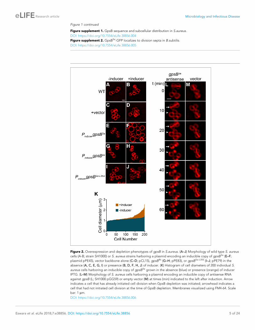
Figure 1 continued
Figure supplement 1. GpsB sequence and subcellular distribution in S.aureus.
DOI: https://doi.org/10.7554/eLife.38856.004
Figure supplement 2. GpsBSa-GFP localizes to division septa in B.subtilis.
DOI: https://doi.org/10.7554/eLife.38856.005
Figure 2. Overexpression and depletion phenotypes of gpsB in S.aureus. (A–J) Morphology of wild type S. aureus
cells (A-B; strain SH1000) or S. aureus strains harboring a plasmid encoding an inducible copy of gpsBSa (E–F;
plasmid pPE45), vector backbone alone (C–D; pCL15), gpsBBs (G–H; pPE83), or gpsBSa-L35S (I–J; pPE79) in the
absence (A, C, E, G, I) or presence (B, D, F, H, J) of inducer. (K) Histogram of cell diameters of 200 individual S.
aureus cells harboring an inducible copy of gpsBSa grown in the absence (blue) or presence (orange) of inducer
IPTG. (L–M) Morphology of S. aureus cells harboring a plasmid encoding an inducible copy of antisense RNA
against gpsB (L; SH1000 pGG59) or empty vector (M) at times (min) indicated to the left after induction. Arrow
indicates a cell that has already initiated cell division when GpsB depletion was initiated; arrowhead indicates a
cell that had not initiated cell division at the time of GpsB depletion. Membranes visualized using FM4-64. Scale
bar: 1 mm.
DOI: https://doi.org/10.7554/eLife.38856.006
Eswara et al. eLife 2018;7:e38856. DOI: https://doi.org/10.7554/eLife.38856 5 of 24
Research article Microbiology and Infectious Disease

GpsB. Quantification of cell diameters of 200 individual cells overproducing GpsBSa revealed a range
of cell diameters higher than 1.2 mm in over half of the cells (Figure 2K). The variation in cell diame-
ters was likely due to unequal expression of gpsBSa in every cell, since control experiments in which
gfp was placed under control of the inducible promoter revealed that only ~34% of cells (n = 263)
produced GFP in the presence of inducer.
The gpsB gene is essential for viability in S. aureus (Santiago et al., 2015). Consistent with this
observation, we were unable to knockout the gene, even in the presence of a complementing multi-
copy plasmid, presumably due to the disruptive overproduction phenotype described above. We
therefore sought to deplete GpsB by overexpressing gpsB antisense RNA under the control of an
inducible promoter from a multicopy plasmid and examined the morphology of cells using fluores-
cence microscopy (antisense resulted in ~2.5-fold reduction in GpsB; Figure 1—figure supplement
1F) . Immediately after addition of the inducer, cells harboring this construct were morphologically
similar to cells harboring the empty vector (Figure 2L–M). At later time points, we observed that
cells harboring the depletion construct that had already elaborated a division septum (Figure 2L,
arrow) did not complete cytokinesis. Instead, the division septa became deformed and membrane
aberrantly accumulated as foci. Cells that had not yet initiated cell division at the time of induction
(Figure 2L, arrowhead) did not elaborate division septa and also accumulated aberrant membrane
foci. In contrast, cells harboring only the empty vector (Figure 2M) elaborated division septa and
completed cytokinesis during the observation period.
The severe growth defect imposed by gpsBSa overexpression in B. subtilis permitted us to isolate
suppressor mutations that could correct this defect. One such mutation, an intragenic single nucleo-
tide change in gpsBSa, altered the specificity of a highly conserved codon at position 35 from Leu to
Ser (Figure 1—figure supplement 1A, boxed residue), and allowed B. subtilis cells overexpressing
gpsBSa-L35S to grow normally. To check if the L35S substitution caused in a major structural change
in the protein, we purified WT GpsBSa and the L35S variant and examined the a-helical content of
both proteins using circular dichroism (CD) spectroscopy (Figure 1—figure supplement 1D). The
CD spectrum revealed similar profiles for each protein, suggesting that the L35S substitution did not
grossly affect the secondary structure of GpsBSa. In the presence of inducer, S. aureus cells harbor-
ing inducible gpsBSa-L35S did not exhibit a cell enlargement defect (Figure 2I–J). Taken together, we
conclude that overproduction of GpsBSa, but not GpsBBs, inhibits cell division in both S. aureus and
B. subtilis, resulting in cell filamentation (in B. subtilis) or cell enlargement (in S. aureus), like the
depletion phenotype of FtsZ (Pinho and Errington, 2003). Depletion of GpsB in S. aureus, however,
arrested cell division without a coincident enlargement of cells and ultimately caused aberrant mem-
brane accumulation. Furthermore, substitution of Leu35 to Ser abolished the toxicity resulting from
GpsB overproduction, suggesting that this residue is critical for GpsBSa function.
GpsB dynamically localizes to mid-cell in S. aureus and co-constrictswith the division septumWe next examined the subcellular localization of GpsBSa-GFP in S. aureus. In non-dividing cells
GpsBSa-GFP (produced at lower levels that did not result in cell division inhibition) localized near the
cell periphery (Figure 3A, arrowhead). In dividing cells, GpsBSa-GFP localized to mid-cell, between
the segregated chromosomes, and co-localized with the constricting membrane (Figure 3A, arrow).
In contrast, GpsBSa-L35S-GFP localized primarily in the cytosol in both dividing and non-dividing cells
(Figure 3A). Likewise, when produced at lower levels in B. subtilis, GpsBSa-GFP accumulated at divi-
sion septa, whereas the L35S variant localized primarily in the cytosol (Figure 1—figure supplement
2). Since the L. monocytogenes GpsB ortholog is membrane-associated (Rismondo et al., 2016), we
next tested if the L35S substitution could have disrupted any intrinsic membrane affinity of GpsBSa
by fractionating S. aureus cell extracts and examining the distribution of GpsBSa-GFP and GpsBSa-
L35S-GFP by immunoblotting (Figure 1—figure supplement 1E). Unlike L. monocytogenes GpsB, we
detected S. aureus GpsB-GFP exclusively in the soluble fraction, suggesting that it does not directly
associate with the Staphylococcal membrane. GpsBSa-L35S-GFP was similarly detected in the cytosolic
fraction. Association of L. monocytogenes GpsB with the membrane is reportedly mediated by
Leu24, since substitution of Leu24 with Ala disrupted membrane association (Rismondo et al.,
2016). Interestingly, the corresponding residue in S. aureus GpsB is Ala (Figure 1—figure supple-
ment 1A, asterisk), consistent with the apparent lack of intrinsic affinity of S. aureus GpsB for the
membrane. We conclude that, unlike L. monocytogenes GpsB, S. aureus GpsB (hereafter, simply
Eswara et al. eLife 2018;7:e38856. DOI: https://doi.org/10.7554/eLife.38856 6 of 24
Research article Microbiology and Infectious Disease

Figure 3. Dynamic redistribution of GpsB to mid-cell and periphery of S.aureus during the cell cycle. (A) Localization of GpsB-GFP (top, SH1000 pPE46)
or GpsBL35S-GFP (bottom, SH1000 pPE80) to mid-cell in actively dividing cells (arrow) and to the periphery of cells that are not dividing (arrowhead).
First panel: membranes visualized using FM4-64; second panel: GFP fluorescence; third panel: chromosomes visualized using DAPI; fourth panel:
overlay of membrane, GFP, and DNA. (B) GpsB-GFP localization in S. aureus cells at various stages of division (i–iv) using structured illumination
microscopy (SIM). First column: membranes visualized using FM4-64; second column: GpsB-GFP fluorescence; third column: overlay, membrane and
GpsB-GFP. Columns 4–6: reconstruction of deconvolved Z-stacks and rotation of the cells in columns 1–3, respectively, around the vertical axis. (C)
Time-lapse fluorescence micrographs of a dividing S. aureus cell taken at the time intervals indicated at the left. Left panels: membranes visualized
using FM4-64; middle panels: GpsB-GFP fluorescence; right panels: overlay, membrane and GpsB-GFP. Depictions of GpsB-GFP localization patterns
are to the right of the panels. Scale bar: 1 mm.
DOI: https://doi.org/10.7554/eLife.38856.007
Eswara et al. eLife 2018;7:e38856. DOI: https://doi.org/10.7554/eLife.38856 7 of 24
Research article Microbiology and Infectious Disease
‘GpsB’) is likely not directly membrane-associated and that cell peripheral localization of S. aureus
GpsB may be mediated by another factor.
To discern if GpsB-GFP co-localized with, or at sites adjacent to, the site of membrane constric-
tion, we employed structured illumination microscopy (SIM) (Gustafsson, 2005), a super-resolution
technique that previously provided enough resolution to discern the localization of DivIVA-GFP on
either side of the ~80 nm division septum (Eswaramoorthy et al., 2011). At the onset of cell divi-
sion, mid-plane images of S. aureus cells displayed only two GpsB-GFP foci that co-localized with
sites of membrane invagination at mid-cell (Figure 3Bi). Reconstruction of deconvolved Z-stacks and
rotation of the reconstructed image around the axis of cell division revealed that GpsB-GFP formed
an irregular ring-shaped structure, reminiscent of the structure of an assembling divisome
(Figure 3Bi) (Lund et al., 2018). In cells that were further advanced in cell division, the two foci of
GpsB-GFP followed the leading edges of the constricting membrane (Figure 3Bii) and formed a ring
structure that was smaller than the diameter of the cell, (Figure 3Bii) (Buss et al., 2015;
Ebersbach et al., 2008), suggesting that the GpsB ring structure co-constricts with the division
machinery. In a cell approaching completion of cytokinesis, GpsB-GFP collapsed into a single focus
at the center of the invaginating membrane (Figure 3Biii), and immediately after the completion of
cell division, we observed that GpsB-GFP localized largely to the cell periphery in the adjacent
daughter cells (Figure 3Biv), suggesting that GpsB may dynamically localize during the cell cycle.
Phototoxicity induced by SIM precluded us from performing super-resolution time lapse experi-
ments of actively dividing cells using this method. To test the dynamic nature of GpsB-GFP localiza-
tion, we followed the fate of GpsB-GFP in individual cells through three rounds of cell division using
diffraction-limited epifluorescence microscopy. At the onset of our measurements, GpsB-GFP local-
ized primarily at mid-cell in a cell that had completed cytokinesis and was poised to separate into
two daughter cells (Figure 3; [Steele et al., 2011]). After cell separation, GpsB-GFP redistributed to
the periphery of each daughter cell (Figure 3). Beginning the next round of cell division, GpsB-GFP
re-localized to the mid-cell of each daughter cell as two foci that coincided with the invaginating
membrane (Figure 3C,t). It again localized with the invaginating membrane, followed by redistribu-
tion of fluorescence to the cell peripheries of the daughter cells (Figure 3). The redistribution of
peripherally-localized GpsB to the division septum is reminiscent of the FtsZ-dependent late localiza-
tion of GpsB reported in S. pneumoniae (Land et al., 2013). We therefore conclude that GpsB local-
izes as a single ring-shaped structure at mid-cell at the onset of cell division, constricts with the
invaginating membrane during cytokinesis, and ultimately, after daughter cell separation, uniformly
redistributes to the periphery of each daughter cell.
GpsB localization and divisome assembly reciprocally influence eachotherAlthough the S. aureus ortholog of GpsB was non-functional in B. subtilis, its ability to localize at
mid-cell suggested that it is capable of recognizing an intrinsic feature of the divisome shared
between B. subtilis and S. aureus (Figure 1—figure supplement 2). The bacterial divisome is com-
posed of approximately 10 core proteins (Lutkenhaus et al., 2012; Margolin, 2005) that direct the
cell wall assembly machinery to mid-cell and mediate cell membrane constriction during cell division.
The core divisome component is the bacterial tubulin homolog, FtsZ (Coltharp et al., 2016;
Osawa and Erickson, 2013), which is a target of cell division regulators in different systems
(Ortiz et al., 2016). To investigate a potential interaction between the divisome and GpsB, we
examined the localization of GpsB-GFP in S. aureus cells grown in the presence and absence of the
PC190723, a small ligand that inhibits GTPase activity of FtsZ and inhibits cell division
(Andreu et al., 2010; Haydon et al., 2008). In the presence of the drug, 92.5% of cells (n = 200)
harboring empty vector exhibited a diameter larger than 1.5 mm, compared to just 30.5% of cells in
the absence of inhibitor, consistent with a block in cell division (Figure 4A–B’). To confirm that the
drug was inhibiting divisome assembly, we examined the localization of ZapA-GFP, a known early
stage cell division protein that assembles concomitantly with FtsZ (Gamba et al., 2009;
Reichmann et al., 2014) and is used as a proxy for localization of FtsZ. In untreated cells, ZapA local-
ized at mid-cell at the onset of cell division (Figure 4C–C’), but in the presence of the inhibitor,
96.5% of cells (n = 200) displayed diffuse and/or punctate localization in the cytosol that was not
located at mid-cell (Figure 4D–D’), indicating that the divisome was not assembling correctly due to
inhibition of FtsZ. In the absence of inhibitor, GpsB-GFP localized at mid-cell or the periphery in 55%
Eswara et al. eLife 2018;7:e38856. DOI: https://doi.org/10.7554/eLife.38856 8 of 24
Research article Microbiology and Infectious Disease

or 30% of cells, respectively, and mis-localized in the cytosol in 15% of cells (n = 200; Figure 4E–E’).
In contrast, in the presence of inhibitor, GpsB did not localize to mid-cell in any cell observed
(n = 200) and instead displayed a combination of diffuse cytosolic localization and aggregation along
the cell periphery (Figure 4F–F’). The data therefore indicated that that GpsB localization to mid-
cell depends directly or indirectly on functional FtsZ.
We next tested how GpsB influences divisome assembly. In otherwise wild type cells producing
ZapA-GFP, no cell enlargement was detected; among them, 53% of cells displayed ZapA-GFP local-
ized to mid-cell (these were cells that were actively undergoing cell division (Figure 4G–G’, arrow)
and 37% of cells displayed ZapA-GFP as a ring that corresponded to the subsequent plane of cell
division in daughter cells that had recently completed cytokinesis (Figure 4G–G’, arrowhead;
n = 200). In contrast, in cells harboring inducible gpsB, addition of inducer resulted in the
Figure 4. FtsZ and GpsB reciprocally influence each other’s subcellular localization pattern. Morphology of wild
type S. aureus cells harboring empty vector (strain SH1000 pCL15) in the absence (A–A’) or presence (B–B’) of FtsZ
inhibitor PC190723 as visualized in the GFP fluorescence channel (A–B) or by differential interference contrast (DIC;
A’–B’). Localization of ZapA-GFP (proxy for FtsZ localization; strain SH1000 pRB42; C–D’) or GpsB-GFP (E-F’;
SH1000 pPE46) in the absence (C-C’; E-E’) or presence (D-D’; F-F’) of S. aureus FtsZ GTPase activity inhibitor
PC190723. (G–I’) Localization of ZapA-GFP in wild type (G-G’; SH1000 pRB42), or in cells harboring an IPTG-
inducible copy of gpsB in the absence (H-H’; SH1000 pPE45 pRB42) or presence (I–I’) of IPTG. Localization of
ZapA-GFP in cells harboring vector only (J-J’; SH1000 pRB42 pEPSA5) or vector producing gpsB antisense RNA (K-
K’; SH1000 pRB42 pGG59). A-K: GFP fluorescence; A’-F’: overlay of GFP fluorescence and DIC; G’-K’: overlay of
GFP fluorescence and membrane. Arrows indicate site of cell division; arrowheads indicate ZapA-GFP localization
at the subsequent plane of cell division. Scale bar: 1 mm.
DOI: https://doi.org/10.7554/eLife.38856.008
Eswara et al. eLife 2018;7:e38856. DOI: https://doi.org/10.7554/eLife.38856 9 of 24
Research article Microbiology and Infectious Disease

enlargement of cells and ZapA-GFP was mis-localized in 86% of the enlarged cells (n = 100;
Figure 4H–I’). Assuming that the FtsZ bundling activity of ZapA is not synergistically participating
with GpsB overexpression, this suggests that the cell enlargement phenotype caused by overpro-
duction of GpsB was due to the mis-assembly of the divisome.
To determine the behavior of the divisome in GpsB-depleted cells, we monitored the localization
of ZapA-GFP. In cells harboring empty vector, ZapA-GFP localized to mid-cell in 82.5% of the cells
(n = 200; Figure 4J–J’). Quantification of fluorescence intensity in individual cells revealed that the
fluorescence of mid-cell-localized ZapA-GFP was 2429 ± 1346 units/cell (n = 75). At earlier time
points after induction to deplete GpsB, before cell lysis, we observed faint ZapA-GFP signals at mid-
cell in 41% of the cells and diffuse localization in the remaining cells (n = 200; Figure 4K–K’), but the
mean fluorescence intensity of the ZapA-GFP ring structure (614 ± 450 units/cell; n = 75) was nearly
four-fold lower than that observed for ZapA-GFP intensity in the absence of gpsB depletion.
Together, the observations suggest that divisome assembly and GpsB localization are reciprocally
influenced: GpsB requires FtsZ for localization to mid-cell; overproduction of GpsB disrupts divisome
assembly; and depletion of GpsB prevents robust divisome assembly at mid-cell that precedes mem-
brane deformities that ultimately lead to cell lysis.
GpsB stimulates GTPase activity of FtsZTo test if GpsB directly influences FtsZ behavior, we purified recombinant S. aureus FtsZ, GpsB, and
GpsBL35S (Figure 5A) and examined the GpsB variants by size exclusion chromatography
(Figure 5B). GpsB eluted in two peaks by size exclusion chromatography, which approximately cor-
responds to the predicted sizes of hexameric (Rismondo et al., 2016) and dodecameric GpsB
(Figure 5B, top), indicating that S. aureus GpsB could potentially exist in two forms. In contrast,
GpsBL35S eluted exclusively as a dodecamer (Figure 5B, bottom), suggesting that its inability to
form hexamers could underlie its loss of function in vivo.
We next measured the GTP hydrolysis activity of purified S. aureus FtsZ with time at increasing
protein concentrations. Unlike the well-characterized E. coli FtsZ, which robustly hydrolyzes GTP
(Arjes et al., 2015; Buske and Levin, 2012; Mukherjee and Lutkenhaus, 1998; Romberg and
Levin, 2003), S. aureus FtsZ hydrolyzed GTP poorly below ~30 mM (Figure 5C) (Anderson et al.,
2012). The rate of hydrolysis continued to increase with FtsZ concentration (Figure 5D), displaying a
behavior more similar to FtsZ from the Gram-positive Streptococcus pneumoniae, which has a critical
concentration above 10 mM, than to E. coli FtsZ (although a lag observed for S. pneumonia FtsZ was
not detected for S. aureus FtsZ) (Salvarelli et al., 2015). It should be noted that this result contrasts
with that of Elsen et al., which reported a low critical concentration for S. aureus FtsZ (~5 mM)
(Elsen et al., 2012). However, a recent report by Wagstaff et al. showed GTP hydrolysis rates at
high S. aureus FtsZ concentration (10 and 20 mM), and similar to the rates reported here
(Wagstaff et al., 2017). These differences could be due to varying populations of conformationally
active FtsZ in different preparations (Elsen et al., 2012).
We next measured the effect of GpsB on the GTP hydrolysis rate of FtsZ. Incubation of 30 mM
FtsZ with increasing amounts of GpsB resulted in a non-linear stimulation of GTP hydrolysis activity,
wherein appreciable stimulation of GTP hydrolysis was only seen above 8 mM GpsB (Figure 5E). At
10 mM GpsB (1:3 ratio of monomeric GpsB:FtsZ; 1:18 ratio of hexameric GpsB:FtsZ; 1:36 ratio of
dodecameric GpsB:FtsZ), GTP hydrolysis was stimulated ~3 fold. As a control, GpsB alone did not
exhibit appreciable GTPase activity (Figure 5E). In contrast, incubation of FtsZ with GpsBL35S did not
appreciably stimulate GTPase activity of FtsZ (Figure 5F), nor did the addition of GpsB, even at
equimolar amounts, to lower concentrations (10 mM) of FtsZ. Thus, wild type GpsB, which purifies as
a hexamer and dodecamer, stimulates the GTPase activity of FtsZ at substoichiometric levels at suffi-
ciently high enough concentrations of FtsZ (above 30 mM), whereas GpsBL35S, which is locked in the
dodecameric form, fails to do so.
GpsB interacts with and promotes lateral interactions between FtsZpolymersWe next investigated if GpsB directly interacts with polymerized FtsZ using a high-speed sedimenta-
tion assay performed with a non-hydrolyzable GTP analog (GMPCPP), which promotes the assembly
of stable FtsZ polymers. In the absence of nucleotide, FtsZ was largely detected in the supernatant
Eswara et al. eLife 2018;7:e38856. DOI: https://doi.org/10.7554/eLife.38856 10 of 24
Research article Microbiology and Infectious Disease

after ultracentrifugation, but in the presence of GMPCPP, more than 50% of FtsZ was detected in
the pellet fraction, indicating that it had polymerized (Figure 6A). When GpsB was incubated with
the reaction, 94% of GpsB co-sedimented with FtsZ, whereas only 20% of the nonfunctional
GpsBL35S co-sedimented with FtsZ. Finally, in the absence of FtsZ, GpsB and GpsBL35S were largely
soluble, suggesting that GpsB, but not GpsBL35S, interacts with FtsZ polymers. To test if GpsB
altered the ultrastructure of assembled FtsZ, we repeated the centrifugation at a slower speed to
distinguish between individual or short FtsZ polymers and larger supramolecular assemblies of FtsZ.
At a slower centrifugation speed, we detected 43% of FtsZ in the pellet fraction in the presence of
GMPCPP (Figure 6—figure supplement 1A). Addition of GpsB increased the fraction of FtsZ in the
Figure 5. GpsB stimulates GTPase activity of FtsZ in vitro. (A) Coomassie-stained gel of purified FtsZ, GpsB, and
GpsBL35S. (B) Size exclusion chromatograms of purified GpsB (top) or GpsBL35S (bottom). Predicted
multimerization states of the purified protein, based on migration of MW standards, is indicated above peaks
(12X, dodecamer; 6X, hexamer). Shown is a representative example of at least 3 independent purifications. (C)
Initial velocities of GTP hydrolysis by FtsZ as a function of time at various FtsZ concentrations. (D) GTP hydrolysis
turnover rate of FtsZ as a function of FtsZ concentration. (E) GTP hydrolysis of increasing concentrations of GpsB
alone (~) or 30 mM FtsZ in the presence of increasing GpsB concentrations (.). Error bars represent SEM (n = 3).
(F) Median GTP hydrolysis rates of 30 mM FtsZ and 10 mM FtsZ in the absence and presence of 10 mM GpsB or
GpsBL35S. The ends of the boxes represent the first and third quartile of measurements; bars represent the entire
range of measurements; line indicates median value; ‘+’ indicates mean value (n = 3).
DOI: https://doi.org/10.7554/eLife.38856.009
Eswara et al. eLife 2018;7:e38856. DOI: https://doi.org/10.7554/eLife.38856 11 of 24
Research article Microbiology and Infectious Disease

pellet to 55%, whereas addition of GpsBL35S did not significantly alter the pelleted fraction of FtsZ.
In the presence of GTP, 29% of FtsZ was detected in the pellet fraction (Figure 6—figure supple-
ment 1B), and this fraction increased to 39% in the presence of GpsB, but not GpsBL35S. The differ-
ential centrifugation patterns suggested that direct interaction with GpsB could alter the assembly
of FtsZ polymers.
To visualize the morphology of purified FtsZ polymers with and without GpsB, we examined puri-
fied proteins in the presence and absence of GTP using negative stain transmission electron micros-
copy (TEM). Purified FtsZ or GpsB alone did not show any distinguishable structures by TEM
(Figure 6B–C,F–G). In the presence of GTP, FtsZ formed linear polymers,~100 nm in length, that
were abundant and scattered in different directions on the grid, indicating that it had polymerized
successfully in a GTP-dependent manner (Figure 6D,H). In the presence GpsB and GTP, however,
Figure 6. GpsB bundles FtsZ polymers in vitro. (A) Co-sedimentation of GpsB with polymerized FtsZ in vitro. 30 mM FtsZ were incubated in the
presence or absence of GMPCPP, and 10 mM GpsB or GpsBL35S as indicated. Polymerized FtsZ was collected by ultracentrifugation and proteins in the
resulting supernatant (S) and pellet (P) were separated by SDS-PAGE and detected by Coomassie staining. Percentage of total FtsZ or GpsB in the
pellet are indicated below. Migration of MW markers are indicated to the right. Shown is a representative gel of 3 independent replicates. (B–E)
Morphology of (B) purified FtsZ, (C) purified GpsB, (D) polymerized FtsZ incubated with GTP, or (E) polymerized FtsZ incubated with GTP and GpsB
visualized using negative stain transmission electron microscopy. Scale bar: 200 nm. (F–I) magnified views of the areas indicated in (B–E), respectively.
Scale bar: 100 nm. (J) Assembly of 10 mM (dotted trace), 20 mM (dashed), 30 mM (gray) or 40 mM (black) FtsZ measured using 90˚ angle light scattering.
(K) FtsZ assembly in the presence (solid) or absence (dash-dot) of GpsB, or GpsB alone (dotted), measured by 90˚ angle light scattering. (L) Assembly
and disassembly of FtsZ in the presence of limiting amount of GTP monitored by 90˚ angle light scattering. Time of GTP addition is indicated with an
arrow. Note the difference in slit width in (L). Shown are representative traces of at least 3 independent experiments.
DOI: https://doi.org/10.7554/eLife.38856.010
The following figure supplements are available for figure 6:
Figure supplement 1. GTP hydrolysis is not required for GpsB-mediated FtsZ bundling.
DOI: https://doi.org/10.7554/eLife.38856.011
Figure supplement 2. Assembly of 30 mM FtsZ measured using 90˚ angle light scattering in the presence of GTP (red trace), GDP (blue), ATP (light
green), or ADP (dark green).
DOI: https://doi.org/10.7554/eLife.38856.012
Eswara et al. eLife 2018;7:e38856. DOI: https://doi.org/10.7554/eLife.38856 12 of 24
Research article Microbiology and Infectious Disease

FtsZ polymers formed long filaments, closer to 1 mm in length, which were oriented in a similar direc-
tion (Figure 6E,I). This pattern of orientation on an EM grid was reminiscent of the bundling behav-
ior reported previously for proteins in E. coli that could promote lateral interactions between FtsZ
filaments in vitro (Hale et al., 2000; Small et al., 2007). With GMPCPP, FtsZ polymers were very
long and in the presence of GpsB also exhibited extensive lateral interactions between FtsZ fila-
ments, indicating that GpsB-mediated bundling of FtsZ did not require GTP hydrolysis (Figure 6—
figure supplement 1C–E).
We next monitored the kinetics of FtsZ assembly in vitro using 90˚ angle light scattering
(Mukherjee and Lutkenhaus, 1999) as a function of FtsZ concentration. Incubation of either 10 mM
or 20 mM purified S. aureus FtsZ with GTP did not result in an appreciable increase in light scattering
(Figure 6J), consistent with the apparent high critical concentration for FtsZ assembly suggested in
GTP turnover experiments (Figure 5C–D). Incubation of 30 mM or 40 mM FtsZ with GTP resulted in a
rapid increase in light scattering that likely corresponds to the assembly of FtsZ polymers. The
increase was followed by a brief plateau, likely reflecting that the reaction was at steady state, then
a decrease in light scattering, corresponding to disassembly of FtsZ polymers coincident with deple-
tion of GTP in the reaction. Such kinetics were not detected when 30 mM FtsZ was incubated with
GDP, ATP, or ADP (Figure 6—figure supplement 2A), suggesting that the light scattering assay
specifically reflects GTP-dependent dynamics of FtsZ assembly. To confirm that the decrease in light
scattering corresponded to the depletion of GTP and accumulation of GDP in the reaction, we
repeated the assay in the presence of a regeneration system to replenish GTP. As expected, addi-
tion of a GTP regeneration system prevented the rapid loss of scatter following the plateau (Fig-
ure 6—figure supplement 2B), suggesting that the decrease in Figure 6J represents the
disassembly of FtsZ polymers as GTP becomes limiting.
Next, we tested the effect of GpsB on FtsZ assembly. Addition of 10 mM GpsB to the reaction
with GTP and 30 mM FtsZ resulted in an initial increase in light scattering that was much more rapid
and larger in amplitude than that of FtsZ alone in the presence of GTP (Figure 6K), whereas incuba-
tion of GpsB alone with GTP did not result in an increase in light scattering. To determine if the
increase in light scattering due to GpsB was reversible, we followed the assembly reaction for a lon-
ger period (Figure 6L). Upon addition of GTP, the reaction containing FtsZ and GpsB displayed a
rapid increase in light scattering, which was not observed when FtsZ was incubated with GpsBL35S.
Note that, due to saturation of the detector in the presence of GpsB, the slit width in Figure 6L was
adjusted, precluding a direct comparison between the signal amplitudes shown in Figure 6J K. After
reaching a plateau, the reaction containing WT GpsB displayed a steady decrease in light scattering,
suggesting that the assembly of the higher order FtsZ structures generated in the presence of GpsB
was reversible, in contrast to the behavior of other FtsZ bundling proteins reported in other systems.
We therefore conclude that GpsB directly interacts with polymerized FtsZ and bundles FtsZ fila-
ments. Taken together with the observation that GpsB also triggers GTP hydrolysis by FtsZ, we pro-
pose that FtsZ bundling by GpsB increases FtsZ local concentration and triggers GTP hydrolysis
which, in the light scattering assay, is linked to the disassembly of FtsZ polymers as GTP is depleted.
DiscussionSince binary fission has been traditionally studied in rod-shaped model organisms, the roles of fac-
tors that participate in cell division of spherical bacteria have been less well characterized
(Eswara and Ramamurthi, 2017). In this report, we investigated the role of a coiled-coil protein,
GpsB, during cell division in the spherical bacterium S. aureus. Unlike the orthologs of GpsB in other
systems, we report that GpsB directly interacts with FtsZ, the core component of the bacterial cell
division machinery and increases the GTPase activity of FtsZ. We also demonstrate that GpsB pro-
motes bundling of FtsZ filaments in vitro. We propose a model in which the bundling of S. aureus
FtsZ by GpsB raises the local concentration of FtsZ transiently so that it may robustly hydrolyze GTP,
and thereby participates in remodeling the constricting divisome during cytokinesis. A recent report
suggested that cell division in S. aureus proceeds in two principal steps: an initial FtsZ treadmilling-
dependent step in which membrane invagination initiates, followed by recruitment of peptidoglycan
remodeling enzymes by later arriving divisome components that mediates the progression and com-
pletion of cell division (Monteiro et al., 2018). We propose that GpsB may participate in the initial
step that stabilizes FtsZ at mid-cell and activates GTP hydrolysis (by increasing the local
Eswara et al. eLife 2018;7:e38856. DOI: https://doi.org/10.7554/eLife.38856 13 of 24
Research article Microbiology and Infectious Disease

concentration of FtsZ via a bundling-like mechanism) to trigger FtsZ treadmilling, which is linked to
constriction of the membrane and concurrent peptidoglycan synthesis (Bisson-Filho et al., 2017;
Yang et al., 2017).
In our model, FtsZ requires a high concentration to polymerize and to hydrolyze GTP efficiently.
In the presence of GpsB, though, FtsZ filaments are laterally bridged to form higher order supramo-
lecular structures (Figure 7A). Intracellular levels of FtsZ and GpsB are reported to be approximately
4452 and 1659 molecules per cell, respectively (Zuhlke et al., 2016) (S. Fuchs, personal communica-
tion). Considering a cell diameter of 0.8 mm, this equates to intracellular concentrations of 28 uM
FtsZ and 10 uM GpsB and corresponds closely with our in vitro reaction conditions.
We show that GpsB is a multimer and propose that it may harbor 6–12 binding sites per multimer
to recruit and bridge multiple FtsZ proteins. We propose that the bridging of FtsZ filaments also
serves to increase the local FtsZ concentration and enhances GTP hydrolysis. Unlike other proteins
that bundle FtsZ irreversibly in vitro by inhibiting GTP hydrolysis, incubation with GpsB ultimately
allows for the subsequent disassembly of FtsZ polymers once GTP is depleted (Figure 7A). To our
knowledge, this is the first report of an FtsZ regulatory protein that promotes both lateral interac-
tions between FtsZ filaments while also stimulating GTP hydrolysis. Furthermore, considering the
intracellular concentrations of FtsZ and GpsB and what we have observed biochemically, FtsZ poly-
mers and regulators appear poised at the threshold between assembly and disassembly, enabling
tight control over the division process.
Our view is supported by multiple lines of evidence. First, overexpression of gpsB resulted in the
enlargement of S. aureus cells, reminiscent of the phenotype caused by depletion of FtsZ
(Pinho and Errington, 2003), likely due to increased FtsZ GTPase activity leading to the inability of
FtsZ to polymerize and treadmill in a concerted fashion. Curiously, overexpression of S. aureus gpsB
in B. subtilis, but not the B. subtilis gpsB ortholog, resulted in filamentation, suggesting that S.
aureus GpsB harbors a unique cell division-modulating activity that is not exhibited by the B. subtilis
Figure 7. Model of GpsB remodeling of FtsZ in S.aureus. (A) Molecular model. FtsZ (green) filaments, which form
upon GTP binding, directly interact with GpsB (red). Filament-bound GpsB molecules promote lateral interactions
between FtsZ filaments, thereby raising the local concentration of FtsZ, which drives GTP hydrolysis that leads to
FtsZ disassembly. (B) Cellular model. FtsZ (green) ring localizes at mid-cell and recruits GpsB (red), which initially
drives lateral interactions between FtsZ filaments to promote Z-ring stabilization at that position. Subsequent
stimulation of FtsZ GTP hydrolysis, caused by a local increase in FtsZ concentration, stimulates FtsZ treadmilling
which is linked to membrane constriction and concurrent peptidoglycan synthesis at mid-cell.
DOI: https://doi.org/10.7554/eLife.38856.013
Eswara et al. eLife 2018;7:e38856. DOI: https://doi.org/10.7554/eLife.38856 14 of 24
Research article Microbiology and Infectious Disease

version. Second, depletion of GpsB in S. aureus resulted in the arrest of cytokinesis and abrogation
of initiation of cell division. Third, we observed that GpsB co-localized with the cell division machin-
ery at the onset of cytokinesis, and co-constricted with the invaginating membrane during cell divi-
sion, consistent with its role in modulating the activity of FtsZ. Fourth, we found that purified GpsB
directly interacted with FtsZ in vitro and stimulated the GTPase activity of FtsZ, consistent with the
ability of GpsB to inhibit cell division in vivo when overproduced. Finally, when incubated with FtsZ
in vitro, GpsB promoted lateral interactions between FtsZ polymers, but also allowed for the ulti-
mate disassembly of FtsZ in vitro once GTP had been depleted.
Our genetic, cytological, and biochemical data in sum suggest a model in which Staphylococcal
FtsZ begins assembling at mid-cell and recruits GpsB to that location (Figure 7B) where GpsB stabil-
izes the Z-ring via a bundling-like mechanism that concentrates and organizes FtsZ polymers. We
propose that this reinforces the faithful and robust assembly of FtsZ at mid-cell at the onset of cell
division and drives an increase in the local concentration of FtsZ, which stimulates its GTPase activity,
which is linked to treadmilling- an activity that is likely coincident with the initial membrane constric-
tion that initiates cytokinesis. After completion of cytokinesis, GpsB redistributes to the cell periph-
ery and awaits the next round of cell division. It is tempting to speculate that this dynamic
redistribution of GpsB, presumably coincident with a dynamic ability to modulate FtsZ activity, is
dependent on the multimerization state of GpsB. In this way, the hexameric and dodecameric popu-
lations of purified GpsB could represent the active and inactive forms, respectively, of the protein
that may mediate its interaction with FtsZ. Consistent with this model, the inactive GpsBL35S was
locked in the dodecameric form and did not exhibit the dynamic redistribution from the cell periph-
ery to the cell division site in vivo. Identifying the factors that regulate the multimerization state of
GpsB could therefore provide an understanding into the temporal regulation of cell division in S.
aureus. Interestingly, depletion of a known interaction partner of GpsB, EzrA, also leads to cell
enlargement, hinting at a possible collaboration between these two proteins in regulating cell divi-
sion (Jorge et al., 2011; Steele et al., 2011).
Several divisome proteins in E. coli and B. subtilis that positively regulate cell division by bundling
FtsZ polymers do so via inhibition of FtsZ GTPase activity (Durand-Heredia et al., 2011; Hale et al.,
2011; Lutkenhaus et al., 2012; Mohammadi et al., 2009; Pacheco-Gomez et al., 2013;
Singh et al., 2008; Small et al., 2007; Tsang and Bernhardt, 2015). In our model, the seemingly
contradictory observation that GpsB stimulates GTP hydrolysis, even though it promotes FtsZ bun-
dling may be explained by the proposition that FtsZ bundling is not the ultimate activity of GpsB.
Rather, we envision that FtsZ bundling is an intermediate step that increases local FtsZ concentration
to stimulate GTP hydrolysis (Figure 7A). This set of opposing activities exhibited in S. aureus in a sin-
gle protein is reminiscent of the model in E. coli, where FtsZ polymers bundled by other proteins
require a separate protein, FtsA, that can disrupt the bundles and destabilize FtsZ polymers
(Conti et al., 2018; Krupka et al., 2017). In a slight variation of this model, since FtsZ bundling in B.
subtilis requires C-terminal positively charged residues (Buske and Levin, 2012), it is conceivable
that GpsB modulates exposure of the C-terminus of FtsZ to promote FtsZ self-interactions or remod-
els FtsZ to stabilize a conformation associated with enhanced GTP hydrolysis. In this way, GpsB, an
essential S. aureus protein, may orchestrate the organization, stabilization, and activity of FtsZ to
remodel the divisome during cell division.
Materials and methods
Key resources table
Reagent type (species)or resource Designation Source or reference Identifiers
Additionalinformation
Strain, strainbackground(Bacillus subtilis)
PY79 Youngman et al. (1984) Wild type
Strain, strainbackground(Bacillus subtilis)
GG7 This paper amyE::Phyperspank-gpsBSa spec
Continued on next page
Eswara et al. eLife 2018;7:e38856. DOI: https://doi.org/10.7554/eLife.38856 15 of 24
Research article Microbiology and Infectious Disease

Continued
Reagent type (species)or resource Designation Source or reference Identifiers
Additionalinformation
Strain, strainbackground(Bacillus subtilis)
GG8 This paper amyE::Phyperspank-gpsBSa
-gfp spec
Strain, strainbackground(Bacillus subtilis)
GG35 This paper, derived fromFG345, Gueiros-Filho andLosick (2002)
DezrA::spec::erm amyE::Phyperspank-gpsBSa spec
Strain, strainbackground(Bacillus subtilis)
CS26 This paper, derivedfrom BKE22320 (BGSC)
DponA::erm amyE::Phyperspank-gpsBSa spec
Strain, strainbackground(Bacillus subtilis)
CS24 This paper, derivedfrom BKE15770 (BGSC)
DprkC::erm amyE::Phyperspank-gpsBSa spec
Strain, strainbackground(Bacillus subtilis)
CS40 This paper DgpsB::tet amyE::Phyperspank-gpsBSa spec
Strain, strainbackground(Bacillus subtilis)
CS94 This paper, derivedfrom KR546,Ramamurthi and Losick (2009)
DdivIVA::erm amyE::Phyperspank-gpsBSa spec
Strain, strainbackground(Bacillus subtilis)
GG9 This paper, derivedfrom AD3007,Eswaramoorthy et al. (2011)
amyE::Phyperspank-gpsBSa
spec ftsAZftsAZ-gfp erm
Strain, strainbackground(Bacillus subtilis)
GG18 This paper amyE::Phyperspank-gpsBBs spec
Strain, strainbackground(Bacillus subtilis)
GG19 This paper amyE::Phyperspank-gpsBBs-gfp spec
Strain, strainbackground(Bacillus subtilis)
PE448 This paper amyE::Phyperspank-gpsBSa-L35S-gfp spec
Strain, strainbackground(Staphylococcus aureus)
SH1000 (aka PL3055) Horsburgh et al. (2002) Wild type
Strain, strainbackground(Staphylococcus aureus)
SH1000 pCL15 Luong and Lee (2006) bla cat
Strain, strainbackground(Staphylococcus aureus)
SH1000 pPE45 This paper pCL15 backbone,Pspac-gpsBSa bla cat
Strain, strainbackground(Staphylococcus aureus)
SH1000 pPE83 This paper pCL15 backbone,Pspac-gpsBBs bla cat
Strain, strainbackground(Staphylococcus aureus)
SH1000 pPE79 This paper pCL15 backbone,Pspac-gpsB
Sa-L35S bla cat
Strain, strainbackground(Staphylococcus aureus)
SH1000 pPE46 This paper pCL15 backbone,Pspac-gpsB
Sa-gfp bla cat
Strain, strainbackground(Staphylococcus aureus)
SH1000 pPE80 This paper pCL15 backbone,Pspac-gpsB
Sa-L35S-gfp bla cat
Strain, strainbackground(Staphylococcus aureus)
SH1000 pEPSA5 Forsyth et al. (2002) bla cat
Strain, strainbackground(Staphylococcus aureus)
SH1000 pGG59 This paper pEPSA5 backbone,Pxyl-gpsB
antisense bla cat
Continued on next page
Eswara et al. eLife 2018;7:e38856. DOI: https://doi.org/10.7554/eLife.38856 16 of 24
Research article Microbiology and Infectious Disease

Continued
Reagent type (species)or resource Designation Source or reference Identifiers
Additionalinformation
Strain, strainbackground(Staphylococcus aureus)
SH1000 pRB42 This paper pJB67 backbone,PCd-zapA
Sa-gfp bla erm
Sequence-basedreagent(oligonucleotide)
oP36 This paper AAAAAGCTTACATAAGGAGGAACTACTATGTCAGATGTTTCATTGAAATTATCAGCA
Sequence-basedreagent(oligonucleotide)
oP37 This paper AAAGCTAGCTTTACCAAATACAGCTTTTTCTAAGTTTGA
Sequence-basedreagent(oligonucleotide)
oP38 This paper AAAGCATGCTTATTTACCAAATACAGCTTTTTCTAAGTTTGA
Sequence-basedreagent(oligonucleotide)
oP46 This paper AAAGCTAGCATGAGTAAAGGAGAAGAACTTTTC
Sequence-basedreagent(oligonucleotide)
oP24 This paper GCCGCATGCTTATTTGTATAGTTCATCCATGCC
Sequence-basedreagent(oligonucleotide)
oP100 This paper AAAGTCGACACATAAGGAGGAACTACTATGCTTGCTGATAAAGTAAAGCTTTCTGCG
Sequence-basedreagent(oligonucleotide)
oP101 This paper AAAGCTAGCATCATAAAGCTTGCTGCCAAAAACGTG
Sequence-basedreagent(oligonucleotide)
oP102 This paper AAAGCTAGCTCAATCATAAAGCTTGCTGCCAAAAACGTG
Sequence-basedreagent(oligonucleotide)
oP195 This paper AAAGGATCCTCAATCATAAAGCTTGCTGCCAAAAACGTG
Sequence-basedreagent(oligonucleotide)
oP187 This paper AAAGAATTCTTATTTACCAAATACAGCTTTTTCTAAGTTTGAAATACGTTTTAAAATATCTAC
Sequence-based reagent(oligonucleotide)
oP188 This paper AAAGGATCCGAGGTGGAAAAAATGTCAGATGTTTCATTGAAATTATCAGC
Sequence-basedreagent(oligonucleotide)
oP236 This paper AAAGTCGACTAATGAGGAGGAAAAAATGACACAGTTTAAAAACAAGGTAAATGTATCAATTAATGATCAG
Sequence-basedreagent(oligonucleotide)
oP237 This paper AAAGCTAGCCGCTGCTGCAATTTGTGAATTTGTTGTTTCAAACGT
Sequence-basedreagent(oligonucleotide)
oP47 This paper AAAGGATCCTTATTTGTATAGTTCATCCATGCC
Antibody anti-GpsB This paper Raised against purifiedGpsB-His6
Antibody anti-SigA Ramamurthi lab Raised against purifiedB. subtilis SigA-His6
Strain construction and general methodsB. subtilis strains used in this study are derivatives of PY79 (Youngman et al., 1984), and S. aureus
strains are derivatives of SH1000 (Horsburgh et al., 2002). To overproduce GpsB or GpsB-GFP
orthologs in B. subtilis, gpsB (HindIII/SphI; primers oP36/oP38, please see Key Resources Table for
Eswara et al. eLife 2018;7:e38856. DOI: https://doi.org/10.7554/eLife.38856 17 of 24
Research article Microbiology and Infectious Disease

primers) or gpsB-gfp (HindIII/NheI; oP36/37 for gpsB without stop codon; and NheI/SphI; oP46/24
for gfp with stop codon) were PCR amplified and cloned into the 5’ HindIII and 3’ NheI restriction
sites in pDR111 (D. Rudner) to place it under control of the isopropyl b-D-1-thiogalactopyranoside
(IPTG)-inducible Phyperspank promoter. The resulting plasmids (pGG3, gpsB; pGG4, gpsB-gfp) were
integrated into the amyE locus in the B. subtilis chromosome by double recombination. Similarly, B.
subtilis gpsB was constructed using primers oP100/102 (gpsB; SalI/NheI) and gpsB-gfp was con-
structed by ligating the products of oP100/101 (gpsB no stop codon; SalI/NheI) and oP46/24 (gfp;
NheI/SphI). To produce S. aureus GpsB or GpsB-GFP in S. aureus, gpsB or gpsB-gfp were PCR
amplified and cloned into the 5’ HindIII and 3’ SphI restriction sites in the pCL15 plasmid
(Luong and Lee, 2006), downstream of the IPTG-inducible Pspac promoter, to create pPE45 and
pPE46, respectively. The L35S substitution was introduced using the QuikChange Site-Directed
Mutagenesis kit (Agilent) using pPE45, pPE46, pGG3, or pGG4 as a template. To express B. subtilis
gpsB in S. aureus, a pCL15-based vector pPE83 was constructed by amplifying and inserting the B.
subtilis gpsB fragment with the help of primer pairs oP100/195 (SalI/BamHI). To express the anti-
sense RNA of the gpsB open reading frame and ribosome binding site under control of a xylose-
inducible promoter, using primers oP187/188 abutted by EcoRI and BamHI restriction sites and
cloned into plasmid pEPSA5 (Forsyth et al., 2002) to create plasmid pGG59. Plasmid pRB42 (zapA-
gfp) was constructed using primers oP236/237 (zapA no stop codon; SalI/NheI) and oP46/47 (gfp;
NheI/BamHI) and inserted into cadmium-inducible plasmid pJB67 (Windham et al., 2016). Plasmids
were first introduced into S. aureus RN4220 by electroporation, then transduced into strain SH1000.
Expression was induced by addition of 1 mM IPTG or 1% xylose or 1.25 mM CdCl2, as required, in
the growth medium.
Cell lysatesFor immunoblot analysis of cell extracts, overnight cultures of S. aureus were diluted 1:50 into 10 ml
tryptic soy broth (TSB) and were grown to mid-logarithmic phase, harvested by centrifugation, and
resuspended in 1 ml buffer A (see below) containing 200 mM KCl, 1 mM dithiothreitol, and 10 mg/
ml lysostaphin and incubated for 15 min at room temperature. Suspensions were then sonicated (3
intervals at 10 s each at 20% power level), then cleared by centrifugation at 14,000 � g for 10 min.
Supernatants were isolated and centrifuged at 100,000 � g for 1 hr to separate soluble (supernatant)
fraction from insoluble (pellet) fraction. Supernatants were removed for analysis. Pellets were resus-
pended in 1 ml buffer (no lysostaphin) containing 0.01% SDS. Samples were separated using 8–16%
SDS-PAGE (BioRad), transferred to nitrocellulose membrane, and probed with rabbit antisera raised
against purified S. aureus GpsB or B. subtilis SigA antibody.
MicroscopyOvernight B. subtilis cultures grown at 22˚C in Luria-Bertani (LB) medium were diluted 1:20 into fresh
LB medium and grown for 2.5 hr at 37˚C. Overnight cultures of S. aureus in TSB, containing 15 mg/
ml chloramphenicol and/or 5 mg/ml erythromycin for plasmid maintenance if necessary, were diluted
into fresh medium and grown to mid-logarithmic phase. 1 mM IPTG was added as required for 3 hr.
1 ml cultures were washed with PBS and resuspended in ~100 ml PBS containing 1 mg/ml fluorescent
dye FM4-64 and/or 2 mg/ml DAPI to visualize membranes and DNA, respectively. 5 ml was spotted
on a glass bottom culture dish (Mattek) and covered with a 1% agarose pad made with distilled
water and imaged at 25˚C. For time lapse, a 5 ml aliquot of SH1000 pGG59 cells grown in TSB/chlor-
amphenicol until mid-log phase was spotted on a glass bottom culture dish and covered with an
agarose pad made with TSB/chloramphenicol containing 1% xylose to induce expression of the
gpsB antisense RNA. After 20 min of equilibration in the microscopy environmental chamber, images
were obtained at 15 min intervals for 4 hr at 25˚C. For FtsZ inhibition experiments, mid-log phase
cells were incubated with 2 mg/ml PC190723 and samples for imaging were collected after 3 hr. Cells
were viewed with a DeltaVision Core microscope system (Applied Precision/GE Healthcare)
equipped with a Photometrics CoolSnap HQ2 camera and an environmental chamber. Seventeen
planes for standard microscopy and four planes for time-lapse microscopy were acquired every 200
nm, and the data were deconvolved using SoftWorx software as described previously (Tan et al.,
2015). For structured illumination microscopy, cells were viewed using a DeltaVision OMX (Applied
Eswara et al. eLife 2018;7:e38856. DOI: https://doi.org/10.7554/eLife.38856 18 of 24
Research article Microbiology and Infectious Disease

Precision/GE Healthcare) comprising an OMX optical microscope (version 4), equipped with a
sCMOS camera.
FtsZ and GpsB purificationTo purify FtsZ, S. aureus ftsZ was PCR amplified and cloned into the pET28a(+) vector (EMD Milli-
pore) using 5’ NdeI and 3’ XhoI restriction sites, resulting in the addition of an N-terminal histidine
tag followed by a thrombin cleavage site. Expression was induced in BL21(lDE3)::DclpP cells grown
in LB broth supplemented with 50 mg/ml Kanamycin for plasmid maintenance, at 30˚C by adding 1
mM IPTG after cells reached an optical density (600 nm) of 1.0. Cells were harvested by centrifuga-
tion, resuspended in buffer A [20 mM HEPES (pH 7.5), 50 mM KCl, 5 mM MgCl2 and 10% glycerol],
and lysed by French press. Soluble extract was collected by centrifugation at 30,000 � g for 30 min
at 4˚C and applied to an IMAC column (TALON Superflow, GE Healthcare), and washed with Buffer
A containing 10 mM imidazole. Untagged FtsZ was eluted with thrombin (4 U; Novagen) and then
0.5 mM phenylmethylsulphonyl fluoride was added to inactivate thrombin.
To purify GpsB-His6, S. aureus gpsB was PCR amplified and cloned into pET28a(+) using 5’ XbaI
and 3’ BamHI restriction sites, using primers to append a His6 tag to the C-terminus. Overproduction
of GpsBSa-His6 in B. subtilis resulted in cell filamentation similar to untagged GpsBSa, suggesting
that the His6-tagged protein was functional. The L35S substitution was introduced using the Quik-
Change Site-Directed Mutagenesis kit (Agilent). Expression was induced in BL21(lDE3)::DclpP cells
grown in LB broth supplemented with 25 mg/ml Kanamycin for plasmid maintenance, at 37˚C by
adding 0.5 mM IPTG for 2 hr after cells reached an optical density (600 nm) of 0.6. Cells were har-
vested by centrifugation and resuspended in 30 ml cold buffer B [50 mM sodium phosphate (pH
8.0), 500 mM NaCl, 20 mM imidazole, 1 mM EDTA, 10% Glycerol, 3 mM DTT] and lysed by sonica-
tion (5 s on/10 s off cycle for 5 min). Lysate was cleared by centrifugation for 30 min at 40,000 � g;
cleared lysate was passed through a Ni2+-NTA column equilibrated with buffer B, washed with 20
column volumes buffer B, and eluted with buffer B containing 200 mM imidazole. Imidazole was
removed with a PD10 desalting column and eluted with buffer A containing 250 mM KCl and 1 mM
DTT. To ensure the final buffer composition the protein was dialyzed over night at 4˚C against buffer
A.
FtsZ Assembly and GTP hydrolysisFtsZ assembly was monitored by 90˚ angle light scattering using an Agilent Eclipse fluorescence
spectrophotometer with excitation and emission wavelengths set to 450 nm and slit widths of 5/5 or
2.5/5, where indicated. FtsZ (30 mM) was added to reactions (80 ml) containing assembly buffer (20
mM HEPES pH 7.5, 140 mM KCl, 5 mM MgCl2) with and without GpsB or GpsBL35S (1 or 10 mM),
where indicated. Baseline readings were collected for 3 min, 2 mM GTP was added and light scatter-
ing was measured for up to 300 min. GMPCPP-stabilized FtsZ polymers were assembled by incubat-
ing FtsZ (30 mM) with 0.5 mM GMPCPP in the absence and presence of GpsB or GpsBL35S (10 mM)
for 10 min and collected by centrifugation either for 30 min at 129,000 x g (Figure 6A), 20 min at
20,000 x g, or 20 min at 90,000 x g (Figure 2—figure supplement 1A–B), as indicated. Where indi-
cated, polymerization was stimulated with GTP (2 mM) and a nucleotide regenerating system con-
taining acetate kinase (25 mg ml�1) and acetyl phosphate (15 mM) was included to prevent GDP
accumulation. Supernatants and pellets were resuspended in equivalent volumes of LDS sample
buffer (Life Technologies) and analyzed by SDS-PAGE and Coomassie staining. The relative amounts
of FtsZ, GpsB and GpsBL35S in supernatant and pellet fractions were quantified by densitometry
using ImageJ (NIH).
FtsZ GTP hydrolysis activity was monitored by detection of free phosphate using Biomol Green
(Enzo Life Sciences). Reactions containing FtsZ (0–40 mM) in the absence and presence of GpsB and
GpsB(L35S) (0–10 mM) were incubated with 2 mM GTP in assembly buffer at room temperature.
Phosphate was measured at 0 and 15 min by comparison to a phosphate standard curve. Rates were
calculated by measuring the amount of free phosphate released during the incubation period. At
low FtsZ concentrations, reactions were incubated for 60 min.
Eswara et al. eLife 2018;7:e38856. DOI: https://doi.org/10.7554/eLife.38856 19 of 24
Research article Microbiology and Infectious Disease

Electron microscopyFtsZ (30 mM) polymers were assembled in buffer (20 mM HEPES pH 7.5, 140 mM KCl, 5 mM MgCl2)
in the presence or absence of GpsB (10 mM) by addition of 2 mM GTP. After 10 min, reactions were
applied to formvar/carbon coated 300 mesh grids, fixed with 2.5% glutaraldehyde in 0.15M sodium
cacodylate buffer (pH 7.4) and stained with 2% aqueous uranyl acetate. Samples were imaged by
transmission electron microscopy using a FEI Tecnai G2 Spirit BioTWIN 80Kv instrument equipped
with a SIS Morada 11 Megapixel camera.
AcknowledgementsWe thank S Gottesman, S Wickner, M Maurizi, and D Chattoraj for suggestions; V Lee and members
of our labs for comments on the manuscript; JP Cooper’s lab (NCI) for use of their SIM microscope;
H Arjes and P Levin for S. aureus strain SH1000 and plasmid pEPSA5; L Shaw for S aureus plasmid
pJB67; D Ziegler (Bacillus Genetic Stock Center) for various B. subtilis strains; and Marc Llaguno and
Xinran Liu at the Center for Cellular and Molecular Imaging at Yale School of Medicine for TEM. This
work was funded by a start-up grant from the University of South Florida (PJE); the National Institute
of General Medical Sciences of the National Institutes of Health #R01GM118927
(JLC), #R01GM128037 (PJE), and #SC2 GM105419 (KMT); USDA National Institute of Food and Agri-
culture, Hatch project #232838 (JLC); Howard University Medical Alumni Association (KMT), sup-
ported in part by: Howard University Research Centers in Minority Institutions, funded by the
National Institute on Minority Health and Health Disparities (G12MD007597), NIH; and the Intramu-
ral Research Program of the NIH, National Cancer Institute, Center for Cancer Research (KSR).
Additional information
Funding
Funder Grant reference number Author
National Institutes of Health R01GM128037 Prahathees J Eswara
University of South Florida Start-up grant Prahathees J Eswara
National Institutes of Health G12MD007597 Karl Thompson
National Institutes of Health SC2 GM105419 Karl Thompson
U.S. Department of Agriculture 232838 Jodi Camberg
National Institutes of Health R01GM118927 Jodi Camberg
National Institutes of Health Intramural ResearchProgram
Kumaran S Ramamurthi
The funders had no role in study design, data collection and interpretation, or the
decision to submit the work for publication.
Author contributions
Prahathees J Eswara, Conceptualization, Formal analysis, Supervision, Funding acquisition, Investiga-
tion, Methodology, Writing—original draft, Project administration; Robert S Brzozowski, Marissa G
Viola, Gianni Graham, Catherine Spanoudis, Catherine Trebino, Jyoti Jha, Joseph I Aubee, Formal
analysis, Investigation, Writing—review and editing; Karl M Thompson, Jodi L Camberg, Formal anal-
ysis, Funding acquisition, Investigation, Project administration, Writing—review and editing;
Kumaran S Ramamurthi, Conceptualization, Formal analysis, Funding acquisition, Writing—original
draft, Project administration
Author ORCIDs
Prahathees J Eswara https://orcid.org/0000-0003-4430-261X
Kumaran S Ramamurthi http://orcid.org/0000-0002-2335-3568
Eswara et al. eLife 2018;7:e38856. DOI: https://doi.org/10.7554/eLife.38856 20 of 24
Research article Microbiology and Infectious Disease

Decision letter and Author response
Decision letter https://doi.org/10.7554/eLife.38856.017
Author response https://doi.org/10.7554/eLife.38856.018
Additional files
Supplementary files. Transparent reporting form
DOI: https://doi.org/10.7554/eLife.38856.014
Data availability
All data generated or analysed during this study are included in the manuscript and supporting files.
ReferencesAdams DW, Errington J. 2009. Bacterial cell division: assembly, maintenance and disassembly of the Z ring.Nature Reviews Microbiology 7:642–653. DOI: https://doi.org/10.1038/nrmicro2198, PMID: 19680248
Anderson DE, Kim MB, Moore JT, O’Brien TE, Sorto NA, Grove CI, Lackner LL, Ames JB, Shaw JT. 2012.Comparison of small molecule inhibitors of the bacterial cell division protein FtsZ and identification of a reliablecross-species inhibitor. ACS Chemical Biology 7:1918–1928. DOI: https://doi.org/10.1021/cb300340j, PMID: 22958099
Andreu JM, Schaffner-Barbero C, Huecas S, Alonso D, Lopez-Rodriguez ML, Ruiz-Avila LB, Nunez-Ramırez R,Llorca O, Martın-Galiano AJ. 2010. The antibacterial cell division inhibitor PC190723 is an FtsZ polymer-stabilizing agent that induces filament assembly and condensation. Journal of Biological Chemistry 285:14239–14246. DOI: https://doi.org/10.1074/jbc.M109.094722, PMID: 20212044
Arjes HA, Lai B, Emelue E, Steinbach A, Levin PA. 2015. Mutations in the bacterial cell division protein FtsZhighlight the role of GTP binding and longitudinal subunit interactions in assembly and function. BMCMicrobiology 15:209. DOI: https://doi.org/10.1186/s12866-015-0544-z, PMID: 26463348
Bisson-Filho AW, Hsu YP, Squyres GR, Kuru E, Wu F, Jukes C, Sun Y, Dekker C, Holden S, VanNieuwenhze MS,Brun YV, Garner EC. 2017. Treadmilling by FtsZ filaments drives peptidoglycan synthesis and bacterial celldivision. Science 355:739–743. DOI: https://doi.org/10.1126/science.aak9973, PMID: 28209898
Buske PJ, Levin PA. 2012. Extreme C terminus of bacterial cytoskeletal protein FtsZ plays fundamental role inassembly independent of modulatory proteins. Journal of Biological Chemistry 287:10945–10957. DOI: https://doi.org/10.1074/jbc.M111.330324, PMID: 22298780
Buss J, Coltharp C, Shtengel G, Yang X, Hess H, Xiao J. 2015. A multi-layered protein network stabilizes theEscherichia coli FtsZ-ring and modulates constriction dynamics. PLoS Genetics 11:e1005128. DOI: https://doi.org/10.1371/journal.pgen.1005128, PMID: 25848771
Claessen D, Emmins R, Hamoen LW, Daniel RA, Errington J, Edwards DH. 2008. Control of the cell elongation-division cycle by shuttling of PBP1 protein in Bacillus subtilis. Molecular Microbiology 68:1029–1046.DOI: https://doi.org/10.1111/j.1365-2958.2008.06210.x, PMID: 18363795
Cleverley RM, Rismondo J, Lockhart-Cairns MP, Van Bentum PT, Egan AJ, Vollmer W, Halbedel S, Baldock C,Breukink E, Lewis RJ. 2016. Subunit arrangement in GpsB, a regulator of cell wall biosynthesis. Microbial DrugResistance 22:446–460. DOI: https://doi.org/10.1089/mdr.2016.0050, PMID: 27257764
Coltharp C, Buss J, Plumer TM, Xiao J. 2016. Defining the rate-limiting processes of bacterial cytokinesis. PNAS113:E1044–E1053. DOI: https://doi.org/10.1073/pnas.1514296113, PMID: 26831086
Conti J, Viola MG, Camberg JL. 2018. FtsA reshapes membrane architecture and remodels the Z-ring inEscherichia coli. Molecular Microbiology 107:558–576. DOI: https://doi.org/10.1111/mmi.13902, PMID: 29280220
Durand-Heredia JM, Yu HH, De Carlo S, Lesser CF, Janakiraman A. 2011. Identification and characterization ofZapC, a stabilizer of the FtsZ ring in Escherichia coli. Journal of Bacteriology 193:1405–1413. DOI: https://doi.org/10.1128/JB.01258-10, PMID: 21216995
Ebersbach G, Galli E, Møller-Jensen J, Lowe J, Gerdes K. 2008. Novel coiled-coil cell division factor ZapBstimulates Z ring assembly and cell division. Molecular Microbiology 68:720–735. DOI: https://doi.org/10.1111/j.1365-2958.2008.06190.x, PMID: 18394147
Edwards DH, Errington J. 1997. The Bacillus subtilis DivIVA protein targets to the division septum and controlsthe site specificity of cell division. Molecular Microbiology 24:905–915. DOI: https://doi.org/10.1046/j.1365-2958.1997.3811764.x, PMID: 9219999
Elsen NL, Lu J, Parthasarathy G, Reid JC, Sharma S, Soisson SM, Lumb KJ. 2012. Mechanism of action of the cell-division inhibitor PC190723: modulation of FtsZ assembly cooperativity. Journal of the American ChemicalSociety 134:12342–12345. DOI: https://doi.org/10.1021/ja303564a, PMID: 22793495
Eswara PJ, Ramamurthi KS. 2017. Bacterial cell division: nonmodels poised to take the Spotlight. Annual Reviewof Microbiology 71:393–411. DOI: https://doi.org/10.1146/annurev-micro-102215-095657, PMID: 28697666
Eswara et al. eLife 2018;7:e38856. DOI: https://doi.org/10.7554/eLife.38856 21 of 24
Research article Microbiology and Infectious Disease

Eswaramoorthy P, Erb ML, Gregory JA, Silverman J, Pogliano K, Pogliano J, Ramamurthi KS. 2011. Cellulararchitecture mediates DivIVA ultrastructure and regulates min activity in Bacillus subtilis. mBio 2. DOI: https://doi.org/10.1128/mBio.00257-11, PMID: 22108385
Fleurie A, Manuse S, Zhao C, Campo N, Cluzel C, Lavergne JP, Freton C, Combet C, Guiral S, Soufi B, Macek B,Kuru E, VanNieuwenhze MS, Brun YV, Di Guilmi AM, Claverys JP, Galinier A, Grangeasse C. 2014. Interplay ofthe serine/threonine-kinase StkP and the paralogs DivIVA and GpsB in pneumococcal cell elongation anddivision. PLoS Genetics 10:e1004275. DOI: https://doi.org/10.1371/journal.pgen.1004275, PMID: 24722178
Forsyth RA, Haselbeck RJ, Ohlsen KL, Yamamoto RT, Xu H, Trawick JD, Wall D, Wang L, Brown-Driver V,Froelich JM, C KG, King P, McCarthy M, Malone C, Misiner B, Robbins D, Tan Z, Zhu Zy ZY, Carr G, Mosca DA,et al. 2002. A genome-wide strategy for the identification of essential genes in Staphylococcus aureus.Molecular Microbiology 43:1387–1400. DOI: https://doi.org/10.1046/j.1365-2958.2002.02832.x, PMID: 11952893
Gamba P, Veening JW, Saunders NJ, Hamoen LW, Daniel RA. 2009. Two-step assembly dynamics of the Bacillussubtilis divisome. Journal of Bacteriology 191:4186–4194. DOI: https://doi.org/10.1128/JB.01758-08, PMID: 19429628
Gueiros-Filho FJ, Losick R. 2002. A widely conserved bacterial cell division protein that promotes assembly ofthe tubulin-like protein FtsZ. Genes & Development 16:2544–2556. DOI: https://doi.org/10.1101/gad.1014102,PMID: 12368265
Gustafsson MG. 2005. Nonlinear structured-illumination microscopy: wide-field fluorescence imaging withtheoretically unlimited resolution. PNAS 102:13081–13086. DOI: https://doi.org/10.1073/pnas.0406877102,PMID: 16141335
Hale CA, Rhee AC, de Boer PA. 2000. ZipA-induced bundling of FtsZ polymers mediated by an interactionbetween C-terminal domains. Journal of Bacteriology 182:5153–5166. DOI: https://doi.org/10.1128/JB.182.18.5153-5166.2000, PMID: 10960100
Hale CA, Shiomi D, Liu B, Bernhardt TG, Margolin W, Niki H, de Boer PA. 2011. Identification of Escherichia coliZapC (YcbW) as a component of the division apparatus that binds and bundles FtsZ polymers. Journal ofBacteriology 193:1393–1404. DOI: https://doi.org/10.1128/JB.01245-10, PMID: 21216997
Haydon DJ, Stokes NR, Ure R, Galbraith G, Bennett JM, Brown DR, Baker PJ, Barynin VV, Rice DW, SedelnikovaSE, Heal JR, Sheridan JM, Aiwale ST, Chauhan PK, Srivastava A, Taneja A, Collins I, Errington J, Czaplewski LG.2008. An inhibitor of FtsZ with potent and selective anti-staphylococcal activity. Science 321:1673–1675.DOI: https://doi.org/10.1126/science.1159961, PMID: 18801997
Horsburgh MJ, Aish JL, White IJ, Shaw L, Lithgow JK, Foster SJ. 2002. sigmaB modulates virulence determinantexpression and stress resistance: characterization of a functional rsbU strain derived from Staphylococcusaureus 8325-4. Journal of Bacteriology 184:5457–5467. DOI: https://doi.org/10.1128/JB.184.19.5457-5467.2002, PMID: 12218034
Jorge AM, Hoiczyk E, Gomes JP, Pinho MG. 2011. EzrA contributes to the regulation of cell size inStaphylococcus aureus. PLoS One 6:e27542. DOI: https://doi.org/10.1371/journal.pone.0027542,PMID: 22110668
Kaval KG, Halbedel S. 2012. Architecturally the same, but playing a different game: the diverse species-specificroles of DivIVA proteins. Virulence 3:406–407. DOI: https://doi.org/10.4161/viru.20747, PMID: 22722244
Klevens RM, Morrison MA, Nadle J, Petit S, Gershman K, Ray S, Harrison LH, Lynfield R, Dumyati G, Townes JM,Craig AS, Zell ER, Fosheim GE, McDougal LK, Carey RB, Fridkin SK, Active Bacterial Core surveillance (ABCs)MRSA Investigators. 2007. Invasive methicillin-resistant Staphylococcus aureus infections in the United States.JAMA 298:1763–1771. DOI: https://doi.org/10.1001/jama.298.15.1763, PMID: 17940231
Krupka M, Rowlett VW, Morado D, Vitrac H, Schoenemann K, Liu J, Margolin W. 2017. Escherichia coli FtsAforms lipid-bound minirings that antagonize lateral interactions between FtsZ protofilaments. NatureCommunications 8:15957. DOI: https://doi.org/10.1038/ncomms15957, PMID: 28695917
Kuehnert MJ, Kruszon-Moran D, Hill HA, McQuillan G, McAllister SK, Fosheim G, McDougal LK, Chaitram J,Jensen B, Fridkin SK, Killgore G, Tenover FC. 2006. Prevalence of Staphylococcus aureus nasal colonization inthe United States, 2001-2002. The Journal of Infectious Diseases 193:172–179. DOI: https://doi.org/10.1086/499632, PMID: 16362880
Land AD, Tsui HC, Kocaoglu O, Vella SA, Shaw SL, Keen SK, Sham LT, Carlson EE, Winkler ME. 2013.Requirement of essential Pbp2x and GpsB for septal ring closure in Streptococcus pneumoniae D39. MolecularMicrobiology 90:939–955. DOI: https://doi.org/10.1111/mmi.12408, PMID: 24118410
Lock RL, Harry EJ. 2008. Cell-division inhibitors: new insights for future antibiotics. Nature Reviews DrugDiscovery 7:324–338. DOI: https://doi.org/10.1038/nrd2510, PMID: 18323848
Lund VA, Wacnik K, Turner RD, Cotterell BE, Walther CG, Fenn SJ, Grein F, Wollman AJ, Leake MC, Olivier N,Cadby A, Mesnage S, Jones S, Foster SJ. 2018. Molecular coordination of Staphylococcus aureus cell division.eLife 7:e32057. DOI: https://doi.org/10.7554/eLife.32057, PMID: 29465397
Luong TT, Lee CY. 2006. The arl locus positively regulates Staphylococcus aureus type 5 capsule via an mgrA-dependent pathway. Microbiology 152:3123–3131. DOI: https://doi.org/10.1099/mic.0.29177-0, PMID: 17005991
Lutkenhaus J, Pichoff S, Du S. 2012. Bacterial cytokinesis: From Z ring to divisome. Cytoskeleton 69:778–790.DOI: https://doi.org/10.1002/cm.21054, PMID: 22888013
Margolin W. 2005. FtsZ and the division of prokaryotic cells and organelles. Nature Reviews Molecular CellBiology 6:862–871. DOI: https://doi.org/10.1038/nrm1745, PMID: 16227976
Eswara et al. eLife 2018;7:e38856. DOI: https://doi.org/10.7554/eLife.38856 22 of 24
Research article Microbiology and Infectious Disease

Mohammadi T, Ploeger GE, Verheul J, Comvalius AD, Martos A, Alfonso C, van Marle J, Rivas G, den BlaauwenT. 2009. The GTPase activity of Escherichia coli FtsZ determines the magnitude of the FtsZ polymer bundlingby ZapA in vitro. Biochemistry 48:11056–11066. DOI: https://doi.org/10.1021/bi901461p, PMID: 19842714
Monteiro JM, Pereira AR, Reichmann NT, Saraiva BM, Fernandes PB, Veiga H, Tavares AC, Santos M, FerreiraMT, Macario V, VanNieuwenhze MS, Filipe SR, Pinho MG. 2018. Peptidoglycan synthesis drives an FtsZ-treadmilling-independent step of cytokinesis. Nature 554:528–532. DOI: https://doi.org/10.1038/nature25506,PMID: 29443967
Mukherjee A, Lutkenhaus J. 1998. Dynamic assembly of FtsZ regulated by GTP hydrolysis. The EMBO Journal17:462–469. DOI: https://doi.org/10.1093/emboj/17.2.462, PMID: 9430638
Mukherjee A, Lutkenhaus J. 1999. Analysis of FtsZ assembly by light scattering and determination of the role ofdivalent metal cations. Journal of Bacteriology 181:823–832. PMID: 9922245
Ortiz C, Natale P, Cueto L, Vicente M. 2016. The keepers of the ring: regulators of FtsZ assembly. FEMSMicrobiology Reviews 40:57–67. DOI: https://doi.org/10.1093/femsre/fuv040, PMID: 26377318
Osawa M, Erickson HP. 2013. Liposome division by a simple bacterial division machinery. PNAS 110:11000–11004. DOI: https://doi.org/10.1073/pnas.1222254110, PMID: 23776220
Pacheco-Gomez R, Cheng X, Hicks MR, Smith CJ, Roper DI, Addinall S, Rodger A, Dafforn TR. 2013.Tetramerization of ZapA is required for FtsZ bundling. Biochemical Journal 449:795–802. DOI: https://doi.org/10.1042/BJ20120140, PMID: 23098212
Pendleton JN, Gorman SP, Gilmore BF. 2013. Clinical relevance of the ESKAPE pathogens. Expert Review ofAnti-infective Therapy 11:297–308. DOI: https://doi.org/10.1586/eri.13.12, PMID: 23458769
Pinho MG, Errington J. 2003. Dispersed mode of Staphylococcus aureus cell wall synthesis in the absence of thedivision machinery. Molecular Microbiology 50:871–881. DOI: https://doi.org/10.1046/j.1365-2958.2003.03719.x, PMID: 14617148
Pinho MG, Kjos M, Veening JW. 2013. How to get (a)round: mechanisms controlling growth and division ofcoccoid bacteria. Nature Reviews Microbiology 11:601–614. DOI: https://doi.org/10.1038/nrmicro3088,PMID: 23949602
Pompeo F, Foulquier E, Serrano B, Grangeasse C, Galinier A. 2015. Phosphorylation of the cell division proteinGpsB regulates PrkC kinase activity through a negative feedback loop in Bacillus subtilis. MolecularMicrobiology 97:139–150. DOI: https://doi.org/10.1111/mmi.13015, PMID: 25845974
Ramamurthi KS, Losick R. 2009. Negative membrane curvature as a cue for subcellular localization of a bacterialprotein. PNAS 106:13541–13545. DOI: https://doi.org/10.1073/pnas.0906851106, PMID: 19666580
Reichmann NT, Picarra Cassona C, Monteiro JM, Bottomley AL, Corrigan RM, Foster SJ, Pinho MG, Grundling A.2014. Differential localization of LTA synthesis proteins and their interaction with the cell division machinery inStaphylococcus aureus. Molecular Microbiology 92:273–286. DOI: https://doi.org/10.1111/mmi.12551,PMID: 24533796
Rismondo J, Cleverley RM, Lane HV, Großhennig S, Steglich A, Moller L, Mannala GK, Hain T, Lewis RJ, HalbedelS. 2016. Structure of the bacterial cell division determinant GpsB and its interaction with penicillin-bindingproteins. Molecular Microbiology 99:978–998. DOI: https://doi.org/10.1111/mmi.13279, PMID: 26575090
Romberg L, Levin PA. 2003. Assembly dynamics of the bacterial cell division protein FTSZ: poised at the edge ofstability. Annual Review of Microbiology 57:125–154. DOI: https://doi.org/10.1146/annurev.micro.57.012903.074300, PMID: 14527275
Rowlett VW, Margolin W. 2015. The bacterial divisome: ready for its close-up. Philosophical Transactions of theRoyal Society B: Biological Sciences 370:20150028. DOI: https://doi.org/10.1098/rstb.2015.0028, PMID: 26370940
Rued BE, Zheng JJ, Mura A, Tsui HT, Boersma MJ, Mazny JL, Corona F, Perez AJ, Fadda D, Doubravova L,Buriankova K, Branny P, Massidda O, Winkler ME. 2017. Suppression and synthetic-lethal genetic relationshipsof dgpsb mutations indicate that GpsB mediates protein phosphorylation and penicillin-binding proteininteractions in Streptococcus pneumoniae D39. Molecular Microbiology 103:931–957. DOI: https://doi.org/10.1111/mmi.13613, PMID: 28010038
Salvarelli E, Krupka M, Rivas G, Mingorance J, Gomez-Puertas P, Alfonso C, Rico AI. 2015. The Cell DivisionProtein FtsZ from Streptococcus pneumoniae Exhibits a GTPase Activity Delay. Journal of Biological Chemistry290:25081–25089. DOI: https://doi.org/10.1074/jbc.M115.650077, PMID: 26330552
Santiago M, Matano LM, Moussa SH, Gilmore MS, Walker S, Meredith TC. 2015. A new platform for ultra-highdensity Staphylococcus aureus transposon libraries. BMC Genomics 16:252. DOI: https://doi.org/10.1186/s12864-015-1361-3, PMID: 25888466
Sass P, Brotz-Oesterhelt H. 2013. Bacterial cell division as a target for new antibiotics. Current Opinion inMicrobiology 16:522–530. DOI: https://doi.org/10.1016/j.mib.2013.07.006, PMID: 23932516
Singh JK, Makde RD, Kumar V, Panda D. 2008. SepF increases the assembly and bundling of FtsZ polymers andstabilizes FtsZ protofilaments by binding along its length. Journal of Biological Chemistry 283:31116–31124.DOI: https://doi.org/10.1074/jbc.M805910200, PMID: 18782755
Small E, Marrington R, Rodger A, Scott DJ, Sloan K, Roper D, Dafforn TR, Addinall SG. 2007. FtsZ polymer-bundling by the Escherichia coli ZapA orthologue, YgfE, involves a conformational change in bound GTP.Journal of Molecular Biology 369:210–221. DOI: https://doi.org/10.1016/j.jmb.2007.03.025, PMID: 17428494
Steele VR, Bottomley AL, Garcia-Lara J, Kasturiarachchi J, Foster SJ. 2011. Multiple essential roles for EzrA in celldivision of Staphylococcus aureus. Molecular Microbiology 80:542–555. DOI: https://doi.org/10.1111/j.1365-2958.2011.07591.x, PMID: 21401734
Eswara et al. eLife 2018;7:e38856. DOI: https://doi.org/10.7554/eLife.38856 23 of 24
Research article Microbiology and Infectious Disease

Tan IS, Weiss CA, Popham DL, Ramamurthi KS. 2015. A Quality-Control mechanism removes unfit cells from apopulation of sporulating Bacteria. Developmental Cell 34:682–693. DOI: https://doi.org/10.1016/j.devcel.2015.08.009, PMID: 26387458
Tavares JR, de Souza RF, Meira GL, Gueiros-Filho FJ. 2008. Cytological characterization of YpsB, a novelcomponent of the Bacillus subtilis divisome. Journal of Bacteriology 190:7096–7107. DOI: https://doi.org/10.1128/JB.00064-08, PMID: 18776011
Tsang MJ, Bernhardt TG. 2015. Guiding divisome assembly and controlling its activity. Current Opinion inMicrobiology 24:60–65. DOI: https://doi.org/10.1016/j.mib.2015.01.002, PMID: 25636132
Wagstaff JM, Tsim M, Oliva MA, Garcıa-Sanchez A, Kureisaite-Ciziene D, Andreu JM, Lowe J. 2017. APolymerization-Associated structural switch in FtsZ that enables treadmilling of model filaments. mBio 8.DOI: https://doi.org/10.1128/mBio.00254-17, PMID: 28465423
Windham IH, Chaudhari SS, Bose JL, Thomas VC, Bayles KW. 2016. SrrAB modulates Staphylococcus aureus celldeath through regulation of cidABC transcription. Journal of Bacteriology 198:1114–1122. DOI: https://doi.org/10.1128/JB.00954-15, PMID: 26811317
Yang X, Lyu Z, Miguel A, McQuillen R, Huang KC, Xiao J. 2017. GTPase activity-coupled treadmilling of thebacterial tubulin FtsZ organizes septal cell wall synthesis. Science 355:744–747. DOI: https://doi.org/10.1126/science.aak9995, PMID: 28209899
Youngman P, Perkins JB, Losick R. 1984. Construction of a cloning site near one end of Tn917 into which foreignDNA may be inserted without affecting transposition in Bacillus subtilis or expression of the transposon-borneerm gene. Plasmid 12:1–9. DOI: https://doi.org/10.1016/0147-619X(84)90061-1, PMID: 6093169
Zuhlke D, Dorries K, Bernhardt J, Maaß S, Muntel J, Liebscher V, Pane-Farre J, Riedel K, Lalk M, Volker U,Engelmann S, Becher D, Fuchs S, Hecker M. 2016. Costs of life - Dynamics of the protein inventory ofStaphylococcus aureus during anaerobiosis. Scientific Reports 6:e28172. DOI: https://doi.org/10.1038/srep28172
Eswara et al. eLife 2018;7:e38856. DOI: https://doi.org/10.7554/eLife.38856 24 of 24
Research article Microbiology and Infectious Disease