an essential gene for fruiting body initiation in the ... · 1present address: laboratorio di...
TRANSCRIPT

Copyright � 2006 by the Genetics Society of AmericaDOI: 10.1534/genetics.105.045542
An Essential Gene for Fruiting Body Initiation in the BasidiomyceteCoprinopsis cinerea Is Homologous to Bacterial Cyclopropane
Fatty Acid Synthase Genes
Yi Liu,*,1 Prayook Srivilai,† Sabine Loos,*,2 Markus Aebi* and Ursula Kues†,3
*Institute for Microbiology, ETH Zurich, CH-8093 Zurich, Switzerland and †Molecular Wood Biotechnology,Institute for Forest Botany, Georg-August University, D-37077 Gottingen, Germany
Manuscript received May 11, 2005Accepted for publication October 28, 2005
ABSTRACT
The self-compatible Coprinopsis cinerea homokaryon AmutBmut produces fruiting bodies without priormating to another strain. Early stages of fruiting body development include the dark-dependentformation of primary hyphal knots and their light-induced transition to the more compact secondaryhyphal knots. The AmutBmut UV mutant 6-031 forms primary hyphal knots, but development arrests atthe transition state by a recessive defect in the cfs1 gene, isolated from a cosmid library by mutant com-plementation. A normal primordia phenotype was achieved when cfs11 was embedded at both sides in atleast 4.0 kb of native flanking DNA. Truncations of the flanking DNA lead to reduction in transformationfrequencies and faults in primordia tissue formation, suggesting that the gene is also acting at later stagesof development. The cfs1 gene encodes a protein highly similar to cyclopropane fatty acid synthases, aclass of enzymes shown in prokaryotes and recently in a plant to convert membrane-bound unsaturatedfatty acids into cyclopropane fatty acids. In C. cinerea 6-031, the mutant cfs1 allele carries a T-to-Gtransversion, leading to an amino acid substitution (Y441D) in a domain suggested to be involved in thecatalytic function of the protein and/or membrane interaction.
THE heterothallic fungus Coprinopsis cinerea servesas a model organism to study fruiting body de-
velopment in higher basidiomycetes. Fruiting bodiesnormally develop on the dikaryon (Kues 2000). How-ever, the presence of two genetically distinct nuclei inthe dikaryotic mycelium is a major drawback to per-forming genetic analysis on fruiting body development.The self-compatible homokaryon AmutBmut, havingspecific mutations in both mating-type loci (A43mutand B43mut), shows characteristics typical of the dikaryon,for example, formation of fused clamp cells at hyphalsepta. It also gives rise to fruiting bodies without priormating to another strain (Swamy et al. 1984; Walser
et al. 2003). This special feature together with the abilityto form unicellular haploid spores (oidia) providesus with an easily accessible genetic system (Kues et al.2004). A series of developmental mutants have beengenerated from strain AmutBmut by UV- and REMI-mutagenesis (Granado et al. 1997; Lu et al. 2003;
U. Kues, J. D. Granado and M. Aebi, unpublishedresults), which serves to isolate genes.
The development of fruiting bodies is a highly or-ganized process, which requires the coordination be-tween genetic, environmental, and physiological factors(Kues 2000). In the dark, upon nutritional depletion,single hyphae locally undergo intense branching toform microscopic primary hyphal knots. When kept inthe dark, these develop into multicellular pigmentedresting bodies called sclerotia (Kues et al. 2002a). Fol-lowing a light signal, radial growth of primary hyphalknots and hyphal interaction lead to the formation ofcompact hyphal aggregates, secondary hyphal knots,which are specific fruiting body initials with undifferen-tiated hyphae. Once tissue differentiation initiates withinthe secondary hyphal knot, the structures are termedprimordia. Cellular differentiation results in the for-mation of distinct cap and stipe tissues. Formation ofthe various tissues within the developing primordiumneeds altering dark and night phases and continuesover 3 days, over which the size of the primordiumenlarges (Boulianne et al. 2000; Kues 2000; Walser
et al. 2003). In the mature primordium cap, induced bya further light signal, karyogamy occurs in specializedcells (basidia) present in the hymenium of the gills.Karyogamy is directly followed by meiosis. In parallel,the stipe elongates and the cap expands, giving rise to afully developed fruiting body (Moore et al. 1979; Kues
2000).
Sequence data from this article have been deposited with the EMBL/GenBank Data Libraries under accession no. AF338438.
1Present address: Laboratorio di Differenziamento Cellulare, Centro diBiotecnologie Avanzate, Largo R. Benzi 10, 16132 Genova, Italy.
2Present address: Hans Knoll Institut Jena e.V., Junior Research GroupBioorganic Synthesis, Beutenbergstr. 11a, D-07745 Jena, Germany.
3Corresponding author: Georg-August University Gottingen, Institute forForest Botany, Section Molecular Wood Biotechnology, Busgenweg 2,D-37077 Gottingen, Germany. E-mail: [email protected]
Genetics 172: 873–884 (February 2006)

So far, little is known about the genetic determinantsthat act in fruiting body initiation and formation. In-duction of primary hyphal knots and the morphologicaltransition from primary into secondary hyphal knotswere shown to be regulated by the A and B mating-typegenes (Kues et al. 1998, 2002b). The gene pcc1, encod-ing an HMG-box transcription factor, likely acts down-stream of the A mating-type gene products and appearsto negatively regulate fruiting body initiation (Murata
et al. 1998). Onset of expression of two genes encodingfruiting body specific galectins (b-galactoside sugar-binding lectins) correlates with the formation of pri-mary and secondary hyphal knots, respectively, andcontinues during primordia development (Boulianne
et al. 2000; Bertossa et al. 2004). In addition, four geneshave been identified that act in cap and stipe tissueformation and stipe elongation, respectively, includingone for a potential photoreceptor (Muraguchi andKamada 1998, 2000; Arima et al. 2004; Terashima et al.2005). Next to the gene described in this study, a yetuncharacterized gene acting in fruiting body initiationhas also been cloned (Clergeot et al. 2003).
Within our mutant collection derived from homo-karyon AmutBmut, we identified two groups of mutantswhose defects link to fruiting body initiation. Membersof one group are termed pkn (primary knotless) mu-tants, because they do not form any primary hyphal knotin the dark. The other group of mutants is arrested atthe transition from primary to secondary hyphal knots.Therefore, they are called skn (secondary knotless) mu-tants (U. Kues, Y. Liu and M. Aebi, unpublished). In thisstudy, we isolated a gene that complemented the defectof fruiting body initiation in the skn1 UV-mutant 6-031.The predicted gene product is highly homologous tocyclopropane fatty acid synthases, a class of enzymescharacterized before in bacteria and recently also in aplant.
MATERIALS AND METHODS
Fungal strains, culture conditions, and transformation:C. cinerea strains were standardly grown at 37� on YMG/Tcomplete medium and minimal medium (Granado et al.1997) supplemented with p-aminobenzoate (PABA, 5 mg/liter) when required. Strain 6-031 (A43mut, B43mut, pab1-1,skn1) is a fruiting body initiation mutant generated fromhomokaryon AmutBmut (A43mut, B43mut, pab1-1) by UV-mutagenesis (U. Kues, J. D. Granado and M. Aebi, un-published results). Monokaryons PG78 (A6, B42, pab1) andJV6 (A42, B42), both unrelated to homokaryon AmutBmut,and the AmutBmut co-isogenic monokaryons PS001-1 (A42,B42) and PS002-1 (A3, B1) were used in crosses (Kertesz-Chaloupkova et al. 1998; P. Srivilai and U. Kues, unpub-lished results). Matings were performed on YMG/T plates byplacing two mycelial blocks of inoculum 5 mm apart. Forgrowth and induction of fruiting bodies, mating plates wereincubated at 12-hr light/12-hr dark, 25�, 90% humidity understandard fruiting conditions (Granado et al. 1997). Randomlyisolated basidiospores were germinated on YMG/T medium at
37�. Progeny of crosses were analyzed on minimal media forPABA auxotrophy. Presence of unfused and fused clamp cells,indicators of activated A and B mating-type pathways, respec-tively (Kues 2000), was determined by microscopy.
Monokaryon JV6 served to confirm mating types in A43mut,B43mut progenies from crosses with PS001-1 and PS002-1 thatsubsequently were submitted to fruiting tests. Frequencies ofphenotypic distributions in A43mut, B43mut progenies weretested by a chi-square method. A skn11, mat1, bad clone (PS-Mu1-3) and a skn11, mat, bad clone (PS-Mu1-2) within the A42,B42 progeny of cross 6-031 3 PS001-1 (defined by crosses withmonokaryon PG78) were identified through mating with mu-tant 6-031. Mating of these two strains with A43mut, B43mutclones of the progeny 6-031 3 PS001-1 that did not initiatefruiting identified homokaryon OU3-1 (A43mut, B43mut,pab1-1, skn1).
The F1 progeny of cross 6-031 3 JV6 was randomly analyzedfor fruiting ability by individually inoculating clones onYMG/T agar, growing them for 4 days at 37� in the dark, andsubsequently transferring them to standard fruiting condi-tions. Dikaryons among the clones were identified by lightinducing oidia production, germinating the spores on YMG/Tagar, and analyzing pab-auxotrophy on minimal medium. Foroidia induction of dikaryons, mutant 6-031 and other A43mut,B43mut strains, dark-grown cultures were exposed to light for 2days (Kertesz-Chaloupkova et al. 1998) and the number ofoidia per plate was determined by a spectrophotometer.
DNA transformation was performed as described (Granado
et al. 1997). For selecting PABA prototrophs in cotransforma-tions, 1 mg of plasmid pPAB1-2 (Granado et al. 1997) wasadded. Upon germination on regeneration agar, transform-ants were individually transferred onto minimal medium forfurther growth. Subsequently, three or four individual trans-formants were inoculated on YMG/Tagar per single petri dishand grown in the dark at 37� for 2 days to a colony size of 3–3.5 cm in diameter. To induce fruiting, plates were moved for2 weeks to standard fruiting conditions. The number and sizeof primordia per transformant were scored. A small piece ofgill tissue from primordia developed upon transformationwith cosmid 40-5A was spread and nuclei in basidia stainedwith hematoxylin (Lu and Raju 1970).
DNA and RNA techniques: An indexed genomic cosmidlibrary derived from homokaryon AmutBmut was transformedinto mutant 6-031 and screened for cosmids that were able torestore fruiting ability in this strain, following a SIB-selectionprocedure. The pab11 wild-type gene of C. cinerea present inthe cosmid backbone was used as a selection marker. CosmidDNAs from 60 pools of each 96-well microtiter dish-arrangedE. coli clones, and from subpools and individual clones ofmicrotiter dish 40 were isolated (Bottoli et al. 1999).
Cloning was performed by standard methods (Sambrook
et al. 1989). Plasmids were propagated in E. coli strain XL1-Blue(Stratagene, La Jolla, CA). Derivative pSphA of cosmid 40-5A isa ligation product between a 16-kb SphI fragment (13-kbgenomic DNA 1 3-kb cosmid backbone) and a 7.5-kb SphIfragment (2-kb genomic DNA 1 5.5-kb cosmid backbone) intheir natural order.NotI fragments of cosmid 40-5A were clonedinto the NotI site of pBC SK (1) (Stratagene). Plasmids pNotB5and pNotB7 contained the same DNA insert but in oppositeorientation. The insert in pNotB5 was sequenced on bothstrands by primer walking (Microsynth, Balgach, Switzerland).Sequences were assembled with the program DNASTAR andanalyzed with OMIGA 2.0, BLAST (NCBI). The whole se-quence but 32 bp originating from the linker of the cosmid wassubmitted to GenBank (AF338438).
pNotB5 and pNotB7 gave rise to the following pBC SK (1)subclones: p5SmaCS and p5BamCS (used in Northern analysis)carry gene arf1 on a 1.4-kb NotI–SmaI and a 3.5-kb NotI–BamHI
874 Y. Liu et al.

fragment, respectively. p5EcoCS and p5XbaCS contain arf1and a truncated cfs11 gene on a 3.8-kb NotI–EcoRI and a 5.5-kbNotI–XbaI fragment, respectively. p5SpeCS includes both arf1and cfs11 on a 7-kb SpeI fragment, whereas cfs11 is truncated inp5KpnCS on the shorter 4.4-kb NotI–KpnI fragment. p7XbaCScarries truncated cfs11 and kin1 copies on a 5-kb XbaI–NotIfragment. p7SpeCS contains a truncated kin1 on a 3.5-kbSpeI–NotI fragment. Subclones constructed in pBluescript KS(�) (Stratagene) were as follows: pPvu8.5 contains an 8.5-kbPvuII fragment covering the complete cfs11 gene and the 39end of kin1. pBam3.5 and pSmaSpe5.5 carry cfs11 on a 3.5-kbBamHI and a 5.5-kb SmaI–SpeI insert, respectively. pEco4.4contains truncated cfs11 and kin1 copies on a 4.4-kb EcoRI frag-ment. Furthermore, the 8.5-kb PvuII fragment, 3.5-kb BamHIfragment, and 4.4-kb EcoRI fragment were also cloned intopPAB1-2 containing the C. cinerea pab11 gene, resulting inpPvu8.5-pab, pBam3.5-pab, and pEco4.4-pab, respectively.
A cut-and-shut strategy using NcoI and plasmid pNotB7resulted in p7NcoCSDcfs with a deletion in cfs11 (Dbp 5296–5476). Similarly, p5BstCSDkin with a deletion in kin1 (Dbp8502–8619) were created from plasmid p5NotB5 by usingBstEII. AnAatII deletion (Dbp 6567–6803) in p5SpeCS yieldedp5SpeCSDgtl. An NruI deletion in arf1 (Dbp 676–972) inp5SmaCS gave rise to p5SmaCSDarf, from which the SmaIinsert was cloned into pSmaSpe5.5 to generate p5SpeCSDarf.The insertion of a 3.5-kb SpeI fragment from pNotB7 at the SpeIsite in p5SpeCSDarf resulted in pNotB5Darf. The T-to-G trans-version found in the cfs1 allele of mutant 6-031 was introducedinto plasmid p5SpeCS by exchanging a 1-kb PCR-amplifiedStuI–NdeI fragment with the wild-type sequence, yieldingp5SpeCS/6-031. pNotB5/6-031 distinguishes from pNotB5by the same T-to-G transversion.
Genomic DNA of C. cinerea strains was isolated frompowdered lyophilized mycelium (Zolan and Pukkila 1986).Two overlapping fragments containing the cfs1 allele of mu-tant 6-031 were independently amplified six times from ge-nomic DNA with specific primers (a 3.1-kb fragment usingprimers 59 TCAAGTCGGGTCGGTAGAAG 39 and 59 TTTGTTTCGGAGCTTGACTG 39 and a 1.1-kb fragment using prim-ers 59 GGACGCTTCAAGATTAGATC 39 and 59 CTCTGAAGGAATCGCTCTTG 39) and sequenced using a ABI PRISMDNA Sequencing Kit and a Model 373A DNA sequencer(Perkin-Elmer). Sequences of PCR products separately ampli-fied with the same primer set were identical. Presence of thesame sequence in p5SpeCS/6-031 has also been verified bysequencing.
Southern blot analysis was performed with 10 mg of ge-nomic DNA per sample following basic protocols (Sambrook
et al. 1989). Total RNA of strain AmutBmut was extracted with aguanidinium isothiocyanate procedure (Chomczynski andSacchi 1987) from powdered lyophilized C. cinerea mycelia ortissues of different fruiting stages. Poly(A)1 RNA was isolatedwith the Oligotex mRNA Midi kit (QIAGEN). Per sample, 10mg of total RNA or 2.5 mg of poly(A)1 RNA were used forNorthern blot analysis (Sambrook et al. 1989). Hybridizationsignals in Southern and Northern blot analyses were producedwith DNA fragments labeled with [a-32P]dCTP by randomprimed DNA labeling (Boehringer Mannheim).
The 59 and 39 cDNA ends of the cfs11 gene were determinedwith the 59/39 RACE kit (Roche Molecular Biochemicals)following the instructions of the manufacturer. Poly(A)1 RNAfrom 5-mm-sized primordia of homokaryon AmutBmut wasused for cDNA synthesis. In the 59 RACE, a cfs1 specific primersp1 (59 ACAATGCACAGGAGTACATC 39) was employed tosynthesize the first strand cDNA. Two cfs1-specific primers, sp2(59 GCAATGGCATTGAGTCGAG 39) and sp3 (59 TAGACGATAGGGTCATCTCC 39), were applied in subsequent PCRreactions. In the 39 RACE, two cfs1-specific primers, sp4 (59
GATTTTGCCCTCAAGCCAC 39) and sp5 (59 CAATTCGAGCCTGCCCAG 39), were used. RACE products were cloned intopBluescript KS (�) by T/A cloning (Marchuk et al. 1991) andsequenced with a Model 373A DNA sequencer. The full codinglength of the cfs11 cDNA was obtained by PCR, using the twoprimers cfsATG (59 ATGCCGGCCCACCACCACCCTTC 39)and cfsREV (59 CGCCGAGGCCGCCGTGTAAACAC 39). Forsequencing, the PCR product was cloned into the EcoRV site intheb-galactosidase gene of pBluescript KS (�) via T/A cloning,resulting in construct pYL28 having the cfs11 cDNA insertedin frame to the b-galactosidase gene.
Computer analysis of protein sequences: Proteomics toolsprovided by ExPaSy Molecular Biology Server (Swiss Instituteof Bioinformatics, Geneva) were used to perform protein pat-tern and profile searches (InterPro), transmembrane regiondetection (TMpred and TMHMM), and secondary structurepredication (PSA and PSIpred). Hydrophilicity profile wascalculated with Goldman/Engelman/Steitz parameters inOMIGA 2.0.
RESULTS
Morphological and genetic analysis of UV-mutant6-031: UV-mutant 6-031 has a growth rate (8 mm/day onYMG/T agar at 37�) and a mycelial morphology indis-tinguishable from its progenitor strain AmutBmut. Likehomokaryon AmutBmut (Kertesz-Chaloupkova et al.1998; Badalyan et al. 2004), the mutant forms fusedclamp cells at the hyphal septa and produces�109 oidia/plate in a light-dependent manner, indicating thatmating-type functions in mutant 6-031 are not affected.Mutant 6-031 forms primary hyphal knots in the darkthat mature into sclerotia when cultures are further keptin the dark (not shown). However, primary hyphal knotsdo not develop into secondary hyphal knots under stan-dard fruiting conditions, suggesting that mutant 6-031has a specific defect in fruiting body initiation (skn1). Asin the case of the wild type, mutant 6-031 is also not ableto initiate fruiting body development in constant dark,in constant light, at other temperatures, or on minimalmedium.
Crosses between strain 6-031 and the AmutBmut co-isogenic monokaryons PS001-1 (A42, B42) and PS001-2(A3, B1) gave rise to mature fruiting bodies, indicatingthat mutant 6-031 carries a recessive defect in fruitingbody initiation. Self-compatible A43mut, B43mut de-scendants of the crosses were subjected to a fruiting test(49 clones from cross PS001-1 3 6-031; 64 clones fromcross PS002-1 3 6-031). A total of 65% of the A43mut,B43mut progeny of cross PS001-1 3 6-031 (i.e., 32/49clones) and 59% of cross PS002-1 3 6-031 A43mut,B43mut progeny (i.e., 38/64 clones) did not form sec-ondary hyphal knots, suggesting that there is one defectin fruiting body initiation in mutant 6-031 (skn1) thatis not linked to the mating type genes (P , 0.05; forcomparison of background failure in fruiting initiation:6 and 5% of A43mut, B43mut progenies of parallelcrosses PS001-1 3 AmutBmut and PS002-1 3 AmutB-mut, respectively, did not initiate fruiting). Ten clones
An Essential Fruiting Initiation Gene 875

(20%) and 12 clones (19%) of the A43mut, B43mut pro-genies of crosses PS001-1 3 6-031 and PS002-1 3 6-031,respectively, arrested primordia development at the ba-sidial stage of prekaryogamy, indicating that mutant6-031 has a second unlinked recessive defect in primor-dia maturation (mat; P , 0.05). The remaining clonessplit into two further groups, one with white mushroomsby a failure in basidiospore formation (two clones, i.e.,4% of A43mut, B43mut progeny of cross PS001-1 3 6-031;seven clones, i.e., 11% of A43mut, B43mut progeny ofcross PS002-1 3 6-031) and one with mushrooms carry-ing mature black basidiospores (five clones and sevenclones, i.e., 11% of A43mut, B43mut progeny of crossPS001-1 3 6-031 and PS002-1 3 6-031, respectively).Therefore, a third unlinked recessive gene is present inmutant 6-031 that acts in basidiospore formation (bad;P , 0.05).
Identification of a cosmid able to restore fruitingbody initiation in mutant 6-031: A genomic library ofC. cinerea monokaryon AmutBmut (Bottoli et al. 1999)was employed to isolate DNA sequences that restoredthe fruiting initiation in mutant 6-031. In a first round oftransformations using pools of 96 different cosmids,one transformant in a total number of 7948 (equivalentto the analysis of �45% of the entire library) formedprimordia up to a size of 5–8 mm (Figure 1). In sub-sequent transformations dividing the positive pool 40into subpools, 12 of 208 transformants of subpool 40-5developed primordia, and in the final round usingindividual cosmids, 27 of 45 tested transformants ofcosmid 40-5A. Basidia within these fully establishedprimordia had either two distinct nuclei at the prekar-yogamy stage (Figure 1) or no nucleus (data not shown).
Transformation activities of subclones derived fromcosmid 40-5A in strain 6-031: Cosmid 40-5A with a 40-kb-sized insert of C. cinerea genomic DNA was digestedwith various restriction enzymes, and digestion mixtureswere transformed into mutant 6-031. Digestions withBamHI, NotI, PvuII, or SphI still allowed initiation offruiting body development in part of the transformants,unlike a number of other enzymes (EcoRI, EcoRV, KpnI,PstI, or XhoI). NotI divides cosmid 40-5A into three C.cinerea genomic DNA fragments (20, 10.5, and 8.9 kb,Figure 2A) plus an extra fragment representing thecosmid backbone (8.9 kb). NotI was chosen to constructpBC SK(1) subclones, which were cotransformed withplasmid pPAB1-2 into mutant 6-031. Plasmids pNotB5and pNotB7 containing the same 10.5-kb insert frag-ment (NotI-B), restored fruiting body initiation (Figure2). Some subclones of this C. cinerea genomic fragment(p5SpeCS, pSmaSpe5.5, pPvu8.5, and pBam3.5) werealso active in fruiting body initiation. However, wenoticed quantitative and qualitative variations in trans-formation activities, related to the length of transformedDNA fragments (Figure 2). The most effective con-structs were cosmid 40-5A and its deletion derivativepSphA carrying the NotI-B fragment together with flank-ing DNA regions. Usually, 20–30% of the transformantsof these cosmids initiated fruiting body developmentand developed primordia up to 5–8 mm in size. Fourplasmids (pNotB5, pNotB7, p5SpeCS, and pSmaSpe5.5,containing a common 5.5-kb sequence) induced fruit-ing body initiation in 5–10% of the transformants, butthe primordia formed were less developed and of amaximal size of only 2–5 mm. Normal cap and stipedifferentiation were observed in these primordia. Thereduction in percentage of transformants initiatingfruiting might relate to the fact that cosmids 40-5Aand pSphA carry the pab11 selection marker, whereasthe pBC SK(1) and pBluescript KS(�) constructs neededto be cotransformed with the pab11 containing plasmidpPAB1-2. In contrast, this difference in the transforma-tion procedure cannot account for the less-developedprimordia obtained with the four plasmids. Moreover,upon cotransformation of plasmids pPAB1-2 and pPvu8.5(having a 5.2-kb C. cinerea sequence in common with theformer four plasmids), only 1–2% of transformants de-veloped primordia, which were malformed and maxi-mal 2–3 mm in size. In these primordia, the internalpileus trama tissue was missing. Plasmid pBam3.5 witha 3.5 kb BamHI fragment was the smallest constructregularly active in cotransformation, with 0.2–1% oftransformants initiating fruiting but development ar-rested shortly after secondary hyphal knot formationat a size of� 1 mm. When solely transforming constructspPvu8.5-pab and pBam3.5-pab containing the pab11
selection marker in addition to the 8.5-kb PvuIIfragment or the 3.5-kb BamHI fragment, neither thetransformation efficiency increased nor the primor-dia development improved in positive transformants
Figure 1.—Cosmid 40-5A restores the defect in fruiting ini-tiation in the Coprinopsis cinerea mutant 6-031 upon trans-formation. Mycelial morphology of mutant 6-031 beforetransformation (top left) and primordium formation aftertransformation (top right). Primordia of transformants areshown enlarged at the bottom left. Their basidia (bottomright) are in a stage of prekaryogamy, as indicated by the pres-ence of the two nuclei (encircled) and their positions withinthe basidia (Kues 2000).
876 Y. Liu et al.

obtained (not shown). The data suggest that the ob-served differences in transformation efficiency and de-gree of primordia maturation obtained with different C.cinerea fragments are not simply a result of variations inthe transformation procedure.
The 3.5-kb BamHI fragment present in pBam3.5originated from the central region of the 10.5-kb NotI-B fragment (Figure 2). In a total of 498 clones obtainedfrom cotransformation of pPAB1-2 and pEco4.4, apartial overlapping 4.4-kb EcoRI fragment gave rise toonly one transformant (0.2%) able to initiate fruiting.Two more transformants with primordia of 200 testedclones were obtained from transforming mutant 6-031with plasmid pEco4.4-pab, containing both the 4.4-kbEcoRI fragment and the C. cinerea pab11 gene. Interest-ingly, primordia of these three transformants developedto a size and shape comparable to that of pNotB5 (notshown). Other plasmids carrying either C. cinerea inserts
from the flanking regions of the 3.5-kb BamHI fragment(p5KpnCS, p5EcoCS, p5SmaCS, and p7SpeCS) orinserts splitting this fragment in half (p5XbaCS andp7XbaCS) were all negative in transformation.
The 3.5-kb BamHI fragment is linked to the skn1mutation in strain 6-031: Strains 6-031 and JV6 have adistinct BglII restriction fragment length polymorphism(RFLP) in the DNA region covered by the 3.5-kb BamHIfragment. Forty-six of 588 randomly isolated descen-dants of a cross between the strains initiated fruitingbody development on YMG/T medium. Forty-one ofthese clones had the A43mut, B43mut mating type andthe RFLP pattern of monokaryon JV6. Both parentalpatterns were detected in the remaining five clones, butanalysis of the A mating-type-linked pab1-1 allele (Kues
et al. 2001) in their oidia identified them as dikaryons(data not shown). Either the JV6 or the 6-031 RFLPpattern was found in 30 randomly isolated non-fruiting
Figure 2.—Identification ofDNA fragments that restore fruit-ing body initiation in C. cinereamutant 6-031. (A) The three C.cinerea genomic NotI fragments(A, B, C) present in cosmid 40-5A are shown, as well as the lengthand position of subfragmentspresent in pSphA. The NotI-Bfragment and subfragments wereinserted into either pBC SK(1) orpBluescript KS(�) (see materials
and methods). For transforma-tion we used either 1 mg of cos-mid DNA or 1 mg DNA of pBCSK(1)- or pBluescript KS(�)-based plasmids plus 1 mg ofpPAB1-2 for cotransformation(experimental setup I). To equal-ize the absolute number of DNAmolecules possibly acting in fruit-ing body initiation, 7 mg of cos-mid 40-5A, 3.3 mg of pSphA, or1 mg/7 kb DNA of pBC SK(1)-or pBluescript KS(�)-based plas-mids plus 1 mg of pPAB1-2 wereapplied (experimental setup II).Per single experiment, between32 and 248 transformants wereobtained. Since percentages oftransformants initiating fruitingwere comparable between differ-ent transformations of the sameDNA construct (not shown),transformants of different experi-ments were added. Transfor-mation with 1 mg of pPAB1-2and transformation with 1 mg of
pPAB1-2 plus 1 mg of pBluecsript KS(�) or pBC SK(1) served as negative controls. From these control transformations, a totalnumber of 1909, 215, and 132 transformants were obtained, respectively, and none of them initiated fruiting body formation. (B)The morphological progress in primordia development declines when reducing the length of DNA fragments in transformation.The phenomenon is indicated by the isolated primordia formed by the cosmid 40-5A, pSphA, pNotB5, pPvu8.5, and pBam3.5transformants (top). Differentiation of cap and stipe tissues is normal in primordia induced by cosmid 40-5A, pSphA, and pNotB5.In contrast, the section through a primordium induced by plasmid pPvu8.5 shows that the internal pileus trama is missing(bottom).
An Essential Fruiting Initiation Gene 877

clones. The data suggest that the 3.5-kb BamHI frag-ment is linked to the fruiting initiation defect (skn1) inmutant 6-031.
Transformation activities of cosmid 40-5A and de-rived subclones in skn1, mat1, bad1 strain OU3-1: Todetermine whether the quantitative and qualitative dif-ferences in complementation activity with differentplasmids in mutant 6-031 were not triggered by the twoother mutations present in the strain, mutant strainOU3-1 carrying only the skn1 mutation was selected fromthe A43mut, B43mut progeny of cross PS001-1 3 6-031.
When transforming either cosmid 40-5A or its de-rivative pSphA into this strain (1 mg DNA per trans-formation), the defect in initiation was complementedin .40% of the transformants (61 positive/145 cosmid40-5A transformants; 56 positive/131 pSphA transfor-mants). Mature fruiting bodies with basidiospores wereobtained in 18 and 13% of the cases, respectively (Figure3A), showing that the arrest in primordia developmentin cosmid 40-5A and pSphA transformants of mutant6-031 was due to its mat mutation.
Transformation efficiencies with strain OU3-1 gener-ally were higher than those with mutant 6-031 andquantitative differences in transformation efficiencieswere less pronounced. Nevertheless, in cotransforma-tion of a pBC SK(1) or a pBluescript KS(�) derivative(usually 1 mg) and pPAB1-2 (1 mg), we again observedqualitative differences in complementation efficiency.Unlike for the cosmids, no mature fruiting bodies wereobtained using any of the plasmid constructs. pNotB7initiated fruiting in strain OU3-1 in 20% of cases (24positive/118 total transformants) but development ar-rested latest at a primordia size of�5–6 mm. pSmaSpe5.5,pPvu8.5, and pBam3.5 were all less efficient in trans-formation with �15% transformants initiating fruiting(16 positive/110 pSmaSpe5.5 transformants, 24 posi-tive/177 pPvu8.5 transformants, 29 positive/213 pBam3.5transformants). Primordia development in the group of
pSmaSpe5.5 transformants arrested at maximum sizesof 3–4 mm, and in the group of pPvu8.5 transformantsarrested at a size of 2 mm. Most positive transformantsof pBam3.5 had primordia of #1 mm in size, but eighttransformants had primordia of 2–3 mm. Primordiafrom pPvu8.5 and pBam3.5 transformants were alwaysmalformed, being more flat than wild-type primordia.Moreover, they lacked inner tissues of the pileus (Figure3, B and C). The results indicate again that pBam3.5carries the gene for fruiting body initiation, althoughsubsequent development does not follow the normalroute. pBluescript KS(�) control transformants (51clones) and control transformants of only pPAB1-2(63 clones) gave no positive transformants. Transform-ants of p5SpecCS (350 ng DNA were used) formednormal-shaped primordia with final sizes of 2–3 mmwith a transformation frequency of 6% (7 positives/105transformants).
Characterization of the 10.5-kb NotI-B region: Thequantitative and qualitative differences in complemen-tation activities with different plasmids led us to se-quence the whole genomic NotI-B region, which is10,526 bp in size. A GenBank BLAST search with thissequence revealed four potential coding regions, whosededuced protein sequences showed highest similaritiesto the human ADP-ribosylation factor-like protein 2ARL2 (67% identity and 82% similarity over a lengthof 185 aa; accession no. P36404), the CFA synthase ofEscherichia coli (32% identity and 48% similarity over alength of 367 aa; accession no. P30010), the galactur-onosyl transferase Cap1E in Streptococcus pneumoniae(32% identity and 49% similarity over a length of 91aa; accession no. L36873), and the C-terminal part ofthe kinesin-like protein UNC-104 in Caenorhabditiselegans (32% identity and 51% similarity over a lengthof 271 aa; accession no. P23678) (Figure 4A). Whereaspotential start codons are present on theNotI-B fragmentfor the potential ARL2- and cfa-like genes, the UNC-104-related sequence is incomplete and 59-truncated bythe NotI site. For the stretch of DNA translating in aCap1E-like sequence, there is no obvious start codon. Inaccordance, a transcription analysis of the entire 10.5-kbNotI-B fragment detected three transcripts (Figure 4B),corresponding in location to the deduced coding re-gions for the ARL2-like protein (gene arf1), for thepotential CFA (gene cfs1), and for the UNC-104-likekinesin (gene kin1). Weak transcripts for kin1 weredetected in Northern blots of total RNA from homo-karyon AmutBmut. When using poly(A)1 RNA to in-crease the sensitivity, transcripts for arf1, cfs1, and kin1were well detected but we never observed a transcript fora further gene.
The arf1 transcript is�0.58 kb in size (Figure 4B). arf1is expressed in all developmental stages tested (myceliagrown in constant dark and constant light, dark-grownmycelium containing primary hyphal knots, 1-mm-sizedprimordia at a prekaryogamy stage of tissue differentiation,
Figure 3.—Fruiting behavior of transformants of homo-karyon OU3-1. (A) Many transformants of cosmid 40-5A formmature fruiting bodies (�4 cm). (B and C) pBam3.5 trans-formants form small, flat primordia (2 mm primordiumshown) that lack inner pileus tissue.
878 Y. Liu et al.

and 1-cm-sized primordia being after karyogamy inmeiosis) at a similar level, but decreases slightly at thestage of primary hyphal knot formation (Figure 4C).The expression of the 1.75-kb-sized cfs1 transcript is lowin mycelium growing in the dark compared to fullygrown cultures with primary hyphal knots and com-pared to older cultures grown in constant light (Figure4B). cfs1 transcription continues in developing primor-dia and is highest in mature primordia (Figure 4C).Gene kin1 has a 5.6-kb transcript (Figure 4B), which ispoorly expressed in all stages except in primordia at themeiotic stage just about to undergo rapid stipe elonga-tion and cap expansion (Figure 4C).
Gene cfs1 is essential for fruiting body initiation: Thetransformation data of mutants 6-031 (shown in Figure2) and OU3-1 did not yet allow us to definitely assign thefruiting restoring ability of the 10.5-kb NotI-B fragmentto a single gene. The higher frequency of transformantsinitiating fruiting and the qualitative progress in pri-mordia development with larger DNA fragments in-dicated that more than one of the cloned genes couldcontribute together or sequentially to fruiting bodyinitiation. Southern blot analysis, using genomic DNAfrom mutant 6-031 and homokaryon AmutBmut di-gested with a number of restriction enzymes and threeDNA probes that together covered the whole NotI-Bfragment (a 4.4-kb NotI–KpnI fragment, a 3.5-kb BamHI
fragment, and a 3.5-kb SpeI–NotI fragment), excludedthe possibility of a large deletion in this region inmutant 6-031 (not shown). Moreover, each probe de-tected only single bands (not shown), indicating thesingle copy nature of all cloned genes.
Next, we constructed plasmids with deletions withinthe coding region of arf1, cfs1, or kin1 and within theuncertain glt1 region (see material and methods) andtested them by transforming mutant 6-031. Only plas-mid p7NcoCSDcfs, carrying a deletion in cfs11, lost theability to restore fruiting body initiation in the mutant(Figure 5), defining cfs1 as the gene active in fruitingbody initiation. Since interruptions within other genesdid not fundamentally influence the transformationefficiency and since primordia formed were of normalshape and 2–4 mm in size as were those obtained withthe unmutated NotI-B fragment in the same series ofexperiments (Figure 5), it is possible that the generalchromosomal environment of gene cfs1 rather thanthe presence of other functional genes influences itsexpression and accounts for the developmental differ-ences observed in our transformation experiments.
In the cfs1 allele from mutant 6-031, a single T-to-Gtransversion was found, which leads to a Y441D aminoacid substitution in the C-terminus of the deducedprotein sequence (Figure 6). When introducing thisT-to-G transversion into the cloned NotI-B fragment and
Figure 4.—Transcription profile ofthe NotI-B region. (A) Sequence analysisof pNotB5 revealed four potential C. cin-erea open reading frames (indicated bythe arrows arf1, cfs1, glt1, and kin1), ofwhich one (kin1) is only partially pres-ent on the NotI-B fragment and one(glt1) has no obvious start codon. Threetranscripts, arf1, cfs1, and kin1 were de-tected in Northern blot analysis of totalRNA and poly(A)1RNA by various DNAprobes (shown as differentially shadedboxes in relation to the respective tran-script detected). No signal was detectedwith two probes (open boxes) in the in-tergenic arf1–cfs1 and cfs1–kin1 regions.(B) Gene expression was analyzed withpoly(A)1RNA isolated from vegetativemycelium grown on YMG/T agar at37� for 4 days in the dark (sample D)or 7 days in the light (sample L). LetterK denotes RNA isolated from culturesgrown for 6 days at 37� in the dark thatdeveloped numerous primary hyphalknots within the aerial mycelium. P1and P2 indicate RNA extracted from iso-
lated 1-mm-sized primordia at a prekaryogamy stage and from isolated 1-cm-sized primordia undergoing meiotic divisions, respec-tively. Primordia were harvested at day 3 (P1) and day 5 (P2) after fully grown YMG/T cultures were incubated at 25� understandard fruiting conditions. The 26S and 18S rRNA are shown to indicate the quality of poly(A)1RNA samples. poly(A)1RNAwas isolated from total RNA only once per developmental stage with comparable yields. The relative strength of signals in blots ofpoly(A)1RNA followed that of earlier total RNA blots. Total RNA has been analyzed before from two to three independent RNAisolations from dark and light incubated mycelium and developing primordia. Parallel blots from the same total and poly(A)1RNAisolations gave similar results. (C) Densitometric quantification of the transcripts shown in B. For calibration, the b1-tubulin (b1-tub) transcript per poly(A)1RNA was used as standard. Transcript levels are given in arbitrary units (transcripts/b1-tub transcript).
An Essential Fruiting Initiation Gene 879

a smaller 7-kb NotI–SpeI fragment, neither of the two re-sulting plasmids, pNotB5/6-031 and p5SpeCS/6-031, wasable to restore fruiting body initiation (Figure 5), indi-cating that the point mutation is indeed the cause of theinability to initiate fruiting body formation in mutant 6-031.
The cfs1 gene encodes a protein highly similar tobacterial cyclopropane fatty acid synthases: The cDNAof the cfs11 gene is 1776 bp in size with a 66-bp-longnucleotide sequence upstream of the first start codonATG and a 300-bp-long nucleotide sequence down-stream of the stop codon TGA. Comparison of the ge-nomic DNA and cDNA sequences revealed an openreading frame (ORF) of 1407 bp interrupted by 10 in-trons of 53–70 bp in size with typical C. cinerea 59- and 39-splice sites and branch-receptor sequences (Boulianne
et al. 2000). The cfs1 promoter region contains a CAATelement 60–57 bp upstream of the transcription initia-tion site. No classical TATA box is found, but an AATAAAAA sequence is 37–30 bp upstream of the trans-cription initiation site. A total of 456 bp are upstream tothe transcription initiation site in the 3.5-kb BamHIfragment having the smallest sequence regularly butinefficiently active in transformation (Figure 2). Thissequence should mediate at least some promoter ac-tivity. Within the cfs1 coding region, 332 bp downstreamof the start codon ATG in the second exon is the EcoRIrestriction site used to construct plasmids pEco4.4 andpEco4.4-pab with 59 truncated cfs11 copies. Homolo-gous recombination at the natural cfs1 locus within theC. cinerea 6-031 genome or in-frame integration intoanother gene might therefore explain the occasionaltransformants initiating primordia development ob-tained with these two plasmids (not shown).
The cfs11 ORF encodes a polypeptide of 469 aminoacid residues with a predicted molecular mass of 52 kD.The Cfs1 protein has an overall high identity to a numberof bacterial CFA synthases (Figure 6), a particular sub-family of the S-adenosyl-l-methionine (SAM)-dependentC-methyltransferases (MTases) (Fauman et al. 1999).NCBI database searches for related proteins amongothers also identified a functional enzyme from the
plant Sterculia foetida (AAM33848) and potential but notyet characterized eukaryotic protein products from thefilamentous ascomycetes Neurospora crassa (EAA32979)and Magnaporthe grisea (EAA50372), the basidiomyceteUstilago maydis (XP_751900.1), the plant Arabidopsis tha-liana (BAB02771.1), the worm C. elegans (T18571), andthe slime mold Dictostelium discoideum (EAL65538). Incontrast, no homologous human or any other mammalsequence seems to exist in the GenBank databases.Within the fungi, no closely related sequences werefound in the ascomycete yeasts Schizosaccharomyces pombeand Saccharomyces cerevisiae. In the yeast S. cerevisiae, themost similar protein is ERG6, a D24-sterol-C-methyl-transferase (31% identity and 48% similarity over alength of 236 aa; 19% identity and 33% similarity overthe whole protein length; S42003; Figure 6).
Consistent with a function as SAM-dependent MTase,a SAM-binding motif is present in Cfs1 between aa 209and 338 (Figure 6). Protein profile programs (PSA andPSIpred) predict this region to adopt the typical 6helices-7 strands configuration of SAM-binding domains(referred to as a SAM-dependent MTase fold) (Fauman
et al., 1999). All but one amino acid shown to contactSAM in structurally characterized SAM-dependent MTases(Fauman et al., 1999) are conserved in Cfs1 (Figure 6).Both programs TMpred and TMHMM predict potentialtransmembrane domains in the C-terminus of the C.cinerea Cfs1 protein (aa 435–453 and aa 437–456),closely behind the SAM-binding motif. The Y441Damino acid substitution in mutant 6-031 is localized inthis region (Figure 6). This substitution overturns theprediction of a transmembrane domain and gives rise tohigher hydrophilicity (Goldman/Engelman/Steitz pre-diction) at the C-terminal region (not shown). Overall,the wild-type Cfs1 protein appears to be hydrophilicwithout any long hydrophobic region.
DISCUSSION
In this study, we characterized the first mutant of thebasidiomycete C. cinerea with a specific defect in fruiting
Figure 5.—Gene cfs1 is essentialfor fruiting body initiation. PlasmidspNotB5Darf, p5SpeCSDarf, p5SpeCSDglt,and p5BstCSDkin containing a deletionin arf1, kin1, or the glt1 region (posi-tions of deletions within subcloned frag-ments are indicated by open triangles)still permitted initiation of fruiting bodyformation when transformed into mu-tant 6-031. Positive transformants devel-oped 2–4 mm primordia with normalmorphology. In contrast, a deletion incfs11 in plasmid p7NcoCSDcfs abolishedthe ability to induce fruiting body for-
mation. Likewise, plasmids pNotB5/6-031 and p5SpeCS/6-031 carrying the T-to-G transversion found in the cfs1 allele of mutant6-031 (positions are marked by arrowheads) did not activate fruiting body formation in mutant 6-031. In all transformations, 1 mgDNA/7 kb length of the test plasmids plus 1 mg of pPAB1-2 have been used.
880 Y. Liu et al.
body initiation at the transition stage from primary tosecondary hyphal knots. Through mutant complemen-tation, we cloned the cfs11 gene that encodes a potentialCFA synthase. Transcription of the gene is low duringgrowth of the mycelium compared to fully grown my-celium susceptible to a light signal for induction ofsecondary hyphal knot formation. Transcription is alsohigher in older cultures grown in constant light, in-dicating a possible age effect in gene expression. Whentransforming the subcloned cfs11 gene into the fruitingdefective mutants 6-031 and OU3-1, we observed quan-titative and qualitative differences in complementationactivities with cfs11-carrying DNA fragments of differentlength. For efficient complementation, a DNA fragmentof .10 kb was needed. Smaller DNAs could induceformation of the unstructured secondary hyphal knotsbut subsequent development arrested early in primor-dia formation and, in some instances, lead to defects in
tissue differentiation. Such effects on primordia de-velopment are not expected if the cfs1 gene is neededonly in induction of secondary hyphal knots. Highexpression of the gene during primordia developmentfurther supports that the gene also functions in laterstages of development.
Interruptions within the open reading frames of theneighboring genes (arf1, kin1, and the non-transcribedglt1 region downstream to the cfs1 gene, Figure 5) pres-ent in the 10.5-kb active NotI-B fragment had no majoreffect on the complementation activity of cfs11. This indi-cates as a likely cause for the differences in transforma-tion activity that the larger chromosomal environmentplays a role in proper cfs1 gene expression. Transcrip-tion of arf1 is somewhat decreased at the onset of cfs1expression during primary hyphal knot formation,and kin1 is specifically transcribed at subsequent stagesof primordia formation when cfs1 transcription is the
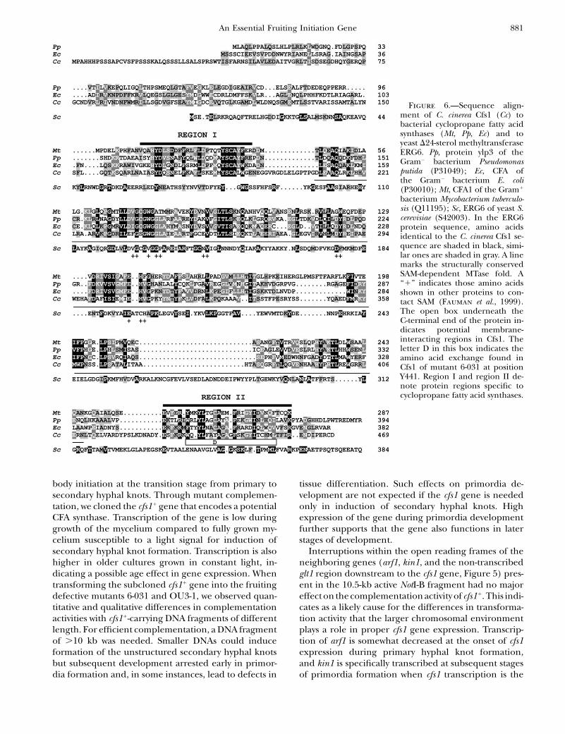
Figure 6.—Sequence align-ment of C. cinerea Cfs1 (Cc) tobacterial cyclopropane fatty acidsynthases (Mt, Pp, Ec) and toyeast D24-sterol methyltransferaseERG6. Pp, protein ylp3 of theGram� bacterium Pseudomonasputida (P31049); Ec, CFA ofthe Gram� bacterium E. coli(P30010); Mt, CFA1 of the Gram1
bacterium Mycobacterium tuberculo-sis (Q11195); Sc, ERG6 of yeast S.cerevisiae (S42003). In the ERG6protein sequence, amino acidsidentical to the C. cinerea Cfs1 se-quence are shaded in black, simi-lar ones are shaded in gray. A linemarks the structurally conservedSAM-dependent MTase fold. A‘‘1’’ indicates those amino acidsshown in other proteins to con-tact SAM (Fauman et al., 1999).The open box underneath theC-terminal end of the protein in-dicates potential membrane-interacting regions in Cfs1. Theletter D in this box indicates theamino acid exchange found inCfs1 of mutant 6-031 at positionY441. Region I and region II de-note protein regions specific tocyclopropane fatty acid synthases.
An Essential Fruiting Initiation Gene 881

highest. Possibly, the transcription profiles of arf1 andkin1 influence the expression of cfs1 during early andlater stages of fruiting body development.
Structure of the Cfs1 protein: Within the C. cinereaCfs1 protein, we identified a potential SAM-bindingdomain, a SAM-dependent MTase fold (Fauman et al.1999). CFA synthases are C-MTases that in bacteriatransfer a methylene group from SAM to a C-atom inunsaturated membrane localized phospholipids, therebyforming a cyclopropane ring (Grogan and Cronan
1997). Plant and fungal D24-sterol C-MTase are the clos-est related to CFA synthases, possibly because their enzymeactivities are both linked to lipid bilayers (Taylor andCronan 1979; Leber et al. 1994). Low homology isfound over the whole protein length between ERG6 ofS. cerevisiae and Cfs1 of C. cinerea, with the highestconservation in the SAM-binding motif (Figure 6). InERG6, directly at the N-terminal end of the SAM-dependent MTase fold is a sequence (DFYEYGWGSSFHFS; residues 77–92) referred to as region I that ishighly specific to all D24-sterol C-MTases, has sterolbinding activity, and forms an a-helix with a loopstructure that targets into the substrate pocket (Nes
et al. 1998, 2004). This sequence is not present in thefamily of CFA synthases (Figure 6). Instead, a highlyspecific sequence occupies the corresponding position(region I consensus: V/I85XXH88Y100D100V/L/I92S77N/D100D/N65F65F/Y100XL/I73W/F85L92D77P54S/T69M/L77T/S58Y100S/T100C85A92Y/F54F/W100E38R/K38; see supplemen-tal material at http://www.genetics.org/supplemental/).In the crystal structures of mycolic acid cyclopropane syn-thases from Mycobacterium tuberculosis, this CFA synthasesignature sequence adopts a helix-loop-helix-loop struc-ture that coats the surface of a hydrophobic tunnel fromthe entrance to active SAM-binding sites. The alkylchains of model substrates enter these pockets in aU-shaped manner, thereby contacting amino acids fromregion I (Huang et al. 2002).
In E. coli, CFA synthase is a soluble protein found inthe cell cytoplasm that uses SAM as a soluble and un-saturated fatty acid (UFA)-containing phospholipid asan insoluble substrate while transiently accessing to bothinner and outer leaflets of intact UFA-containing mem-branes (Taylor and Cronan 1979). The substrate C-double bond, positioned at 9–11 carbon units from theglycerol backbone of the phospholipid molecule, islocated deeply within the hydrophobic core of themembrane bilayer (Gally et al. 1979; Seelig and Seelig
1980). Inhibitor studies with sulfhydryl-modifying re-agents and C-terminal truncation (50 aa) suggested thatthe C-terminus and possibly C354 within the C-terminushave a role in catalysis or interaction with the membrane(Wang et al. 1992). However, changing C354 to analanine or serine did not result in loss of function(Grogan and Cronan 1997; Courtois et al. 2004). TheC-terminal region (region II in Figure 6, consensus: XV/M/L/I50XXQ/E73XXXR/K65V/M/L/I54Y/W/F100XXY96L/
M73XXC69A58XXF100K/R58XG58XL/I/V81D/N58V/L73XQ77V/M/L/I62T50XK/R96; see supplemental ma-terial at http://www.genetics.org/supplemental/) is nev-ertheless important, since the Y441D substitution in ourmutant 6-031 (corresponding to A353 in E. coli Cfa)resulted in a loss of function. Computer programs pre-dict the wild-type Cfs1 of C. cinerea being a cytoplasmicprotein, like the CFA synthase of E. coli, with two trans-membrane domains in the C-terminus. Possibly the C-terminus functions in transiently anchoring the Cfs1protein to the membrane and/or represents part ofthe catalytic domain. In the crystallized M. tuberculosisenzymes, the C-terminal end with region II forms ana-helix and a b-sheet. C269 within this a-helix (corre-sponding to C354 in E. coli Cfa) is in the vicinity of theactive site, while the b-sheet dangles away from the site(Huang et al. 2002).
Occurrence of cyclopropane fatty acid synthases andtheir products: CFA synthases have been found in abroad range of bacteria, with cis-9,10-methylenehexade-canoic acid (17CFA), cis-9,10-methyleneoctadecanoicacid [MOA, dihydrosterculic acid (DHSA), C19], andcis-11,12-methyleneoctadecanoic acid (lactobacillic acid,C19) being characteristic bacterial CFAs (for review seeGrogan and Cronan 1997). In E. coli, the CFA synthaseis not essential for growth under an assortment of ex-perimental conditions but improves survival in low-pHenvironments (Chang and Cronan 1999). In other bac-teria, the production of CFAs also relates to stress condi-tions. As a consequence of CFA production, membraneproperties, in particular membrane fluidity, alter withenhanced bacterial stress tolerance (Couto et al. 1996;Sajbidor 1997; Chang and Cronan 1999). Phospholip-ids containing CFAs have a broader transition tempera-ture range and increased rigidity than those containingUFAs, which confers more resistance of the membranelipid matrix to environmental perturbations (Dufourc
et al. 1984; Perly et al. 1985).In eukaryotes, CFAs have only sporadically been re-
ported, while the chemical structures are far morediverse. For example, cyclopropyl hydroxy-eicosanoidswere described in a red alga (Nagle and Gerwick
1990), cyclopropanated C19 straight-chain fatty acid(cladocroic acid) in a sponge (D’Auria et al. 1993), cy-clopropane containing eicosanoid (C20) in a soft coral(White and Jensen 1993), and CFAs with 17, 18 and19 C-atoms in females and eggs of millipedes (Oudejans
and van der Horst 1978). DHSA has been identifiedin trypanosomatid protozoa and CFA synthase activityhas been demonstrated (Li et al. 1993). DHSA synthesisfrom oleoyl phospatidylcholines has recently also beenobserved in tobacco cells after transformation of agene from the plant Sterculia foetida (Bao et al. 2003).DHSA and cis-9,10-methyleneheptadecanoic acid occurin roots of S. foetida (Kaimal and Lakshminarayana,1970). Biological roles of CFAs in eukaryotes have still tobe clarified. Functions in cold hardiness and drought
882 Y. Liu et al.
resistance in plants and desiccation tolerance in milli-pedes have been discussed (Grogan and Cronan 1997).In slime molds, CFA levels have been linked to cellularaggregation and cAMP metabolism (Saito and Ochiai
1998; Matsuoka et al. 2004). These latter observationscan be of special interest to our study since in C. cinereathere is an increase in levels of cAMP at the onset offruiting and cAMP has been shown in particular mutantsto induce fruiting (Kues et al. 2004).
To our knowledge, no cyclopropanated moiety has sofar been reported in higher fungal lipids. However, inthe higher basidiomycetes, the unsaturated linoleic acidis the major constituent of fatty acids (Solberg 1989;Bonzom et al. 1999; Sakai and Kajiwara 2004). Therelated oleic acid is shown in E. coli to be a substrate forthe action of CFA synthase (Marinari et al. 1974) andpreliminary expression studies suggest that the E. coliand C. cinerea enzymes are at least partially interchange-able in function (S. Loos, M. Aebi and U. Kues, un-published results).
In this study, we show that the C. cinerea cfs1 geneis superfluous for vegetative mycelial growth, but es-sential for fruiting body development. As in bacteria,the physical properties of cellular membranes mayalter through production of CFAs by Cfs1 and thiscould be the trigger to initiate fruiting body morphogen-esis in the fungus. Accordingly, in feeding experimentsmembrane-interactive compounds such as sucrose es-ters of fatty acids, plant saponins, and cerebrosidesinduced fruiting body development in various basidio-mycetes (Kawai 1989; Oita and Yanagi 1993; Mizushina
et al. 1998; Magae et al. 2004). From such studies, mem-brane alteration has been postulated to be a stress signalthat promotes the fungus to shift from vegetative toreproductive growth.
We are very grateful to Ben Lu for help with nuclear staining.P.S. holds a Ph.D. studentship from the Mahasarakham University,Thailand. Parts of this work were financed by the Swiss NationalScience Foundation (grants 31-46940.96, 31-46940.96/2 and 31-59157.99)and the Eidgenossische Technische Hochschule-Zurich. The lab inGottingen was funded by the Deutsche Bundesstiftung Umwelt.
LITERATURE CITED
Arima, T., M. Yamamoto, A. Hirata, S. Kawano and T. Kamada,2004 The eln3 gene involved in fruiting body morphogenesisof Coprinus cinereus encodes a putative membrane protein witha general glycosyltransferase domain. Fungal Genet. Biol. 41:805–812.
Badalyan, S. M., E. Polak, R. Hermann, M. Aebi and U. Kues,2004 Role of peg formation in clamp cell fusion in homobasid-iomycete fungi. J. Basic Microbiol. 44: 167–177.
Bao, X., J. J. Thelen, G. Bonaventura and J. B. Ohlrogge,2003 Characterization of cyclopropane fatty-acid synthase fromSterculia foetida. J. Biol. Chem. 278: 12846–12853.
Bertossa, R. C., U. Kues, M. Aebi and M. Kunzler, 2004 Promoteranalysis of cgl2, a galectin encoding gene transcribed during fruit-ing body formation in Coprinopsis cinerea (Coprinus cinereus). Fun-gal Genet. Biol. 41: 1120–1131.
Bonzom, P. M. A.,A. Nicolaou, M. Zloh,W. Baldeo andW. A. Gibbons,1999 NMR lipid profile of Agaricus bisporus. Phytochemistry 50:1311–1321.
Bottoli, A.P. F., K. Kertesz-Chaloupkova, R. P. Boulianne, J. D.Granado, M. Aebi et al., 1999 Rapid isolation of genes froman indexed genomic library of C. cinereus in a novel pab11 cosmid.J. Microbiol. Methods 35: 129–141.
Boulianne, R. P., Y. Liu, M. Aebi, B. C. Lu and U. Kues,2000 Fruiting body development in Coprinus cinereus: regulatedexpression of two galectins secreted by a non-classical pathway.Microbiology 146: 1841–1853.
Chang, Y. Y., and J. E. Cronan, Jr., 1999 Membrane cyclopropanefatty acid content is a major factor in acid resistance of Escherichiacoli. Mol. Microbiol. 33: 249–259.
Chomczynski, P., and N. Sacchi, 1987 Single-step method of RNAisolation by acid guanidinium thiocyanate-phenol-chloroformextraction. Anal. Biochem. 162: 156–159.
Clergeot, P.-H., G. Ruprich-Robert, Y. Liu, S. Loos, P. Srivilai
et al., 2003 Mutants in initiation of fruiting body developmentof the basidiomycete Coprinus cinereus. Fungal Genet. Newsl.50(Suppl.): 68.
Courtois, F., C. Guerard, X. Thomas and O. Ploux, 2004 Es-cherichia coli cyclopropane fatty acid synthase. Mechanistic andsite-directed mutagenic studies. Eur. J. Biochem. 271: 4769–4778.
Couto, J. A., N. Rozes and T. Hogg, 1996 Ethanol-induced changesin the fatty acid composition of Lactobacillus hilgardii, its effects onplasma membrane fluidity and relationship with ethanol toler-ance. J. Appl. Bacteriol. 81: 126–132.
D’Auria, M. V., L. G. Paloma, L. Minale, R. Riccio and A. Zampella,1993 Metabolites of the New Caledonian sponge Cladocroce in-curvata. J. Nat. Prod. 56: 418–423.
Dufourc, E. J., I. C. Smith and H. C. Jarrell, 1984 Role of cyclo-propane moieties in the lipid properties of biological mem-branes: a 2H NMR structural and dynamical approach.Biochemistry 23: 2300–2309.
Fauman, E. B., R. M. Blumenthal and X. D. Cheng, 1999 Structureand evolution of AdoMet-dependent methyltransferases, pp.1–38 in S-Adenosylmethionine-Dependent Methyltransferases: Structuresand Functions, edited by X. D. Cheng and R. M. Blumenthal.World Scientific, Singapore.
Gally, H. U., G. Pluschke, P. Overath and J. Seelig, 1979 Struc-ture of Escherichia coli membranes. Phospholipid conformation inmodel membranes and cells as studied by deuterium magneticresonance. Biochemistry 18: 5605–5610.
Granado, J. D., K. Kertesz-Chaloupkova, M. Aebi and U. Kues,1997 Restriction enzyme-mediated DNA intergration in Copri-nus cinereus. Mol. Gen. Genet. 256: 28–36.
Grogan, D. W., and J. E. Cronan, Jr., 1997 Cyclopropane ring for-mation in membrane lipids of bacteria. Microbiol. Mol. Biol. Rev.61: 429–441.
Huang, C., C. V. Smith, M. S. Glickman, W. R. Jacobs, Jr. and J. C.Sacchettini, 2002 Crystal structure of myolic acid cyclopro-pane synthase from Mycobacterium tubercolosis. J. Biol. Chem.277: 11559–11569.
Kaimal, T. N. B., and G. Lakshminarayana, 1970 Fatty acid com-positions of lipids isolated from different parts of Ceiba pentandra,Sterculia foetida and Hydnocarpus wightiana. Phytochem. 9: 2225–2229.
Kawai, G., 1989 Molecular species of cerebrosides in fruiting bodiesof Lentinus edodes and their biological activity. Biochim. Biophys.Acta 1001: 185–190.
Kertesz-Chaloupkova, K., P. J. Walser, J. D. Granado, M. Aebi andU. Kues, 1998 Blue light overrides repression of asexual spor-ulation by mating type genes in the basidiomycete Coprinus cin-ereus. Fungal Genet. Biol. 23: 95–109.
Kues, U., 2000 Life history and developmental processes in the basid-iomycete Coprinus cinereus. Microbiol. Mol. Biol. Rev. 64: 316–353.
Kues, U., J. D. Granado, R. Hermann, R. P. Boulianne, K. Kertesz-Chaloupkova et al., 1998 The A mating type and blue lightregulate all known differentiation processes in the basidiomyceteCoprinus cinereus. Mol. Gen. Genet. 260: 81–91.
Kues, U., T. Y. James, R. Vilgalys and M. P. Challen, 2001 Thechromosomal region containing pab-1, mip, and the A matingtype locus of the secondary homothallic homobasidiomyceteCoprinus bilanatus. Curr. Genet. 39: 16–24.
Kues, U., E. Polak, A. P. F. Bottoli, M. Hollenstein, P. J. Walser
et al., 2002a Vegetative development in Coprinus cinereus,
An Essential Fruiting Initiation Gene 883

pp. 133–164 in Molecular Biology of Fungal Development, edited byH. D. Osiewacz. Marcel Dekker, New York.
Kues, U., P. J. Walser, M. J. Klaus and M. Aebi, 2002b Influenceof activated A and B mating type pathways on developmentalprocesses in the basidiomycete Coprinus cinereus. Mol. Genet.Genomics 268: 262–271.
Kues, U., M. Kunzler, A. P. F. Bottoli, P. J. Walser, J. D. Granado
et al., 2004 Mushroom development in higher basidiomycetes;implications for human and animal health, pp. 431–470 inFungi in Human and Animal Health, edited by R. K. S. Kushwaha.Scientific Publishers, Jodhpur, India.
Leber, R., E. Zinser, G. Zellnig, F. Paltauf and G. Daum,1994 Characterization of lipid particles of the yeast, Saccharomycescerevisiae. Yeast 10: 1421–1428.
Li, R. X., S. Ganguli and R. A. Pascal, Jr., 1993 Synthesis of sulfur-substituted phosphatidylethanolamines and inhibition of proto-zoan cyclopropane fatty acid synthase. Tetrahedron Lett. 34:1279–1282.
Lu, B. C., and N. B. Raju, 1970 Meiosis in Coprinus. II. Chromosomepairing and the lampbrush diplotene stage of meiotic prophase.Chromosoma 29: 305–316.
Lu, B. C., N. Gallo and U. Kues, 2003 White-cap mutants and mei-otic apoptosis in the basidiomycete Coprinus cinereus. FungalGenet. Biol. 39: 82–93.
Magae, Y., T. Nishimura and S. Ohara, 2004 3-O-alkyl-D-glucosederivatives induce fruit bodies of Pleurotus. Mycol. Res. 109:374–376.
Marchuk, D., M. Drumm, A. Saulino and F. S. Collins, 1991 Con-struction of T-vectors, a rapid and general system for directcloning of unmodified PCR products. Nucleic Acids Res. 19:1154.
Marinari, L. A., H. Goldfine and C. Panos, 1974 Specificity ofcyclopropane fatty acid synthesis in Escherichia coli. Utilizationof isomers of monounsaturated fatty acid. Biochemistry 13:1978–1983.
Matsuoka, S., H. Kuwayama, D. Ikeno, M. Oyama and M. Maeda,2004 Defect in peroximal multifunctional enzyme MFE1 affectscAMP relay in Dictyostelium. Dev. Growth Differ. 46: 195–199.
Mizushina, Y., L. Hanashima, T. Yamaguchi, M. Takemura,F. Sugawara et al., 1998 A mushroom fruiting body-inducingsubstance inhibits activities of replicative DNA polymerases.Biochem. Biophys. Res. Commun. 249: 17–22.
Moore, D., M. M. Y. Elhiti and R. D. Butler, 1979 Morphogenesisof the carpophore of Coprinus cinereus. New Phytol. 83: 695–722.
Muraguchi, H., and T. Kamada, 1998 The ich1 gene of the mush-room Coprinus cinereus is essential for pileus formation in fruiting.Development 125: 3133–3141.
Muraguchi, H., and T. Kamada, 2000 A mutation in the eln2 geneencoding a cytochrome P450 of Coprinus cinereus affects mush-room morphogenesis. Fungal Genet. Biol. 29: 49–59.
Murata, Y., M. Fujii, M. E. Zolan and T. Kamada, 1998 Molecularanalysis of pcc1, a gene that leads to A-regulated sexual morpho-genesis in Coprinus cinereus. Genetics 149: 1753–1761.
Nagle, D. G., and W. H. Gerwick, 1990 Isolation and structure ofconstanolactones A and B, new cyclopropyl hydroxyeicosanoidsfrom the temperate red alga Constantinea simplex. TetrahedronLett. 31: 2995–2998.
Nes, W. D., B. S. McCourt, W. X. Zhou, J. Ma, J. A. Marshall et al.,1998 Overexpression, purification, and stereochemical studies
of the recombinant (S)-adenosyl-L-methionine: delta 24(25)- todelta 24(28)-sterol methyl transferase enzyme from Saccharomycescerevisiae. Arch. Biochem. Biophys. 353: 297–311.
Nes, W. D., P. Jayasimha, W. Zhou, R. Kanagasabai, C. Jin et al.,2004 Sterol methyltransferase: functional analysis of highlyconserved residues by site-directed mutagenesis. Biochemistry43: 569–576.
Oita, S., and S. O. Yanagi, 1993 Stimulation of Schizophyllum com-mune fruit body formation by inhibitor of membrane functionand cell wall synthesis. Biosci. Biotechnol. Biochem. 57: 1270–1274.
Oudejans, R. C. H. M., and D. J. van der Horst, 1978 Cy-clopropane fatty acids in millipedes: their occurrence and metab-olism. Abh. Naturwiss. Ver. Hamburg 21/22: 345–348.
Perly, B., I. C. Smith and H. C. Jarrell, 1985 Effects of replace-ment of a double bond by a cyclopropane ring in phosphatidy-lethanolamines: a 2H NMR study of phase transitions andmolecular organization. Biochemistry 24: 1055–1063.
Saito, T., and H. Ochiai, 1998 Fatty acid composition of the cellu-lar slime mold Polysphondylium pallidum. Lipids 33: 327–332.
Sajbidor, J., 1997 Effect of some environmental factors on the con-tent and composition of microbial membrane lipids. Crit. Rev.Biotechnol. 17: 87–103.
Sakai, H, and S. Kajiwara, 2004 Membrane lipid profile of anedible basidiomycete Lentinula edodes during growth and cell dif-ferentiation. Lipids 39: 67–73.
Sambrook, J., E. F. Fritsch and T. Maniatis, 1989 Molecular Clon-ing: A Laboratory Manual, Ed. 2. Cold Spring Harbor LaboratoryPress, Cold Spring Harbor, NY.
Seelig, J., and A. Seelig, 1980 Lipid conformation in model mem-branes and biological membranes. Q. Rev. Biophys. 13: 19–61.
Solberg, Y., 1989 A literature review of the lipid constituents ofhigher fungi, new investigation of Agaricus species. Int. J. Mycol.Lichenol. 4: 137–154.
Swamy, S., I. Uno and T. Ishikawa, 1984 Morphogenetic effectsof mutations at the A and B incompatibility factors in Coprinuscinereus. J. Gen. Microbiol. 130: 3219–3224.
Taylor, F. R., and J. E. Cronan, Jr., 1979 Cyclopropane fatty acidsynthase of Escherichia coli: stabilization, purification, and interac-tion with phospholipid vesicles. Biochemistry 18: 3292–3300.
Terashima, K., K. Yuki, H. Muraguchi, M. Akiyama and T. Kamada,2005 The dst1 gene involved in mushroom photomorphogene-sis of Coprinus cinereus encodes a putative photoreceptor for bluelight. Genetics 171: 101–108.
Walser, P. J., R. Velagapudi, M. Aebi and U. Kues, 2003 Ex-tracellular matrix proteins in mushroom development. RecentRes. Devel. Microbiol. 7: 381–415.
Wang, A. Y., D. W. Grogan and J. E. Cronan, Jr., 1992 Cyclo-propane fatty acid synthase of Escherichia coli: deduced aminoacid sequence, purification, and studies of the enzyme active site.Biochemistry 31: 11020–11028.
White, J. D., and M. S. Jensen, 1993 Biomimetic synthesis of acyclopropane containing eicosanoid from the coral Plexaurahomomalla. Assignment of relative configuration. J. Am. Chem.Soc. 115: 2970–2971.
Zolan, M. E., and P. J. Pukkila, 1986 Inheritance of DNA methyl-ation in Coprinus cinereus. Mol. Cell. Biol. 6: 195–200.
Communicating editor: J. J. Loros
884 Y. Liu et al.