ameloblastoma of the jaws: a retrospective analysis of 340...
TRANSCRIPT
Itaeo
PATHOLOGY
J Oral Maxillofac Surg70:608-615, 2012
Ameloblastoma of the Jaws: ARetrospective Analysis of 340 Cases in a
Malaysian PopulationChong Huat Siar, BDS, MSc, FDSRCPS, FRCPath,*
Shin Hin Lau, BDS, MSc, FDSRCS,† and
Kok Han Ng, BDS, MSc, FDSRCPS, FRCPath‡
Purpose: Ameloblastoma of the human jaw is an uncommon but clinically significant odontogenicepithelial neoplasm. The aim was to analyze the clinicopathologic characteristics of ameloblastoma in aMalaysian population.
Materials and Methods: This is a retrospective study (1993 through 2008) of consecutive ameloblas-toma cases accessioned in 2 main oral pathology diagnostic centers: the Unit of Stomatology, Institute forMedical Research and the Department of Oral Pathology, Oral Medicine, and Periodontology, Faculty ofDentistry, University of Malaya, Kuala Lumpur, Malaysia. Data on patient demographics, tumor location,symptomology, duration, radiographic appearance, preoperative diagnosis, clinicopathologic subtypes,treatment, and recurrence were analyzed.
Results: Three hundred forty cases of ameloblastoma were reviewed. These were from 197 male patients(57.9%) and 143 female patients (42.1%), with a male-to-female ratio of 1.4:1. A wide age range (7 to 85 years),mean onset age of 30.3 � 16.3 years, and peak incidence in the second decade of life were recorded. Mostwere mandibular tumors (n � 311/340, 91.5%). These consisted of 95 (28%) unicystic ameloblastomas, 221(65%) solid/multicystic ameloblastomas, 22 (6.4%) desmoplastic ameloblastoma, and 2 (0.6%) peripheralameloblastomas. Unicystic ameloblastoma (41.1%) and solid/multicystic ameloblastoma (52.0%) mostly af-fected Malays patients, whereas desmoplastic ameloblastoma (59.1%) was prevalent in Chinese patients.Unicystic ameloblastoma (56.8%) and solid/multicystic ameloblastoma (47.1%) occurred predominantly in thebody and posterior mandible, whereas desmoplastic ameloblastoma (36.4%) preferentially involved theanterior jaw segment. Most tumors presented as multilocular radiolucencies (36.8%). Enucleation (n � 42/92,45.7%) was the treatment of choice. About 18 cases (13.3%) presented with recurrence.
Conclusions: Because ameloblastoma subsets differ in their biologic behavior, the present data aresignificant as baseline references for clinicians and pathologists.© 2012 American Association of Oral and Maxillofacial Surgeons
J Oral Maxillofac Surg 70:608-615, 20120
n the recent histologic classification of odontogenicumors from the World Health Organization (WHO),meloblastoma is defined as a benign, locally invasivepithelial odontogenic neoplasm of putative enamelrgan origin.1,2 It is the second most common odon-
togenic neoplasm and accounts for approximately
*Professor and Head, Department of Oral Pathology, Oral Medi-
cine, and Periodontology, Faculty of Dentistry, University of Ma-
laya, Kuala Lumpur, Malaysia.
†Consultant, Oral Pathologist and Head, Unit of Stomatology, Insti-
tute for Medical Research, Jalan Pahang, Kuala Lumpur, Malaysia.
‡Former Consultant, Oral Pathologist and Director, Institute for
Medical Research, Jalan Pahang, Kuala Lumpur, Malaysia. d
608
11% to 18% of all odontogenic tumors. There are 4distinct clinicopathologic subtypes: unicystic ameloblas-toma (UA), solid/multicystic ameloblastoma (SMA), andperipheral and malignant forms.1 SMA and UA formthe 2 major subsets.1,2 SMA has great infiltrative po-tential and a higher recurrence rate. In contrast, UA
Address correspondence and reprint requests to Dr Siar:
Department of Oral Pathology, Oral Medicine, and Periodontology,
Faculty of Dentistry, University of Malaya, 50603 Kuala Lumpur,
Malaysia; e-mail: [email protected]
© 2012 American Association of Oral and Maxillofacial Surgeons
278-2391/12/7003-0$36.00/0
oi:10.1016/j.joms.2011.02.039
tTl
c5ssw
SIAR, LAU, AND NG 609
has an odontogenic cystlike behavior, occurs at ayounger age, and has a lower recurrence rate.
Malaysia, located at the southernmost tip of theAsian continent, is a multiracial, multicultural nationwith a population of about 28 million. The 3 mainracial groups are the Malays (65.0%), Chinese (26.0%),and Indians (8.0%). In the Malaysian capital city ofKuala Lumpur, there are 2 main oral pathology biopsydiagnostic centers. The Unit of Stomatology at theInstitute for Medical Research was established in 1967and subserves as the main oral pathology diagnosticservice center for most government-based dental clin-ics and hospitals in Malaysia. The other diagnosticcenter is the Department of Oral Pathology, OralMedicine, and Periodontology at the Faculty of Den-tistry, University of Malaya. This department wasformed when the first dental school in Malaysia wasestablished in 1972. It offers an oral pathology diag-nostic service not only for patients accessioned in thedental school but also for referral cases in the KlangValley district. The latter refers to Kuala Lumpur andits suburbs and the adjoining cities and towns in thestate of Selangor.
Ameloblastoma is a clinically significant tumor inthis region and has been the subject of considerablestudies.3-7 A previous study3 examined 401 ameloblas-oma cases based on the WHO’s 1971 Histologicalyping of Odontogenic Tumours, Jaw Cysts and Al-ied Lesions.8 However, little is known about amelo-
blastoma and its variants based on the new 2005World Health Organization Classification of Odon-togenic Tumours.1
The aim of the present study was to retrospectivelyanalyze ameloblastoma cases diagnosed in the 2 afore-mentioned centers from 1993 through 2008, classifythem according to criteria of the 2005 WHO classifi-cation,1 and determine their clinicopathologic char-acteristics.
Materials and Methods
The surgical biopsy records of all histologically di-agnosed cases of ameloblastoma archived from Janu-ary 1993 through December 2008 inclusive were re-trieved from the files of the Unit of Stomatology,Institute for Medical Research and the Department ofOral Pathology, Oral Medicine, and Periodontology,Faculty of Dentistry, University of Malaya, Kuala Lum-pur, Malaysia. This was a retrospective study that wasexempted from institutional review board approval(research grant RG83/09HTM). A patients’ age, gen-der, race, location, clinical signs and symptoms, du-ration, radiographic appearance, preoperative diagno-sis, treatment, and recurrence were abstracted fromthe case summaries accompanying the biopsy speci-
mens. For analysis of mandibular ameloblastomas, siteof occurrence was categorized into anterior (incisal–canine), body (premolar–molar region), posterior(distal to third molar), and bilateral (across midline)regions. For the mandible, the posterior area alsoincluded the ramus, angle, coronoid process, and con-dyle. For maxillary tumors, site was subdivided intoanterior (incisal–canine) and posterior (distal to ca-nine) regions. Any tumor involving 2 or more siteswas assigned to the region approximating the centerof the lesion. Any recurrent tumors arising from thesecases were not considered as separate or additionalcases. Sections of all primary and recurrent tumorsstained with hematoxylin and eosin were retrievedand reviewed to reclassify them according to criteriaof the recent WHO classification of odontogenic tu-mors.1
Descriptive statistics were performed to calculatethe frequency and percentages of these variables. Agewas stratified into various groups at 10-year intervals.Statistical analysis was carried out using SPSS 12.0(SPSS, Inc, Chicago, IL). The Fisher exact test fordifferences between 2 groups and the Kruskal-Wallistest for differences among 3 or more groups wereused as appropriate. The level of statistical signifi-cance was set at P � .05.
Results
CLINICAL FINDINGS
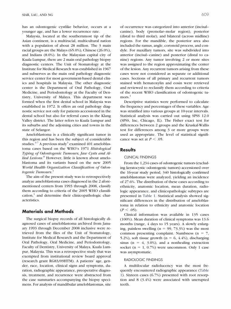
From the 1,234 cases of odontogenic tumors (exclud-ing keratocystic odontogenic tumors) accessioned overthe 16-year study period, 340 histologically confirmedameloblastomas were analyzed, yielding an incidenceof 27.6%. The distribution of these cases according toethnicity, anatomic location, mean duration, radio-logic appearance, and clinicopathologic subtypes arepresented in Table 1. Statistical analysis showed sig-nificant differences in the distribution of ameloblas-toma in relation to ethnicity and anatomic location(P � .05).
Clinical information was available in 135 cases(100%). Mean duration of clinical symptoms was 13.6months (range, 4 days to 15 years). A slowly enlarg-ing, painless swelling (n � 99, 73.3%) was the mostommon presenting complaint. Numbness (n � 7,.2%), soft tissue growth (n � 6, 4.4%), discharginginus (n � 4, 3.0%), and a nonhealing extractionocket (n � 1, 0.7%) were uncommon. Only 1 caseas asymptomatic.
RADIOLOGIC FINDINGS
A multilocular radiolucency was the most fre-quently encountered radiographic appearance (Table1). Sixteen cases (6.7%) presented with root resorp-tion and 8 (3.4%) were associated with unerupted
teeth.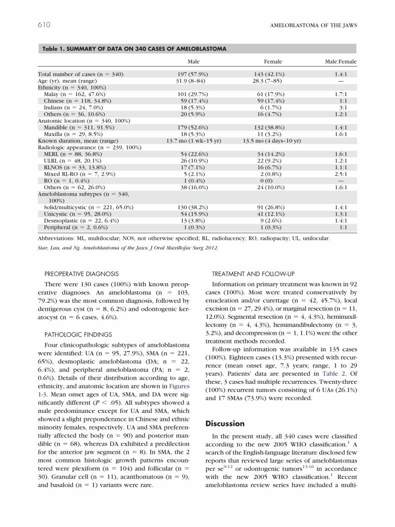
0e1nmsmtdf
3a
T
Surg 2
610 AMELOBLASTOMA OF THE JAWS
PREOPERATIVE DIAGNOSIS
There were 130 cases (100%) with known preop-erative diagnoses. An ameloblastoma (n � 103,79.2%) was the most common diagnosis, followed bydentigerous cyst (n � 8, 6.2%) and odontogenic ker-atocyst (n � 6 cases, 4.6%).
PATHOLOGIC FINDINGS
Four clinicopathologic subtypes of ameloblastomawere identified: UA (n � 95, 27.9%), SMA (n � 221,65%), desmoplastic ameloblastoma (DA; n � 22,6.4%), and peripheral ameloblastoma (PA; n � 2,.6%). Details of their distribution according to age,thnicity, and anatomic location are shown in Figures-3. Mean onset ages of UA, SMA, and DA were sig-ificantly different (P � .05). All subtypes showed aale predominance except for UA and SMA, which
howed a slight preponderance in Chinese and ethnicinority females, respectively. UA and SMA preferen-
ially affected the body (n � 90) and posterior man-ible (n � 68), whereas DA exhibited a predilectionor the anterior jaw segment (n � 8). In SMA, the 2
most common histologic growth patterns encoun-tered were plexiform (n � 104) and follicular (n �0). Granular cell (n � 11), acanthomatous (n � 9),
Table 1. SUMMARY OF DATA ON 340 CASES OF AMEL
M
otal number of cases (n � 340) 197 (Age (yr), mean (range) 31.9 (Ethnicity (n � 340, 100%)
Malay (n � 162, 47.6%) 101 (Chinese (n � 118, 34.8%) 59 (Indians (n � 24, 7.0%) 18 (Others (n � 36, 10.6%) 20 (
Anatomic location (n � 340, 100%)Mandible (n � 311, 91.5%) 179 (Maxilla (n � 29, 8.5%) 18 (
Known duration, mean (range) 13.7 mo (Radiologic appearance (n � 239, 100%)
MLRL (n � 88, 36.8%) 54 (ULRL (n � 48, 20.1%) 26 (RLNOS (n � 33, 13.8%) 17 (Mixed RL-RO (n � 7, 2.9%) 5 (RO (n � 1, 0.4%) 1 (Others (n � 62, 26.0%) 38 (
Ameloblastoma subtypes (n � 340,100%)
Solid/multicystic (n � 221, 65.0%) 130 (Unicystic (n � 95, 28.0%) 54 (Desmoplastic (n � 22, 6.4%) 13 (Peripheral (n � 2, 0.6%) 1 (
Abbreviations: ML, multilocular; NOS, not otherwise specifi
Siar, Lau, and Ng. Ameloblastoma of the Jaws. J Oral Maxillofac
nd basaloid (n � 1) variants were rare. a
TREATMENT AND FOLLOW-UP
Information on primary treatment was known in 92cases (100%). Most were treated conservatively byenucleation and/or curettage (n � 42, 45.7%), localexcision (n � 27, 29.4%), or marginal resection (n � 11,12.0%). Segmental resection (n � 4, 4.3%), hemimaxil-lectomy (n � 4, 4.3%), hemimandibulectomy (n � 3,3.2%), and decompression (n � 1, 1.1%) were the othertreatment methods recorded.
Follow-up information was available in 135 cases(100%). Eighteen cases (13.3%) presented with recur-rence (mean onset age, 7.3 years; range, 1 to 29years). Patients’ data are presented in Table 2. Ofthese, 3 cases had multiple recurrences. Twenty-three(100%) recurrent tumors consisting of 6 UAs (26.1%)and 17 SMAs (73.9%) were recorded.
Discussion
In the present study, all 340 cases were classifiedaccording to the new 2005 WHO classification.1 Asearch of the English-language literature disclosed fewreports that reviewed large series of ameloblastomasper se9-12 or odontogenic tumors13-16 in accordancewith the new 2005 WHO classification.1 Recent
STOMA
Female Male:Female
) 143 (42.1%) 1.4:128.3 (7–85) —
) 61 (17.9%) 1.7:1) 59 (17.4%) 1:1
6 (1.7%) 3:116 (4.7%) 1.2:1
) 132 (38.8%) 1.4:111 (3.2%) 1.6:1
15 yr) 13.5 mo (4 days–10 yr)
) 34 (14.2%) 1.6:1) 22 (9.2%) 1.2:1
16 (6.7%) 1.1:12 (0.8%) 2.5:10 (0) —
) 24 (10.0%) 1.6:1
) 91 (26.8%) 1.4:1) 41 (12.1%) 1.3:1
9 (2.6%) 1.4:11 (0.3%) 1:1
, radiolucency; RO, radiopacity; UL, unilocular.
012.
OBLA
ale
57.9%8–84)
29.7%17.4%5.3%)5.9%)
52.6%5.3%)1 wk–
22.6%10.9%7.1%)2.1%)0.4%)16.0%
38.2%15.9%3.8%)0.3%)
ed; RL
meloblastoma review series have included a multi-

SIAR, LAU, AND NG 611
FIGURE 1. Age distribution of ameloblastoma subtypes. DA, desmoplastic ameloblastoma; PA, peripheral ameloblastoma; SMA, solid/multicystic ameloblastoma; UA, unicystic ameloblastoma.
Siar, Lau, and Ng. Ameloblastoma of the Jaws. J Oral Maxillofac Surg 2012.
FIGURE 2. Ethnic distribution of ameloblastoma subtypes. CF, Chinese female; CM, Chinese male; DA, desmoplastic ameloblastoma; IF,Indian female; IM, Indian male; MF, Malay female; MM, Malay male; OF, other females; OM, other males; SMA, solid/multicysticameloblastoma; UA, unicystic ameloblastoma.
Siar, Lau, and Ng. Ameloblastoma of the Jaws. J Oral Maxillofac Surg 2012.
(m
gc1C
gE
si
stdtCfr
mat
p
Surg 2
612 AMELOBLASTOMA OF THE JAWS
centric Latin-American study on 163 cases of amelo-blastoma,9 a critical appraisal of 25 cases from a singleinstitution,10 a comparative analysis of mandibular251 cases) versus maxillary (31 cases) ameloblasto-as in Sri Lanka,11 and a prevalence study of 37 cases
in Chinese children and adolescents.12 For odonto-enic tumor series based on the 2005 WHO classifi-ation, these have included a retrospective study of,642 cases (inclusive of 661 ameloblastomas) in ahinese population,13 an analysis of 238 odontogenic
tumors (inclusive of 57 ameloblastomas) in Brazil,14 areview of 1,309 cases of odontogenic tumors (inclu-sive of 478 ameloblastomas) in a northern Chinesepopulation,15 and a prevalence study of 82 odonto-enic tumors (inclusive of 34 ameloblastomas) ingypt.16 From these published reports, certain simi-
larities and differences compared with the presentdata were identified.
It is well-known that the demographic profile ofameloblastoma exhibits considerable geographic vari-ation, and this has been extensively discussed in ameta-analysis2 and in other published series.9-23 Bylimiting the present comparison with those studiesbased on the 2005 WHO classification, the Malaysianameloblastomas were found to correlate well withother Asian and, to a lesser extent, Egyptian amelo-blastomas in showing a high relative frequency,13,15,16
a wide age distribution,13,15,16 a peak incidence in theecond decade of life,11,13,15 and slight male predom-
FIGURE 3. Site distribution of A, UA and B, SMA subtypes. DA,otherwise specified; SMA, solid/multicystic ameloblastoma; UA, u
Siar, Lau, and Ng. Ameloblastoma of the Jaws. J Oral Maxillofac
nance.13,15,16 In contrast, in Latin American amelo- c
blastomas, the tumors occurred at a lower frequency,exhibited an almost even gender distribution, and awide age range.9 The present data concurred withprevious studies in showing that individuals with UAtended to be younger than those with SMA,1,15
whereas patients with DA belonged to an even olderage group.8,15
It has been suggested that ameloblastomas aremore prevalent in blacks and people of Asian de-scent.2 In the present study, although a statisticallyignificant difference in the distribution of ameloblas-oma in relation to ethnicity was observed, this racialistribution pattern reflected the local racial popula-ion ratio.3 In contrast, DA occurred predominantly inhinese (59.1%), even though this ethnic group
ormed 26.0% of the local racial population.3,8 Theeason for this racial predilection is unclear.
It is well recognized that ameloblastomas occurore frequently in the mandible than in the maxilla,
nd that these tumors are located predominantly inhe body and posterior mandible.2,9,13-15,17,19 In the
present study, more than 90% of ameloblastomaswere found in the mandible and most of these tumorswere also located in the body and posterior regions.The observed prevalence of DA for the anterior jawsegment agrees with other reported data in the liter-ature.1,4,8,11,15 A Chinese study found that the more
osterior in the mandible an ameloblastoma was lo-
lastic ameloblastoma; Mand, mandible; Max, maxilla; NOS, notameloblastoma.
012.
desmopnicystic
ated, the younger the patient was at diagnosis.15 In
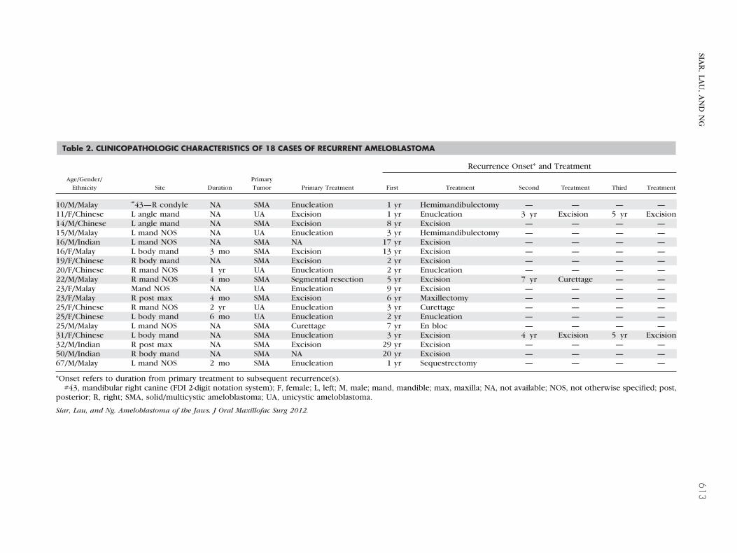
Table 2. CLINICOPATHOLOGIC CHARACTERISTICS OF 18 CASES OF RECURRENT AMELOBLASTOMA
Recurrence Onset* and Treatment
Age/Gender/Ethnicity Site Duration
PrimaryTumor Primary Treatment First Treatment Second Treatment Third Treatment
10/M/Malay #43—R condyle NA SMA Enucleation 1 yr Hemimandibulectomy — — — —11/F/Chinese L angle mand NA UA Excision 1 yr Enucleation 3 yr Excision 5 yr Excision14/M/Chinese L angle mand NA SMA Excision 8 yr Excision — — — —15/M/Malay L mand NOS NA UA Enucleation 3 yr Hemimandibulectomy — — — —16/M/Indian L mand NOS NA SMA NA 17 yr Excision — — — —16/F/Malay L body mand 3 mo SMA Excision 13 yr Excision — — — —19/F/Chinese R body mand NA SMA Excision 2 yr Excision — — — —20/F/Chinese R mand NOS 1 yr UA Enucleation 2 yr Enucleation — — — —22/M/Malay R mand NOS 4 mo SMA Segmental resection 5 yr Excision 7 yr Curettage — —23/F/Malay Mand NOS NA UA Enucleation 9 yr Excision — — — —23/F/Malay R post max 4 mo SMA Excision 6 yr Maxillectomy — — — —25/F/Chinese R mand NOS 2 yr UA Enucleation 3 yr Curettage — — — —25/F/Chinese L body mand 6 mo UA Enucleation 2 yr Enucleation — — — —25/M/Malay L mand NOS NA SMA Curettage 7 yr En bloc — — — —31/F/Chinese L body mand NA SMA Enucleation 3 yr Excision 4 yr Excision 5 yr Excision32/M/Indian R post max NA SMA Excision 29 yr Excision — — — —50/M/Indian R body mand NA SMA NA 20 yr Excision — — — —67/M/Malay L mand NOS 2 mo SMA Enucleation 1 yr Sequestrectomy — — — —
*Onset refers to duration from primary treatment to subsequent recurrence(s).#43, mandibular right canine (FDI 2-digit notation system); F, female; L, left; M, male; mand, mandible; max, maxilla; NA, not available; NOS, not otherwise specified; post,
posterior; R, right; SMA, solid/multicystic ameloblastoma; UA, unicystic ameloblastoma.
Siar, Lau, and Ng. Ameloblastoma of the Jaws. J Oral Maxillofac Surg 2012.
SIAR
,LA
U,
AN
DN
G613
BaE
at
sw
ftv
bf
o
etoogettmrnt(gbbrc(
(atac
614 AMELOBLASTOMA OF THE JAWS
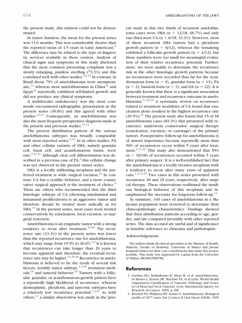
the present study, this relation could not be demon-strated.
In tumor duration, the mean for the present serieswas 13.6 months. This was considerably shorter thanthe reported mean of 4.5 years in Latin Americans.9
The difference may be related to the type of diagnos-tic services available in these centers. Analysis ofclinical signs and symptoms in this study disclosedthat the most common presenting complaint was aslowly enlarging, painless swelling (73.3%) and thiscorrelated well with other studies.2,9,13 In contrast, in
razil about 79% of ameloblastoma were asymptom-tic,14 whereas most ameloblastomas in China13 andgypt16 reportedly exhibited self-limited growth and
did not produce any clinical symptoms.A multilocular radiolucency was the most com-
monly encountered radiographic presentation in thepresent series (36.8%) and this agreed with otherstudies.12,13 Consequently, an ameloblastoma waslso the most frequent preoperative diagnosis made inhe present and previous studies.12,13
The present distribution pattern of the variousameloblastoma subtypes was broadly comparablewith most reported series.9,16 As in other studies, PAand other cellular variants of SMA, namely granularcell, basal cell, and acanthomatous forms, wererare.9,12,16 Although clear cell differentiation was de-cribed in a previous case of PA,4 this cellular changeas not observed in the present tumor series.SMA is a locally infiltrating neoplasm and the pre-
erred treatment is wide surgical excision.10 In con-rast, UA has a cystlike behavior, and a more conser-ative surgical approach is the treatment of choice.9
There are others who recommended that the thirdhistologic subtype of UA (showing intraluminal andintramural proliferations) is an aggressive tumor andtherefore should be treated more radically as forSMA.13 In the present series, most cases were treatedconservatively by enucleation, local excision, or mar-ginal resection.
Ameloblastoma is an enigmatic tumor with a strongtendency to recur after treatment.24-28 The recur-rence rate (13.3%) in the present series was lowerthan the reported recurrence rate for ameloblastoma,which may range from 15.9% to 20.6%.2,9 It is knownthat recurrences can take longer than 20 years tobecome apparent and, therefore, the eventual recur-rence rate may be higher.12,29,30 Recurrence in amelo-
lastoma is believed to be the result of several riskactors, notably tumor subtype,1,2,26 treatment meth-
ods,26 and tumoral behavior.29 Tumors with a follic-ular, granular, or acanthomatous growth pattern havea reportedly high likelihood of recurrence, whereasdesmoplastic, plexiform, and unicystic subtypes havea relatively low recurrence potential.1,2,26 As with
thers,11 a similar observation was made in the pres-
nt study in that two thirds of recurrent ameloblas-oma cases were SMA (n � 12/18, 66.7%) and onlyne third were UA (n � 6/18, 33.3%). However, mostf these recurrent SMA tumors had a plexiformrowth pattern (n � 8/12), whereas the remainingxhibited a follicular growth pattern (n � 4/12), buthese numbers were too small for meaningful evalua-ion of their relative recurrence potential. Further-ore, we were unable to determine the recurrence
isk in the other histologic growth patterns becauseo recurrences were recorded thus far for the acan-homatous form (n � 9), granular form (n � 11), PAn � 2), basaloid form (n � 1), and DA (n � 22). It isenerally known that there is a significant associationetween treatment and recurrence outcome in amelo-lastoma.11,25,30 A systematic review on recurrenceelated to treatment modalities of UA found that enu-leation alone resulted in the highest recurrence rate30.5%).25 The present study also found that 15 of 18
ameloblastoma cases (83.3%) that presented with re-currence underwent conservative surgical therapy(enucleation, excision, or curettage) of the primarytumors. Postoperative follow-up for ameloblastoma isof utmost importance because reportedly more than50% of recurrences occur within 5 years after treat-ment.2,12,24 This study also demonstrated that 55%n � 10/18) of recurrences occurred within 5 yearsfter primary surgery. It is a well-established fact thathe ameloblastoma is a locally invasive neoplasm with
tendency to recur after many years of apparenture.1,2,12,25 Two cases in this series presented with
recurrence 20 and 29 years, respectively, after surgi-cal therapy. These observations reaffirmed the insidi-ous biological behavior of this neoplasm and re-emphasized the necessity for long-term follow-up.12
In summary, 340 cases of ameloblastoma in a Ma-laysian population were reviewed to determine theirclinicopathologic characteristics. Findings showedthat their distribution patterns according to age, gen-der, and site compared favorably with other reportedseries. The data accrued are useful and of significanceas baseline reference to clinicians and pathologists.
Acknowledgments
The authors thank all clinical specialists at the Ministry of Health,Malaysia, Faculty of Dentistry, University of Malaya and privatehospitals/clinics for their case contributions that made this projectpossible. This study was supported by a grant from the Universityof Malaya (RG083/09HTM).
References1. Gardner DG, Heikinheimo K, Shear M, et al: Ameloblastomas,
in Barnes L, Eveson JW, Reichart PA, et al (eds): World HealthOrganization Classification of Tumours. Pathology and Genet-ics of Head and Neck Tumours. Lyon, International Agency forResearch on Cancer, 2005, p 296
2. Reichart PA, Philipsen HP, Sonner S: Ameloblastoma: Biological
profile of 3677 cases. Eur J Cancer B Oral Oncol 31B:86, 1995
SIAR, LAU, AND NG 615
3. Siar CH, Ng KH: Ameloblastoma in Malaysia—A 25-year review.Ann Acad Med Singapore 22:856, 1993
4. Ng KH, Siar CH: Peripheral ameloblastoma with clear celldifferentiation. Oral Surg Oral Med Oral Pathol 70:210, 1990
5. Siar CH, Ng KH: Calcifying and keratinizing ameloblastoma ofthe maxilla. J Laryngol Otol 105:971, 1991
6. Siar CH, Ng KH: Combined ameloblastoma and odontogenickeratocyst or keratinising ameloblastoma. Br J Oral MaxillofacSurg 31:183, 1993
7. Ng KH, Siar CH: Desmoplastic variant of ameloblastoma inMalaysians. Br J Oral Maxillofac Surg 31:299, 1993
8. Pindborg JJ, Kramer IRH, Torloni H: Histological Typing ofOdontogenic Tumours, Jaw Cysts and Allied Lesions. Interna-tional Histological Classification of Tumours. No 5. Geneva,World Health Organization, 1971
9. Ledesma-Montes C, Mosqueda-Taylor A, Carlos-Bregni R, et al:Ameloblastomas: A regional Latin-American multicentric study.Oral Dis 13:303, 2007
10. Hertog D, van der Waal I: Ameloblastoma of the jaws: A criticalreappraisal based on a 40-years single institution experience.Oral Oncol 46:61, 2010
11. Gunawardhana KSND, Jayasooriya PK, Rambukeweka IK, et al:A clinico-pathological comparison between mandibular andmaxillary ameloblastomas in Sri Lanka. J Oral Pathol Med 39:236, 2010
12. Zhang J, Gu Z, Jiang L, et al: Ameloblastoma in children andadolescents. Br J Oral Maxillofac Surg 48:549, 2010
13. Jing W, Xuan M, Lin Y, et al: Odontogenic tumours: A retro-spective study of 1642 cases in a Chinese population. Int J OralMaxillofac Surg 36:20, 2007
14. Avelar RL, Antunes AA, Santos T, et al: Odontogenic tumors:Clinical and pathology of 238 cases. Braz J Otorhinolaryngol74:668, 2008
15. Luo HY, Li TJ: Odontogenic tumors: A study of 1309 cases in aChinese population. Oral Oncol 45:706, 2009
16. Tawfik MA, Zyada MM: Odontogenic tumors in Dakahlia,Egypt: Analysis of 82 cases. Oral Surg Oral Med Oral Pathol OralRadiol Endod 109:e67, 2010
17. Fernandes AM, Duarte EC, Pimenta FJ, et al: Odontogenic
tumors: A study of 340 cases in a Brazilian population. J OralPathol Med 34:583, 200518. Olgac V, Koseoglu BG, Aksakalli N: Odontogenic tumours inIstanbul: 527 cases. Br J Oral Maxillofac Surg 44:386, 2006
19. Okada H, Yamamoto H, Tilakaratne WM: Odontogenic tumorsin Sri Lanka: Analysis of 226 cases. J Oral Maxillofac Surg65:875, 2007
20. Siriam G, Shetty R: Odontogenic tumors: A study of 250 casesin an Indian teaching hospital. Oral Surg Oral Med Oral PatholOral Radiol Endod 105:e14, 2008
21. Poon CSP, Wu PC, So MKP: Ameloblastoma in Hong KongChinese. Hong Kong Med J 2:172, 1996
22. Lu Y, Xuan M, Takata T, et al: Odontogenic tumors. A demo-graphic study of 759 cases in a Chinese population. Oral SurgOral Med Oral Pathol Oral Radiol Endod 86:707, 1998
23. Adebiyi KE, Ugboko VI, Omoniyi-Esan GO, et al: Clinico-pathological analysis of histological variants of ameloblas-toma in a suburban Nigerian population. Head Face Med2:42, 2006
24. Olaitan AA, Arole G, Adekeye EO: Recurrent ameloblastoma ofthe jaws. A follow-up study. Int J Oral Maxillofac Surg 27:456,1998
25. Lau SL, Samman N: Recurrence related to treatment modalitiesof unicystic ameloblastoma: A systematic review. Int J OralMaxillofac Surg 35:681, 2006
26. Hong J, Yun PY, Chung IH, et al: Long-term follow up onrecurrence of 305 ameloblastoma cases. Int J Oral MaxillofacSurg 36:283, 2007
27. Huang IY, Lai ST, Chen CH, et al: Surgical management ofameloblastoma in children. Oral Surg Oral Med Oral PatholOral Radiol Endod 104:478, 2007
28. Sammartino G, Zarrelli C, Urciuolo V, et al: Effectiveness of anew decisional algorithm in managing mandibular ameloblas-tomas: A 10-years experience. Br J Oral Maxillofac Surg 45:306,2007
29. Martins WD, Martins D: Recurrence of an ameloblastoma in anautogenous iliac bone graft. Oral Surg Oral Med Oral PatholOral Radiol Endod 98:657, 2007
30. Eckardt AM, Kokemüller H, Flemming P, et al: Recurrent
ameloblastoma following osseous reconstruction—A review oftwenty years. J Craniomaxillofac Surg 37:36, 2009