am report 11/24/09 amy auerbach peak onset between 20 and 30 years form of spondyloarthritis...
TRANSCRIPT


AM Report 11/24/09Amy Auerbach

Peak onset between 20 and 30 years Form of spondyloarthritis (cause
inflammation around site of ligament insertion into bone) and association with HLA-B27
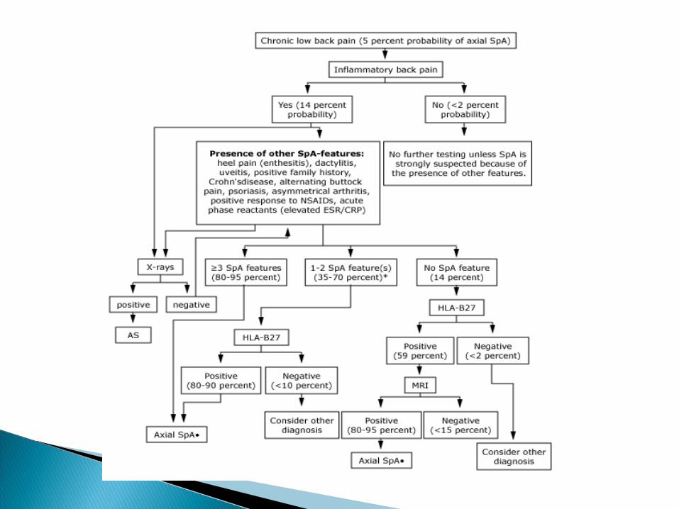
Prevalence as high as 5% in adults with chronic low back pain
Male to female ratio 2-3:1
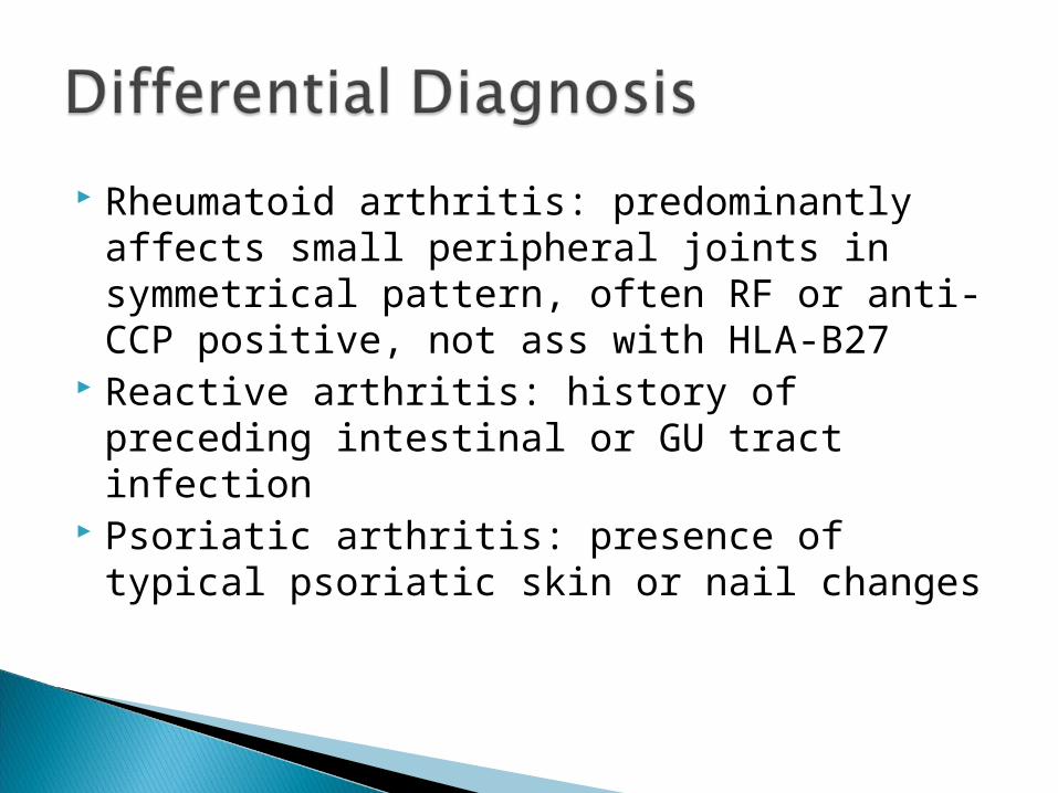
Rheumatoid arthritis: predominantly affects small peripheral joints in symmetrical pattern, often RF or anti-CCP positive, not ass with HLA-B27
Reactive arthritis: history of preceding intestinal or GU tract infection
Psoriatic arthritis: presence of typical psoriatic skin or nail changes

Reiter's syndromeRheumatoid arthritis
Gonococcal arthritis
Psoriatic arthritis
Age Young Middle Young Middle
Gender Male>femaleFemale>male
Female>male No effect
Onset Abrupt Insidious Abrupt Insidious
Joint numbr Oligoarthritis PolyarthritisMonoarthritis or oligoarthritis
Oligoarthritis
Symmetry of arthritis
No Yes No No
Sausage digits
Yes No No Yes
Back pain Yes No No Yes
Urethritis Yes No Yes No
Skin lesionsPalms and soles in 10 percent
Subcutaneous nodules
Pustular, nodular or vesicular
Psoriasis
Gonococcus No No Yes No
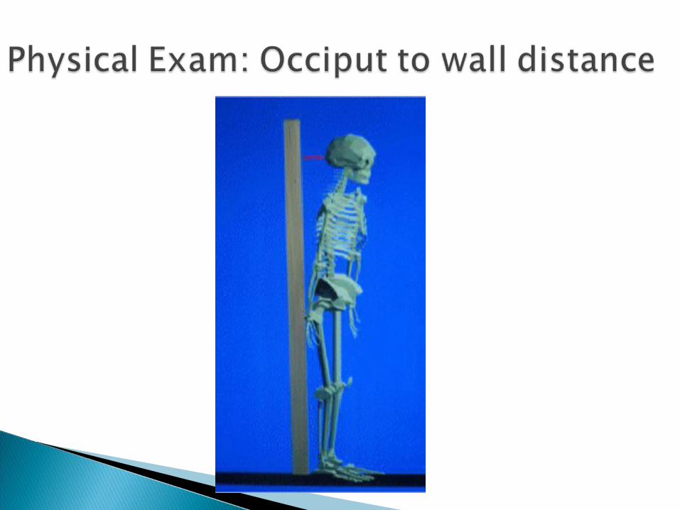
Onset of back pain before age 40 Insidious onset Improvement with exercise No improvement with rest Pain at night

Chest expansion: expansion of less than 2.5cm abnormal (5cm considered normal)
Sacroiliac joint tenderness Hip joint involvement Peripheral joint involvement (dactylitis-
“sausage toes”)


CRP typically elevated HLA-B27: present in 8% of population,
prevalence in HLA-B27 positive population is only 5%

Widening, erosions, sclerosis, or ankylosis of sacroiliac joint
Early signs: squaring of vertebral bodies due to anterior and posterior spondylitis
Late stages: proliferative changes, anterior atlantoaxial subluxation
MRI: more sensitive- can use in patients who do not have sacroiliitis on plain radiographs (can see “bone marrow edema”

Acute anterior uveitis: occurs in 25-40% of patients
- Presents as acute unilateral pain, photophobia, and blurring of vision
Neurologic symptoms: fracture of ankylosed spine, atlantoaxial-axial subluxation, cauda equina syndrome
Cardiovascular disease: increased risk Pulmonary disease: restriction secondary to
restriction in chest expansion Renal disease: IgA nephropathy and secondary
amyloidosis (only in patients with longstanding active inflammation)
Bowel lesions: Inflammatory bowel disease Osteopenia (in patients with persistent active
disease)
Clinical: 1) Low back pain and stiffness >3 months improves with
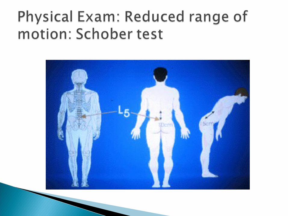
exercise and not relieved by rest2) Limitation of motion of lumbar spine3) Limitation of chest expansion relative to normal values
correlated for age and sex
Radiologic:1) Sacroiliitis grade >2 BL or 3 to 4 unilaterally
Spinal and sacroiliac involvement Hip and shoulder involvement Costovertebral, sternoclavicular,
costochondral inflammation Inflammation of extraspinal entheses

Low back pain (inflammatory in nature) Buttock pain (may be indicative of sacroiliac
involvement) Limited spine mobility and chest expansion Hip pain TMJ involvement Enthesitis

Symptomatic relief Restore function Prevent joint damage Prevent spinal fusion (prevent progressive
bony erosions and ankylosis of the spine) Minimize extraspinal and extraarticular
manifestations Prevent complications of spinal disease
Global pain Axial pain Degree and duration of morning stiffness Activities that are limited
ESR or CRP are useful as laboratory parameters of active disease
Hip arthritis Dactylitis Poor efficacy of NSAIDs High ESR Limitation in ROM of lumbar spine Oligoarthritis Onset less than 16 years of age
Also associated with poor outcome: cigarette smoking, severe radiographic changes, functional impairment

Physical therapy: can help maintain function and partially relieve symptoms
Local application of heat/cold Pharmacologic therapy: Analgesics, NSAIDs,
sulfasalazine, MTX, anti-TNF agents - 70-80% of patients report substantial relief
with NSAIDs. Continuous use may reduce radiographic progression

Typically have rapid response: improvement in pain, functional assessment, degree of inflammation
Patients with good functional ability, elevated ESR/CRP, and HLA-B27 positive respond best
Need to be wary of possibility of reactivation of latent TB