alternativesplicingandcaspase-mediatedcleavage...
TRANSCRIPT

Alternative Splicing and Caspase-Mediated CleavageGenerate Antagonistic Variants of the StressOncoprotein LEDGF/p75
Terry A. Brown-Bryan,1,2 Lai S. Leoh,1,2 Vidya Ganapathy,1,2 Fabio J. Pacheco,1,6
Melanie Mediavilla-Varela,1,2 Maria Filippova,1,3 Thomas A. Linkhart,5
Rik Gijsbers,7 Zeger Debyser,7 and Carlos A. Casiano1,2,4
1Center for Health Disparities and Molecular Medicine, 2Division of Microbiology and Molecular Genetics, 3Divisionof Biochemistry, and 4Department of Medicine, Loma Linda University School of Medicine; 5MuskuloskeletalDisease Center, Jerry L. Pettis Memorial Veterans Affairs Medical Center, Loma Linda, California;6Department of Biological Sciences, Centro Universitario Adventista de Sao Paulo, Sao Paulo,Brazil; and 7Molecular Medicine, Katholieke Universiteit Leuven, B-3000, Leuven, Belgium
AbstractThere is increasing evidence that an augmented state of
cellular oxidative stress modulates the expression of
stress genes implicated in diseases associated with
health disparities such as certain cancers and diabetes.
Lens epithelium–derived growth factor p75 (LEDGF/
p75), also known as DFS70 autoantigen, is emerging as a
survival oncoprotein that promotes resistance to
oxidative stress–induced cell death and chemotherapy.
We previously showed that LEDGF/p75 is targeted by
autoantibodies in prostate cancer patients and is
overexpressed in prostate tumors, and that its stress
survival activity is abrogated during apoptosis. LEDGF/
p75 has a COOH-terminally truncated splice variant, p52,
whose role in stress survival and apoptosis has not been
thoroughly investigated. We observed unbalanced
expression of these proteins in a panel of tumor cell
lines, with LEDGF/p75 generally expressed at higher
levels. During apoptosis, caspase-3 cleaved p52 to
generate a p38 fragment that lacked the NH2-terminal
PWWP domain and failed to transactivate the Hsp27
promoter in reporter assays. However, p38 retained
chromatin association properties and repressed the
transactivation potential of LEDGF/p75. Overexpression
of p52 or its variants with truncated PWWP domains in
several tumor cell lines induced apoptosis, an activity
that was linked to the presence of an intron-derived
COOH-terminal sequence. These results implicate the
PWWP domain of p52 in transcription function but not
in chromatin association and proapoptotic activities.
Consistent with their unbalanced expression in
tumor cells, LEDGF/p75 and p52 seem to play
antagonistic roles in the cellular stress response and
could serve as targets for novel antitumor therapies.
(Mol Cancer Res 2008;6(8):1293–307)
IntroductionEmerging evidence links the augmented state of cellular
oxidative stress with the pathogenesis of diseases associated
with health disparities, including cancer and type II diabetes
(1-3). Oxidative stress–modulated signaling pathways have
been implicated in cancer development and resistance to
therapy and may offer attractive targets for therapeutic
interventions (4-6). The lens epithelium–derived growth factor
p75 (LEDGF/p75), also known as transcription coactivator
p75 (TCP75), PC4 and SFRS1 interacting protein (PSIP), and
dense fine speckled autoantigen of 70 kDa (DFS70), is
emerging as a key player in the cellular response to oxidative
stress (7-12). LEDGF/p75 is induced by oxidative stress
and is presumed to promote resistance to stress-induced cell
death via transcriptional activation of stress and antioxidant
genes (8, 9, 13-16). LEDGF/p75 has also been identified as a
target of autoantibodies in various autoimmune and inflam-
matory conditions (17-19) and has emerged as an important
cellular cofactor for the chromosomal tethering of HIV-1
(20-24).
A role for LEDGF/p75 in malignancy was first hinted by its
homology to members of the hepatoma-derived growth factor
family (25) and the observation that chromosomal trans-
locations in leukemias may result in LEDGF/NUP98 fusion
Received 3/6/08; revised 4/20/08; accepted 4/23/08.Grant support: National Center for Minority Health. and Health DisparitiesProject EXPORT Program 5P20MD001632/Project 2 (C.A. Casiano), NationalInstitute of Allergy and Infectious Diseases AI44088 (C.A. Casiano), NationalInstitute of General Medical Sciences Initiative for Maximizing Student Diversity5R25GM60507 (M. Mediavilla-Varela), National Cancer Institute Ruth Kirsch-stein Minority Predoctoral Fellowship 1F31CA117742-01A1 (M. Mediavilla-Varela), Department of Veterans Affairs Medical Research Merit Review Grant(T. Linkhart), the European Commission LSHB-CT-2003-503480/TRIoH project(R. Gijsbers and Z. Debyser), Basic Research Support Grant from Loma LindaUniversity School of Medicine (C.A. Casiano), and a predoctoral fellowship fromCentro Universitario Adventista de Sao Paulo (F.J. Pacheco).The costs of publication of this article were defrayed in part by the payment ofpage charges. This article must therefore be hereby marked advertisement inaccordance with 18 U.S.C. Section 1734 solely to indicate this fact.Note: T.A. Brown-Bryan and L.S. Leoh contributed equally to this work andshare first authorship.Present address for V. Ganapathy: Department of Medical Oncology, CancerInstitute of New Jersey, New Brunswick, New Jersey. Present address for T.A.Brown-Bryan: Medical Sciences Institute, Charles Drew University of Medicineand Science, Las Angeles, California.Requests for reprints: Carlos A. Casiano, Center for Health Disparities andMolecular Medicine, Loma Linda University School of Medicine, Mortensen Hall146, 11085 Campus Street, Loma Linda, CA 92350. Phone: 909-558-1000, ext.42759; Fax: 909-558-0196. E-mail: [email protected] D 2008 American Association for Cancer Research.doi:10.1158/1541-7786.MCR-08-0125
Mol Cancer Res 2008;6(8). August 2008 1293on May 5, 2019. © 2008 American Association for Cancer Research. mcr.aacrjournals.org Downloaded from

proteins with potentially altered transcription functions (26-29).
We reported that overexpression of LEDGF/p75 in HepG2 liver
tumor cells enhanced their proliferation and protected them
from stress-induced death (30). We also identified LEDGF/p75
as an autoantigen in prostate cancer whose expression is
elevated in advanced stage tumors, most likely as a result of
the augmented state of cellular oxidative stress in the prostate
tumor microenvironment (31). More recently, Daugaard et al.
(32) reported that LEDGF/p75 increases the tumorigenic
potential of human cancer cell lines in murine models, and
that its expression is increased in human breast and bladder
carcinomas. These investigators also provided evidence that
overexpression of LEDGF/p75 in HeLa and MCF-7 cells
increases chemoresistance. Furthermore, Huang et al. (33)
reported increased LEDGF/p75 expression in blasts from
chemotherapy-resistant human acute myelocytic leukemia
patients.
LEDGF/p75 is composed of 530 amino acids and has an
alternative splice variant designated LEDGF/p52 (333 amino
acids; hereafter called p52), which also functions as a
transcription coactivator of RNA polymerase II and has been
implicated in coupling general transcription with mRNA
splicing (10, 34, 35). These splice variants share NH2-terminal
amino acids 1 to 325; however, p52 has a unique intron-derived
COOH-terminal tail (amino acids 326-333; refs. 10, 34). The
NH2 terminus of both proteins contains a PWWP domain
(amino acids 1-93), a highly conserved entity implicated in
DNA binding, transcriptional repression, and methylation
(30, 36-40). The NH2 terminus also has three charged domains,
a nuclear localization signal and two AT-hook sequences, all
important for DNA binding (41, 42). The COOH terminus of
LEDGF/p75 has a domain (amino acids 347-429) that shares
sequence homology with hepatoma-derived growth factor–
related protein 2, and encompasses both the HIV-1 integrase
binding domain and the autoepitope recognized by human anti-
LEDGF/p75 autoantibodies (30, 43-45). Both the NH2- and
COOH-terminal domains of LEDGF/p75 contribute to its
transcription and stress survival functions (30, 33, 46).
Alternative splicing of genes involved in cell death and
survival often generates protein isoforms that differ in their
domain structure and have antagonistic functions, thus
providing regulatory networks that determine the cell fate in
response to survival or stress signals (47, 48). Another
mechanism for generating protein diversity is the caspase-
mediated removal of structural domains in proteins involved in
key cellular functions, resulting in the generation of cleavage
fragments with dominant-interfering functions that suppress
survival pathways and amplify cell death (49). We report here
that during apoptosis, caspase-3 removes the PWWP domain of
p52 to generate a p38 fragment that interferes with the
transactivation potential of LEDGF/p75. Disruption of this
domain inhibits the transcriptional function of p52 but not its
interaction with chromatin. We also observed that transient
overexpression of p52 in various tumor cell lines induced
apoptosis, and that this activity was independent of the PWWP
domain. These studies provide initial clues for a mechanistic
understanding of how alternative splicing and caspase-mediated
cleavage of LEDGF modulate tumor cell survival and death
under stress.
ResultsUnbalanced Intracellular Expression of LEDGF/p75 andp52
Elevated LEDGF/p75 protein levels have been detected in
various cancer cell lines (31-33); however, the relative protein
expression levels of LEDGF/p75 and p52 in tumor cells have
not been analyzed. Immunoblotting analysis with a monoclonal
antibody directed against the common NH2-terminal domain
revealed unbalanced expression of these splice isoforms in
various tumor and nontumor cell lines, with p52 protein levels
generally lower than those of LEDGF/p75 (Fig. 1). The highest
p52 levels were observed in the HCT116, U2OS, and 22Rv1
cell lines, which also had relatively high LEDGF/p75
expression. Cell lines with relatively low LEDGF/p75 expres-
sion (e.g., HepG2, PrEC, and PrSC) displayed negligible p52
protein levels.
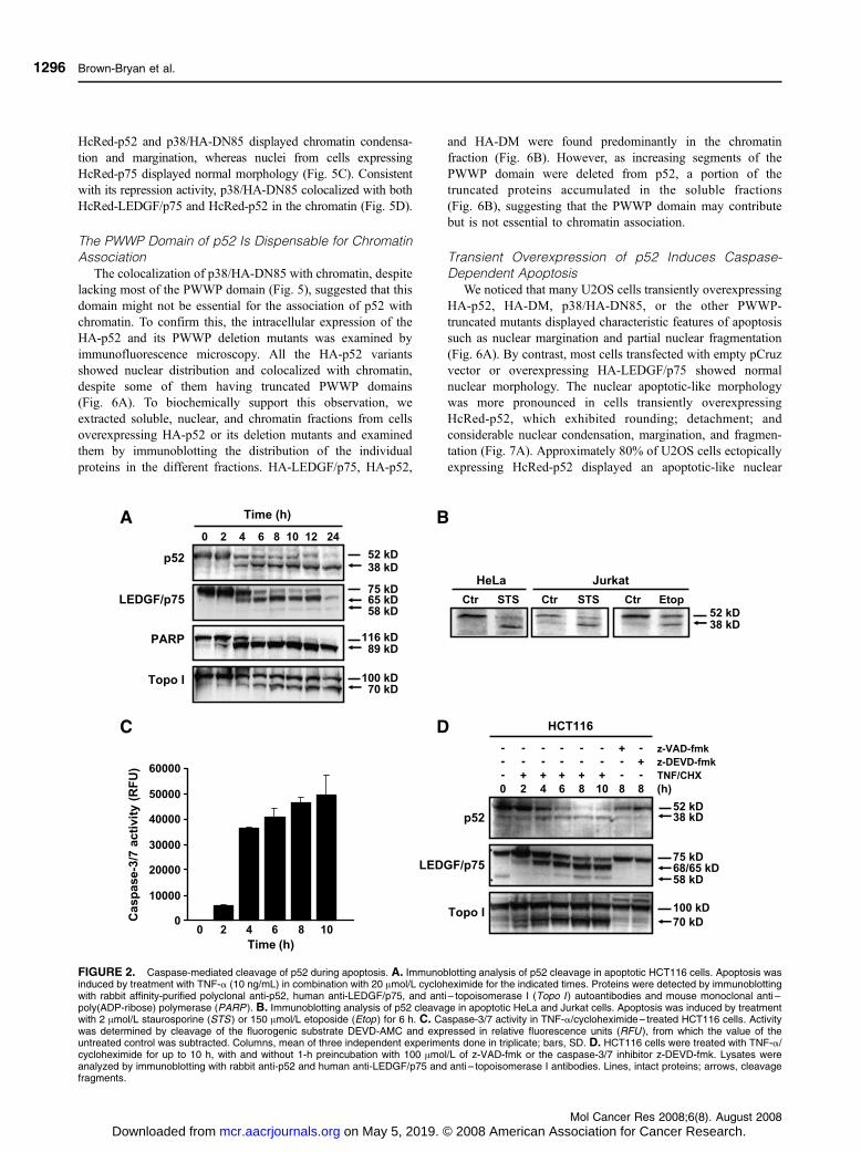
LEDGF/p52 Is Cleaved by Effector Caspases at thePWWP Domain
We previously showed that caspase-3 disrupts the NH2-
terminal PWWP and COOH-terminal domains of LEDGF/p75,
abrogating its ability to protect against stress-induced cell death
(30). Because LEDGF/p75 and p52 share identical PWWP
domains, we hypothesized that the cellular functions of p52 may
also be modulated by caspases during apoptosis. Cleavage of
p52 into a 38-kDa fragment (hereafter called p38) was detected
in various human tumor cell lines undergoing apoptosis (Fig. 2A
and B) and coincided with increased caspase-3/7 activity
(Fig. 2C). The caspase dependency of this cleavage was
confirmed by its inhibition with the pan-caspase inhibitor
benzylozycarbonyl-Val-Ala-Asp-fluoromethylketone (z-VAD-
fmk) and the caspase-3/7 inhibitor benzylozycarbonyl-Asp-
Glu-Val-Asp-fluoromethylketone (z-DEVD-fmk; Fig. 2D).
To investigate the mechanism of apoptotic p52 cleavage,
in vitro translated p52 fused to a hemagglutinin (HA) tag was
incubated with individual recombinant caspases. Caspase-3 and
caspase-7, but not caspase-6, caspase-8, and caspase-9, cleaved
HA-p52 into p48 and p38 fragments, although caspase-7 seemed
to be less efficient in generating p38 (Fig. 3A). LEDGF/p75,
which we previously established as a substrate of caspase-3 and
caspase-7 but not of caspase-6 (28), was used in control
experiments (Fig. 3A). Caspase activity assays indicated that
each recombinant caspase was active (data not shown). Caspase-
3 cleaved HA-p52 sequentially into p48 and p38 in a dose- and
time-dependent manner (Fig. 3B). The p48 fragment could be
distinguished from HA-p52 in vitro but was not consistently
resolved from endogenous p52 in apoptotic cells (Fig. 2). Based
on the known caspase cleavage sites in the PWWP domain of
LEDGF/p75 (30), we identified two candidate cleavage sites in
the p52 PWWP domain, DEVPD30 and WEID85, which would
generate p48 and p38 (Fig. 3C). To map these sites, we
substituted aspartic acids at positions 30 and 85 in HA-p52 with
alanines to generate partial caspase-resistant mutants, HA-
DEVPA and HA-WEIA, and a complete caspase-resistant double
mutant, HA-DM. The mutations were confirmed by DNA
sequencing. In vitro cleavage of HA-DEVPA by caspase-3
generated p38, consistent with cleavage at the available WEID85
site. Cleavage of HA-WEIA generated p48, consistent with
cleavage at the available DEVPD30 site (Fig. 3D). Incubation of
Brown-Bryan et al.
Mol Cancer Res 2008;6(8). August 2008
1294
on May 5, 2019. © 2008 American Association for Cancer Research. mcr.aacrjournals.org Downloaded from

HA-DM with caspase-3 failed to generate p48 and p38,
consistent with blockade of both cleavage sites.
For further exploration of the cleavage sites and subsequent
experiments, we generated HA-tagged truncated variants of p52
corresponding to p48 and p38 by deleting amino acids 1 to 30
(HA-DN30) and 1 to 85 (HA-DN85), respectively. The
deletions were confirmed by DNA sequencing. In vitro
translated HA-p52 migrated slightly slower than endogenous
p52 due to the presence of the tag (Fig. 3E, compare lanes 1
and 3); however, its cleavage by caspase-3 generated the p48
and p38 fragments (Fig. 3E, compare lanes 2 and 4). In vitro
translated HA-DN30 and HA-DN85 also displayed slightly
slower electrophoretic migration than their corresponding
cleavage fragments p48 and p38 (Fig. 3E, compare lane 4
with lanes 5 and 7). However, incubation of HA-DN30 with
caspase-3 generated fragments that corresponded to p48 and
p38 (Fig. 3E, compare lanes 4 and 6), suggesting that this
protease cleaved the HA tag to produce p48 and then removed
most of the PWWP domain to produce p38. Likewise,
incubation of HA-DN85 with caspase-3 generated a slightly
faster migrating fragment corresponding to p38 (Fig. 3E,
compare lanes 4 and 8), again consistent with cleavage of the
HA tag. A parallel experiment in which the blots were
incubated with an anti-HA antibody revealed loss of HA tag
immunoreactivity in the HA-tagged proteins incubated with
caspase-3 (Fig. 3F).
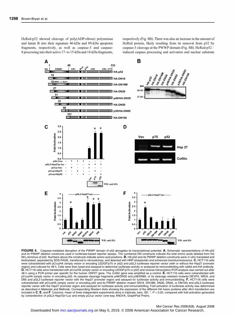
Disruption of the PWWP Domain Abrogates p52 Trans-activation Potential
Based on previous observations that the NH2 terminus of
LEDGF/p75 (amino acids 1-187) transactivates the Hsp27
promoter (Hsp27pr) by binding to stress-response elements
(STRE) present in this promoter (46), we predicted that p52
would also transactivate this promoter in transient transfection
assays with a luciferase (Luc) reporter construct. This system
was used to examine the effect of caspase-mediated disruption
of the PWWP domain on p52 transactivation potential. A
schematic representation of the p52 constructs used in the
reporter assays is depicted in Fig. 4A. The electrophoretic
migration of the in vitro translated constructs is shown in
Fig. 4B. Colorectal carcinoma HCT116 cells were transiently
cotransfected with the pGL3 basic plasmid, with or without
a proximal Hsp27pr, and effector pCruz plasmids encoding
HA-LEDGF/p75 or HA-p52. Both fusion proteins showed
significant induction of Luc activity (P < 0.05) as well as
comparable expression levels (Fig. 4C). Similar results were
obtained in U2OS osteosarcoma cells (data not shown). The
induction of endogenous Hsp27 mRNA by ectopically
expressed HA-LEDGFp75 and HA-p52 was confirmed by
reverse transcription-PCR (Fig. 4D).
Next, we analyzed the transactivation potential of the
cleavage fragments (p48/HA-DN30 and p38/HA-DN85) and
the caspase-resistant mutants (HA-DEVPA, HA-WEIA, and
HA-DM). P48/HA-DN30 and p38/HA-DN85 did not increase
Hsp27pr-Luc activity above the basal level observed in empty
pCruz vector– transfected cells, whereas the caspase-resistant
mutants (all of which had an intact PWWP domain) were as
effective as HA-p52 in transactivating the promoter (Fig. 4E).
Other PWWP deletion constructs of p52 (i.e., HA-DN18, HA-
DN18M, HA-DN26, HA-DN50, and HA-DN100) also
showed decreased transactivation potential (Fig. 4F). HA-
DN18M, which contains a tryptophan21 to alanine21 mutation
in the PHWP motif (amino acids 19-22), the most conserved
sequence within the PWWP domain (30), showed slightly less
activity than HA-DN18, but the mean difference was not
significant.
p38 Interferes with the Transactivation Potential ofLEDGF/p75
The p38/HA-DN85 fragment seemed to be relatively stable
in apoptotic cells and lacked the ability to transactivate
Hsp27pr-Luc (Figs. 2-4). To determine whether this fragment
might possess transcription repression properties, we examined
its influence on the transactivation potential of LEDGF/p75 and
p52. The p38/HA-DN85 construct was cotransfected in
HCT116 cells with either HA-LEDGF/p75 or HA-p52 in the
presence of the luciferase reporter plasmids. P38/HA-DN85
significantly repressed the transactivation potential of HA-
LEDGF/p75 (P < 0.05) but not that of HA-p52 (Fig. 5A).
Although the p38/HA-DN85 construct was transfected at
submaximal doses, it seemed to be highly stable because its
protein expression was consistently higher than those of
LEDGF/p75 and p52 (Fig. 5B).
To determine if p38/HA-DN85 colocalized with both
LEDGF/p75 and p52 in the chromatin, we examined their
nuclear localization in U2OS cells. Nuclei from cells expressing
FIGURE 1. Immunoblotting analysis of endogenous protein expression of LEDGF/p75 and p52 in a panel of human normal and cancer cell lines. Cell linesused were HCT116 (colon carcinoma), U2OS (osteosarcoma), HepG2 (hepatocarcinoma), 293T (embryonic kidney), PrEC (normal prostate epithelium),PrSC (normal prostate stroma), DU145 (prostate carcinoma), PC3 (prostate adenocarcinoma), LnCAP (prostate carcinoma), BRF-41T (prostateadenocarcinoma), 22RV1 (prostate carcinoma), RWPE-2 (transformed normal prostate epithelial cell line, tumorigenic), PWR-1E (transformed normalepithelial cell line, nontumorigenic), RWPE-1 (transformed normal epithelial cell line, nontumorigenic), and BRF-55T (benign prostatic hyperplasia). Proteinsin whole-cell lysates were separated by SDS-PAGE, transferred to nitrocellulose, and detected by chemiluminescence with a monoclonal antibody thatrecognizes both LEDGF/p75 and p52. Monoclonal antibody to h-actin was used as a loading control.
Antagonistic Roles of LEDGF Variants
Mol Cancer Res 2008;6(8). August 2008
1295
on May 5, 2019. © 2008 American Association for Cancer Research. mcr.aacrjournals.org Downloaded from
HcRed-p52 and p38/HA-DN85 displayed chromatin condensa-
tion and margination, whereas nuclei from cells expressing
HcRed-p75 displayed normal morphology (Fig. 5C). Consistent
with its repression activity, p38/HA-DN85 colocalized with both
HcRed-LEDGF/p75 and HcRed-p52 in the chromatin (Fig. 5D).
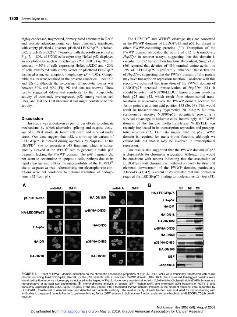
The PWWP Domain of p52 Is Dispensable for ChromatinAssociation
The colocalization of p38/HA-DN85 with chromatin, despite
lacking most of the PWWP domain (Fig. 5), suggested that this
domain might not be essential for the association of p52 with
chromatin. To confirm this, the intracellular expression of the
HA-p52 and its PWWP deletion mutants was examined by
immunofluorescence microscopy. All the HA-p52 variants
showed nuclear distribution and colocalized with chromatin,
despite some of them having truncated PWWP domains
(Fig. 6A). To biochemically support this observation, we
extracted soluble, nuclear, and chromatin fractions from cells
overexpressing HA-p52 or its deletion mutants and examined
them by immunoblotting the distribution of the individual
proteins in the different fractions. HA-LEDGF/p75, HA-p52,
and HA-DM were found predominantly in the chromatin
fraction (Fig. 6B). However, as increasing segments of the
PWWP domain were deleted from p52, a portion of the
truncated proteins accumulated in the soluble fractions
(Fig. 6B), suggesting that the PWWP domain may contribute
but is not essential to chromatin association.
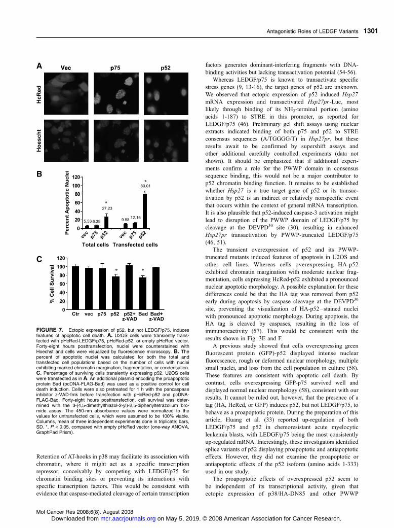
Transient Overexpression of p52 Induces Caspase-Dependent Apoptosis
We noticed that many U2OS cells transiently overexpressing
HA-p52, HA-DM, p38/HA-DN85, or the other PWWP-
truncated mutants displayed characteristic features of apoptosis
such as nuclear margination and partial nuclear fragmentation
(Fig. 6A). By contrast, most cells transfected with empty pCruz
vector or overexpressing HA-LEDGF/p75 showed normal
nuclear morphology. The nuclear apoptotic-like morphology
was more pronounced in cells transiently overexpressing
HcRed-p52, which exhibited rounding; detachment; and
considerable nuclear condensation, margination, and fragmen-
tation (Fig. 7A). Approximately 80% of U2OS cells ectopically
expressing HcRed-p52 displayed an apoptotic-like nuclear
FIGURE 2. Caspase-mediated cleavage of p52 during apoptosis. A. Immunoblotting analysis of p52 cleavage in apoptotic HCT116 cells. Apoptosis wasinduced by treatment with TNF-a (10 ng/mL) in combination with 20 Amol/L cycloheximide for the indicated times. Proteins were detected by immunoblottingwith rabbit affinity-purified polyclonal anti-p52, human anti-LEDGF/p75, and anti – topoisomerase I (Topo I ) autoantibodies and mouse monoclonal anti –poly(ADP-ribose) polymerase (PARP ). B. Immunoblotting analysis of p52 cleavage in apoptotic HeLa and Jurkat cells. Apoptosis was induced by treatmentwith 2 Amol/L staurosporine (STS ) or 150 Amol/L etoposide (Etop ) for 6 h. C. Caspase-3/7 activity in TNF-a/cycloheximide – treated HCT116 cells. Activitywas determined by cleavage of the fluorogenic substrate DEVD-AMC and expressed in relative fluorescence units (RFU ), from which the value of theuntreated control was subtracted. Columns, mean of three independent experiments done in triplicate; bars, SD. D. HCT116 cells were treated with TNF-a/cycloheximide for up to 10 h, with and without 1-h preincubation with 100 Amol/L of z-VAD-fmk or the caspase-3/7 inhibitor z-DEVD-fmk. Lysates wereanalyzed by immunoblotting with rabbit anti-p52 and human anti-LEDGF/p75 and anti – topoisomerase I antibodies. Lines, intact proteins; arrows, cleavagefragments.
Brown-Bryan et al.
Mol Cancer Res 2008;6(8). August 2008
1296
on May 5, 2019. © 2008 American Association for Cancer Research. mcr.aacrjournals.org Downloaded from

morphology, compared with 12% of cells expressing HcRed-
LEDGF/p75 or 10% of cells transfected with empty pHcRed C1
vector (P < 0.001; Fig. 7B). In the total cell population, f27%
of cells transfected with pHcRed-p52 exhibited a nuclear
apoptotic morphology, compared with 6% of total cells
transfected with pHcRed-p75. Transfection efficiency, mea-
sured by the number of cells expressing the HcRed fusion
proteins, was f30% using the Fugene 6 reagent, which was the
method of choice for these experiments due to its minimal
cytotoxicity (50). These results were reproduced in HCT116
and HeLa cells (data not shown).
Cell survival, measured by reduction of 3-(4,5-dimethylth-
iazol-2-yl)-2,5-diphenyltetrazolium bromide, was significantly
reduced in the total U2OS cell population transfected with
pHcRed-p52, compared with cells transfected with empty
pHcRed-C1 vector or pHcRed-LEDGF/p75 (P < 0.05;
Fig. 7C). This reduction was reversed by z-VAD-fmk (Fig. 7C),
suggesting that pHcRed-p52–induced apoptosis was caspase
dependent. Comparable results were obtained in control cells
overexpressing the proapoptotic protein Bad (Fig. 7C). To further
confirm that HcRed-p52–induced cell death was caspase
dependent, we measured caspase activation in U2OS cells
transiently transfected with HcRed-p52. Significant activation
of caspase-3, caspase-8, and caspase-9 and abolition of caspase
activity by z-VAD-fmk were observed (Fig. 8A). Furthermore,
immunoblotting of U2OS whole-cell lysates expressing
FIGURE 3. Caspase-3 and caspase-7 cleave p52 at two sites in vitro to generate p48 and p38 fragments. A. In vitro translated biotinylated HA-p52 wasincubated with 20 ng/mL of purified recombinant caspase-3, caspase-6, caspase-7, caspase-8, and caspase-9 for 2 h. HA-LEDGF/p75 was included as apositive control. Some experiments were done in the presence or absence of 10 Amol/L z-VAD-fmk or z-DEVD-fmk. Translated biotinylated proteins wereseparated by SDS-PAGE, transferred to nitrocellulose, and detected with HRP-streptavidin and enhanced chemiluminescence. B. In vitro translatedbiotinylated HA-p52 was treated with increasing concentrations of caspase-3 or with 20 ng/mL caspase-3 for the indicated times and analyzed as describedabove. C. Schematic representation of LEDGF/p75 and p52 showing their domain structure, the location of the putative caspase cleavage sites (DEVD30 andWEID85), and the p52 cleavage fragments (p48/DN30 and p38/DN85). CR, charged region; NLS, nuclear localization region, ATH, AT-hooks; IBD, integrasebinding domain; CTT, COOH-terminal tail. D. In vitro translated HA-p52 and its mutants HA-DEVPA, HA-WEIA, and HA-DM were exposed to recombinantcaspase-3 for 2 h and analyzed as described above. E. Comparison of the migration in SDS-PAGE of endogenous p52 and its cleavage products derivedfrom control and apoptotic HCT116 cells (lanes 1 and 2 ) with the migration of intact and caspase-3 cleaved HA-p52 (lanes 3 and 4), HA-DN30 (lanes 5 and6), and HA-DN85 (lanes 7 and 8). Lanes 1 and 2, endogenous proteins detected by immunoblotting with anti-p52 antibody; lanes 3 to 8, translatedbiotinylated proteins detected in blots with HRP-streptavidin. Lanes 1 to 6 are from the same blot, whereas lanes 7 and 8 are from a blot done in parallel underidentical conditions. F. Immunoblot, using an anti-HA antibody, of translated HA-tagged proteins incubated with and without caspase-3.
Antagonistic Roles of LEDGF Variants
Mol Cancer Res 2008;6(8). August 2008
1297
on May 5, 2019. © 2008 American Association for Cancer Research. mcr.aacrjournals.org Downloaded from
HcRed-p52 showed cleavage of poly(ADP-ribose) polymerase
and lamin B into their signature 46-kDa and 89-kDa apoptotic
fragments, respectively, as well as caspase-3 and caspase-
8 processing into their active 17- to 15-kDa and 18-kDa fragments,
respectively (Fig. 8B). There was also an increase in the amount of
HcRed protein, likely resulting from its removal from p52 by
caspase-3 cleavage at the PWWP domain (Fig. 8B). HcRed-p52–
induced caspase processing and activation and nuclear substrate
FIGURE 4. Caspase-mediated disruption of the PWWP domain of p52 abrogates its transcriptional potential. A. Schematic representations of HA-p52and its PWWP deletion constructs used in luciferase-based reporter assays. The numbered DN constructs indicate the total amino acids deleted from theNH2 terminus of p52. Numbers above the constructs indicate amino acid positions. B. HA-p52 and its PWWP deletion constructs were in vitro translated andbiotinylated, separated by SDS-PAGE, transferred to nitrocellulose, and detected with HRP-streptavidin and enhanced chemiluminescence. C. HCT116 cellswere cotransfected with pCruzHA (empty vector or encoding LEDGF/p75 or p52) and pGL3 luciferase reporter vector (with or without the Hsp27 promoterregion) and cultured for 48 h. Cells were then lysed and assayed to determine luciferase activity or analyzed by immunoblotting with rabbit anti-HA antibody.D. HCT116 cells were transfected with pCruzHA (empty vector or encoding LEDGF/p75 or p52) and reverse transcription-PCR analysis was carried out after48 h using a PCR primer pair specific for the human HSP27 gene. The Cofilin gene was amplified as a control. E. HCT116 cells were cotransfected withpCruzHA (empty vector or encoding p52, its caspase cleavage fragments p48/DN30 and p38/DN85, or its cleavage resistant mutants DEVPA, WEIA, andDM) and pGL3 luciferase reporter vector with the Hsp27 promoter region and assayed for luciferase activity and immunoblotting. F. HCT116 cells werecotransfected with pCruzHA (empty vector or encoding p52 and its PWWP deletion mutant DN18, DN18M, DN26, DN50, or DN100) and pGL3 luciferasereporter vector with the Hsp27 promoter region and assayed for luciferase activity and immunoblotting. Fold activation of luciferase activity was determinedas described in Materials and Methods. Corresponding Western blots showing the expression of the different HA fusion proteins after 48-h transfection areincluded in C, E, and F. Columns, mean of three independent experiments done in triplicate; bars, SD. *, P < 0.05, compared with fold activation generatedby cotransfection of pGL3-Hsp27pr -Luc and empty pCruz vector (one-way ANOVA, GraphPad Prism).
Brown-Bryan et al.
Mol Cancer Res 2008;6(8). August 2008
1298
on May 5, 2019. © 2008 American Association for Cancer Research. mcr.aacrjournals.org Downloaded from

cleavage were not as dramatic as those triggered by over-
expression of p53 or treatment with tumor necrosis factor (TNF)
or staurosporine (Fig. 8A and B). This might be due to the
relatively low transfection efficiency (<30%) attained when using
pHcRed plasmids. Lamin B and poly(ADP-ribose) polymerase
cleavages were not observed in cells transfected with empty
pHcRed vector or pHcRed-LEDGF/p75 (Fig. 8C).
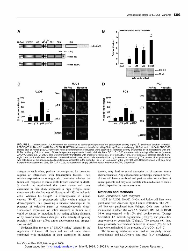
The COOH-Terminal Tail Contributes to p52 ApoptoticActivity but not to Its Transactivation Potential
The observation that transient overexpression of p52 and its
PWWP-truncated variants induced a nuclear apoptotic morphol-
ogy suggested that the PWWP domain is dispensable for
proapoptotic activity. However, because all the p52 constructs
retained the intron-derived COOH-terminal tail, we investigated
whether this sequence might contribute to proapoptotic activity
and transactivation potential. For these experiments, we
generated an HcRed-p52DC construct lacking the COOH-
terminal tail (Fig. 9A). We observed that HcRed-p52DC
transactivated Hsp27pr-Luc at levels comparable with those of
HcRed-p52 and HcRed-LEDGF/p75 (P < 0.05; Fig. 9B),
suggesting that the COOH-terminal tail is not required for
transactivation potential. To determine if the COOH-terminal tail
is required for proapoptotic activity, we counted nuclei with
FIGURE 5. The p38 cleav-age fragment interferes withthe transactivation potential ofLEDGF/p75. A. Either 1 Ag(1�) or 2 Ag (2�) of pCruzHA-LEDGF/p75 or pCruzHA-p52DNA were cotransfected with0.2 Ag (1�) or 0.4 Ag (2�) ofpCruzHA-DN85 or empty vec-tor (Vec ), together with thepGL3-Hsp27pr -Luc reporterplasmid. HCT116 cells werelysed after 48 h and assayedfor fold induction of luciferaseactivity. Columns, mean ofthree independent experi-ments done in quadruplicate;bars, SD. *, P < 0.05, one-wayANOVA with Bonferroni’s mul-tiple comparison test (Graph-Pad Prism).B.Correspondingimmunoblot showing recombi-nant protein expression,detected with anti-HA anti-body, 48 h after transfectionin HCT116 cells. C. U2OScells were transiently trans-fected with pHcRed-LEDGF/p 7 5 , p H c R e d - p 5 2 , o rpCruzHA-p38/DN85 and thengrown in coverslips. After48 h, recombinant protein ex-pression in transfected cellswas visualized by fluores-cence microscopy. After fixa-tion and permeabilization,HcRed-LEDGF/p75 andHcRed-p52 were visualizeddirectly, whereas HA-p38/DN85 was detected with pri-mary rabbit anti-HA antibodywith secondary Alexa 488 –labeled goat anti-rabbit anti-body. Nuclei were counter-stained with 4¶,6-diamidino-2-phenylindole (DAPI ). D.Cells were cotransfected withpCruzHA-p38/DN85 (0.5 Ag)and pHcRed-LEDGF/p75(1 Ag) or pHcRed-p52 (1 Ag),stained with anti-HA antibod-ies, and visualized by fluores-cence microscopy.
Antagonistic Roles of LEDGF Variants
Mol Cancer Res 2008;6(8). August 2008
1299
on May 5, 2019. © 2008 American Association for Cancer Research. mcr.aacrjournals.org Downloaded from
highly condensed, fragmented, or marginated chromatin in U2OS
and prostate adenocarcinoma cell lines transiently transfected
with empty pHcRed-C1 vector, pHcRed-LEDGF/p75, pHcRed-
p52, or pHcRed-p52DC. Consistent with the results presented in
Fig. 7, f80% of U2OS cells expressing HcRed-p52 displayed
an apoptotic-like nuclear morphology (P < 0.001; Fig. 9C). In
contrast, f30% of cells expressing HcRed-p52DC and <20%
of cells transfected with empty vector or pHcRed-LEDGF/p75
displayed a nuclear apoptotic morphology (P < 0.05). Compa-
rable results were obtained in the prostate cancer cell lines PC3
and 22rv1, although the percentage of apoptotic nuclei was
between 50% and 60% (Fig. 9D and data not shown). These
results suggested differential sensitivity to the proapoptotic
activity of transiently overexpressed p52 among various cell
lines, and that the COOH-terminal tail might contribute to this
activity.
DiscussionThis study was undertaken as part of our efforts to delineate
mechanisms by which alternative splicing and caspase cleav-
age of LEDGF modulate tumor cell death and survival under
stress. Our data suggest that p52, a short splice variant of
LEDGF/p75, is cleaved during apoptosis by caspase-3 at the
DEVPD30 site to generate a p48 fragment, which is subse-
quently cleaved at the WEID85 site to generate a stable p38
fragment lacking the PWWP domain. The p48 fragment did
not seem to accumulate in apoptotic cells, perhaps due to its
rapid cleavage into p38 or the inaccessibility of the DEVPD30
site to caspases in vivo . Alternatively, our electrophoretic con-
ditions were not conducive to optimal resolution of endoge-
nous p52 from p48.
The DEVPD30 and WEID85 cleavage sites are conserved
in the PWWP domains of LEDGF/p75 and p52 but absent in
other PWWP-containing proteins (30). Disruption of the
PWWP domain abrogated the ability of p52 to transactivate
Hsp27pr in reporter assays, suggesting that this domain is
essential for p52 transcription function. By contrast, Singh et al.
(46) reported that deletion of NH2-terminal amino acids 1 to
198 of LEDGF/p75 significantly enhanced transactivation
of Hsp27pr, suggesting that the PWWP domain of this protein
may have transcription repression function. Consistent with this
report, we observed that truncation of the PWWP domain of
LEDGF/p75 increased transactivation of Hsp27pr (51). It
should be noted that NUP98-LEDGF fusion proteins involving
both p75 and p52, which result from chromosomal trans-
locations in leukemias, lack the PWWP domain because the
fusion point is at amino acid position 153 (26, 33). This would
result in transcriptionally hyperactive NUP98-p75 but tran-
scriptionally inactive NUP98-p52, potentially providing a
survival advantage to leukemic cells. Interestingly, the PWWP
domain of the histone methyltransferase WHISTLE was
recently implicated in its transcription repression and proapop-
totic activities (52). Our data suggest that the p52 PWWP
domain is required for transcription function, although we
cannot rule out that it may be involved in transcriptional
repression.
Our results also suggested that the PWWP domain of p52
is dispensable for chromatin association. Although this would
be consistent with reports indicating that the association of
LEDGF/p75 with chromatin is mediated primarily by structural
elements downstream of the PWWP domain, particularly
AT-hooks (41, 42), a recent study revealed that this domain is
required for LEDGF/p75 binding to nucleosomes in vitro (53).
FIGURE 6. Effect of PWWP domain disruption on the chromatin association properties of p52. A. U2OS cells were transiently transfected with pCruzplasmid encoding HA-LEDGF/p75, HA-p52, or the p52 variants with a truncated PWWP domain. After 48 h, the expressed HA-tagged proteins werevisualized by fluorescence microscopy as indicated in the legend of Fig. 5. Nuclei were counterstained with 4¶,6-diamidino-2-phenylindole (DAPI). Images arerepresentative of at least two experiments. B. Immunoblotting analysis of soluble (SF ), nuclear (NF ), and chromatin (CF) fractions of HCT116 cellstransiently expressing HA-LEDGF/p75, HA-p52, or the p52 variant with a truncated PWWP domain. Proteins in the different fractions were separated bySDS-PAGE, transferred to nitrocellulose, and detected with anti-HA antibody. The relative purity of each fraction was evaluated by immunoblotting withantibodies to caspase-8 (soluble fraction), upstream binding factor (UBF ; present in both nuclear fraction and chromatin fraction), and LEDGF/p75 (chromatinfraction).
Brown-Bryan et al.
Mol Cancer Res 2008;6(8). August 2008
1300
on May 5, 2019. © 2008 American Association for Cancer Research. mcr.aacrjournals.org Downloaded from
Retention of AT-hooks in p38 may facilitate its association with
chromatin, where it might act as a specific transcription
repressor, conceivably by competing with LEDGF/p75 for
chromatin binding sites or preventing its interactions with
specific transcription factors. This would be consistent with
evidence that caspase-mediated cleavage of certain transcription
factors generates dominant-interfering fragments with DNA-
binding activities but lacking transactivation potential (54-56).
Whereas LEDGF/p75 is known to transactivate specific
stress genes (9, 13-16), the target genes of p52 are unknown.
We observed that ectopic expression of p52 induced Hsp27
mRNA expression and transactivated Hsp27pr-Luc, most
likely through binding of its NH2-terminal portion (amino
acids 1-187) to STRE in this promoter, as reported for
LEDGF/p75 (46). Preliminary gel shift assays using nuclear
extracts indicated binding of both p75 and p52 to STRE
consensus sequences (A/TGGGG/T) in Hsp27pr, but these
results await to be confirmed by supershift assays and
other additional carefully controlled experiments (data not
shown). It should be emphasized that if additional experi-
ments confirm a role for the PWWP domain in consensus
sequence binding, this would not be a major contributor to
p52 chromatin binding function. It remains to be established
whether Hsp27 is a true target gene of p52 or its transac-
tivation by p52 is an indirect or relatively nonspecific event
that occurs within the context of general mRNA transcription.
It is also plausible that p52-induced caspase-3 activation might
lead to disruption of the PWWP domain of LEDGF/p75 by
cleavage at the DEVPD30 site (30), resulting in enhanced
Hsp27pr transactivation by PWWP-truncated LEDGF/p75
(46, 51).
The transient overexpression of p52 and its PWWP-
truncated mutants induced features of apoptosis in U2OS and
other cell lines. Whereas cells overexpressing HA-p52
exhibited chromatin margination with moderate nuclear frag-
mentation, cells expressing HcRed-p52 exhibited a pronounced
nuclear apoptotic morphology. A possible explanation for these
differences could be that the HA tag was removed from p52
early during apoptosis by caspase cleavage at the DEVPD30
site, preventing the visualization of HA-p52–stained nuclei
with pronounced apoptotic morphology. During apoptosis, the
HA tag is cleaved by caspases, resulting in the loss of
immunoreactivity (57). This would be consistent with the
results shown in Fig. 3E and F.
A previous study showed that cells overexpressing green
fluorescent protein (GFP)-p52 displayed intense nuclear
fluorescence, rough or deformed nuclear morphology, multiple
small nuclei, and loss from the cell population in culture (58).
These features are consistent with apoptotic cell death. By
contrast, cells overexpressing GFP-p75 survived well and
displayed normal nuclear morphology (58), consistent with our
results. It cannot be ruled out, however, that the presence of a
tag (HA, HcRed, or GFP) induces p52, but not LEDGF/p75, to
behave as a proapoptotic protein. During the preparation of this
article, Huang et al. (33) reported up-regulation of both
LEDGF/p75 and p52 in chemoresistant acute myelocytic
leukemia blasts, with LEDGF/p75 being the most consistently
up-regulated mRNA. Interestingly, these investigators identified
splice variants of p52 displaying proapoptotic and antiapoptotic
effects. However, they did not examine the proapoptotic or
antiapoptotic effects of the p52 isoform (amino acids 1-333)
used in our study.
The proapoptotic effects of overexpressed p52 seem to
be independent of its transcriptional activity, given that
ectopic expression of p38/HA-DN85 and other PWWP
FIGURE 7. Ectopic expression of p52, but not LEDGF/p75, inducesfeatures of apoptotic cell death. A. U2OS cells were transiently trans-fected with pHcRed-LEDGF/p75, pHcRed-p52, or empty pHcRed vector.Forty-eight hours posttransfection, nuclei were counterstained withHoechst and cells were visualized by fluorescence microscopy. B. Thepercent of apoptotic nuclei was calculated for both the total andtransfected cell populations based on the number of cells with nucleiexhibiting marked chromatin margination, fragmentation, or condensation.C. Percentage of surviving cells transiently expressing p52. U2OS cellswere transfected as in A. An additional plasmid encoding the proapoptoticprotein Bad (pcDNA-FLAG-Bad) was used as a positive control for celldeath induction. Cells were also pretreated for 1 h with the pancaspaseinhibitor z-VAD-fmk before transfection with pHcRed-p52 and pcDNA-FLAG-Bad. Forty-eight hours posttransfection, cell survival was deter-mined with the 3-(4,5-dimethylthiazol-2-yl)-2,5-diphenyltetrazolium bro-mide assay. The 450-nm absorbance values were normalized to thevalues for untransfected cells, which were assumed to be 100% viable.Columns, mean of three independent experiments done in triplicate; bars,SD. *, P < 0.05, compared with empty pHcRed vector (one-way ANOVA,GraphPad Prism).
Antagonistic Roles of LEDGF Variants
Mol Cancer Res 2008;6(8). August 2008
1301
on May 5, 2019. © 2008 American Association for Cancer Research. mcr.aacrjournals.org Downloaded from

deletion mutants also induced apoptotic nuclear morphology.
It cannot be ruled out, however, that under stress condi-
tions p52 might transactivate proapoptotic genes or repress
survival genes, leading to caspase activation and conse-
quently inducing its own cleavage, as well as that of
LEDGF/p75, into proapoptotic dominant-interfering frag-
ments that amplify the cell death process. This would be
consistent with our previous observation that stable over-
expression of a p65 caspase cleavage fragment of LEDGF/p75
with disrupted PWWP domain and COOH terminus in HepG2
tumor cells did not affect viability under normal growth
conditions but sensitized cells to serum starvation– induced
death, which is known to be mediated by oxidative stress
(30, 59).
The retention of intronic sequences during alternative
splicing is also known to contribute to functional diversity
among spliced variants (60). Our data suggest that the
intron-derived COOH-terminal tail sequence (amino acids
326-333) in p52, retained in all the PWWP deletion
mutants, may contribute to its proapoptotic properties
but is not essential because its deletion failed to completely
reduce the number of transfected cells with apoptotic nuclei.
The mechanism by which the COOH-terminal tail may
contribute to the proapoptotic function of p52 remains to
be elucidated.
It is tempting to speculate that LEDGF splice variants
may play multiple roles depending on the cell type and
environment. Some of these variants may function as general
transcription coactivators under normal growth condi-
tions, in which their prosurvival or proapoptotic func-
tions are kept in check by strict compartmentalization or by
the level and type of posttranslational modifications.
However, under increased oxidative stress conditions, such
as those found in many tumors, these variants may
FIGURE 8. Ectopic ex-pression of p52, but notLEDGF/p75, induces caspaseactivation and cleavage ofprototype caspase-3 sub-strates. A. U2OS cells weretransiently transfected for 48 hwith empty pHcRed vector(vec ), pHcRed-LEDGF/p75,pHcRed-p52, or the positivecontrol plasmids pcDNA-FLAG-Bad and pcDNA-3p-p53. Treatment with TNF wasalso used as a control forcaspase activation. Cells werealso pretreated with z-VAD-fmk before transfection. Forty-eight hours after transfection,caspase activity was assayedand fold activation determinedas described in Materials andMethods. Columns, mean ofat least three independentexperiments done in triplicate;bars, SD. *, P < 0.05, com-pared with empty pHcRedvector (one-way ANOVA,GraphPad Prism). B. U2OScells were transiently trans-fected for up to 60 h withpHcRed-p52. Whole-cel llysates were analyzed bySDS-PAGE and immunoblot-ting with antibodies to HcRed,poly(ADP-ribose) polymerase,lamin B, caspase-3, caspase-8, and h-actin. C. U2OS cellswere transiently transfectedfor 48 h wi th pHcRed-LEDGF/p75, pHcRed-p52, orempty pHcRed vector. Whole-cell lysates were analyzed bySDS-PAGE and immunoblot-ting with antibodies to poly(ADP-ribose) polymerase,lamin B, and h-actin. Lines,intact proteins; arrows, cleav-age fragments.
Brown-Bryan et al.
Mol Cancer Res 2008;6(8). August 2008
1302
on May 5, 2019. © 2008 American Association for Cancer Research. mcr.aacrjournals.org Downloaded from
antagonize each other, perhaps by competing for promoter
regions or interactions with transcription factors. Their
relative expression ratio might also determine whether the
tumor cell response to stress shifts toward survival or death.
It should be emphasized that most cancer cell lines
examined in this study expressed a high p75/p52 ratio,
consistent with the findings of Huang et al. (33) in leukemic
cells. Whereas LEDGF/p75 is overexpressed in human
cancers (30-33), its proapoptotic splice variants might be
down-regulated, thus providing a survival advantage in the
presence of oxidative stress or chemotherapeutic drugs.
Unbalanced expression of splice isoforms in tumor cells
could be caused by mutations in cis -acting splicing elements
or by environment-driven changes in the activity of splicing
proteins, which may affect tumor development and progres-
sion (60-62).
Understanding the role of LEDGF splice variants in the
regulation of tumor cell death and survival under stress,
combined with modulation of their expression in human
tumors, may lead to novel strategies to circumvent tumor
chemoresistance. Any enhancement of therapy-induced surviv-
al time will have a profound and positive effect on the lives of
cancer patients and may also translate into a reduction of racial/
ethnic disparities in cancer mortality.
Materials and MethodsCells, Antibodies, and Reagents
HCT116, U2OS, HepG2, HeLa, and Jurkat cell lines were
purchased from American Type Culture Collection. The 293T
cell line was purchased from Orbigen. Cells were routinely
maintained in either McCoy’s 5A medium, DMEM, or RPMI
1640, supplemented with 10% fetal bovine serum (Omega
Scientific), 1.5 mmol/L L-glutamine (Cellgro), and penicillin/
streptomycin or gentamicin (Cellgro). The prostate cell lines
were previously described and cultured as indicated (29). All cell
lines were maintained in the presence of 5% CO2 at 37jC.
The following antibodies were used in this study: mouse
monoclonals anti-LEDGF/p75 and p52 (BD Biosciences),
FIGURE 9. Contribution of COOH-terminal tail sequence to transcriptional potential and proapoptotic activity of p52. A. Schematic diagram of HcRed-LEDGF/p75, HcRed-p52, and HcRed-p52DC. B. HCT116 cells were cotransfected with pGL3-Hsp27pr-Luc and empty pHcRed vector, HcRed-LEDGF/p75,HcRed-p52, or HcRed-p52DC. Forty-eight hours posttransfection, cell lysates were assayed for luciferase activity or analyzed by immunoblotting with anti-HcRed antibody. Columns, mean of three independent experiments done in triplicate; bars, SD. *, P < 0.05, compared with empty pHcRed vector (one-wayANOVA, GraphPad). C. U2OS cells were transiently transfected with empty pHcRed vector, pHcRed-LEDGF/p75, pHcRed-p52, or pHcRed-p52DC. Forty-eight hours posttransfection, nuclei were counterstained with Hoechst and cells were visualized by fluorescence microscopy. The percent of apoptotic nucleiwas calculated for the transfected cell populations as indicated in the legend of Fig. 7. D. Same as in C but with PC3 cells. Columns, mean of at least threeindependent experiments; bars, SD. *, P < 0.05, compared with empty pHcRed vector (one-way ANOVA, GraphPad).
Antagonistic Roles of LEDGF Variants
Mol Cancer Res 2008;6(8). August 2008
1303
on May 5, 2019. © 2008 American Association for Cancer Research. mcr.aacrjournals.org Downloaded from

anti –h-actin (Sigma), anti – poly(ADP-ribose) polymerase
(R&D Systems), anti–caspase-3 (PharMingen), and anti–
caspase-8 (Alexis); rabbit polyclonals anti-HA (Santa Cruz
Biotechnology) and anti-HcRed (Clontech); goat anti– lamin B
(Santa Cruz Biotechnology), Alexa Flour 488–conjugated goat
anti-rabbit IgG (Molecular Probes), and rat monoclonal
horseradish peroxidase (HRP)–conjugated anti-HA antibody
(Roche Diagnostics). All other HRP-conjugated secondary
antibodies were from Zymed. Rabbit anti-p52 antibody was
raised against a synthetic 15-mer peptide containing the
COOH-terminal tail (amino acids 318-333; Biosource Interna-
tional). Human autoantibodies to LEDGF/p75, topoisomerase I,
and upstream binding factor were a kind gift from Dr. Eng M.
Tan (Scripps Research Institute, La Jolla, CA). Human TNF-a,
etoposide, cycloheximide, and 4¶,6-diamidino-2-phenylindole
were from Sigma-Aldrich. Hoechst 33342 was from Molecular
Probes. Staurosporine and the caspase substrates Asp-Glu-Val-
Asp-7-amino-4-methyl coumarin (DEVD-AMC), Ile-Glu-
Thr-Asp-AMC (IETD-AMC), and Leu-Glu-His-Asp-AMC
(LEHD-AMC) were from Alexis. z-VAD-fmk was from Biomol
International and z-DEVD-fmk was from Enzyme Systems
Products. Purified caspases were from PharMingen.
Apoptosis AssaysApoptosis was induced in HCT116 cells with 10 ng/mL
TNF-a in combination with 20 Amol/L cycloheximide.
HCT116 cells were also preincubated with either 100 Amol/L
z-VAD-fmk or z-DEVD-fmk for 1 h before exposure to TNF-a.
Apoptosis was induced in HeLa and Jurkat cells with 2 Amol/L
staurosporine and 150 Amol/L etoposide, respectively. In some
experiments, cells were transiently transfected with the appro-
priate expression plasmids using Fugene 6 (Roche Diagnostics)
or Amaxa Nucleofection (Amaxa, Inc.). The medium was
removed 24 h posttransfection to discard cells killed during the
transfection procedure; at 36 to 48 h posttransfection, cells were
visualized under fluorescence microscopy, processed for deter-
mination of caspase activity or viability, or processed for SDS-
PAGE (NuPAGE 4-12%, Invitrogen) and immunoblotting
analysis. For quantification of apoptotic nuclei, cells were
cultured in 35-mm dishes to f60% to 70% confluency
and transfected with plasmids encoding HcRed-fusion proteins.
Thirty six to 48 h posttransfection, cells were coun-
terstained with Hoechst 33342 and visualized directly
using a 60� water immersion objective under an Olympus
BX50 epifluorescence microscope (Scientific Instruments)
equipped with a digital SPOT camera system (Diagnostic
Instruments). Nuclei of cells expressing HcRed-tagged
proteins that exhibited marked chromatin condensation,
margination, or fragmentation were counted as apoptotic.
Approximately 200 nuclei distributed in >10 different fields
were counted in at least four independent double-blind
experiments.
One-step cellular caspase activity assays were done in
transfected cells cultured in 96-well plates in the presence of the
appropriate caspase substrate, as previously described (63). Cell
survival was determined using the standard 3-(4,5-dimethylth-
iazol-2-yl)-2,5-diphenyltetrazolium bromide assay (Sigma-
Aldrich). All immunoblotting studies were done as previously
described (30).
Immunofluorescence MicroscopyCells seeded on coverslips and transfected with pCruzHA
plasmids were fixed for 15 min at room temperature with
3.7% paraformaldehyde and permeabilized in PBS-0.2%
Triton X-100 for 5 min. Coverslips were then incubated with
rabbit anti-HA antibody for 2 h. Following three washes
with PBS, cells were incubated with Alexa 488 goat anti-
rabbit for 1 h, washed with PBS, mounted on glass slide
with Vectashield Mounting Medium containing 4¶,6-diami-
dino-2-phenylindole, and examined under a fluorescence
microscope.
Reverse Transcription-PCRTotal RNA from HCT116 cells was isolated 48 h after
transfection with pCruzHA empty vector, pCruzHA-p75, or
pCruzHA-p52 using Fugene 6 (Roche Diagnostics). cDNA was
synthesized using the Invitrogen SuperScript III first-strand
cDNA synthesis system. Hsp27 forward primer (5¶-GAGAT-
CACCGGCAAGCACGAG-3¶) and Hsp27 reverse primer (5¶-CGGCAGTCTCATCGGATTTTGC-3¶) were used to amplify
Hsp-27 PCR products (255 bp). Cofilin 1 (CFL1) forward
primer (5¶-CCTTCCCAAACTGCTTTTGAT-3¶) and CFL1
reverse primer (5¶-CTGGTCCTGCTTCCATGAGTA-3¶) were
used to amplify cofilin PCR products (287 bp). Briefly, cDNA
was added into a 50-AL reaction mixture containing 12.5 AL
of PCR master mix and 500 nmol/L of primers. Reverse
transcription was carried out at 50jC for 50 min, followed by
incubation with RNase H at 37jC for 20 min. The PCR
parameters were preheating at 94jC for 2 min, followed by
35 cycles of 94jC for 30 s, 57jC for 1 min 30 s, and 72jC for
1 min 30 s. Final elongation was at 72jC for 10 min. PCR
reactions were separated on a 1.5% agarose gel and PCR
products were visualized under UV light after ethidium bromide
staining. Images were obtained using the Alpha Innotech
imaging system.
Plasmid ConstructsA cDNA encoding p52 was amplified by PCR using pET28a-
ledgf/p75 template DNA (30) and the primers 5¶-CGGAATT-
CATCACTCGCGATTTCAAACCTGGAG-3¶ (forward) and 5¶-GCAGATCTTACTGTAGATTACATGTTGTTTGGTGCT-
CAG-3¶ (reverse). The amplified fragment was ligated directly
into the EcoRI-Bgl II sites of the pCruzHA mammalian
expression vector (Santa Cruz Biotech). The LEDGF/p75
cDNA was subcloned from a pcDNA3.1-ledgf/p75 construct
(30) into pCruzHA using EcoRI-NotI digestion. The cDNAs
encoding NH2-terminal truncated p52 mutants were generated
by PCR using a pET28a-ledgf/p75 template DNA; the p52
reve rse pr imer 5 ¶-GCAGATCTTACTGTAGATTA-
CATGTTGTTTGGTGCTCAG-3¶; and the following forward
primers: 5¶DN18, 5¶-ACCCCATTGGCCAGCTCGAGTA-3¶;5 ¶DN18M, 5 ¶-ACCACATGCGCCAGCTCGAGTA-3 ¶;5¶DN26, 5¶-GCCGGGGATATCGAAGTTCCTGATGGAGCTG-
TAAAG-3¶; 5¶DN30, 5¶-AGGAGCTGTAAAGCCACCCA-
CAAAC-3; 5¶DN50, 5¶-AGCTTTTTTAGGACCAAAG-3¶;5¶DN85, 5¶-AAACAATCCAAAAGTGAAATTCTCAAGC-
CAACAG-3¶; and 5¶DN100, 5¶-ACAATCAAATGCATCATCT-
GATG-3¶. PCR products corresponding to truncated fragments of
Brown-Bryan et al.
Mol Cancer Res 2008;6(8). August 2008
1304
on May 5, 2019. © 2008 American Association for Cancer Research. mcr.aacrjournals.org Downloaded from

p52 were subcloned into the EcoRV/HpaI site of pCruzHA. The
pGL3-Hsp27pr-Luc promoter construct was obtained by remov-
ing the Hsp27 promoter region from a pGL2-Luc construct
followed by ligation into the pGL3-Luc reporter vector using
SmaI-BglII digestion. The plasmids pcDNA-FLAG-Bad and
pcDNA-3p-p53 were a kind gift from Dr. Marina Zemskova
(Loma Linda University, Loma Linda, CA).
Site-Directed MutagenesisMultiple rounds of PCR-based mutagenesis were done using
the QuickChange Site-Directed Mutagenesis Kit (Stratagene) to
substitute aspartic acids to alanines in the putative cleavage sites
of p52. The pCruzHA-p52 construct was used as a template and
the following primers were used to generate the HA-DEVPA
and HA-WEIA constructs: 5¶-CCCCATTGGCCAGCTCGAG-
TAGACGAAGTTCCTGCGGGAGCTGTAAAGCCACCCA-
CAAACAAACTACCC-3¶ (5¶DEVPA, forward), 5¶-GGGTAG-
TTTGTTTGTGGGTGGCTTTACAGCTCCCGCAGGA-
ACTTCGTCTACTCGAGCGGCCAATGGGG-3¶ (3¶DEVPA,
reverse), 5¶-GGCAAACCAAATAAAAGAAAAGGTTTTAAT-
GAAGGTTTATGGGAGGCGGCGGGCGGCCCAAAGT-
GAAATTTTCAAGTCAACAGGCAGC-3¶ (5¶WEIA, forward),
and 5 ¶-GCTGCCTGTTGACTTGAAAATTTCACTT-
TTGGGCCGCCCGCCGCCTCCCATAAACCTTCATTA-
AAACCTTTTCTTTTATTTGGTTTGCC-3¶ (3¶WEIA, reverse).
For the generation of the pCruzHA-p52DM construct, the
pCruzHA-p52WEIA template and the p52DEVPA primer
pairs were used. The pHcRed-p52DC construct was gener-
ated by inserting a stop codon at amino acid position 326 within
the p52 nucleotide sequence. For this, pHcRed-p52 was used
as template with the following primers: 5¶-CGCAAGCAAG-
AGGAACAAATGGAAACTGAGTAGCAAGCAACGCG-
CAATCTACAGTAAGATCTAGAGGGCCC-3 (forward) and
5¶-GGGCCCTCTAGATCTTACTGTAGATTGCGCGTTG-
CTTGCTACTCAGTTTCCATTTGTTCCTCTTGCTTGCG-3¶(reverse).
In vitro Transcription/TranslationcDNAs were transcribed and translated in vitro using
the TNT T7 Coupled Reticulocyte Lysate System and
Transcend Non-Radioactive Translation Detection System
(Promega) at 30jC for 90 min as indicated previously (30).
Using these systems, biotinylated lysine was added to the
transcription-translation reaction as a precharged epsilon-
labeled biotinylated lysine-tRNA complex (provided by the
manufacturer). Biotinylated translated proteins were incu-
bated with caspase cleavage buffer [100 mmol/L NaCl,
20 mmol/L PIPES (pH 7.2), 1 mmol/L EDTA, 10% sucrose,
0.1% CHAPS, 100 mmol/L DTT] with or without purified
caspases. Biotinylated translated proteins were separated
by SDS-PAGE, transferred to nitrocellulose, and detected
with HRP-streptavidin and enhanced chemiluminescence
(Perkin-Elmer).
Luciferase-Based Transcription Reporter AssaysHCT116 or U2OS cells were cotransfected with plasmid
constructs encoding the proteins of interest and pGL3 basic
luciferase reporter plasmids, with or without the Hsp27pr.
At 48 h posttransfection, cells were lysed in reporter lysis
buffer and luciferase assays were done using the Luciferase
Assay System (Promega). The assays were done in opaque
luminometer-compatible microplates and relative light units
were obtained in a MicroLumatPlus Lb 96V luminometer
(Berthold Tech). Luciferase values were normalized to the
amount of protein in each sample, and fold induction
was calculated by normalizing to the luciferase activity in
lysates from cells cotransfected with the empty vectors
(pCruzHA, pcDNA3.1+, or pHcRed) and pGL3-Hsp27pr-
Luc or pGL3-Luc.
Chromatin FractionationChromatin fractionation was done essentially as described
previously (64). Briefly, cells transiently transfected with
different plasmid constructs were collected after tryp-
sinization, washed twice with PBS (2 min, 1,700 � g, 4jC),
and resuspended in 200 AL of buffer A [10 mmol/L HEPES (pH
7.9), 10 mmol/L KCl, 1.5 mmol/L MgCl2, 0.34 mol/L sucrose,
10% glycerol, 1 mmol/L DTT, 0.5 Ag/mL of pepstatin A, and
protease inhibitor cocktail]. This cell suspension was treated
with Triton X-100 (0.1% final concentration) and incubated on
ice for 8 min; the nuclei were collected by centrifugation
(5 min, 1,300 � g , 4jC). The supernatant was further
centrifuged (5 min, 20,000 � g , 4jC) and the resulting
supernatant, called soluble fraction, was collected. The nuclear
fraction was washed once in 200-AL buffer A and lysed for
30 min in 100-AL buffer B (3 mmol/L EDTA, 0.2 mmol/L
EGTA, 1 mmol/L DTT, 0.5 Ag/mL of pepstatin A, and protease
inhibitor cocktail). The insoluble chromatin fraction was
collected by centrifugation (5 min, 1,700 � g , 4jC)
and washed once with buffer B. Fractions were mixed
with SDS sample buffer and boiled for 10 min. Proteins were
then separated by SDS-PAGE and analyzed by immunoblotting.
Disclosure of Potential Conflicts of InterestNo potential conflicts of interest were disclosed.
AcknowledgmentsWe thank the following colleagues from Loma Linda University School ofMedicine for valuable discussions and suggestions and for technical advice:Marino De Leon, Penny Duerksen-Hughes, Hansel Fletcher, Mark Johnson,Michael B. Lilly, and Marina Zemskova.
References1. Borek C. Dietary antioxidants and human cancer. Integr Cancer Ther 2004;3:333 – 41.
2. Pathak SK, Sharma RA, Steward WP, Mellon JK, Griffiths TR, Gescher AJ.Oxidative stress and cyclooxygenase activity in prostate carcinogenesis: targetsfor chemopreventive strategies. Eur J Cancer 2005;41:61 – 70.
3. Robertson RP, Harmon JS. Diabetes, glucose toxicity, and oxidative stress: Acase of double jeopardy for the pancreatic islet beta cell. Free Radic Biol Med2006;41:177 – 84.
4. Giles GI. The redox regulation of thiol dependent signaling pathways incancer. Curr Pharm Des 2006;12:4427 – 43.
5. Kinnula VL, Paakko P, Soini Y. Antioxidant enzymes and redoxregulating thiol proteins in malignancies of human lung. FEBS Lett 2004;569:1 – 6.
6. Pennington JD, Wang TJ, Nguyen P, Sun L, Bisht K, Smart D, Gius D. Redox-sensitive signaling factors as novel molecular targets for cancer therapy. DrugResist Updat 2005;8:322 – 30.
Antagonistic Roles of LEDGF Variants
Mol Cancer Res 2008;6(8). August 2008
1305
on May 5, 2019. © 2008 American Association for Cancer Research. mcr.aacrjournals.org Downloaded from

7. Ganapathy V, Daniels T, Casiano CA. LEDGF/p75: a novel nuclearautoantigen at the crossroads of cell survival and apoptosis. Autoimmun Rev2003;2:290 – 7.
8. Shinohara T, Singh DP and Fatma N. LEDGF, a survival factor, activatesstress-related genes. Prog Retin Eye Res 2002;21:341 – 58.
9. Takamura Y, Fatma N, Kubo E, Singh DP. Regulation of heavy subunitchain of ;-glutamylcysteine synthetase by tumor necrosis factor-A in lensepithelial cells: role of LEDGF/p75. Am J Physiol Cell Physiol 2006;290:C554 – 66.
10. Ge H, Si Y, Roeder RG. Isolation of cDNAs encoding novel transcriptioncoactivators p52 and p75 reveals an alternate regulatory mechanism oftranscriptional activation. EMBO J 1998;17:6723 – 9.
11. Ochs RL, Muro Y, Si Y, Ge H, Chan EK, Tan EM. Autoantibodies to DFS70 kd/transcription coactivator p75 in atopic dermatitis and other conditions.J Allergy Clin Immunol 2000;105:1211 – 20.
12. Sutherland HG, Newton K, Brownstein DG, et al. Disruption of Ledgf/Psip1results in perinatal mortality and homeotic skeletal transformations. Mol Cell Biol2006;26:7201 – 10.
13. Fatma N, Singh DP, Shinohara T, Chrylack LT, Jr. Transcriptional regulationof the antioxidant protein 2 gene, a thiol-specific antioxidant, by lens epithelium-derived growth factor to protect cells from oxidative stress. J Biol Chem 2001;276:48899 – 907.
14. Fatma N, Kubo E, Chylack LT, Jr., Shinohara T, Akagi Y, Singh DP. LEDGFregulation of alcohol and aldehyde dehydrogenases in lens epithelial cells:stimulation of retinoic acid production and protection from ethanol toxicity. Am JPhysiol Cell Physiol 2004;287:C508 – 16.
15. Sharma P, Singh DP, Fatma N, Chylack LT, Jr., Shinohara T. Activation ofLEDGF gene by thermal-and oxidative-stresses. Biochem Biophys Res Commun2000;276:1320 – 4.
16. Singh DP, Fatma N, Kimura A, Chylack LT, Jr., Shinohara T. LEDGF bindsto heat shock and stress-related element to activate the expression of stress-relatedgenes. Biochem Biophys Res Commun 2001;283:943 – 55.
17. Dellavance A, Viana VS, Leon EP, Bonfa ES, Andrade LE, Leser PG.The clinical spectrum of antinuclear antibodies associated with the nucleardense fine speckled immunofluorescence pattern. J Rheumatol 2005;32:2144 – 9.
18. Ganapathy V, Casiano CA. Autoimmunity to the nuclear autoantigen DFS70(LEDGF): what exactly are the autoantibodies trying to tell us? Arthritis Rheum2004;50:684 – 8.
19. Muro Y, Ogawa Y, Sugiura K, Tomita Y. HLA-associated production of anti-DFS70/LEDGF autoantibodies and systemic autoimmune disease. J Autoimmun2006;26:252 – 7.
20. Ciuffi A, Bushman FD. Retroviral DNA integration: HIV and the role ofLEDGF/p75. Trends Genet 2006;22:388 – 95.
21. Hombrouck A, De Rijck J, Hendrix J, et al. Virus evolution reveals anexclusive role for LEDGF/p75 in chromosomal tethering of HIV. PLoS Pathog2007;3:e47.
22. Van Maele B, Busschots K, Vandekerckhove L, Christ F, DebyserZ. Cellular co-factors of HIV-1 integration. Trends Biochem Sci 2006;31:98 – 105.
23. Shun MC, Raghavendra NK, Vandegraaff N, et al. LEDGF/p75 functionsdownstream from preintegration complex formation to effect gene-specific HIV-1integration. Genes Dev 2007;21:1767 – 78.
24. Marshall HM, Ronen K, Berry C, et al. Role of PSIP1/LEDGF/p75 in lentiviral infectivity and integration targeting. PLoS ONE 2007;2:e1340.
25. Dietz F, Franken S, Yoshida K, Nakamura H, Kappler J, Gieselmann V.The family of hepatoma-derived growth factor proteins: characterization of anew member HRP-4 and classification of its subfamilies. Biochem J 2002;366:491 – 500.
26. Ahuja HG, Hong J, Aplan PD, Tcheurekdjian L, Forman SY,Slovak ML. t(9;11)(p22;p15) in acute myeloid leukemia results in a fusionbetween NUP98 and the gene encoding transcriptional coactivators p52and p75-lens epithelium-derived growth factor (LEDGF). Cancer Res 2000;60:6227 – 9.
27. Grand FH, Koduru P, Cross NC, Allen SL. NUP98-LEDGF fusionand t(9;11) in transformed chronic myeloid leukemia. Leuk Res 2005;29:1469 – 72.
28. Hussey DJ, Moore S, Nicola M, Dobrovic A. Fusion of the NUP98 gene withthe LEDGF/p52 gene defines a recurrent acute myeloid leukemia translocation.BMC Genet 2001;2:20.
29. Morerio C, Acquila M, Rosanda C, et al. t(9;11)(p22;p15) with NUP98-
LEDGF fusion gene in pediatric acute myeloid leukemia. Leuk Res 2005;29:467 – 70.
30. Wu X, Daniels T, Molinaro C, Lilly MB, Casiano CA. Caspase cleavage ofthe nuclear autoantigen LEDGF/p75 abrogates its pro-survival function:implications for autoimmunity in atopic disorders. Cell Death Differ 2002;9:915 – 25.
31. Daniels T, Zhang J, Gutierrez I, et al. Antinuclear autoantibodiesin prostate cancer: immunity to LEDGF/p75, a survival protein highlyexpressed in prostate tumors and cleaved during apoptosis. Prostate 2005;62:14 – 26.
32. Daugaard M, Kirkegaard-Sorensen T, Ostenfeld MS, et al. Lens epithelium-derived growth factor is an Hsp70-2 regulated guardian of lysosomal stability inhuman cancer. Cancer Res 2007;67:2559 – 67.
33. Huang TS, Myklebust LM, Kjarland E, et al. LEDGF/p75 has increasedexpression in blasts from chemotherapy-resistant human acute myelogenicleukemia patients and protects leukemia cells from apoptosis in vitro . MolCancer 2007;6:31.
34. Singh DP, Kimura A, Chylack LT, Jr. Shinohara T. Lens epithelium-derivedgrowth factor (LEDGF/p75) and p52 are derived from a single gene by alternativesplicing. Gene 2000;242:265 – 73.
35. Ge H, Si Y, Wolffe AP. A novel transcriptional coactivator, p52, func-tionally interacts with the essential splicing factor ASF/SF2. Mol Cell 1998;2:751 – 9.
36. Ge YZ, Pu MT, Gowher H, et al. Chromatin targeting of de novoDNA methyltransferases by the PWWP domain. J Biol Chem 2004;279:25447 – 54.
37. Chen T, Tsujimoto N, Li E. The PWWP domain of Dnmt3a and Dnmt3b isrequired for directing DNA methylation to the major satellite repeats at pericentricheterochromatin. Mol Cell Biol 2004;24:9048 – 58.
38. Lukasik SM, Cierpicki T, Borloz M, Grembecka J, Everett A, Bushweller JH.High resolution structure of the HDGF PWWP domain: a potential DNA bindingdomain. Protein Sci 2006;15:314 – 23.
39. Nameki N, Tochio N, Koshiba S, et al. Solution structure of the PWWPdomain of the hepatoma-derived growth factor family. Protein Sci 2005;14:756 – 64.
40. Qiu C, Sawada K, Zhang X, Cheng X. The PWWP domain of mammalianDNA methyltransferase Dnmt3b defines a new family of DNA-binding folds. NatStruct Biol 2002;9:217 – 24.
41. Llano M, Vanegas M, Hutchins N, Thompson D, Delgado S, Poeschla EM.Identification and characterization of the chromatin-binding domains of the HIV-1integrase interactor LEDGF/p75. J Mol Biol 2006;360:760 – 73.
42. Turlure F, Maertens G, Rahman S, Cherepanov P, Engelman A. A tripartiteDNA-binding element, comprised of the nuclear localization signal and two AT-hook motifs, mediates the association of LEDGF/p75 with chromatin in vivo .Nucleic Acids Res 2006;34:1653 – 75.
43. Cheperanov P, Devroe E, Silver PA, Engelman A. Identification of anevolutionarily conserved domain in human lens epithelium-derived growth factor/transcriptional co-activator p75 (LEDGF/p75) that binds HIV-1 integrase. J BiolChem 2004;279:48883 – 92.
44. Ogawa Y, Sugiura K, Watanabe A, et al. Autoantigenicity of DFS70 isrestricted to the conformational epitope of C-terminal a-helical domain.J Autoimmun 2004;23:221 – 31.
45. Vanegas M, Llano M, Delgado S, Thompson D, Peretz M, Poeschla E.Identification of the LEDGF/p75 HIV-1 integrase-interaction domain andNLS reveals NLS-independent chromatin tethering. J Cell Sci 2005;118:1733 – 43.
46. Singh DP, Kubo E, Takamura Y, et al. DNA binding domains and nuclearlocalization signal of LEDGF: contribution of two helix-turn-helix (HTH)-likedomains and a stretch of 58 amino acids of the N-terminal to the trans-activationpotential of LEDGF. J Mol Biol 2006;355:379 – 94.
47. Schwerk C, Schulze-Osthoff K. Regulation of apoptosis by alternative pre-mRNA splicing. Mol Cell 2005;19:1 – 13.
48. Venables JP. Unbalanced alternative splicing and its significance in cancer.Bioessays 2006;28:378 – 86.
49. Fischer U, Janicke RU, Schulze-Osthoff K. Many cuts to ruin: acomprehensive update of caspase substrates. Cell Death Differ 2003;10:76 – 100.
50. Jacobsen LB, Calvin SA, Colvin KE, Wright M. FuGENE 6 TransfectionReagent: the gentle power. Methods 2004;33:104 – 12.
51. Mediavilla-Varela M, Leoh LS, Basu A, Ganapathy V, Casiano CA.LEDGFp75/DFS70, a stress response autoantigen with multiple functions andbroad clinical relevance. In: Conrad K Chan, EKL, Fritzler MJ. SackU, Shoenfeld Y, Wiik A (eds), From Etiopathogenesis to the Prediction of
Brown-Bryan et al.
Mol Cancer Res 2008;6(8). August 2008
1306
on May 5, 2019. © 2008 American Association for Cancer Research. mcr.aacrjournals.org Downloaded from
Autoimmune Diseases: Relevance of Autoantibodies. Autoantigens, Autoanti-bodies and Autoimmunity series. Vol. 5. Lengerich (Germany) PABS SciencePublishers. 2007 p.146 – 65.
52. Kim SM, Kee HJ, Choe N, et al. The histone methyltransferase activity ofWHISTLE is important for the induction of apoptosis and HDAC1-mediatedtranscriptional repression. Exp Cell Res 2007;313:975 – 83.
53. Botbol Y, Raghavendra NK, Rahman S, Engelman A, Lavigne M.Chromatinized templates reveal the requirement for the LEDGF/p75 PWWPdomain during HIV-1 integration in vitro . Nucleic Acids Res 2008;36:1237 – 46.
54. Charvet C, Alberti I, Luciano F, et al. Proteolytic regulation of Forkheadtranscription factor FOXO3a by caspase-3-like proteases. Oncogene 2003;22:4557 – 68.
55. Kim W, Kook S, Kim DJ, Teodorof C, Song WK. The 31-kDa caspase-generated cleavage product of p130cas functions as a transcriptional repressor ofE2A in apoptotic cells. J Biol Chem 2004;279:8333 – 42.
56. Steinhusen U, Badock V, Bauer A, et al. Apoptosis-induced cleavageof h-catenin by caspase-3 results in proteolytic fragments with reducedtransactivation potential. J Biol Chem 2000;275:16345 – 53.
57. Schembri L, Dalibart R, Tomasello F, Legembre P, Ichas F, De Giorgi F. TheHA tag is cleaved and loses immunoreactivity during apoptosis. Nat Methods2007;4:107 – 8.
58. Nishizawa Y, Usukura J, Singh DP, Chylack LT, Shinohara T. Spatial andtemporal dynamics of two alternatively spliced regulatory factors, lensepithelium-derived growth factor (ledgf/p75) and p52, in the nucleus. Cell TissueRes 2001;305:107 – 14.
59. Kang S, Song J, Kang H, Kim S, Lee Y, Park D. Insulin can block apoptosisby decreasing oxidative stress via phosphatidylinositol 3-kinase- and extracellularsignal-regulated protein kinase-dependent signaling pathways in HepG2 cells.Eur J Endocrinol 2003;148:147 – 55.
60. Srebrow A, Kornblihtt AR. The connection between splicing and cancer.J Cell Sci 2006;119:2635 – 41.
61. Wu JY, Tang H, Havlioglu N. Alternative pre-mRNA splicing and regulationof programmed cell death. Prog Mol Subcell Biol 2003;31:153 – 85.
62. Ebihara K, Masuhiro Y, Kitamoto T, et al. Intron retention generates anovel isoform of the murine vitamin D receptor that acts in a dominantnegative way on the vitamin D signaling pathway. Mol Cell Biol 1996;16:3393 – 400.
63. Carrasco RA, Stamm NB, Patel BK. One-step cellular caspase-3/7 assay.Biotechniques 2003;34:1064 – 7.
64. Wysocka J, Reilly PT, Herr W. Loss of HCF-1-chromatin associationprecedes temperature-induced growth arrest of tsBN67 cells. Mol Cell Biol 2001;21:3820 – 9.
Antagonistic Roles of LEDGF Variants
Mol Cancer Res 2008;6(8). August 2008
1307
on May 5, 2019. © 2008 American Association for Cancer Research. mcr.aacrjournals.org Downloaded from

2008;6:1293-1307. Mol Cancer Res Terry A. Brown-Bryan, Lai S. Leoh, Vidya Ganapathy, et al. LEDGF/p75Generate Antagonistic Variants of the Stress Oncoprotein Alternative Splicing and Caspase-Mediated Cleavage
Updated version
http://mcr.aacrjournals.org/content/6/8/1293
Access the most recent version of this article at:
Cited articles
http://mcr.aacrjournals.org/content/6/8/1293.full#ref-list-1
This article cites 63 articles, 17 of which you can access for free at:
Citing articles
http://mcr.aacrjournals.org/content/6/8/1293.full#related-urls
This article has been cited by 4 HighWire-hosted articles. Access the articles at:
E-mail alerts related to this article or journal.Sign up to receive free email-alerts
Subscriptions
Reprints and
To order reprints of this article or to subscribe to the journal, contact the AACR Publications
Permissions
Rightslink site. (CCC)Click on "Request Permissions" which will take you to the Copyright Clearance Center's
.http://mcr.aacrjournals.org/content/6/8/1293To request permission to re-use all or part of this article, use this link
on May 5, 2019. © 2008 American Association for Cancer Research. mcr.aacrjournals.org Downloaded from