altered mrna expression of the rb and p16 tumor suppressor genes
TRANSCRIPT
Abstract. Based on the concept that tumor suppressor genesare involved in the pathogenesis of urinary bladdercarcinogenesis, we analysed the mRNA expression of theretinoblastoma (Rb) and p16 (CDKN2, INK4A, MTS1) genesas well as of the proto-oncogene cyclin D-dependent kinase 4(CDK4) in 71 transitional cell carcinomas (TCC) of theurinary bladder in relation to the tumor grades and stages, andwith reference to certain lifestyle and occupational risk factors.Using real-time quantitative reverse transcription-polymerasechain reaction, high-stage muscle invasive TCC expressed theRb, p16 and CDK4 mRNA at lower levels than low-stagesuperficial cancers, indicating down-regulation to be linkedwith tumor progression. The drop of the expression in the groupof grade 2 TCC when invading the muscle layer compared tograde 2 carcinomas with a superficial pattern of growth isconsidered to represent a key event in promoting urothelialcarcinogenesis in this subset of carcinomas. The proteinexpression of the Rb gene evaluated by immunohistochemistryproved to be closely related to the tumor grades and stages aswell as to the mRNA expression, high-grade and high-stageTCC disclosing a lower rate of positive immunoreactivity thanlow-grade and low-stage carcinomas. The p16 protein productwas expressed at a lower level in grade 3 than in grade 1 TCC,but there was no correlation with the tumor stages or themRNA expression. TCC with loss of heterozygosity (LOH) atthe INK4A region showed a decreased expression of p16mRNA compared to those without an allelic loss. Tobacco
smoke was not identified to substantially modulate theRb/p16/CDK4 pathways, except for a ten-fold elevated mRNAexpression of the p16 gene in TCC of light compared to heavysmokers. Heavy coffee consumption was associated with areduced expression of CDK4 mRNA. Among occupationalexposures, TCC of patients in contact with stone dust, paintsand lacquer, plastics, wood and wood preservers and chemicalsolvents and adhesives displayed altered partly elevated, partlyreduced levels of Rb, p16 and CDK4 mRNA compared to non-exposed subjects. Although the underlying molecular-geneticpathways are not yet fully understood, the current resultssuggest functional reduction of the tumor suppressor genes Rband p16 to be associated with progression of bladder cancer toa more malignant and aggressive behaviour.
Tumor suppressor genes are assumed to be implicated in the
pathogenesis of carcinomas of the urinary bladder (for review
of the literature see 1-3). To gain more insights into possible
pathways of suppressor genes during early and late phases of
urothelial carcinogenesis, it was the purpose of the current
molecular-genetic study to comparatively analyse the mRNA
expression of the retinoblastoma (Rb) and p16 (CDKN2,
INK4A, MTS1) tumor suppressor genes as well as of the
proto-oncogene cyclin D-dependent kinase 4 (CDK4) in low-
grade superficial and high-grade muscle invasive transitional
cell carcinomas (TCC) of the urinary bladder using real-time
quantitative reverse transcription-polymerase chain reaction
(RT-PCR). The Rb-gene – located on chromosome 13q14 –
encodes a protein that is involved in the control of the cell
cycle by inhibiting progression of proliferating cells through the
presynthetic G1-phase to the DNA synthesis phase. The
growth suppressive function of the Rb protein is inactivated by
phosphorylation, which is catalysed by the cyclin D-dependent
kinase 4 releasing transcription factors E2F (4-6). The protein
product of the p16 gene – mapped to chromosome 9p21 –
binds to the CDK complexes and blocks phosphorylation of
the Rb gene, thus negatively regulating the cell cycle. Both the
1011
Correspondence to: Prof. Dr. E. Kunze, Centre of Pathology,
University of Göttingen, Robert-Koch-Str. 40, D-37075 Göttingen,
Germany. Fax: +49 551 392233, e-mail: [email protected]
goettingen.de
Key Words: Transitional cell carcinomas, urinary bladder, RT-PCR,
mRNA, retinoblastoma gene, p16 gene, cyclin D-dependent kinase
4, LOH, lifestyle and occupational risk factors.
ANTICANCER RESEARCH 24: 1011-1024 (2004)
Altered mRNA Expression of the Rb and p16 Tumor SuppressorGenes and of CDK4 in Transitional Cell Carcinomas of the
Urinary Bladder Associated with Tumor ProgressionTHOMAS QUENTIN1, CHRISTIAN HENKE1, MONIKA KORABIOWSKA2,
THILO SCHLOTT2, BRITT ZIMMERMAN1 and EKKEHARD KUNZE1
1Department of Osteopathology and Hematopathology and 2Department of Cytopathology, Centre of Pathology, Georg-August-University, Göttingen, Germany
0250-7005/2004 $2.00+.40

Rb and p16 tumor suppressor genes have been documented to
frequently show heterozygous and homozygous deletions,
proposed to be responsible for functional loss of these genes
(7-15). Only a few studies are available dealing with the
expression of p16 mRNA (16, 17) and of CDK4 (18) in TCC
based on semiquantitative RT-PCR, whereas the mRNA
expression of the Rb gene has not yet been studied. Since little
is known about a possible relationship between hazardous
exogenous exposures and genomic instabilities in bladder
cancer, it was a further objective of this combined molecular-
genetic and epidemiological study to explore whether known
or suspected lifestyle and occupational bladder cancer risk
factors are associated with an altered – possibly exposure-
specific – pattern of the mRNA expression of the two tumor
suppressor genes and of CDK4.
Materials and Methods
Patients and sample collection. Transitional cell carcinomas of the
urinary bladder were obtained from 71 patients who had
undergone transurethral resection or, rarely, radical cystectomy.
Non-neoplastic, normal-appearing bladder mucosa was obtained
from cystectomy specimens of six tumor patients. Immediately after
surgical removal, the tissue samples were covered with "RNAlater"
(Ambion, Austin, USA) and subsequently snap-frozen at -70ÆC.
For accurate histopathological diagnosis, additional tumor
specimens were formalin-fixed and paraffin-embedded; sections of
4Ìm thickness were prepared and routinely stained with
hematoxylin and eosin. The TCC were graded using the
classification of the World Health Organisation (19) and staged
according to the guidelines of the International Union Against
Cancer (20). The study was performed according to the instructions
of the local Ethics Committee with the informed patient’s consent.
Epidemiological inquiry. In an attempt to identify patterns of
altered gene expression potentially associated with known or
suspected lifestyle and occupational risk factors of bladder cancer,
the patients were interviewed at the hospital according to a
standardized questionnaire that was previously used in
epidemiological case-control studies (21-23). Respondents were
asked about their smoking habits including the average number of
cigarettes/cigars or pipes smoked per day, duration of consumption
and the time they eventually stopped smoking. One cigar was
defined to equal 7 and one pipe 3 cigarettes. Current smokers were
those who regulary had consumed at least 1 cigarette daily for at
least 1 year before diagnosis of their bladder cancer. Current
smokers were stratified into 4 categories: those smoking between
1 and 20 or more than 20 cigarettes per day, and those smoking for
a duration of between 1 and 30 or longer than 30 years.
Multiplication of the number of cigarettes consumed per day and
the duration of smoking in years yielded the so-called smoking
index. Ex-smokers had terminated smoking for at least 10 years
prior to cancer diagnosis. Subjects who had never consumed
tobacco were classified as non-smokers. Patients were also asked
to account for their lifetime occupational history. Detailed
information was obtained on every employment held for at least 1
year; in addition, data were collected about occupational exposure
to hazardous chemicals, dusts or fumes lasting for 1 year or longer.
Study participants subjected to several exposures are represented
in more than one occupation category (see corresponding figures).
The interview included, finally, questions on drinking of coffee,
referring to the number of cups consumed per day.
Immunohistochemistry. Sections of 4Ìm thickness were prepared
from the formalin-fixed, paraffin-embedded samples,
deparaffinized in xylene, rehydrated in graded ethanol solutions
and washed with distilled water. For immunohistochemical
demonstration of the retinoblastoma gene protein status, sections
were subjected to microwaves (750 W) 3 times for 5 minutes each
in citrate buffer (pH 6.0) and thereafter cooled down to room
temperature. Following washing in distilled water and TBS (pH7.4;
0.05M), the primary antibody (mouse monoclonal, clone 1FG,
IgG1; Novocastra Laboratories, Newcastle upon Tyne, UK) was
applied diluted 1:100 in TBS for 45 minutes at room temperature
in a wet chamber. After thorough washing with TBS, the sections
were incubated with the secondary antibody (streptavidin-biotin
alkaline phosphatase Dako Chem Mateì Detectionkit code-
No.5005; Dako Diagnostika, Hamburg, Germany) for 30 minutes
in a wet chamber at room temperature. Following rinsing in TBS,
the sections were treated with alkaline phosphatase conjugated
streptavidin. The color reaction was developed with fast red as
chromogen for 30 minutes and Mayer’s hemalaun as counterstain.
For negative control staining the primary antibody was omitted.
For detection of p16 gene protein product, sections of the same
specimens used for staining the RB gene protein were pretreated 3
times with microwaves (700 W) for 5 minutes each in citrate buffer
(pH 6). The sections were then cooled down for 20 minutes, washed in
distilled water and placed in 3% hydrogen peroxide for 10 minutes at
room temperature. Following washing in distilled water and TBS (pH
7.4; 0.05 M), the sections were covered with bovine serum albumin
(10% in bi-distilled water) for 15 minutes at room temperature. The
primary antibody (mouse monoclonal p16 tumor suppressor
oncoprotein F-12: sc 1661, IgG2a, Santa Cruz Biotechnology Inc,
Heidelberg, Germany) was applied at a dilution 1:50 in TBS at 4ÆC
overnight. After rinsing with TBS, the sections were subjected to the
Dako EnVision-ì System containing the secondary antibody (Chem
Mateì EnVisionì HRP, anti-mouse rabbit; code-No. K 5007; Dako
Diagnostika) for 30 minutes. Following washing with TBS, the slides
were processed for staining using 3,3’-diaminobenzedene-
tetrahydrochloride (DAB; Dako Diagnostika) as chromogen to
visualize the sites of immunoprecipitation. The sections were finally
washed in warm distilled water and counterstained with Mayer’s
hemalaun. Negative control staining was obtained by omission of the
primary antibody.
Nuclear reactivity of p16 and Rb protein was semiquantitatively
stratified into three categories: less than 10%, between 10% and
50%, and more than 50% of moderately or strongly positive cells.
Real-time quantitative reverse transcription-polymerase chain reaction
RNA preparation. The snap-frozen surgical specimens were
pulverized using a microdismembrator (B.Braun Biotech
International, Melsungen, Germany). Smears were prepared from
the pulverized material, stained with May-Grünwald-Giemsa and
checked by light microscopy for sufficient tumor material (at least
70% tumor cells). Total RNA was extracted with Tri Reagent
(Sigma-Alderich Chemie, Taufkirchen, Germany) according to the
recommendations of the manufacturer. The extracted RNA was
purified using the Qiagen RNeasy Kit (Qiagen, Hilden, Germany).
ANTICANCER RESEARCH 24: 1011-1024 (2004)
1012

Integrity of the RNA was assessed by separating on a 1.5% agarose
gel and staining with ethidium bromide (5 Ìl/40 ml agarose). The
RNA was stored at -70ÆC until use.
cDNA synthesis. The 20 Ìl reaction mixture for reverse
transcription contained 1 Ìg RNA, 200 units Superscript II RNase
H-reverse transcriptase (Invitrogen Corp. Karlsruhe, Germany), 4
Ìl 5 x first-strand buffer [250 mMTris-HCl (pH 8.3), 375 mM KCl,
15 mM MgCl2], 2 Ìl DTT solution (0.1 M), dNTP’s in a
concentration of 500 Ìm each (Roche Molecular Biochemicals,
Mannheim, Germany), 3 Ìg random primers (Invitrogen) and 40
units RNaseOUT recombinant Ribonuclease inhibitor. The
reaction mixture was incubated at 42ÆC for 50 minutes and
subsequently inactivated by heating at 70ÆC for 15 minutes.
Standard curves. Standard curves were constructed from serial
dilutions of gene-specific PCR-fragments. These fragments had
been amplified from human cDNA by PCR in a thermocycler
(Biometra, Göttingen, Germany) and were purified from agarose
gel using the QIAquick gel extraction kit (Qiagen). The PCR
reactions were performed in 50 Ìl volumes containing 2 Ìl cDNA,
2.5 units Taq DNA polymerase (Amersham Pharmacia Biotech,
Freiburg, Germany), 5 Ìl 5 x reaction buffer [100 mM Tris-HCl
(pH 9.0), 500 mM KCl, 15 mM MgCl2], dNTP’s (Roche Molecular
Quentin et al: Rb and p16 Tumor Suppressor Genes and CDK4 in Bladder Cancer
1013
Figure 1. Real-time RT-PCR standard curves of ß-actin, Rb, p16 and CDK4 mRNA at serial dilutions (100, 10, 1, 0.1, and 0.01 attomol gene-specificPCR fragments) demonstrating high amplification efficiency.

Biochemicals) in a concentration of 200 Ìm each, and 5 pmol sense
and 5 pmol antisense primer. A standard curve was established
from four PCR reactions containing 0.1, 1.0, 10 and 100 attomol
gene-specific PCR product, respectively, which showed a high
amplification efficiency ranging from 82 to 87% (Figure 1).
Primers. RT-PCR was performed using the following primers: ‚-
actin, sense 5’-CAT CAC CAT TGG CAA TGA GC-3’, antisense
5’-TCG TCA TAC TCC TGC TTG C-3’ (product size 351 bp); Rb,
sense 5’-GTG TTC CAT GTA TGG CAT ATG-3’, antisense 5’-
GGT ATT GGT GAC AAG GTA GG-3’ (product size 233bp);
p16, sense 5’-AAC GCA CCG AAT AGT TAC GG-3’, antisense
5’- CAC CAG CGT GTC CAG GAA G-3’ (product size 170bp);
CDK4, sense 5’-ATG TTG TCC GGC TGA TGG A-3’, antisense
5’-CDK4 CAC CAG GGT TAC CTT GAT CTC-3’ (product size
60bp). The primers and the amplification conditions for CDK4 were
those applied by Korz and coworkers (24). The CDK4 fragments
were too short in order to purify them from an agarose gel using
the QIAquick gel extraction kit. To obtain purified CDK4
fragments which could serve as templates in the PCR reactions
generating the standard curves, elongated primers (sense 5’-GGA
TCC TAA TAC GAC TCA CTA TAG GGA GGA TGT TGT
CCG GCT GAT GGA-3’, antisense 5’ –ATT AAC CCT CAC TAA
AGG GAC ACC AGG GTT ACC TTG ATC TC-3’) were used
yielding an CDK4-fragment of 109bp. Each primer pair produced a
PCR fragment spanning at least one exon boundary. All primers
were obtained from MWG-Biotechnology (Ebersberg, Germany).
PCR amplification. Quantitative real-time RT-PCR reactions were
performed in a volume of 20 Ìl using an i-Cycler (Bio-RAD
Laboratories, München, Germany). Each reaction mixture contained
50 ng cDNA, 1 unit HotStarTaq DNA polymerase (Qiagen), 2 Ìl 10
x reaction buffer [Tris-HCl (pH 8.7), KCl, NH42S04, 15 mM MgCl2],
dNTP’s (Roche Molecular Biochemicals) in a concentration of 200
Ìm each, 5 pmol sense - and 5 pmol antisense primer, SYBR green
I (Molecular Probes Incorporation, Eugene, USA) in a final dilution
1:40.000, and fluorescein calibration dye (Bio-RAD Laboratories)
diluted 1:10.000. The thermal cycling conditions for the i-Cycler
consisted of an initial 15 minutes denaturation step at 95ÆC to
activate the HotStarTaq-DNA polymerase. The amplification
conditions for the various genes were as follows. ‚-actin:
denaturation at 94ÆC for 45s, annealing at 60ÆC for 45s, extension
at 72ÆC for 45s; Rb: denaturation at 94ÆC for 45s, annealing at 58ÆC
for 45s, extension at 72ÆC for 30s; p16: denaturation at 94ÆC for 45s,
annealing at 56ÆC for 45s, extension at 72ÆC for 30s; CDK4:
denaturation at 95ÆC for 15s, annealing at 60ÆC for 60s. Examples of
the amplified PCR products are demonstrated in Figure 2.
Expression of the mRNA was determined simultaneously for all
tumor samples and the normal probes in two separate runs. The
mean expression value of the threshold cycles from the two runs
was standardized to an external gene-specific standard curve. The
calculated values were divided through the expression value of a
housekeeping ‚-actin and finally depicted as ‚-actin/mRNA ratio.
Microsatellite analysis at p16 locus for detection of LOH. Loss of
heterozygosity on 9p21 was examined using the two microsatellite
markers D9S171 and D9S162, flanking the CDKN2/INK4A region.
The sequences of the primers were recruited from the NCBI-
Databank (D9S171 sense: 5-AGC TAA GTG AAC CTC ATC TCT
GTC T-3, antisense 5-ACC CTA GCA CTG ATG GTA TAG
TCT-3, 158-177bp; D9S162 sense: 5-GCA ATG ACC AGT TAA
GGT TC-3, antisense 5-AAT TCC CAC AAC AAA TCT CC-3,
172-196bp). The primers were purchased from MWG-Biotech. Both
sense primers were labelled with the fluorescence dye HEX. DNA
from peripheral nuclear blood cells served as control. The
sequences of the markers from normal and tumor DNA were
amplified by PCR using the AccuPrime TaqDNA Polymerase
System (Invitrogen Corporation); the marker-specific primers were
added in a final concentration of 1 pmol/ Ìl. The PCR started with
an initial denaturation step at 92ÆC for 5 minutes, followed by 30
cycles (D9S171: 92ÆC for 30s, 60ÆC for 60s, 72ÆC for 60s; D9S162:
92ÆC for 30s, 57ÆC for 60Æs, 72ÆC for 60s). The PCR products were
separated and analyzed with the ABI PRISM 310 GeneScan System
(PE Biosystems, Forster City, USA). LOH were scored, if the signal
intensitiy from 1 allele was reduced in the tumor DNA by at least
50% compared to the DNA of the peripheral blood cells.
Statistical analysis. For statistical analysis, the non-parametric two-
sided Wilcoxon rank sum test for paired group comparisons was
applied. The data are presented as box plots demonstrating the
median value, 25th and 75th percentiles and the minimum and
maximum values. A p-value of at least 0.05 was considered to be
statistically significant.
Results
Histopathological findings. Among the 71 urothelial
carcinomas of the urinary baldder studied, there were 52
transitional cell carcinomas (TCC) with a papillary pattern of
growth of various grades and stages (26 grade 1, 22 grade 2
and 4 grade 3; 42 pTa, 4 pT1, 5 pT2 and 1 pT3) and 19
nonpapillary (solid) carcinomas (5 grade 2 and 14 grade 3; 17
pT2 and 2 pT3). Ten of the papillary and 3 of the
ANTICANCER RESEARCH 24: 1011-1024 (2004)
1014
Figure 2. Agarose gel electrophoresis of PCR products showing specifityof real-time RT-PCR for the angiogenic mediators analyzed.

Quentin et al: Rb and p16 Tumor Suppressor Genes and CDK4 in Bladder Cancer
1015
Figure 3. Expression of Rb mRNAin normally appearing non-neoplastic urothelium and in TCCpresented in box plots (boxesencompass the 25th to 75thpercentiles, the bars encompass the5th to 95th percentiles; thick lineswithin the boxes represent themedian values; open circlesdocument cases with maximumexpression) showing decreasinglevels with increasing grades andstages. Note drop of expressionwithin the subset of grade 2 TCCwhen infiltrating the muscle layer(G2, pT2/3) compared to thosewith a superficial pattern of growth(G2, pTa/1).
Figure 4. Expression of p16 mRNAin normal urothelium (presented inbox plots, for details compare Figure3) and in TCC of various gradesand stages demonstrating lowerlevels in muscle invasive (pT2/3)than in superficial (pTa/1) as wellas in high-grade, high-stage (G3,pT2/3) versus low-grade, low-stage(G1/2, pTa/1) carcinomas. Notetwo-fold decreased expression inmuscle invasive (G2, pT2/3)compared to superficial (G2,pTa/1) grade 2 TCC.

ANTICANCER RESEARCH 24: 1011-1024 (2004)
1016
Figure 6. TCC documenting p16 protein immunohistochemistry. Nonpapillary (solid) muscle invasive TCC with approximately 90 % of cells with strongintranuclear immunoprecipitation (A and B). Papillary superficial TCC consisting of approximately 50 % of positive cells (C and D).
Figure 5. TCC showing RB protein immunohistochemistry. Nonpapillary (solid) muscle invasive TCC with strong positive nuclear staining of most of thetumor cells (A and B). Papillary superficial TCC demonstrating nearly all cells to be positive (C). Papillary superficial TCC with only approximately 15% of tumor cells exhibiting nuclear accumulation (D).

Quentin et al: Rb and p16 Tumor Suppressor Genes and CDK4 in Bladder Cancer
1017
Table I. Immunohistochemical expression of the Rb protein in TCC ofthe urinary bladder related to grades and stages.
Number of TCC with positive cells (%)
Variables Total No. <10% 10 - 50% >50%
of cases
Grades
G1 26 1 (4) 7 (27) 18 (69)
G2 26 5 (19) 9 (35) 12 (46)
G3 18 4 (22) 7 (39) 7 (39)
Stages
pTa / pT1 45 4 (9) 14 (31) 27 (60)
pT2 / pT3 25 6 (24) 9 (36) 10 (40)
Grades and stages
combined
G1 / G2 43 3 (7) 14 (33) 26 (60)
pTa / pT1
G2 16 2 (12) 6 (38) 8 (50)
pTa / pT1
G2 10 3 (30) 3 (30) 4 (40)
pT2 / pT3
G3 15 3 (20) 6 (40) 6 (40)
pT2 / pT3
Table II. Immunohistochemical expression of the p16 protein in TCC ofthe urinary bladder related to grades and stages.
Number of TCC with positive cells (%)
Variables Total No. <10% 10 - 50% >50%
of cases
Grades
G1 25 2 (8) 3 (12) 20 (80)
G2 27 4 (15) 9 (33) 14 (52)
G3 18 3 (17) 4 (22) 11 (61)
Stages
pTa / pT1 45 5 (11) 10 (22) 30 (67)
pT2 / pT3 25 4 (16) 5 (20) 16 (64)
Grades and stages
combined
G1 / G2
pTa / pT1 42 4 (10) 9 (21) 29 (69)
G2
pTa / pT1 17 2 (12) 7 (41) 8 (47)
G2
pT2 / pT3 10 2 (20) 2 (20) 6 (60)
G3
pT2 / pT3 15 2 (13) 3 (20) 10 (67)
nonpapillary cancers represented recurrent tumors. The age
of the patients ranged from 35 to 92 years (mean age 73
years). Fifty-four patients were men and 17 women.
Expression of Rb mRNA. Expression of Rb mRNA was slightly
higher in TCC including all grades and stages (median 0.066;
N=69) than in the normal-appearing, non-neoplastic mucosa
of the bladder (median 0.045; N=6). Referring to the various
grades of cellular malignancy (Figure 3), grade 3 carcinomas
showed the lowest (median 0.048) and grade 1 cancers the
highest expression (median 0.073), but the difference lacked
statistical significance (p=0.31). High-stage muscle invasive
(pT2 and pT3) TCC revealed a significantly (p=0.03)
decreased expression (median 0.047) compared to low-stage
superficial (pTa and pT1) tumors (median 0.077; Figure 3).
High-grade and high-stage TCC combined (grade 3, pT2 and
pT3) expressed the Rb mRNA at a lower level (median 0.043)
relative to low-grade, low-stage carcinomas (grades 1 and 2,
pTa and pT1; median 0.077; p=0.14). Within the group of
TCC grade 2, those infiltrating the muscle layer showed a
lower expression (median 0.051) than those with a superficial
pattern of growth (median 0.080), but the difference was not
statistically significant (p=0.15). Recurrent tumors did not
substantially differ in their expression (median 0.076; N=12)
from primary TCC (median 0.064; N=56).
Immunohistochemical expression of the Rb protein.Immunohistochemically, 37 out of 70 transitional cell
carcinomas including all grades and stages (53%) exhibited
more than 50% of tumor cells with nuclear accumulation of
the Rb protein, 11 (16%) revealed less than 10% and 22
(31%) between 10% and 50% positive cells (Figure 5A-5D).
The protein expression was closely related to the grades and
stages of the TCC (Table I). Only 39% of grade 3 carcinomas
showed more than 50% positive cells, while 69% of grade 1
cancers. More than 50% positively-stained cells were observed
in 40% of high-stage muscle invasive (pT2 and pT3) TCC, and
in 60% of the low-stage superficial (pTa and pT1) carcinomas.
Staining of more than 50% of the cells was exhibited in 40%
of high-grade (grade 3), high-stage TCC and in 60% of low-
grade (grade 1 and 2), low-stage tumors. The protein
expression correlated with the expression of the mRNA. TCC
with intranuclear staining of more than 50% of the tumor cells
showed the highest (median 0.077), those with less than 10%
ANTICANCER RESEARCH 24: 1011-1024 (2004)
1018
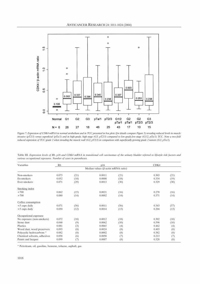
Table III. Expression levels of Rb, p16 and CDK4 mRNA in transitional cell carcinomas of the urinary bladder referred to lifestyle risk factors andvarious occupational exposures. Number of cases in parentheses.
Variables Rb p16 CDK4
Median values (‚-actin mRNA ratio)
Non-smokers 0.073 (21) 0.0011 (21) 0.303 (21)
Ex-smokers 0.052 (18) 0.0008 (18) 0.334 (19)
Ever-smokers 0.071 (29) 0.0013 (30) 0.329 (30)
Smoking index
<700 0.062 (15) 0.0021 (16) 0.278 (16)
>700 0.080 (14) 0.0002 (14) 0.371 (14)
Coffee consumption
<5 cups daily 0.071 (56) 0.0011 (56) 0.343 (57)
>5 cups daily 0.050 (12) 0.0014 (13) 0.204 (13)
Occupational exposure
No exposure (non-smokers) 0.072 (18) 0.0012 (18) 0.302 (18)
Stone dust 0.048 (9) 0.0062 (10) 0.598 (10)
Plastics 0.081 (3) 0.0065 (4) 0.462 (4)
Wood dust, wood preservers 0.093 (8) 0.0024 (8) 0.403 (8)
Polycyclic hydrocarbons * 0.082 (8) 0.0002 (8) 0.382 (8)
Chemical solvents, adhesives 0.058 (6) 0.0050 (7) 0.213 (7)
Paints and lacquer 0.099 (7) 0.0007 (8) 0.328 (8)
* Petroleum, oil, gasoline, benzene, toluene, asphalt, gas
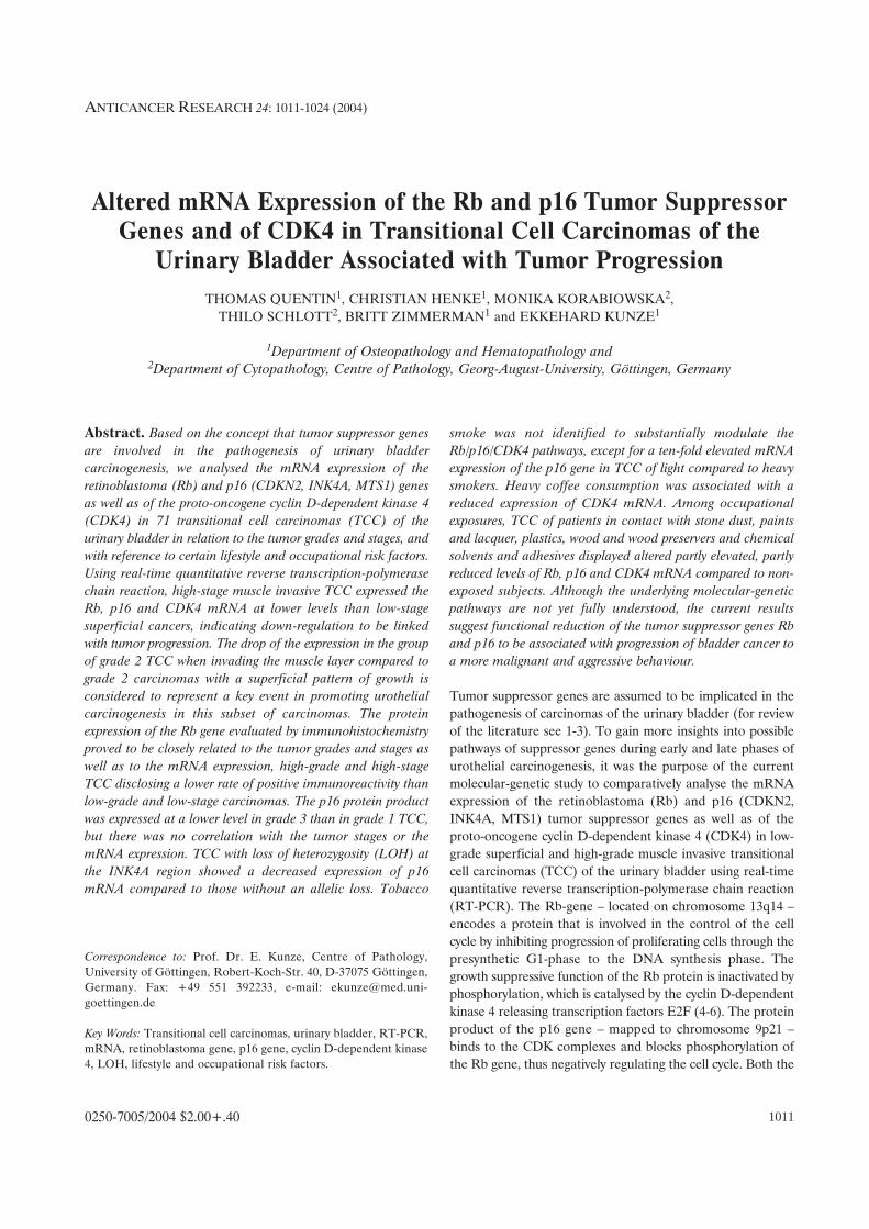
Figure 7. Expression of CDK4 mRNA in normal urothelium and in TCC presented in box plots (for details compare Figure 3) revealing reduced levels in muscleinvasive (pT2/3) versus superficial (pTa/1) and in high-grade, high-stage (G3, pT2/3) compared to low-grade,low-stage (G1/2, pTa/1) TCC. Note a two-foldreduced expression of TCC grade 2 when invading the muscle wall (G2, pT2/3) in comparison with superficially growing grade 2 tumors (G2, pTa/1).

positive cells the lowest (median 0.028) expression of Rb
mRNA, the difference of the expression levels being
statistically significant (p<0.001). Carcinomas with between
10% to 50% positive cells expressed the mRNA marginally
significantly (p=0.06) twice as high (median 0.064) as those
containing less than 10% cells with a positive
immunoreactivity.
Expression of p16 mRNA. Expression of mRNA of the p16
gene was five-fold higher in TCC of all grades and stages
(median 0.0011; N=70) than in the non-neoplastic bladder
mucosa (median 0.0002; N=6). The expression levels of the
carcinomas varied considerably, most probably reflecting the
cellular and molecular-pathologic heterogeneity of the tumors
independently of their grades and stages (Figure 4). High-
grade (grade 3) TCC produced the mRNA at a three-fold
higher level (median 0.0030) compared to grade 1 (median
0.0010; p=0.39) and grade 2 (median 0.0012) carcinomas.
High-stage muscle invasive (pT2 and pT3) TCC showed an
approximately five-fold lower expression (median 0.0002) than
low-stage superficial (pTa and pT1) tumors (median 0.0012),
but the difference lacked statistical significance (p=0.97).
High-grade, high-stage TCC expressed the p16 mRNA at a
five-fold lower level (median 0.0002) compared to low-grade,
low-stage cancers (median 0.0011; p=0.79). In the group of
grade 2 TCC, the muscle invasive carcinomas disclosed a two-
fold lower (median 0.0006) activity of the p16 gene than those
with a superficial pattern of growth (median 0.0014; p=0.98).
Recurrent tumors revealed a two-fold decreased expression
level (median 0.0007; N=13) compared to primary carcinomas
(median 0.0012; N=57; p=0.26).
Immunohistochemical expression of p16 protein. The data
showing the immunoreactivity of the TCC for p16 protein are
summarized in Table II. The incidence of grade 3 carcinomas
revealing more than 50% tumor cells with a strong intranuclear
positivity (Figure 6A-6D) was lower (61%) compared to grade
1 carcinomas (80%). Conversely, the frequency of TCC grade 3
with less than 10% positive cells proved to be higher (17%)
than that of grade 1 cancers (8%). The protein expression was
not observed to be linked with the tumor stages. There was also
no correlation between the protein and the mRNA expression
of the p16 gene, but there existed an inverse association
between the expression of the Rb protein and the expression of
the p16 mRNA: TCC disclosing less than 10% of Rb-positive
cells showed a significantly higher expression of p16 mRNA
(median 0.0132) than cancers with 10-15% (median 0.0015) and
more than 50% (median 0.0002) positive cells (p=0.01 and
0.0005, respectively).
Microsatellite analysis at p16 locus. Analysing the
microsatellite marker D9S171, 32 of the 66 TCC examined
proved to be informative, while the remaining 34 were non-
informative. Fourteen (44%) of the informative cases had
LOH, 16 (50%) showed retention of heterozygosity and 2 a
microsatellite instability. LOH at the D9S162 displayed 14
out of 30 (48%) informative cases among 67 TCC studied; 1
carcinoma revealed a microsatellite instability. Four tumors
exhibited allelic loss at both microsatelitte markers. TCC with
LOH at D9S171 expressed the p16 mRNA at a considerably
lower – though not significantly (p=0.10) – level (median
0.0001; N=14), compared to tumors lacking allelic loss
(median 0.0021; N=16). Accordingly, TCC revealing LOH at
D9S162 showed an insignificantly (p=0.48) lower expression
of p 16 mRNA (median 0.0001; N=14) than carcinomas
without LOH (median 0.001; N=15). LOH was not observed
to be related to the tumor grades (data not shown). High-
stage muscle invasive TCC disclosed a higher incidence of
LOH at marker D9S162 (7 out of 11 informative cases; 64%)
compared to low-stage superficial carcinomas (7 out of 19
informative cases; 37%), whereas LOH at D9S171 was not
identified to be linked to the tumor stages (data not shown).
Expression of cyclin D-dependent kinase 4 mRNA. CDK4
mRNA was expressed at a three-fold higher level in TCC of
all grades and stages (median 0.339; N=71) than in the non-
neoplastic bladder mucosa (median 0.108; N=6).
Expression levels did not substantially differ in the TCC of
the various grades, the median values ranging between 0.296
and 0.302 (Figure 7). High-stage muscle invasive (pT2 and
pT3) carcinomas showed a decreased expression (median
0.238) compared to low-stage superficial (pTa and pT1)
tumors (median 0.340), but statistical significance was
lacking (p=0.31; Figure 7). High-grade (grade 3), high-stage
TCC exhibited an insignificantly (p=0.64) lower expression
(median 0.238) than low-grade (grades 1 and 2), low-stage
carcinomas (median 0.337). TCC grade 2 with a superficial
pattern of growth expressed the CDK4 mRNA twice as high
(median 0.503) as carcinomas which had infiltrated the
muscle layer (median 0.253; p=0.16). No difference existed
between the expression levels of primary (median 0.336;
N=57) and recurrent tumors (median 0.320; N=13).
Expression of Rb, p16 and CDK4 mRNA referred to lifestyle riskfactors and occupational exposure. Analysing the effects of
lifestyle bladder cancer risk factors (Table III), carcinomas of
habitual smokers including all categories of consumption and
referred to the smoking index expressed the Rb, p16 and CDK4
mRNA at levels largely similar to those of non-smokers and ex-
smokers, except for a ten-fold higher expression of p16 mRNA
in carcinomas of subjects with a smoking index lower than 700
compared to an index greater than 700 (p=0.21). Stratifying for
the various consumption categories, we were unable to observe
a dose- or time-relationship between smoking and expression
of Rb and CDK4 mRNA (data not shown). However, smoking
of between 1-20 cigarettes daily was associated with a nine-fold
Quentin et al: Rb and p16 Tumor Suppressor Genes and CDK4 in Bladder Cancer
1019

elevated expression level of p16 mRNA compared to smoking
of more than 20 cigarettes (p=0.81). Heavy coffee consumption
(5 cups per day or more) was linked with a nearly significantly
(p=0.06) decreased expression of CDK4 mRNA relative to
drinkers of less than 5 cups.
Analysing the significance of occupational exposures (Table
III), increased expression levels of Rb mRNA were observed in
TCC of patients in contact with paints and lacquer (p=0.24),
and wood dust and wood preservers (p=0.12) referred to non-
exposed and non-smoking individuals, while decreased levels
were determined in cancers of subjects exposed to stone dust
(p=0.78). The expression of p16 mRNA was significantly
elevated in TCC of patients who had worked in the plastic
manufacturing industry (p=0.003) or in contact with stone dust
(p=0.031), but insignificantly increased in tumors of individuals
exposed to chemical solvents and adhesives (p=0.27) as well
as to wood dust and wood preservers; the expression was lower
following exposure to polycyclic hydrocarbons or paints and
lacquer. The expression of CDK4 mRNA proved to be two-
fold higher in TCC of individuals in contact with stone dust
(p=0.12) and slightly higher in carcinomas of workers in the
plastic industry (p=0.84) compared to the expression in tumors
of non-exposed subjects. Other exposures analysed could not
be identified to alter the expression profiles (data not shown).
Discussion
Most transitional cell carcinomas of the urinary bladder
(between 70% and 80%) show, at presentation, a superficial
papillary pattern of growth and either maintain a low
malignant biological potential or progress to invade the
muscle wall and to metastatic disease, implying a poorer
clinical outcome. A major problem in applying optimal
therapeutic strategies consists in the fact that, from a
histopathological point of view, it is impossible to accurately
predict the prospective clinical behaviour in the individual
case. Therefore, many attempts using various methodological
approaches have been made to elucidate and understand the
molecular-genetic pathways underlying progression of
bladder cancer from initially low-grade to finally high-grade
malignancy (for review of the literature see 1, 2, 25-27).
The objective of the current combined histopathological,
immunohistochemical, molecular-genetic and epidemiological
study was to analyse the mRNA expression of the Rb and p16
genes and of the cyclin D-dependent kinase 4 during early and
late phases of urothelial carcinogenesis to possibly identify
alterations in the expression patterns indicating tumor
progression. Involvement of the cell cycle controlling tumor
suppressor genes Rb and p16 in the pathogenesis of
transitional cell carcinomas of the urinary bladder is supported
mainly by studies at the DNA level, revealing homozygous and
heterozygous deletions as a possibly underlying mechanism of
inactivation of the suppressor function. Thus, loss of
heterozygosity of the Rb gene was reported to occur in
between 21% and 33% of cases (12, 14, 15), while point
mutations were rarely observed (15). Similarly, LOH at the
CDKN2 locus was detected in between 33% and 48% (8, 11,
13, 28) and homozygous deletions in between 7% and 33% (7,
9-11, 13) of TCC, whereas point mutations were not (7, 8, 16)
or rarely (9, 11, 29-31) identified.
Using real-time quantitative RT-PCR, we were able to
document lower expression levels of Rb mRNA in high-grade
(grade 3) as well as in high-stage (pT2 and pT3) compared to
low-grade (grades 1 and 2) and low-stage (pTa and pT1) TCC,
the difference being statistically significant between superficial
and muscle invasive carcinomas. Initial up-regulation of the Rb
mRNA expression in superficial low-grade and subsequent
down-regulation in advanced muscle invasive high-grade
carcinomas to a level largely similar to that of the normal
appearing, non-neoplastic urothelium provides evidence for a
deregulation of the Rb pathway during late phases of urothelial
carcinogenesis, indicating progression of bladder cancer to a
more malignant and aggressive behavior. The drop in the
expression level within the cohort of TCC grade 2 when
invading the muscle layer might represent a key event
responsible for a switch from a low to a high malignancy in this
subset of tumors. The protein expression of the Rb gene
determined by immunohistochemistry was found to be closely
associated with the expression of the mRNA, high-grade, high-
stage TCC disclosing more frequently less than 50% of positive
cells compared to low-grade, low-stage carcinomas. The
findings of other authors are hard to compare with each other
and the current study, mainly because of differing definitions of
positivity. Some authors categorised tumors without any
positive nuclear staining (32-35) or those with less than 50% of
positive cells (36, 37) as having an abnormal expression, others
regarded both loss or overexpression (more than 50% positive
cells) as abnormal (38-40). Thus, an altered pRb expression was
reported in a wide range of TCC (29% - 74%), regardless of
the tumor grades and stages (33, 34, 36, 37, 40). Only a few
immunohistochemical studies dealt with the expression of pRb
in relation to the tumor grades and stages. While some authors
reported alteration of pRb expression to occur more frequently
in muscle invasive than in superficial TCC (32, 37), others were
unable to detect such an association (34, 40). In contrast to our
data, no relationship was found to exist between the protein
expression and the tumor grades in other studies (34, 37, 40).
Paralleling the expression pattern of the Rb-gene, the
mRNA expression of p16 was found to be down-regulated in
high-grade, high-stage TCC compared to low-grade, low-stage
carcinomas. The reduction of both the p16 and Rb mRNA
expression in advanced cancers following initial up-regulation
does not fit into the widely accepted concept of an inverse
correlation between the activity of the two tumor suppressor
genes. The reduced expression of p16 mRNA in conjunction
with the decreased mRNA expression of Rb in advanced
ANTICANCER RESEARCH 24: 1011-1024 (2004)
1020

muscle invasive compared to superficial TCC grade 2 support
the hypothesis that the shift in the expression pattern occurring
within the group of grade 2 TCC may reflect an important step
toward development of a more malignant potential of bladder
cancer. Asamoto and coworkers (16) observed a decreased p16
mRNA expression in 39% of TCC using semiquantitative RT-
PCR with "no histological differences" between cases with
normal and reduced expression. By contrast, Le Frère-Belda
and coworkers (17) reported increasing expression levels of p16
mRNA with increasing grades and stages of the TCC. The
protein expression of the p16 gene was slightly correlated in the
current study with the tumor grades – grade 3 TCC exhibiting
less frequently more than 50% positive cells than grade 1
cancers – but failed to be linked with the tumor stages and the
expression of the mRNA as well. In another study, neither
tumor grades nor stages were found to be related to the
expression of the p16 protein (28), while Korkolopoulou and
coworkers (37) found a decreased p16 protein expression in
29% of TCC in association with advanced stages, but not with
the histological grades. Primdahl and coworkers (41) recently
reported a significantly lower level of p16 protein in primarily
muscle invasive TCC compared to those having developed
secondarily from precursors with a superficial pattern of growth.
These immunohistochemical findings are in line with the
current results of a reduced production of p16 mRNA in
advanced TCC. While we could not observe an
interrelationship between the immunohistochemical expression
of p16 and pRb, others demonstrated an overexpression of pRb
in association with a loss of p16 and vice versa (42), supporting
an inverse correlation between the activity of the two tumor
suppressor genes. In favor of a negative regulation are our
findings of a significantly diminished expression of p16 mRNA
in TCC which showed an increased immunoreactivity for pRb,
suggesting that pRb inhibits the function of the p16 gene.
Analysing the deletion status of the p16 gene using the
microsatellite markers D9S171 and D9S162, we were able to
detect loss of heterozygosity in nearly half of the informative
carcinomas. Other authors, using between 5 and 12
microsatellite markers, identified LOH at the CDKN2 locus
in 33% to 48% (8, 11, 13, 28) and homozygous deletions in
7% to 33% (7, 9-11, 13) of TCC. In the current study, LOH
at both microsatellite markers examined was found to be
linked with a considerably reduced expression of p16 mRNA,
compatible with an altered transcription caused by allelic loss.
LOH at D9S162 occurred with a considerably higher
incidence in high-stage muscle invasive than in low-stage
superficial TCC, while there was no such correlation of LOH
at D9S171. Heterozygosity was not observed to be related to
the tumor grades. In another study (7), low-grade cancers
showed a higher frequency of p16 deletions (44%) than high-
grade tumors (10%), but others were unable to correlate
LOH or homozygous deletions at 9p21 with the tumor grades
and stages (9, 13).
The only study analysing the mRNA expression of CDK4
in bladder cancers available at present using quantitative RT-
PCR did not identify any differences between normal mucosa
and tumors, or between low-stage and high-stage TCC (18).
Simon and coworkers (43) recently described amplification of
CDK4 in a very small number (2.3%) of 303 TCC, the
amplifications associated with increasing tumor stages and
grades. We were able to document a three-fold higher
expression of CDK4 mRNA in carcinomas in comparison with
non-neoplastic urothelium and a lower level in high-grade,
high-stage than in low-grade, low-stage TCC. In accordance
with the expression patterns of the Rb and p16 genes, the
most prominent difference was observed in the subset of TCC
grade 2 showing a two-fold lower expression when they had
developed a muscle invasive growth in comparison to those
with only a superficial pattern of growth. In line with this,
cyclin D1 mRNA was reported to be expressed at a higher
level in superficial versus invasive TCC (44). Accordingly, the
protein product as evaluated by immunohistochemistry proved
to be overexpressed exclusively in superficial low-stage and
low-grade cancers (45). Our finding of an increased expression
of CDK4 mRNA during early phases of urothelial
carcinogenesis and, in turn, of a decreased expression during
late stages similar to the expression patterns of the Rb and
p16 mRNA – although an inverse relationship could be
expected – suggests that other pathways independently of the
CDK4 complex may be involved in the regulation of the
tumor suppressor activity of the Rb and p16 genes during
bladder tumor development.
In a first attempt to explore the effect of lifestyle risk
factors of bladder cancer, tobacco smoke could not be
identified to substantially modulate the Rb/p16/CDK4
pathways. However, involvement of the p16 gene is
indicated by a ten-fold higher expression of the mRNA in
TCC of relatively light smokers with a smoking index lower
than 700 compared to smokers with an index higher than
700. So far, the effect of smoking on vesical tumor
development has been studied nearly exclusively with
respect to the p53 tumor suppressor gene, yielding
conflicting results. While some authors reported an
association between the spectrum of p53 mutations and
smoking habits (46-48), others failed to detect smoking-
specific mutations (49, 50). Zhang et al. (51) observed
LOH and microsatelite alterations of chromosome 9 in
82% of TCC of smokers, but only in 56% of never-smokers
with a dose-response trend. Heavy coffee consumption (at
least 5 cups or more per day) as a further known lifestyle
risk factor of bladder cancer (for review of the literature
see 23) was found to be associated with a borderline
significantly reduced mRNA expression of CDK4.
Among occupational exposures, TCC of patients in
contact with stone dust, plastics, paints and lacquer, chemical
solvents and adhesives, and wood and woodpreservers
Quentin et al: Rb and p16 Tumor Suppressor Genes and CDK4 in Bladder Cancer
1021

showed partly increased, partly decreased expression levels
of Rb, p16 and CDK4 mRNA compared to non-exposed
individuals, but in varying combinations which do not yet
allow a reliable interpretation. We do not believe that the
differences have arisen by pure chance, although a statistical
significance was obtained only for the elevated expression of
the p16 mRNA in cancers following exposure to stone dust
and plastics, both exposures previously demonstrated to be
associated with an increased relative risk for bladder cancer
in an epidemiological case-control study. Albeit the number
of cases in the various occupational categories are limited to
allow definite conclusions, our results suggest that hazardous
exogenous agents may play a role in modulating the
expression of tumor suppressor genes, encouraging further
combined molecular-genetic and epidemiological studies to
extend our findings.
In conclusion, we were able to document a reduced
expression of Rb, p16 and CDK4 mRNA in advanced muscle
invasive compared to superficial TCC of the urinary bladder,
indicating down-regulation to be associated with tumor
progression. The drop of expression in TCC grade 2 when
invading the muscle layer is regarded as a crucial event in
promoting bladder carcinogenesis in this subset of
carcinomas. Tumors with LOH at the CDKN2/INK4A
region showed a considerably lower mRNA expression of the
p16 gene than those without heterozygous deletions.
Analysing the effect of bladder cancer risk factors, habitual
smoking was not identified as substantially altering the
Rb/p16/CDK4 pathways, except for an elevated expression
of the p16 mRNA in TCC of light compared to heavy
smokers. Heavy coffee consumption proved to be associated
with a decreased expression of the CDK4 mRNA. Certain
hazardous occupational exposures may play a role in
modulating the activity of the Rb and p16 tumor suppressor
genes. Our findings indicate that deregulation of the
Rb/p16/CDK4 pathway is implicated in urothelial
carcinogenesis and suggest reduced mRNA production to be
linked with progression of bladder cancer to a more
malignant aggressive behaviour.
Acknowledgements
The authors thank Prof. Dr. R.-H: Ringert, Department of
Urology, University of Göttingen, Dr. H. Bartels, Department of
Urology, Evangelisches Krankenhaus Göttingen-Weende and Prof.
Dr. M. Blech, Kreiskrankenhaus Northeim, Germany, for supplying
fresh tumors tissues and allowing us to interview their patients. The
authors also wish to express their gratitude to Dipl. Ing. S. Kellner
for performing statistical analysis and to Miss B. Jünemann for
exellent technical processing of the immunohistochemical stainings.
This work was supported by the Else Kröner-Fresenius-
Foundation, Bad Homburg v. h. Höhe, Germany (grant numbers
13344660 and 1344170).
References
1 Cordon-Cardo C and Reuter VE: Alterations of tumor suppressor
genes in bladder cancer. Seminars Diagnostic Pathology 14: 123-132,
1997.
2 Knowles MA: The genetics of transitional cell carcinoma: progress and
potential clinical application. BJU Int 84: 412-427, 1999.
3 Rabbani F and Cordon-Cardo C: Mutation of cell cycle regulators and
their impact on superficial bladder cancer. Urologic Clinics North
America 27: 83-102, 2000.
4 Bartek J, Bartokova J and Lukas J: The retinoblastoma protein
pathway in cell cycle control and cancer. Exp Cell Res 237: 1-6, 1997.
5 Mulligan G and Jacks T: The retinoblastoma gene family: cousins with
overlapping interests. Trends Genet 14: 221-228, 1998.
6 Sellers WR and Kaelin WG: Role of the retinoblastoma protein in the
pathogenesis of human cancer. J Clin Oncol 15: 3301-3312, 1997.
7 Orlow I, Lacombe L, Hannon, GJ, Serrano M, Pellicer I, Dalbagni G,
Reuter VE, Zhang ZF, Beach D and Cordon-Cardo C: Deletion of the
p16 and p15 genes in human bladder tumors. J Int Cancer Inst 87:
1524-1529, 1995.
8 Packenham JP, Taylor JA, Anna CH, White CM and Devereux TR:
Homozygous deletions but no sequence mutations in coding regions
of p15 or p16 in human primary bladder tumors. Mol Carcinogen 14:
147-151, 1995.
9 Akao T, Kakehi Y, Itoh N, Özdemir E, Shimizu T, Tachibana A,
Sasaki MS and Yoshida O: A high prevalence of functional inactivation
by methylation modification of p16INK4A/CDKN2/ MTS1 gene in
primary urothelial cancers. Jpn J Cancer Res 88: 1078-1086, 1997.
10 Böhm M, Kirch H, Otto T, Rübben H and Wieland I: Deletion
analysis at the Del-27, APC and MTS1 loci in bladder cancer: LOH at
the Del-27 locus on 5p13-12 is a prognostic marker of tumor
progression. Int J Cancer 74: 291-295, 1997.
11 Baud E, Catilina P and Bignon YJ: p16 involvement in primary
bladder tumors: analysis of deletions and mutations. Int J Oncol 14:
441-445, 1999.
12 Wada T, Louhelainen J, Hemminki K, Adolfsson J, Wijkström H,
Norming U, Borgström E, Hansson J, Sandstedt B and Steineck G:
Bladder cancer: allelic deletions at and around the retinoblastoma
suppressor gene in relation to stage and grade. Clin Cancer Res 6: 610-
615, 2000.
13 Florl AR, Franke KH, Niederader D, Gerharz CD, Seifert HH and
Schulz WA: DNA methylation and the mechanisms of CDKN2A
inactivation in transitional cell carcinomas of the urinary bladder. Lab
Invest 80: 1513-1522, 2000.
14 Cairns P, Proctor AJ and Knowles MA: Loss of heterozygosity at the
RB locus is frequent and correlates with muscle invasion in bladder
carcinoma. Oncogene 6: 2305-2309, 1991.
15 Miyamoto H, Shuin T, Iwasaki Y and Kubota Y: Retinoblastoma
gene mutations in primary human bladder cancer. Br J Cancer 71:
831-835, 1995.
16 Asamoto M, Iwadori Y, Okamura T, Shirai T and Tsuda H: Decreased
expression of the p16/MTS1 gene without mutation is frequent in
human bladder carcinomas. Jpn J Clin Oncol 27: 22-25, 1997.
17 Le Frère-Belda MA, Cappellen D, Daher A, Gil-Diez-de-Medina S,
Besse F, Abbou CC, Thiery JP, Zafrani ES, Chopin DK and Radvanyi
F: p15INK4b in bladder carcinomas: decreased expression in
superficial tumours. Br J Cancer 85: 1515-1521, 2001.
18 Oya M, Schmidt B, Schmitz-Dräger BJ and Schulz WA: Expression of
G1 → S transition regulatory molecules in human urothelial cancer.
Jpn J Cancer Res 89: 719-726, 1998.
19 World Health Organization. International Histological Classification
of Tumors. Mostofi FK, Davis CJ, Sesterhenn IA, Sobin LH, eds
Berlin, Heidelberg, New York: Springer-Verlag, 1999.
ANTICANCER RESEARCH 24: 1011-1024 (2004)
1022
20 International Union against Cancer. TNM Klassifikation Maligner
Tumoren. Wittekind CH, Wagner G, eds Berlin, Heidelberg, New
York: Springer-Verlag, 1997.
21 Claude J, Kunze E, Frentzel-Beyme R, Paczkowski K, Schneider J and
Schubert H: Life-style and occupational risk factors in cancer of the
lower urinary tract. Am J Epidemiol 124: 578-581, 1986.
22 Claude J, Frentzel-Beyme R and Kunze E: Occupation and risk of
cancer of the lower urinary tract among men. A case-control study. Int
J Cancer 41: 371-379, 1988.
23 Kunze E, Chang-Claude J and Frentzel-Beyme R: Lifestyle and
occupational risk factors of bladder cancer in Germany. Cancer 69:
1776-1790, 1992.
24 Korz C, Pscherer A, Benner A, Mertens D, Schaffner C, Leupolt E,
Döhner H, Stilgenbauer S and Lichter P: Evidence for distinct
pathomechanisms in B-cell chronic lymphocytic leukemia and mantle
cell lymphoma by quantitative expression analysis of cell cycle and
apoptosis-associated genes. Blood 99: 4554-4560, 2002.
25 Stein JP, Grossfeld GD, Ginsberg DA, Esrig D, Freeman JA, Figueroa
AJ, Skinner DG and Cote RJ: Prognostic markers in bladder cancer: A
contemporary review of the literature. J Urol 160: 645-659, 1998.
26 Orntoft F and Wolf H: Molecular alterations in bladder cancer. Urol
Res 26: 223-233, 1998.
27 Diaz-Cano S, Blanes A, Rubio J, Matilla A and Wolfe HJ: Molecular
evolution and intratumor heterogeneity by topographic compartments
in muscle-invasive transitional cell carcinoma of the urinary bladder.
Lab Invest 80: 279-289, 2000.
28 Friedrich MG, Blind C, Milde-Langosch K, Erbesdobler A,
Conrad S, Löning T, Hammerer P and Huland H: Frequent
p16/MTS1 inactivation in early stages of urothelial carcinoma of
the bladder is not associated with tumor recurrence. Eur Urol 40:
518-524, 2001.
29 Kai M, Arakawa H, Sugimoto Y, Murata Y, Ogawa M and Nakamura
Y: Infrequent somatic mutation of the MTS1 gene in primary bladder
carcinomas. Jpn J Cancer Res 86: 249-251, 1995.
30 Miyamoto H, Kubota Y, Fujinami K, Dobashi Y, Kondo K, Yao
M, Shuin T and Hosaka M: Infrequent somatic mutations of the
p16 and p15 genes in human bladder cancer: p16 mutations occur
only in low-grade and superficial bladder cancers. Oncology Res 7:
327-330, 1995.
31 Sorlie T, Martel-Planche G, Hainaut P, Lewalter J, Holm R, Borresen-
Dale AL and Montesano R: Analysis of p53, p16MTS1, p21WAF1 and
H-ras in archived bladder tumours from workers exposed to aromatic
amines. Br J Cancer 77: 1573-1579, 1998.
32 Logothetis CJ, Xu HJ, Ro JY, Hu SX, Sahin A, Ordonez N and
Benedict WF: Altered expression of retinoblastoma protein and known
prognostic variables in locally advanced bladder cancer. J Natl Cancer
Inst 84: 1256-1261, 1992.
33 Xu HJ, Cairns P, Hu SX, Knowles MA and Benedict WF: Loss of RB
protein expression in primary bladder cancer correlates with loss of
heterozygosity at the RB locus and tumor progression. Int J Cancer 53:
781-784, 1993.
34 Pollack A, Czerniak B, Zagars GK, Hu SX, Wu CS, Dinney CPN,
Chyle V and Benedict WF: Retinoblastoma protein expression and
radiation response in muscle-invasive bladder cancer. Int J Rad Oncol
Biol Phys 39: 687-695, 1997.
35 Niehans GA, Kratzke RA, Froberg MK, Aeppli DM, Nguyen PL and
Geradts J: G1 checkpoint protein and p53 abnormalities occur in most
invasive transitional cell carcinomas of the urinary bladder. Br J Cancer
80: 1175-1184, 1999.
36 Cordon-Cardo C, Wartinger D, Petrylak D, Dalbagni G, Fair WR,
Fuks Z and Reuter VE: Altered expression of the retinoblastoma gene
product: prognostic indicator in bladder cancer. J Natl Cancer Inst 84:
1251-1256, 1992.
37 Korkolopoulou P, Christodoulu P, Lazaris A, Thomas-Tsagli E,
Kapralos P, Papanikolaou A, Kalliteraki I and Davaris P: Prognostic
implications of aberrations in p16/pRB pathway in urothelial bladder
carcinomas: a multivariate analysis including p53 expression and
proliferation markers. Eur Urol 39: 167-177, 2001.
38 Cote RJ, Duun MD, Chatterjee SJ, Stein JP, Shi SR, Tran QC, Hu SX,
Xu HJ, Groshen S,Taylor CR, Skinner DG and Benedict WF: Elevated
and absent pRB expression is associated with bladder cancer progression
and has cooperative effects with p53. Cancer Res 58: 1090-1094, 1998.
39 Grossman HB, Liebert M, Antelo M, Dinney CPN, Hu SX, Palmer JL
and Benedict WF: p53 and RB expression predict progression in T1
bladder cancer. Clin Cancer Res 4: 829-834, 1998.
40 Tokunaga H, Shariat SF, Green AE, Brown RM, Zhou JH, Benedict
WF and Lerner SP: Correlation of immunohistochemical molecular
staging of bladder biopsies and radical cystectomy specimens. Int J Rad
Oncol Biol Phys 51: 16-22, 2001.
41 Primdahl H, von der Maase H, Sorensen FB, Wolf H and Orntroft TF:
Immunohistochemical study of the expression of cell cycle regulating
proteins at different stages of bladder cancer. J Cancer Res Clin Oncol
128: 295-301, 2002.
42 Benedict WF, Lerner SP, Zhou J, Shen X, Tokunaga H and Czerniak
B: Level of retinoblastoma protein expression correlates with p16
(MTS-1/INK4A/CDKN2) status in bladder cancer. Oncogene 18: 1197-
1203, 1999.
43 Simon R, Struckmann K, Schraml P, Wagner U, Forster T, Moch A,
Fijan A, Bruderer J, Wilber K, Mihatsch MJ, Gasser T and Sauter G:
Amplification pattern of 12q13-q15 genes (MDM2, CDK4, GLI) in
urinary bladder cancer. Oncogene 21: 2476-2483, 2002.
44 Bringuier PP, Tamimi Y, Schuuring E and Schalken J: Expression of
cyclin D1 and EMS1 in bladder tumours; relationship with chromosome
11q13 amplification. Oncogene 12: 1747-1753, 1996.
45 Lee CCR, Yamamoto S, Morimura K, Wanibuchi H, Nishisaka N,
Ikemoto S, Nakatani T, Wada S, Kishimoto T and Fukushima S:
Significance of cyclin D1 overexpression in transitional cell carcinomas
of the urinary bladder and its correlation with histopathologic features.
Cancer 79: 780-789, 1997.
46 Kannio A, Ridanpää M, Koskinen H, Partanen T, Antilla S, Collan Y,
Hietanen E, Vainio H and Husgafvel-Pursiainen K: A molecular and
epidemiological study on bladder cancer: p53 mutations, tobacco
smoking, and occupational exposure to asbestos. Cancer Epidemiol
Biomarkers and Prevention 5: 33-39, 1996.
47 LaRue H, Allard P, Simeneau M, Normand C, Pfister C, Moore L,
Meyer F, Têtu B and Fradet Y: p53 point mutations in initial superficial
bladder cancer occur only in tumors from current or recent cigarette
smokers. Carcinogenesis 21: 101-106, 2000.
48 Bernardini S, Adessi GL, Chezy E, Billery C, Carbillet JP and Bittard
H: Influence of cigarette smoking on p53 gene mutations in bladder
carcinomas. Anticancer Res 21: 3001-3004, 2001.
49 Xu X, Stower MJ, Reid LN, Garner RC and Burns PA: A hot spot for
53 mutation in transitional cell carcinoma of the bladder: clues to the
etiology of bladder cancer. Cancer Epidemiol Biomarkers and
Prevention 6: 611-616, 1997.
50 Martone T, Airoldi L, Magagnotti C, Coda R, Randone D, Malaveille
C, Avanzi G, Merletti F, Hautefeuille A and Vineis P: 4-aminobiphenyl-
DNA adducts and p53 mutations in bladder cancer. Int J Cancer 75:
512-516, 1998.
51 Zhang ZF, Shu XM, Cordon-Cardo C, Orlow I, Lu ML, Millon TV,
Cao PQ, Connolly-Jenks C, Dalbagni G, Lianes P, Lacombe L, Reuter
VE and Scher H: Cigarette smoking and chromosome 9 alterations in
bladder cancer. Cancer Epidemiol Biomarkers and Prevention 6: 321-
326, 1997.
Received August 29, 2003Accepted February 2, 2004
Quentin et al: Rb and p16 Tumor Suppressor Genes and CDK4 in Bladder Cancer
1023