altered cd4 t cell homing to the gut impairs mucosal ... · christophe pasquier,1,2 pascale klopp,3...
TRANSCRIPT
Research article
62 TheJournalofClinicalInvestigation http://www.jci.org Volume 122 Number 1 January 2012
Altered CD4+ T cell homing to the gut impairs mucosal immune reconstitution
in treated HIV-infected individualsMaud Mavigner,1 Michelle Cazabat,1,2 Martine Dubois,1,2 Fatima-Ezzahra L’Faqihi,1 Mary Requena,1
Christophe Pasquier,1,2 Pascale Klopp,3 Jacques Amar,3,4 Laurent Alric,5,6 Karl Barange,6 Jean-Pierre Vinel,6 Bruno Marchou,7 Patrice Massip,7 Jacques Izopet,1,2 and Pierre Delobel1,7
1INSERM, UMR1043, Toulouse, France. 2Centre Hospitalier Universitaire de Toulouse, Hôpital Purpan, Laboratoire de Virologie, Toulouse, France. 3INSERM, UMR1048, Toulouse, France. 4Centre Hospitalier Universitaire de Toulouse, Hôpital Rangueil, Service de Médecine Interne et Hypertension Artérielle,
Toulouse, France. 5UMR-MD3 EA2405, Toulouse, France. 6Centre Hospitalier Universitaire de Toulouse, Hôpital Purpan, Pôle Digestif, Toulouse, France. 7Centre Hospitalier Universitaire de Toulouse, Hôpital Purpan, Service des Maladies Infectieuses et Tropicales, Toulouse, France.
DepletionofCD4+TcellsfromthegutoccursrapidlyduringacuteHIV-1infection.Thishasbeenlinkedtosystemicinflammationanddiseaseprogressionasaresultoftranslocationofmicrobialproductsfromthegutlumenintothebloodstream.Combinedantiretroviraltherapy(cART)substantiallyrestoresCD4+Tcellnumbersinperipheralblood,butthegutcompartmentremainslargelydepletedofsuchcellsforpoorlyunderstoodreasons.Here,weshowthatalackofrecruitmentofCD4+TcellstothegutcouldbeinvolvedintheincompletemucosalimmunereconstitutionofcART-treatedHIV-infectedindividuals.WeinvestigatedthetraffickingofCD4+Tcellsexpressingthegut-homingreceptorsCCR9andintegrinα4β7andfoundthatmanyoftheseTcellsremainedinthecirculationratherthanrepopulatingthemucosaofthesmallintestine.ThisislikelybecauseexpressionoftheCCR9ligandCCL25waslowerinthesmallintestineofHIV-infectedindividuals.ThedefectiveguthomingofCCR9+β7+CD4+Tcells—apopulationthatwefoundincludedmostgut-homingTh17cells,whichhaveacriticalroleinmucosalimmunedefense—correlatedwithhighplasmaconcentrationsofmarkersofmucosaldamage,microbialtranslocation,andsystemicTcellactivation.OurresultsthusdescribealterationsinCD4+TcellhomingtothegutthatcouldpreventefficientmucosalimmunereconstitutioninHIV-infectedindividualsdespiteeffectivecART.
IntroductionThe immune responses to the antigens encountered along the intestinal mucosa surfaces are mainly initiated in inductive sites of the gut-associated lymphoid tissue (GALT), Peyer’s patches, and mesenteric lymph nodes. The lymphocytes primed in these sec-ondary lymphoid organs then express high levels of gut-homing receptors, integrin α4β7 and CCR9, to subsequently direct their migration from the blood to the effector sites of the gut mucosa, the lamina propria and epithelium (1, 2). The ligand of α4β7 inte-grin, mucosal addressin cell adhesion molecule-1 (MAdCAM-1), is expressed by endothelial cells of the lamina propria and associated lymphoid tissues along the whole intestine (3). By contrast, the ligand of CCR9, the chemokine CCL25, is expressed only by small intestine endothelial and epithelial cells (4, 5). Thus, the combined expression of CCR9 and α4β7 delineates a T cell subset prone to migrate to the small intestine mucosa.
Most of the CD4+ T cells in the gut mucosa are activated effec-tor memory cells that express the HIV-1 entry coreceptor CCR5, thus providing a large pool of HIV-1 target cells (6, 7). The CD4+ T cells in the gut mucosa are rapidly and deeply depleted dur-ing acute HIV-1 infection, due to the direct killing of target cells by the virus and bystander apoptosis (8, 9). The ability of HIV-1 envelope glycoprotein (gp120) to bind to and signal through α4β7 could contribute to the selective tropism of HIV-1 for CD4+ T cells in the gut mucosa (10, 11).
The α4β7hi CD4+ T cell subset includes most gut-homing Th17 cells (12). These specialized cells play a critical role in gut mucosal immune defense (13, 14). Their depletion in the gut mucosa after HIV-1 and pathogenic SIV infections could compromise the integ-rity of the gut mucosal barrier (15). The subsequent translocation of microbial products from the gut lumen into the bloodstream has been associated with systemic inflammation and disease pro-gression in HIV-infected individuals and SIV-infected macaques (16–20). By contrast, the Th17 cell subset in the gut mucosa is selectively preserved in natural SIV hosts, like sooty mangabeys and African green monkeys (21). This could contribute to the absence of microbial translocation and systemic inflammation in nonpathogenic SIV infections (20). Thus, the restoration of an efficient mucosal immune barrier in HIV-infected individuals receiving combined antiretroviral therapy (cART) would be critical for reducing systemic inflammation.
Gut CD4+ T cells are restored much more slowly than those in the peripheral blood of treated HIV-infected individuals (22, 23). Many HIV-infected individuals still have effector sites of the gut mucosa that are severely lacking CD4+ T cells despite sustained effective cART, in marked contrast to the significant restoration of CD4+ T cells in their peripheral blood and immune inductive sites (24). This could be because HIV-1 replication is incompletely suppressed in the gut despite cART. A lack of recruitment of CD4+ T cells to the gut could also contribute to this incomplete restora-tion of CD4+ T cells in the gut mucosa. However, we know little of how CD4+ T cells migrate to repopulate the gut during immune reconstitution in response to cART.
Conflictofinterest: The authors have declared that no conflict of interest exists.
Citationforthisarticle: J Clin Invest. 2012;122(1):62–69. doi:10.1172/JCI59011.

research article
TheJournalofClinicalInvestigation http://www.jci.org Volume 122 Number 1 January 2012 63
We have therefore investigated CD4+ T cells trafficking between the blood and gut compartments in HIV-infected indi-viduals on sustained effective cART, focusing on CCR9+α4β7hi CD4+ T cells as a traceable phenotype for cells that home to the small intestine mucosa.
ResultsCD4+ T cells remain depleted and HIV-1 persists in the gut mucosa of HIV-infected individuals despite prolonged cART. We assessed CD4+ T cell reconstitution in the peripheral blood and small intestine mucosa of treated HIV-infected individuals by flow cytometry and immunohistochemistry. The frequency of CD4+ T cells remained lower in the peripheral blood of treated HIV-infected individuals than in uninfected controls (40.7% vs. 64.8%, P < 0.0001; Figure 1A). CD4+ T cell depletion also persisted in the small intestine mucosa of HIV-infected individuals compared with that in uninfected controls, as analyzed by flow cytometry (median fre-quency of 28.9% vs. 48.4%, respectively, P = 0.002; Figure 1A) and immunohistochemistry (median absolute number of 362 cells/mm2 vs. 660 cells/mm2, respectively, P = 0.0001; Figure 1B). The fre-
quency of CD4+ T cells in the gut was positively correlated with the frequency in the peripheral blood (correlation coefficient [ρ] = 0.67, P = 0.0001; Figure 1C). Almost all the CD4+ T cells in the gut mucosa were memory CD45RO+CCR5+ CD4+ T cells, 88% of which had an effector phenotype (Supplemental Figure 1; supplemental material available online with this article; doi:10.1172/JCI59011DS1).
Activated memory CCR5+ CD4+ T cells in the gut could be highly per-missive to HIV-1 infection. We there-fore quantified the HIV-1 DNA and RNA in samples of jejunum muco-sa. The median HIV-1 DNA load in CD4+ T cells in the gut mucosa was 6-times greater than that in CD4+ T cells in the peripheral blood (P = 0.013; Figure 1D), and their numbers in the 2 compartments were positively correlated (ρ = 0.54, P = 0.013; data not shown). HIV-1 RNA was also detected in the jeju-num mucosa of all the individuals tested (n = 16), albeit at low frequen-cy (mean of 2.5 copies per mg tissue; data not shown).
The magnitude of CD4+ T cell restoration in the small intestine mucosa of patients on cART var-ied greatly from one individual to another. Good and poor immuno-logical responders were divided on the basis of whether the percentage of CD4+ T cells in their gut mucosa was above or below the median. The good immunological respond-
ers had lower gut DNA viral loads than the poor immunological responders (P = 0.038). They also had a higher frequency of CD4+ T cells (P = 0.019) and a lower frequency of activated HLA-DR+ CD8+ T cells (P = 0.023) in the blood than the poor immunologi-cal responders (data not shown).
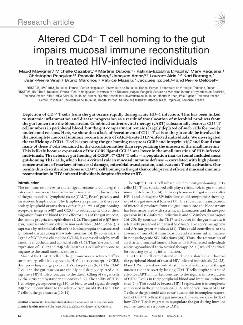
CCR9+β7hi CD4+ T cells are inversely distributed in the blood and gut compartments of HIV-infected individuals. We assessed CD4+ T cells trafficking between the peripheral blood and small intestine mucosa by measuring CD4+ T cells expressing gut-homing recep-tors, α4β7 and CCR9, in the 2 compartments (Figure 2A and Supplemental Figure 2, A–C). The frequencies of CCR9+β7hi in CD4+ T cells in the peripheral blood and small intestine mucosa were inversely correlated in HIV-infected individuals (ρ = –0.53, P = 0.017) and in uninfected controls (ρ = –0.75, P = 0.019; Figure 2B). But the distribution pattern of CCR9+β7hi CD4+ T cells between the blood and gut compartments of HIV-infected individuals was the inverse of that in uninfected controls. There were more CCR9+β7hi in CD4+ T cells in the peripheral blood (P = 0.002) and fewer cells in the small intestine mucosa (P = 0.014) of HIV-infected individuals than in those of the controls (Figure 2C).
Figure 1CD4+ T cell depletion and HIV-1 persistence in the gut mucosa of HIV-infected individuals despite sustained effective cART. (A) Frequencies of CD4+ in CD3+ T cells in the peripheral blood (PB) and jejunum mucosa of HIV-infected individuals (n = 20) and uninfected individuals (n = 9). Percentages of cells were determined by flow cytometry. Horizontal lines indicate median values. Each symbol represents an individual. (B) Absolute numbers of CD4+ T cells per surface unit of jejunum mucosa in HIV-infected individuals (n = 20) and uninfected individuals (n = 9). Horizontal lines indicate median values. Each symbol represents an individual. Immunohistochemical detection of intestinal CD4+ T cells (brown staining) in a representative HIV-infected individual and an uninfected control are shown. Original magnification, ×100. (C) Correlation between the frequency of CD4+ in CD3+ T cells in periph-eral blood and jejunum mucosa (n = 20 HIV-infected individuals, and n = 9 uninfected individuals). Percentages of cells were determined by flow cytometry. Each symbol represents an individual. (D) Paired HIV-1 DNA loads in CD4+ T cells of the peripheral blood and jejunum mucosa (n = 20). HIV-1 DNA was quantified by real-time PCR.
research article
64 TheJournalofClinicalInvestigation http://www.jci.org Volume 122 Number 1 January 2012
Thus, there appear to be significantly more CCR9+β7hi CD4+ T cells circulating in HIV-infected individuals than are localizing in the gut mucosa, in contrast to those in uninfected controls.
The CCL25-CCR9 axis driving CD4+ T cell gut homing is altered in HIV-infected individuals. The chemoattraction of CCR9+ lymphocytes is governed by their ligand, CCL25 (5). We therefore assessed wheth-er the abnormal distribution of CCR9+β7hi CD4+ T cells between the blood and gut compartments of HIV-infected individuals could be linked to differences in CCL25 expression in the small intestine mucosa. We measured CCL25 mRNA in small intestine epithelial cells using real-time RT-PCR. The median amount of CCL25 mRNA in HIV-infected individuals was 5.4-times lower than that in uninfected controls (P = 0.0001; Figure 3A). We also confirmed the reduced expression of the CCL25 chemo-kine in the small intestine mucosa of HIV-infected individuals by immunohistochemistry on jejunal tissue (automatic quantifica-tion of CCL25 per unit surface area, P = 0.006; Figure 3B). It was correlated with the amount of CCL25 mRNA (ρ = 0.45, P = 0.026; data not shown). The homing of CCR9+β7hi CD4+ T cells to the small intestine mucosa appears to be mainly driven by the CCL25-CCR9 axis. The amount of CCL25 mRNA was positively correlat-ed with the frequency of CCR9+β7hi in CD4+ T cells in the small intestine mucosa (ρ = 0.45, P = 0.026, Figure 3C) and conversely negatively correlated with the frequency of CCR9+β7hi in CD4+ T cells in the peripheral blood (ρ = –0.63, P = 0.0008; Figure 3D). The same positive and negative correlations were obtained when we measured CCL25 using immunohistochemistry (ρ = 0.35, P = 0.064 and ρ = –0.46, P = 0.013, respectively; data not shown). These results strongly suggest that a reduced expression of the che-
mokine CCL25 in the small intestine mucosa is involved in the altered homing of CCR9+β7hi CD4+ T cells to the gut of HIV-infected individuals.
In contrast to the depletion of CD4+ T cells observed in the jeju-num mucosa, the absolute numbers of CD8+ T cells in HIV-infected individuals (783 cells/mm2) and healthy controls (797 cells/mm2) were similar (P = 0.673). Thus, the defect in the CCL25-CCR9 axis seems to affect the CD4+ and CD8+ T cell populations differ-ently in HIV-infected individuals. We found that CD8+ T cells in the jejunum mucosa expressed much higher levels of β7 integrin than the CD4+ T cells (P < 0.0001; Supplemental Figure 3). We assessed the expression of MAdCAM-1 on endothelial cells in the lamina propria using immunohistochemistry. The amounts of MAdCAM-1 in the guts of HIV-infected individuals and healthy controls were similar (P = 0.493; Supplemental Figure 3). The pre-served absolute numbers of CD8+ T cells, despite the altered levels of CCL25, could thus be due to CD8+ T cells being more depen-dent on β7 integrin than on CCR9 in their trafficking to the gut during HIV-1 infection.
CCR9+β7hi CD4+ T cells include most gut-homing Th17 cells. Th17 cells play a critical role in the immune defense of the gut mucosa. Their depletion in the gut after HIV-1 infection could compromise the integrity of the gut mucosal barrier (15). The α4β7hi CD4+ T cell sub-set has been shown to harbor most of the gut-homing Th17 cells (12). We assessed the frequency of Th17 cells in the CCR9+ subset of α4β7hi CD4+ T cells in peripheral blood. The chemokine receptor CCR6 is a marker of Th17 lineage polarization that contributes to the Th17 migration to the gut (25–28). We therefore first measured the frequen-cy of CCR6+ cells among CCR9+β7hi and CCR9–β7hi CD4+ T cell sub-
Figure 2Distribution of CCR9+β7hi CD4+ T cells in the peripheral blood and jejunum compartments. (A) Flow cytometry analysis of CCR9 and β7 on CD4+ T cells in the peripheral blood and jejunum mucosa of a representative HIV-infected individual and an uninfected control. Numbers indi-cate the percentage of gated cells. (B) Correlation between the frequency of CCR9+β7hi in CD4+ T cells in peripheral blood and jejunum mucosa of HIV-infected individuals (n = 20) and uninfected individuals (n = 9). Percentages of cells were determined by flow cytometry. Each symbol represents an individual. (C) Frequency of CCR9+β7hi in CD4+ T cells in the peripheral blood and jejunum mucosa of HIV-infected individuals (n = 20) and uninfected individuals (n = 9). Percentages of cells were determined by flow cytometry. Horizontal lines indicate median values. Each symbol represents an individual.

research article
TheJournalofClinicalInvestigation http://www.jci.org Volume 122 Number 1 January 2012 65
sets using flow cytometry. There were more CCR6+ cells in CCR9+β7hi T cells than in CCR9–β7hi CD4+ T cells (P < 0.0001; Figure 4A). We then looked directly for Th17 and Th1 cells in 5 HIV-infected individuals and 5 uninfected controls by measuring the frequency of IL-17– and IFN-γ–producing cells in the CCR9+β7hi and CCR9–β7hi CD4+ T cell subsets sorted by flow cytometry (Supplemental Figure 4). The CCR9+β7hi CD4+ T cell subset was richer in Th17 cells than the CCR9–β7hi subset (P = 0.006; Figure 4B), while both subsets had simi-lar frequencies of Th1 cells (data not shown). Thus, most gut-hom-ing Th17 cells are in the CCR9+β7hi CD4+ T cell subset.
Defective homing of CCR9+β7hi CD4+ T cells to the gut correlates with mucosal damage, microbial translocation, and systemic T cell activation. We found that a significant fraction of CCR9+α4β7hi CD4+ T cells remains circulating rather than repopulating the gut of HIV-infected individuals. We also showed that this abnormally large circulating cell subset contains most of the gut-homing Th17 cells, which play an important role in the immune defense of the gut mucosa. We therefore looked at the association between this defect in gut hom-ing and any weakness in the gut mucosal barrier, microbial translo-cation, and increased T cell activation in HIV-infected individuals.
We evaluated enterocyte damage by measuring the plasma con-centration of intestinal-fatty acid–binding protein (I-FABP), which leaks out of damaged small intestine epithelial cells (29). The medi-
an plasma concentration of I-FABP was higher in treated HIV-infected individuals (238.3 pg/ml) than that in uninfected controls (54.5 pg/ml, P = 0.038; Figure 5A). The defec-tive homing of CCR9+β7hi CD4+ T cells to the gut was associated with enterocyte damage, as the plasma I-FABP concentration increased as the frequency of CCR9+β7hi in CD4+ T cells in the gut mucosa decreased (ρ = –0.51, P = 0.005; data not shown).
We assessed the translocation of bacterial products through the gut mucosal barrier by measuring the plasma concentrations of LPS, a component of the cell wall of Gram-negative bacteria, and soluble CD14 (sCD14), a marker of monocyte activation after stimulation by LPS (30). The median plasma concentra-tions of LPS (4.29 pg/ml) and sCD14 (1560 ng/ml) were higher in treated HIV-infected individuals than they were in uninfected controls (3.69 pg/ml for LPS, P = 0.004; 860 ng/ml for sCD14, P = 0.005; Figure 5A), sug-gesting that microbial translocation persists in HIV-infected individuals despite cART. Microbial transloca-tion was associated with enterocyte damage, because the concentrations of sCD14 and I-FABP were positively correlated (ρ = 0.41, P = 0.027; data not shown). The correlations between the concentrations of LPS and sCD14 and the frequency of CCR9+β7hi in CD4+
T cells in the blood (ρ = 0.57, P = 0.002 for LPS; ρ = 0.48, P = 0.008 for sCD14; Figure 5B) and the inverse correlations of these concen-trations and the frequency of CCR9+β7hi in CD4+ T cells in the gut mucosa (ρ = –0.48, P = 0.011 for LPS; ρ = –0.48, P = 0.008 for sCD14; Figure 5C) indicated that microbial translocation increased when more CCR9+β7hi CD4+ T cells were circulating rather than localizing in the gut. This was supported by our finding that the concentrations of LPS and sCD14 in the plasma were both inversely correlated with the amount of CCL25 mRNA in the small intestine mucosa (ρ = –0.58, P = 0.003 for LPS; ρ = –0.56, P = 0.004 for sCD14; Figure 5D).
Lastly, we examined the relationship between the dysregulated gut homing of CCR9+β7hi CD4+ T cells and an increase in T cell activa-tion in HIV-infected individuals. The degree of CD4+ T cell activation in peripheral blood, measured by Ki67 expression, was strongly cor-related with the plasma concentration of LPS in HIV-infected indi-viduals (ρ = 0.69, P = 0.001; data not shown). Systemic T cell activa-tion could be favored by the alterations in gut homing of CCR9+β7hi CD4+ T cells, because the frequency of Ki67+ in CD4+ T cells in the peripheral blood was positively correlated with the frequency of cir-culating CCR9+β7hi in CD4+ T cells (ρ = 0.64, P = 0.003; Figure 5E) but inversely correlated with the amount of CCL25 mRNA in the small intestine mucosa (ρ = –0.61, P = 0.007; Figure 5F). Thus our data strongly suggest that the defective homing of CCR9+β7hi CD4+
Figure 3Alterations in the CCL25-CCR9 axis that drives the gut homing of CCR9+β7hi CD4+ T cells in HIV-infect-ed individuals. (A) CCL25 mRNA expression in the jejunum epithelial cells of HIV-infected individuals (n = 18) and uninfected individuals (n = 7). Horizontal lines indicate median values. CCL25 mRNA was quantified by real-time RT-PCR and normalized to GAPDH. (B) CCL25 chemokine expression in the jejunum mucosa of HIV-infected individuals (n = 19) and uninfected individuals (n = 9). Horizontal lines indicate median values. A representative HIV-infected individual and an uninfected control are shown. CCL25 chemokine (brown) was stained by immunohistochemistry and automatically quantified with NIS-element (Nikon). Original magnification, ×400. (C) Correlation between CCL25 mRNA expression and the frequency of CCR9+β7hi in CD4+ T cells in the jejunum mucosa (n = 18 HIV-infected individu-als, and n = 7 uninfected individuals). (D) Correlation between CCL25 mRNA expression and the frequency of CCR9+β7hi in CD4+ T cells in the peripheral blood (n = 18 HIV-infected individuals, and n = 7 uninfected individuals). Throughout, each symbol represents an individual.

research article
66 TheJournalofClinicalInvestigation http://www.jci.org Volume 122 Number 1 January 2012
T cells to the gut in HIV-infected individuals is correlated with muco-sal damage, microbial translocation, and systemic T cell activation.
DiscussionWe have described alterations in the homing of CD4+ T cells to the gut that could contribute to the lack of mucosal immune reconsti-tution in HIV-infected individuals despite cART. CD4+ T cells in the small intestine mucosa of the subjects studied remained depleted, despite the subjects being on effective cART for more than 5 years. This is in marked contrast to a substantial restoration of their blood CD4+ T cell count. The persistence of microbial transloca-tion and systemic inflammation in these subjects underlines the importance of restoring an efficient mucosal immune barrier.
The immune inductive sites of the GALT are better reconsti-tuted than effector sites in response to cART (24). But unlike the ileum and colon, the mucosa of the upper small intestine is devoid of lymphoid follicles. We have focused on CCR9+α4β7hi CD4+ T cells trafficking to the jejunum mucosa. This allows us to assess the immune reconstitution of the effector sites of the gut during cART, without bias due to the presence of immune inductive sites, as is the case for the ileum or colon mucosa.
We found that a significant proportion of CCR9+β7hi CD4+ T cells are in the circulation of HIV-infected individuals rather than localizing in the gut, as they are in uninfected controls. Similarly, the distribution of CCR9+ T cells between the blood and small intestine compartments appears to be the inverse in inflammatory bowel diseases, as in celiac and Crohn’s diseases, with the frequency of CCR9+ T cells in the peripheral blood being markedly increased and that in the small intestine mucosa being reduced (31, 32). It has been suggested that the preferential apoptosis of CCR9+ T cells or the downregulation of CCR9 expression after T cell activation in the small intestine mucosa may explain the reduced frequency of CCR9+ T cells in the gut mucosa during celiac or Crohn’s diseases. The ex vivo activation of T cells downregulates CCR9 expression (31, 32). However, we found no expansion of the CCR9–β7hi CD4+ T cell subset accompanying the reduction of the CCR9+β7hi CD4+ T cell subset in the gut of HIV-infected individuals (data not shown). Some downregulation of CCR9 on T cells could have occurred dur-ing tissue processing, but it cannot account for the difference we
found between HIV-infected individuals and healthy individuals. The preferential activation-induced death of CCR9+ T cells could contribute to their depletion in the gut mucosa. But our data better support a mechanism involving a lack of recruitment of CCR9+β7hi CD4+ T cells to the gut, as the amount of CCL25 in the small intes-tine mucosa of HIV-infected individuals was much lower than that in uninfected controls. Moreover, the amount of CCL25 appears to be positively correlated with the frequency of CCR9+β7hi CD4+ T cells in the gut mucosa but inversely correlated with their fre-quency in the peripheral blood. The expression of CCL25 is altered in Crohn’s disease, with patchy increases in areas of lymphocyte infiltration but reduced amounts in inflamed and ulcerative areas of the small intestine mucosa (31). By contrast, we found a diffuse reduction in CCL25 in the jejunum mucosa of HIV-infected indi-viduals. We detected CCL25 expression by immunohistochemistry in both the crypt and villous epithelial cells. Some published stud-ies found more CCL25 in crypt epithelial cells (4, 5), while others found more CCL25 in villous cells (33). Ericsson et al. isolated epi-thelial cells from crypt and villous regions by laser capture micros-copy and found high amounts of CCL25 mRNA in both crypt and villous enterocytes (34). A reduction in the amount of CCL25 mRNA has also been observed in macaques lymph nodes during SIV infection (35). Epithelial cells are the main source of CCL25 in the small intestine mucosa. The reduced expression of CCL25 could thus be due to persistent enterocyte damage in HIV-infected individuals despite cART. This, in turn, could impair the CCL25-mediated recruitment of CCR9+β7hi CD4+ T cells to the gut, setting up a vicious circle that prevents efficient mucosal reconstitution.
Gene expression profiles of gut mucosal tissue have revealed increased activity of the genes involved in inflammation and apop-tosis in individuals with poor CD4+ T cell restoration in the gut, while the genes involved in mucosal repair and regeneration are more active in those having an efficient immune restoration (36). Residual HIV-1 replication in the gut despite cART could result in the intestine mucosa being persistently inflamed. Some virus pro-teins might have bystander effects, notably gp120 signaling through GPR15/Bob on the enterocytes could lead to their apoptosis (37, 38). Intraepithelial T cells bearing the Fas ligand could also induce Fas-mediated apoptosis of the enterocytes. The resulting damage to enterocytes could be responsible for the reduced expression of CCL25 in the small intestine mucosa and the associated defective homing of CD4+ T cells to the gut in HIV-infected individuals.
Other mechanisms are probably involved in this incomplete mucosal immune reconstitution, notably, in the colon, in which the CCL25-CCR9 axis does not play a major role. The chemokines CCL28, CCL20, and CXCL12 and the ligands of CXCR3 could play a role in the homing of T cells to the gut, but little is known regarding a potential dysfunction of this chemokine network in the setting of HIV infection (39, 40). The depletion of CD4+ T cells in treated HIV-infected individuals appears much more pronounced in the small intestine than in the colon (41). Thus, while the defect in the CCL25-CCR9 axis is probably not the sole mechanism involved — as it can-not explain the lack of immune reconstitution of the colonic mucosa — its impairment could contribute significantly to the particular depletion of CD4+ T cells observed in the small intestine mucosa.
The disrupted integrity of the gut mucosal barrier allows trans-location of microbial products from the gut lumen into the blood-stream during HIV-1 infection (20, 42, 43), as in inflammatory bowel diseases (44). HIV-infected individuals have been reported to have increased plasma levels of I-FABP and LPS, reflecting ongoing
Figure 4CCR9+β7hi CD4+ T cells include most gut-homing Th17 cells. (A) Paired frequencies of CCR6+ cells in CCR9+β7hi and CCR9–β7hi CD4+ T cell subsets in the peripheral blood (n = 20 HIV-infected individuals, and n = 9 uninfected individuals). Percentages of cells were deter-mined using flow cytometry. (B) Paired frequencies of IL-17–producing cells in CCR9+β7hi and CCR9–β7hi CD4+ T cell subsets in the peripheral blood (n = 5 HIV-infected individuals, and n = 5 uninfected individuals). Percentages of cells were determined by flow cytometry.

research article
TheJournalofClinicalInvestigation http://www.jci.org Volume 122 Number 1 January 2012 67
Figure 5Correlation between defective homing of CCR9+β7hi CD4+ T cells to the gut and mucosal damage, microbial translocation, and systemic T cell activation. (A) Concentrations of I-FABP, LPS, and sCD14 in HIV-infected individuals (n = 20 for I-FABP and sCD14 concentrations, and n = 19 for LPS concentrations) and uninfected individuals (n = 9). Plasma I-FABP and sCD14 concentrations were measured by ELISA. Plasma LPS was measured by the Limulus amoebocyte lysate assay. Horizontal lines indicate median values. (B) Correlation between the frequency of CCR9+β7hi in CD4+ T cells in the peripheral blood and the plasma concentrations of LPS (n = 19 HIV-infected individuals, and n = 9 uninfected individuals) and sCD14 (n = 20 HIV-infected individuals, and n = 9 uninfected individuals). (C) Correlation between the frequency of CCR9+β7hi in CD4+ T cells in the jejunum mucosa and the plasma concentrations of LPS (n = 19 HIV-infected individuals, and n = 9 uninfected individuals) and sCD14 (n = 20 HIV-infected individuals, and n = 9 uninfected individuals). (D) Correlation between CCL25 mRNA expression in the jejunum mucosa and the plasma concentrations of LPS (n = 17 HIV-infected individuals, and n = 7 uninfected individuals) and sCD14 (n = 18 HIV-infected individuals, and n = 7 uninfected individuals). (E) Correlation between the frequency of CCR9+β7hi in CD4+ T cells in the peripheral blood and the frequency of Ki67+ in CD4+ T cells in the peripheral blood (n = 20 HIV-infected individuals). (F) Correlation between CCL25 mRNA expres-sion in jejunum epithelial cells and the frequency of Ki67+ in CD4+ T cells in peripheral blood (n = 18 HIV-infected individuals). Throughout, each symbol represents an individual.
research article
68 TheJournalofClinicalInvestigation http://www.jci.org Volume 122 Number 1 January 2012
enterocyte death and bacterial translocation. Further, high levels of sCD14, a marker of monocyte activation after stimulation by LPS, have been associated with an increased risk of mortality in HIV-1 infection (18). Th17 cells play a critical role in the immune defenses of the gut mucosa. We found that most gut-homing Th17 cells are within the CCR9+β7hi CD4+ T cell subset and that the defective hom-ing of this subset to the gut is associated with microbial transloca-tion. The lack of recruitment of Th17 cells to the gut could thus be involved in the persistence of a “leaky” intestinal mucosal barrier.
In conclusion, we find that the defective homing of CCR9+β7hi CD4+ T cells to the gut could impair mucosal immune reconsti-tution in treated HIV-infected individuals. A vicious circle could arise among mucosal inflammation, enterocyte damage, reduced CCL25 expression, and defective homing of CCR9+β7hi CD4+ T cells to the gut. The lack of recruitment of CCR9+β7hi CD4+ T cells to the gut was associated with persistent microbial translocation and systemic T cell activation despite cART. Monitoring the fre-quency of circulating CCR9+β7hi CD4+ T cells can provide a sur-rogate marker of poor immune reconstitution of the gut mucosa in treated HIV-infected individuals.
MethodsStudy subjects and samples. Twenty HIV-1–infected individuals and ten unin-fected controls were enrolled in this study at the Toulouse University Hos-pital, Toulouse, France. All 20 HIV-1–infected individuals initiated cART at the chronic stage of infection, with a median nadir CD4+ T cell count of 185 cells/μl (interquartile range [IQR], 123–221 cells/μl). They had been on cART for a median of 66 months (IQR, 49–67 mo.). All had sustained plasma HIV-1 RNA of less than 20 copies per ml. Their median CD4+ T cell count was 668 cells/μl (IQR, 451–849 cells/μl) at the time of enrollment. Samples (80 ml) of peripheral blood were collected from each participant. Jejunal mucosa (8 biopsies) was obtained from each participant during upper endoscopy. All individuals were free of inflammatory or lymphopro-liferative bowel diseases on histopathologic examination. One uninfected control was excluded because of a concomitant angiocholitis.
Isolation of small intestine mucosal lymphocytes. Jejunal biopsies (n = 5) were digested with 0.5 mg/ml collagenase type II-S (Sigma-Aldrich) in RPMI by incubation at 37°C, with shaking, for 4 periods of 30 minutes. The cells were then filtered through 70-μm gauze, and intestinal T lymphocytes were isolated by positive selection (EasySep Human CD3 Positive Selection Kit, Stemcell Technologies Inc.) and immediately processed.
HIV-1 RNA extraction from small intestine mucosa. RNA was extracted from one biopsy that had been snap-frozen on dry ice. Each sample was weighed and homogenized, and RNA was extracted (QIAamp RNeasy Mini Kit, Qiagen) with on-column DNase treatment (RNase-Free DNase Set, Qiagen).
Quantification of HIV-1 in peripheral blood and small intestine mucosa. HIV-1 DNA was quantified from sorted CD4+ T cells by real-time PCR on a Light-Cycler (Roche), as previously described (45). The HIV-1 RNA in gut tissue was quantified by real-time RT-PCR using the SuperScript III One-Step RT-PCR System (Invitrogen) on a LightCycler 480 (Roche). This assay, adapted from a previously published procedure (46), has a sensitivity of 1 copy per reac-tion. HIV-1 RNA is expressed as copies per milligram of gut tissue.
Quantification of CCL25 mRNA in small intestine epithelial cells. RNA was extracted from small intestine epithelial cells (CD3– fraction of the digest-ed cell suspension) using the RNeasy Mini Kit (Qiagen). The primers were designed to amplify a fragment encompassing a spliced region of CCL25 mRNA. RT-PCR was performed on a LightCycler 480 (Roche). Data are given as fold increase in CCL25 mRNA normalized to the GAPDH control.
Histopathology and immunohistochemistry. Fresh tissues were fixed in 4% neutral buffered formalin and embedded in paraffin. Sections (3 μm) were
stained with hematoxylin and eosin. Immunohistochemistry was per-formed using anti-CD3 and CD4 (both from Novacastra); CD8 (Dako); CCL25 (R&D Systems); MAdCAM-1 (Serotec) mAb; and the appropriate secondary antibodies. Quantification was performed using LAS v3.7 (Leica microsystems) and NIS-element (Nikon).
Immunophenotyping of T lymphocytes. Flow cytometry analyses were per-formed on a BD LSRII driven by the FACSDiva software (BD Biosciences). Intracellular staining was performed using Cytofix/Cytoperm (BD Biosci-ences). We used anti–CD3-PECy7, CD4-ECD, and CD45RA-FITC (all from Beckman-Coulter); CD8-Pacific blue, CD27-PECy5, CD45RO-PECy5, α4-PE, β7-APC, Ki67-FITC, and CCR5-PE (all from BD Biosciences); and CCR6-PE, CCR7-PE, and CCR9-FITC (all from R&D Systems) mAbs. Con-trol experiments showed that more than 99% of peripheral CCR9+β7hi CD4+ T cells also expressed the α4 chain (Supplemental Figure 2A). The α4 and β7 chains were closely associated on CCR9+ CD4+ T cells, as dem-onstrated by FRET experiments (Supplemental Figure 2B). CCR9+β7hi CD4+ T cells were colabeled with the α4β7 mAb produced from the Act-1 clone (obtained through the NIH AIDS Research and Reference Reagent Program from A.A. Ansari, Emory University School of Medicine, Atlanta, Georgia, USA) that recognizes an epitope on β7 that is specific to the α4β7 heterodimer (Supplemental Figure 2C).
Detection of IL-17– and IFN-γ–producing cells. CD4+ T cells were stained with anti–CD3-PECy7, CD4-ECD, CCR9-FITC, and β7-APC mAbs. Unfixed cells were sorted on a BD FACSAria (BD Biosciences). Cells were stimulated by incubation with 10 ng/ml PMA and 500 ng/ml ionomycin (Sigma-Aldrich) for 1 hour at 37°C. Monensin was then added (GolgiStop, BD Biosciences), and the cells were incubated for a further 5 hours at 37°C. The cells were then washed, fixed/permeabilized, labeled with anti–IL-17–PE (eBiosci-ence) and IFN-γ–Alexa Fluor 700 (BD Biosciences) mAbs, and analyzed using a BD LSRII (Supplemental Figure 4).
Measurement of soluble markers of enterocyte injury and microbial translocation. The plasma concentrations of I-FABP and sCD14 were determined by ELISA (Cell Sciences and R&D Systems). Plasma was diluted 50% for I-FABP assays and 0.5% for sCD14 assays. LPS was measured in plasma diluted to 5% and heated at 70°C using the Limulus amoebocyte lysate assay (Lonza). All assays were performed in duplicate.
Statistics. Quantitative variables were compared using the Wilcoxon rank-sum test. Correlations were estimated by calculating Spearman’s rank cor-relation coefficients. The Wilcoxon signed-rank test was used for matched pairs. All tests were 2 sided, and P values of less than 0.05 were considered statistically significant. Statistical analyses were performed with Stata 9.2.
Study approval. The study was approved by the Institutional Review Board CPP Sud-Ouest et Outre-Mer II. All participants provided written informed consent (trial registration no. NCT01038401).
AcknowledgmentsThis project was supported by grant EP44 from the French National Agency for Research on AIDS and Viral Hepatitis. We are indebted to the patients who took part in this study. We also thank L. Cuzin, F. Balsarin, S. Lagarrigue, and A. Fooladi for their help in monitoring the study; T. al Saati and F. Capilla for histopathologic analyses; R. Burcelin and C. Garret for LPS and sCD14 assays; F. Barré-Sinoussi for helpful discussions; and O. Parkes for checking the English text.
Received for publication May 16, 2011, and accepted in revised form November 2, 2011.
Address correspondence to: Pierre Delobel, INSERM, UMR1043, Toulouse, F-31300 France. Phone: 33.5.61.77.75.08; Fax: 33.5.61. 77.21.38; E-mail: [email protected].
research article
TheJournalofClinicalInvestigation http://www.jci.org Volume 122 Number 1 January 2012 69
1. Johansson-Lindbom B, Svensson M, Wurbel MA, Malissen B, Marquez G, Agace W. Selective genera-tion of gut tropic T cells in gut-associated lymphoid tissue (GALT): requirement for GALT dendritic cells and adjuvant. J Exp Med. 2003;198(6):963–969.
2. Mora JR, et al. Selective imprinting of gut-homing T cells by Peyer’s patch dendritic cells. Nature. 2003; 424(6944):88–93.
3. Streeter PR, Berg EL, Rouse BT, Bargatze RF, Butcher EC. A tissue-specific endothelial cell mole-cule involved in lymphocyte homing. Nature. 1988; 331(6151):41–46.
4. Kunkel EJ, et al. Lymphocyte CC chemokine recep-tor 9 and epithelial thymus-expressed chemokine (TECK) expression distinguish the small intestinal immune compartment: Epithelial expression of tis-sue-specific chemokines as an organizing principle in regional immunity. J Exp Med. 2000;192(5):761–768.
5. Papadakis KA, et al. The role of thymus-expressed chemokine and its receptor CCR9 on lymphocytes in the regional specialization of the mucosal immune system. J Immunol. 2000;165(9):5069–5076.
6. Anton PA, et al. Enhanced levels of functional HIV-1 co-receptors on human mucosal T cells demon-strated using intestinal biopsy tissue. AIDS. 2000; 14(12):1761–1765.
7. Poles MA, Elliott J, Taing P, Anton PA, Chen IS. A preponderance of CCR5(+) CXCR4(+) mononucle-ar cells enhances gastrointestinal mucosal suscep-tibility to human immunodeficiency virus type 1 infection. J Virol. 2001;75(18):8390–8399.
8. Li Q, et al. Peak SIV replication in resting memory CD4+ T cells depletes gut lamina propria CD4+ T cells. Nature. 2005;434(7037):1148–1152.
9. Mattapallil JJ, Douek DC, Hill B, Nishimura Y, Martin M, Roederer M. Massive infection and loss of memory CD4+ T cells in multiple tis-sues during acute SIV infection. Nature. 2005; 434(7037):1093–1097.
10. Arthos J, et al. HIV-1 envelope protein binds to and signals through integrin alpha4beta7, the gut mucosal homing receptor for peripheral T cells. Nat Immunol. 2008;9(3):301–309.
11. Cicala C, et al. The integrin alpha4beta7 forms a com-plex with cell-surface CD4 and defines a T-cell subset that is highly susceptible to infection by HIV-1. Proc Natl Acad Sci U S A. 2009;106(49):20877–20882.
12. Kader M, et al. Alpha4(+)beta7(hi)CD4(+) memory T cells harbor most Th-17 cells and are preferen-tially infected during acute SIV infection. Mucosal Immunol. 2009;2(5):439–449.
13. Ye P, et al. Requirement of interleukin 17 receptor signaling for lung CXC chemokine and granulo-cyte colony-stimulating factor expression, neutro-phil recruitment, and host defense. J Exp Med. 2001; 194(4):519–527.
14. Mangan PR, et al. Transforming growth factor-beta induces development of the T(H)17 lineage. Nature. 2006;441(7090):231–234.
15. Raffatellu M, et al. Simian immunodeficiency virus-induced mucosal interleukin-17 deficiency promotes Salmonella dissemination from the gut. Nat Med. 2008;14(4):421–428.
16. Marchetti G, et al. Microbial translocation is associated with sustained failure in CD4+ T-cell reconstitution in HIV-infected patients on long-term highly active antiretroviral therapy. AIDS. 2008;22(15):2035–2038.
17. Pandrea I, et al. Cutting edge: Experimentally induced immune activation in natural hosts of simian immunodeficiency virus induces significant increases in viral replication and CD4+ T cell deple-tion. J Immunol. 2008;181(10):6687–6691.
18. Sandler NG, et al. Plasma levels of soluble CD14 independently predict mortality in HIV infection. J Infect Dis. 2011;203(6):780–790.
19. Ancuta P, et al. Microbial translocation is associat-ed with increased monocyte activation and demen-tia in AIDS patients. PLoS One. 2008;3(6):e2516.
20. Brenchley JM, et al. Microbial translocation is a cause of systemic immune activation in chronic HIV infection. Nat Med. 2006;12(12):1365–1371.
21. Brenchley JM, et al. Differential Th17 CD4 T-cell depletion in pathogenic and nonpathogenic lenti-viral infections. Blood. 2008;112(7):2826–2835.
22. Guadalupe M, et al. Severe CD4+ T-cell depletion in gut lymphoid tissue during primary human immu-nodeficiency virus type 1 infection and substantial delay in restoration following highly active antiret-roviral therapy. J Virol. 2003;77(21):11708–11717.
23. Mehandru S, et al. Lack of mucosal immune recon-stitution during prolonged treatment of acute and early HIV-1 infection. PLoS Med. 2006;3(12):e484.
24. Mehandru S, et al. Primary HIV-1 infection is asso-ciated with preferential depletion of CD4+ T lym-phocytes from effector sites in the gastrointestinal tract. J Exp Med. 2004;200(6):761–770.
25. Acosta-Rodriguez EV, et al. Surface phenotype and antigenic specificity of human interleukin 17-pro-ducing T helper memory cells. Nat Immunol. 2007; 8(6):639–646.
26. Annunziato F, et al. Phenotypic and functional features of human Th17 cells. J Exp Med. 2007; 204(8):1849–1861.
27. El Hed A, et al. Susceptibility of human Th17 cells to human immunodeficiency virus and their perturbation during infection. J Infect Dis. 2010; 201(6):843–854.
28. Singh SP, Zhang HH, Foley JF, Hedrick MN, Farber JM. Human T cells that are able to produce IL-17 express the chemokine receptor CCR6. J Immunol. 2008;180(1):214–221.
29. Pelsers MM, et al. Intestinal-type and liver-type fatty acid-binding protein in the intestine. Tis-sue distribution and clinical utility. Clin Biochem. 2003;36(7):529–535.
30. Anderson KV. Toll signaling pathways in the innate immune response. Curr Opin Immunol. 2000; 12(1):13–19.
31. Papadakis KA, et al. CCR9-positive lymphocytes and thymus-expressed chemokine distinguish small bowel from colonic Crohn’s disease. Gastro-enterology. 2001;121(2):246–254.
32. Olaussen RW, Karlsson MR, Lundin KE, Jahnsen J, Brandtzaeg P, Farstad IN. Reduced chemokine receptor 9 on intraepithelial lymphocytes in celiac
disease suggests persistent epithelial activation. Gastroenterology. 2007;132(7):2371–2382.
33. Wurbel MA, et al. The chemokine TECK is expressed by thymic and intestinal epithelial cells and attracts double- and single-positive thymo-cytes expressing the TECK receptor CCR9. Eur J Immunol. 2000;30(1):262–271.
34. Ericsson A, Kotarsky K, Svensson M, Sigvardsson M, Agace W. Functional characterization of the CCL25 promoter in small intestinal epithelial cells suggests a regulatory role for caudal-related homeo-box (Cdx) transcription factors. J Immunol. 2006; 176(6):3642–3651.
35. Qin S, Sui Y, Murphey-Corb MA, Reinhart TA. Association between decreased CXCL12 and CCL25 expression and increased apoptosis in lym-phoid tissues of cynomolgus macaques during SIV infection. J Med Primatol. 2008;37(suppl 2):46–54.
36. Guadalupe M, et al. Viral suppression and immune restoration in the gastrointestinal mucosa of human immunodeficiency virus type 1-infected patients initiating therapy during primary or chronic infection. J Virol. 2006;80(16):8236–8247.
37. Clayton F, et al. Gp120-induced Bob/GPR15 activation: a possible cause of human immuno-deficiency virus enteropathy. Am J Pathol. 2001; 159(5):1933–1939.
38. Li Q, et al. Simian immunodeficiency virus-induced intestinal cell apoptosis is the underlying mechanism of the regenerative enteropathy of early infection. J Infect Dis. 2008;197(3):420–429.
39. Kunkel EJ, Campbell DJ, Butcher EC. Chemokines in lymphocyte trafficking and intestinal immunity. Microcirculation. 2003;10(3–4):313–323.
40. Agace WW. T-cell recruitment to the intestinal mucosa. Trends Immunol. 2008;29(11):514–522.
41. Yukl SA, et al. Differences in HIV burden and immune activation within the gut of HIV-positive patients receiving suppressive antiretroviral therapy. J Infect Dis. 2010;202(10):1553–1561.
42. Cassol E, et al. Persistent microbial translocation and immune activation in HIV-1-infected South Afri-cans receiving combination antiretroviral therapy. J Infect Dis. 2010;202(5):723–733.
43. Jiang W, et al. Plasma levels of bacterial DNA cor-relate with immune activation and the magnitude of immune restoration in persons with antiret-roviral-treated HIV infection. J Infect Dis. 2009; 199(8):1177–1185.
44. Caradonna L, Amati L, Magrone T, Pellegrino NM, Jirillo E, Caccavo D. Enteric bacteria, lipopolysac-charides and related cytokines in inflammatory bowel disease: biological and clinical significance. J Endotoxin Res. 2000;6(3):205–214.
45. Delobel P, et al. Naive T-cell depletion related to infection by X4 human immunodeficiency virus type 1 in poor immunological responders to highly active antiretroviral therapy. J Virol. 2006; 80(20):10229–10236.
46. Mavigner M, et al. HIV-1 residual viremia correlates with persistent T-cell activation in poor immuno-logical responders to combination antiretroviral therapy. PLoS One. 2009;4(10):e7658.