allergic fungal frontal sinusitis - st. paul's sinus …...urgent endoscopic sinus surgery to...
TRANSCRIPT

Title: Fungal Frontal Sinusitis - Allergic and
Non-Allergic Authors: Fahad Al-Asousi1, MB BCh, BAO, SB-ORL Anali Dadgostar1, MD, FRCSC Amin Javer1, MD, FRCSC, FARS Carl Philpott2, MB ChB, FRCS(ORL-HNS), MD, PGCME
1Author Affiliation: St. Paul's Sinus Centre, Vancouver, British Columbia, Canada
2Author Affiliation: Norwich Medical School, University of East Anglia, United Kingdom
Corresponding Author:
Professor Carl Philpott Norwich Medical School, University of East Anglia, Norwich, NR4 7TJ UK

Contents Introduction 3 .....................................................................................
Classification 3 ....................................................................................
Invasive 3 ..........................................................................................
Acute Invasive Fungal Rhinosinusitis (AIFRS) 3 ............................................
Chronic Invasive Fungal Rhinosinusitis (CIFRS) 4 ..........................................
Granulomatous Invasive Fungal Sinusitis (GIFS) 5 .........................................
Noninvasive 5 ......................................................................................
Eosinophilic fungal sinus disease 5 ..........................................................
Allergic Fungal Rhinosinusitis (AFRS) 5 ...................................................
Eosinophilic Mucinous Rhinosinusitis (EMRS) 6 ..........................................
Sinus mycelia 7 .................................................................................
Special considerations in frontal sinus fungal disease 7 ....................................
Medical Management 8 ..........................................................................
Surgical and Post-operative Management in the Frontal Sinus 8 ..........................
Case example 9 ...................................................................................
Conclusion 13 ......................................................................................
References: 14 ....................................................................................
Table 1: Diagnostic Criteria for AFRS – Bent & Kuhn¥/Vancouver* 17 ..................
Table 2a and 2b: Philpott-Javer Endoscopic Staging system for AFRS 17..............

Introduction Fungal sinusitis can be a significant disorder in any of the sinuses and may prove
problematic in the frontal sinus when severe. This chapter will delineate the
different forms of fungal sinusitis and their discerning features as well as consider
how the management of these conditions in the frontal sinus specifically may
require additional challenges. To help illustrate this, a specific case example is
included, that highlights these challenges.
Classification
Based on the clinical picture, imaging and histology, fungal sinusitis can be broadly
classified into invasive and non-invasive fungal rhinosinusitis.
Invasive
Acute Invasive Fungal Rhinosinusitis (AIFRS)
Acute Fulminant Invasive Fungal Rhinosinusitis (AIFRS) is a potentially lethal
disease entity with low survival rate (49.7%)1. It primarily affects patients with
conditions associated with severe neutropenia (absolute neutrophil count [ANC] <
500/µL)2 and/or impaired neutrophil function; i.e. patient undergoing
transplantation, leukemia, uncontrolled diabetic ketoacidosis, patients receiving
chemotherapy and hæmochromatosis3 4 and especially those in receipt of bone
marrow transplantation5 6. Although nonspecific, red flags for AIFRS include pyrexia
and symptoms of localization to the paranasal sinus area (e.g. facial pain and
pressure, nasal congestion, orbital swelling). Symptoms of greater concern include
visual disturbances, paraesthesia and cranial neuropathy, indicating late
presentation and more advanced disease. On endoscopic examination, the findings
can range from oedema to dry or pale mucosa in the early stages to frank necrosis
in the advance stages. The middle turbinate (67%) and the nasal septum (24%) are
the most common sites to show clinical findings4. The genera of Aspergillus and
Mucor are the most common organisms that have been associated with AIFRS2 7,

hence why the condition is also sometimes known as mucormycosis.
Histopathological features include fungi invading the mucosal barriers and tissue
necrosis8 9. Management requires an attempt at reversal of the underlying
immunocompromised state and multidisciplinary approach is needed with both
medical and surgical interventions. It is therefore important to have high index of
suspicion in patients who are considered to be high-risk populations. This is
accomplished by prompt biopsies being taken and pathologic evaluation in such
patients. High-resolution, non-contrasted CT scan is crucial part for the work-up
and MRI is recommended in patients who present with orbital or intracranial
involvement signs or symptoms. Thickening of the peri-antral fat plane has been
reported as an early indicator of AIFRS10.
Chronic Invasive Fungal Rhinosinusitis (CIFRS)
CIFRS is encountered in patients who are not or with limited immunocompromised
status such as diabetic or patients on long term corticosteroids9. It is slowly
destructive disease over a time course of more than 12 weeks and can reach up to
12 months duration. Granulomatous Invasive Fungal Rhinosinusitis (GIFRS) is a
subtype of CIFRS that is more commonly encountered in healthy and immune
competent patients at the Middle East, North Africa and India3. Orbital and CNS
involvement is less common than in AIFRS, although orbital apex syndrome is
possible. Radiological findings include hyperdense soft tissue and bony involvement
on CT scanning and very hypointense T2 signal on MRI with possible evidence of
intracranial involvement.
CIFRS is distinguished histologically by the formation of non-caseating granuloma in
which giant cells contain the hyphae reside11. Aspergillus flavus, Aspergillus
fumigatus, Alternaria, P. boydii, Sporothrix schenckii are reported to be the
organisms associated with CIFRS. The presentation does not differ from AIFRS but
it is suspected when the symptoms of CRS are refractory to medical management
and progressing in severity, especially persistent headache, visual disturbance or
development of cranial nerve deficits. Tissue biopsy is the only definitive tool to
diagnose CIFS12 but radiological imaging will help and with surgical planning.
Urgent endoscopic sinus surgery to debride the affected areas is needed along with
systemic antifungals and once controlled they should continue on itraconazole for
up to 1 year. Recurrence is common and thus long-term follow up is needed to
ensure the disease remains controlled.
Granulomatous Invasive Fungal Sinusitis (GIFS) This form of invasive fungal sinusitis is attributed to infection with Aspergillus
flavus and is principally seen in North Africa, India and Pakistan. The infection
manifests as a locally invasive disease over at least 3 months duration but usually
in immunocompetent patients. Typical presenting symptoms include those of CRS
and possibly of proptosis or an enlarging mass in the affected sinus. Histological
examination of material will show a pattern of non-caseating granulomas and of
foreign body/Langhans giant cells with central necrosis. These cases need surgical
debridement followed by systemic antifungal medication. Disease recurrence is
uncommon and GIFS has a good prognosis.
Noninvasive
Eosinophilic fungal sinus disease
Eosinophilic fungal sinus disease, secondary to an over-responsiveness to fungus
with or without fungal hypersensitivity, can be subdivided into:
• Allergic fungal rhinosinusitis (AFRS)
• Eosinophilic mucinous/fungal rhinosinusitis (EMRS/EFRS)
Allergic Fungal Rhinosinusitis (AFRS)
Allergic fungal rhinosinusitis (AFRS) may be considered a form of chronic
rhinosinusitis (CRS) and accounts for 7-10% of CRS. In the late 1970’s AFRS was
recognized as an upper airway manifestation of allergic bronchopulmonary
aspergillosis (ABPA)13 14. It was in 1994 that Bent & Kuhn defined the 5 diagnostic
criteria for AFRS (table 1)15 16. The name itself may indeed be a misnomer as a type
I hypersensitivity reaction is not always proven despite the evidence of the other
key clinical features and a modified version has been proposed whereby
immunocompetence replaces type I hypersensitivity, reflecting the group of

characteristic patients seen in rhinologic practice17. The constant features in these
patients are a distinct clinical pattern of recurrent nasal poylposis and
accumulation of fungal mucin. AFRS classically involves all sinus cavities with
impacted thick mucin, polyps and chronic inflammation with pushing bony
margins. Radiological changes include double densities with a railtrack pattern in
the sinus, sinus expansion, remodeling of the sinus wall and bony erosion on CT
imaging and hypointense areas on T1 and signal voids on T2 on MRI18.
Isolated fungal frontal sinusitis is rare and only few case reports are available in
the literature19. Their consistent clinical pattern is the key factor in their
management as, unlike the management of classical CRS, the cornerstone for
treatment of AFRS is surgery. They require meticulous and complete endoscopic
sinus surgery (see below), along with careful and regular follow-up in the
outpatient clinic in order to try and prevent the polyp reformation and
accumulation of mucin and a comprehensive postoperative medical regimen is
almost always a necessity20.
Eosinophilic Mucinous Rhinosinusitis (EMRS)
In a study conducted by Ponikau et al. on CRS patients, 93% of the patients
undergoing sinus surgery had both eosinophilic mucin and fungus21. However less
than half of their sample of almost 100 patients in whom eosinophilic mucin and
fungus were present were allergic. They demonstrated the movement of
eosinophils out of blood vessels and into the sinus cavity to engulf fungal hyphae.
They proposed that a cell-mediated response provoked by fungi in susceptible
hosts was responsible and coined the term EMRS. Some studies attempted to
differentiate EMRS and AFRS patients based on demographics. They suggested EMRS
patients to be relatively younger, less likely to have asthma and aspirin sensitivity
and more likely to have bilateral disease when compared with AFRS patients22.
However, in reality, there is overlap in the clinical pictures between EMRS and
ARFS23. Orbital involvement and higher IgE levels have been found to be more
common in AFRS patients. This may be due to variations in climate, genetic
susceptibility and socioeconomic factors. Orlandi et al carried out a microarray
gene analysis between these two subgroups24. They showed 38 genes or potential
genes were differentially expressed in AFRS patients, while 10 genes were
differentially expressed in EMRS patients.
Sinus mycelia Sinus mycelia, also known as fungal balls, represent an accumultation of fungal
material in which the immune system is neither over- nor under-responsive. They
are mainly caused by Aspergillus species e.g. Aspergillus fumigatus in
immunocompetent patients. Demographics include being more common in middle-
aged and elderly females, in contrast to all forms of invasive and chronic
aspergillosis, which are more common in males. Typical presentation is with
symptoms relating to chronic sinusitis of one sinus, which is usually the maxillary
and less commonly the sphenoid sinus, but they can be incidental findings on CT
scans requested for non-sinugenic causes/symptoms. Typical symptoms, if present,
include nasal discharge, nasal obstruction, headache, facial pain and cacosmia,
the latter of which may be a predominant symptom. Occasionally they can be
associated with unilateral proptosis and facial hypoaesthesia.
Radiological imaging (CT scan) will demonstrate a unilateral, single sinus disease
with heterogeneous opacification (Figure 2). Fungal cultures are positive in less
than one third of patients despite fungal elements on histopathology in more than
90% of those affected. There is no predominance of eosinophils or granulomata or
allergic mucin, and no histopathological evidence of fungal invasion of mucosa.
Treatment is surgical, invariable an endoscopic approach to remove fungal ball and
open affected sinus; however in an asymptomatic patient a discussion about
watchful waiting may be needed depending on the age and co-morbidities of the
patient. At surgery the sinus full of dense brown/green material which requires
irrigation to help dislodge (Figure 3). Following removal of the fungal ball, no
antifungal treatment is required and no long-term follow-up required once patency
and healing of sinus is confirmed endoscopically in clinic.
Special considerations in frontal sinus fungal disease The frontal sinus is the least susceptible to fungal infection due to the location of
the ostium. Acute fulminant and chronic invasive fungal sinusitis therefore rarely

involve the frontal sinus with only 14.8% of cases being reported to involve the
frontal sinus in a large case series25; other series have reported slightly higher
levels of involvement (17-21%)26 27. Fungal ball involvement of the frontal sinus is
also rare19 28. There are few case reports in the English literature describing
primary sinus mycelia in the frontal sinus, with several authors advocating an
external approach for its management19 29 30 .
In contrast, it is estimated that 71% of AFRS cases have frontal sinus involvement
31. The relatively thin bones that are in close proximity to the frontal sinus (lamina
papyracia, cribriform plate) are more susceptible to changes in a manner
equivalent to pressure necrosis secondary of the accumulation of dense
eosinophilic fungal mucin. This may result in erosion and extension of the disease
to the orbit and intracranial space32, but is a more indolent process than is seen in
the invasive forms of fungal disease.
Medical Management Acute Invasive Fungal Rhinosinusitis is routinely managed both medically with
systematic antifungal therapy in conjunction with surgical intervention.
Amphotericin-B in liposomal formulation is the mainstay therapy for the past 50
years4 33. Topical antifungal therapy should be considered as well34. A patient who
recovers their neutrophil count and function has a better prognosis35.
Surgical and Post-operative Management in the Frontal Sinus Acute Invasive Fungal Rhinosinusitis depends highly on surgical debridement.
Resection of gross necrotic tissue is required and sometimes requires stages
approach. Aggressive debridement and early diagnosis are associated with positive
prognostic factors36.
Fungal balls involving the frontal sinuses have been treated traditionally with
external approaches. However, with the advancement in surgical techniques and
instrumentation, endoscopic eradication is achievable with or without external
trephination in an “above and below” fashion37. The surgical management of CIFRS
does not differ from that of AIFRS and radical surgical resection and intravenous

amphotericin B is recommended 38-41.
In AFRS, the surgery aims to eradicate all eosinophilic mucin and fungal debris,
provide adequate ventilation and drainage to the sinus, and facilitate
postoperative access for debridement and monitoring of disease32. In cases with
extensive fungal disease that is difficult to control post-operatively in the clinic,
revision surgery with Draf IIb or III may be required. Frontal sinus obliteration must
not be used if frontal osteoplastic flap is considered as it is almost impossible to
eradicate all mucosal disease and recurrence is high42. Monitoring of disease
status requires a combination of symptom reporting with patient reported outcome
measures such as the SNOT-22 and endoscopic examination and staging systems can
help to track fluctuations between visits17. Table 2 shows the Philpott-Javer staging
system devised as a more specific method of tracking all sinus cavities and the
olfactory cleft in AFRS. Patients with AFRS appear to show good correlation
between subjective and objective measures of disease, especially with reference
to olfaction43.
Case example A 24-year-old male presented with severe allergic fungal rhinosinusitis (AFRS).
Initial assessment revealed grade 4 polyposis bilaterally with evidence of allergic
mucin. IgE levels were consistently over 5000 throughout the course of medical
and surgical management. He underwent primary complete bilateral computer-
assisted sinus surgery (BiCASS) in 2012 where he was identified to have left-sided
orbital and intracranial extension. Despite aggressive topical and oral therapies
and appropriate medical management, he continued to have severe allergic mucin
and development of polyposis, 3 months post-surgical intervention. He underwent
revision surgery one year later, where he was identified to have significant fungus
with extensive disease in the left supra-orbital ethmoid cells, lateral left frontal
recess and extending into the crista galli. There was evidence of dehiscence of the
anterior and posterior tables of the left frontal sinus. A frontal trephine and wide
frontal sinostomy was required to access the deep and lateral cavities. Post-
operative maintenance therapy included a low dose daily oral Prednisone (5mg),
topical pulmicort via MAD syringe, and oral itraconazole. Unfortunately the patient

was lost to follow-up for 1 year and upon presentation, he was identified to have
severe recurrence of AFRS. He was commenced on oral and topical therapy but was
again lost to follow-up. He presented in 2015 with left frontal facial swelling and
significant left frontal headache. He was subsequently taken to the operating room
for revision BiCASS and left frontal resection of mucocoele and fungal mucin. The
frontal sinus recess was resected and marsupialized bilaterally and the lateral
recess was debrided bilaterally and cleared of inflammatory disease. Despite the
use of angled 70 and 90-degree scopes and accompanying angled instrumentation
specifically designed for use in the frontal sinus, it was challenging to visualize and
completely remove all fungal debris due to its extension laterally within the
frontal sinus, as well as the presence of deep cavities harbouring disease. In such
cases, it is prudent to utilize angled instrumentation and scopes to visualize and
reach such extensive frontal sinus disease in order to ensure complete removal of
fungal debris. Post-operatively the patient did well and at his last visit 6 months
post-operatively, he was identified to have completely clear sinus cavities
bilaterally, with no evidence of mucin or polyposis.

Figure 1: Frontal sinus filled with dense fungal mucin
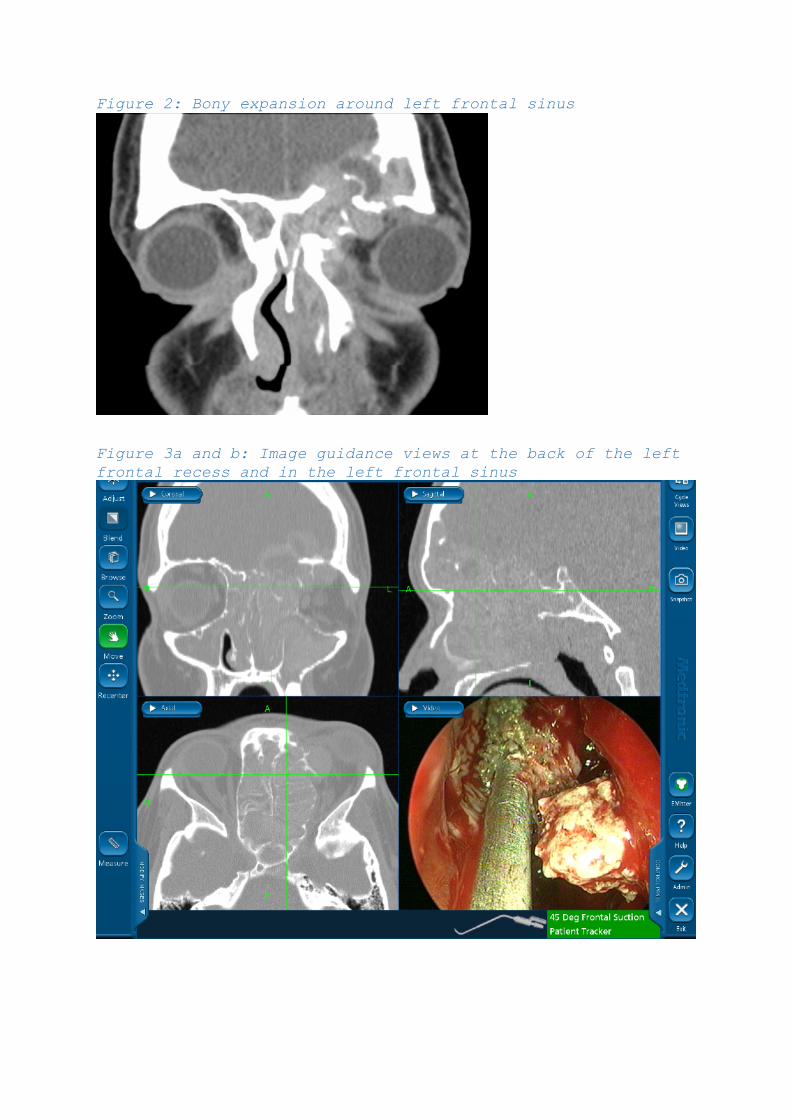
Figure 2: Bony expansion around left frontal sinus
Figure 3a and b: Image guidance views at the back of the left frontal recess and in the left frontal sinus

Conclusion Fungal sinus disease involves a spectrum of severity from invasive and potentially
fatal infection to benign affectation with poor quality of life and high rates of
potential relapse. Ultimately most scenarios involve the need for some form of
surgical debridement; in the frontal sinus this will bring specific challenges for
access and the surgeon tasked with these cases must have the skills and equipment
to be able to tackle the varying scenarios to ensure success. In the case of AFRS,
long-term follow up of patients will be needed to maintain control of the disease
with an emphasis on compliance for the patients. With increasing understanding of
disease endotypes, perhaps in the future we will see more focused treatment from
the outset in such patients.

References: 1. Turner JH, Soudry E, Nayak JV, et al. Survival outcomes in acute invasive fungal
sinusitis: a systematic review and quantitative synthesis of published evidence. Laryngoscope 2013;123(5):1112-8. doi: 10.1002/lary.23912
2. Valera FC, do Lago T, Tamashiro E, et al. Prognosis of acute invasive fungal rhinosinusitis related to underlying disease. Int J Infect Dis 2011;15(12):e841-4. doi: 10.1016/j.ijid.2011.08.005
3. deShazo RD. Fungal sinusitis. Am J Med Sci 1998;316(1):39-45. 4. Gillespie MB, O'Malley BW. An algorithmic approach to the diagnosis and
management of invasive fungal rhinosinusitis in the immunocompromised patient. Otolaryngol Clin North Am 2000;33(2):323-34. [published Online First: 2000/03/29]
5. Chen CY, Sheng WH, Cheng A, et al. Invasive fungal sinusitis in patients with hematological malignancy: 15 years experience in a single university hospital in Taiwan. BMC Infect Dis 2011;11:250. doi: 10.1186/1471-2334-11-250
6. Drakos PE, Nagler A, Or R, et al. Invasive fungal sinusitis in patients undergoing bone marrow transplantation. Bone Marrow Transplant 1993;12(3):203-8.
7. Cho HJ, Jang MS, Hong SD, et al. Prognostic factors for survival in patients with acute invasive fungal rhinosinusitis. Am J Rhinol Allergy 2015;29(1):48-53. doi: 10.2500/ajra.2015.29.4115
8. Chakrabarti A, Denning DW, Ferguson BJ, et al. Fungal rhinosinusitis: a categorization and definitional schema addressing current controversies. Laryngoscope 2009;119(9):1809-18. doi: 10.1002/lary.20520
9. deShazo RD, O'Brien M, Chapin K, et al. A new classification and diagnostic criteria for invasive fungal sinusitis. Archives of otolaryngology--head & neck surgery 1997;123(11):1181-8.
10. DelGaudio JM, Swain RE, Jr., Kingdom TT, et al. Computed tomographic findings in patients with invasive fungal sinusitis. Archives of otolaryngology--head & neck surgery 2003;129(2):236-40.
11. Halderman A, Shrestha R, Sindwani R. Chronic granulomatous invasive fungal sinusitis: an evolving approach to management. Int Forum Allergy Rhinol 2014;4(4):280-3. doi: 10.1002/alr.21299
12. Challa S, Pamidi U, Uppin SG, et al. Diagnostic accuracy of morphologic identification of filamentous fungi in paraffin embedded tissue sections: correlation of histological and culture diagnosis. Indian J Pathol Microbiol 2014;57(4):583-7. doi: 10.4103/0377-4929.142673
13. McCarthy DS. Bronchiectasis in allergic bronchopulmonary aspergillosis. Proc R Soc Med 1968;61(5):503-6.
14. Safirstein BH. Allergic bronchopulmonary aspergillosis with obstruction of the upper respiratory tract. Chest 1976;70(6):788-90. [published Online First: 1976/12/01]
15. Bent JP, 3rd, Kuhn FA. Diagnosis of allergic fungal sinusitis. Otolaryngol Head Neck Surg 1994;111(5):580-8. doi: S0194599894001075 [pii] [published Online First: 1994/11/01]
16. deShazo RD, Swain RE. Diagnostic criteria for allergic fungal sinusitis. J Allergy Clin Immunol 1995;96(1):24-35. doi: S0091-6749(95)70029-3 [pii] [published Online First: 1995/07/01]

17. Philpott CM, Javer AR, Clark A. Allergic fungal rhinosinusitis - a new staging system. Rhinology 2011;49(3):318-23. doi: 10.4193/Rhin [published Online First: 2011/08/23]
18. Meltzer EO, Hamilos DL, Hadley JA, et al. Rhinosinusitis: establishing definitions for clinical research and patient care. J Allergy Clin Immunol 2004;114(6 Suppl):155-212. doi: 10.1016/j.jaci.2004.09.029
19. Gupta R, Gupta AK. Isolated primary frontal sinus aspergillosis: role of endonasal endoscopic approach. J Laryngol Otol 2013;127(3):274-8. doi: 10.1017/S0022215112003179
20. Marple BF. Allergic fungal rhinosinusitis: A review of clinical manifestations and current treatment strategies. Medical Mycology 2006;44:S277-S84. doi: 10.1080/13693780600778650
21. Ponikau JU, Sherris DA, Kern EB, et al. The diagnosis and incidence of allergic fungal sinusitis. Mayo Clin Proc 1999;74(9):877-84. doi: 10.4065/74.9.877
22. Ferguson BJ. Eosinophilic mucin rhinosinusitis: a distinct clinicopathological entity. Laryngoscope 2000;110(5 Pt 1):799-813. doi: 10.1097/00005537-200005000-00010 [published Online First: 2000/05/12]
23. Saravanan K, Panda NK, Chakrabarti A, et al. Allergic fungal rhinosinusitis: an attempt to resolve the diagnostic dilemma. Archives of Otolaryngology Head & Neck Surgery 2006;132(2):173-8.
24. Orlandi RR, Thibeault SL, Ferguson BJ. Microarray analysis of allergic fungal sinusitis and eosinophilic mucin rhinosinusitis. Otolaryngol Head Neck Surg 2007;136(5):707-13. doi: S0194-5998(06)03528-5 [pii]
10.1016/j.otohns.2006.11.033 [published Online First: 2007/05/05] 25. Foshee J, Luminais C, Casey J, et al. An evaluation of invasive fungal sinusitis
outcomes with subsite analysis and use of frozen section analysis. Int Forum Allergy Rhinol 2016;6(8):807-11. doi: 10.1002/alr.21714
26. Pagella F, De Bernardi F, Dalla Gasperina D, et al. Invasive fungal rhinosinusitis in adult patients: Our experience in diagnosis and management. J Craniomaxillofac Surg 2016;44(4):512-20. doi: 10.1016/j.jcms.2015.12.016
27. Monroe MM, McLean M, Sautter N, et al. Invasive fungal rhinosinusitis: a 15-year experience with 29 patients. Laryngoscope 2013;123(7):1583-7. doi: 10.1002/lary.23978
28. Bernardini E, Karligkiotis A, Fortunato S, et al. Surgical and pathogenetic considerations of frontal sinus fungus ball. European Archives of Oto-Rhino-Laryngology 2017;274(6):2493-97. doi: 10.1007/s00405-017-4531-x
29. Chen IH, Chen TM. Isolated frontal sinus aspergillosis. Otolaryngol Head Neck Surg 2000;122(3):460-1. doi: 10.1067/mhn.2000.99036
30. Kodama S, Moriyama M, Okamoto T, et al. Isolated frontal sinus aspergillosis treated by endoscopic modified Lothrop procedure. Auris Nasus Larynx 2009;36(1):88-91. doi: 10.1016/j.anl.2008.02.004
31. Mukherji SK, Figueroa RE, Ginsberg LE, et al. Allergic fungal sinusitis: CT findings. Radiology 1998;207(2):417-22.
32. Marple BF. Allergic fungal rhinosinusitis: current theories and management strategies. Laryngoscope 2001;111(6):1006-19.
33. Snidvongs K, Pratt E, Chin D, et al. Corticosteroid nasal irrigations after endoscopic sinus surgery in the management of chronic rhinosinusitis. Int Forum Allergy Rhinol 2012;2(5):415-21. doi: 10.1002/alr.21047

34. Ferguson BJ. Mucormycosis of the nose and paranasal sinuses. Otolaryngol Clin North Am 2000;33(2):349-65.
35. Kennedy CA, Adams GL, Neglia JP, et al. Impact of surgical treatment on paranasal fungal infections in bone marrow transplant patients. Otolaryngol Head Neck Surg 1997;116(6 Pt 1):610-6. doi: 10.1016/S0194-59989770236-5
36. Saedi B, Sadeghi M, Seilani P. Endoscopic management of rhinocerebral mucormycosis with topical and intravenous amphotericin B. J Laryngol Otol 2011;125(8):807-10. doi: 10.1017/S0022215111001289
37. Klossek JM, Serrano E, Peloquin L, et al. Functional endoscopic sinus surgery and 109 mycetomas of paranasal sinuses. Laryngoscope 1997;107(1):112-7.
38. Busaba NY, Colden DG, Faquin WC, et al. Chronic invasive fungal sinusitis: a report of two atypical cases. Ear Nose Throat J 2002;81(7):462-6. [published Online First: 2002/08/02]
39. Li Y, Li Y, Li P, et al. Diagnosis and endoscopic surgery of chronic invasive fungal rhinosinusitis. Am J Rhinol Allergy 2009;23(6):622-5. doi: 10.2500/ajra.2009.23.3361
40. Stringer SP, Ryan MW. Chronic invasive fungal rhinosinusitis. Otolaryngol Clin North Am 2000;33(2):375-87. [published Online First: 2000/03/29]
41. D'Anza B, Stokken J, Greene JS, et al. Chronic invasive fungal sinusitis: characterization and shift in management of a rare disease. Int Forum Allergy Rhinol 2016;6(12):1294-300. doi: 10.1002/alr.21828
42. Kuhn FA, Swain R, Jr. Allergic fungal sinusitis: diagnosis and treatment. Curr Opin Otolaryngol Head Neck Surg 2003;11(1):1-5. [published Online First: 2003/09/30]
43. Philpott CM, Thamboo A, Lai L, et al. Olfactory dysfunction in allergic fungal rhinosinusitis. Archives of otolaryngology--head & neck surgery 2011;137(7):694-7. doi: 137/7/694 [pii]
10.1001/archoto.2011.105 [published Online First: 2011/07/20]

Table 1: Diagnostic Criteria for AFRS – Bent & Kuhn¥/Vancouver*
Table 2a and 2b: Philpott-Javer Endoscopic Staging system for AFRS
Major Minor
Type 1 hypersensitivity¥/Immunocompetence*
Asthma
Nasal polyposis Unilateral disease
Characteristic CT findings Bone erosion
Eosinophilic mucin without invasion Fungal cultures
Positive fungal stain Charcot-Leyden crystals
Serum eosinophilia
Grading State of mucosa
0 No oedema
1-3 Mucosal oedema (mild/moderate/severe)
4-6 Polypoid oedema (mild/moderate/severe)
7-9 Frank polyps (mild/moderate/severe)
Sinus cavity Right Mucin Left Mucin
Olfactory cleft 0-9 1 0-9 1
Frontal 0-9 1 0-9 1
Ethmoid 0-9 1 0-9 1
Maxillary 0-9 1 0-9 1
Sphenoid 0-9 1 0-9 1
Total (maximum score) 50 50

Bilateral total 100