alexithymia and apathy in parkinson’s disease...
TRANSCRIPT

Behavioural Neurology 27 (2013) 535–545 535DOI 10.3233/BEN-129021IOS Press
Research Report
Alexithymia and apathy in Parkinson’sdisease: Neurocognitive correlates
Yelena Bogdanovaa,b,c,d,∗ and Alice Cronin-Golombb
aPsychology Research, VA Boston Healthcare System, Boston, MA, USAbDepartment of Psychology, Boston University, Boston, MA, USAcDepartment of Psychiatry, Boston University School of Medicine, Boston, MA, USAdDepartment of Psychiatry, Harvard Medical School, Boston, MA, USA
Abstract. Non-motor symptoms such as neuropsychiatric and cognitive dysfunction have been found to be common in Parkinson’sdisease (PD) but the relation between such symptoms is poorly understood. We focused on alexithymia, an impairment ofaffective and cognitive emotional processing, as there is evidence for its interaction with cognition in other disorders. Twenty-twonon-demented PD patients and 22 matched normal control adults (NC) were administered rating scales assessing neuropsychiatricstatus, including alexithymia, apathy, and depression, and a series of neuropsychological tests. As expected, PD patients showedmore alexithymia than NC, and there was a significant association between alexithymia and disease stage. Alexithymia wasassociated with performance on non-verbally mediated measures of executive and visuospatial function, but not on verballymediated tasks. By contrast, there was no correlation between cognition and ratings of either depression or apathy. Our findingsdemonstrate a distinct association of alexithymia with non-verbal cognition in PD, implicating right hemisphere processes, anddifferentiate between alexithymia and other neuropsychiatric symptoms in regard to PD cognition.
Keywords: Non-motor symptoms, basal ganglia, ACC, frontostriatal, right hemisphere
1. Introduction
Parkinson’s disease (PD) is traditionally character-ized as a motor disorder, but the existence of non-motorsymptoms is attracting increasing attention from clin-icians and researchers because of their impact on pa-tients’ quality of life. Among others, the non-motorsymptoms include cognitive dysfunction [1–4] and neu-ropsychiatric symptoms such as apathy [5–13], depres-sion [14–19], and alexithymia [20,21]. PD-associatedcognitive impairments have been attributed to reduceddopaminergic innervation in the nigrostriatal andmeso-
∗Corresponding author: Yelena Bogdanova, Ph.D., VA BostonHealthcare System, Psychology Research (151-A), 150 South Hunt-ington Avenue, Boston, MA 02130, USA. E-mail: [email protected].
cortical dopaminergic systems, leading to dysfunctionof neural circuits including the basal ganglia and corti-cal projection areas (reviewed in [3]). PET studies haveshown decreased network activity [22] and metabolicabnormalities [23] in prefrontal and parietal brain re-gions in PD patients with mild cognitive impairment,consistent with the cognitive domains commonly af-fected in PD: executive and visuospatial function [24].
Alexithymia, an impairment of affective and cog-nitive emotional processing, is a common neuropsy-chiatric concomitant of PD [25], with an estimatedprevalence of 21% in non-demented patients [21,26].Dopamine depletion in brain areas critical for emotion-al cognition, such as anterior cingulate cortex (ACC)and orbitofrontal cortex [27,28], may underlie the man-ifestation of alexithymia in this disorder. Though under-studied, it has been suggested that alexithymia shouldbe considered and treated as an important non-motor
ISSN 0953-4180/13/$27.50 2013 – IOS Press and the authors. All rights reserved

536 Y. Bogdanova and A. Cronin-Golomb / Alexithymia in PD
symptom of PD [20]. The multifaceted construct ofalexithymia is characterized by the following: a) dif-ficulty identifying and distinguishing between feelingsand bodily sensations of emotional arousal, b) difficul-ty describing feelings, c) reduced imaginal processes,and d) a stimulus-bound, externally-oriented cognitivestyle [29]. Alexithymia characteristics reflect deficitsin the cognitive processing and regulation of emotions,such as the inability to apply adaptive processes foraffect regulation (e.g., modulating arousal, expressingor suppressing emotions, and cognitive assimilation),which may in turn contribute to the development andcourse of various psychiatric and medical disorders,such as depression and diabetes [30–32], and affecttreatment outcome [33–35].
According to a current model, alexithymia derivesfrom dysfunction of frontal areas [36,37], specifical-ly the ACC and prefrontal cortex [38]. Support forthis model comes from neuroimaging studies [39–43],which also have related alexithymia to predominant-ly right hemisphere dysfunction. Scores on the stan-dard measure of alexithymia, the Toronto AlexithymiaScale (TAS-20 total), were reported to be significantlycorrelated with gray matter volume of the right, butnot left, pregenual ACC in a sample of healthy olderadults [44]. In another study of normal aging, a signif-icant correlation was found between right rostral ACCvolume and alexithymia ratings, with more advancedage corresponding to higher alexithymia ratings andto smaller overall right ACC total gray matter volumein rostral and dorsal subregions as well as in the leftrostral subregion [45]. Alexithymia has also been re-lated to right-hemisphere lesions in individuals withstroke [46].
There is little known in regard to the relation be-tween alexithymia and cognitive dysfunction in PD. Inone study alexithymia was associated with poor perfor-mance on a cognitive (visuospatial) test [47]. In indi-vidualswithHIV, which is like PD in that it affects fron-tostriatal areas, associations were documented betweenalexithymia, but not depression, and performance onmeasures of executive and visuospatial function [48].
While alexithymia is often associated with depres-sion [49–51], previous research has indicated that theyshould be considered distinct constructs [52–55]. InPD, some studies also related alexithymia and depres-sion [26,56], while others showed that the two condi-tions may be reliably dissociated in this disorder [20,21], suggesting that alexithymia is at least partly inde-pendent of depression in PD andmay be directly relatedto the disease process [20].
Apathy, like alexithymia, is associated with dam-age to the frontal lobes and disruption of the frontal-subcortical anterior cingulate circuits [57–59]. Neu-roimaging research has shown that the severity of ap-athy is associated with the volume of the nucleus ac-cumbens, one of the central structures in the anteriorcingulate frontal-subcortical circuit [60]. Apathy, likealexithymia, also appears to be more common in pa-tients with right- than left-hemisphere damage [9,61].Neuroimaging research has shown that apathy is asso-ciated with decreased gray matter volume in the rightanterior cingulate in older adults [62]. In PD, apathywas correlated with low gray matter density in bilat-eral inferior frontal gyrus and inferior parietal gyrus,right cingulate and right precuneus in one study [63];and with low volume of the right medial temporal lobein another study [5]. Also in PD, apathy has been re-lated to emotional cognition, specifically the ability toattribute emotional states to others [64]. Similarly, ap-athy has been related to alexithymia in patients withfrontostriatal dysfunction arising from neurodegenera-tive disease [44] and HIV [48]. The anatomical overlapin neural networks involved in alexithymia and apathy,together with the neuropsychological findings, raisesthe question of potential overlap in the expressions ofalexithymia and apathy in PD.
The present study aimed to explore the expressionof neuropsychiatric symptoms in PD without dementiaby assessing the effect of neuropsychiatric status, andalexithymia in particular, on cognitive function. Exam-ining the relation between emotional processing andcognitive function is important from both clinical andtheoretical perspectives, as it may provide insights intothe interaction of emotional and cognitive neural sys-tems. The present study had two main aims. The firstwas to explore the association between alexithymia andcognition in PD and to consider the mechanisms un-derlying an association. The ACC connections with theprefrontal and parietal cortices are important for bothcognitive (executive in particular) and emotional func-tion [65–67]. We hypothesized that alexithymia levelshould be associated with cognitive dysfunction asso-ciated with these brain areas, including attention, exec-utive function, and visuospatial processing. To addressan ongoing debate in the literature regarding lateralized(left vs. right hemisphere) expression of alexithymiaand follow the lead of one neuropsychological study ofPD that suggested an association between alexithymiaand cognitive abilitiesmediated by the right hemisphere(i.e., visuospatial function) [47], in the present studywe related alexithymia to material-specific cognitive

Y. Bogdanova and A. Cronin-Golomb / Alexithymia in PD 537
domains (verbal vs. non-verbal). We also explored therelation between cognitive performance and various as-pects (factors) of alexithymia in PD, as neuroimagingstudies have suggested differentiation between neuralsystems associated with specific aspects of alexithymia.The second aim was to examine the potential over-lap between alexithymia and apathy in PD. Based onthe results of imaging and behavioral studies as de-scribed above, we hypothesized that if apathy and alex-ithymia share common neurophysiological substrates,there should be an association between the levels ofapathy and alexithymia. We also examined the extentof overlap between alexithymia and apathy in regard totheir relation to cognition in PD.
2. Methods
2.1. Participants
Twenty-two consecutively-enrolled non-dementedindividuals with idiopathic PD (10 men, 12 women)and 22 normal control adults (NC) (10 men, 12 wom-en) matched on socio-demographic variables partici-pated in the study. All participants scored 28 or higheron the Mini-Mental State Examination (MMSE) [68]and were not demented. PD participants were recruitedfrom the ParkinsonClinic of the Department of Neurol-ogy, Boston Medical Center and through local supportgroups. NC were recruited from the community. Theresearch was approved by Boston University’s Institu-tional Review Board. Participants were required to benative speakers of English. Exclusion criteria includedco-existing cancer, serious cardiac disease, other seri-ous chronic medical illness, prior intracranial surgery,history of traumatic brain injury, psychiatric diagnosis(exclusive of depression or anxiety) or neurological di-agnoses other than PD; history of alcoholism or otherdrug abuse; history of eye disease or other abnormal-ities; and use of psychoactive medications, except foruse of antidepressants and anxiolytics in the PD group,which are commonly prescribed. PD clinical stagingwas determined by the patient’s neurologist based onthe widely used index of motor disability, the Hoehnand Yahr scale (H&Y) [69]. All PD participants werestages I–III (mild to moderate bilateral). The averageduration of disease was 7.8 years (SD = 3.1). PD di-agnosis was made by patients’ neurologists, using UKParkinson’s Disease Society Brain Bank clinical diag-nostic criteria [70]. Side of motor symptom onset wasobtained by patient report and fromneurologist records.
Eleven patients had initial motor symptom on the rightbody side (RPD: 5 men, 6 women) and 11 had left-side onset (LPD: 5 men, 6 women). RPD and LPDgroups did not differ in age, education, MMSE, H&Ystage, or disease duration. There were no group dif-ferences between male and female PD participants forage, education, MMSE, H&Y stage, or disease dura-tion. All PD participants received daily dopamine re-placement therapy and /or dopamine receptor agonists.Levodopa equivalent dosages (LED) were calculatedbased on previous reports with LED: (regular levodopadose× 1) + (levodopa controlled-release dose× 0.75)+ (pramipexole dose × 67.0) + (ropinirole dose ×16.67) + (rotigotine × 16.67) + (pergolide dose andcabergoline dose × 67.0) + (bromocriptine dose ×10) + ([regular levodopa dose + levodopa controlled-release dose × 0.75] × 0.25, if taking tolcapone orentacapone) [71]. None of the PD participants weretaking anticholinergic medications. Three were takingsome form of sleep medication. PD participants weretested while on their anti-parkinsonian medications (intheir “on” state).
2.2. Procedure
Study participants were administered standardizedmeasures of neuropsychiatric functioning and a seriesof standardized neuropsychologicalmeasures sensitiveto PD-associated cognitive impairments. The cognitivetests were chosen to sample both verbal and non-verbaldomains. Because the PDandNCgroupswerematchedon age, education, and male:female ratio, we comparedand report raw scores for all tests. Before participating,each individual provided written informed consent.
2.2.1. Neuropsychiatric status assessmentThe 20-itemToronto Alexithymia Scale (TAS-20)was
administered to evaluate alexithymia [72]. It compris-es three subscales that reflect distinct facets of alex-ithymia: Factor 1 (difficulty identifying feelings anddistinguishing them from bodily sensations of emotion,DIF), Factor 2 (difficulty describing feelings, DDF),and Factor 3 (externally oriented thinking, EOT). Itemresponses were rated on a five-point Likert scale rang-ing from 1 to 5. Higher scores reflect more severe alex-ithymia. Based on the total score, individuals are cate-gorized as non-alexithymic (scores ranging from 20 to51), borderline alexithymic (scores ranging from 52 to60), or alexithymic (scores � 61) [72].
Apathy was assessed using the modified 14-item Ap-athy Scale (AS) [10,73]. Items are rated on a 0-to-3Lik-

538 Y. Bogdanova and A. Cronin-Golomb / Alexithymia in PD
ert scale. Scores range from 0 to 42, with higher scoresindicative of more severe apathy. Total score was thedependent measure. AS and its modified version werereported to have excellent psychometric properties andhave been used to study PD [10].
The Beck Depression Inventory, Second Edition(BDI-II) [74] is a 21-item self-report instrument thatassesses the existence and severity of symptoms of de-pression as listed in the American Psychiatric Asso-ciation’s Diagnostic and Statistical Manual of MentalDisorders, Fourth Edition [DSM-IV; [75]]. There is afour-point scale for each item ranging from0 to 3. Totalscore in the range of 0–13 is considered indicative ofminimal depression, 14–19 is mild, 20–28 is moderate,and 29–63 is severe. The BDI is considered a reliableand valid measure of depression in PD [76].
2.2.2. Cognitive functioning assessmentThe neuropsychological series included a number of
tests that we expected to be sensitive to frontostriataland parietal dysfunction (attention, executive function,visuospatial ability) [3,8,48]. A focus of interest wason the potential dissociation between correlations ofneuropsychiatric status and performance on the verbal-ly and non-verbally mediated cognitive tests.
2.2.2.1. Verbal domainControlled Oral Word Association Test [77]. This is
a test of verbal fluency: (1) phonemic fluency, in whichparticipants were required to generate words beginningwith a particular letter (F, A, S); and (2) category flu-ency, in which participants generate words that belongto a particular category (Animals). Total number cor-rect within a 60-s time period for each condition wasrecorded.
Digit Span, Wechsler Memory Scale III [78], is ameasure of the efficiency of attention (Forward Span)and working memory (Backward Span) in the verbaldomain [79]. The total score was used for the groupcomparison.
Subtracting from 100 by 7 [80] is a task of sequentialarithmetic operation. A neuroimaging study demon-strated bilateral prefrontal and posterior parietal cortexactivation during silent subtraction by sevens in healthyadults [81]. The task is presented verbally and requiresa spoken verbal answer. Time to completion and num-ber of errors was recorded.
2.2.2.2. Non-verbal domainThe Trail Making Test (TMT) [82] is a test of psy-
chomotor speed, attention, and executive functioning
that consists of two subtests. Trails A is a test of simpleattention and psychomotor speed, in which participantsconnect numbered circles in ascending order (1-2-3,etc.). Trails B is a measure of combined visual search,psychomotor speed, and cognitive flexibility, assessingthe ability to shift and maintain the response set. Par-ticipants sequentially alternate between alphanumericsequences (1-A-2-B, etc.). Time to completion andnumber of errors were recorded.
Raven’sColouredProgressiveMatrices (RCPM) [83].This is a measure of visuospatial skills and reasoningability. The task is to choose one of six possible com-pletions of an incomplete pattern matrix. Total score(the number correct out of 36 items) was recorded.
Clock reading test [80]. Participants are asked toidentify and mark the time shown on each of the10 clocks presented on a standard paper. Time to com-pletion and number of errors were recorded.
Visual symbol search test [84] provides a measure ofvisual scanning abilities and sustained attention. Par-ticipants search and cancel the target symbol in thenon-verbal array. Time to completion was recorded.
Boston Visuospatial Quantitative Battery (BVSQB;[85]). On the Drawing to Command subtest, the partic-ipant is required to draw six objects, one at a time. To-tal number of correct points was recorded. Right-LeftOrientation subtest requires the identification of rightand left on 20 different body parts and objects drawn invarious positions. The number of errors was recorded.
2.2.3. Statistical analysesTo analyze differences between the PD and NC
groups, independent samples t-tests (2-tailed) wereused. Alpha was set to 0.01 to account for multiplecomparisons. Analyses of performance of men andwomen in the PD and NC groups revealed no signifi-cant differences in neuropsychological profile or moodratings, and data were accordingly collapsed acrossthese subgroups. Pearson correlations were performedto examine associations between neuropsychiatric sta-tus and neuropsychological performance. Linear re-gression analyses were then performed to examine therole of neuropsychiatric and neuropsychological fac-tors in predicting alexithymia in PD, with alexithymiaas the dependent variable and neuropsychiatric testscores (apathy and depression) and neuropsychologi-cal test scores (those correlated with alexithymia) asindependent variables.
3. Results
This study used a mixed design, with each partici-pant receiving all assessments. The results are divided
Y. Bogdanova and A. Cronin-Golomb / Alexithymia in PD 539
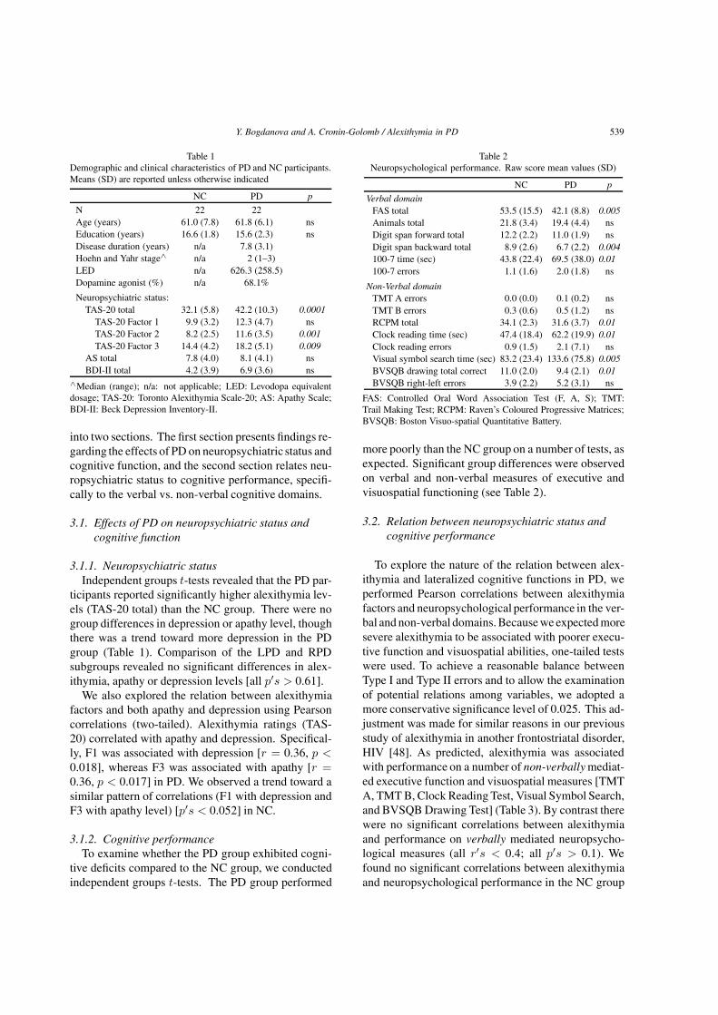
Table 1Demographic and clinical characteristics of PD and NC participants.Means (SD) are reported unless otherwise indicated
NC PD p
N 22 22Age (years) 61.0 (7.8) 61.8 (6.1) nsEducation (years) 16.6 (1.8) 15.6 (2.3) nsDisease duration (years) n/a 7.8 (3.1)Hoehn and Yahr stage∧ n/a 2 (1–3)LED n/a 626.3 (258.5)Dopamine agonist (%) n/a 68.1%
Neuropsychiatric status:TAS-20 total 32.1 (5.8) 42.2 (10.3) 0.0001
TAS-20 Factor 1 9.9 (3.2) 12.3 (4.7) nsTAS-20 Factor 2 8.2 (2.5) 11.6 (3.5) 0.001TAS-20 Factor 3 14.4 (4.2) 18.2 (5.1) 0.009
AS total 7.8 (4.0) 8.1 (4.1) nsBDI-II total 4.2 (3.9) 6.9 (3.6) ns
∧Median (range); n/a: not applicable; LED: Levodopa equivalentdosage; TAS-20: Toronto Alexithymia Scale-20; AS: Apathy Scale;BDI-II: Beck Depression Inventory-II.
into two sections. The first section presents findings re-garding the effects of PD on neuropsychiatric status andcognitive function, and the second section relates neu-ropsychiatric status to cognitive performance, specifi-cally to the verbal vs. non-verbal cognitive domains.
3.1. Effects of PD on neuropsychiatric status andcognitive function
3.1.1. Neuropsychiatric statusIndependent groups t-tests revealed that the PD par-
ticipants reported significantly higher alexithymia lev-els (TAS-20 total) than the NC group. There were nogroup differences in depression or apathy level, thoughthere was a trend toward more depression in the PDgroup (Table 1). Comparison of the LPD and RPDsubgroups revealed no significant differences in alex-ithymia, apathy or depression levels [all p′s > 0.61].
We also explored the relation between alexithymiafactors and both apathy and depression using Pearsoncorrelations (two-tailed). Alexithymia ratings (TAS-20) correlated with apathy and depression. Specifical-ly, F1 was associated with depression [r = 0.36, p <0.018], whereas F3 was associated with apathy [r =0.36, p < 0.017] in PD. We observed a trend toward asimilar pattern of correlations (F1 with depression andF3 with apathy level) [p′s < 0.052] in NC.
3.1.2. Cognitive performanceTo examine whether the PD group exhibited cogni-
tive deficits compared to the NC group, we conductedindependent groups t-tests. The PD group performed
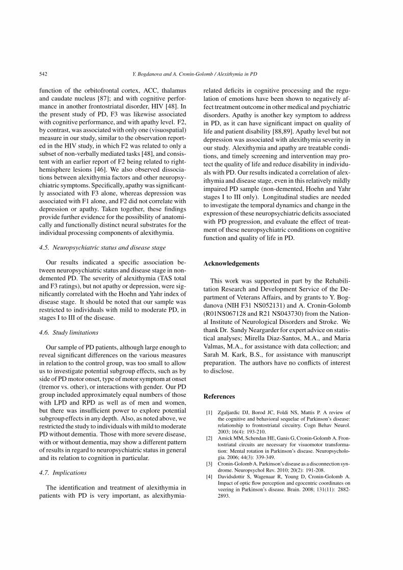
Table 2Neuropsychological performance. Raw score mean values (SD)
NC PD p
Verbal domainFAS total 53.5 (15.5) 42.1 (8.8) 0.005Animals total 21.8 (3.4) 19.4 (4.4) nsDigit span forward total 12.2 (2.2) 11.0 (1.9) nsDigit span backward total 8.9 (2.6) 6.7 (2.2) 0.004100-7 time (sec) 43.8 (22.4) 69.5 (38.0) 0.01100-7 errors 1.1 (1.6) 2.0 (1.8) ns
Non-Verbal domainTMT A errors 0.0 (0.0) 0.1 (0.2) nsTMT B errors 0.3 (0.6) 0.5 (1.2) nsRCPM total 34.1 (2.3) 31.6 (3.7) 0.01Clock reading time (sec) 47.4 (18.4) 62.2 (19.9) 0.01Clock reading errors 0.9 (1.5) 2.1 (7.1) nsVisual symbol search time (sec) 83.2 (23.4) 133.6 (75.8) 0.005BVSQB drawing total correct 11.0 (2.0) 9.4 (2.1) 0.01BVSQB right-left errors 3.9 (2.2) 5.2 (3.1) ns
FAS: Controlled Oral Word Association Test (F, A, S); TMT:Trail Making Test; RCPM: Raven’s Coloured Progressive Matrices;BVSQB: Boston Visuo-spatial Quantitative Battery.
more poorly than the NC group on a number of tests, asexpected. Significant group differences were observedon verbal and non-verbal measures of executive andvisuospatial functioning (see Table 2).
3.2. Relation between neuropsychiatric status andcognitive performance
To explore the nature of the relation between alex-ithymia and lateralized cognitive functions in PD, weperformed Pearson correlations between alexithymiafactors and neuropsychological performance in the ver-bal and non-verbal domains.Becausewe expectedmoresevere alexithymia to be associated with poorer execu-tive function and visuospatial abilities, one-tailed testswere used. To achieve a reasonable balance betweenType I and Type II errors and to allow the examinationof potential relations among variables, we adopted amore conservative significance level of 0.025. This ad-justment was made for similar reasons in our previousstudy of alexithymia in another frontostriatal disorder,HIV [48]. As predicted, alexithymia was associatedwith performance on a number of non-verbally mediat-ed executive function and visuospatial measures [TMTA, TMT B, Clock Reading Test, Visual Symbol Search,and BVSQB Drawing Test] (Table 3). By contrast therewere no significant correlations between alexithymiaand performance on verbally mediated neuropsycho-logical measures (all r′s < 0.4; all p′s > 0.1). Wefound no significant correlations between alexithymiaand neuropsychological performance in the NC group

540 Y. Bogdanova and A. Cronin-Golomb / Alexithymia in PD
Table 3Correlations between alexithymia factor scores and performance onnon-verbal neuropsychological tests in PD
TAS-20 TAS-20 TAS-20Factor 1 Factor 2 Factor 3
TMT A errors 0.667*** ns nsTMT B errors ns ns 0.529**RCPM total ns ns nsClock reading errors ns ns 0.435*Visual symbol search time ns 0.434* nsBVSQB drawing total correct −0.535∗∗ ns nsBVSQB right-left errors 0.523** ns ns
TAS-20: Toronto Alexithymia Scale-20; Factor 1: difficulty iden-tifying feelings; Factor 2: difficulty describing feelings; Factor 3:externally oriented thinking; FAS: Controlled Oral Word AssociationTest (F, A, S); TMT: Trail Making Test; RCPM: Raven’s ColouredProgressive Matrices; BVSQB: Boston Visuo-spatial QuantitativeBattery; Note: There were no significant correlations between alex-ithymia factor scores and performance on neuropsychological testsin the verbal domain. *p < 0.25, **p < 0.01, **p < 0.001.
(all r′s < 0.3; all p′s > 0.15). There were no cor-relations between apathy (AS) or depression (BDI-II)ratings and neuropsychological performance for eithergroup (all r′s < 0.3; all p′s > 0.17).
To further examine the role of neuropsychiatric andneuropsychological factors in predicting alexithymia(TAS-20 total) in PD,we performedmultiple regressionanalyses. The first variable entered into the equationwas apathy (AS) [slope = 0.542, R2 = 0.294, t(20) =2.4, p = 0.03], indicating an expected increase of 0.54in TAS-20 total score for each point increase on AS.In the second step, TMT A (errors) was entered andsignificantly contributed to predicting the level of alex-ithymia [slope = 0.763, R2 change = 0.289, t(19) =3.0, p = 0.01]. Patients who had more errors on TMTA showed a higher alexithymia level, with expectedincrease of 0.76 for each point increase on TMT Aerrors.
We also assessed the association between degreeof alexithymia and disease stage (H&Y) using multi-ple Spearman’s correlations (two-tailed). Alexithymiaratings significantly correlated with disease stage asshown for the overall alexithymia rating (TAS-20 total:r = 0.56, p < 0.020) and for F3 specifically (exter-nally oriented thinking) (r = 0.59, p < 0.013). Bycontrast, there were no significant correlations betweenH&Y stage and severity of apathy (AS) or depression(BDI-II) (all r′s < 0.39; all p′s > 0.12).
4. Discussion
We examined the effect of PD on alexithymia andits relation to cognition in non-demented individuals
with PD. First, as expected, we found the expectedPD-related deficits in verbal and non-verbal cognitivedomains, including attention, executive function andvisuospatial function, which is reflective of frontostri-atal and parietal dysfunction associated with the dis-order. Second, also as predicted, PD patients reportedsignificantly higher levels of alexithymia than the NCgroup. The group differences applied to the total alex-ithymia score and to two of the three alexithymia pro-cessing components, F2 (difficulty describing feelings)and F3 (externally oriented thinking). The extent ofalexithymia and each of its processing components (F1,F2, and F3) but not the extent of depression or apathy,significantly correlated with performance on neuropsy-chological measures of attention, executive and visu-ospatial function in this sample. There was a differen-tial association between alexithymia and performanceof tests in the non-verbal, but not the verbal, cognitivedomain, supporting our hypothesis that alexithymia isassociated with cognitive deficits mediated predomi-nantly by the right hemisphere. The results are in ac-cord with those of a study that showed an association ofalexithymia and performance on a visuospatial task in ahospitalized non-demented PD patient sample [47]. Toour knowledge, ours is the first study to demonstrate adifferential association of alexithymia and apathy withcognitive dysfunction in PD in relation to the lateral-ized cognitive domains. While there was a significantassociation between alexithymia factors, apathy anddepression in PD, neither apathy nor depression levelcorrelated with cognitive performance.
4.1. Potential brain substrates of alexithymia in PD
The extent of alexithymia significantly correlatedwith attention, executive and visuospatial function,cognitive domains associated with frontostriatal andparietal pathology. This finding supports the current“frontal” alexithymia model, which postulates thatalexithymia is the result of dysfunctional mechanismsin the frontal cortex [36–38].The visuospatial cognitivedomain reflecting parietal dysfunction is also affectedby PDandwas associatedwith alexithymia in our study.The ACC connections with the prefrontal and parietalcortex are important for both cognitive and emotionalfunction [65–67], which may explain the significant re-lation between visuospatial dysfunction and the extentof alexithymia in PD.
4.2. Apathy and alexithymia in PD
Previous imaging and behavioral research (reviewedin the Introduction) have suggested that apathy and

Y. Bogdanova and A. Cronin-Golomb / Alexithymia in PD 541
alexithymia may share some common neurophysiolog-ical substrates within frontostriatal circuits, and relatedapathy and alexithymia to right-hemisphere dysfunc-tion. We found that apathy and alexithymia differential-ly correlated with performance on neuropsychologicalmeasures. Whereas alexithymia was significantly as-sociated with performance on non-verbal measures ofexecutive function and visuospatial ability, apathy wasnot associated with performance on any of the tasks.The fact that apathy and alexithymia can be dissociatedin PD implies that they have at least partially distinctneural substrates within frontostriatal circuits.
The observed dissociation between the two neu-ropsychiatric conditions in relation to cognition sug-gests at least two potential interpretations: (1) the un-derlying neural substrates for apathy and alexithymiaare differentially affected by PDneuropathological pro-cesses; (2) it was too early in the course of the diseaseto detect the apathy-associated changes in our sample.In line with this argument, we found no differences inapathy levels between our non-demented PD and NCgroups, which is consistent with the notion that apathymay be a reflection of more advanced (moderate to se-vere) stage of PD [25]. There was, however, a signif-icant correlation between apathy level and severity ofalexithymia (F3) in our sample. Previous neuroimag-ing research in healthy older adults has found a sig-nificant correlation between right rostral ACC volumeand alexithymia ratings (TAS-20) for both total scoreand F3 [45]. Taken together, these findings suggest apotential overlap in the neural substrates (the rostralsubregion of ACC) of apathy and alexithymia, specificto F3.
4.3. Depression and alexithymia in PD
We found no significant group difference betweenPD and NC for depression, though there was a trendfor the PD group to be more depressed. The severity ofdepression correlated with alexithymia level (TAS-20,F1) but did not correlate with performance on any ofthe cognitive tests, meaning that the neuropsychologi-cal abnormalities observed in our sample could not beattributed to depression.
The results of multiple regression analyses furtherclarified the association between alexithymia, apathyand depression in PD. We found that apathy, but not de-pression, predicted higher levels of alexithymia. Thesefindings are consistent with previous studies that indi-cated that although alexithymia may be associated with
depression, these neuropsychiatric conditions can bedissociated in patients with PD [20,21].
Of note, in contrast to what was found in our PDgroup, in our healthy control group there were no corre-lations between alexithymia (or apathy or depression)and performance on the neuropsychological measuresin NC group in our study. It is possible that the interac-tion between the processing componentsof alexithymiaand cognitive dysfunction occurs only in the presenceof cognitive deficits. These results are consistent withour findings of a significant correlation between alex-ithymia and cognition in patients with HIV but not intheir matched healthy control group [48].
4.4. PD and the individual processing components ofalexithymia
We explored the association between cognition andthe individual processing components of alexithymiain PD, as neuroimaging and behavioral studies havesuggested distinct neural mechanisms underlying thecomponents. In one study of normal aging, there was asignificant correlation between right rostral ACC vol-ume and alexithymia ratings on TAS-20 F3 (External-ly Oriented Thinking, EOT) [45]. These findings sug-gested that deterioration in the rostral subregion of theACC with age may contribute to alexithymia, and morespecifically to F3. In a study of patients with stroke, F1,Difficulty Identifying Feelings (DIF), and F2, Difficul-ty Describing Feelings (DDF), were related to right-hemisphere lesions [46]. Though limited in number,these studies providedevidence that individual process-ing components of alexithymia may be associated withdistinct brain regions. Specifically, F3 is thought to de-pend on the rostral ACC. Our results support this no-tion, as we observed group differences between PD andNC on F3 but not F1. See Bogdanova et al. [48] for fur-ther consideration of the role of the ACC in emotionalcognition.
Dissociations between cognitive and affective as-pects of alexithymia (reflected by alexithymia factors)have been reported previously in clinical and non-clinical populations [86]. F1 (DIF) reflects the affectivedimension of alexithymia, and was found to be strong-ly associated with depression ratings in a clinically de-pressed group [87], which is consistent with our find-ings. F3 (EOT, considered the cognitive component)was found to be significantly associated with smallerrostral ACC volume in healthy older adults [45]; withdiagnosis of obsessive-compulsive disorder, a fronto-striato-thalamocortical disorder associated with dys-
542 Y. Bogdanova and A. Cronin-Golomb / Alexithymia in PD
function of the orbitofrontal cortex, ACC, thalamusand caudate nucleus [87]; and with cognitive perfor-mance in another frontostriatal disorder, HIV [48]. Inthe present study of PD, F3 was likewise associatedwith cognitive performance, and with apathy level. F2,by contrast, was associated with only one (visuospatial)measure in our study, similar to the observation report-ed in the HIV study, in which F2 was related to only asubset of non-verballymediated tasks [48], and consis-tent with an earlier report of F2 being related to right-hemisphere lesions [46]. We also observed dissocia-tions between alexithymia factors and other neuropsy-chiatric symptoms. Specifically, apathywas significant-ly associated with F3 alone, whereas depression wasassociated with F1 alone, and F2 did not correlate withdepression or apathy. Taken together, these findingsprovide further evidence for the possibility of anatomi-cally and functionally distinct neural substrates for theindividual processing components of alexithymia.
4.5. Neuropsychiatric status and disease stage
Our results indicated a specific association be-tween neuropsychiatric status and disease stage in non-demented PD. The severity of alexithymia (TAS totaland F3 ratings), but not apathy or depression, were sig-nificantly correlated with the Hoehn and Yahr index ofdisease stage. It should be noted that our sample wasrestricted to individuals with mild to moderate PD, instages I to III of the disease.
4.6. Study limitations
Our sample of PD patients, although large enough toreveal significant differences on the various measuresin relation to the control group, was too small to allowus to investigate potential subgroup effects, such as byside of PD motor onset, type of motor symptom at onset(tremor vs. other), or interactions with gender. Our PDgroup included approximately equal numbers of thosewith LPD and RPD as well as of men and women,but there was insufficient power to explore potentialsubgroupeffects in any depth. Also, as noted above,werestricted the study to individualswithmild to moderatePD without dementia. Those with more severe disease,with or without dementia, may show a different patternof results in regard to neuropsychiatric status in generaland its relation to cognition in particular.
4.7. Implications
The identification and treatment of alexithymia inpatients with PD is very important, as alexithymia-
related deficits in cognitive processing and the regu-lation of emotions have been shown to negatively af-fect treatment outcome in othermedical and psychiatricdisorders. Apathy is another key symptom to addressin PD, as it can have significant impact on quality oflife and patient disability [88,89]. Apathy level but notdepression was associated with alexithymia severity inour study. Alexithymia and apathy are treatable condi-tions, and timely screening and intervention may pro-tect the quality of life and reduce disability in individu-als with PD. Our results indicated a correlation of alex-ithymia and disease stage, even in this relatively mildlyimpaired PD sample (non-demented, Hoehn and Yahrstages I to III only). Longitudinal studies are neededto investigate the temporal dynamics and change in theexpression of these neuropsychiatric deficits associatedwith PD progression, and evaluate the effect of treat-ment of these neuropsychiatric conditions on cognitivefunction and quality of life in PD.
Acknowledgements
This work was supported in part by the Rehabili-tation Research and Development Service of the De-partment of Veterans Affairs, and by grants to Y. Bog-danova (NIH F31 NS052131) and A. Cronin-Golomb(R01NS067128 and R21 NS043730) from the Nation-al Institute of Neurological Disorders and Stroke. Wethank Dr. Sandy Neargarder for expert advice on statis-tical analyses; Mirella Diaz-Santos, M.A., and MariaValmas, M.A., for assistance with data collection; andSarah M. Kark, B.S., for assistance with manuscriptpreparation. The authors have no conflicts of interestto disclose.
References
[1] Zgaljardic DJ, Borod JC, Foldi NS, Mattis P. A review ofthe cognitive and behavioral sequelae of Parkinson’s disease:relationship to frontostriatal circuitry. Cogn Behav Neurol.2003; 16(4): 193-210.
[2] Amick MM, Schendan HE, Ganis G, Cronin-Golomb A. Fron-tostriatal circuits are necessary for visuomotor transforma-tion: Mental rotation in Parkinson’s disease. Neuropsycholo-gia. 2006; 44(3): 339-349.
[3] Cronin-GolombA. Parkinson’s disease as a disconnection syn-drome. Neuropsychol Rev. 2010; 20(2): 191-208.
[4] Davidsdottir S, Wagenaar R, Young D, Cronin-Golomb A.Impact of optic flow perception and egocentric coordinates onveering in Parkinson’s disease. Brain. 2008; 131(11): 2882-2893.

Y. Bogdanova and A. Cronin-Golomb / Alexithymia in PD 543
[5] Isella V, Melzi P, Grimaldi M, Iurlaro S, Piolti R, FerrareseC, et al. Clinical, neuropsychological, and morphometric cor-relates of apathy in Parkinson’s disease. Mov Disord. 2002;17(2): 366-371.
[6] Pluck GC, Brown RG. Apathy in Parkinson’s disease. J NeurolNeurosurg Psychiatry. 2002; 73(6): 636-642.
[7] Kirsch-Darrow L, Fernandez HH, Marsiske M, Okun MS,Bowers D. Dissociating apathy and depression in Parkinsondisease. Neurology. 2006; 67(1): 33-38.
[8] Bogdanova Y, Cronin-Golomb A. Neurocognitive correlatesof apathy and anxiety in Parkinson’s disease. Parkinsons Dis.2012; 2012.
[9] Aarsland D, Larsen JP, Lim NG, Janvin C, Karlsen K, Tand-berg E, et al. Range of neuropsychiatric disturbances in pa-tients with Parkinson’s disease. J Neurol Neurosurg Psychia-try. 1999; 67(4): 492-496.
[10] Starkstein SE, Mayberg HS, Preziosi TJ, Andrezejewski P,Leiguarda R, Robinson RG. Reliability, validity, and clinicalcorrelates of apathy in Parkinson’s disease. J NeuropsychiatryClin Neurosci. 1992; 4(2): 134-139.
[11] Starkstein SE. The riddle of psychiatric disorders in Parkinsondisease: from phenomenology to treatment. Am J GeriatrPsychiatry. 2012; 20(2): 99-103.
[12] Aarsland D, Litvan I, Larsen JP. Neuropsychiatric symptomsof patients with progressive supranuclear palsy and Parkin-son’s disease. J Neuropsychiatry Clin Neurosci. 2001; 13(1):42-49.
[13] Levy ML, Cummings JL, Fairbanks LA, Masterman D, MillerBL, Craig AH, et al. Apathy is not depression. J Neuropsychi-atry Clin Neurosci. 1998; 10(3): 314-319.
[14] Leentjens AFG. Depression in Parkinson’s disease: conceptu-al issues and clinical challenges. J Geriatr Psychiatry Neurol.2004; 17(3): 120-126.
[15] McDonald WM, Richard IH, DeLong MR. Prevalence, etiol-ogy, and treatment of depression in Parkinson’s disease. BiolPsychiatry. 2003; 54(3): 363-375.
[16] Reijnders JS, Ehrt U, Weber WE, Aarsland D, Leentjens AFG.A systematic review of prevalence studies of depression inParkinson’s disease. Mov Disord. 2008; 23(2): 183-189.
[17] Cimino CR, Siders CA, Zesiewicz TA. Depressive symptomsin Parkinson disease: degree of association and rate of agree-ment of clinician-based and self-report measures. J GeriatrPsychiatry Neurol. 2011; 24(4): 199-205.
[18] Kirsch-Darrow L, Marsiske M, Okun MS, Bauer R, BowersD. Apathy and depression: separate factors in Parkinson’sdisease. J Int Neuropsychol Soc. 2011; 17(6): 1058-1066.
[19] Santangelo G, Vitale C, Trojano L, Longo K, Cozzolino A,Grossi D, et al. Relationship between depression and cogni-tive dysfunctions in Parkinson’s disease without dementia. JNeurol. 2009; 256(4): 632-638.
[20] Assogna F, Palmer K, Pontieri FE, Pierantozzi M, StefaniA, Gianni W, et al. Alexithymia Is a Non-Motor Symptomof Parkinson Disease. Am J Geriatr Psychiatry. 2012; 20(2):133-141.
[21] Costa A, Peppe A, Carlesimo GA, Salamone G, Caltagirone C.Prevalence and Characteristics of Alexithymia in Parkinson’sDisease. Psychosomatics. 2010; 51(1): 22-28.
[22] Huang C, Tang C, Feigin A, Lesser M, Ma Y, Pourfar M,et al. Changes in network activity with the progression ofParkinson’s disease. Brain. 2007; 130(Pt 7): 1834-1846.
[23] Huang C, Mattis P, Perrine K, Brown N, Dhawan V, EidelbergD. Metabolic abnormalities associated with mild cognitiveimpairment in Parkinson disease. Neurology. 2008; 70(16 Pt2): 1470-1477.
[24] Huber SJ, Bornstein RA, editors. Neuropsychological evalu-ation of Parkinson’s disease. New York: Oxford UniversityPress; 1992.
[25] Poletti M, De Rosa A, Bonuccelli U. Affective symptoms andcognitive functions in Parkinson’s disease. J Neurol Sci. 2012;317(1–2): 97-102.
[26] Costa A, Peppe A, Carlesimo GA, Pasqualetti P, CaltagironeC. Alexithymia in Parkinson’s disease is related to severity ofdepressive symptoms. Eur J Neurol. 2006; 13(8): 836-841.
[27] Bermond B, Vorst HC, Moormann PP. Cognitive neuropsy-chology of alexithymia: implications for personality typology.Cogn Neuropsychiatry. 2006; 11(3): 332-360.
[28] Lane RD. Neural Correlates of Conscious Emotional Experi-ence. In: Lane RD, Nadel L, editors. Cognitive Neuroscienceof Emotion. New York: Oxford University Press; 2000. p.359–360.
[29] Bagby RM, Taylor GJ. Affect dysregulation and alexithymia.In: Taylor GJ, Bagby RM, Parker JDA, editors. Disordersof affect regulation Alexithymia in medical and psychiatricillness. Cambridge (UK): Cambridge University Press; 1997;pp. 26-45.
[30] Lumley MA, Stettner L, Wehmer F. How are alexithymia andphysical illness linked? A review and critique of pathways. JPsychosom Res. 1996; 41(6): 505-518.
[31] Lumley MA, Neely LC, Burger AJ. The Assessment of Alex-ithymia in Medical Settings: Implications for Understandingand Treating Health Problems. J Pers Assess. 2007; 89(3):230-246.
[32] Taylor GJ, Bagby RM, Parker JD. Disorders of affect regu-lation: Alexithymia in medical and psychiatric illness. NewYork: Cambridge University Press; 1997.
[33] Ogrodniczuk JS, Piper WE, JoyceAS. Effect of alexithymia onthe process and outcome of psychotherapy: a programmaticreview. Psychiatry Res. 2011; 190(1): 43-48.
[34] Parker JD, Keefer KV, Taylor GJ, Bagby RM. Latent struc-ture of the alexithymia construct: a taxometric investigation.Psychol Assess. 2008; 20(4): 385-396.
[35] Porcelli P, Tulipani C, Maiello E, Cilenti G, Todarello O.Alexithymia, coping, and illness behavior correlates of painexperience in cancer patients. Psychooncology. 2007; 16(7):644-650.
[36] Davidson RJ, Ekman P, Saron CD, Senulis JA, Friesen WV.Approach-withdrawal and cerebral asymmetry: emotional ex-pression and brain physiology. I. J Pers Soc Psychol. 1990;58(2): 330-341.
[37] Gainotti G. Disorders of emotions and affect in patients withunilateral brain damage. In: Boller F, Grafman J, editors.Handbook of Neuropsychology. Amsterdam: Elsevier; 1989;pp. 345-358.
[38] Lane RD, Ahern GL, Schwartz GE, Kaszniak AW. Is alex-ithymia the emotional equivalent of blindsight? Biol Psychi-atry. 1997; 42(9): 834.
[39] Borsci G, Boccardi M, Rossi R, Rossi G, Perez J, Bonetti M,et al. Alexithymia in healthy women: A brain morphologystudy. J Affect Disord. 2009; 114(1–3): 208-215.
[40] Gundel H, Lopez-Sala A, Ceballos-Baumann AO, Deus J,Cardoner N, Marte B. Alexithymia correlates with the sizeof the right anterior cingulate. Psychosom Med. 2004; 56(6):609-610.
[41] Huber M, Herholz K, Habedank B, Thiel A, Muller-KuppersM, Ebel H, et al. Different patterns of regional brain activationduring emotional stimulation in alexithymics in comparisonwith normal controls. Psychother Psychosom Med Psychol.2002; 52(11): 469-478.
544 Y. Bogdanova and A. Cronin-Golomb / Alexithymia in PD
[42] Kano M, Fukudo S, Gyoba J, Kamachi M, Tagawa M,MochizukiH. Specific brain processing of facial expressions inpeople with alexithymia: an H215O-PET study. Brain. 2003;126(6): 1474-1484.
[43] Karlsson H, Naatanen P, Stenman H. Cortical activation inalexithymia as a response to emotional stimuli. Br J Psychiatry.2008; 192(1): 32-38.
[44] Sturm VE, Levenson RW. Alexithymia in neurodegenerativedisease. Neurocase. 2011; 17(3): 242-250.
[45] Paradiso S, Vaidya JG, McCormick LM, Jones A, RobinsonRG. Aging andAlexithymia: AssociationWithReducedRightRostral Cingulate Volume. Am J Geriatr Psychiatry. 2008;16(9): 760-769.
[46] Spalletta G, Pasini A, Costa A, De Angelis D, Ramundo N,Paolucci S, et al. Alexithymic Features in Stroke: Effects ofLaterality and Gender. Psychosom Med. 2001; 63(6): 944-950.
[47] Costa A, Peppe A, Carlesimo GA, Salamone G, Caltagirone C.Neuropsychological correlates of alexithymia in Parkinson’sdisease. J Int Neuropsychol Soc. 2007; 13(06): 980-992.
[48] Bogdanova Y, Dıaz-Santos M, Cronin-Golomb A. Neurocog-nitive correlates of alexithymia in asymptomatic individualswith HIV. Neuropsychologia. 2010; 48(5): 1295-1304.
[49] Honkalampi K, Hintikka J, Saarinen P, Lehtonen J, ViinamakiH. Is alexithymia a permanent feature in depressed patients?Psychother Psychosom. 2000; 69(6): 303-308.
[50] Saarijarvi S, Salminen JK, Toikka TB. Alexithymia and de-pression: A 1-year follow-up study in outpatients with majordepression. J Psychosom Res. 2001; 51(6): 729-733.
[51] Muller J, Buhner M, Ellgring H. Relationship and DifferentialValidity of Alexithymia and Depression: A Comparison ofthe Toronto Alexithymia and Self-Rating Depression Scales.Psychopathology. 2003; 36(2): 71-77.
[52] Parker JD,BagbyRM,TaylorGJ.Alexithymia anddepression:Distinct or overlapping constructs? Compr Psychiatry. 1991;32(5): 387-394.
[53] Wise TN, Jani NN, Kass E, et al. Alexithymia: relationshipto severity of medical illness and depression. Psychother Psy-chosom. 1988; 50(2): 68-71.
[54] Wise TN, Mann LS, Hill B. Alexithymia and depressed moodin the psychiatric patient. Psychother Psychosom. 1990; 54(1):26-31.
[55] Wood RL, Williams C. Neuropsychological correlates of or-ganic alexithymia. J Int Neuropsychol Soc. 2007; 13(3): 471-479.
[56] Poletti M, Frosini D, Pagni C, Lucetti C, Del Dotto P, TognoniG, et al. Alexithymia may modulate decision making in pa-tients with de novo Parkinson’s disease. Funct Neurol. 2011;26(3): 127-131.
[57] Cummings JL. Frontal-subcortical circuits and human behav-ior. Arch Neurol. 1993; 50(8): 873-880.
[58] Tekin S, Cummings JL. Frontal-subcortical neuronal circuitsand clinical neuropsychiatry: an update. J Psychosom Res.2002; 53(2): 647-654.
[59] Levy R, Dubois B. Apathy and the Functional Anatomy ofthe Prefrontal Cortex – Basal Ganglia Circuits. Cereb Cortex.2006; 16(7): 916-928.
[60] Paul RH, BrickmanAM, NaviaB, HinkinC, Malloy PF, Jeffer-son AL, et al. Apathy is associated with volume of the nucleusaccumbens in patients infected with HIV. J NeuropsychiatryClin Neurosci. 2005; 17(2): 167-171.
[61] McAllister TW. Apathy. Semin Clin Neuropsychiatry. 2000;5(4): 275-282.
[62] Lavretsky H, Ballmaier M, Pham D, Toga A, Kumar A. Neu-roanatomical characteristics of geriatric apathy and depres-sion: a magnetic resonance imaging study. Am J Geriatr Psy-chiatry. 2007; 15(5): 386-394.
[63] Reijnders JS, Scholtissen B, Weber WE, Aalten P, VerheyFR, Leentjens AF. Neuroanatomical correlates of apathy inParkinson’s disease: A magnetic resonance imaging studyusing voxel-based morphometry. Mov Disord. 2010; 25(14):2318-2325.
[64] Santangelo G, Vitale C, Trojano L, Errico D, Amboni M,Barbarulo AM, et al. Neuropsychological correlates of theoryofmind in patientswith early Parkinson’s disease.MovDisord.2012; 27(1): 98-105.
[65] Bush G, Luu P, Posner MI. Cognitive and emotional influencesin anterior cingulate cortex. Trends Cogn Sci. 2000; 4(6):215-222.
[66] Nieuwenhuis S, Ridderinkhof KR, Blom J, Band GP, KokA. Error-related brain potentials are differentially related toawareness of response errors: evidence from an antisaccadetask. Psychophysiology. 2001; 38(5): 752-760.
[67] Posner MI, DiGirolamo GJ. Executive attention: Conflict,target detection and cognitive control. In: Parasuraman R,editor. The attentive brain. Cambridge (MA):MITPress; 1998;pp. 401-423.
[68] Folstein MF, Folstein SE, McHugh PR. “Mini-mental state”.A practical method for grading the cognitive state of patientsfor the clinician. J Psychiatr Res. 1975; 12(3): 189-198.
[69] Hoehn MM, Yahr MD. Parkinsonism: onset, progression andmortality. Neurology. 1967; 17(5): 427-442.
[70] Hughes AJ, Daniel SE, Kilford L, Lees AJ. Accuracy of clin-ical diagnosis of idiopathic Parkinson’s disease: a clinico-pathological study of 100 cases. J Neurol Neurosurg Psychia-try. 1992; 55(3): 181-184.
[71] Gjerstad MD, Boeve B, Wentzel-Larsen T, Aarsland D, LarsenJP. Occurrence and clinical correlates of REM sleep behaviourdisorder in patients with Parkinson’s disease over time. J Neu-rol Neurosurg Psychiatry. 2008; 79(4): 387-391.
[72] Bagby RM, Parker JDA, Taylor GJ. The twenty-item TorontoAlexithymia Scale-I. Item selection and cross-validation of thefactor structure. J Psychosom Res. 1994; 38(1): 23-32.
[73] Marin RS, Biedrzycki RC, Firinciogullari S. Reliability andvalidity of the Apathy Evaluation Scale. Psychiatry Res. 1991;38(2): 143-162.
[74] Beck AT, Steer RA, Brown GK. Beck Depression Inventory-II.San Antonio (TX): The Psychological Corporation; 1996.
[75] American Psychiatric Association. Diagnostic and StatisticalManual of MentalDisorders. 4th ed., Washington (DC):Amer-ican Psychiatric Association; 1994.
[76] LevinBE, LlabreMM,WeinerWJ. Parkinson’s disease andde-pression: psychometric properties of the Beck Depression In-ventory. J Neurol Neurosurg Psychiatry. 1988; 51(11): 1401-1404.
[77] Benton AL, Hamsher K. Multilingual Aphasia Examination.2nd ed., Iowa City (IA): AJA Associates, Inc; 1989.
[78] Wechsler D. The Wechsler Memory Scale-III Manual. 3rd ed.,San Antonio (TX): The Psychological Corporation; 1997.
[79] Lezak MD, Howieson DB, Loring DW. Neuropsychologicalassessment. 4th ed., New York: Oxford University Press;2004.
[80] Luria AR. Higher Cortical Functions in Man. Moscow:Moscow University Press; 1962.
[81] Rueckert L, Lange N, Partiot A, Appollonio I, Litvan I, LeBihan D, et al. Visualizing cortical activation during mental
Y. Bogdanova and A. Cronin-Golomb / Alexithymia in PD 545
calculation with functional MRI. Neuroimage. 1996; 3(2): 97-103.
[82] Armitage SG. An analysis of certain psychological tests usedfor the evaluation of brain injury. Psychol Monogr. 1946; 60(1,Serial No. 277).
[83] Raven JC. Guide to using the Coloured Progressive Matrices.London: H.K. Lewis; 1965.
[84] Mesulam MM. Attention, confusional state, and attention.In: Mesulam MM, editor. Principles of behavioral neurology.Philadelphia: F.A. Davis Company; 1985.
[85] Goodglass H, Kaplan E. The assessment of aphasia and relateddisorders. 2nd ed., Philadelphia: Lea and Febiger; 1983.
[86] Larsen JK, Brand N, Bermond B, Hijman R. Cognitive and
emotional characteristics of alexithymia: a review of neurobi-ological studies. J Psychosom Res. 2003; 54(6): 533-541.
[87] Bankier B, Aigner M, Bach M. Alexithymia in DSM-IV dis-order: Comparative evaluation of somatoform disorder, pan-ic disorder, obsessive-compulsive disorder, and depression.Psychosomatics. 2001; 42: 235-240.
[88] Antonini A, Barone P, Marconi R, Morgante L, Zappulla S,Pontieri F, et al. The progression of non-motor symptoms inParkinson’s disease and their contribution to motor disabilityand quality of life. J Neurol. 2012: 1-11.
[89] Lauterbach EC. The neuropsychiatry of Parkinson’s disease.Minerva Med. 2005; 96(3): 155-173.

Submit your manuscripts athttp://www.hindawi.com
Stem CellsInternational
Hindawi Publishing Corporationhttp://www.hindawi.com Volume 2014
Hindawi Publishing Corporationhttp://www.hindawi.com Volume 2014
MEDIATORSINFLAMMATION
of
Hindawi Publishing Corporationhttp://www.hindawi.com Volume 2014
Behavioural Neurology
EndocrinologyInternational Journal of
Hindawi Publishing Corporationhttp://www.hindawi.com Volume 2014
Hindawi Publishing Corporationhttp://www.hindawi.com Volume 2014
Disease Markers
Hindawi Publishing Corporationhttp://www.hindawi.com Volume 2014
BioMed Research International
OncologyJournal of
Hindawi Publishing Corporationhttp://www.hindawi.com Volume 2014
Hindawi Publishing Corporationhttp://www.hindawi.com Volume 2014
Oxidative Medicine and Cellular Longevity
Hindawi Publishing Corporationhttp://www.hindawi.com Volume 2014
PPAR Research
The Scientific World JournalHindawi Publishing Corporation http://www.hindawi.com Volume 2014
Immunology ResearchHindawi Publishing Corporationhttp://www.hindawi.com Volume 2014
Journal of
ObesityJournal of
Hindawi Publishing Corporationhttp://www.hindawi.com Volume 2014
Hindawi Publishing Corporationhttp://www.hindawi.com Volume 2014
Computational and Mathematical Methods in Medicine
OphthalmologyJournal of
Hindawi Publishing Corporationhttp://www.hindawi.com Volume 2014
Diabetes ResearchJournal of
Hindawi Publishing Corporationhttp://www.hindawi.com Volume 2014
Hindawi Publishing Corporationhttp://www.hindawi.com Volume 2014
Research and TreatmentAIDS
Hindawi Publishing Corporationhttp://www.hindawi.com Volume 2014
Gastroenterology Research and Practice
Hindawi Publishing Corporationhttp://www.hindawi.com Volume 2014
Parkinson’s Disease
Evidence-Based Complementary and Alternative Medicine
Volume 2014Hindawi Publishing Corporationhttp://www.hindawi.com