airway assessment - · pdf file3 airway assessment part 1. introduction this airway assessment...
TRANSCRIPT

Airway Assessment Authors: Dr Pierre BradleyDr Gordon ChapmanDr Ben CrookeDr Keith Greenland
August 2016

2 Airway Assessment
Contents
Part 1. Introduction 3
Part 2. The traditional approach to normal and difficult airway assessment 6
Part 3. The anatomical basis for airway assessment and management 36
Part 4. Airway device selection based on the two-curve theory and three-column assessment model 48
DISCLAIMER
This document is provided as an educational resource by ANZCA and represents the views of the authors. Statements therein do not represent College policy unless supported by ANZCA professional documents.
Professor David A Scott, President, ANZCA

3 Airway Assessment
Part 1. IntroductionThis airway assessment resource has been produced for use by ANZCA Fellows and trainees to improve understanding and guide management of airway assessment and difficult airways. It is the first of an airway resource series and complements the Transition to CICO resource document (and ANZCA professional document PS61), which are available on the ANZCA website.
There are four components to this resource:
Part 1. Introduction.
Part 2. The traditional approach to normal and difficult airway assessment.
Part 3. The anatomical basis for airway assessment and management:
i) The “two-curve” theory.
ii) The “three-column” approach.
Part 4. Airway device selection based on the two-curve theory and three-column assessment model.
OVERVIEW
The role of airway assessment is to identify potential problems with the maintenance of oxygenation and ventilation during airway management. It is the first step in formulating an appropriate airway plan, which should incorporate a staged approach to manage an unexpected difficult airway or the institution of emergency airway management.
Airway assessment should be done for all anaesthesia encounters, including regional anaesthesia or monitored-care cases. This is despite evidence that current airway-assessment tools have a low positive predictive value for a difficult airway, because of low test sensitivity, modest test specificity, and the low prevalence of difficult or failed intubation in the general population. The aim of the assessment is to ensure any abnormalities are detected and an appropriate safe strategy is considered and employed, very much like making a diagnosis (assessment) and then treating (airway plan). Yentis’s editorial made this point very eloquently in 20021.
• The UK National Audit Project of Difficult Airway Management (NAP4) noted2.
• There were deficiencies in the undertaking and/or recording of an airway assessment.
• Even when abnormalities were detected, the strategies adopted were not always likely to manage the problem successfully.
Poor judgement was the most common contributory factor.
In an audit of 850 anaesthesia records, airway assessment documentation was deemed to be compliant in 59 per cent of cases and intraoperative airway device documentation was complete in only 76 per cent of cases3. This is not reassuring or helpful for subsequent anaesthesia on the same patient.
In the event of a difficult airway being encountered, it is mandatory to provide written information to the patient and their medical practitioner, as well as to advise them to get a medical alert bracelet.

4 Airway Assessment
Part 2 of this resource identifies the following nine core airway management considerations, which should be used to determine the most appropriate airway plan in any patient:
1. Is there information about any previous airway difficulties?
2. Is there any altered cardiorespiratory physiology?
3. What is the impact of the surgery on the airway?
4. How difficult will it be to bag-and mask ventilate?
5. How difficult is it to place a supraglottic airway?
6. How difficult will it be to intubate the patient?
7. How difficult will it be to perform an infraglottic airway?
8. What is the risk of aspiration?
9. How easy will they be to extubate safely?
The essential components of the routine airway assessment should include:
1. Presence of any previous anaesthesia issues.
2. Presence of any gastric reflux.
3. Presence of any obstructive sleep apnoea.
4. Body mass index.
5. Mouth opening.
6. Modified Mallampati score.
7. Dental status.
8. Thyro-mental distance.
9. Jaw protrusion.
10. Cervical spine movement.
Parts 3 and 4 outline airway assessment expanding on the Model for Direct Laryngoscopy and Tracheal Intubation4. This is a functional classification based on a deconstruction of direct laryngoscopy and tracheal intubation5. It provides a structured approach to airway assessment and is relevant preoperatively as well as reassessment when an unexpected difficult airway is encountered. Part 4 uses this approach to lay a foundation for diagnosis and implementing management.
AUTHORS
The authors’ substantial contributions are gratefully acknowledged. They are identified for each section. The authors are:
• Dr Pierre Bradley.
• Dr Gordon Chapman.
• Dr Ben Crooke.
• Dr Keith Greenland.

5 Airway Assessment
REFERENCES
1. Yentis SM. Predicting difficult intubation – worthwhile exercise or pointless ritual? Anaesthesia. 2002;57: pp. 105-109.
2. Cook TM, Woodall N, Frerk C. Fourth National Audit Project. Major complications of airway management in the UK: results of the Fourth National Audit Project of the Royal College of Anaesthetists and the Difficult Airway Society. Part 1: Anaesthesia. Br J Anaesth. Oxford University Press; 2011;106: pp. 617-631.
3. Elhalawani I, Jenkins S, Newman N. Perioperative anesthetic documentation: adherence to current Australian guidelines. J Anaesthesiol Clin Pharmacol. 2013; 29(2): pp. 211-215.
4. Greenland K. A proposed model of direct laryngoscopy and tracheal intubation. Anaesthesia 2008; 63: pp. 156-161.
5. Greenland KB. Airway assessment based on a three column model of direct laryngoscopy. Anaesth Intensive Care 2010; 38: pp. 14-19.

6 Airway Assessment
Part 2. The traditional approach to normal and difficult airway assessmentDr Pierre Bradley
MBChB, FANZCA
• Specialist anaesthetist, Department of Anaesthesia and Perioperative Medicine, The Alfred, Melbourne, Vic.
Dr Gordon Chapman
MBChB, FRCA, FANZCA, MD
• Consultant anaesthetist, Royal Perth Hospital, Perth, WA.
Dr Keith Greenland
MB BS, MD, FANZCA, FHKAM
• Consultant anaesthetist, Wesley Anaesthesia and Pain Management, Qld.
• Honorary associate professor, Department of Anaesthesiology, University of Hong Kong, Hong Kong SAR.
• Senior staff anaesthetist, Department of Anaesthesia and Perioperative Medicine, Royal Brisbane and Women’s Hospital, Qld.

7 Airway Assessment
SUMMARY
Preoperative airway assessment and tests to determine difficult intubation should ideally be simple, quick, and cost-effective to perform with high sensitivity, specificity and positive predictive value. The diagnostic accuracy of various screening tests has varied greatly as a result of differences in definition, incidence of difficult intubation in the study, inadequate statistical power, different test thresholds and differences in patient characteristics. For the most part, the sensitivity is low, the specificity modest and since the prevalence of difficult or failed intubation in the general population is low, the positive predictive value will always be low.
Given the low prevalence of failed intubation and CICO in the general population, no test is likely to accurately predict it. This makes the unexpected difficult airway a fact of life and it is therefore essential that every anaesthetist be equipped to deal with it1.
Both the Royal College of Anaesthetists (RCoA) UK, and Australian and New Zealand College of Anaesthetists (ANZCA) stress the importance of conducting an adequate preoperative history and thorough examination of the airway during a pre-anaesthesia consultation. ANZCA has not specified individual assessments that should be included in a pre-operative airway assessment2.
The role of airway assessment is to identify predicted problems with the maintenance of oxygenation during airway management and to formulate an airway plan in the event of the unexpected difficult airway or emergency airway management.
A thorough preoperative airway assessment should answer the following questions.
Table 1. ANZCA specific airway overview questions.
Specific airway overview questions1 Is there documentation regarding previous airway difficulty?
2 What is the impact of surgery on the airway?
3 How difficult is bag and mask ventilation?
4 How difficult is it place a supraglottic airway device?
5 How difficult is it to intubate the patient?
6 How difficult is it to perform an infraglottic airway?
7 What is the aspiration risk?
8 Is there any altered cardio-respiratory physiology?
9 How easy will they be to extubate?
This will ultimately determine the plan for airway management.
The RCoA recently published a compendium regarding best practice and recommended standards for pre-operative airway assessment3. These are based on expert opinion and the meta-analysis of 35 studies by Shiga and colleagues4.
The most valuable independent predictors of difficult mask ventilation as per RCoA analysis are:
• Age >55yrs.
• BMI >26.
• Lack of dentition.
• Facial hair.
• History of snoring.

8 Airway Assessment
The strongest independent predictors of difficult intubation identified by meta-analysis of 35 studies were a combination of Mallampati and thyromental distance. However, this was still associated with low sensitivity 36 per cent (14-59 per cent). Therefore, the RCoA recommended the combination of Mallampati, thyromental distance and thorough patient history to improve sensitivity and predictive power.
Screening in general is unrewarding unless there are specific abnormalities, disease process or symptoms associated with difficulty. Calder has suggested the following for routine preoperative airway assessment: a thorough history and review of previous notes, mouth opening followed by examination of the teeth and assessment of inter-dental (incisor) distance. A measurement of less than 37mm lies outside the normal range. Some indication of cranio-cervical movement may be obtained by observing the head movement during maximal mouth opening. Mouth opening should be 37mm or more in young adults with normal jaw protrusion5.
Thus it is important to have an airway plan based upon the history, examination and special investigations available. The plan should be tailored to the individual patient and their diagnosis accounting for the skills and ability of the anaesthetist involved. Be vigilant, assess carefully, be realistic about your abilities and have a sound, methodical plan in place. The answers to the specific overview questions will guide you to the most appropriate plan.
Early recognition of the failed airway should result in a timely assessment of the likely problem and a shift in the airway strategy to a technique likely to optimise oxygenation and ensure a good chance of succeeding as determined by the preoperative assessment.
Table 2. ANZCA recommended components of preoperative airway assessment.
Recommended components of airway assessment
1 Previous history of any previous anaesthesia issues including difficult intubation (DI)
2 Presence of gastro-oesophageal reflux
3 Presence of obstructive sleep apnoea
4 Body mass index
5 Mouth opening and interincisor gap (IIG)
6 Modified Mallampati score
7 Teeth examination
8 Upper lip bite test (ULBT)/Mandible protrusion test
9 Thyromental distance (TMD)
10 Cervical spine movement
It cannot be emphasised enough that documentation of any airway management undertaken provides useful information for subsequent anaesthesia and in the perfect situation all of the ten recommend components be documented.
Unfortunately, as explained by Yentis’ editorial6, screening tests perform badly in the general population where the prevalence is low. In this case high sensitivity (positive in disease) and high specificity (negative in health) do not go together. Thus we tend to have either a test that identifies an aggravating number of false positives (low positive predictive value) or one that misses too many true positives. The reason is related to the low prevalence of difficult airway in the so-called “normal” general population.
9 Airway Assessment
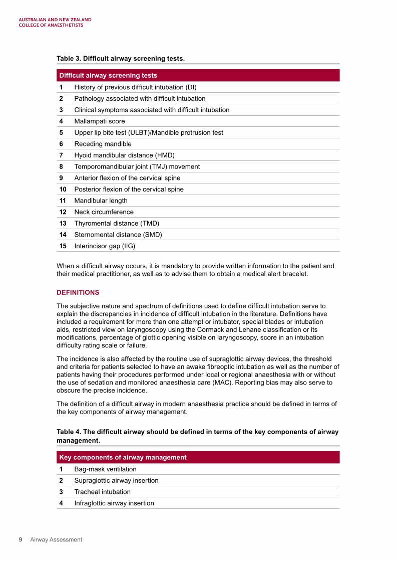
Table 3. Difficult airway screening tests.
Difficult airway screening tests
1 History of previous difficult intubation (DI)
2 Pathology associated with difficult intubation
3 Clinical symptoms associated with difficult intubation
4 Mallampati score
5 Upper lip bite test (ULBT)/Mandible protrusion test
6 Receding mandible
7 Hyoid mandibular distance (HMD)
8 Temporomandibular joint (TMJ) movement
9 Anterior flexion of the cervical spine
10 Posterior flexion of the cervical spine
11 Mandibular length
12 Neck circumference
13 Thyromental distance (TMD)
14 Sternomental distance (SMD)
15 Interincisor gap (IIG)
When a difficult airway occurs, it is mandatory to provide written information to the patient and their medical practitioner, as well as to advise them to obtain a medical alert bracelet.
DEFINITIONS
The subjective nature and spectrum of definitions used to define difficult intubation serve to explain the discrepancies in incidence of difficult intubation in the literature. Definitions have included a requirement for more than one attempt or intubator, special blades or intubation aids, restricted view on laryngoscopy using the Cormack and Lehane classification or its modifications, percentage of glottic opening visible on laryngoscopy, score in an intubation difficulty rating scale or failure.
The incidence is also affected by the routine use of supraglottic airway devices, the threshold and criteria for patients selected to have an awake fibreoptic intubation as well as the number of patients having their procedures performed under local or regional anaesthesia with or without the use of sedation and monitored anaesthesia care (MAC). Reporting bias may also serve to obscure the precise incidence.
The definition of a difficult airway in modern anaesthesia practice should be defined in terms of the key components of airway management.
Table 4. The difficult airway should be defined in terms of the key components of airway management.
Key components of airway management
1 Bag-mask ventilation
2 Supraglottic airway insertion
3 Tracheal intubation
4 Infraglottic airway insertion

10 Airway Assessment
DIFFICULT BAG-MASK VENTILATION
The subjective perception of difficulty together with the operator skill and technique dependent nature of the ability to perform bag-mask ventilation (BMV), make it difficult to define and objectively measure7. Causes of difficulty may be technique or airway-related, and include inadequate mask seal, excessive gas leak, and excessive resistance to inspiratory or expiratory airflow.
Several definitions exist for difficult bag-mask ventilation. The American Society of Anesthesiologists (ASA) defined difficult bag-mask ventilation as the inability of the unassisted anaesthetist to maintain the saturations (SpO2) above 90 per cent using a FiO2 of 100 per cent and positive pressure ventilation where the patients’ saturations were above 90 per cent prior to intervention or the inability of said anaesthetist to prevent or reverse the signs of inadequate ventilation during positive pressure ventilation8.
Han and colleagues9 proposed a scale to grade and classify bag-mask ventilation. This is useful for clinical description but has not been validated or shown to be reproducible. It may also not be sensitive enough for data comparisons or research purposes7. Difficult facemask ventilation is regarded as grade 3 mask ventilation.
Table 5. Han’s mask ventilation grading scale. Difficult facemask ventilation is regarded as grade 3 mask ventilation. Grade 4 represents impossible mask ventilation. The number of patients (N) and percentage of those studied (%) is given for each grade of mask ventilation.
Classification Description N (%)
Grade 1 Ventilated by mask 17,535 (77.4)
Grade 2 Ventilated by mask plus oral airway adjuvant +/-muscle relaxant
4775 (21.1)
Grade 3 Difficult to mask ventilate despite above, inadequate or unstable, requiring two providers
313 (1.4)
Grade 4 Unable to mask ventilate with or without the use of muscle relaxants.
37 (0.16)
INCIDENCE OF DIFFICULT BAG-MASK VENTILATION
The incidence of difficult bag-mask ventilation ranges from 0.08 per cent to 15 per cent depending upon the definition used and patient population selected7,10.
Langeron and colleagues10 defined difficult bag-mask ventilation (BMV) as when the anaesthetist considered the difficulty to be clinically relevant and potentially problematic if BMV were to be maintained for a longer period of time. They reported difficulty in 75 cases from 1502 patients with an incidence of 5 per cent (95 per cent CI 3.9 – 6.1 per cent). One patient proved impossible to mask ventilate. Interestingly, difficulty in mask ventilation was only anticipated in 13 (17 per cent) of those patients found to be difficult.
Kheterpal11 reported an incidence of difficult bag-mask ventilation (grade 3) of 1.4 per cent. This large database study included 22,660 attempts at bag bask ventilation from 61,252 adult anaesthetic cases over two years. Their findings were similar to the 1.6 per cent reported by Han9 in 1405 patients and consistent with those of Asai12 1.4 per cent and Rose and Cohen13 0.9 per cent. Asai12 reported failure to anticipate difficult mask ventilation in 57 per cent of patients who proved to be difficult to BMV.
One may conclude that even experienced anaesthetists may not predict difficult mask ventilation10.

11 Airway Assessment
PREDICTORS OF DIFFICULT BAG-MASK VENTILATION
Langeron10 identified five risk factors for difficult bag-mask ventilation. The presence of any two risk factors predicted difficulty with a sensitivity of 72 per cent and specificity of 73 per cent and a likelihood ratio of 2.5.
Table 6. Langeron’s independent risk factors for difficult mask ventilation10
Factor Odds ratio (95% CI) P valueAge >55 2.26 (1.34-3.81) 0.002
BMI >26 kg.m-2 2.75 (1.64-4.62) <0.001
Facial hair (beard) 3.18 (1.39-7.27) 0.006
Edentulous 2.28 (1.26-4.10) 0.006
Kheterpal and colleagues identified BMI greater than 30kg.m-2, presence of a beard, Mallampati three or four, age older than 57, severely limited mandibular protrusion and snoring as independent predictors of difficulty bag-mask ventilation11. Snoring and thyromental distance less than 6cm were identified as independent risk factors for grade 4 bag-mask ventilation. Abnormal neck anatomy, obstructive sleep apnoea (OSA) and snoring were independent predictors of grade 3 or 4 bag-mask ventilation and difficult intubation. However, many of the high-risk patients with anticipated difficulties were excluded and managed with a rapid sequence induction, an awake fibreoptic intubation (AFOI), procedure under regional or sedation with monitored anaesthesia care (MAC). A lower threshold for proceeding with an AFOI may also be partly responsible. The authors acknowledge this as patients who had an AFOI had significantly increased rates of risk factors for grade 3 mask ventilation when compared to the general study population (p<0.01). Importantly, the cases of airway difficulty experienced in this study were truly unexpected and unpredicted.
Table 7. Kheterpal and colleagues risk factors for difficult (grade 3) mask ventilation8.
Factor P valueBMI ≥ 30kg.m-2 <0.0001
Beard <0.0001
Mallampati 3 or 4 <0.0001
Age ≥57 0.002
Severely limited jaw protrusion 0.018
Snoring 0.019
Table 8. Kheterpal and colleagues risk factors for difficult (grade 3 or 4) mask ventilation and difficult intubation11.
Factor P valueLimited jaw protrusion <0.0001
Thick, obese neck anatomy 0.019
Sleep apnoea 0.036
Snoring 0.049
BMI ≥30 kg.m-2 0.053

12 Airway Assessment
Table 9. Murphy and Walls’s difficult bag-mask ventilation mnemonic, MOANS.
DescriptionM Mask seal. Facial features such as beards, saliva or blood, anatomical disruptions such
as facial fractures or retrognathia
O Obesity. BMI >26kg.m-2, Parturient or at-term mothers
A Age >55 years
N No teeth, edentulous
S Snoring or stiff. OSA, bronchospasm. Neck radiation changes
Murphy and Walls’s Manual of Emergency Airway Management 3rd edition (Lippincott Williams and Wilkins), describe five indicators of difficult bag-mask ventilation using the mnemonic MOANS. While bag-mask ventilation devices commonly generate 50-100cm H2O pressure, this requires an adequate seal and compliance to ensure ventilation. Conditions where this may not be possible are listed below. Facial features such as beards, saliva, blood, or anatomical facial anatomy and disruptions such as facial fractures and retrognathia may make obtaining a satisfactory mask seal difficult. Mask design is also important14. Improperly inflated cushion or wrong size may preclude a good seal. High volume, low-pressure cushions serve to improve mask performance15. Trauma, burns, swelling, infections, haematomas of the mouth, tongue, larynx, pharynx, trachea or neck may result in poor mask seal. BMV may be difficult in the face of decreased pulmonary compliance, for example, pulmonary fibrosis, oedema or severe bronchospasm. Snoring has been identified as a significant risk factor for difficult mask ventilation10,16. Suboptimal head and neck positioning may result in difficult bag-mask ventilation15. The “sniffing” position is reported to be best7. Cricoid pressure, particularly if improperly applied, may also serve to make BMV difficult17.
REMEDIABLE FACTORS
Shaving a beard and leaving the patient’s dentures in place during bag-mask ventilation represent the only easily remediable factors10. Removal of a beard may also uncover underlying anatomical or pathological changes camouflaged by the presence of facial hair such as a small, receding chin.
The use of creams or gels for patients with beards to improve mask seal has the potential to make the whole face oily and slippery and cannot be recommended. Saline soaked gauze or wide sticking plaster tape or adhesive film applied in a triangular fashion around the nose and mouth serve to improve the mask seal without the complications of the mask and hands sliding.
Alternatively, a supraglottic airway may be used as an alternative to bag-mask ventilation prior to intubation. Weight loss should be encouraged although significant improvement in bag-mask ventilation requires considerable time and patient effort.
IMPOSSIBLE MASK VENTILATION
Defined as the inability to guarantee gas exchange during attempts at bag-mask ventilation despite multiple providers, airway adjuvants with or without the use of neuromuscular blockade18.
The relative unimportance of muscle relaxants in this setting is attributed to the work of Goodwin and colleagues19. They measured the difference in inspired and expired tidal volumes before and after muscle relaxants in 30 patients with normal airways and found no significant difference in the ratio as a measure of efficiency of ventilation. The conclusion from this study, however, is not universally accepted. Calder and Yentis support the correct use of neuromuscular blocking agents in this situation20. Muscle relaxants make intubation easier and serve to ensure patency of the glottis excluding laryngospasm as cause for failure to achieve oxygenation. There is also a clinical impression that ventilation improves following muscle relaxation.

13 Airway Assessment
INCIDENCE OF IMPOSSIBLE MASK VENTILATION
Impossible or grade 4 mask ventilation is a rare occurrence with the incidence reported between 0.07 per cent and 1.4 per cent10,12,13. Kheterpal and colleagues reported an incidence of 0.15 per cent (one in 690 patients)11. This included 77 cases of impossible mask ventilation in 53,041 patients. They identified five independent predictors of impossible mask ventilation (Table 10).
Table 10. Predictors of impossible mask ventilation as identified by Kheterpal and colleagues.
Predictors of impossible mask ventilation1 Neck radiation changes
2 Male gender
3 Obstructive sleep apnoea
4 Mallampati 3 or 4
5 Presence of a beard
Of the 77 patients who were impossible to bag-mask ventilate, 19 (25 per cent) were also reportedly difficult to intubate. Of these, 15 were successfully intubated. Two patients required surgical airways; two were woken up and had an awake, fibreoptic intubation. In six cases an attempt was made to insert a non-intubating LMA. All but four of the 77 received neuromuscular blockade (65 patients receiving succinylcholine and a non-depolarising neuromuscular junction blocker used in the remaining eight cases).
Again, many of the patients predicted to be difficult would have been excluded from this study. They would have had an awake fibreoptic intubation, a regional procedure or surgical procedure under sedation with monitored anaesthesia care. Thus the incidence of 0.15 per cent could be described as conservative. This highlights the fact that even with a relatively low threshold for managing the airway by an AFOI technique, unanticipated difficult facemask ventilation remains a clinical problem.
Kheterpal and colleagues highlighted the importance of reduced mandibular protrusion as an important predictor of both difficult mask ventilation and difficult intubation18.
DIFFICULT LARYNGEAL MASK VENTILATION
The laryngeal mask airway (LMA) is a well-established means by which the airway is managed in anaesthesia practice for both spontaneous and controlled ventilation. It has been shown to have a high success rate and low complication rate. The LMA has also been shown to be a useful adjuvant in cases of failed intubation21. The “classic” LMA has also been reported to be useful in managing the unanticipated difficult airway in obese patients14,22.
DEFINITION
Difficult laryngeal mask ventilation has been defined as the inability to, within three insertions, place the laryngeal mask in a satisfactory position to allow clinically adequate ventilation and airway patency. Clinically adequate ventilation was defined as greater than 7ml.kg-1 with a leak pressure no greater than 15-20cm H2O pressure.
INCIDENCE
The incidence of difficult LMA insertion has been reported to be between 0.16 per cent and 0.9 per cent21.

14 Airway Assessment
PREDICTION OF DIFFICULT SUPRAGLOTTIC AIRWAY DEVICE INSERTION
The anaesthetist should assess the suitability and likely success of a supraglottic airway device in all patients, not just those where this is considered the airway device of choice, as this may form the mainstay of rescue treatment to provide oxygenation in the event of failure of bag-mask ventilation and endotracheal intubation.
Table 11. Murphy and Walls’s difficult supraglottic device insertion mnemonic RODS.
DescriptionR Reduced mouth opening: small mouth opening will limit the ease of placement of a
laryngeal mask airway.
O Obstruction: airway obstruction at or below the level of the glottis will not be relieved by the insertion of a supraglottic device.
D Distorted airway: unusual airway anatomy may prevent the supraglottic device from seating properly and hence the seal will be compromised.
S
Stiff neck or lungs: Decreased lung compliance, for example, asthma may make ventilation impossible with a supraglottic device. Beware the patient with limited neck movement, where a supraglottic device may form a poor seal.
LMA OR SUPRAGLOTTIC AIRWAY DEVICE FAILURE
Definition
LMA failure is defined as an airway related event requiring the removal of the LMA and tracheal intubation23.
Incidence
The incidence of LMA failure is reported to be 0.16 per cent to 4.7 per cent21.
Differences in incidence may reflect suboptimal usage or differing thresholds for accepting failure or success24. Risk factors for LMA failure have been identified as: advanced age, increased body mass index (BMI), male sex, reduced thyromental distance (TMD), thick neck (neck circumference), poor dentition, smoking and surgical table rotation23.
LMA failure, while uncommon, is not without significant consequences. Ramachandran and colleagues23 identified 170 incidences of LMA failure in 15,795 patients yielding an incidence of 1.1 per cent. Of these, there were 106 (60 per cent) significant episodes of hypoxia, hypercapnoea or airway obstruction whilst 42 per cent reported inadequate ventilation due to a leak. Two patients required unplanned admission to the intensive care unit. The authors identified four risk factors for LMA failure: surgical bed rotation, male patients, poor dentition and increased BMI. The increased pharyngeal resistance, upper airway narrowing, obstruction and OSA may be responsible for the increased incidence of difficult mask ventilation and failed LMA insertion in male patients. Unsurprisingly bag-mask ventilation is also reported to be more difficult in patients with demonstrated LMA failure (incidence 5.6 per cent, or a threefold increase). Difficulty with one technique heralds difficulty with another as many of the risk factors over lap.

15 Airway Assessment
DIFFICULT ENDOTRACHEAL INTUBATION
Definition
Difficult intubation may be defined in terms of the time taken, number of attempts at direct laryngoscopy required to achieve intubation, view at laryngoscopy, or the requirement for specialised equipment or techniques. Many of the definitions are vague, practically meaningless and not directed at the cause of the problem.
Incidence of difficult intubation
The incidence of difficult intubation depends upon the definition used24.
The ASA originally defined difficult intubation as requiring more than three attempts or taking longer than 10 minutes to complete. The incidence was reported to be 1.8 per cent in 18,205 patients and 1.9 per cent in 3325 patients13.
The incidence of difficult intubation is reported to be 5.8 per cent (95 per cent CI, 4.5-7.5 per cent) for the general patient population. This was 6.2 per cent (CI, 4.6-8.3 per cent) for normal patients excluding obese and obstetric patients, 3.1 per cent (CI, 1.7-5.5 per cent) for obstetric patients and 15.8 per cent (95 per cent CI, 14.3-17.5 per cent) for obese patients4. These figures are consistent with those reported by Khan25 (5 per cent), Rose and Cohen13,24 (4.7 per cent) and similar to the incidence of 5.9 per cent in the initial study by Wilson and then 1.5 per cent in the prospective study26.
The Australian Critical Incident Monitoring Study (AIMS) identified four variables associated with difficult intubation: limited mouth opening, obesity, limited neck extension and lack of a trained assistant27.
DIFFICULT LARYNGOSCOPY
Difficult intubation may imply difficulty with visualising the glottis, that is, direct laryngoscopy or difficulty with endotracheal tube placement due to laryngeal or tracheal distortion or narrowing. The distinction is important since difficult laryngoscopy does not preclude successful placement of the endotracheal tube, which may be passed without visualising the glottis. Difficult laryngoscopy best describes the problem of establishing a suitable laryngeal view to enable intubation.
Murphy and Walls’s mnemonic LEMON is a useful bedside guide to prediction of difficult intubation (Table 6). The score is calculated by assigning one point for each of the following LEMON criteria. The score has a maximum of 10 points. Patients who are difficult to intubate have higher LEMON scores. This simple scoring system was devised by the US national emergency airway management course and is designed for use in an emergency room setting28. LEMON is an acronym for “Look-Evaluate-Mallampati-Obstruction-Neck”.
Table 12. Murphy and Walls’s bedside predictors of difficult intubation with direct laryngoscopy.
DescriptionL Look externally
E Evaluation:Mouth opening <5cmThyromental distance <6cm
M Mallampati class
O Obstruction
N Neck mobility

16 Airway Assessment
BEDSIDE PREDICTORS OF DIFFICULTY WITH INTUBATION VIA DIRECT LARYNGOSCOPY
A variety of features on external examination suggest difficult laryngoscopy: small or recessed mandible, poor dentition, a short neck, facial disruption, presence of a halo-thoracic brace or cervical spine collar and a large tongue are just some features that suggest difficult direct laryngoscopy. Mallampati class one and two patients are associated with low intubation failure rates. Class three patients, however, have an intubation failure rate greater than 10 per cent. Signs of impending obstruction include stridor, voice changes and failure to swallow secretions. Presence of stridor indicates that the airway diameter has been reduced to 4.0mm or less. Positioning of the head and neck is vital for successful direct laryngoscopy. Classic positioning advice is to place the patient in the “sniffing the morning air” position – that is neck flexion and head extension. Practitioners should beware in patients with limited neck extension – for example, patients with cervical spine injuries, pathologies such as ankylosing spondylitis, radiation changes or patients in cervical spine immobilisation collars.
CORMACK AND LEHANE
Cormack and Lehane defined laryngoscopy in terms of the best view of the glottis during conventional laryngoscopy with a direct view and performed as a best attempt29. The optimal laryngeal view includes external manipulation. Yentis and Lee30 added a modified version that subdivided grade 2 into grade 2a and 2b. This is known as the modified Cormack and Lehane classification. Cook’s modification31 subdivided grade 3 depending on whether the epiglottis could be elevated from the posterior pharyngeal wall using a bougie or introducer (Table 7).
The Cormack and Lehane laryngoscopic grading has been used to define difficult intubation with grade 3 and grade 4 equated with difficulty, although it was never intended for this purpose30.
Benumof32 defined the best attempt at laryngoscopy as that performed by a reasonably skilled and experienced practitioner, using the optimum type and length of laryngoscope blade and the patient in the optimal “sniffing” position with no significant muscle tone together with the use of external laryngeal manipulation as appropriate. When confronted with the unexpected difficult intubation it is necessary to ensure that laryngoscopy conditions as above are optimal.
Table 13. Modified Cormack and Lehane classification.
Classification Description Frequency (%)
Possibility of intubation failure
(%)
Grade 1 Full view of the glottis 68 <1
Grade 2a Partial view of the glottis 24 4.3Grade 2b Only posterior portion of glottis or
arytenoid cartilages6.5 67.4
Grade 3a Epiglottis can be lifted from the posterior pharyngeal wall
1.2 87.5
Grade 3b Epiglottis cannot be lifted from the posterior pharyngeal wall
Very rare Very likely
Grade 4 Neither glottis or epiglottis can be seen Very rare Very likely
PERCENTAGE OF VISIBLE GLOTTIC OPENING (POGO)
Levitan devised an alternative description of the laryngeal view in terms of the percentage of visible glottic opening33. This score can be used for direct and indirect laryngoscopy, or techniques where standard positioning is not used. One disadvantage of this description is that it requires the observer to estimate how much of the glottis is not visualised. The POGO score has not been widely adopted34.

17 Airway Assessment
Figure 1. A 100 per cent POGO score corresponds to visualisation of the entire glottic opening from the anterior commissure of vocal cords to the inter-arytenoid notch between the posterior cartilages.
INTUBATION DIFFICULTY SCALE
Adnet35 introduced an intubation difficulty scale. This may be useful for communicating the total intubating difficulty and for researching the predictive power of a specific variable in identical patient groups32.
Table 14. Intubation difficulty score (IDS).
Parameter ScoreNumber of attempts >1Every additional attempt adds one point.
N1
Number of operators >1Each additional operator adds one point.
N2
Number of alternative techniques.Each alternative technique adds one point. Repositioning the patient, change of materials (blade, ET tube, addition of a stylette), change in approach (nasotracheal/orotracheal), or use of another technique (fibreoptic or intubation through LMA).
N3
Cormack and Lehane grade – 1Apply Cormack and Lehane grade for first oral attempt. For successful blind intubation N4 = 0.
N4
Lifting force requiredNormalIncreased
N5 = 0N5 = 1
Laryngeal pressure NoneApplied
N6 = 0N6 = 1
Position of vocal cords during laryngoscopyAbductionAdduction
N7 = 0N7 = 1
Total intubation difficulty score (IDS) = sum of all the scores. N1-N7

18 Airway Assessment
Table 15. Intubating difficulty score.
IDS score Degree of difficulty0 Easy
IDS ≤ 5 Slight difficulty
IDS > 5 Moderate to major difficulty
IDS = ¥ Impossible
FAILED INTUBATION
Failed intubation is defined as the inability to site an endotracheal tube after multiple attempts. The incidence is estimated at 0.05-0.3 per cent36, 37. Failure to intubate is in itself not a problem but failure to maintain oxygenation may be catastrophic.
DISCUSSION
Even the most comprehensive preoperative assessment will also not predict every case of difficult airway1,26. In particular, we are unable to reliably identify difficult cases in the general population who look normal with no history of previous airway difficulties.
Since we cannot rely on preoperative airway assessments to accurately predict the difficult airway, we need to be equipped to manage the unexpected difficult or failed airway1. The possibility of a failed airway should be recognised early, accepted and managed appropriately. Fortunately, the incidence of the failed airway is low. However, this means limited exposure, training and experience in managing this complication of anaesthesia.
Preoperative airway assessment is poorly taught despite the importance of airway management in daily practice. The topic of airway assessment hardly features more than a couple of pages in major anaesthesia texts. Yentis6 questions the “pointless ritual” of airway examination, as serious airway difficulty is rare in “normal” patients and the screening of a “normal” population is unrewarding. So the question was asked, “why bother?”
In spite of advances in technology and training, difficult intubation and airway management remain an important cause of perioperative morbidity and mortality related to anaesthesia38-41. Given the seriousness of the problem, we need to take a professional approach and attempt to reduce this. Should an airway problem occur, it reflects badly on the practitioner if an assessment has not been done, however inaccurate the methods of assessment have been shown to be3. While we in anaesthesia may debate the merits of various tests, their sensitivities and specificities, when there is serious morbidity and mortality involved, little sympathy will be offered to the practitioner whose airway assessment has been anything less than a thorough, concerted attempt to identify the difficult airway.
Anaesthetists are a group of professionals caring for individuals, human beings. We render patients unconscious and unable to look after themselves and take on that responsibility ourselves. When things go wrong, the low incidence of mortality and morbidity are little comfort to the affected relatives whose loss is real. On review, the practitioner will be considered as a professional and his actions assessed by his peers and experts in the field. Simply stated, a preoperative history and clinical examination represent good medical practice.
By practicing and routinely performing an airway assessment, we improve our individual skills in terms of assessing who is easy or difficult. Understanding the patient’s peculiar airway geometry enables us to focus our attention on which instrument or tool may be best suited to manage their airway. Thus the history and examination should direct our airway plan and particular choice of equipment to manage the patient’s airway.

19 Airway Assessment
When the patient has risk factors, it seems reasonable to try and increase the probability of a diagnosis of difficult intubation. Without a history and a simple examination one is unlikely to uncover or elicit such risk factors.
The preoperative airway assessment should include identification of the predictors for each of the following:
1. Patients who are difficult to intubate.
2. Patients who are difficult to oxygenate by bag-mask ventilation.
3. Patients who are difficult to oxygenate by a supraglottic airway.
4. Patients who are difficult to oxygenate by an infraglottic airway.
5. Patients at risk of aspiration.
6. Patients with altered cardiorespiratory physiology.
7. Patients who may be an extubation risk.
In addition to the above, the effect of the surgery and any previous airway documentation should help direct the airway plan. This plan should be documented in advance. Following airway instrumentation, a detailed note should be made informing future care. Hopefully we agree that an airway assessment should be done. The difficulty then is determining which tests should be included.
Screening tests should be easy to perform, timely, cost effective and applicable to most patients. The RCoA suggests that history of difficult intubation, Mallampati and thyromental distance should be assessed as part of the preoperative airway assessment in all patients3.
Predicted difficult with airway management approaches
B&M
SGA ETT
The challenging trinity of difficult airway predictors
Consider an awake technique

20 Airway Assessment
HISTORY
A detailed patient history is recommended in all patients in an attempt to identify patient, anaesthesia, and surgical factors suggestive of a difficult airway42. Obtaining a patient history and review of the available patient records or notes represents good medical practice.
A history of a previous difficult intubation should not be taken lightly although it may be of limited significance. While conditions may improve, in general they get worse as the mobility of the cervical spine and temporomandibular joints decrease with age. A record of previous ease thus does not guarantee future success.
Patients themselves may volunteer information. This might include dental or airway trauma (pharyngeal, oesophageal or tracheal perforation) related to previous anaesthesia, or verbal recollections of prior difficulty. A bruised or split lip, loose, chipped, damaged or missing front teeth or unexplained oral bleeding following previous anaesthesia may signal previous problematic airway management. Unexpected ICU admission should raise the possibility of airway issues either at induction or extubation. An awake fibreoptic intubation may be revealed upon by questioning. Look for a Medic alert bracelet; inquire about difficult airway letter, review previous anaesthesia records, patient or hospital notes where available.
History of any conditions associated with difficulty should be sought. This places patients in a different risk group, for example, intermittent hoarseness and stridor in rheumatoid arthritis and acromegaly or OSA in acromegaly and obesity. Patients in these different risk groups warrant further inspection.
Knowledge regarding the potential for regurgitation and aspiration is important, particularly in a patient with a failed airway during a rapid sequence intubation43. The intubation strategy may need to be modified in light of the risk for aspiration, such as pregnancy. Cricoid pressure may impair laryngoscope insertion, successful passage of an airway introducer or bougie, and may cause inadvertent airway obstruction especially if poorly applied. In the event of an unanticipated difficult airway during a rapid sequence intubation, the contribution of cricoid pressure should be assessed. If deemed necessary, then cricoid pressure may be cautiously reduced with suction ready43.
EXAMINATION
A detailed examination of the airway should be performed in all patients. This may need to be modified in certain circumstances but should be tailored to detect physical findings associated with a difficult airway. Obvious syndromes and related conditions should not be missed.
No single anatomical factor is able to confidently predict a difficult airway or intubation and current rating systems are only of modest sensitivity and specificity in predicting the difficult airway.
MOUTH OPENING AND INTER-INCISOR GAP
Mouth opening is central to airway management and intubation. Adequacy of mouth opening should be assessed in all patients. In extreme cases, patients may not be able to open their mouths at all. The difficulty lies in determining whether this is mechanical or functional as the latter may improve with general anaesthesia and muscle relaxation. Mouth opening of less than 3.7cm falls outside the normal range5. An inter-incisor distance of less than 5cm or two to three finger breadths may make conventional laryngoscopy difficult. Mouth opening of 1.5cm or less than one finger breadth may impair the insertion of a supraglottic airway device or laryngoscope and an inter-incisor distance of 2cm is required to insert the intubating LMA (ILMA). At least 2.5cm is required to insert an LMA. An IIG of 5cm for intubation and 4cm for insertion of an LMA is a simple test with a relatively high predictive value44. While limited mouth opening may impair bag-mask ventilation, the insertion of a supraglottic airway device, and direct laryngoscopy, it does not appear to be a useful singular predictor of difficult intubation4.

21 Airway Assessment
TONGUE ABNORMALITIES
Direct laryngoscopy requires the tongue to be mobile and retracted from the line of vision. Problems arise when the tongue is abnormally large or poorly compliant. Rosenstock and Kristensen suggest that tongue mobility may be a sensitive indicator of difficult laryngoscopy45.
MALLAMPATI
Three classes were initially described by Mallampati with Samsoon and Young46 adding a fourth later47,48. The use of the latter four-class classification is often referred to as a modified Mallampati score (Table 16). The score is based on the visibility of pharyngeal structures with the patient sitting directly opposite the examiner, mouth open as widely as possible and the tongue maximally extruded and no vocalisation (Figure 2). The test serves to estimate the relative size of the tongue to the oral cavity and likely ease or difficulty in displacing the tongue with a standard laryngoscope in order to view the glottis26,49,50.
Figure 2. Modified Mallampati scoring system.
Table 16. Modified Mallampati scoring system.
Class Pharyngeal structures visible1 Faucial pillars, soft palate and uvula visible
2 Faucial pillars and soft palate visible. Uvula obscured by tongue
3 Only the soft palate is visible
4 Soft palate not visible

22 Airway Assessment
The Mallampati score, however, suffers from poor sensitivity, specificity and positive predictive value. On its own it predicts only about half of the cases of difficult laryngoscopy and is also unreliable with a high incidence of false positives. This is highlighted by a likelihood ratio of between 1.5 and six in the general surgical population42.
A meta-analysis involving 177,088 patients reported that only 35 per cent of patients with a difficult intubation were identified as Mallampati 3 or 451. The pooled sensitivity and specificity were 0.35 (95 per cent confidence interval (CI, 0.34-0.36) and 0.91 (CI, 0.91-0.91) respectively. The odds ratio for a difficult intubation with a Mallampati score of 3 or 4 was 5.89 (CI, 4.74-7.32). The positive and negative likelihood ratios were 4.13 (CI, 3.60-4.66) and 0.70 (CI, 0.65-0.75) respectively. A clinical test is considered diagnostically accurate with a positive likelihood ratio greater than 1052.
There is also considerable inter-observer variability and patients may inadvertently phonate during the test altering the findings. The findings are limited to laryngoscopy using the standard laryngoscope blade with the role of the Mallampati test changing with the introduction of various video laryngoscopes. The test may not be appropriate in the emergency scenario with a supine patient although there is evidence to support its use53. The Mallampati test is thus not suitable as a stand-alone test, but may be of some value as part of a multivariate model for the prediction of difficult laryngoscopy and intubation51.
The importance of the Mallampati test is that it initiates the airway assessment and draws the anaesthetist’s attention to the oral cavity and airway pathway important to accessing the glottis. It is universally appreciated and easily performed. It gets the patient to open their mouth, which may reveal information about the access to and contents of the oral cavity that may otherwise have been missed, such as poor mouth opening, awful dentition, gross abnormalities or tumours. Incredibly, Williamson reported an incident of airway difficulty with unexpected limited mouth opening discovered only upon difficulty inserting the laryngoscope27.
THYROMENTAL DISTANCE
The thyromental distance (TMD) was defined by Patil as the distance from the thyroid notch to mental prominence with the head fully extended. It is considered a test of mandibular space and reflects the ease by which the tongue may be displaced using a standard laryngoscope blade4,49. A distance greater then 6.5cm is rarely associated with difficulty. Distances between 6-6.5cm may be associated with difficult laryngoscopy but intubation is usually possible. A TMD of less than 6cm suggests that intubation using conventional, direct laryngoscopy may be very difficult or impossible.
The cut-off values, however, are disputed and range widely between studies54. Also, both short and long TMD measurements may be associated with difficult intubation. The positive predictive value (PPV) is low when used alone. Accuracy may be improved if correcting for patient height and weight. Importantly, however, it focuses the examiner’s attention on the geometry of the airway.
JAW PROTRUSION (PROGNATHISM/SUBLUXATION)
Temporomandibular joint mobility is assessed by asking the patient to open their mouth fully and then asking them to place their lower incisors as far forward relative to their lower incisors as possible (subluxation). This may be easier than performing the upper lip bite test52. There are several publications supporting its use as a single test. It is also included in several scoring systems, for example, Wilson or Arne26,44,54. Grade C is rare but diagnostic for difficult intubation. Grade B however has poor PPV.

23 Airway Assessment
Class Position of the lower teeth in relation to the upperA Lower teeth may be placed in front of the upper teeth
B Lower teeth may be placed in line with the upper teeth
C Lower teeth cannot be placed in line with the upper teeth
UPPER LIP BITE TEST (ULBT)
Khan supports the use of the ULBT as a measure for ease of intubation44. The test has a sensitivity of 78.95 per cent, specificity of 91.96 per cent and accuracy of 91.05 per cent. Inter-observer reliability of the ULBT is higher than that of the Mallampati score55. Some patients find it difficult to understand and elderly patients with dentures may have difficulty performing the test25.
Table 17. Upper lip bite test classification.
Class Position of the lower incisors1 Lower incisors can bite above the vermilion border of the upper lip
2 Lower incisors can bite the vermilion border of the upper lip
3 Unable to bite the upper lip
STERNOMENTAL DISTANCE
The sternomental distance (SMD) is defined as the distance between the mentum and sternum with the head fully extended and the mouth closed. The threshold value of less than 12.5-13.5cm is used to predict a higher incidence of difficult intubation. SMD is considered an indicator of head and neck mobility with head extension playing an important part in obtaining a good laryngeal view with a conventional laryngoscope56.
In the meta-analysis by Shiga, there were limited studies reporting on sternomental distance4. This prevented the authors from commenting further on the diagnostic performance of the test but it appeared to be the most useful singular test for excluding difficult intubation.
In a study of 523 obstetric patients the prevalence of grade 3 or 4 laryngeal views was found to be 3.5 per cent34. The predictive power of the threshold value of 13.5cm or less gave a sensitivity of 67 per cent and a specificity of 71 per cent returning a likelihood ratio of two. This low score explains why it is rarely used as a solo score in contemporary anaesthesia practice42.
CERVICAL SPINE MOBILITY
An attempt to assess cranio-cervical mobility should be attempted. Calder suggests this may be done when observing head movement during maximum mouth opening. A cervical spine range of movement should be more than 90 degrees (anterior plus posterior flexion) to ensure easy intubation26.
Fritscherova reported on the statistical significance of c-spine mobility in their study but also commented on the fact that several control patients were unable to achieve this54. The test is limited in clinical practice by subjective error and availability of equipment (goniometer) to measure it.
HORIZONTAL LENGTH OF THE MANDIBLE (HLM)
HLM has little predictive value if used on its own. Measurement is subject to error although a value greater than 9cm suggests easy intubation57.

24 Airway Assessment
NECK CIRCUMFERENCE
On its own, neck circumference has not been shown to be predictive of difficult intubation. However, greater neck circumference may be associated with increased body mass and several studies have shown the significance of obesity on difficult bag-mask ventilation and intubation26, 58-60.
HYOMENTAL DISTANCE
A short (less than 4cm) hyomental distance (HMD) has been reported to be a significant finding in patients who were unexpectedly found to be difficult to intubate54. However, this may be difficult to assess in obese patients.
COMBINED TESTS
Individual tests perform poorly. Unfortunately, combination scores also prove unsuccessful in low prevalence populations. Combined tests may represent sensible practice in high prevalence populations.
In a review by Shiga and colleagues4 reporting on 35 studies including 50,760 patients, the overall incidence of difficult intubation was reported to be 5.8 per cent (CI, 4.5-7.5 per cent). Screening tests were reported with poor to moderate sensitivity (20-62 per cent) and moderate to fair specificity (82-97 per cent). In an attempt to improve upon the predictive power of singular tests and measurements, combined tests and scoring systems have been investigated. The combination of the Mallampati and Thyromental distance are more predictive than either test alone61. Shiga and colleagues identified the combination of Mallampati and Thyromental distance as the most useful bedside test for prediction of difficult laryngoscopy (positive likelihood ratio 9.9; 95 per cent confidence interval 3.1-31.9).
The Wilson and Arne rating systems have a sensitivity of about 75-94 per cent, but low positive predictive value. The Naguib model was reported to have a sensitivity of 81.4 per cent but a positive predictive value of just 15.3 per cent62.
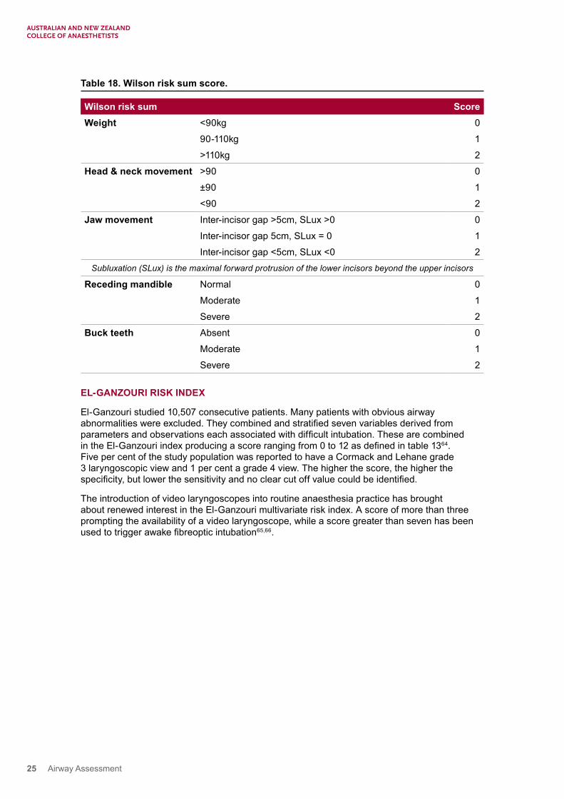
WILSON RISK SUM SCORE
The Wilson risk sum score is based upon five risk factors: weight, head movement, neck and jaw movement, mandibular recession, and buck teeth with three possible scores for each (0,1,2) yielding a total score between zero and 10 (Table 12 Wilson Risk sum)26. A score greater than two predicts 75 per cent of difficulties but includes a high number of false positives. Shiga and colleagues reported a low true-positive rate and a low false positive rate indicating that the Wilson score correctly identified patients in whom intubation was easy4. A score greater than four reduces the number of false positives but at the cost of missing some of the difficult intubations.
The importance of the Wilson risk sum score is highlighted in a study of 372 obstetric patients undergoing caesarean section where a score greater than two produced a likelihood ratio of more than 2063. The score has been shown to be highly reproducible4. Predictive powers of both the Mallampati and the Wilson risk score are similarly poor, however there may be less inter-observer variation with the Wilson risk score26.
25 Airway Assessment
Table 18. Wilson risk sum score.
Wilson risk sum ScoreWeight <90kg 0
90-110kg 1
>110kg 2
Head & neck movement >90 0
±90 1
<90 2
Jaw movement Inter-incisor gap >5cm, SLux >0 0
Inter-incisor gap 5cm, SLux = 0 1
Inter-incisor gap <5cm, SLux <0 2Subluxation (SLux) is the maximal forward protrusion of the lower incisors beyond the upper incisors
Receding mandible Normal 0
Moderate 1
Severe 2
Buck teeth Absent 0
Moderate 1
Severe 2
EL-GANZOURI RISK INDEX
El-Ganzouri studied 10,507 consecutive patients. Many patients with obvious airway abnormalities were excluded. They combined and stratified seven variables derived from parameters and observations each associated with difficult intubation. These are combined in the El-Ganzouri index producing a score ranging from 0 to 12 as defined in table 1364. Five per cent of the study population was reported to have a Cormack and Lehane grade 3 laryngoscopic view and 1 per cent a grade 4 view. The higher the score, the higher the specificity, but lower the sensitivity and no clear cut off value could be identified.
The introduction of video laryngoscopes into routine anaesthesia practice has brought about renewed interest in the El-Ganzouri multivariate risk index. A score of more than three prompting the availability of a video laryngoscope, while a score greater than seven has been used to trigger awake fibreoptic intubation65,66.

26 Airway Assessment
Table 19. El-Ganzouri risk index.
El-Ganzouri risk index ScoreMouth opening >4cm 0
4cm 1
<4cm 2
TMD >6cm 0
6-6.5cm 1
<6cm 2
Mallampati 1 0
2 1
3 2
Neck movement >90 degrees 0
80-90 degrees 1
<80 degrees 2
Jaw protrusion Yes 0
No 1
Weight <90kg 0
90-110kg 1
>110kg 2
History of difficult intubation None 0
Questionable 1
Definite 2
ARNE RISK INDEX
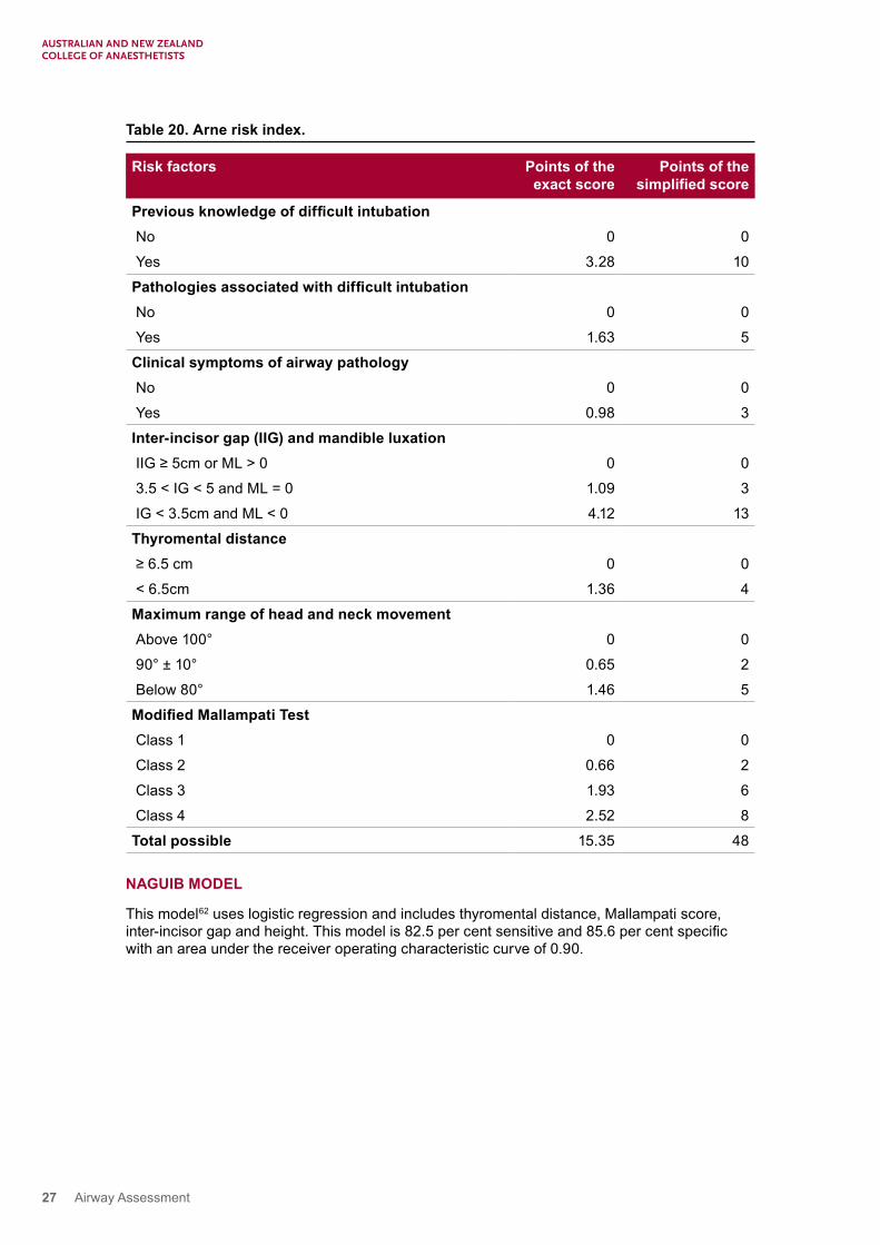
Arne and colleagues studied 1200 consecutive ear, nose and throat (ENT) and general surgical patients. They defined difficulty in intubation as requiring the use of a bougie, the Bullard laryngoscope, fibreoptic intubation and Piquet-Crinquette-Vilette (PCV) laryngoscope67. They developed a risk index (Table 14) and determined a threshold value of 11. They then prospectively studied a further 1090 patients with a risk index score of 11 or more. The threshold score of 11 identified difficult intubation with a sensitivity of 94 per cent and specificity of 96 per cent in general surgery patients, a sensitivity of 90 per cent and specificity of 93 per cent for non-cancer ENT surgery patients and a sensitivity of 92 per cent and specificity of 66 per cent in cancer ENT surgery patients.
27 Airway Assessment
Table 20. Arne risk index.
Risk factors Points of the exact score
Points of the simplified score
Previous knowledge of difficult intubation No 0 0
Yes 3.28 10
Pathologies associated with difficult intubation No 0 0
Yes 1.63 5
Clinical symptoms of airway pathology No 0 0
Yes 0.98 3
Inter-incisor gap (IIG) and mandible luxation IIG ≥ 5cm or ML > 0 0 0
3.5 < IG < 5 and ML = 0 1.09 3
IG < 3.5cm and ML < 0 4.12 13
Thyromental distance ≥ 6.5 cm 0 0
< 6.5cm 1.36 4
Maximum range of head and neck movement Above 100° 0 0
90° ± 10° 0.65 2
Below 80° 1.46 5
Modified Mallampati Test Class 1 0 0
Class 2 0.66 2
Class 3 1.93 6
Class 4 2.52 8
Total possible 15.35 48
NAGUIB MODEL
This model62 uses logistic regression and includes thyromental distance, Mallampati score, inter-incisor gap and height. This model is 82.5 per cent sensitive and 85.6 per cent specific with an area under the receiver operating characteristic curve of 0.90.

28 Airway Assessment
SUMMARY OF TESTS
Table 21. Summary of the sensitivity, specificity and positive predictive value of the commonly used airway assessment tests.
Sensitivity % Specificity % Positive predictive value %Mallampati 42-60 81-89 4-21
Modified Mallampati 65-81 66-82 8-9
Thyromental distance 65-91 81-82 8-15
Sternomental distance 82 89 27
Wilson 42-55 86-92 6-9
Arne 80-98 91-94 25-42
Mouth opening 26-47 94-95 7-25
Jaw protrusion 17-26 95-96 5-21
PREDICTORS OF DIFFICULT INFRAGLOTTIC AIRWAY
When assessing the airway, the anaesthetist should evaluate the ease or difficulty of citing an infraglottic device, as this forms the mainstay of rescue treatment to provide oxygenation in the event of failure of bag-mask ventilation, placement of a supraglottic device and endotracheal intubation.
Table 22. Murphy and Walls’s predictors of difficult cricothyroidotomy mnemonic SHORT.
DescriptionS Surgery on the neck
H Haematoma or infection
O Obesity
R Radiation
T Tumour
ULTRASOUND USE IN AIRWAY ASSESSMENT
Ultrasound has been used to assess both the anatomy of the neck and importantly the airway68. Singh and colleagues used ultrasound to detail the normal anterior airway anatomy including that of the vocal cords69. Ultrasound has been reported to facilitate the rapid identification of the cricothyroid membrane by emergency physicians69. Ultrasound has been used to confirm endotracheal tube position in critically ill patients68. Successful intubation was assessed in 24 patients using either foam or saline filled endotracheal cuffs. In an emergency department setting, the sensitivity for detecting successful intubation was 96.3 per cent with a specificity of 100 per cent. More recently in obese patients it was identified that ultrasound was faster than capnography and auscultation to confirm endotracheal tube placement67-72.
Ultrasound also has been used to detect and assess pharyngeal or laryngeal pathology including abscesses, tumours (subglottic haemangiomas, laryngeal cysts), infection (epiglottitis), obstructive sleep apnoea, laryngeal stenosis, and pneumothorax73-77.
Ultrasound may be useful in assessing the location of the trachea in patients with deep tissue neck infections, tumours and dysmorphia. Knowledge regarding the depth and deviation of the trachea may serve to inform the percutaneous approach to the airway. An example where this might be relevant is the patient with extensive submandibular abscess and stridor. Awake

29 Airway Assessment
fibreoptic intubation has a finite failure rate and the option of performing a tracheostomy under local anaesthesia may be technically challenging. In such circumstances, the local anaesthetic is likely to be less effective and the trachea highly mobile.
Fasting status may be assessed and gastric volume determined by ultrasound. This may be of use in determining issues with compliance or delayed gastric emptying78,79.
Ultrasound is reported to have a strong correlation with MRI when used to measure the subglottic airway diameter in healthy volunteers80. Ultrasound also has been used to determine endotracheal tube size including that for double lumen tubes68,81,82.
There have been reports in the literature linking the presence of abundant neck soft tissue in the pharynx, retropharynx and suprascapular regions as measured by CT and MRI with that of a difficult airway83. Ultrasound imaging has been shown to correlate with that of the MRI for the estimation of fat depth84. Thus there has been keen interest in the role of ultrasound in the assessment of the difficult airway.
However, the role of ultrasound in the assessment and prediction of a difficult airway has not been fully elucidated. In 50 morbidly obese patients, difficult intubation by direct laryngoscopy was predicted by pre-tracheal soft tissue thickness of 28mm or more and a neck circumference greater than 50cm at the level of the vocal cords85. However, a subsequent study by Komatasu and colleagues was unable to replicate these findings86. This has been attributed to the difference in population groups between the two studies and the possible difference in fat distribution between Middle Eastern obese patients in Israel and those in the obese American population.
Another pilot study enrolled 51 elective surgical patients where a 1.69cm or greater thickness at the level of the hyoid bone and a 3.47cm thickness or greater at the thyrohyoid membrane corresponded to a difficult airway87.
Ultrasound has been used to predict extubation outcomes and post extubation stridor in ICU patients88,89. When the diaphragmatic displacement value was determined to be 1.1cm or more the prediction for successful extubation was more likely. This was believed to reflect the assessment of respiratory muscles global function. For stridor prediction, an air column width of 4.5 (0.8) mm during cuff deflation was found to be statistically significant. A total of 51 patients were assessed with only four of them developing stridor.
NASOPHARYNGOSCOPY IN AIRWAY ASSESSMENT
Flexible, fibreoptic nasopharyngoscopy is a relatively simple technique used to visualise the upper airway for diagnosis, treatment, or both. It may be used to evaluate foreign bodies, congenital abnormalities and potential airway obstruction from neoplasm and epiglottitis, obstructive sleep apnoea, dysphagia, dysphonia, tonsillar hypertrophy, glossoptosis, or laryngomalacia90. Vocal cords pathology, including polyps, nodules, masses and paralysis, also can be identified.
Otolaryngologists often perform nasopharyngoscopy in the outpatient and emergency department. This may be repeated later in theatre if required, enabling an ongoing, dynamic assessment of the airway and impact of pathology and obstruction.
When performed as part of a preoperative surgical work up this may need to be repeated, as a rapidly changing lesion may result from a previously unappreciated abnormality. The surgical assessment is also focused towards diagnosis and treatment and does not necessarily include the anaesthesia issues of bag-mask ventilation, insertion of a supraglottic airway device or intubation and impact of positive pressure ventilation. For example, a laryngeal mass or anatomical distortion may preclude the optimal positioning of a laryngeal mask airway and a vascular or friable anterior airway lesion could be traumatised by direct or video laryngoscopy.

30 Airway Assessment
Preoperative endoscopic airway examination is an underutilised technique in anaesthesia that may provide useful information regarding the supraglottic airway architecture in patients with suspected or known abnormal anatomy from either deep neck infections, such as retropharyngeal abscess and tumours. Preoperative endoscopic airway examination may improve theatre efficiency and patient experience by reducing the number of unnecessary awake fibreoptic intubations. Lesions of the tongue base, epiglottis, or larynx may not be fully appreciated following a routine preoperative airway assessment as clinical signs and symptoms may be unreliable indicators of the significance of these lesions91. Rosenblatt and colleagues found the additional information afforded by flexible endoscopy altered clinical practice. Anaesthetists reassured by relatively normal findings on endoscopic examination in patients with suspected airway difficulty where an awake intubation had initially been planned, resulted in a safe, standard induction. Also, where the additional information suggested difficulty, a standard induction was abandoned in favour of an awake technique92.
Diagnostic endoscopy can be easily and rapidly performed with minimal fuss and preparation93. The nares are not always symmetrical and selection of the more patent nostril may reduce trauma. Topical anaesthesia together with a vasoconstrictor may improve patient compliance and minimises the risk of bleeding secondary to instrumentation94. The glottis should be inspected from above without local anaesthetic being applied directly, as this may result in laryngospasm.
Nasopharyngoscopy is a relatively safe procedure with few contraindications and complications in experienced hands. Extreme caution should be used in epiglottitis and then by experienced personnel only, as it may result in laryngospasm and subsequent airway compromise95. Significant bleeding may occur in patients with a coagulopathy even from minor trauma. In the setting of craniofacial trauma, the benefits should be carefully weighed against the risks of inadvertent intracranial instrumentation and exacerbation of nasopharyngeal injuries. Mucosal trauma may result in epistaxis (a smaller diameter scope may help mitigate this problem. Tearing, coughing and, less frequently, a residual foreign body sensation, transient laryngospasm and vasovagal syncope are also potential complications. Laryngospasm is rare, incidence approximately 1 per cent, and is usually self-limited but can lead to desaturation and airway obstruction96.
REFERENCES
1. Cobley M, and Vaughan RS. Recognition and management of difficult airway problems. British Journal of Anaesthesia, 1992. 68(1): pp. 90-7.
2. PS07 Recommendations for the Pre-Anaesthesia Consultation. Australian and New Zealand College of Anaesthetists 2008. ABN 82 055 042 852.
3. Raising the Standard: a compendium of audit recipes (3rd edition 2012). Dr B McGuire. Section 1.6 Preoperative care: Preoperative airway assessment. Royal College of Anaesthetists. Available from: www.rcoa.ac.uk/ARB2012.
4. Shiga T, et al. Predicting difficult intubation in apparently normal patients: a meta-analysis of bedside screening test performance. Anesthesiology, 2005. 103(2): pp. 429-37.
5. Core Topics in Airway Management 2nd edition ed. Ian Calder and Adrian Pearce. Chapter 7 Difficult airways: causation and identification. Published by Cambridge University Press, 2011.
6. Yentis SM. Predicting difficult intubation - worthwhile exercise or pointless ritual? Anaesthesia, 2002. 57(2): pp. 105-9.
7. El-Orbany M, and Woehlck HJ. Difficult mask ventilation. Anesth Analg, 2009. 109(6): pp. p1870-80.

31 Airway Assessment
8. Practice guidelines for management of the difficult airway. A report by the American Society of Anesthesiologists Task Force on Management of the Difficult Airway. Anesthesiology, 1993. 78(3): pp. 597-602.
9. Han R, et al. Grading scale for mask ventilation. Anesthesiology, 2004. 101(1): pp. 267.
10. Langeron O, et al. Prediction of difficult mask ventilation. Anesthesiology, 2000. 92(5): pp. 1229-36.
11. Kheterpal S, et al. Incidence and predictors of difficult and impossible mask ventilation. Anesthesiology, 2006. 105(5): pp. 885-91.
12. Asai T, Koga, K, and Vaughan, RS. Respiratory complications associated with tracheal intubation and extubation. British Journal of Anaesthesia, 1998. 80: pp. 767-675.
13. Rose DK, and Cohen, MM. The airway: problems and predictions in 18,500 patients. Canadian Journal of Anaesthesia, 1994. 41(5 Pt 1): pp. 372-83.
14. Redfern D, et al. Comparison of face masks in the bag-mask ventilation of a manikin. Eur J Anaesthesiol, 2006. 23(2): pp. 169-72.
15. Greenberg RS. Facemask, nasal and oral airway devices. Anesthesiol Clin North America, 2002. 20(4): pp. 833-61.
16. Yildiz TS, Solak M, and Toker K. The incidence and risk factors of difficult mask ventilation. J Anesth, 2005. 19(1): pp. 7-11.
17. Ho AM, et al. Airway difficulties caused by improperly applied cricoid pressure. J Emerg Med, 2001. 20(1): pp. 29-31.
18. Kheterpal S, et al. Prediction and outcomes of impossible mask ventilation: a review of 50,000 anesthetics. Anesthesiology, 2009. 110(4): pp. 891-7.
19. Goodwin MW, et al. The effect of neuromuscular blockade on the efficiency of mask ventilation of the lungs. Anaesthesia, 2003. 58(1): pp. 60-3.
20. Calder I, and Yentis SM. Could “safe practice” be compromising safe practice? Should anaesthetists have to demonstrate that face mask ventilation is possible before giving a neuromuscular blocker? Anaesthesia, 2008. 63(2): pp. 113-5.
21. Verghese C, and Brimacombe JR. Survey of laryngeal mask airway usage in 11,910 patients: safety and efficacy for conventional and nonconventional usage. Anesth Analg, 1996. 82(1): pp. 129-33.
22. Airway management in the anesthetised adult, except for difficult intubation. Annales Francaises d’Anesthesie et de Reanimation, 2003. 22 Suppl 1: pp. 3s-17s.
23. Ramachandran SK, et al. Predictors and clinical outcomes from failed laryngeal mask airway unique: a study of 15,795 patients. Anesthesiology, 2012. 116(6): pp. 1217-26.
24. Rose DK, and Cohen MM. The incidence of airway problems depends on the definition used. Canadian Journal of Anaesthesia, 1996. 43(1): pp. 30-4.
25. Khan ZH, Kashfi A, and Ebrahimkhani E. A comparison of the upper lip bite test (a simple new technique) with modified Mallampati classification in predicting difficulty in endotracheal intubation: a prospective blinded study. Anesth Analg, 2003. 96(2): pp. 595-9, table of contents.
26. Wilson ME, et al. Predicting difficult intubation. British Journal of Anaesthesia, 1988. 61: pp. 211-6.

32 Airway Assessment
27. Williamson JA, et al. The Australian Incident Monitoring Study. Difficult intubation: an analysis of 2000 incident reports. Anaesth Intensive Care, 1993. 21(5): pp. 602-7.
28. Reed MJ, Dunn MJ, and McKeown DW. Can an airway assessment score predict difficulty at intubation in the emergency department? Emerg Med J, 2005. 22(2): pp. 99-102.
29. Cormack RS, and Lehane J. Difficult tracheal intubation in obstetrics. Anaesthesia, 1984. 39: pp. 1105-11.
30. Yentis SM, and Lee DJ. Evaluation of an improved scoring system for the grading of direct laryngoscopy. Anaesthesia, 1998. 53(11): pp. 1041-4.
31. Cook TM. A new practical classification of laryngeal view. Anaesthesia, 2000. 55: pp. 274-9.
32. Benumof JL. Intubation difficulty scale: anticipated best use. Anesthesiology, 1997. 87(6): pp. 1273-4.
33. Levitan RM, et al. Assessment of airway visualisation: validation of the percentage of glottic opening (POGO) scale. Acad Emerg Med, 1998. 5(9): pp. 919-23.
34. Ochroch EA, et al. Assessment of laryngeal view: percentage of glottic opening score vs Cormack and Lehane grading. Can J Anaesth, 1999. 46(10): pp. 987-90.
35. Adnet F, et al. The intubation difficulty scale (IDS): proposal and evaluation of a new score characterising the complexity of endotracheal intubation. Anesthesiology, 1997. 87(6): pp. 1290-7.
36. Lyons G. Failed intubation. Six years’ experience in a teaching maternity unit. Anaesthesia, 1985. 40(8): pp. 759-62.
37. Hawthorne L, et al. Failed intubation revisited: 17-yr experience in a teaching maternity unit. Br J Anaesth, 1996. 76(5): pp. 680-4.
38. Cooper GM, and McClure JH, Anaesthesia chapter from Saving mothers’ lives; reviewing maternal deaths to make pregnancy safer. Br J Anaesth, 2008. 100(1): pp. 17-22.
39. Peterson G, et al, Management of the difficult airway: a closed claims analysis. Anesthesiology, 2005. 103: pp. 33-9.
40. Hove LD, et al, Analysis of deaths related to anesthesia in the period 1996-2004 from closed claims registered by the Danish Patient Insurance Association. Anesthesiology, 2007. 106(4): pp. 675-80.
41. Rosenstock C, Moller J, and Hauberg A. Complaints related to respiratory events in anaesthesia and intensive care medicine from 1994 to 1998 in Denmark. Acta Anaesthesiol Scand, 2001. 45(1): pp. 53-8.
42. Pearce A. Evaluation of the airway and preparation for difficulty. Best Pract Res Clin Anaesthesiol, 2005. 19(4): pp. 559-79.
43. Henderson JJ, et al. Difficult Airway Society guidelines for management of the unanticipated difficult intubation. Anaesthesia, 2004. 59(7): pp. 675-694.
44. Khan ZH, et al. The diagnostic value of the upper lip bite test combined with sternomental distance, thyromental distance, and interincisor distance for prediction of easy laryngoscopy and intubation: a prospective study. Anesth Analg, 2009. 109(3): pp. 822-4.
45. Rosenstock C, and Kristensen MS. Decreased tongue mobility – an explanation for difficult endotracheal intubation? Acta Anaesthesiol Scand, 2005. 49(1): pp. 92-4.

33 Airway Assessment
46. Samsoon GL, and Young JR. Difficult tracheal intubation: a retrospective study. Anaesthesia, 1987. 42(5): pp. 487-90.
47. Mallampati SR. Clinical sign to predict difficult tracheal intubation (hypothesis). Can Anaesth Soc J, 1983. 30(3 Pt 1): pp. 316-7.
48. Mallampati SR, et al. A clinical sign to predict difficult tracheal intubation: a prospective study. Can Anaesth Soc J, 1985. 32(4): pp. 429-34.
49. Randell T. Prediction of difficult intubation. Acta Anaesthesiologica Scandinavica, 1996. 40: pp. 1016-23.
50. Janssens M, and Hartstein G. Management of difficult intubation. Eur J Anaesthesiol, 2001. 18(1): pp. 3-12.
51. Lundstrom LH, et al. Poor prognostic value of the modified Mallampati score: a meta-analysis involving 177 088 patients. Br J Anaesth, 2011. 107(5): pp. 659-67.
52. Jaeschke R, Guyatt GH, and Sackett DL. Users’ guides to the medical literature. III. How to use an article about a diagnostic test. B. What are the results and will they help me in caring for my patients? The Evidence-Based Medicine Working Group. JAMA, 1994. 271(9): pp. 703-7.
53. Tham EJ, et al. Effects of posture, phonation and observer on Mallampati classification. Br J Anaesth, 1992. 68(1): pp. 32-8.
54. Fritscherova S, et al. Can difficult intubation be easily and rapidly predicted? Biomed Pap Med Fac Univ Palacky Olomouc Czech Repub, 2011. 155(2): pp. 165-71.
55. Eberhart LH, et al. The reliability and validity of the upper lip bite test compared with the Mallampati classification to predict difficult laryngoscopy: an external prospective evaluation. Anesth Analg, 2005. 101(1): pp. 284-9, table of contents.
56. Al Ramadhani S, et al. Sternomental distance as the sole predictor of difficult laryngoscopy in obstetric anaesthesia. Br J Anaesth, 1996. 77(3): pp. 312-6.
57. Merah NA, et al. Prediction of difficult laryngoscopy in a population of Nigerian obstetric patients. West Afr J Med, 2004. 23(1): pp. 38-41.
58. Yildiz TS, et al. Prediction of difficult tracheal intubation in Turkish patients: a multi-center methodological study. Eur J Anaesthesiol, 2007. 24(12): pp. 1034-40.
59. Juvin P, et al. Difficult tracheal intubation is more common in obese than in lean patients. Anesth Analg, 2003. 97(2): pp. 595-600, table of contents.
60. Eberhart LH, et al. A simplified risk score to predict difficult intubation: development and prospective evaluation in 3763 patients. Eur J Anaesthesiol, 2010. 27(11): pp. 935-40.
61. Frerk CM, Predicting difficult intubation. Anaesthesia, 1991. 46(12): pp. 1005-8.
62. Naguib M, et al. Predictive performance of three multivariate difficult tracheal intubation models: a double-blind, case-controlled study. Anesth Analg, 2006. 102(3): pp. 818-24.
63. Gupta S, Pareek S, and Dulara SC. Comparison of two methods for predicting difficult intubation in obstetric patients. Middle East J Anesthesiol, 2003. 17(2): pp. 275-85.
64. El-Ganzouri A, et al. Preoperative Airway Assessment: Predictive Value of a Multivariate Risk Index. Anesthesia & Analgesia, 1996. 82: pp. 1197-1204.
65. Cortellazzi P, et al. Predictive value of the El-Ganzouri multivariate risk index for difficult tracheal intubation: a comparison of Glidescope videolaryngoscopy and conventional Macintosh laryngoscopy. Br J Anaesth, 2007. 99(6): pp906-11.

34 Airway Assessment
66. Caldiroli D, and Cortellazzi P. A new difficult airway management algorithm based upon the El-Ganzouri Risk Index and GlideScope(R) videolaryngoscope: a new look for intubation? Minerva Anestesiol, 2011. 77(10): pp. 1011-7.
67. Arne J, et al. Preoperative assessment for difficult intubation in general and ENT surgery: predictive value of a clinical multivariate risk index. Br J Anaesth, 1998. 80(2): pp. 140-6.
68. Sustic A. Role of ultrasound in the airway management of critically ill patients. Crit Care Med, 2007. 35(5 Suppl): pp. S173-7.
69. Nicholls SE, et al. Bedside sonography by emergency physicians for the rapid identification of landmarks relevant to cricothyrotomy. Am J Emerg Med, 2008. 26(8): pp. 852-6.
70. Raphael DT, and Conard FU, 3rd. Ultrasound confirmation of endotracheal tube placement. J Clin Ultrasound, 1987. 15(7): pp. 459-62.
71. Park SC, et al. Confirmation of endotracheal intubation by combined ultrasonographic methods in the Emergency Department. Emerg Med Australas, 2009. 21(4): pp. 293-7.
72. Pfeiffer P, et al. Verification of endotracheal intubation in obese patients – temporal comparison of ultrasound vs. auscultation and capnography. Acta Anaesthesiol Scand, 2012. 56(5): pp. 571-6.
73. Garel C, et al. Laryngeal ultrasonography in infants and children: a new way of investigating. Normal and pathological findings. Int J Pediatr Otorhinolaryngol, 1992. 23(2): pp. 107-15.
74. Bektas F, et al. Sonographic diagnosis of epiglottal enlargement. Emerg Med J, 2010. 27(3): pp. 224-5.
75. Liu KH, et al. Sonographic measurement of lateral parapharyngeal wall thickness in patients with obstructive sleep apnea. Sleep, 2007. 30(11): pp. 1503-8.
76. Lichtenstein DA, and Menu Y. A bedside ultrasound sign ruling out pneumothorax in the critically ill. Lung sliding. Chest, 1995. 108(5): pp. 1345-8.
77. Lahav Y, et al. Tongue base ultrasound: a diagnostic tool for predicting obstructive sleep apnea. Ann Otol Rhinol Laryngol, 2009. 118(3): pp. 179-84.
78. Jacoby J, et al. Bedside ultrasound to determine prandial status. Am J Emerg Med, 2003. 21(3): pp. 216-9.
79. Perlas A, et al. Ultrasound assessment of gastric content and volume. Anesthesiology, 2009. 111(1): pp. 82-9.
80. Lakhal K, et al. The feasibility of ultrasound to assess subglottic diameter. Anesth Analg, 2007. 104(3): pp. 611-4.
81. Shibasaki M, et al. Prediction of pediatric endotracheal tube size by ultrasonography. Anesthesiology, 2010. 113(4): pp. 819-24.
82. Sustic A, et al. Can ultrasound be useful for predicting the size of a left double-lumen bronchial tube? Tracheal width as measured by ultrasonography versus computed tomography. J Clin Anesth, 2008. 20(4): pp. 247-52.
83. Caballero P, et al. CT in the evaluation of the upper airway in healthy subjects and in patients with obstructive sleep apnea syndrome. Chest, 1998. 113(1): pp. 111-6.
84. Abe, T, et al. Use of B-mode ultrasound for visceral fat mass evaluation: comparisons with magnetic resonance imaging. Appl Human Sci, 1995. 14(3): pp. 133-9.
85. Ezri T, et al. Prediction of difficult laryngoscopy in obese patients by ultrasound quantification of anterior neck soft tissue. Anaesthesia, 2003. 58(11): pp. 1111-4.

35 Airway Assessment
86. Komatsu R, et al. Ultrasound quantification of anterior soft tissue thickness fails to predict difficult laryngoscopy in obese patients. Anaesth Intensive Care, 2007. 35(1): pp. 32-7.
87. Adhikari S, et al. Pilot study to determine the utility of point-of-care ultrasound in the assessment of difficult laryngoscopy. Acad Emerg Med, 2011. 18(7): pp. 754-8.
88. Jiang JR, et al. Ultrasonographic evaluation of liver/spleen movements and extubation outcome. Chest, 2004. 126(1): pp. 179-85.
89. Ding LW, et al. Laryngeal ultrasound: a useful method in predicting post-extubation stridor. A pilot study. Eur Respir J, 2006. 27(2): pp. 384-9.
90. Tan AK, and Manoukian JJ. Hospitalised croup (bacterial and viral): the role of rigid endoscopy. J Otolaryngol, 1992. 21(1): pp. 48-53.
91. Moorthy SS, et al. Management of airway in patients with laryngeal tumors. J Clin Anesth, 2005. 17(8): pp. 604-9.
92. Rosenblatt W, et al. Preoperative endoscopic airway examination (PEAE) provides superior airway information and may reduce the use of unnecessary awake intubation. Anesth Analg, 2011. 112(3): pp. 602-7.
93. Leder SB, et al. A prospective, double-blind, randomised study on the use of a topical anesthetic, vasoconstrictor, and placebo during transnasal flexible fiberoptic endoscopy. J Speech Lang Hear Res, 1997. 40(6): pp. 1352-7.
94. Patton DD. Office procedures. Nasopharyngoscopy. Prim Care, 1997. 24(2): pp. 359-74.
95. Cantrell RW, Bell RA, and Morioka WT. Acute epiglottitis: intubation versus tracheostomy. Laryngoscope, 1978. 88(6): pp. 994-1005.
96. Alalami AA, Ayoub CM, and Baraka AS. Laryngospasm: review of different prevention and treatment modalities. Paediatr Anaesth, 2008. 18(4): pp. 281-8.

36 Airway Assessment
Part 3. The anatomical basis for airway assessment and managementDr Ben Crooke
BSc, MBBS, FANZCA, PG Cert Clinical Ultrasound
• Consultant anaesthetist, Department of Anaesthesia and Perioperative Medicine, Royal Brisbane and Women’s Hospital, Qld.
Dr Keith B Greenland
MB BS, MD, FANZCA, FHKAM
• Consultant anaesthetist, Wesley Anaesthesia and Pain Management, Qld.
• Honorary associate professor, Department of Anaesthesiology, University of Hong Kong, Hong Kong SAR.
• Senior staff anaesthetist, Department of Anaesthesia and Perioperative Medicine, Royal Brisbane and Women’s Hospital, Qld.

37 Airway Assessment
RE-EXAMINING THE FOUNDATIONS OF AIRWAY ASSESSMENT AND MANAGEMENT
In airway assessment, the discovery of abnormal signs and symptoms often leads to the presumption of the “difficult airway”. The determination of an abnormality during the assessment phase is only the first step towards a working diagnosis. Unless we understand the fundamentals of airway anatomy, laryngoscopy and intubation, evaluation of an airway is fruitless.
With the advent of supraglottic airway devices, more intuitive video laryngoscopes and other developments in airway equipment, airway management is becoming easier. This trend could lead to the incorrect assumption that airway assessment is less important. This is far from the truth.
There is a disconnect between assessment and its application. There is a divergence in teaching between pattern recognition and conceptual understanding. This article presents an anatomical foundation for successful direct laryngoscopy and tracheal intubation and a model for structured airway assessment.
I) THE TWO-CURVE THEORY
In 1944, Banister and Macbeth explored the anatomical basis of direct laryngoscopy via the description of the “three-axis alignment theory”1. The three-axis theory was a refinement of Magill’s formal description of the “sniffing position” as the preferred intubating position2. Although contentious, the theory has been a mainstay of airway teaching3-8.
A novel anatomical airway concept was first proposed in 20089. A Bézier spline, consisting of primary (oro-pharyngeal) and secondary (pharyngo-glotto-tracheal) curves, serves as the foundation of the anatomical model (Figure 1)10. The inflection point of these curves occurs in the laryngeal vestibule – the space from the epiglottis to the glottis10. Successful direct laryngoscopy requires alignment of the primary curve and laryngeal vestibule with the line of sight10,11. Intubation of the trachea is facilitated by alignment of the secondary curve and laryngeal vestibule10.
Figure 1. The airway passage is divided into two curves (primary and secondary). The meeting point of these curves occurs in the laryngeal vestibule10.

38 Airway Assessment
A magnetic resonance imaging study demonstrated the effects of altered head and neck position on the primary and secondary curves and the laryngeal vestibule axis (tangent to the point of inflection) (Figure 2)10. Head and neck extension generates a flattening of the primary curve, with minimal effect on the secondary curve (Figure 2c)9,10, whereas head lift produces a flattening of the secondary curve, with nominal effect on the primary curve (Figure 2b)9,10.
Figure 2 (a, b, c and d) Airway passage (solid curved line) superimposed over MRI scan showing primary and secondary curves10. The line of sight (dashed line) drawn from top front incisors to glottis. Dotted line is the laryngeal vestibule axis. (a) Neutral position, (b) head lift, (c) extension-extension, and (d) ‘sniffing’ position10.
Magill’s original report regarding the benefits of the sniffing position in facilitating direct laryngoscopy was reinforced by quantitative data in this study2. Compared to neutral position, the sniffing position significantly reduces the distance between the direct line of sight and the airway curve (Figures 2 and 3). This occurs due to clockwise rotation of the line of sight, flattening of the primary curve and anti-clockwise rotation of the laryngeal vestibule axis10. Moreover, the sniffing position flattens the secondary curve and reduces the angle between the laryngeal vestibule axis and the horizontal plane10.

39 Airway Assessment
Figure 3. Stylised orientatation of the airway curves in the “neutral position” (left) and the “sniffing position” (right). The “sniffing position” demonstrates a reduction in the distance between the line of sight and the vertex of the primary curve and anti-clockwise rotation of the laryngeal vestibule axis.
The clinical application of the two-curve theory is best described by separating direct laryngoscopy into a static and dynamic phase12,13. The aim of the static phase of direct laryngoscopy is to orientate the head and neck in the sniffing position so that the distance between the vertex of the primary curve and the direct line of sight is minimised. The dynamic phase of laryngoscopy involves the use of a laryngoscope blade to compress and displace the primary curve anterior to the line of sight, such that a direct view of the glottis is procured (Figure 4). Subsequent tracheal intubation is facilitated by appropriate positioning to flatten the secondary curve and align the line of sight, laryngeal vestibule axis and the trachea.

40 Airway Assessment
Figure 4. Dynamic phase of laryngoscopy. The primary and secondary curves (red line) are flattened by compression and translocation of tissue using a laryngoscope with a Macintosh blade.
The application of this theory to the dynamic phase of direct laryngoscopy permits the operator to predict issues associated with positioning. The inability to achieve the sniffing position in the static phase may result in difficult laryngoscopy and intubation. There is more “work” (work = force x distance) required to antero-laterally displace the primary curve to attain a line of sight to the glottis. Moreover, the accentuated secondary curve and angulation of the laryngeal vestibular axis may result in difficult passage of an endotracheal tube or airway assist device, because of impaction on the anterior wall of the subglottic space.
Although there is objective evidence supporting the application of the two-curve theory to the sniffing position, extrapolation of this concept to explain the abnormal airway remains theoretical. An understanding of the foundations of the anatomical model should, however, permit the operator to predict the clinical implications of a given airway problem and, furthermore, the airway devices that will facilitate laryngoscopy and intubation.
For example, retrognathia (reduction in TMJ-incisor distance) should cause translocation of the primary curve posteriorly (Figure 5). This increases the distance between the airway curve and the line of sight to the glottis. Therefore, the required anterior displacement of the mandible and the submandibular tissues during the dynamic phase of laryngoscopy is greater. In severe retrognathia, a direct line of sight may not be achievable, regardless of the force applied. Retrognathia should also cause clockwise rotation of the laryngeal vestibule axis with accentuation the secondary curve. Consequently, tracheal intubation is rendered difficult. A similar alteration in airway curvature occurs in patients with a reduced incisor-hyoid (thyromental) distance (Figure 6). The application of the two-curve theory with regard to anatomical variances and the use of airway devices will be comprehensively discussed in a subsequent paper.

41 Airway Assessment
Figure 5. Retrognathic patient (left) with stylised changes in the orientation of the primary and secondary curves (right). There is accentuation of the primary curve resulting in an increased distance between the line of sight and the vertex of the primary curve. There is accentuation of the secondary curve with clockwise rotation of the laryngeal vestibule axis.
Figure 6. Patient with a short thyromental distance (left) with stylised changes in the orientation of the primary and secondary curves (right). There is accentuation of the primary curve resulting in an increased distance between the line of sight and the vertex of the primary curve. There is accentuation of the secondary curve with clockwise rotation of the laryngeal vestibule axis.
II) THE THREE-COLUMN APPROACH TO AIRWAY ASSESSMENT
Airway assessment should be refined to not only identify possible difficult airways but to identify factors that contribute to potential difficulty and to plan strategies to address these factors specifically. The positive predictive value of an individual airway assessment test is inevitably low because of the rarity of the true difficult airway14,15. Rather than focusing unduly on individual or any combination of airway assessment tests, an airway management concept should be taught that allows an understanding of how the airway passage may be manipulated to optimise air movement. Airway assessment is critically important, but it must be interpreted in context, that is for planned intubation, the assessor must endeavour to understand the implications of the assessment as they apply to the static and dynamic phases of laryngoscopy.

42 Airway Assessment
A structured approach to airway assessment has been advocated by Greenland based on the proposed “model for direct laryngoscopy and tracheal intubation”12,13. Each phase of direct laryngoscopy has implications for airway assessment. The static phase requires the assessment of ideal positioning the head and neck to optimise laryngoscopy and intubating conditions, as well as supraglottic airway placement, bag-mask ventilation and during patient sedation. Dynamic phase assessment is developed from the laryngoscopy and intubation process. It consists of an evaluation of a number of movements including mouth opening, anterior displacement of the mandible and anterolateral compression/displacement of the submandibular tissues in relation to the maxilla. With reference to each phase, airway assessment is undertaken to evaluate three stylised columns – anterior, middle and posterior – using traditional airway assessment techniques (Figure 7)12,13.
Figure 7. The three columns – anterior column (blue triangle), middle column (green line) and posterior column (red line).
Reorganising our established airway assessment tests into this compartmentalised evaluation of the static and dynamic phases of direct laryngoscopy allows the operator to answer two important questions:
1. Can laryngoscopy and intubating conditions be further optimised prior to the procedure?
2. Are there any anticipated “difficulties” associated with the manoeuvres required to visualise the glottis and intubate the trachea?
Interpretation of the airway assessment findings permits the generation of an airway plan devised to combat the potential problems identified. This process involves a simultaneous evaluation of the three columns and reflection on the two airway curves.

43 Airway Assessment
POSTERIOR COLUMN
The original descriptions of the “sniffing position” emphasised the importance of appropriate head and neck positioning prior to laryngoscopy1,2. The latter work by Greenland and colleagues reinforced the efficacy of the sniffing position in optimising direct laryngoscopy and intubating conditions10. Achieving the sniffing position is governed by the ability to flex the lower cervical spine and extend the occipito-atlanto-axial complex. Horton asserted the “ideal angle” for upper-neck extension and lower-neck flexion to be 15 degrees and 35 degrees, respectively (Figure 8)16. To achieve a true sniffing position in obese patients “ramping” may be required. The external auditory meatus (as a surrogate marker for the clivus or C1 vertebrae) and the sternal notch are aligned in the horizontal plane in the ramped position17-19.
Figure 8. The sniffing position. The lower neck is flexed to 35 degrees from the torso and the head is extended to at the antlantooccipital joint, producing a 15 degree angle between the facial and horizontal plane8.
Assessment of the range of movement of the occipito-atlanto-axial complex and the lower cervical spine forms the basis for evaluation of the Posterior Column12,13. The static phase of direct laryngoscopy is directly coupled to this assessment. Failure to position the patient in an ideal sniffing position during the static phase will have consequences during the dynamic phase. Airway operators are at an immediate disadvantage due to the accentuated angulation of the primary and secondary curves in a neutral position compared to the optimal sniffing position10.
ANTERIOR COLUMN
Evaluation of the anterior column allows the assessor to make inferences regarding the dynamic phase of laryngoscopy. The anterior column of the neck is bounded by the two temporo-mandibular joints (TMJ), the mandibular incisors and the hyoid bone – forming an imaginary inverted triangular pyramid (Figure 9)12,13. The base of the pyramid is formed by the cephalic surface of the tongue and floor of mouth. The contents of anterior complex are predominantly muscles. The genioglossus anteriorly, and the hyoglossus, styloglossus and stylopharyngeus posteriorly.

44 Airway Assessment
Figure 9. The boundaries of the Anterior Column12,13.
The dynamic phase of direct laryngoscopy requires both joint movement and translocation/compression of anterior column contents to achieve a direct line of sight to the glottis. Various factors can affect this performance. The anterior column can be classified by the:
• Volume of the submandibular space (relative and absolute).
• Compliance of the submandibular tissues.
• Range of movement of the TMJ.
• Range of movement of the stylohyoid ligament.
Volume of the submandibular space
The absolute volume of the anterior column is estimated via assessment of each of the boundaries of the pyramid. A reduction in the TMJ-incisor distance correlates with micrognathia or retrognathia13. A reduction in the incisor-hyoid distance is akin to a short thyromental distance13. Both these abnormalities result in accentuated airway curvatures with increased “work” required to achieve a direct line of sight to the glottis and difficulty intubating the trachea using conventional methods. A reduction in the TMJ-TMJ distance suggests a narrow palate, which has similar consequences, but additionally may result in difficult laryngoscope blade insertion13.
A relative reduction in anterior column volume is associated with a large tongue relative to the volume of the bony limits of the pyramid13. This may be suggested by a modified Mallampati score III or IV. The likely effect of a large tongue includes difficult laryngoscope insertion, accentuation of the primary curve and possible reduction in tissue compliance due to greater tissue bulk. Prominent maxillary incisors (“buck teeth”) create a “relative retrognathia”, requiring greater anterior displacement of the mandible to achieve a direct line of sight to the glottis13.

45 Airway Assessment
It is postulated that any reduction in the volume of the anterior column will result in displacement of the pyramid’s contents predominantly posteriorly into the airway conduit. Lateral and anterior movement of the tissue is limited by the rigid borders of the mandible and the less compliant muscles of the submental and submandibular triangles. Pathological processes, such as tumours or haematomas, may also displace anterior column contents and accentuate the airway curves. Ultimately, these anatomical and pathological processes can result in a greater distance between the primary curve and glottic line of sight. This increases the potential for difficult laryngoscopy. Moreover, there is concomitant accentuation of the secondary curve with clockwise rotation of the laryngeal vestibule axis relative to the line of sight. This increases the potential for difficult intubation.
Compliance of the submandibular space
There are no objective measures of submandibular tissue compliance. A qualitative assessment based on history and examination remains the foundation of compliance evaluation. Reduced compliance is commonly associated with scarring of submandibular tissues in association with radiotherapy or burns and the presence of less compliant material including haematomas, inflammatory exudates and tumours. The consequences of a reduction in submandibular compliance pertain to the compression and anterolateral translocation of the anterior column tissues that can be achieved with a given force. Thus, despite optimisation of the primary and secondary curve orientation during the static phase, the dynamic phase of laryngoscopy can be impeded by an inability to further flatten the airway curves.
Range of joint movement
Abnormalities of TMJ function include both hinge movement limitation (which impedes mouth opening and blade insertion) and restriction of anterior subluxation (which reduces anterior column compliance). Typical examination of TMJ function includes assessment of inter-incisor distance (hinge movement) and active prognathism (anterior subluxation)20,21. Active assessment of TMJ joint movement preoperatively may not correlate with the range of movement in an anaesthetised, paralysed patient.
Stylohyoid ligament calcification has been reported as a possible causative factor for difficult laryngoscopy22,23. It is likely associated with reduction of anterior column compliance due to restriction of anterior movement of the hyoid. Preoperative identification of reduction of stylohyoid ligament compliance is difficult. Calcification identified on a lateral neck x-ray and restriction of active hyoid movement may assist in the diagnosis. However, it is rare and the association with difficult laryngoscopy has yet to be validated.
The anterior column is the most complex system, conceptually, in both assessment and implications for airway management. A combination of existing airway assessment tests to evaluate the components of the anterior column in a structured format will allow the assessor to make inferences regarding the likely problems to be encountered during direct laryngoscopy and intubation. Understanding of the two-curve theory is essential for the development of a system of critical reasoning when constructing an airway plan for patients with anterior column pathology.
Middle column
Evaluation of the middle column or airway passage by existing assessment techniques is limited as the majority of this column is not directly visible. History (including previous laryngoscopy grading) and examination form the initial middle column assessment12,13.
Pathology, including epiglottitis, retropharyngeal abscess, and laryngeal tumours or oedema, may encroach on the patent airway and cause difficulties with both laryngoscopy and intubation13. Static radiological assessment of the airway is combined with dynamic assessment with a fibreoptic naso-endoscope. Airway configuration may change with altered positioning and variation in muscular tone in an anaesthetised patient, rendering the clinical relevance of pre-anaesthetic assessment unreliable.

46 Airway Assessment
It is essential to understand that abnormalities of the anterior and/or posterior columns may have effects on the middle column. Accentuated airway curvatures from posterior column abnormalities may reduce airway patency by encroachment of the curves towards the posterior pharyngeal wall. Anterior column abnormalities displacing the primary curve posteriorly can further limit the diameter of the airway conduit.
Pathology involving the middle column is typically the most concerning due to our infrequent exposure to these problems, the difficulty in assessment and the dynamic airway changes that occur with anaesthesia.
THE NEXT STEP: APPLICATION OF THE “TWO CURVES AND THREE COLUMNS”
An anatomical airway model for direct laryngoscopy must be logical and clinically applicable. Its usefulness is limited unless every airway can be appropriately assessed with reference to the model. This is one of the strengths of the two-curve theory.
Airway assessment and management are inseparable processes. First we must define the problem. The next step is determining the solution. The application of this model allows the operator to rationally appraise problems likely to be encountered during direct laryngoscopy. Careful consideration is necessary to determine the optimal method of safely securing an airway given a single or multiple simultaneous pathologies. Appropriate airway equipment and manoeuvres employed to combat anterior, middle and posterior column abnormalities will be comprehensively discussed in Part 4.
ACKNOWLEDGEMENTS
The authors thank Dr Phil Allen and Professor David A Scott for their assistance with the preparation of this article.
ONLINE RESOURCES
Revising our approach to airway assessment (Greenland, K. 25 minutes): www.youtube.com/watch?v=y4-F-usCCFk
Reinventing our approach to direct laryngoscopy (Greenland, K. 25 minutes): www.youtube.com/watch?v=el2-3bdAuug
REFERENCES
1. Banister F, Macbeth R. Direct laryngoscopy and tracheal intubation. Lancet 1944; 244: pp. 651-654,
2. Magill IW. Endotracheal anaesthesia. American Journal of Surgery 1936; 34: pp. 450-455.
3. Adnet F, Borron SW, Lapostolle F, Lapandry C. The three-axis alignment theory and the “sniffing position”: perpetuation of an anatomic myth? Anesthesiology 1999; 91: pp. 1964–1665.
4. Adnet F, Borron SW, Dumas JL, Lapostolle F, Cupa M, Lapandry C. Study of the “sniffing position” by magnetic resonance imaging. Anesthesiology 2001; 94: pp. 83-6.
5. Adnet F, Baillard C, Borron SW, Denantes C, Lefebvre L, Galinski M, et al. Randomised study comparing the “sniffing position” with simple head extension for laryngoscopic view in elective surgery patients. Anesthesiology 2001; 95: pp. 836-41.
6. Adnet F. A reconsideration of three axes alignment theory and sniffing position. Anesthesiology 2002; 97: pp. 754.

47 Airway Assessment
7. Chou HC, Wu TL. A reconsideration of three axes alignment theory and sniffing position. Anesthesiology 2002; 97: pp. 753-754.
8. El-Orbany M, Woehlck H, Salem MR. Head and neck position for direct laryngoscopy. Anesthesia and Analgesia 2011; 113(1): pp. 103-9.
9. Greenland KB. The sniffing and extension-extension position: the need to develop the clinical relevance. Anaesthesia 2008; 63: pp. 1013-1014.
10. Greenland KB, Edwards MJ, Hutton NJ, Challis VJ, Irwin MG, Sleigh JW. Changes in airway configuration with different head and neck positions using magnetic resonance imaging of normal airways: a new concept with possible clinical applications. British Journal of Anaesthesia 2010; 105(5): pp. 683-90.
11. Marks RRD, Hancock R, Charters P. An analysis of laryngoscope blade shape and design: new criteria for laryngoscopic evaluation. Canadian Journal of Anaesthesia 1993; 40: pp. 262-70.
12. Greenland KB. A proposed model for direct laryngoscopy and tracheal intubation. Anaesthesia 2008; 63: pp. 156-161.
13. Greenland KB. Airway assessment based on a three-column model of direct laryngoscopy. Anaesthesia and Intensive Care 2010; 38: pp. 14-19.
14. Arné J, Descoins P, Fusciardi J, Ingrand P, Ferrier B, Boudigues D, Ariés J. Preoperative assessment for difficult intubation in general and ENT surgery: predictive value of a clinical and multivariate risk index. British Journal of Anaesthesia 1998; 80: pp. 140-146.
15. Shiga T, Wajima Z, Inoue T, Sakamoto A. Predicting difficult intubation in apparently normal patients: a meta-analysis of bedside screening test performance. Anesthesiology 2005; 103: pp. 429-37.
16. Horton WA, Fahy L, Charters P. Defining a standard intubating position using “angle finder”. British Journal of Anaesthesia 1989; 62: pp. 6-12.
17. Greenland KB, Edwards MJ, Hutton NJ. External auditory meatus-sternal notch relationship in adults in the sniffing position: a magnetic resonance imaging study. British Journal of Anaesthesia 2010; 104(2): pp. 268-269.
18. Benumof JL. Comparison of intubating positions: the end point for position should be measured. Anesthesiology 2002; 97: p. 750.
19. Collins J, Lemmens H, Brodsky J, Brock-Utne J, Levitan R. Laryngoscopy and morbid obesity: a comparison of the “sniff” and “ramped” positions. Obesity Surgery 2004; 14: pp. 1171-1175.
20. Khan ZH, Kashfi A, Ebrahimkhani E. A comparison of the upper lip bite test (a simple new technique) with modified Mallampati classification in predicting difficulty in endotracheal intubation: A prospective blinded study. Anesthesia and Analgesia 2003; 96: pp. 595-599.
21. Myneni N, O’Leary AM, Sandison M, Roberts K. Evaluation of the upper lip bite test in predicting difficult laryngoscopy. Journal of Clinical Anaesthesia 2010; 22(3): pp. 174-8.
22. Sharwood-Smith GH. Difficulty in intubation. Calcified stylo-hyoid ligament. Anaesthesia 1976; 31: pp. 508-510.
23. Walls RD, Timmis DP, Finucane BT. Difficult intubation associated with calcified stylohyoid ligament. Anaesthesia and Intensive Care 1990; 18: pp. 110-112.

48 Airway Assessment
Part 4. Airway device selection based on the two-curve theory and three-column assessment modelDr Keith B Greenland
MB BS, MD, FANZCA, FHKAM
• Consultant anaesthetist, Wesley Anaesthesia and Pain Management, Qld.
• Honorary associate professor, Department of Anaesthesiology, University of Hong Kong, Hong Kong SAR.
• Senior staff anaesthetist, Department of Anaesthesia and Perioperative Medicine, Royal Brisbane and Women’s Hospital, Qld.
Dr Ben Crooke
BSc, MBBS, FANZCA, PG Cert Clinical Ultrasound
• Consultant anaesthetist, Department of Anaesthesia and Perioperative Medicine, Royal Brisbane and Women’s Hospital, Qld.

49 Airway Assessment
CHANGING THE PARADIGM: THE NEXT STEP – WHAT, WHEN AND HOW?
Much of difficult airway practice is based on anaesthesia folklore, time-honoured techniques, local expert opinion and a paucity of case reports. Devising a dictum for airway management will always be controversial. The authors wish to stimulate discussion by outlining what they do and teach. It is important to lay down an infrastructure for choosing the “right tool for the right job”. The selection process is built on seven axioms. These are outlined in Table 1.
Table 1: The basis of a selection process for difficult airway devices for an anaesthesia department.
Book of Proverbs, 9:1: “Wisdom hath builded her house, she hath hewn out her seven pillars.”
1. Oxygenation, not intubation, is the priority at all times.
2. The provision of difficult airway equipment should be for use by the least experienced personnel and not the most experienced. Devices should be intuitive and user-friendly with a short training period.
3. Devices should have sufficient reliable research to support their clinical role.
4. Rescue devices should have a high success rate to ensure the minimal number of steps when securing the airway. A device with a high success rate in routine use may have a lower success rate when used as a rescue manoeuvre, especially when the difficult airway is unexpected. The urgency and the possibility of morbidity or mortality are likely to negatively influence its success.
5. Devices should be trialled over an adequate period of time (several weeks in most cases) to ensure that the device is used for a variety of airway problems and by an adequate cross-section of staff.
6. Successful intubation should be followed by successful extubation. A range of tube exchange catheters for use in difficult airway extubation should be available.
7. Provision of devices for oxygenation followed by technical and non-technical training is mandatory for all areas where anaesthesia is conducted.
The two-curve theory provides an understanding of normal airway morphology and its variants. The three-column model is a functional classification of difficult airways based on the various steps taken during direct laryngoscopy and intubation. This classification then provides the operator an understanding of the various causes of difficult airways and how they are related to each other and airway morphology (two-curve theory). These two concepts form the basis for understanding the problem at hand and allow the operator to devise a logical airway plan.
The following represents an extrapolation of the two-curve theory based on an airway assessment employing the three-column model.
Posterior column pathology – normal anterior column
For example, manual in-line neck stabilisation (MILNS), ankylosing spondylitis and morbid obesity.
Common issue: lack of extension of the upper cervical spine causing poor flattening of the primary curve

50 Airway Assessment
Elective patients with normal anterior column requiring manual in-line neck stabilisation (MILNS):
These patients cannot be placed in the sniffing position. The curvature of the primary and secondary curves is accentuated in the neutral compared to the sniffing position (Figure 1 and 2)1. Thus, a greater amount of submandibular tissue displacement is required during the dynamic phase of direct laryngoscopy. As a result, there is an increased incidence of Cormack and Lehane grade 3 and 4 views during MILNS using a standard Macintosh laryngoscope blade2-5. Moreover, the clockwise rotation of the laryngeal axis (from the line of sight) results in a more anterior trajectory of the endotracheal tube (ETT) during the intubation phase, increasing the risk of impaction of the tube on the anterior wall of the subglottic space.
Figure 1. MRI image demonstrating orientation of the airway curves (solid line) in the “neutral position” (left) and the “sniffing position” (right) with superimposed neutral position airway curves (dotted line)1.
Figure 2. Stylised orientatation of the airway curves in the “neutral position” (left) and the “sniffing position” (right). The “sniffing position” demonstrates a reduction in the distance between the line of sight and the vertex of the primary curve and anti-clockwise rotation of the laryngeal vestibule axis.

51 Airway Assessment
Compression of submandibular structures and anterior translocation of the airway curve beyond the direct line of sight to the glottis may be achieved with the application of greater force in patients with a normal anterior column. Excessive force may result in more significant upper cervical spine movement6-8. Methods suggested for intubation in the “neutral position” include a curved blade laryngoscope with a curve-tipped bougie to permit tracheal intubation with a restricted glottic view and insertion of an ETT with an anterior trajectory. Laryngoscopy with a levered laryngoscope blade (for example, McCoy), video laryngoscopy with a modified Macintosh blade (for example, C-Mac video laryngoscopeTM – Karl Storz, Germany) and techniques that follow the primary and secondary curves without displacement of the submandibular tissues (for example, various video laryngoscopes or flexible fibreoptic bronchoscopy) are also acceptable techniques. Ensuring the hard collar is released prior to the dynamic phase of laryngoscopy is recommended to avoid reduction of compliance in the anterior column.
The levered laryngoscope (for example, McCoyTM, FlexibladeTM), when activated, produces additional pressure at the base of the tongue, which increases tension on the hyo-epiglottic ligament. This elevates the epiglottis in patients with normal anterior column compliance and flattens both the primary and importantly, the secondary curve. It has been used successfully in patients requiring MILNS or wearing cervical collars9-11. In contrast, the McCoy blade would not be expected to perform well in instances of reduced anterior column compliance or volume, as the levered device is unlikely to compress the non-compliant tongue. It is possible that the blade may be displaced posteriorly making the laryngoscopy view worse12. The McCoy blade is therefore contraindicated for anterior column problems.
Devices that manoeuvre around the primary curve without displacing it include video laryngoscopes designed for midline insertion, the intubating laryngeal mask and flexible fibreoptic bronchoscopy. Video laryngoscopes may be considered either as first line or rescue devices where previous options have failed13-16.
Pentax-AWSTM (Pentax Corp, Japan), AirtraqTM (Prodol, Spain), McGrath Series 5™ (Aircraft Medical, UK) and GlideScope video laryngoscopeTM (Verathon Medical Inc, US) act to contour, rather than displace, the primary curve. The blades of these devices are designed to be placed in the midline, over dorsum of tongue. The operator does not lift the mandible or submandibular tissues (Figure 3). Consequently, the passage to the glottis is often not visualised directly (that is, only on the video screen) and tracheal intubation may require the assistance of an integrated ETT conduit, appropriately shaped stylet or airway-assist catheter to traverse the accentuated secondary curve. This design can result in excellent video-assisted laryngoscopy conditions, but difficult intubation secondary to anterior subglottic wall impingement of an ETT (Figure 4). Once positioned in the subglottic region, rotation of the ETT tip 180 degrees can orientate the tube in the direction of the secondary curve and assists with its passage into the trachea. A bevel-tipped ETT (for example, Parker Flex-Tip®, Parker Medical, US) does not require this rotation and can result in improved passage through the glottis compared to conventional ETTs during midline video laryngoscope-assisted intubation (Figure 5)17.

52 Airway Assessment
Figure 3. The mechanism of laryngoscopy and tracheal intubation by a midline video laryngoscope demonstrated with an AirtraqTM superimposed on a magnetic resonance scan of head and neck in neutral position. The red line indicates the primary and secondary curves. The black arrow represents the line of sight from the distal end of the device to the glottis.
Figure 4. Impaction of the ETT tip on the anterior subglottic wall.

53 Airway Assessment
Figure 5. Passage of a bevel-tipped ETT through the glottis with an accentuated secondary curve.
The C-Mac video laryngoscopeTM has blades designed to either follow (the “Dorges” or D blade) or compress the airway curves (the modified Macintosh blade). Use of the modified Macintosh blade in patients with posterior column pathology permits flattening of the primary and secondary curves, with the benefit of video-assisted glottic visualisation from further along the primary curve. Consequently, the operator may need to apply less force to visualise the glottis compared to a standard Macintosh blade. The C-MacTM should improve intubating conditions compared to contouring video laryngoscope blades, due to flattening of the secondary curve (anti-clockwise rotation of the laryngeal vestibule axis). The application of this device is intuitive, and it is recommended to operators with infrequent exposure to video laryngoscopes.
The Classic LMATM (cLMA, LMA North America, Inc, US) with fibreoptic bronchoscope/Aintree Intubating CatheterTM18 (AIC; Cook Critical Care, US), intubating laryngeal mask19-21 and flexible fibreoptic intubation are other possible alternatives. Intubation can be assisted via the placement of a Classic LMATM in the extraglottic region, permitting oxygenation +/- ventilation. Subsequently, a fibreoptic bronchoscope loaded with an AICTM is inserted through the LMA until the glottis is visualised (Figure 6). Once the AICTM positioned in the trachea under fibreoptic vision, the cLMATM and the bronchoscope are removed. Intubation with an ETT over the AICTM is facilitated with a curved-blade laryngoscope to flatten the primary and secondary curves.

54 Airway Assessment
Figure 6. Intubation of the trachea with fibreoptic bronchoscope loaded with Aintree Intubating CatheterTM via a Classic LMATM.
Of these devices, the following may be considered “user-friendly”:
1. McCoy laryngoscope.
2. Intubating laryngeal mask or Classic LMATM with fibreoptic bronchoscope/Aintree Intubating CatheterTM.
3. C-Mac video laryngoscopeTM.
ANKYLOSING SPONDYLITIS
This condition combines lower cervical spine flexion with failure of extension of the occipito-atlanto-axial complex. The latter is the main cause of difficult intubation in this group. The fixed-flexion deformity often associated with ankylosing spondylitis would be predicted to flatten the secondary curve and laryngeal vestibule axis, similar to the head lift (lower cervical flexion) element of the sniffing position (Figure 7)1. Restriction of extension at the occipito-atlanto-axial complex prevents flattening of the primary curve and, thus, a greater amount of work (work = force x distance) is required to attain a direct glottic view during laryngoscopy (Figure 7).

55 Airway Assessment
Figure 7. Predicted alteration in primary and secondary curves in ankylosing spondylitis with a fixed flexion deformity of the cervical spine.
Extreme lower cervical spine flexion limits mouth opening when the chin is close to the sternum22. This can restrict the insertion of laryngoscopes with long handles. When mouth opening is adequate, classic laryngeal mask, intubating laryngeal mask23,24 and GlideScope video laryngoscopeTM25-27 may be appropriate as they contour the primary curve. Intubation is facilitated by a flattened secondary curve (due to lower cervical flexion) and fibreoptic visualisation of the glottis. When mouth opening in inadequate, awake fibreoptic intubation is recommended.
Extreme lower cervical spine flexion can contribute to a reduction in compliance of the anterior column. The sternum can restrict the translocation of the mandible and submandibular tissues during the dynamic phase of laryngoscope and limit flattening of the already accentuated primary curve. Direct or video-assisted glottic visualisation with a Macintosh blade is rendered difficult due to the multiple column pathology.
MORBID OBESITY
Obesity is primarily a posterior column problem. These patients need special attention to achieve optimal positioning for laryngoscopy and intubation. Excessive tissue in the upper back puts the supine obese patient’s lower cervical spine in an extended position when their head is resting on a standard pillow. Repositioning the patient, with the external auditory meatus (as surrogate for the clivus) in the same horizontal plane as the sternal notch, optimises the two airway curves for direct laryngoscopy (Figure 8)28. This positioning is often called the “ramped position” but is essentially the “sniffing position for the obese patient”29.

56 Airway Assessment
Figure 8. MRI slices through the external auditory meatus (left) and through the midline (right) in the sniffing position. The following points are marked: (A) external auditory meatus, (B) clivus, (C) nasopharynx, (D) glottis, and (E) sternal notch. The external auditory meatus overlies the clivus28.
Direct laryngoscopy with a curved or levered blade should provide adequate glottic visualisation in the absence of anterior or middle column pathologies when the patient is “ramped”. The intubating laryngeal mask also functions well in obese patients because of the normal orientation of the airway curves in the “ramped position” and is a viable method of intubation30,31.
The middle column may be affected by redundant adipose tissue in naso, oro and laryngo-pharynx. This can increase the likelihood of pharyngeal wall collapse during the dynamic phase of laryngoscopy32,33. The compliance of the anterior column can be reduced by excessive tissue within or outside its margins. This may explain the association between increased neck circumference and difficult laryngoscopy and intubation34,35. If, however, the patient is obese but does not have obstructive sleep apnoea or other indicators of a difficult airway, then body mass index alone is not associated with difficult laryngoscopy36.
Anterior column pathology – normal posterior column
With anterior column problems, optimisation of head and neck position is essential to ensure flattening of the secondary curve before focusing on the primary curve. The primary curve is then the major focus of management with reduced volume (Figure 9) or compliance of the anterior column. In such cases, a greater force needs to be applied to displace the submandibular tissues and flatten the primary curve.

57 Airway Assessment
Figure 9. Accentuation of primary and secondary curve associated with anterior column volume reduction (solid red line). The dotted lines show the orientation of the airway curves and laryngeal vestibule axis in a patient with normal anterior column volume.
The potential management plans for dealing with primary curve problems include:
1. Straight laryngoscope blade either by midline, paraglossal or retromolar insertion.
2. Using a device to follow the primary curve without displacing the submandibular tissues.
3. Laryngeal mask airway-facilitated intubation.
We have found a straight laryngoscope blade more effective than curved blades for tracheal intubation of patients with reduced anterior column volume (for example, retrognathia and short thyromental distance) or low compliance (for example, radiotherapy to the submandibular area)37. The straight blade is narrower than the curved blade, allowing the operator to exert more pressure on the submandibular tissues for the same force (pressure = force/area2). In anterior column problems it is difficult to exert adequate pressure with a curved blade on the hyo-epiglottic ligament to indirectly lift the epiglottis upwards. The straight blade succeeds by lifting the epiglottis directly (Figure 10).
Figure 10. The curved laryngoscope blade tip is positioned in the valeculla (left). The straight blade laryngoscope tip is positioned behind the epiglottis (right).

58 Airway Assessment
MIDLINE INSERTION OF A STRAIGHT LARYNGOSCOPE
A narrow laryngoscope blade allows glottic exposure using minimal force to flatten the primary curve38. Suspension laryngoscopy with a narrow straight laryngoscope blade uses a portion of the patient’s weight to compress the submandibular tissues on the blade39. A straight blade lifts the epiglottis directly rather than applying tension to the hyo-epiglottic ligament to elevate it indirectly40. In keeping with the two-curve theory, glottic visualisation is improved and oropharyngeal tissue pressure is reduced during suspension laryngoscopy in the sniffing position compared to extended position41. Thus, the sniffing position is recommended prior to proceeding with the dynamic phase of laryngoscopy when using a straight blade.
The midline straight blade technique displaces the anterior column structures and flattens the primary and secondary curve more efficiently in patients with anterior column pathology. In instances where the primary curve cannot be translocated beyond the line of sight to the glottis regardless of the force applied, the midline approach will be unsuccessful and a paraglossal or retromolar technique should be employed.
PARAGLOSSAL OR RETROMOLAR INSERTION OF A STRAIGHT LARYNGOSCOPE
Magill described what was later called by Bonfil42 “homolateral retromolar intubation”, which allows the operator to bypass the primary curve and enter the supraglottic space (rostral part of secondary curve). Since then several other workers43-46 have described similar techniques. Henderson47,48 re-visited this concept, using a low-profile straight blade with a paraglossal approach rather than the midline for a patient with a hypoplastic mandible48,49 and limited forward movement of the hyoid48.
This technique can be employed in cases where the primary curve is grossly accentuated (for example, severe retrognathia) or with non-compliant submandibular tissues. The line of sight is moved from the midline, so the maxillary bone and upper incisors no longer intrude on glottic visualisation. The straight blade creates a narrowed window to the glottis, and the insertion of an airway catheter (for example, bougie) is recommended to assist intubation with the resultant loss of the field of view when the ETT is inserted.
Neither of the straight blade techniques is recommended for novice users in difficult airway situations. Considerable training with the technique is required and therefore remains an advanced airway management technique.
FOLLOWING THE PRIMARY CURVE WITHOUT DISPLACING THE SUBMANDIBULAR TISSUES
Methods that manoeuvre around the primary curve without causing its displacement include flexible fibreoptic bronchoscopy and video laryngoscopes designed for midline insertion. The GlideScope video laryngoscopeTM has been described for successful tracheal intubation of patients with reduced anterior column volume50,51. The usefulness of the GlideScope video laryngoscopeTM in patients with neck anatomy distorted due to surgical scarring, radiation changes, or masses has been questioned52. The contouring video laryngoscope blades require an essentially normal primary curve to permit the blade tip to be optimally positioned in the direction of the glottis. Pathologies that grossly alter the primary curve can render these devices unsuitable.
Impaction of the tube tip on the anterior wall of the subglottic space is a frustrating problem of successful laryngoscopy but difficult intubation when using midline video laryngoscopes, resulting in problems advancing the ETT off the stylet or bougie53. This is the result of absent flattening of the primary and secondary curves. It is more likely to happen when the radius of curvature of the secondary curve is short (for example, MILNS and retrognathia) and less likely when it is long (for example, ankylosing spondylitis). Available methods to avoid compressing the primary curve include two-operator technique using a fibreoptic bronchoscope and direct laryngoscopy/video laryngoscopy or fibreoptic bronchoscopy with an Aintree Intubating CatheterTM through a supraglottic airway.

59 Airway Assessment
LARYNGEAL MASK-FACILITATED INTUBATION
The fixed curvature of the intubating laryngeal mask is likely to make placement difficult in patients with abnormal primary curves. This has been demonstrated with macroglossia in acromegalic patients54, post-burn contractures of the neck55 and in patients after neck radiotherapy56. It may be unwise to rely on this device as a rescue manoeuvre. The Classic LMATM should, however, follow an abnormal primary curve and permit oxygenation +/-ventilation. Intubation can then be achieved with the assistance of a fibreoptic bronchoscope/Aintree intubating catheterTM.
MIDDLE COLUMN PATHOLOGY – NORMAL ANTERIOR AND POSTERIOR COLUMN
Purely middle column problems generally lead to narrowing of the upper airway. Devices with a wide field of view assist the operator to negotiate the upper airway during laryngoscopy and intubation. It is predictable that fibreoptic devices, including fibreoptic bronchoscopes and optical stylets, may be more difficult to use. Although there are several reports of their successful use in the literature, they are often by “experts”. Therefore, extensive practice will be required before such devices can be used as rescue techniques. Moreover, awake fibreoptic intubation in patients with significant middle column pathology shoulders the risk of total airway occlusion with the insertion of the bronchoscope through the narrowed airway.
If the upper airway is narrowed, maintenance of spontaneous ventilation is generally advocated to avoid the absolute loss of pharyngeal tone. Direct laryngoscopy is recommended to provide a wide view of the upper airway. A deep level of anaesthesia is required to allow this to occur. If there are co-existing anterior and/or posterior column problems, then video laryngoscopes similar to the Macintosh blade may provide a better view of the glottis. The operator should be well trained in their use and attempt to gently follow anatomical landmarks until successful laryngoscopy is achieved.
If there is generalised upper airway oedema, a supraglottic device may be inserted into the upper airway to provide a conduit for fibreoptic bronchoscope/Aintree intubating catheterTM intubation. Localised lesions such as epiglottitis, lingual tonsil57 and tumours may make placement of supraglottic devices difficult or dangerous. Case selection and clinical judgment is critical in device selection.
Neck haematomas and abscesses, though infrequent, may seriously complicate airway management. Reduction in anterior column compliance and a distorted primary and/or secondary curve contribute to difficult laryngoscopy. In addition to the reduction in anterior column compliance and the possible distortion of the airway curves, the middle column can be significantly affected. Firstly, the haematoma or abscess may displace tissue and narrow airway lumen. Secondly, the localised pressure effects may impair venous and lymphatic flow and contribute to perilaryngeal oedema. Thus, a narrowed airway lumen, especially at the glottis, contributes to difficult tracheal intubation.
In situations where the operator considers successful laryngoscopy and intubation unlikely or impossible, awake tracheostomy under local anaesthesia is an appropriate alternative.
CONCLUSION
This is not a prescription, but a process. The application of the two-curve theory offers what other anatomical models have been missing, a logical approach to a clinical problem.
ACKNOWLEDGEMENTS
The authors thank Dr Phil Allen and Professor David A Scott for their assistance with preparing this article.

60 Airway Assessment
REFERENCES
1. Greenland KB, Edwards MJ, Hutton NJ, Challis VJ, Irwin MG, Sleigh JW. Changes in airway configuration with different head and neck positions using magnetic resonance imaging of normal airways: a new concept with possible clinical applications. British Journal of Anaesthesia 2010; 105(5): pp. 683-90.
2. Nolan JP, Wilson ME. Orotracheal intubation in patients with potential cervical spine injuries: an indication for the gum elastic bougie. Anaesthesia 1993; 48: pp. 630-3.
3. Smith CE, Pinchak AB, Sidhu TS, Radesic BP, Pinchak AC, Hagen JF. Evaluation of tracheal intubation difficulty in patients with cervical spine immobilisation: fiberoptic (WuScope) versus conventional laryngoscopy. Anesthesiology 1999; 91: pp. 1253-9.
4. Heath KJ. The effect of laryngoscopy of different cervical spine immobilisation techniques. Anaesthesia 1994; 49: pp. 843-5.
5. McElwain J, Laffey JG. Comparison of the C-MAC®, Airtraq®, and Macintosh laryngoscopes in patients undergoing tracheal intubation with cervical spine immobilisation. British Journal of Anaesthesia 2011; 107(2): pp. 258-264.
6. Sawin PD, Todd MM, Traynelis VC, Farrell SB, Nader A, Sato Y, Clausen JD, Goel VK. Cervical spine motion with direct laryngoscopy and orotracheal intubation. An in vivo cinefluoroscopic study of subjects without cervical abnormality. Anesthesiology. 1996; 85: pp. 26-36.
7. Robitaille A, Williams SR, Tremblay M, Guilbert F, Thériault M, Drolet P. Cervical spine motion during tracheal intubation with manual in-line stabilisation: Direct laryngoscopy versus GlideScope® video laryngoscopy. Anesthesia & Analgesia 2008; 106: pp. 935-941.
8. Turkstra TP, Craen RA, Pelz DM, Gelb AW. Cervical spine motion: a fluoroscopic comparison during intubation with lighted stylet, GlideScope, and Macintosh laryngoscope. Anesthesia & Analgesia 2005; 101: pp. 910-915.
9. Gabbott DA. Laryngoscopy using the McCoy laryngoscope after application of a cervical collar. Anaesthesia 1996; 51: pp. 812-4.
10. Laurent SC, de Melo AE, Alexander-Williams JM. The use of the McCoy laryngoscope in patients with simulated cervical spine injuries. Anaesthesia 1996; 51: pp. 74-5.
11. Uchida T, Hikawa Y, Saito Y, Yasuda K. The McCoy levering laryngoscope in patients with limited neck extension. Can J Anaesth 1997; 44: pp. 674-6.
12. Levitan RM, Ochroch EA. Explaining the variable effect on laryngeal view obtained with the McCoy laryngoscope. Anaesthesia 1999; 54: pp. 599-601.
13. Enomoto Y, Asai T, Arai T, Kamishima K, Okuda Y. Pentax-AWS, a new video laryngoscope, is more effective than the Macintosh laryngoscope for tracheal intubation in patients with restricted neck movements: a randomised comparative study. Br J Anaesth 2008; 100: pp. 544-548.
14. Maharaj CH, Buckley E, Harte BH, Laffey JG. Endotracheal intubation in patients with cervical spine immobilisation: a comparison of Macintosh and Airtraq laryngoscopes. Anesthesiology 2007; 107: pp. 53-9.
15. Agro F, Barzoi G, Montecchia F. Tracheal intubation using a Macintosh laryngoscope or a GlideScope in 15 patients with cervical spine immobilisation. Br J Anaesth 2003; 90: pp. 705-6.
16. Malik MA, Maharaj CH, Harte BH, Laffey JG. Comparison of Macintosh, Truview EVO2, Glidescope, and Airwayscope laryngoscope use in patients with cervical spine immobilisation. Br J Anaesth 2008; 101: pp. 723-730.

61 Airway Assessment
17. Sanuki T, Sugioka S, Hirokane M, Kishimoto N, Matsuda Y, Kotani J. Comparison of Parker-tipped and Murphy-tipped tracheal tubes in Airway Scope-assisted intubation in a manikin. Journal of Anesthesia 2009; 23: pp. 530-533.
18. Blair EJ, Mihai R, Cook TM. Tracheal intubation via the Classic and Proseal laryngeal mask airways: a manikin study using the Aintree Intubating Catheter. Anaesthesia 2007; 62: pp. 385-7.
19. Ferson DZ, Brimacombe J, Brain AI, Verghese C. The intubating laryngeal mask airway. Int Anesthesiol Clin 1998; 36: pp. 183-209.
20. Komatsu R, Nagata O, Kamata K, Yamagata K, Sessler DI, Ozaki M. Intubating laryngeal mask airway allows tracheal intubation when the cervical spine is immobilised by a rigid collar. Br J Anaesth 2004; 93: pp. 655-9.
21. Rosenblatt WH, Murphy M. The intubating laryngeal mask: use of a new ventilating-intubating device in the emergency department. Ann Emerg Med 1999; 33: pp. 234-8.
22. Gunaydin B, Gungor I, Yigit N, Celebi H. The Glidescope for tracheal intubation in patients with ankylosing spondylitis. Br J Anaesth 2007; 98: pp. 408-9.
23. Hsin ST, Chen CH, Juan CH, Tseng KW, Oh CH, Tsou MY, et al. A modified method for intubation of a patient with ankylosing spondylitis using intubating laryngeal mask airway (LMA-Fastrach)--a case report. Acta Anaesthesiol Sin 2001; 39: pp. 179-82.
24. Borg PA, Stuart C, Dercksen B, Eindhoven GB. Anaesthetic management of the airway in The Netherlands: a postal survey. Eur J Anaesthesiol 2001; 18: pp. 730-8.
25. Wang PK, Luo PH, Chen A, Chen TY, Lai HY. Emergency tracheal intubation in an ankylosing spondylitis patient in the lateral position using the Glidescope. Acta Anaesthesiol Taiwan 2008; 46: pp. 80-1.
26. Lai HY, Chen IH, Chen A, Hwang FY, Lee Y. The use of the GlideScope for tracheal intubation in patients with ankylosing spondylitis. Br J Anaesth 2006; 97: pp. 419-22.
27. Gooden CK. Successful first time use of the portable GlideScope video laryngoscope in a patient with severe ankylosing spondylitis. Can J Anaesth 2005; 52: pp. 777-8.
28. Greenland KB. The ramped position and its relationship to the two-curve theory. Anesthesia & Analgesia 2011; 113: 1524-1525.
29. Collins J, Lemmens H, Brodsky J, Brock-Utne J, Levitan R. Laryngoscopy and morbid obesity: a comparison of the “sniff” and “ramped” positions. Obesity Surgery 2004; 14: pp. 1171-5.
30. Frappier J, Guenoun T, Journois D, Philippe H, Aka E, Cadi P, et al. Airway management using the intubating laryngeal mask airway for the morbidly obese patient. Anesth Analg 2003; 96: pp. 1510-5, table of contents.
31. Combes X, Sauvat S, Leroux B, Dumerat M, Sherrer E, Motamed C, et al. Intubating laryngeal mask airway in morbidly obese and lean patients: a comparative study. Anesthesiology 2005; 102: pp. 1106-9; discussion 5A.
32. Benumof JL. Obesity, sleep apnea, the airway and anesthesia. Current Opinion in Anaesthesiology 2004; 17: pp. 21-30.
33. Benumof JL. Obstructive sleep apnea in the adult obese patient: implications for airway management. Anesthesiology Clinics of North America 2002; 20: pp. 789-811.
34. Gonzalez H, Minville V, Delanoue K, Mazerolles M, Concina D, Fourcade O. The importance of increased neck circumference to intubation difficulties in obese patients. Anesthesia & Analgesia 2008; 106: pp. 1132-1136.

62 Airway Assessment
35. Brodsky JB, Lemmens HJ, Brock-Utne JG, Vierra M, Saidman LJ. Morbid obesity and tracheal intubation. Anesthesia & Analgesia 2002; 94: pp. 732-736.
36. Ezri T, Medalion B, Weisenberg M, Szmuk P, Warters RD, Charuzi I. Increased body mass index per se is not a predictor of difficult laryngoscopy. Canadian Journal of Anaesthesia 2003; 50: pp. 179-83.
37. Edwards MJ, Greenland KB. The correct laryngoscope blade for the job. Anaesthesia 2009; 64: p. 95.
38. Hochman II, Zeitels SM, Heaton JT. Analysis of the forces and position required for direct laryngoscopic exposure of the anterior vocal folds. Ann Otol Rhinol Laryngol 1999; 108: pp. 715-24.
39. Zeitels SM, Burns JA, Dailey SH. Suspension laryngoscopy revisited. Ann Otol Rhinol Laryngol 2004; 113: pp. 16-22.
40. Beckmann LA, Edwards MJ, Greenland KB. Differences in two new rigid indirect laryngoscopes – letter. Anaesthesia 2008; 63: pp. 1385-1386.
41. Friedrich G, Gugatschka M. Influence of head positioning on the forces occurring during microlaryngoscopy. European Archives of Oto-Rhino-Laryngology 2009; 266: pp. 999-1003.
42. Bonfils P. Difficult intubation in Pierre-Robin children, a new method: the retromolar route. Der Anaesthesist 1983; 32: pp. 363-7.
43. Hex Venn P. The gum elastic bougie. Anaesthesia 1993; 48: 274-5.
44. Phillips OC, Duerksen RL. Endotracheal intubation: a new blade for direct laryngoscopy. Anesth Analg 1973; 52: pp. 691-8.
45. Crinquette V, Vilette B, Solanet C, Louguet F, Chevalier D, Piquet JJ, et al. Use of the PVC, a laryngoscope for difficult intubation. Ann Fr Anesth Reanim 1991; 10: pp. 589-594.
46. Morfey D. Difficult laryngoscopy improved with a Miller blade. Anaesthesia 2008; 63: pp. 890-891.
47. Henderson JJ. ENT vs anaesthesia “straight” laryngoscopes. Anaesth Intensive Care 2002; 30: pp. 250-1.
48. Henderson JJ. The use of paraglossal straight blade laryngoscopy in difficult tracheal intubation. Anaesthesia 1997; 52: pp. 552-60.
49. Semjen F, Bordes M, Cros AM. Intubation of infants with Pierre Robin syndrome: the use of the paraglossal approach combined with a gum-elastic bougie in six consecutive cases. Anaesthesia 2008; 63: pp. 147-50.
50. Milne AD, Dower AM, Hackmann T. Airway management using the pediatric GlideScope in a child with Goldenhar syndrome and atypical plasma cholinesterase. Paediatr Anaesth 2007; 17: pp. 484-7.
51. Vitin AA, Erdman JE. A difficult airway case with GlideScope-assisted fiberoptic intubation. J Clin Anesth 2007; 19: pp. 564-5.
52. Aziz MF, Healy D, Kheterpal S, Fu RF, Dillman D, Brambrink AM. Routine clinical practice effectiveness of the Glidescope in difficult airway management: an analysis of 2,004 Glidescope intubations, complications, and failures from two institutions. Anesthesiology 2011; 114: pp. 34-41.
53. Dow WA, Parsons DG. “Reverse loading” to facilitate Glidescope intubation. Can J Anaesth 2007; 54: pp. 161-162.

63 Airway Assessment
54. Law-Koune JD, Liu N, Szekely B, Fischler M. Using the intubating laryngeal mask airway for ventilation and endotracheal intubation in anesthetised and unparalysed acromegalic patients. J Neuro Anesth 2004; 16: pp. 11-13.
55. Kumar B, Vadaje K, Sethi S, Singh A. Failed intubating laryngeal mask airway-guided blind endotracheal intubation in a severe postburn contractured neck patient. Acta Anaesthesiol Belg 2011; 62: pp. 95-9.
56. Langeron O, Semjen F, Bourgain JL, Marsac A, Cros AM. Comparison of the intubating laryngeal mask airway with the fiberoptic intubation in anticipated difficult airway management. Anesthesiology 2001; 94: pp. 968-72.
57. Asai T, Hirose T, Shingu K. Failed tracheal intubation using a laryngoscope and intubating laryngeal mask. Can J Anaesth 2000; 47: pp. 325-8.