aims of the session
DESCRIPTION
Aims Of The Session. To gain knowledge and understanding of the anatomy of the brain To gain knowledge and understanding about head injuries To gain knowledge and understanding of neurological assessment and the skills involved in assessing patients. Introduction. - PowerPoint PPT PresentationTRANSCRIPT
Aims Of The SessionAims Of The Session
To gain knowledge and understanding of To gain knowledge and understanding of the anatomy of the brainthe anatomy of the brain
To gain knowledge and understanding To gain knowledge and understanding about head injuriesabout head injuries
To gain knowledge and understanding of To gain knowledge and understanding of neurological assessment and the skills neurological assessment and the skills involved in assessing patientsinvolved in assessing patients

IntroductionIntroduction
Each year 1.4 million people in the UK Each year 1.4 million people in the UK suffer head injury, 150,000 will be suffer head injury, 150,000 will be admitted to hospital with most being admitted to hospital with most being discharged within 48 hours.discharged within 48 hours.

Indications For AdmissionIndications For Admission
Patients who are unwell or who have a risk of Patients who are unwell or who have a risk of later deterioration from an intracranial later deterioration from an intracranial haematomahaematoma
Patients who have lost consciousness or who Patients who have lost consciousness or who have suffered amnesia of more than 5 minhave suffered amnesia of more than 5 min
Presence of abnormal neurological findingsPresence of abnormal neurological findings
Skull fracturesSkull fractures

Indications For SurgeryIndications For Surgery
Elevation of depressed skull fractureElevation of depressed skull fracture
Evacuation of a haematomaEvacuation of a haematoma
Arrest of a cerebral bleedArrest of a cerebral bleed

Anatomy - Bones Of The SkullAnatomy - Bones Of The Skull
Support and protect Support and protect the brainthe brain FrontalFrontal TemporalTemporal ParietalParietal OccipitalOccipital
The Coverings of the BrainThe Coverings of the Brain
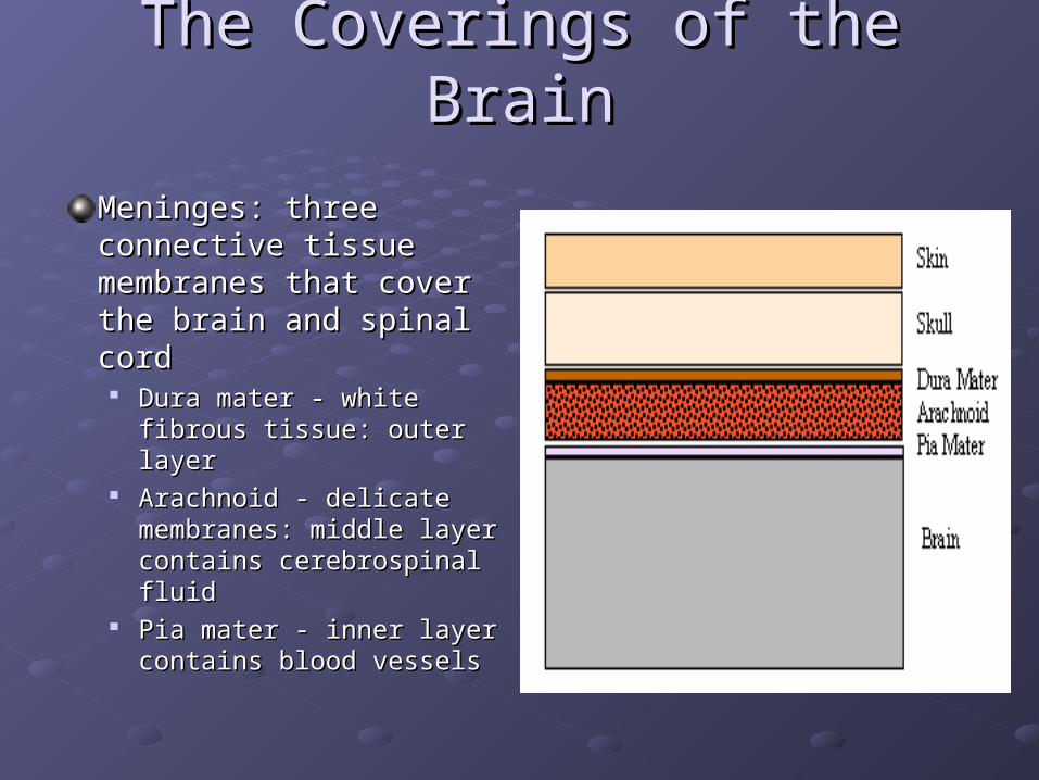
Meninges: three Meninges: three connective tissue connective tissue membranes that cover membranes that cover the brain and spinal cordthe brain and spinal cord
Dura mater - white fibrous Dura mater - white fibrous tissue: outer layertissue: outer layer
Arachnoid - delicate Arachnoid - delicate membranes: middle layer membranes: middle layer contains cerebrospinal fluidcontains cerebrospinal fluid
Pia mater - inner layer Pia mater - inner layer contains blood vesselscontains blood vessels

Major Parts Of The BrainMajor Parts Of The Brain
CerebrumCerebrum Largest area of the brainLargest area of the brain Divided into left and right Divided into left and right
hemisphereshemispheres Right cerebral hemisphere Right cerebral hemisphere
controls the left side of the controls the left side of the bodybody
Left cerebral hemisphere Left cerebral hemisphere controls the right side of the controls the right side of the body body
Each hemisphere is divided Each hemisphere is divided into four lobes – frontal, into four lobes – frontal, parietal, temporal, occipitalparietal, temporal, occipital

Lobes Of The BrainLobes Of The Brain
Frontal LobeFrontal Lobe associated with reasoning, associated with reasoning,
planning, parts of speech, planning, parts of speech, movement, emotions, and movement, emotions, and problem solving problem solving
Parietal LobeParietal Lobe associated with movement, associated with movement,
orientation, recognition, orientation, recognition, perception of stimuli perception of stimuli
Occipital LobeOccipital Lobe associated with visual associated with visual
processing processing
Temporal LobeTemporal Lobe associated with perception and associated with perception and
recognition of auditory stimuli, recognition of auditory stimuli, memory, and speechmemory, and speech

Major Parts Of The BrainMajor Parts Of The Brain
CerebellumCerebellum Second largest part of Second largest part of
the brainthe brain It is connected to the It is connected to the
brain stembrain stem Helps provide smooth Helps provide smooth
coordinated body coordinated body movementmovement

Major Parts Of The BrainMajor Parts Of The Brain
Brain StemBrain Stem is responsible for basic is responsible for basic
vital life functions such vital life functions such as breathing, as breathing, heartbeat, and blood heartbeat, and blood pressure. pressure.
MidbrainMidbrain
Pons Pons
Medulla oblongataMedulla oblongata


VentriclesVentricles
CSF And VentriclesCSF And Ventricles
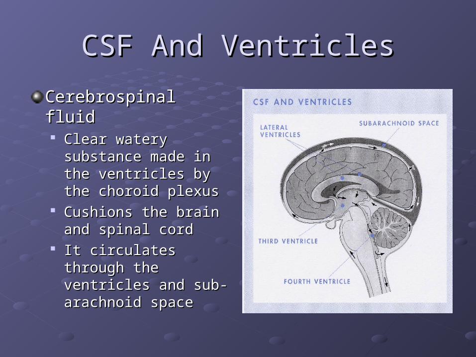
Cerebrospinal fluid Cerebrospinal fluid Clear watery Clear watery
substance made in the substance made in the ventricles by the ventricles by the choroid plexuschoroid plexus
Cushions the brain Cushions the brain and spinal cordand spinal cord
It circulates through It circulates through the ventricles and sub-the ventricles and sub-arachnoid spacearachnoid space

Intra-cranial PressureIntra-cranial Pressure
When intra-cranial pressure begins to rise, the When intra-cranial pressure begins to rise, the body’s own compensatory mechanisms include body’s own compensatory mechanisms include decreasing the production of CSF and restricting decreasing the production of CSF and restricting the blood flow to the brain (by vasoconstriction). the blood flow to the brain (by vasoconstriction).
Once the capacity of these compensatory Once the capacity of these compensatory mechanisms is exceeded, the intra-cranial mechanisms is exceeded, the intra-cranial pressure can continue to rise. pressure can continue to rise.
In addition, as intra-cranial pressure rises, the In addition, as intra-cranial pressure rises, the cerebral blood vessels are constricted, reducing cerebral blood vessels are constricted, reducing blood flow further.blood flow further.

Intra-cranial PressureIntra-cranial Pressure
Normal intracranial pressure (ICP), usually Normal intracranial pressure (ICP), usually measured as a mean pressure, is often measured as a mean pressure, is often cited as 0-10mmHg cited as 0-10mmHg
Sustained high pressures can cause Sustained high pressures can cause 'coning' (tentorial herniation), when 'coning' (tentorial herniation), when brainstem tissue is forced through the brainstem tissue is forced through the foramen magnum into the spinal cord. foramen magnum into the spinal cord.

Cushing’s ResponseCushing’s Response
The following three symptoms are known The following three symptoms are known collectively as Cushing's response triadcollectively as Cushing's response triad Hypertension. Hypertension. Bradycardia. Bradycardia. Abnormal respiratory pattern. Abnormal respiratory pattern.
They indicate brainstem dysfunction and They indicate brainstem dysfunction and exhaustion of compliance (Hickey 1997a); exhaustion of compliance (Hickey 1997a); without urgent intervention, patients are likely to without urgent intervention, patients are likely to die. die.

Causes Of Raised Intra-Cranial Causes Of Raised Intra-Cranial PressurePressure
Anything that increases the volume of brain Anything that increases the volume of brain tissue, blood or CSF within the skull will raise tissue, blood or CSF within the skull will raise intra-cranial pressure:intra-cranial pressure: volume of brain (cerebral oedema): - injury volume of brain (cerebral oedema): - injury infection infection hypoxia hypoxia CSF (eg due to obstruction to drainage) CSF (eg due to obstruction to drainage) haemorrhage (eg subarachnoid) haemorrhage (eg subarachnoid) tumour tumour haematoma haematoma

Head InjuriesHead Injuries
Head injury is most likely to happen to young Head injury is most likely to happen to young men, with an average age of 30 who are men, with an average age of 30 who are involved in road traffic accidents involved in road traffic accidents
Other causes of adult injuries include contact Other causes of adult injuries include contact sports, such as rugby and boxing sports, such as rugby and boxing
Children often suffer head injury from bicycle Children often suffer head injury from bicycle accidents or pedestrian-vehicle collisions and accidents or pedestrian-vehicle collisions and very young children and old adults can suffer very young children and old adults can suffer injury from falls injury from falls

Head InjuriesHead Injuries
The head is vulnerable to injuryThe head is vulnerable to injury Analogy for a head injuryAnalogy for a head injury
Blancmange (brain)Blancmange (brain)Wrapped in cling film (arachnoid mater)Wrapped in cling film (arachnoid mater)In a paper bag (dura mater)In a paper bag (dura mater)Inside a cardboard box (skull)Inside a cardboard box (skull)Wrapped in brown paper (skin)Wrapped in brown paper (skin)
Any layer may be damaged byAny layer may be damaged byDirect impact on the box (blow)Direct impact on the box (blow)Dropping the box (fall)Dropping the box (fall)Shaking the box (acceleration/deceleration)Shaking the box (acceleration/deceleration)
Head InjuriesHead Injuries
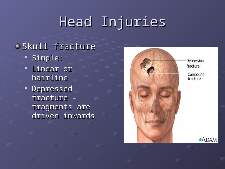
Skull fractureSkull fracture Simple: Simple: Linear or hairlineLinear or hairline Depressed fracture – Depressed fracture –
fragments are driven fragments are driven inwardsinwards

Head InjuriesHead Injuries
Intracranial haemorrhageIntracranial haemorrhage The dura and arachnoid membranes and their The dura and arachnoid membranes and their
associated blood vessels are readily torn by impact or associated blood vessels are readily torn by impact or fractured bone fragmentsfractured bone fragments
There are four types of intracranial haemorrhagesThere are four types of intracranial haemorrhagesExtradural Extradural
Subdural Subdural
SubarachnoidSubarachnoid
Intracerebral Intracerebral
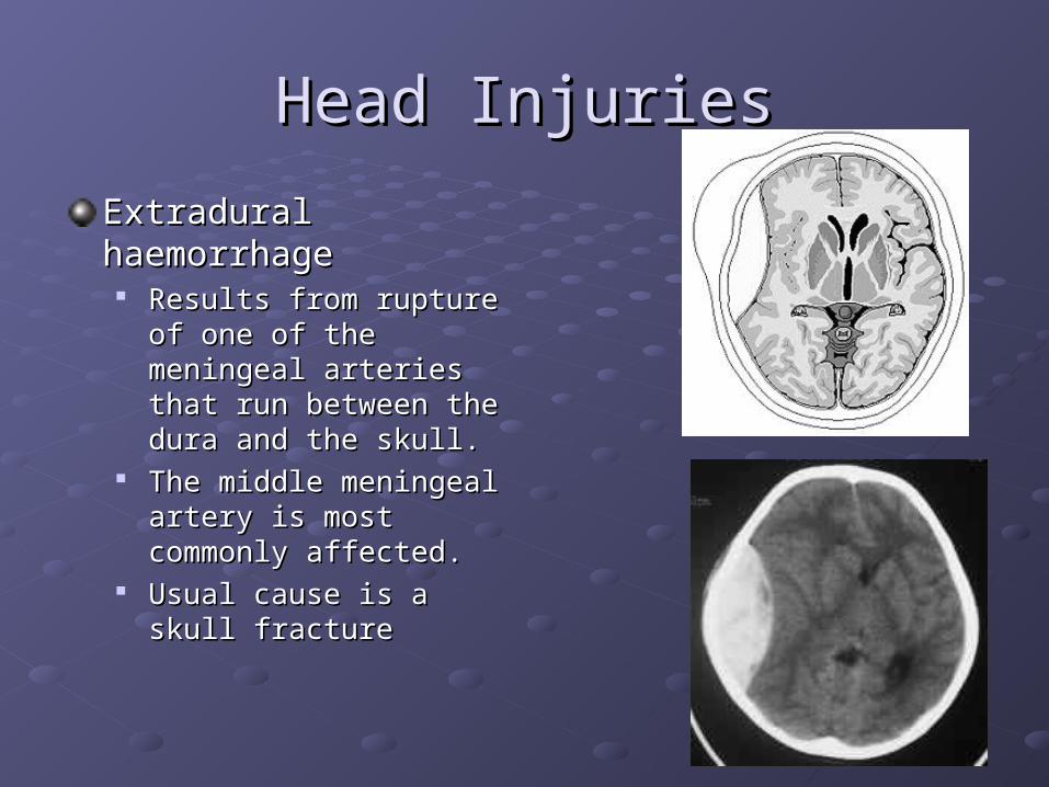
Head InjuriesHead Injuries
Extradural haemorrhageExtradural haemorrhage Results from rupture of one Results from rupture of one
of the meningeal arteries of the meningeal arteries that run between the dura that run between the dura and the skull.and the skull.
The middle meningeal The middle meningeal artery is most commonly artery is most commonly affected.affected.
Usual cause is a skull Usual cause is a skull fracturefracture

Head InjuriesHead Injuries
Subdural haemorrhageSubdural haemorrhage More common than More common than
extradural haemorrageextradural haemorrage Associated with sudden Associated with sudden
jarring or rotation of the jarring or rotation of the headhead
Shears and tears the small Shears and tears the small veins which bridge the gap veins which bridge the gap between the dura and between the dura and cortical surface of the braincortical surface of the brain

Head InjuriesHead Injuries
Intracerebral haemorrhage: Intracerebral haemorrhage: May beMay be natural, due to spontaneous rupture natural, due to spontaneous rupture
of a small blood vessel which has been of a small blood vessel which has been weakened by the effects long-standing high weakened by the effects long-standing high blood pressure. blood pressure.
Traumatic due to extension of haemorrhage Traumatic due to extension of haemorrhage from surface contusions deep into the from surface contusions deep into the substance of the brain. substance of the brain.

Assessment Of Head InjuriesAssessment Of Head Injuries
Glasgow Coma Scale (GCS)Glasgow Coma Scale (GCS)Scoring system originally described for Scoring system originally described for patients with head injury; now applied to patients with head injury; now applied to other causes of coma other causes of coma The Glasgow coma scale (GCS) is a The Glasgow coma scale (GCS) is a reliable and universally comparable way of reliable and universally comparable way of recording the conscious state of a person. recording the conscious state of a person.
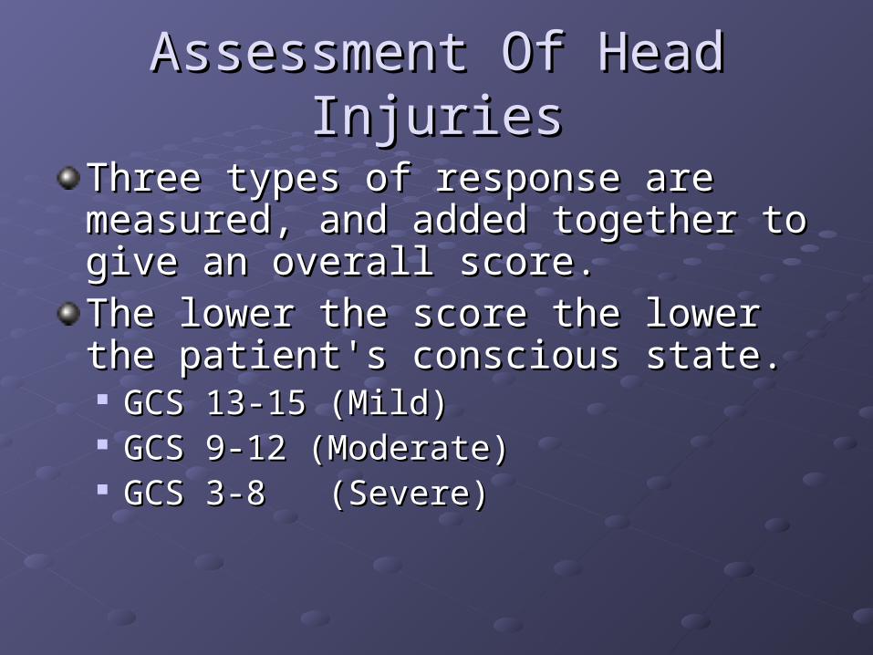
Assessment Of Head InjuriesAssessment Of Head Injuries
Three types of response are measured, Three types of response are measured, and added together to give an overall and added together to give an overall score. score. The lower the score the lower the patient's The lower the score the lower the patient's conscious state. conscious state. GCS 13-15 (Mild) GCS 13-15 (Mild) GCS 9-12 (Moderate) GCS 9-12 (Moderate) GCS 3-8 (Severe) GCS 3-8 (Severe)
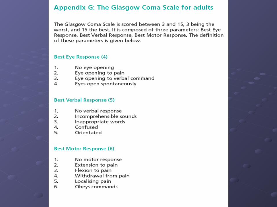

Eye OpeningEye Opening EE
spontaneousspontaneous 44
to speechto speech 33
to painto pain 22
no responseno response 11
Best Motor ResponseBest Motor Response MM
To Verbal Command:To Verbal Command:
obeysobeys 66
To Painful Stimulus:To Painful Stimulus:
localizes painlocalizes pain 55
flexion-withdrawalflexion-withdrawal 44
flexion-abnormalflexion-abnormal 33
extensionextension 22
no responseno response 11
Best Verbal ResponseBest Verbal Response VV
oriented and conversesoriented and converses 55
disoriented and conversesdisoriented and converses 44
inappropriate wordsinappropriate words 33
incomprehensible soundsincomprehensible sounds 22
no responseno response 11
E + M + V = 3 to 15•8 is the critical score •Less than or equal to 8 at 6 hours - 50% die •9-11 = moderate severity •Greater than or equal to 12 = minor injury Coma is defined as: (1) not opening eyes, (2) not obeying commands (3) not uttering understandable words.

DECORTICATEDECORTICATE
Decorticate posturing is also called Decorticate posturing is also called decorticate responsedecorticate response, , decorticate decorticate rigidityrigidity, , flexor posturingflexor posturing

DECEREBRATEDECEREBRATEDecerebrate posturing Decerebrate posturing ::typically the head typically the head is arched back, the arms are extended by is arched back, the arms are extended by the sides, and the legs are extendedthe sides, and the legs are extended

Neurological ObservationsNeurological Observations
Assess conscious levelAssess conscious level SpeechSpeech Mental stateMental state
EyesEyes Can the patient seeCan the patient see Is there an eye injury – eye maybe closedIs there an eye injury – eye maybe closed Can the patient focusCan the patient focus

Neurological AssessmentNeurological Assessment
It is important to assess a patient’s neurological It is important to assess a patient’s neurological state if a patient has a head injury, in a coma or state if a patient has a head injury, in a coma or have had neuro surgery performedhave had neuro surgery performedThis assessment can indicate quite quickly a This assessment can indicate quite quickly a need for interventionneed for interventionNeurological assessment may be carried out Neurological assessment may be carried out every fifteen minutes or half hourly depending every fifteen minutes or half hourly depending on the condition of the patienton the condition of the patientThe most serious situation is the deterioration of The most serious situation is the deterioration of conscious level due to raised intracranial conscious level due to raised intracranial pressurepressure
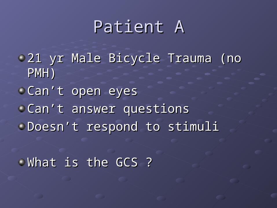
Patient APatient A
21 yr Male Bicycle Trauma (no PMH)21 yr Male Bicycle Trauma (no PMH)
Can’t open eyesCan’t open eyes
Can’t answer questionsCan’t answer questions
Doesn’t respond to stimuliDoesn’t respond to stimuli
What is the GCS ?What is the GCS ?

Patient BPatient B
52 Female (2 week history of headache)52 Female (2 week history of headache)
Responds to verbal commandsResponds to verbal commands
Responds to questions is coherent but Responds to questions is coherent but confusedconfused
Localises to pain (moves hand away from Localises to pain (moves hand away from site)site)
What is the GCS ?What is the GCS ?
Neurological ObservationsNeurological Observations
Pupillary observationsPupillary observations What is their size – normal, moderately What is their size – normal, moderately
dilated or fully dilateddilated or fully dilated What is the pupil reaction to light – brisk, What is the pupil reaction to light – brisk,
sluggish or fixedsluggish or fixed

Neurological ObservationsNeurological Observations
Limb movement & toneLimb movement & tone Can the patient move their limbs on commandCan the patient move their limbs on command
Movement is it normal, weak, severely weak or Movement is it normal, weak, severely weak or absentabsent
If absent does the patient respond to painful stimuli If absent does the patient respond to painful stimuli Is there any abnormal involuntary movementIs there any abnormal involuntary movement

Neurological ObservationsNeurological Observations
Blood pressureBlood pressure
PulsePulse
RespirationRespiration
TemperatureTemperature

Signs Of Raised ICPSigns Of Raised ICP
HeadacheHeadacheVomitingVomitingIncreasing Increasing drowsinessdrowsinessDeterioration in Deterioration in mental and verbal mental and verbal responseresponseInequality of the Inequality of the pupils with sluggish pupils with sluggish reaction to lightreaction to light
Development of Development of hemiparesishemiparesisIncontinenceIncontinencePulse rate becomes Pulse rate becomes slowerslowerBlood pressure risesBlood pressure risesRespiration – depth, Respiration – depth, rate and rhythm rate and rhythm change when patient change when patient loses consciousness loses consciousness

Any Questions?Any Questions?

BibliographyBibliography
Verran B, Aisbett P.(1988) Neurological Verran B, Aisbett P.(1988) Neurological and Neurosurgical Nursing. London and Neurosurgical Nursing. London Edward Arnold PublishingEdward Arnold Publishing