aidap prescribing guidelines
TRANSCRIPT

ANTIBIOTICPRESCRIBINGDETAILED GUIDELINES
AUSTRALASIAN INFECTIOUS DISEASES ADVISORY PANELAIDAP

Dr Steve Holloway BVSc (Syd), MVS, PhD (Melb), DACVIM, MACVSc
Steve graduated from the University of Sydney in 1983 and after several years in practice, journeyed along the pathway to becoming a specialist in internal medicine. In 1991 Steve became a Diplomate of the American College of Internal Medicine. In 1994 Steven commenced his PhD studying herpesviruses of horses at the University of Melbourne, completing in early 1998. Between 1999 and 2009 Steve lectured in infectious diseases of small animals at the Faculty of Veterinary Science at the University of Melbourne. Steven is currently a specialist in internal medicine in private practice and regularly consults on infectious disease problems in small animal patients.
Dr Darren Trott BSc (Hon), BVMS (Hon), PhD
Dr Trott is a veterinarian with 20 years experience in bacterial disease research focused on zoonotic infections, enteric diseases, gastrointestinal microbial ecology and antibiotic resistance. In 2010, Darren accepted a position in the new School of Animal and Veterinary Sciences at The University of Adelaide and aims to establish a new Research Centre focused on Comparative Gastrointestinal Health in animals and humans.
Dr Mike Shipstone BVSc, FACVSC, DACVD
Dr Shipstone graduated from Queensland University in 1984 and has worked in a number of different private practice and industry positions. In 1995 he started a residency at the Animal Skin and Allergy Clinic in Melbourne, with additional periods of study at the University of California, Davis and Louisiana State University, Baton Rouge. Mike is principal and director of a specialist dermatology referral practice and adjunct Associate Professor at the University of Queensland. Mike is a Fellow of the Australian College of Veterinary Scientists (Veterinary Dermatology) and a Diplomate of the American College of Veterinary Dermatology, the only dual boarded veterinary dermatologist in Australia. Mike has published in Australia and overseas and has presented in Australia, South East Asia and North America.
Associate Professor Vanessa Barrs BVSc (hons), MVetClinStud, FACVSc (Feline Medicine), GradCertEd (Higher Ed)
Associate Professor Vanessa Barrs is the Director of the University Veterinary Teaching Hospital and Head of Small Animal Medicine at the University of Sydney. She is a registered Specialist in Feline Medicine and has worked in University and Private Referral Practices in London and Sydney. She has represented the profession in many roles including as President of the Feline Chapter of the Australian College of Veterinary Scientists, Specialist representative of the NSW Board of Veterinary Practitioners and trustee of the Australian Feline Health Research Fund. She enjoys teaching and was awarded the Australian Veterinary Association Excellence in Teaching Award in 2007. Her research interests include lymphoma and infectious diseases, especially fungal diseases, for which she was awarded Distinguished Scientific Award in 2009 by the Australian Small Animal Veterinary Association. She has over 70 refereed publications and book chapters. Most of all A/Professor Barrs loves cats and feline medicine.
Dr Richard Malik DVSc, DipVetAn, MVetClinStud, PhD, FACVSc, FASM
Richard Malik graduated from the University of Sydney, trained in Anaesthesia and Intensive Care, and then moved to ANU where he completed a PhD in pharmacology at the John Curtin School of Medical Research. He then completed a Postdoctoral fellowship at the Neurobiology Research Centre before returning to his alma mater where he remained there for 16 years in a variety of positions (1995 to 2002). Since 2003 Richard has worked as a consultant for the Centre of Veterinary Education and he finds time also to see cases in a number of practices in the Eastern suburbs of Sydney. Richard has varied research interests, most notably infectious diseases, genetic diseases and diseases of cats in general. He is a Fellow of the Australian Society of Microbiology, a member of the Australian Society of Infectious Diseases and an Adjunct Professor of Veterinary Medicine at Charles Sturt University.
Dr Mandy Burrows BSc, BVMS Murd, MACVSc, FACVSc (Dermatology)
Mandy is a Fellow of the Australian College of Veterinary Scientists in Veterinary Dermatology and a registered specialist in veterinary dermatology. She is a consultant in veterinary dermatology and has two dermatology practices in Perth, Western Australia that provide secondary and tertiary referral advice for skin, ear and allergy problems in dogs, cats and horses. She lectures in dermatology at Murdoch University Veterinary Hospital and she teaches undergraduate veterinary students and the dermatology unit of the Masters in Veterinary Medicine at both Murdoch and Massey University, New Zealand. She is currently the Chief Examiner and serves on the Council and the Board of Examiners of the Australian and New Zealand College of Veterinary Scientists and is a member of the Advisory Committee for the Registration of Veterinary Specialists. She is a member of the Australian Advisory Board for Infectious Diseases in companion animals and is the current Australian and New Zealand representative and the Secretary of the World Association for Veterinary Dermatology. She has extensive experience with clinical dermatology in companion animals and she enjoys teaching dermatology to veterinary undergraduate and postgraduate students.
AUSTRALASIAN INFECTIOUS DISEASES ADVISORY PANEL

Antimicrobial resistance is a critical problem in human medicine around the world, both in hospitals and in the wider community. It is emerging as a problem in veterinary medicine, especially in the USA. Although the situation in Australia is currently much better than in North America, multi-resistant E. coli and some methicillin-resistant Staphs have appeared in Australian small animal practices over the last 10 years. Although detailed discussion and analysis of this problem is currently beyond the scope of AIDAP, the group thought some pertinent practical tips would be a good step towards improved antimicrobial stewardship, which is the best way to prevent the emergence of a more widespread resistance problem.
1. Choose antimicrobials based on the most likely pathogen(s) that are associated with particular infectious disease settings (e.g. E. coli from a lower urinary tract infections or S. pseudintermedius from canine pyoderma). Published susceptibility profiles for any given pathogen should be used to make an informed decision as to the antibiotic to be selected. In situations where it is not possible to accurately predict the likely pathogens and/or their likely antibiograms, then culture and susceptibility testing should be performed as soon as practical. Where finances preclude this, an in-practice Gram stain can sometimes be very informative.
2. If empiric antibiotic therapy is instituted but has failed, then ideally perform culture and susceptibility testing. For example, urinary tract infections or staphylococcal pyoderma cases that fail to respond to empiric antimicrobial therapy justify culture. If finances preclude this, choose another class of agent likely to be effective against the putative pathogen.
3. Avoid empiric use of fluoroquinolones for treating chronic Staph spp. infections in dogs or uncomplicated UTI. Amoxicillin clavulanate is a superior choice to fluoroquinolones for empiric therapy of UTIs in the opinion of this panel.
4. Avoid using combination therapy unless there is clearly a life-threatening infection present and/or an unpredictable antibiotic susceptibility of the pathogen(s) involved. For example, life-threatening sepsis in a dog that has peritonitis from a ruptured bowel is an indication for 4-quadrant antibiotic therapy until the results of culture are known.
6. Ensure the length of treatment with antibiotics is appropriate. Serious infections generally justify at least two-weeks of therapy. Identify where owner or patient compliance is likely to be an issue and take appropriate measures to achieve compliance.
7. In the hospital setting, be vigilant for the occurrence of infections attributable to an unusual organism (e.g. Serratia spp.) or common pathogens (e.g. E. coli) with a consistent antibiograms, often with a multi-resistant profile. Such organisms should ideally be forwarded to a suitable reference laboratory (e.g. Darren Trott’s laboratory, the University of Adelaide or VPDS at the University of Sydney) for archiving and possibly additional molecular testing. If such case clustering occurs, consider consultation with an infectious disease expert to try to track down potential sources of infection e.g. foam bedding, a staff member who is a chronic carrier of Staph. aureus.
8. Develop in-house infection control guidelines for every veterinary hospital. These should include signs and policies that encourage regular hand washing with alcohol-based hand preparations.
AIDAP
RESISTANCE TIPS
AIDAP: ANTIBIOTIC PRESCRIBING DETAILED GUIDELINES
DOGS SUBCUTANEOUS ABSCESS/CELLULITIS p1
PERIODONTAL DISEASE p5
ACUTE URT DISEASE/INFECTIOUS TRACHEOBRONCHITIS p9 CHRONIC RHINOSINUSITIS p13
ACUTE LRT INFECTION p16 PYOTHORAX p20
ACUTE LOWER UTI/CYSTITIS (FIRST OCCURRENCE) p24 COMPLICATED UTIs: RECURRENT LOWER UTI/CYSTITIS AND CKD WITH PYURIA p28
ACUTE FEBRILE ILLNESS p33
ACUTE ABDOMINAL PAIN AND PYREXIA/ABDOMINAL INFECTION AND LEUKOPENIA p36
ANTIBIOTIC USE AFTER ROUTINE DESEXING p39
USE OF ANTIBIOTICS IN DENTAL PROPHYLAXIS p41
SURFACE BACTERIAL INFECTIONS: (i.e. INTERTRIGO e.g. LIP FOLD, TAIL FOLD) p44 SUPERFICIAL BACTERIAL INFECTIONS: (i.e. MUCOCUTANEOUS PYODERMA,
BACTERIAL FOLLICULITIS, BACTERIAL OVERGROWTH) p47 DEEP BACTERIAL INFECTIONS: (i.e. FURUNCULOSIS WITH DRAINING TRACTS p52 MYCOBACTERIA AND NOCARDIA AS CAUSES OF DEEP DRAINING SINUS TRACTS p58 DERMATOPHYTE INFECTIONS (e.g. MICROSPORUM OR TRICHOPHYTON) p62 SUPERFICIAL YEAST (MALASSEZIA) INFECTIONS OF THE SKIN (NOT INCLUDING EARS) p71
OTITIS EXTERNA (UNCOMPLICATED, FIRST EPISODE AND COMPLICATED, RECURRENT) p77
SOFT TISSUE
ORAL
UPPER RESPIRATORY TRACT
LOWER RESPIRATORY TRACT
URINARY TRACT
PYREXIA
ABDOMINAL
DESEXING
DENTAL
SKIN/SOFT TISSUE
AURAL
CATS SUBCUTANEOUS ABSCESS/CELLULITIS p83
CHRONIC GINGIVOSTOMATITIS/FAUCITIS p87
ACUTE URT DISEASE p91 CHRONIC RHINOSINUSITIS p95
ACUTE LRT INFECTION p99 PYOTHORAX p103
ACUTE LOWER UTI/CYSTITIS (FIRST OCCURRENCE) p107 COMPLICATED UTIs: RECURRENT LOWER UTI/CYSTITIS AND CKD WITH PYURIA p112
ACUTE FEBRILE ILLNESS p116
ACUTE ABDOMINAL PAIN AND PYREXIA/ABDOMINAL INFECTION AND LEUKOPENIA p120
ANTIBIOTIC USE AFTER ROUTINE DESEXING p123
USE OF ANTIBIOTICS IN DENTAL PROPHYLAXIS p125
MYCOBACTERIA AND NOCARDIA AS CAUSES OF DEEP DRAINING SINUS TRACTS p128 DERMATOPHYTE INFECTIONS (e.g. MICROSPORUM OR TRICHOPHYTON) p132
OTITIS EXTERNA (UNCOMPLICATED, FIRST EPISODE AND COMPLICATED, RECURRENT) p141
SOFT TISSUE
ORAL
UPPER RESPIRATORY TRACT
LOWER RESPIRATORY TRACT
URINARY TRACT
PYREXIA
ABDOMINAL
DESEXING
DENTAL
SKIN/SOFT TISSUE
AURAL
GLOSSARY
abx = antibiotics BAL = bronchioalveolar lavage C+S = culture and susceptibility CKD = chronic kidney diseaseE. coli = Escherichia coli FB = foreign body FC = feline calicivirusFHV-1 = feline herpes virus-type 1 FQ = fluoroquinolone GA = general anaesthetic hrs = hours IM = intramuscular IV = intravenous KCS = keratoconjunctivitis sicca LRT = lower respiratory tract LRTI = lower respiratory tract infection MDR = multi-drug resistant N/A = not applicable NSAID = non-steroidal anti-inflammatory drug PCR = polymerase chain reaction Rx = treatment SC = subcutaneous spp. = species TMS = Trimethoprim-sulfonamide TTW = transtracheal wash UA = urinalysis URT = upper respiratory tract URTI = upper respiratory tract infection UTI = urinary tract infection
† = off label‡ = not registered for animal use
For easy access to the AIDAP guidelines, download the Vets Australia iPhone and iPad application.
SO
FT
TIS
SU
EO
RA
LU
RT
LR
TU
RIN
AR
Y T
RA
CT
PY
RE
XIA
AB
DO
MIN
AL
DE
SE
XIN
GD
EN
TAL
SKIN
/SO
FT T
ISSU
EA
UR
AL
CA
T C
ON
TE
NT
SPECIES: DOG
SPECIES: DOG 1SECTION: SOFT TISSUE
CONDITION: SUBCUTANEOUS ABSCESS/CELLULITIS
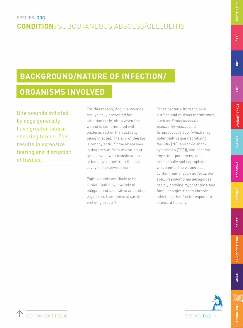
For this reason, dog bite wounds are typically presented for attention early, often when the wound is contaminated with bacteria, rather than actually being infected. The aim of therapy is prophylactic. Some abscesses in dogs result from migration of grass awns, with translocation of bacteria either from the oral cavity or the environment.
Fight wounds are likely to be contaminated by a variety of obligate and facultative anaerobic organisms from the oral cavity and gingival cleft.
Other bacteria from the skin surface and mucous membranes, such as Staphylococcus pseudintermedius and Streptococcus spp. (which may potentially cause necrotising fasciitis [NF] and toxic shock syndromes [TSS]), can become important pathogens, and occasionally soil saprophytes which enter the wounds as contaminants (such as Nocardia spp., Pseudomonas aeruginosa, rapidly growing mycobacteria and fungi) can give rise to chronic infections that fail to respond to standard therapy.
Bite wounds inflicted by dogs generally have greater lateral shearing forces. This results in extensive tearing and disruption of tissues.
BACKGROUND/NATURE OF INFECTION/
ORGANISMS INVOLVED

SO
FT
TIS
SU
EO
RA
LU
RT
LR
TU
RIN
AR
Y T
RA
CT
PY
RE
XIA
AB
DO
MIN
AL
DE
SE
XIN
GD
EN
TAL
SKIN
/SO
FT T
ISSU
EA
UR
AL
CA
T C
ON
TE
NT
SPECIES: DOG
SPECIES: DOG 2SECTION: SOFT TISSUE
CONDITION: SUBCUTANEOUS ABSCESS/CELLULITIS
It can be helpful to make smears of purulent exudate, when present. Subsequent Gram or DiffQuik staining may demonstrate pathogenic bacteria. C+S testing may be helpful, especially in cases that have failed to respond to empiric therapy.
Radiology (with contrast i.e. fistulogram), ultrasonography and occasionally advanced cross-sectional imaging may be useful to detect inciting foreign bodies such as grass seeds, wood splinters, teeth or metallic fragments.
KEY ISSUES
01 Dog fight wounds involve lateral shearing forces, major disruption of tissues and an open draining wound.
02 There is variable contamination with a variety of different bacteria.
03 Streptococcus species can sometimes be important pathogens in this setting.
04 Occasionally, subcutaneous infections occur due to migrating plant foreign bodies such as grass seeds and awns.
TESTS FOR DIAGNOSIS
Latex drain placed to allow ongoing gravitational drainage.
Photo courtesy of Dr Anne Fawcett.
SO
FT
TIS
SU
EO
RA
LU
RT
LR
TU
RIN
AR
Y T
RA
CT
PY
RE
XIA
AB
DO
MIN
AL
DE
SE
XIN
GD
EN
TAL
SKIN
/SO
FT T
ISSU
EA
UR
AL
CA
T C
ON
TE
NT
SPECIES: DOG
SPECIES: DOG 3SECTION: SOFT TISSUE
CONDITION: SUBCUTANEOUS ABSCESS/CELLULITIS
TREATMENT
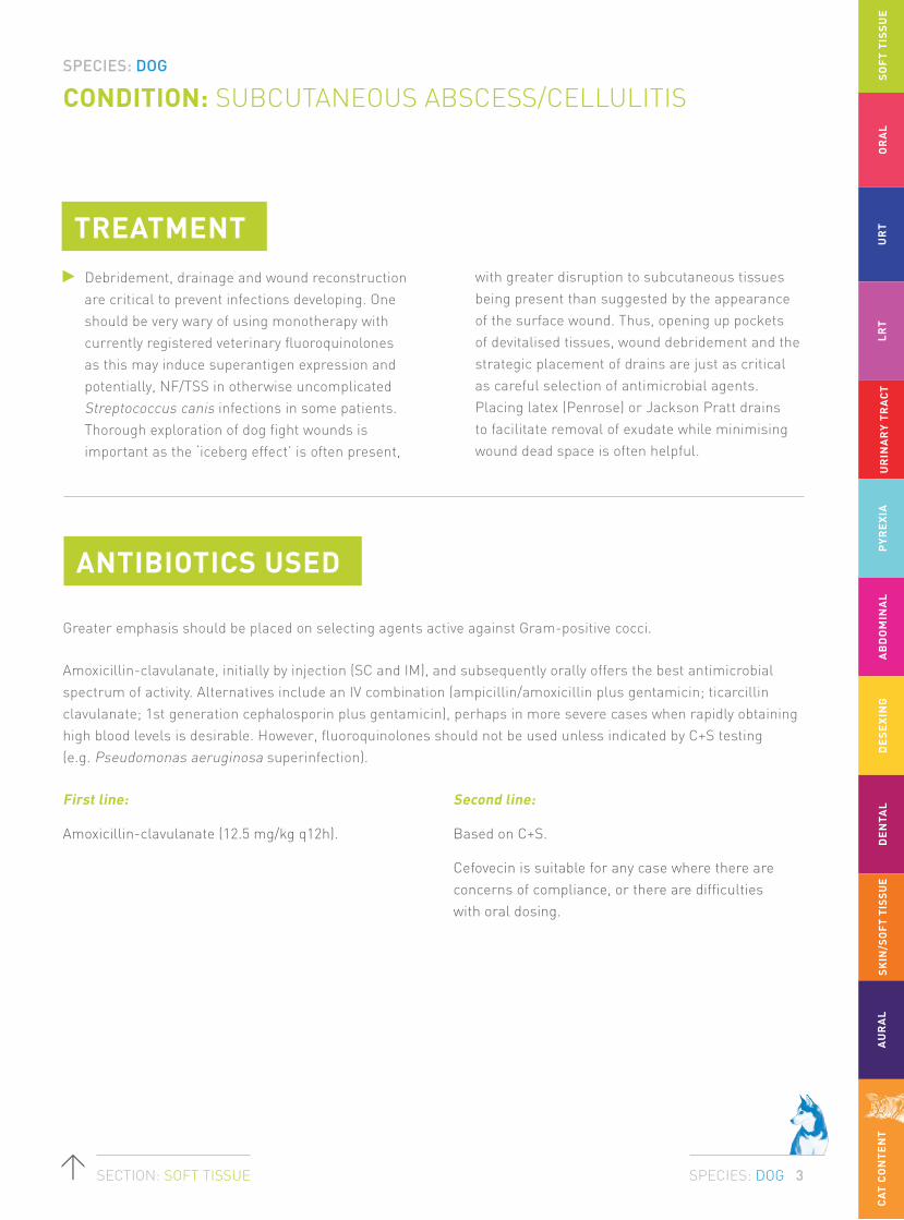
Debridement, drainage and wound reconstruction are critical to prevent infections developing. One should be very wary of using monotherapy with currently registered veterinary fluoroquinolones as this may induce superantigen expression and potentially, NF/TSS in otherwise uncomplicated Streptococcus canis infections in some patients. Thorough exploration of dog fight wounds is important as the ‘iceberg effect’ is often present,
with greater disruption to subcutaneous tissues being present than suggested by the appearance of the surface wound. Thus, opening up pockets of devitalised tissues, wound debridement and the strategic placement of drains are just as critical as careful selection of antimicrobial agents. Placing latex (Penrose) or Jackson Pratt drains to facilitate removal of exudate while minimising wound dead space is often helpful.
Greater emphasis should be placed on selecting agents active against Gram-positive cocci.
Amoxicillin-clavulanate, initially by injection (SC and IM), and subsequently orally offers the best antimicrobial spectrum of activity. Alternatives include an IV combination (ampicillin/amoxicillin plus gentamicin; ticarcillin clavulanate; 1st generation cephalosporin plus gentamicin), perhaps in more severe cases when rapidly obtaining high blood levels is desirable. However, fluoroquinolones should not be used unless indicated by C+S testing (e.g. Pseudomonas aeruginosa superinfection).
First line:
Amoxicillin-clavulanate (12.5 mg/kg q12h).
Second line:
Based on C+S.
Cefovecin is suitable for any case where there are concerns of compliance, or there are difficulties with oral dosing.
ANTIBIOTICS USED
SO
FT
TIS
SU
EO
RA
LU
RT
LR
TU
RIN
AR
Y T
RA
CT
PY
RE
XIA
AB
DO
MIN
AL
DE
SE
XIN
GD
EN
TAL
SKIN
/SO
FT T
ISSU
EA
UR
AL
CA
T C
ON
TE
NT
SPECIES: DOG
SPECIES: DOG 4SECTION: SOFT TISSUE
CONDITION: SUBCUTANEOUS ABSCESS/CELLULITIS
AIDAP TOP TIPS
USAGE RECOMMENDATION
There is no evidence-base to guide recommendations regarding treatment duration.
From experience AIDAP recommend at least 4 days and ideally 7-14 days.
Avoid empiric use of
fluoroquinolones in
dogs with fight wounds,
as they may predispose
to the development
of life-threatening
streptococcal infections.
Key references:
1. Hall M. J Am Vet Med Assoc 2010; 236(6): 620
2. Brook I. Curr Infect Dis Rep 2009; 11(5): 389-395
3. Six R, Cherni J, Chesebrough R, et al. J Am Vet Med Assoc 2008; 233(3): 433-439
4. Csiszer AB, Towle HA, Daly CM. J Am Anim Hosp Assoc 2010; 46(6): 433-438
5. Weese JS, Poma R, James F, et al. Can Vet J 2009; 50(6): 655-656
6. Dendle C and Looke D. Aust Fam Physician. 2009; 38(11): 868-874

SPECIES: DOG
SPECIES: DOG 5SECTION: ORAL
CONDITION: PERIODONTAL DISEASE
SO
FT
TIS
SU
EO
RA
LU
RT
LR
TU
RIN
AR
Y T
RA
CT
PY
RE
XIA
AB
DO
MIN
AL
DE
SE
XIN
GD
EN
TAL
SKIN
/SO
FT T
ISSU
EA
UR
AL
CA
T C
ON
TE
NT
It usually is a result of feeding soft food or highly refined carbohydrate (that is readily fermented by gingival anaerobes) that leads to a shift in the host: bacteria relationship with overgrowth of pathogenic organisms specifically Porphyromonas spp. closely related to P. gingivalis.
At some level, this is a natural disease condition, in which dietary factors, host factors (including crowding of teeth, retention of deciduous teeth, malocclusion due to abnormal head conformation [brachycephalic dogs]) and microbial factors interplay in a complex manner. The bacteria involved are normal constituents of the normal microbiota of the gingival cleft, although they can behave as true pathogens in this scenario.
In this entity, the amount of inflammation in the gum is usually commensurate with the extent of the build-up of plaque and tartar, although irreversible changes in local anatomy (gingival recession with exposure of dentine) contribute.
Typically, the extent of the inflammation is usually proportionate to the extent of plaque and tartar accumulation, although in certain breeds (e.g. Maltese) there seems to be an exaggerated host response. This suggests that genetic factors in this and certain other breeds may play a role in this multi-factorial disease complex. Faucitis – the entity which occurs in cats – has no equivalent entity in the dog, presumably because calicivirus infection does not occur in the dog.
Periodontal disease when it occurs as a single entity in the older dog in association with a build-up of plaque (a biofilm of obligate anaerobes and salivary mucoproteins), tartar (mineralised plaque) and gingival recession.
BACKGROUND/NATURE OF INFECTION/
ORGANISMS INVOLVED
SPECIES: DOG
SPECIES: DOG 6SECTION: ORAL
CONDITION: PERIODONTAL DISEASE
SO
FT
TIS
SU
EO
RA
LU
RT
LR
TU
RIN
AR
Y T
RA
CT
PY
RE
XIA
AB
DO
MIN
AL
DE
SE
XIN
GD
EN
TAL
SKIN
/SO
FT T
ISSU
EA
UR
AL
CA
T C
ON
TE
NT
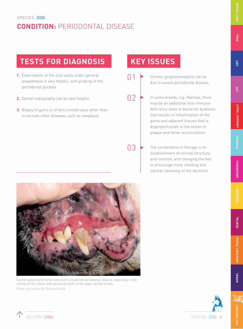
1. Examination of the oral cavity under general anaesthesia is very helpful, with probing of the periodontal pockets.
2. Dental radiography can be very helpful.
3. Biopsy of gums is of very limited value other than to exclude other diseases such as neoplasia.
KEY ISSUES
01 Chronic gingivostomatitis can be due to severe periodontal disease.
02 In some breeds, e.g. Maltese, there may be an additional host immune deficiency state or bacterial dysbiosis that results in inflammation of the gums and adjacent tissues that is disproportionate to the extent of plaque and tartar accumulation.
03 The cornerstone of therapy is re-establishment of normal structure and function, and changing the diet to encourage more chewing and natural cleansing of the dentition.
TESTS FOR DIAGNOSIS
Canine patient with tartar and mild to moderate periodontal disease, especially in the vicinity of the canine and carnassial teeth of the upper dental arcade.
Photo courtesy of Dr Richard Malik.

SPECIES: DOG
SPECIES: DOG 7SECTION: ORAL
CONDITION: PERIODONTAL DISEASE
SO
FT
TIS
SU
EO
RA
LU
RT
LR
TU
RIN
AR
Y T
RA
CT
PY
RE
XIA
AB
DO
MIN
AL
DE
SE
XIN
GD
EN
TAL
SKIN
/SO
FT T
ISSU
EA
UR
AL
CA
T C
ON
TE
NT
TREATMENT
Remove tartar and plaque, scale and polish enamel and exposed dentine, remove unsalvageable teeth, and administer (perhaps) a 2-week course of antimicrobials highly effective against obligate anaerobes likely to be involved.
Clindamycin, metronidazole (with or without spiramycin) and doxycycline are preferred over amoxicillin-clavulanate because they reach more effective levels within the biofilm in the vicinity of the periodontal space.
First line:
Doxycycline monohydrate (5 mg/kg q12h†) or clindamycin (5-11 mg/kg q12h).
Second line:
Metronidazole (10 mg/kg q12h).
Cefovecin is suitable for any case where there are concerns of compliance, or there are difficulties with oral dosing.
ANTIBIOTICS USED

SPECIES: DOG
SPECIES: DOG 8SECTION: ORAL
CONDITION: PERIODONTAL DISEASE
SO
FT
TIS
SU
EO
RA
LU
RT
LR
TU
RIN
AR
Y T
RA
CT
PY
RE
XIA
AB
DO
MIN
AL
DE
SE
XIN
GD
EN
TAL
SKIN
/SO
FT T
ISSU
EA
UR
AL
CA
T C
ON
TE
NT
AIDAP TOP TIPS
USAGE RECOMMENDATION
Ideally, start therapy a few days prior to anaesthesia for dental radiography, extractions, scaling, polishing, and continue for 2 weeks following the procedure or until there is good healing of the periodontal tissues. Ensure doxycycline given with food or water bowl provided.
Key references:
1. Lyon KF. Vet Clin North Am Small Anim Pract 2005; 35(4): 891-911
2. Johnston TP, Mondal P, Pal D, et al. J Vet Dent 2011; 28(4): 224-229
3. Radice M, Martino PA, Reiter AM. J Vet Dent 2006; 23(4): 219-224
4. Stegemann MR, Passmore CA, Sherington J, et al. Antimicrob Agents Chemother 2006; 50(7): 2286-2292
5. Zetner K and Rothmueller G. Vet Ther 2002; 3(4): 441-452
6. Zetner K, Pum G, Rausch WD, et al. Vet Ther 2002; 3(2): 177-188
7. Nielsen D, Walser C, Kodan G, et al. Vet Ther. 2000; 1(3): 150-158
8. Warrick JM, Inskeep GA, Yonkers TD, et al. Vet Ther 2000; 1(1): 5-16
9. Harvey CE, Thornsberry C, Miller BR, et al. J Vet Dent 1995; 12(4): 151-155
10. Jones RL, Godinho KS, Palmer GH. Br Vet J 1994; 150(4): 385-388
11. Sarkiala E, Harvey C. Semin Vet Med Surg (Small Anim) 1993; 8(3): 197-203
12. Giboin H, Becskei C, Civil J, et al. Open J Vet Med 2012; 2: 89-97
Chewing encourages a scissors action of the carnassials on fresh meat and ‘flossing’ by stripping muscle from periosteum and bone, use of specially formulated kibble that minimises tartar accumulation and using adjunctive products and procedures such as dental chews, brushing, chlorhexidine etc.
Further therapy is
directed at changing
the diet to include
more chewing.
SPECIES: DOG
SPECIES: DOG 9SECTION: URT
CONDITION: ACUTE URT DISEASE/INFECTIOUS TRACHEOBRONCHITIS
SO
FT
TIS
SU
EO
RA
LU
RT
LR
TU
RIN
AR
Y T
RA
CT
PY
RE
XIA
AB
DO
MIN
AL
DE
SE
XIN
GD
EN
TAL
SKIN
/SO
FT T
ISSU
EA
UR
AL
CA
T C
ON
TE
NT
The term ‘acute URT disease’ in dogs, may be associated with primary viral URT disease (with secondary bacterial involvement) or disease due to Bordetella bronchiseptica, which can cause tracheobronchitis and pneumonia, and sometimes nasal discharge. Viruses that can cause primary URT disease include distemper, canine adenovirus, parainfluenza, true influenza. These are very rare indeed in contemporary suburban practice in Australian cities. Bordetella can be a primary pathogen or a secondary invader after primary viral disease. There is some new information on canine herpesvirus type 1 as a cause of ocular and also URT disease, but there is no consensus on this or any research data from Australia.
So the question comes down to treatment of ‘canine cough’ or ‘kennel cough’. Nasal foreign bodies e.g. twigs, grass awns, can cause peracute onset of URT signs. Acute cryptococcal rhinosinusitis does occur in dogs, and it is therefore worth examining nasal discharge cytologically or using special fungal media like bird seed agar. Nasal aspergillosis is usually subacute to chronic disease, but should be considered also as a diagnostic possibility.
Primary bacterial rhinitis in dogs does probably not exist as a clinical entity.
BACKGROUND/NATURE OF INFECTION/
ORGANISMS INVOLVED

SPECIES: DOG
SPECIES: DOG 10SECTION: URT
CONDITION: ACUTE URT DISEASE/INFECTIOUS TRACHEOBRONCHITIS
SO
FT
TIS
SU
EO
RA
LU
RT
LR
TU
RIN
AR
Y T
RA
CT
PY
RE
XIA
AB
DO
MIN
AL
DE
SE
XIN
GD
EN
TAL
SKIN
/SO
FT T
ISSU
EA
UR
AL
CA
T C
ON
TE
NT
The new range of multiplex PCR panels may have a use in the clinical evaluation of such cases, although we do not yet have sufficient experience to comment, and they are expensive and slow to give a result.
If cough was the main problem and the owner and/or clinician was interested in further diagnostics, thoracic radiographs and airway (BAL) cytology, Gram stain and bacterial C+S testing might be helpful if you wanted to ‘rule in’ or ‘rule out’ Bordetella and other bacteria, or further characterise the nature of the infectious respiratory disease.
C+S of nasal discharge is unlikely to be helpful, although is some cases one might observe a heavy pure growth of Bordetella which might be clinically significant. Cytology of nasal discharge is useful for assessment of cryptococcal rhinosinusitis, a rare diagnostic possibility.
KEY ISSUES
01 Consider nasal foreign bodies in peracute cases of sneezing and/or nasal discharge. Consider anterior and posterior rhinoscopy in this setting.
02 Consider using a multiplex PCR panel to ‘rule in’ or ‘rule out’ potential viral pathogens and Bordetella using pharyngeal swabs. The problem is such tests are expensive, especially in comparison with the cost of empiric treatment.
03 Consider thoracic radiography and unguided BAL and C+S in dogs with severe cough, especially when fever or dyspnoea is present.
04 Consider empiric treatment with doxycycline monohydrate especially if there is a history of potential contagion from dogs with ‘canine cough’ e.g. from a shelter or pound.
05 Consider a full investigation (radiology, rhinoscopy, bronchoscopy and CT) for cases that fail to respond to empiric therapy. This is more likely to be useful than the selection of a second antimicrobial agent.
Consider that acute cryptococcal rhinosinusitis does occur in dogs, and also consider nasal aspergillosis as a diagnostic possibility.
TESTS FOR DIAGNOSIS

SPECIES: DOG
SPECIES: DOG 11SECTION: URT
CONDITION: ACUTE URT DISEASE/INFECTIOUS TRACHEOBRONCHITIS
SO
FT
TIS
SU
EO
RA
LU
RT
LR
TU
RIN
AR
Y T
RA
CT
PY
RE
XIA
AB
DO
MIN
AL
DE
SE
XIN
GD
EN
TAL
SKIN
/SO
FT T
ISSU
EA
UR
AL
CA
T C
ON
TE
NT
TREATMENT
A thorough history (access to kennels, pounds, other dogs at show), a vaccination history, perhaps thoracic radiography (in certain circumstances) and then trial empiric therapy using doxycycline monohydrate. This drug is arguably the most effective and reliable agent against Bordetella because of its high penetration of respiratory mucous. It also has good efficacy for Pasteurella species and obligate anaerobes involved as secondary respiratory pathogens.
Cough suppressants (e.g. opioids) may be appropriate under some circumstances (persistent dry cough). Nebulisation therapy (using saline with or without gentamicin) may also be helpful. Amoxicillin-clavulanate is a less satisfactory choice because, being charged and water soluble, it tends not to reach sufficiently high levels in respiratory mucous. In young animals, fluoroquinolones should not be used because of their effects on growing cartilage.
First line:
Doxycycline monohydrate (5 mg/kg q12h†).
Second line:
Based on C+S of material collected carefully from the airways (TTW, unguided BAL etc.).
ANTIBIOTICS USED

SPECIES: DOG
SPECIES: DOG 12SECTION: URT
CONDITION: ACUTE URT DISEASE/INFECTIOUS TRACHEOBRONCHITIS
SO
FT
TIS
SU
EO
RA
LU
RT
LR
TU
RIN
AR
Y T
RA
CT
PY
RE
XIA
AB
DO
MIN
AL
DE
SE
XIN
GD
EN
TAL
SKIN
/SO
FT T
ISSU
EA
UR
AL
CA
T C
ON
TE
NT
USAGE RECOMMENDATION
Empiric treatment with doxycycline monohydrate plus nebulisation with saline [with or without gentamicin 1% (10 mg/mL) solution†] for 1-2 weeks, or longer, depending on response to therapy.
Ensure doxycycline given with food or water bowl provided.
Key references:
1. Van Pelt DR and McKiernan BC. Vet Clin North Am Small Anim Pract 1994; 24(5): 789-806
2. Marretta SM. Vet Clin North Am Small Anim Pract 1992; 22(5): 1101-1117
3. Williams M, Olver C, Thrall MA. Vet Clin Pathol 2006; 35(4): 471-473
4. Schwarz S, Alesík E, Grobbel M, et al. Berl Munch Tierarztl Wochenschr. 2007; 120(9-10): 423-430
5. Datz C. Compend Contin Educ Pract Vet 2003; 25(12): 902-914
6. Speakmana AJ, Dawsonb S, Corkillc JE, et al. Vet Microbiol 2000; 71(3-4): 193-200
Profuse nasal discharge, crusting and depigmentation of the nasal planum in a dog with sinonasal aspergillosis.
Photos courtesy of Dr Vanessa Barrs.
Close-up of the dog in previous photo after removal of nasal discharge, illustrating crusting and depigmentation of the nasal planum, which are common findings in dogs with sinonasal aspergillosis – an important differential diagnosis for rhinitis in dogs.

SPECIES: DOG
SPECIES: DOG 13SECTION: URT
CONDITION: CHRONIC RHINOSINUSITIS
SO
FT
TIS
SU
EO
RA
LU
RT
LR
TU
RIN
AR
Y T
RA
CT
PY
RE
XIA
AB
DO
MIN
AL
DE
SE
XIN
GD
EN
TAL
SKIN
/SO
FT T
ISSU
EA
UR
AL
CA
T C
ON
TE
NT
Causes of chronic nasal disease include lymphocytic-plasmacytic rhinitis, foreign bodies (fragmented grass awns, twigs, grass blades etc.), fungal infections (cryptococcosis, low grade aspergillosis), neoplasia and tooth root infections.
Primary viral rhinitis in the dog appears rarely. Nasal cavity disease in the dog is often not primarily due to an infectious agent, apart from mycotic rhinosinusitis.
Chronic rhinosinusitis is rare as a primary entity in the dog.
BACKGROUND/NATURE OF INFECTION/
ORGANISMS INVOLVED
SPECIES: DOG
SPECIES: DOG 14SECTION: URT
CONDITION: CHRONIC RHINOSINUSITIS
SO
FT
TIS
SU
EO
RA
LU
RT
LR
TU
RIN
AR
Y T
RA
CT
PY
RE
XIA
AB
DO
MIN
AL
DE
SE
XIN
GD
EN
TAL
SKIN
/SO
FT T
ISSU
EA
UR
AL
CA
T C
ON
TE
NT
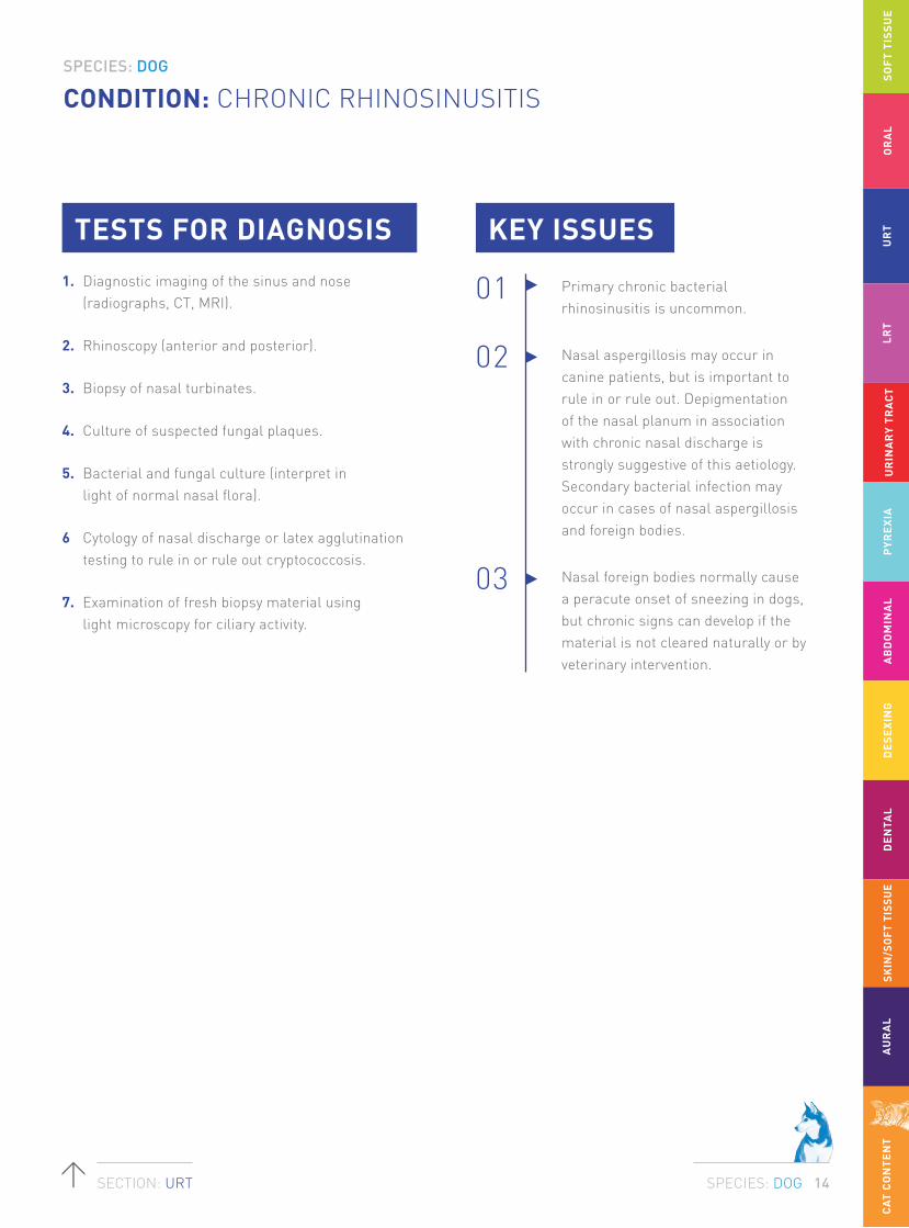
1. Diagnostic imaging of the sinus and nose (radiographs, CT, MRI).
2. Rhinoscopy (anterior and posterior).
3. Biopsy of nasal turbinates.
4. Culture of suspected fungal plaques.
5. Bacterial and fungal culture (interpret in light of normal nasal flora).
6 Cytology of nasal discharge or latex agglutination testing to rule in or rule out cryptococcosis.
7. Examination of fresh biopsy material using light microscopy for ciliary activity.
KEY ISSUES
01 Primary chronic bacterial rhinosinusitis is uncommon.
02 Nasal aspergillosis may occur in canine patients, but is important to rule in or rule out. Depigmentation of the nasal planum in association with chronic nasal discharge is strongly suggestive of this aetiology. Secondary bacterial infection may occur in cases of nasal aspergillosis and foreign bodies.
03 Nasal foreign bodies normally cause a peracute onset of sneezing in dogs, but chronic signs can develop if the material is not cleared naturally or by veterinary intervention.
TESTS FOR DIAGNOSIS
SPECIES: DOG
SPECIES: DOG 15SECTION: URT
CONDITION: CHRONIC RHINOSINUSITIS
SO
FT
TIS
SU
EO
RA
LU
RT
LR
TU
RIN
AR
Y T
RA
CT
PY
RE
XIA
AB
DO
MIN
AL
DE
SE
XIN
GD
EN
TAL
SKIN
/SO
FT T
ISSU
EA
UR
AL
CA
T C
ON
TE
NT
TREATMENT
Do not routinely prescribe antimicrobials when confronted with a dog with nasal cavity signs, unless there is some known event e.g. vomition into the nasal cavity, inhalation of small foreign bodies.
First line:
None empirically.
Second line:
May be appropriate after investigation and C+S e.g. nasal aspergillosis may need antimicrobial therapy for secondary bacterial infection.
ANTIBIOTICS USED
AIDAP TOP TIPS
Nasal discharge should make the clinician pursue diagnostic investigations rather than trialing empiric antimicrobial therapy.
Primary rhinitis is not a
clinical entity in the dog.
Key references:
1. Edwards DF, Patton CS, Kennedy JR. Probl Vet Med 1992; 4(2): 291-319
2. Sharman M, Paul A, Davies D, et al. J Small Anim Pract 2010; 51(8): 423-427
3. Nelson HS. J Allergy Clin Immunol 2007; 119(4): 872-880.
4. Malik R, Dill-Macky E, Martin P, et al. J Med Vet Mycol 1995; 33(5): 291-297

SPECIES: DOG
SPECIES: DOG 16SECTION: LRT
CONDITION: ACUTE LRT INFECTION
SO
FT
TIS
SU
EO
RA
LU
RT
LR
TU
RIN
AR
Y T
RA
CT
PY
RE
XIA
AB
DO
MIN
AL
DE
SE
XIN
GD
EN
TAL
SKIN
/SO
FT T
ISSU
EA
UR
AL
CA
T C
ON
TE
NT
In animals presenting with dyspnoea and coughing, alternative diagnoses must be also considered including pulmonary oedema, aspiration pneumonia, allergic or hypersensitivity disease, haemorrhage and neoplasia. The remarkable number of non-infectious causes of acute respiratory disease makes definitive diagnosis of acute LRT disease difficult. Furthermore, secondary pneumonia may occur in the setting of prior LRT disease due to a non-infectious cause. Clinical signs suggesting infection might include a moist cough, harsh breath sounds, fever, purulent appearing expectorant and sometimes purulent nasal discharge. Radiographs are strongly recommended to investigate all cases presenting with such clinical signs. Haematologic findings suggesting infection include neutrophilia and perhaps left shifting to band neutrophils. In severe acute infection, neutropenia might be seen due to overwhelming demand.
For initial treatment of life-threatening acute infectious LRT disease, consideration of the route of delivery should be made.
For hospitalised patients, parenteral therapy to obtain adequate blood levels of antibiotics immediately should be considered. In such cases, therapy with IV antibiotics with a high likelihood of efficacy against Gram-positive and Gram-negative bacteria/aerobic and anaerobic (four-quadrant therapy) should be given. This includes the use of IV amoxicillin or ampicillin combined with either gentamicin or an injectable fluoroquinolone. Many clinicians in Australia use ticarcillin-clavulanate in this setting, however it should be remembered this drug is more effective against Gram-negative enteric bacteria, but has less intrinsic activity against Gram-positive organisms and anaerobes than amoxicillin.
Identification of bacteria in trans-tracheal wash (TTW) or BAL samples may be influenced by prior antibiotic therapy and ongoing therapy with oral antibiotics. In such cases, where successful emergency therapy has been given, treatment with amoxicillin-clavulanic acid can be used with close monitoring of patient responses.
Inflammation of the LRT in the dog is a common finding in small animal practice. The presenting complaints and clinical signs in cases involving the LRT may be acute and severe in nature. Signs include coughing, dyspnoea, tachypnoea, fever and lethargy. Additionally, auscultation may demonstrate crackles, wheezes and harsh breath sounds.
BACKGROUND/NATURE OF INFECTION/
ORGANISMS INVOLVED

SPECIES: DOG
SPECIES: DOG 17SECTION: LRT
CONDITION: ACUTE LRT INFECTION
SO
FT
TIS
SU
EO
RA
LU
RT
LR
TU
RIN
AR
Y T
RA
CT
PY
RE
XIA
AB
DO
MIN
AL
DE
SE
XIN
GD
EN
TAL
SKIN
/SO
FT T
ISSU
EA
UR
AL
CA
T C
ON
TE
NT
In animals with suspected infectious pneumonia, sampling of the cellular and fluid content of the lower bronchi and alveolar space is strongly recommended. This may be accomplished by guided or unguided BAL methods.
Alternately, in larger dogs a suitable sample can be obtained by TTW. TTW is technically more challenging but is suitable in dogs that are compliant and has the added advantages of not requiring anaesthesia and having less risk of oropharyngeal contamination. Sampling will assist in confirmation of the presence of infectious agents and allow for the culture of bacteria and the identification of antibiograms.
KEY ISSUES
01 Careful clinical examination to rule out non-infectious respiratory diseases.
02 Thoracic imaging (radiography first, then possibly CT where available).
03 TTW and BAL samples for C+S testing.
04 Bronchoscopy, where indicated.
TESTS FOR DIAGNOSIS
Radiograph of a 12 year-old dog with bronchopneumonia. Air bronchograms are evident. Previous metal sutures are from surgery for PDA as a puppy. This dog had a TTW with a heavy growth of Klebsiella pneumoniae isolated.
Photo courtesy of Dr Steve Holloway.

SPECIES: DOG
SPECIES: DOG 18SECTION: LRT
CONDITION: ACUTE LRT INFECTION
SO
FT
TIS
SU
EO
RA
LU
RT
LR
TU
RIN
AR
Y T
RA
CT
PY
RE
XIA
AB
DO
MIN
AL
DE
SE
XIN
GD
EN
TAL
SKIN
/SO
FT T
ISSU
EA
UR
AL
CA
T C
ON
TE
NT
TREATMENT
It is recognised that in some cases due to financial considerations or owner compliance that sampling of the LRT will not be feasible. In severely affected dogs institution of appropriate broad-spectrum ‘four quadrant’ antibiotic therapy (i.e. effective coverage of both Gram-positive and Gram-negative causes, as well as both aerobes and anaerobes) will be required. The rational choice of antibiotics in such cases is best made with knowledge of the likely bacteria that may be present and their likely antibiograms. In this setting, the clinician and pet owner need to be aware that there is a possibility of treatment failure and that close observation of the response to therapy is required.
It is recognised that due to the nature and severity of acute LRT disease that some animals may be at risk from anaesthesia and TTW/BAL. In such cases, empiric antibiotic therapy is often instituted and may be lifesaving. Correct identification of the most appropriate antibiotic(s) for severe life threatening primary pneumonia is challenging. Multiple bacterial species have been identified as potential pathogens. These bacterial species show a wide variety of antibiotic susceptibilities making a single antibiotic choice problematic. For this reason, many authorities recommend combinations of two or three agents. In severe life threatening primary pneumonia it is important to consider the use of antibiotics with activity against Gram-positive pathogens such as
Staphylococcus spp., Streptococcus spp., and Gram-negative pathogens such as Pasteurella multocida, Bordetella bronchiseptica, E. coli and Klebsiella pneumoniae. In many cases, obligate anaerobes are also involved, especially following aspiration. In particular, infections with E. coli and K. pneumoniae can be associated with severe pneumonia associated with unpredictable (and often resistant) susceptibility patterns. In such cases data provided by C+S testing is essential.
Small animals with LRT infections greatly benefit by nebulisation with saline followed by appropriate physiotherapy, viz. exercise (in dogs), percussion, coupage and elevating the hindquarters to facilitate expectoration of inflammatory exudate from the airways. Some clinicians advocate the addition of gentamicin (1% solution or 10 mg/mL) to the nebulisation chamber. Gentamicin is not absorbed systemically when delivered via a nebuliser. Nebulisation of gentamicin may have efficacy against bacteria located on the surface of the ciliary epithelium that may not be exposed to sufficient levels of antibiotics delivered systemically. However it is not suitable for monotherapy in cases of bacterial pneumonia and adequate coverage with systemic antibiotic therapy is essential.

SPECIES: DOG
SPECIES: DOG 19SECTION: LRT
CONDITION: ACUTE LRT INFECTION
SO
FT
TIS
SU
EO
RA
LU
RT
LR
TU
RIN
AR
Y T
RA
CT
PY
RE
XIA
AB
DO
MIN
AL
DE
SE
XIN
GD
EN
TAL
SKIN
/SO
FT T
ISSU
EA
UR
AL
CA
T C
ON
TE
NT
AIDAP TOP TIPS ANTIBIOTICS USED
USAGE RECOMMENDATION
In non-life-threatening infections:
First line: Doxycycline (5 mg/kg q12h†).
Second line: Amoxicillin-clavulanate (12.5 mg/kg q12h).
In severe infections:
Four quadrant parenteral coverage with amoxicillin clavulanate, [enrofloxacin (5 mg/kg q24h) or gentamicin (6 mg/kg q24h IV/SC in a well hydrated patient)] and metronidazole (10 mg/kg q12hrs).
Oral for non-life-threatening infections. Parenteral IV if life threatening. Ensure doxycycline given with food or water bowl provided.
There are only a few published studies or reviews on the treatment of dogs with broncho-pneumonia, the bacteria isolated and their common antibiotic susceptibilities. This emphasises the need for C+S to be performed in all severe or chronic cases.
Key references:
1. Jameson PH, King LA, Lappin MR, et al. J Am Vet Med Assoc 1995; 206(2): 206-209
2. Chandler JC, Lappin MR. J Am Anim Hosp Assoc 2002; 38(2): 111-119
3. Tart KM, Babski DM, Lee JA. J Vet Emerg Crit Care (San Antonio) 2010; 20(3): 319-329

SPECIES: DOG
SPECIES: DOG 20SECTION: LRT
CONDITION: PYOTHORAX
SO
FT
TIS
SU
EO
RA
LU
RT
LR
TU
RIN
AR
Y T
RA
CT
PY
RE
XIA
AB
DO
MIN
AL
DE
SE
XIN
GD
EN
TAL
SKIN
/SO
FT T
ISSU
EA
UR
AL
CA
T C
ON
TE
NT
Critically, there are some key differences compared with cats where the disease is observed more commonly – most notably, grass awn inhalation is a common cause of the disease in many dogs (especially where Actinomyces spp. and related bacteria are cultured).
For this reason, surgical exploration may be warranted fairly early in therapy, unless there is a prompt response to medical therapy. Also, Enterobacteriaceae are more commonly implicated in infections, (E. coli in up to 54% of cases compared with 0-7% of cats), and Nocardia spp. infections are more common (up to 22%, compared with 0-7% in cats). Therefore, empiric ‘four quadrant’ antimicrobial therapy is warranted until results of antimicrobial susceptibility testing become available.
Because of the wide range of organisms that may be isolated from canine pyothorax cases, C+S testing is mandatory and cost effective, as it permits targeted therapy of the specific pathogen involved.
Pyothorax is an uncommon disease in dogs.
BACKGROUND/NATURE OF INFECTION/
ORGANISMS INVOLVED
SPECIES: DOG
SPECIES: DOG 21SECTION: LRT
CONDITION: PYOTHORAX
SO
FT
TIS
SU
EO
RA
LU
RT
LR
TU
RIN
AR
Y T
RA
CT
PY
RE
XIA
AB
DO
MIN
AL
DE
SE
XIN
GD
EN
TAL
SKIN
/SO
FT T
ISSU
EA
UR
AL
CA
T C
ON
TE
NT
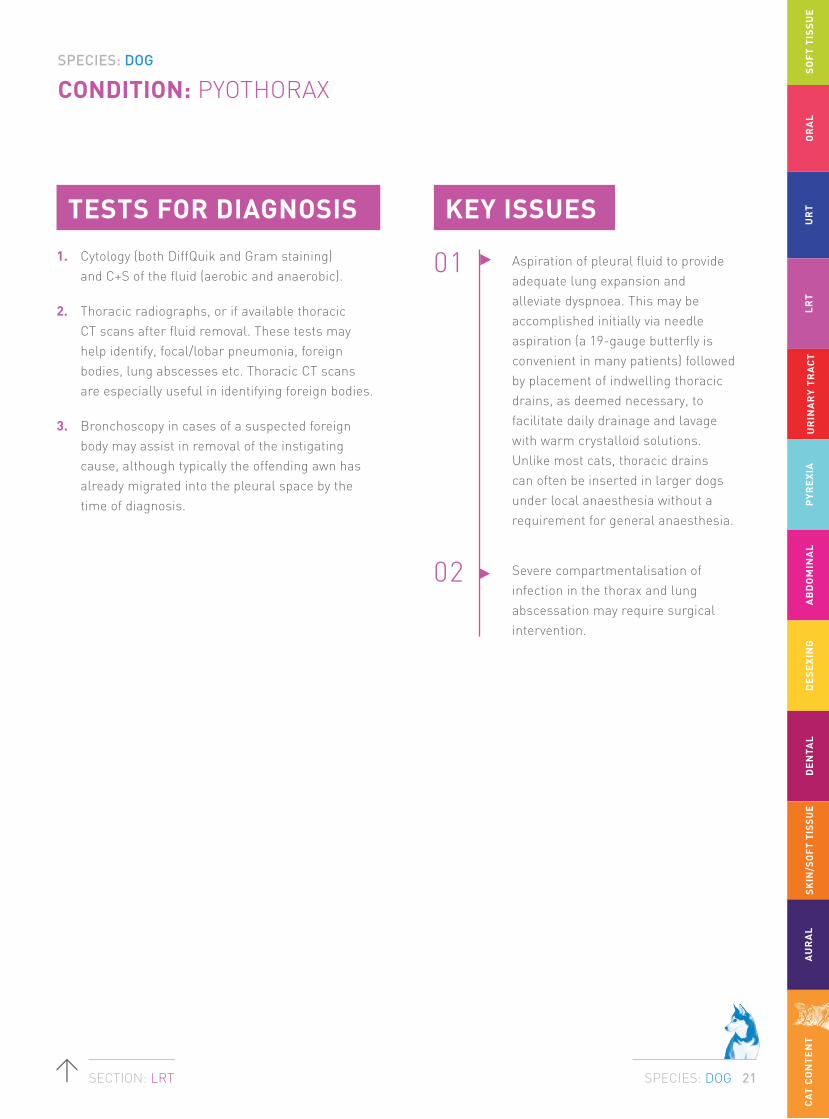
1. Cytology (both DiffQuik and Gram staining) and C+S of the fluid (aerobic and anaerobic).
2. Thoracic radiographs, or if available thoracic CT scans after fluid removal. These tests may help identify, focal/lobar pneumonia, foreign bodies, lung abscesses etc. Thoracic CT scans are especially useful in identifying foreign bodies.
3. Bronchoscopy in cases of a suspected foreign body may assist in removal of the instigating cause, although typically the offending awn has already migrated into the pleural space by the time of diagnosis.
KEY ISSUES
01 Aspiration of pleural fluid to provide adequate lung expansion and alleviate dyspnoea. This may be accomplished initially via needle aspiration (a 19-gauge butterfly is convenient in many patients) followed by placement of indwelling thoracic drains, as deemed necessary, to facilitate daily drainage and lavage with warm crystalloid solutions. Unlike most cats, thoracic drains can often be inserted in larger dogs under local anaesthesia without a requirement for general anaesthesia.
02 Severe compartmentalisation of infection in the thorax and lung abscessation may require surgical intervention.
TESTS FOR DIAGNOSIS

SPECIES: DOG
SPECIES: DOG 22SECTION: LRT
CONDITION: PYOTHORAX
SO
FT
TIS
SU
EO
RA
LU
RT
LR
TU
RIN
AR
Y T
RA
CT
PY
RE
XIA
AB
DO
MIN
AL
DE
SE
XIN
GD
EN
TAL
SKIN
/SO
FT T
ISSU
EA
UR
AL
CA
T C
ON
TE
NT
TREATMENT
Initial antimicrobial therapy is empiric and based on cytology and Gram staining of the pleural fluid. Gram positive filamentous rods are suggestive of Actinomyces, Nocardia and occasionally some obligate anaerobes. Therapy should be modified, if necessary, in the light of C+S data which can take up to 3 days to become available. Factors to consider when choosing an antibiotic regimen for initial treatment are whether to use a bactericidal or bacteriostatic antimicrobial, spectrum of activity, combination therapy, dose, route, frequency and duration of administration. A recent study by Boothe et al (2010) would suggest a wide variety of bacterial species may be isolated from dogs with pyothorax. Furthermore, polymicrobial infections with facultative anaerobic and obligate anaerobic bacteria were common. The use of antibiotics with efficacy against obligate anaerobes is strongly advocated for all cases. Many people also like to choose agents that adequately penetrate into devitalised tissues that might be poorly perfused, such as metronidazole. Adding antimicrobials, such as penicillin, to thoracic lavage solution is controversial and would appear to offer no advantage because comparable tissue levels are attained with IV administration. Some people add small quantities of heparin to the lavage fluid, although this too is controversial.
DiffQuik stained smear of purulent exudates from a dog which presented with tension pneumothorax and purulent pleurisy attributable to migration of grass awn(s).
Photo courtesy of Dr Shane Raidal.
Tension pnuemothorax and lobar pneumonia in a ‘pig dog’ following migration of a grass awn.
Photo courtesy of Dr Peter Young.

SPECIES: DOG
SPECIES: DOG 23SECTION: LRT
CONDITION: PYOTHORAX
SO
FT
TIS
SU
EO
RA
LU
RT
LR
TU
RIN
AR
Y T
RA
CT
PY
RE
XIA
AB
DO
MIN
AL
DE
SE
XIN
GD
EN
TAL
SKIN
/SO
FT T
ISSU
EA
UR
AL
CA
T C
ON
TE
NT
ANTIBIOTICS USED
USAGE RECOMMENDATION
First line empiric therapy with three agents:
(i) Cefazolin (20 mg/kg q8h‡) or amoxicillin (10-20 mg/kg q6h‡),
(ii) Aminoglycoside (gentamicin 6 mg/kg q24h IV to a well hydrated patient) or fluoroquinolone (enrofloxacin, 5 mg/kg q24h SC),
(iii) Metronidazole (10 mg/kg q12h) — four quadrant therapy.
If branching filamentous rods are observed on staining of specimens of pleural fluid, trimethoprim-sulfonamide may also be effective but a C+S is recommended.
IV initially then based on C+S and clinical response. Change to oral antibiotics after a few days, after the animal is responding and has commenced eating. Prolonged therapy after discharge from hospital may be required if significant lobar pneumonia is present.
Key references:
1. Boothe HW, Howe LM, Boothe DM, et al. J Am Vet Med Assoc 2010; 236(6): 657-663
SPECIES: DOG
SPECIES: DOG 24SECTION: URINARY TRACT
CONDITION: ACUTE LOWER UTI/CYSTITIS(FIRST OCCURRENCE)
SO
FT
TIS
SU
EO
RA
LU
RT
LR
TU
RIN
AR
Y T
RA
CT
PY
RE
XIA
AB
DO
MIN
AL
DE
SE
XIN
GD
EN
TAL
SKIN
/SO
FT T
ISSU
EA
UR
AL
CA
T C
ON
TE
NT
Infections occur most commonly in older female dogs, but in contrast to cats uncomplicated UTIs are common in dogs and a low urine specific gravity has not been identified as a risk factor for UTI in dogs. The median age of diagnosis for UTI in dogs is 7 to 8 years.
Dogs may also present with LUT signs due to urolithiasis, bladder neoplasia or prostatic disease (prostatitis, prostatic neoplasia, benign prostatic hypertrophy).
E. coli is the most common pathogen and most common Gram-negative isolate in canine UTIs accounting for 33-55% of isolates, while Streptococcus/Enterococcus are the most common Gram-positive isolates in canine UTIs. Most UTIs are thought to result from ascending infections of colonic microbiota as opposed to infections from haematogenous/lymphatic spread.
Urease-positive mycoplasmas that are adapted for life in the urogenital tract (Ureaplasma spp.) can be uncommon but important causes of UTI in dogs, and may be clinically relevant as they are resistant to β-lactams. Mycoplasma infection should be considered in dogs with high
urine pH ± crystalluria and a high white cell count that returns a diagnostic microbiology report yielding no significant growth.
Whilst research has been undertaken on a small number of highly resistant Gram-negative organisms causing canine UTIs in Australia, apart from focused studies often involving a single class of antimicrobial, there is an absence of data on both the prevalence of specific pathogens causing UTI and the susceptibility profiles of the isolates obtained. In one Australian study involving 162 E. coli isolates obtained mainly from the urogenital system of dogs, resistance prevalence was as follows: amoxicillin (44%); cephalothin (22%); amoxicillin-clavulanate (20%); tetracycline (17%); trimethoprim-sulphonamide (14%); enrofloxacin (10%); third generation cephalosporin (9%); gentamicin (3%). A total of 20% of isolates were classified as MDR i.e. resistant to four or more of nine tested antimicrobial agents. However, it must be remembered that the majority of the cases were presented to specialist rather than primary accession veterinary practices and thus may be more likely to have been obtained from dogs with complicated rather than uncomplicated UTI.
Similar to cats, simple uncomplicated UTI is a sporadic bacterial infection of the bladder in an otherwise healthy individual with normal urinary tract anatomy and function.
BACKGROUND/NATURE OF INFECTION/
ORGANISMS INVOLVED
SPECIES: DOG
SPECIES: DOG 25SECTION: URINARY TRACT
CONDITION: ACUTE LOWER UTI/CYSTITIS(FIRST OCCURRENCE)
SO
FT
TIS
SU
EO
RA
LU
RT
LR
TU
RIN
AR
Y T
RA
CT
PY
RE
XIA
AB
DO
MIN
AL
DE
SE
XIN
GD
EN
TAL
SKIN
/SO
FT T
ISSU
EA
UR
AL
CA
T C
ON
TE
NT
1. Rectal examination is important in male dogs to assess for presence of prostatic disease while vulval examination is important in female dogs.
2. A full urinalysis is recommended for all dogs presenting with LUT signs, using a urine sample collected by cystocentesis or using a sterile urinary catheter.
3. Diagnosis of an uncomplicated UTI should be made on the basis of presence of LUT signs with supporting evidence of UTI (epithelial cells, RBC, WBC and bacteria) on a full urinalysis including examination of Gram or DiffQuik stained urine sediment. Urine collected for urinalysis should also be submitted for C+S testing since false positive diagnoses can be made on urine sediments, usually due to the presence of stain precipitates mimicking bacteria.
4. Free-catch urine samples are inferior and should generally be avoided.
KEY ISSUES
01 Increasing age and female gender are risk factors for uncomplicated UTIs in dogs.
02 Where subclinical bacteriuria is identified (positive urine culture in the absence of clinical and cytological evidence of UTI) treatment is generally not recommended in otherwise healthy patients with normal urinary tract anatomy and function.
03 In dogs with first occurrence of cystitis look for markers of underlying disease or of bladder dysfunction/abnormalities (e.g. prostatic enlargement or PU/PD and cutaneous abnormalities suggestive of hyperadrenocortism) so an assessment can be made as to whether the UTI is most likely uncomplicated or complicated.
TESTS FOR DIAGNOSIS

SPECIES: DOG
SPECIES: DOG 26SECTION: URINARY TRACT
CONDITION: ACUTE LOWER UTI/CYSTITIS(FIRST OCCURRENCE)
SO
FT
TIS
SU
EO
RA
LU
RT
LR
TU
RIN
AR
Y T
RA
CT
PY
RE
XIA
AB
DO
MIN
AL
DE
SE
XIN
GD
EN
TAL
SKIN
/SO
FT T
ISSU
EA
UR
AL
CA
T C
ON
TE
NT
TREATMENT1. Empiric antimicrobial therapy is appropriate
pending urine culture results.
2. Recommended first-line choices for empiric therapy are amoxicillin or trimethoprim-sulfonamide (for Gram-positive infections) and trimethoprim-sulfonamide or amoxicillin-clavulanate (for Gram-negative infections). Adverse side effects associated with trimethoprim-sulfonamide in dogs include keratoconjunctivitis sicca, gastrointestinal side effects, fever, haemolytic anaemia, urticaria, polyarthropathy, facial swelling, PUPD and cholestasis. Also, hypothyroidism can occur after chronic treatment with potentiated sulfonamides. Consideration should be given to using another antimicrobial in large-breed dogs including Doberman Pinschers due to increased risk of hypersensitivity reactions such as idiosyncratic hepatopathy.
3. The recommended treatment duration is 7 to 14 days, although shorter treatment times may be effective.
4. For prostatitis, trimethoprim-sulfonamide and fluoroquinolones reach adequate tissue concentrations in the prostate gland.
5. Be aware of administration of trimethoprim-sulfonamide to large-breed dogs such as Rottweilers and Dobermans as they are prone to idiosyncratic reactions including hypersensitivity and keratoconjunctivitis sicca.

SPECIES: DOG
SPECIES: DOG 27SECTION: URINARY TRACT
CONDITION: ACUTE LOWER UTI/CYSTITIS(FIRST OCCURRENCE)
SO
FT
TIS
SU
EO
RA
LU
RT
LR
TU
RIN
AR
Y T
RA
CT
PY
RE
XIA
AB
DO
MIN
AL
DE
SE
XIN
GD
EN
TAL
SKIN
/SO
FT T
ISSU
EA
UR
AL
CA
T C
ON
TE
NT
AIDAP TOP TIPS ANTIBIOTICS USED
USAGE RECOMMENDATION
First line:
Amoxicillin (11-15 mg/kg q8h PO†); Amoxicillin-clavulanate (12.5-20 mg/kg q8-12h PO – give with food to minimise vomiting).
Second line:
Consider a fluoroquinolone (enrofloxacin 5 mg/kg q24h, marbofloxacin 2.75-5.5 mg/kg q24hrs) on the basis of C+S if the bacteria are resistant to first-line therapy or if the infection is serious; or trimethoprim-sulfonamide (15 mg/kg q12h PO†) – be aware of hypersensitivity reactions in Rottweilers and Dobermans and keratoconjunctivitis sicca.
Cefovecin should only be used where compliance is an issue, or there are difficulties with medicating orally.
Recommended duration of therapy for uncomplicated UTIs is 7 to 14 days.
For treating first time UTIs in dogs, consider administering amoxicillin-clavulanate towards the upper dose limit of 20 mg/kg q12h† (give with food to minimise vomiting). Amoxicillin-clavulanate reaches high concentrations in the urine and increasing the dose rate is one of the best ways to prevent resistance emergence and will exceed the MIC for most UTI pathogens.
Key references:
1. Thompson MF, Litster AL, Platell JL et al. Vet J 2011; 190: 22-27
2. Weese JS, Blondeau JM, Boothe D, et al. Vet Med Int 2011; 2011: 263768
3. Gottlieb S, Wigney DI, Martin PA, et al. Aust Vet J 2008; 86(4): 147-152

SPECIES: DOG
SPECIES: DOG 28SECTION: URINARY TRACT
CONDITION: COMPLICATED UTIs: RECURRENT LOWER UTI /CYSTITIS AND CKD WITH PYURIA
SO
FT
TIS
SU
EO
RA
LU
RT
LR
TU
RIN
AR
Y T
RA
CT
PY
RE
XIA
AB
DO
MIN
AL
DE
SE
XIN
GD
EN
TAL
SKIN
/SO
FT T
ISSU
EA
UR
AL
CA
T C
ON
TE
NT
Complicated UTIs occur where there is an underlying anatomic or functional abnormality or where there is a concurrent disease that predisposes to UTI, for example CKD. Recurrent UTIs, occurring within six months after successful treatment of the first infection, can be re-infections caused by a different bacterial species to the original isolate or relapses caused by the same bacterial species as the original isolate. Relapses are common in persistent or recurrent canine UTIs, and similar with complicated infections in cats are often asymptomatic.
Pseudomonas aeruginosa and Enterococcus spp., are isolated more frequently in complicated canine UTIs as compared to uncomplicated UTIs. The six most prevalent bacterial isolates in complicated canine UTIs are E. coli, Klebsiella spp., Staphylococcus spp., Enterococcus spp., Proteus spp. and Pseudomonas aeruginosa. In addition, multiple isolates are common in complicated canine UTIs.
Since MDR faecal E. coli are increasingly prevalent, accurate identification of uropathogens is important and urine collection by cystocentesis should be performed to prevent contamination with faecal bacteria and a false-positive diagnosis of a MDR E. coli UTI.
Causes of complicated UTIs in dogs include malformations of the genitourinary tract including ectopic ureter, urethral sphincter mechanism incompetence, diverticuli or fistulas, struvite urolithiasis and prostatitis, as well as underlying disease including hyperadrenocorticism, diabetes mellitus and CKD. Although indwelling urinary catheters predispose to UTI, asymptomatic bacteriuria in the absence of cytological and clinical signs of UTI does not warrant treatment in catheterised dogs.
In several studies involving large datasets, E. coli, Klebsiella spp.,Staphylococcus spp., Enterococcus spp., Proteus spp. and Pseudomonas spp.
As for cats, recurrent UTIs in dogs are a subset of complicated UTIs.
BACKGROUND/NATURE OF INFECTION/
ORGANISMS INVOLVED

SPECIES: DOG
SPECIES: DOG 29SECTION: URINARY TRACT
CONDITION: COMPLICATED UTIs: RECURRENT LOWER UTI /CYSTITIS AND CKD WITH PYURIA
SO
FT
TIS
SU
EO
RA
LU
RT
LR
TU
RIN
AR
Y T
RA
CT
PY
RE
XIA
AB
DO
MIN
AL
DE
SE
XIN
GD
EN
TAL
SKIN
/SO
FT T
ISSU
EA
UR
AL
CA
T C
ON
TE
NT
were identified as the most common bacterial pathogens encountered in cases of chronic or recurrent cystitis in dogs. In a case study of 37 dogs with MDR E. coli or Enterobacter infections, the majority of which were urogenital, most dogs had underlying diseases predisposing them to complicated infections. Whilst a small number of dogs were successfully treated with carbepenems, the majority also responded to 25 mg/kg of amoxicillin-clavulanate even though disc diffusion tests indicated in vitro resistance to this combination.
The global emergence and spread of E. coli serotype O25b sequence type (ST) 131 as a cause of urosepsis in humans is of grave medical concern. These strains are highly virulent, MDR and often show combined resistance to fluoroquinolones and third generation cephalosporins. E. coli ST131 UTI has recently been identified in dogs internationally as well as in Australia and the strains are often indistinguishable from human strains. Over 50% of FQ-resistant E. coli isolates from humans in Australia belong to ST131 compared to <10% from dogs. These extraintestinal pathogenic E. coli should therefore be considered to be zooanthroponotic.
E. coli strain 83972 achieves persistent asymptomatic bacteriuria when administered by urinary catheter in humans with spinal injuries who are prone to chronic, recurrent UTI and may be an alternative method of prevention in dogs prone to similar UTI infections. The organism has been shown to temporarily colonise the normal canine bladder and clinical trials are currently underway in Australian dogs with complicated cystitis.

SPECIES: DOG
SPECIES: DOG 30SECTION: URINARY TRACT
CONDITION: COMPLICATED UTIs: RECURRENT LOWER UTI /CYSTITIS AND CKD WITH PYURIA
SO
FT
TIS
SU
EO
RA
LU
RT
LR
TU
RIN
AR
Y T
RA
CT
PY
RE
XIA
AB
DO
MIN
AL
DE
SE
XIN
GD
EN
TAL
SKIN
/SO
FT T
ISSU
EA
UR
AL
CA
T C
ON
TE
NT
1. Clinical signs alone should not be used for diagnosis.
2. Diagnosis should not be based on examination of urine sediment alone.
3. C+S testing (aerobic) should be performed in all cases.
4. Cystocentesis should be used for urine collection where possible.
5. Investigation for underlying disease (comorbidity) should include determination of owner compliance with previous antimicrobial therapy, rectal/vulval exam, CBC/biochemistry, urinalysis, imaging, ± endocrine testing.
6. Where concurrent disease or an underlying bladder abnormality is not identified, consideration should be given to referral for further investigations, e.g. cystoscopy.
7. Where subclinical bacteriuria is identified (positive urine culture in the absence of clinical and cytological evidence of UTI) treatment is based on the risk of ascending or systemic infection.
8. In dogs with indwelling urinary tract catheters that develop cytological or clinical signs of UTI the urinary catheter should be removed permanently. If this is not possible the catheter should be replaced. Urine for urinalysis and C+S testing should ideally be collected by cystocentesis in these patients when the bladder has refilled after catheter removal. Culture of urine from urine collection bags is contraindicated and culture of urinary catheter tips is not recommended since results do not predict catheter-associated UTI.
KEY ISSUES
01 In dogs with recurrent cystitis, careful consideration must be given to detection of an underlying bladder abnormality (e.g. urolithiasis, neoplasia), prostatic disease (prostatitis) or concurrent disease that predisposes to recurrent UTIs e.g. hyperadrenocorticism, diabetes mellitus, CKD.
02 Recurrent UTIs in young dogs should arouse suspicion of an underlying abnormality or dysfunction of the LUT, e.g. ectopic ureter.
03 Because MDR pathogens such as ST131 are becoming increasingly recognised globally and have been identified in dogs, careful consideration must be given to selection of antimicrobials used for treatment of UTI.
04 Prophylactic antimicrobials should not be administered to dogs with indwelling urinary catheters or after catheter removal in dogs without clinical or cytological evidence of UTI.
TESTS FOR DIAGNOSIS
SPECIES: DOG
SPECIES: DOG 31SECTION: URINARY TRACT
CONDITION: COMPLICATED UTIs: RECURRENT LOWER UTI /CYSTITIS AND CKD WITH PYURIA
SO
FT
TIS
SU
EO
RA
LU
RT
LR
TU
RIN
AR
Y T
RA
CT
PY
RE
XIA
AB
DO
MIN
AL
DE
SE
XIN
GD
EN
TAL
SKIN
/SO
FT T
ISSU
EA
UR
AL
CA
T C
ON
TE
NT
TREATMENT1. Empiric antimicrobial therapy is not recommended
unless clinical signs necessitate it. Recommended antimicrobials for empiric therapy are as for uncomplicated UTIs but may often involve treatment with a fluoroquinolone if indicated by C+S. Where possible the drug class selected should be different from that used to treat the original UTI. If the bacterial isolate is resistant to the antimicrobial chosen for empiric therapy, treatment should be changed to an antimicrobial to which the isolate is susceptible and if possible is excreted in its active form primarily in urine. Adverse side effects associated with trimethoprim-sulfonamide in dogs include keratoconjunctivitis sicca, gastrointestinal side-effects, fever, haemolytic anaemia, urticaria, polyarthropathy, facial swelling, PUPD and cholestasis. Also, hypothyroidism can occur after chronic treatment with potentiated sulfonamides. Consideration should be given to using another antimicrobial in large-breed dogs including Doberman Pinschers due to increased risk of hypersensitivity reactions such as idiosyncratic hepatopathy.
2. In dogs with asymptomatic bacteriuria and underlying disease (e.g. CKD) wait until C+S test results are available to initiate treatment using an appropriate antimicrobial.
3. For mixed infections consisting of Enterococcus spp. and another bacterial isolate infection by the former will often resolve when the other organism is successfully treated. Ideally a single antimicrobial or antimicrobial combination effective against both organisms should be selected, however if this is not possible due to resistance antimicrobial therapy should be based on efficacy against the organism perceived to be most clinically relevant.
4. Where MDR organisms are identified, consultation with colleagues with expertise in infectious diseases is recommended. Antimicrobials including carbapenems, vancomycin and linezolid should never be used for treatment of subclinical bacteriuria and are reserved for treatment of complicated UTI diagnosed by C+S testing of a urine sample obtained by cystocentesis in patients with treatable diseases in which all other possible antimicrobials have been considered.
5. Obtaining an antimicrobial minimum inhibitory concentration for the isolated pathogen (or a disc diffusion zone diameter size) from the diagnostic laboratory may indicate that a drug that shows in vitro resistance but effectively concentrates in the urine, such as amoxicillin-clavulanate may be used at the maximum dose rate of 25 mg/kg to clear the infection.
(Note that some dogs may vomit after receiving high oral doses of amoxicillin-clavulanate).

SPECIES: DOG
SPECIES: DOG 32SECTION: URINARY TRACT
CONDITION: COMPLICATED UTIs: RECURRENT LOWER UTI /CYSTITIS AND CKD WITH PYURIA
SO
FT
TIS
SU
EO
RA
LU
RT
LR
TU
RIN
AR
Y T
RA
CT
PY
RE
XIA
AB
DO
MIN
AL
DE
SE
XIN
GD
EN
TAL
SKIN
/SO
FT T
ISSU
EA
UR
AL
CA
T C
ON
TE
NT
AIDAP TOP TIPS ANTIBIOTICS USED
USAGE RECOMMENDATION
First line:
Guided by C+S testing, but consider amoxicillin (11-15 mg/kg q8h PO†); amoxicillin-clavulanate (12.5-20 mg/kg q8-12h PO); or trimethoprim-sulfonamide (15 mg/kg q12h PO†).
Cefovecin should only be used where compliance is an issue, or there are difficulties with medicating orally.
Second line:
Consider a fluoroquinolone (marbofloxacin 2.75-5.5 mg/kg q24hrs, enrofloxacin 5 mg/kg q24hrs) on the basis of C+S if the bacteria are resistant to first-line therapy or if the infection is serious; or trimethoprim-sulfonamide (15 mg/kg q12h PO) – be aware of hypersensitivity reactions in Rottweilers and Dobermans and keratoconjunctivitis sicca.
The recommended treatment duration is four weeks although shorter treatment times may be effective. For recurrent infections, consider urine culture 5 to 7 days after starting therapy and 7 days after stopping oral therapy.
In cases of complicated cystitis in dogs where a fluoroquinolone is indicated on the basis of C+S, administer at the higher end of the registered dose rate.
Key references:
1. Gibson JS, Morton JM, Cobbold RN, et al. J Vet Intern Med 2008; 22(4): 844-850
2. Platell JL, Trott DJ, Wetzstein HG, et al. J Vet Sci Technol 2008; S6: 1
3. Thompson MF, Schembri MA, Mills PC, et al. Vet Microbiol 2012; 158(3-4): 446-450
4. Thompson MD, LItster AL, Platell JL et al. vet J 2011; 190: 22-27
5. Platell JL, Cobbold RN, Johnson JR, et al. J Antimicrob Chemother 2010; 65(9): 1936-1938
6. Platell JL, Cobbold RN, Johnson JR et al Antimicro Agents Chemother 2011; 55(8): 3782-3787
SPECIES: DOG
SPECIES: DOG 33SECTION: PYREXIA
CONDITION: ACUTE FEBRILE ILLNESS
SO
FT
TIS
SU
EO
RA
LU
RT
LR
TU
RIN
AR
Y T
RA
CT
PY
RE
XIA
AB
DO
MIN
AL
DE
SE
XIN
GD
EN
TAL
SKIN
/SO
FT T
ISSU
EA
UR
AL
CA
T C
ON
TE
NT
On average, fever is less commonly due to infectious agents than in the cat, where occult fight injuries and other bacterial and viral infections are quite common. Cats may respond to empiric antimicrobial therapy when presented with fever and no localising signs, whereas dogs less commonly do so.
In the dog, bacterial infections causing fever include endocarditis, peritonitis, pneumonia, prostatitis, pyothorax, pyometra, abscesses (which may be in body cavities). In certain breeds (e.g. German Shepherds), disseminated fungal disease should be considered also in the differential diagnosis.
Thus, although infections should always be considered as the potential cause of acute febrile illness, immune-mediated and neoplastic diseases may also cause fever. Immune-mediated diseases have been noted to represent up to 36% of febrile diseases of the dog in one study.
Diseases such as corticosteroid-responsive meningitis (also known as aseptic suppurative meningitis), immune-mediated polyarthritis, metaphyseal osteopathy (also known as hypertrophic osteodystrophy), panosteitis etc. should also be considered. Pancreatitis is another common cause of fever in the dog. In some dogs, unexplained neutropenia may occur due to immune-mediated mechanisms, further complicating the diagnosis. Therefore, a thorough search for an infectious cause should be undertaken, but with the knowledge that some dogs may have immune-mediated disease as the primary cause of fever.
In young dogs, acute febrile diseases may be infectious and in such instances viral diseases such as parvovirus may be responsible. However, acute bacterial enteritis (Salmonella, Campylobacter, some E. coli) may also occur. In these scenarios, antibiotic selection and efficacy is problematic.
Acute febrile illness in the dog may be infectious, immune-mediated or attributable to malignancy.
BACKGROUND/NATURE OF INFECTION/
ORGANISMS INVOLVED
SPECIES: DOG
SPECIES: DOG 34SECTION: PYREXIA
CONDITION: ACUTE FEBRILE ILLNESS
SO
FT
TIS
SU
EO
RA
LU
RT
LR
TU
RIN
AR
Y T
RA
CT
PY
RE
XIA
AB
DO
MIN
AL
DE
SE
XIN
GD
EN
TAL
SKIN
/SO
FT T
ISSU
EA
UR
AL
CA
T C
ON
TE
NT
Ideally samples for bacterial culture should be collected prior to empiric antibiotic use.
Blood and joint fluid culture may require enrichment culture bottles to facilitate bacterial growth. Many laboratories will give culture bottles to veterinarians, so they are immediately on hand when such cases are presented.
KEY ISSUES
01 A thorough physical examination is mandatory. Pay particular attention to identifying regions of pain such as neck pain or joint pain. New cardiac murmurs may signal development of bacterial endocarditis. It is important to look at the eye for uveitis, and retinal examination can occasionally be rewarding.
02 Haematology, biochemistry, urinalysis and urine culture are required, unless physical findings point to a specific anatomical region as a potential cause of the infection.
03 Diagnostic imaging of the thorax/abdomen – radiographs, ultrasonography and cross-sectional imaging all have their place.
04 Blood and urine cultures may be appropriate.
05 If considered appropriate, specific searches for infectious agents/immune diseases may be required such as cardiac ultrasound for endocarditis, CSF analysis, joint fluid analysis. Culture or PCR may be used for the detection of Bartonella species and other tick-borne agents in some cases, especially in areas where such diseases are endemic (e.g. far north Queensland and the Northern Territory).
06 In young dogs with febrile disease and lameness, radiographs of long bones are required to investigate for metaphyseal osteopathy, panosteitis and fungal osteomyelitis.
TESTS FOR DIAGNOSIS
TREATMENT
If a high index of suspicion is present for infection then antibiotic selection should be based on likely pathogenic bacteria and a bactericidal, broad-spectrum antibiotic should be selected. If overwhelming sepsis is suspected, ‘four-quadrant therapy’ may be instituted after collection of appropriate samples for testing.
SPECIES: DOG
SPECIES: DOG 35SECTION: PYREXIA
CONDITION: ACUTE FEBRILE ILLNESS
SO
FT
TIS
SU
EO
RA
LU
RT
LR
TU
RIN
AR
Y T
RA
CT
PY
RE
XIA
AB
DO
MIN
AL
DE
SE
XIN
GD
EN
TAL
SKIN
/SO
FT T
ISSU
EA
UR
AL
CA
T C
ON
TE
NT
AIDAP TOP TIPS ANTIBIOTICS USED
USAGE RECOMMENDATION
First line:
Amoxicillin clavulanic acid 12.5 mg/kg SC or PO q12h.
Second line:
If overwhelming sepsis is suspected then four-quadrant therapy with IV antibiotics is suggested. E.g. IV amoxicillin (20 mg/kg q6-8h†), metronidazole 10 mg/kg q12h and gentamicin (6 mg/kg q24h in a well hydrated patient) / injectable fluoroquinolone (enrofloxacin 5 mg/kg q24h).
Antibiotic use may interfere with subsequent diagnostics and should be reserved for severe cases with a high index of suspicion for an infectious cause.
Acute abdominal inflammation is a special subcategory of these where multiple species of bacteria may be identified. Therefore, if there is danger of intestinal perforation or translocation of enteric organisms, then the use of ‘four quadrant’ therapy may be required until appropriate testing and surgical therapy is undertaken.
Key references:
1. Dunn KJ and Dunn JK. J Small Anim Pract 1998; 39(12): 574-580
2. Battersby IA, Murphy KF, Tasker S, et al. J Small Anim Pract 2006; 47(7): 370-376

SPECIES: DOG
SPECIES: DOG 36SECTION: ABDOMINAL
CONDITION: ACUTE ABDOMINAL PAIN AND PYREXIA /ABDOMINAL INFECTION AND LEUKOPENIA
SO
FT
TIS
SU
EO
RA
LU
RT
LR
TU
RIN
AR
Y T
RA
CT
PY
RE
XIA
AB
DO
MIN
AL
DE
SE
XIN
GD
EN
TAL
SKIN
/SO
FT T
ISSU
EA
UR
AL
CA
T C
ON
TE
NT
The source of the infection is often bowel leakage, but can be from pyometra, prostatitis, pyothorax, hepatic or kidney abscess rupture. Migrating grass awns and metallic foreign bodies (needles) may also instigate peritonitis. A ruptured gall bladder may occur in animals with infectious cholecystitis. Penetrating bite wounds may also cause peritonitis. In cats and dogs primary bacterial peritonitis is also reported where no predisposing cause is identified. Mortality rate is high and delayed treatment and diagnosis may result in a poor outcome.
This situation is clearly a severe life or death scenario with no time for C+S data prior to initiation of therapy.
BACKGROUND/NATURE OF INFECTION/
ORGANISMS INVOLVED
SPECIES: DOG
SPECIES: DOG 37SECTION: ABDOMINAL
CONDITION: ACUTE ABDOMINAL PAIN AND PYREXIA /ABDOMINAL INFECTION AND LEUKOPENIA
SO
FT
TIS
SU
EO
RA
LU
RT
LR
TU
RIN
AR
Y T
RA
CT
PY
RE
XIA
AB
DO
MIN
AL
DE
SE
XIN
GD
EN
TAL
SKIN
/SO
FT T
ISSU
EA
UR
AL
CA
T C
ON
TE
NT
1. Abdominal fluid should be obtained for cytology and for C+S. Immediate treatment should then be started. Aerobic and anaerobic cultures need to be performed.
2. Surgical exploration of the abdomen is usually required for diagnostic and treatment purposes.
3. Surgical drainage of the abdomen may be required using Jackson Pratt drains; alternately, in some cases an ‘open abdomen’ method for drainage is applied.
KEY ISSUES
01 Immediate antibiotics given parenterally, preferably IV at maximal safe doses.
02 Coverage of Gram-negatives, Gram-positives, obligate anaerobes with a high possibility of antibiotic resistant bacteria being involved.
03 Immediate fluid support and probably surgery for correction of underlying issue.
TESTS FOR DIAGNOSIS
SPECIES: DOG
SPECIES: DOG 38SECTION: ABDOMINAL
CONDITION: ACUTE ABDOMINAL PAIN AND PYREXIA /ABDOMINAL INFECTION AND LEUKOPENIA
SO
FT
TIS
SU
EO
RA
LU
RT
LR
TU
RIN
AR
Y T
RA
CT
PY
RE
XIA
AB
DO
MIN
AL
DE
SE
XIN
GD
EN
TAL
SKIN
/SO
FT T
ISSU
EA
UR
AL
CA
T C
ON
TE
NT
TREATMENT
1. Four-quadrant IV antibiotic therapy.
2. Surgical drainage.
3. Management of sepsis syndrome with fluids, plasma or colloids, sometimes a whole blood transfusion and inotropic support, as needed.
1. Amoxicillin 20 mg/kg IV q6h† or ampicillin (20 mg/kg IV q6-8h‡) or cefazolin (22 mg/kg IV q8h‡) /cefoxitin (30 mg/kg IV q8h‡).
2. Enrofloxacin 5 mg/kg IV or SQ q24h or use an aminoglycoside (e.g. gentamicin 6 mg/kg IV q24h.
3. Metronidazole 10 mg/kg IV q8h.
ANTIBIOTICS USED
Key references:
1. Ruthrauff CM, Smith J, Glerum L. J Am Anim Hosp Assoc 2009; 45(6): 268-276
2. Culp WT, Zeldis TE, Reese MS, Drobatz KJ. J Am Vet Med Assoc 2009; 234(7): 906-913
3. Mueller MG, Ludwig LL, Barton LJ. J Am Vet Med Assoc. 2001; 219(6):789-794
2 weeks post-recovery
USAGE RECOMMENDATION
The upper limit of the recommended dose range should be given.
Note that beta-lactams (penicillins and cephalosporins) and aminoglycosides (gentamicin, amikacin) need to be given separately as they precipitate in the fluid line if given simultaneously. These agents should be given by slow IV push over several minutes, separated by a flush with saline.

SPECIES: DOG
SPECIES: DOG 39SECTION: DESEXING
CONDITION: ANTIBIOTIC USE AFTER ROUTINE DESEXING
SO
FT
TIS
SU
EO
RA
LU
RT
LR
TU
RIN
AR
Y T
RA
CT
PY
RE
XIA
AB
DO
MIN
AL
DE
SE
XIN
GD
EN
TAL
SKIN
/SO
FT T
ISSU
EA
UR
AL
CA
T C
ON
TE
NT
As a general rule desexing operations conducted using sterile technique and not taking longer than average for an experienced veterinarian to complete would not be given antibiotics prophylactically.
In one study, peri operative antimicrobial prophylaxis decreased postoperative infection rate in dogs undergoing elective orthopaedic surgery, compared with the infection rate in control dogs. Cefazolin was not more efficacious than potassium penicillin G in these dogs.
This would suggest that prolonged surgery times for routine desexing might be considered an increased risk of infection, as might a breach in aseptic technique. Definitive studies of what time limits are associated with increased infection in desexing operations are lacking. However, antibiotics with higher efficacy for Staphylococcus spp. would be considered in the event of a prolonged desexing operation.
Several studies have shown that length of time of surgery and the more people in the room at the time of surgery, the greater the risk of infections.
BACKGROUND/NATURE OF INFECTION/
ORGANISMS INVOLVED
SPECIES: DOG
SPECIES: DOG 40SECTION: DESEXING
CONDITION: ANTIBIOTIC USE AFTER ROUTINE DESEXING
SO
FT
TIS
SU
EO
RA
LU
RT
LR
TU
RIN
AR
Y T
RA
CT
PY
RE
XIA
AB
DO
MIN
AL
DE
SE
XIN
GD
EN
TAL
SKIN
/SO
FT T
ISSU
EA
UR
AL
CA
T C
ON
TE
NT
KEY ISSUES
01 There is no need for prophylactic antimicrobials for routine desexing.
02 If the procedure is unduly prolonged, or there is a breach in asepsis, then a single injection of amoxicillin-clavulanate or a 1st generation cephalosporin might be appropriate.
TREATMENT
Antibiotics are considered unnecessary in routine short surgery conducted under sterile conditions. Given the use of gloves and sterile conditions, the routine use of prophylactic antibiotics for spays is not required. Also given that most potential contaminants arise from the skin of the dog or the veterinary staff, a single shot of procaine penicillin offers insufficient coverage for Staphylococcus pseudintermedius or Staphylococcus aureus infection.
Routine use of antibiotics not suggested.
ANTIBIOTICS USED
USAGE RECOMMENDATION
N/A.
SPECIES: DOG
SPECIES: DOG 41SECTION: DENTAL
CONDITION: USE OF ANTIBIOTICS IN DENTAL PROPHYLAXIS
SO
FT
TIS
SU
EO
RA
LU
RT
LR
TU
RIN
AR
Y T
RA
CT
PY
RE
XIA
AB
DO
MIN
AL
DE
SE
XIN
GD
EN
TAL
SKIN
/SO
FT T
ISSU
EA
UR
AL
CA
T C
ON
TE
NT
Pasteurella multocida, and anaerobic Gram-negative rods including Capnocytophaga are frequently involved and these are all sensitive to penicillins, including benzyl penicillin and amoxicillin-clavulanate.
There is a very small risk that bacteraemia associated with the use of ultrasonic scaling devices and extractions could produce infections elsewhere, such as bacterial endocarditis. This is most unlikely in normal patients, but the risk is increased with structural heart disease, especially subaortic stenosis which has been associated with increased risk for the development of bacterial endocarditis.
For this reason, it may be prudent to administer prophylactic bactericidal antibiotics so that high blood levels are obtained during and immediately after the dental procedures.
Most bacteria found in the mouths of dogs (and cats) are similar to what is recovered in bite wounds.
BACKGROUND/NATURE OF INFECTION/
ORGANISMS INVOLVED
SPECIES: DOG
SPECIES: DOG 42SECTION: DENTAL
CONDITION: USE OF ANTIBIOTICS IN DENTAL PROPHYLAXIS
SO
FT
TIS
SU
EO
RA
LU
RT
LR
TU
RIN
AR
Y T
RA
CT
PY
RE
XIA
AB
DO
MIN
AL
DE
SE
XIN
GD
EN
TAL
SKIN
/SO
FT T
ISSU
EA
UR
AL
CA
T C
ON
TE
NT
Histopathology and culture of infected tissue is suggested if initial prophylaxis fails to cure an ulcerated mouth lesion.
KEY ISSUES
01 Prophylactic antibiotics are best administered prior to the procedure e.g. procaine penicillin or amoxicillin-clavulanate administered SC or IM after premedication or immediately after anaesthetic induction.
TESTS FOR DIAGNOSIS
TREATMENT
N/A.
Amoxicillin or amoxicillin-clavulanate would cover the great majority of potential pathogens in this setting.
First line:
Amoxicillin 10 mg/kg q12h/amoxicillin-clavulanate (12.5 mg/kg q12h).
Second line:
Clindamycin (5-11 mg/kg q12h) or doxycycline monohydrate (5 mg/kg q12h†).
Cefovecin is suitable for any case where there are concerns of compliance, or there are difficulties with oral dosing.
ANTIBIOTICS USED
SPECIES: DOG
SPECIES: DOG 43SECTION: DENTAL
CONDITION: USE OF ANTIBIOTICS IN DENTAL PROPHYLAXIS
SO
FT
TIS
SU
EO
RA
LU
RT
LR
TU
RIN
AR
Y T
RA
CT
PY
RE
XIA
AB
DO
MIN
AL
DE
SE
XIN
GD
EN
TAL
SKIN
/SO
FT T
ISSU
EA
UR
AL
CA
T C
ON
TE
NT
USAGE RECOMMENDATION
If there are extractions or bleeding, which occurs in most cases, then a 7-10 day course of antibiotics is required depending on the healing period. Ensure doxycycline given with food or water bowl provided.
Key references:
1. Love D 1990 et al (isolated lots of anaerobes but possibly no data on their sensitivities)
2. Bowersock TL, Wu CC, Inskeep GA, Thoracicer ST. J Vet Dent 2000; 17(1): 11-16
3. Zetner K and Rothmueller G. Vet Ther 2002; 3(4): 441-452
4. Nielsen D, Walser C, Kodan G, et al. Vet Ther 2000; 1(3): 150-158
5. Warrick JM, Inskeep GA, Yonkers TD, et al. Vet Ther 2000; 1(1): 5-16
6. Sarkiala E and Harvey C. Semin Vet Med Surg (Small Anim) 1993; 8(3): 197-203
7. Zetner K and Thiemann G. J Vet Dent 1993; 10(2): 6-9
8. Wilcke JR. Probl Vet Med 1990; 2(2): 298-311
SPECIES: DOG
SPECIES: DOG 44SECTION: SKIN/SOFT TISSUE
CONDITION: SURFACE BACTERIAL INFECTIONS: (i.e. INTERTRIGO e.g. LIP FOLD, TAIL FOLD)
SO
FT
TIS
SU
EO
RA
LU
RT
LR
TU
RIN
AR
Y T
RA
CT
PY
RE
XIA
AB
DO
MIN
AL
DE
SE
XIN
GD
EN
TAL
SKIN
/SO
FT T
ISSU
EA
UR
AL
CA
T C
ON
TE
NT
Surface pyodermas involve the epidermis. As infection does not cross the basement membrane the dermis remains intact. The great majority of bacterial skin infections involve Gram-positive organisms and particularly the coagulase positive staphylococci. The most common of these is Staphylococcus pseudintermedius which represents over 90% of infections in dogs.
The new species, Staphylococcus pseudintermedius, was first described in 2005 and was formed by the division of isolates previously known as S. intermedius into two species. The name S. intermedius is now applied to isolates that are principally found in pigeons. Infections with S. aureus are relatively uncommon (around 5% or less). S. schleiferi coagulans and S. schleiferi schleiferi (coagulase negative) are less often recognised as causes of infection, but are found particularly in otitis externa and in certain geographic regions.
Other coagulase-negative staphylococci are rarely involved and normally only if immunity is greatly reduced or when implants are used. Gram-negative bacteria are sometimes found in pyoderma, particularly when lesions are moist. Organisms such as Proteus spp. And coliforms may be secondary invaders that fail to persist when more significant pathogens are removed. Pseudomonas aeruginosa is more serious and requires specific therapy.
The most common surface pyodermas are skin fold-pyoderma (intertrigo) and pyotraumatic dermatitis. These clinical entities are only reported in dogs. These are types of surface pyoderma with secondary bacterial involvement. These infections typically respond favourably to topical therapy, but recurrence is frequent if a primary disease process cannot be identified and controlled.
The more recent literature classifies bacterial skin infections based on lesion depth and distribution pattern and this relies heavily on clinical features in combination with histopathological data on depth of infection. It can be used to give a prognosis and suggest possible therapy.
BACKGROUND/NATURE OF INFECTION/
ORGANISMS INVOLVED

SPECIES: DOG
SPECIES: DOG 45SECTION: SKIN/SOFT TISSUE
CONDITION: SURFACE BACTERIAL INFECTIONS: (i.e. INTERTRIGO e.g. LIP FOLD, TAIL FOLD)
SO
FT
TIS
SU
EO
RA
LU
RT
LR
TU
RIN
AR
Y T
RA
CT
PY
RE
XIA
AB
DO
MIN
AL
DE
SE
XIN
GD
EN
TAL
SKIN
/SO
FT T
ISSU
EA
UR
AL
CA
T C
ON
TE
NT
We would recommend that practitioners perform an impression smear for cytological evaluation. Examine under the microscope using immersion oil (x100) and evaluate for the presence of healthy or degenerate neutrophils (swollen and pale nuclei) and extracellular cocci and bacilli and intracellular phagocytosed bacteria. If there are abundant bacilli, and if there is no response to empirical antibacterial therapy then we would recommend that practitioners submit a swab of the exudate for C+S. Collecting a skin biopsy to rule out immune mediated disease may be indicated if the lesions fail to respond to appropriate topical and systemic antimicrobial therapy, however these diseases are rare.
KEY STEPS
01 Identify bacterial overgrowth and/or infection via surface cytology.
02 Commence trial treatment with appropriate topical and/or systemic antibiotic.
03 Consider bacterial C+S testing if failure to respond to therapy.
04 Correct underlying anatomical abnormality.
TESTS FOR DIAGNOSIS
Facial fold pyoderma.
Photos courtesy of Dr Mandy Burrows & Dr Mike Shipstone.
Tail fold pyoderma.
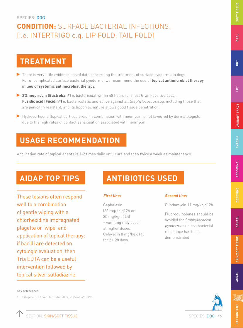
SPECIES: DOG
SPECIES: DOG 46SECTION: SKIN/SOFT TISSUE
CONDITION: SURFACE BACTERIAL INFECTIONS: (i.e. INTERTRIGO e.g. LIP FOLD, TAIL FOLD)
SO
FT
TIS
SU
EO
RA
LU
RT
LR
TU
RIN
AR
Y T
RA
CT
PY
RE
XIA
AB
DO
MIN
AL
DE
SE
XIN
GD
EN
TAL
SKIN
/SO
FT T
ISSU
EA
UR
AL
CA
T C
ON
TE
NT
TREATMENT
There is very little evidence based data concerning the treatment of surface pyoderma in dogs. For uncomplicated surface bacterial pyoderma, we recommend the use of topical antimicrobial therapy in lieu of systemic antimicrobial therapy.
2% mupirocin (Bactroban®) is bactericidal within 48 hours for most Gram-positive cocci. Fusidic acid (Fucidin®) is bacteriostatic and active against all Staphylococcus spp. including those that are penicillin resistant, and its lipophilic nature allows good tissue penetration.
Hydrocortisone (topical corticosteroid) in combination with neomycin is not favoured by dermatologists due to the high rates of contact sensitisation associated with neomycin.
AIDAP TOP TIPS ANTIBIOTICS USED
USAGE RECOMMENDATION
First line:
Cephalexin (22 mg/kg q12h or 30 mg/kg q24h) – vomiting may occur at higher doses; Cefovecin 8 mg/kg q14d for 21-28 days.
Second line:
Clindamycin 11 mg/kg q12h.
Fluoroquinolones should be avoided for Staphylococcal pyodermas unless bacterial resistance has been demonstrated.
Application rate of topical agents is 1-2 times daily until cure and then twice a week as maintenance.
These lesions often respond well to a combination of gentle wiping with a chlorhexidine impregnated plagette or ‘wipe’ and application of topical therapy; if bacilli are detected on cytologic evaluation, then Tris EDTA can be a useful intervention followed by topical silver sulfadiazine.
Key references:
1. Fitzgerald JR. Vet Dermatol 2009; 20(5-6): 490-495
SPECIES: DOG
SPECIES: DOG 47SECTION: SKIN/SOFT TISSUE
CONDITION: SUPERFICIAL BACTERIAL INFECTIONS: (i.e. MUCOCUTANEOUS PYODERMA, BACTERIAL FOLLICULITIS, BACTERIAL OVERGROWTH)
SO
FT
TIS
SU
EO
RA
LU
RT
LR
TU
RIN
AR
Y T
RA
CT
PY
RE
XIA
AB
DO
MIN
AL
DE
SE
XIN
GD
EN
TAL
SKIN
/SO
FT T
ISSU
EA
UR
AL
CA
T C
ON
TE
NT
Superficial bacterial folliculitis: is a bacterial skin infection affecting the hair follicle. Staphylococcus pseudintermedius is the primary pathogen. Most bacterial folliculitis is seen secondary to coexistent disease or other predisposing factors. Triggering diseases or syndromes include atopic dermatitis, flea allergy dermatitis, food allergy, hypothyroidism, spontaneous and iatrogenic hyperglucocorticoidism, primary keratinisation disorders and genodermatosis such as colour dilution alopecia. Other predisposing factors include pruritus of any origin, defects in the immune system and poor grooming.
Distribution: axillae, inguinal region, dorsal trunk, interdigital
Skin lesions: follicular pustules with a central protruding hair (unless the hair has been shed). Pustules are fragile and transient and rupture forming crusted papules. After pustules rupture, collarettes may form with peripheral scaling and post inflammatory hyperpigmentation. Alopecia is variable but common and distinct, circular patches of transient alopecia may form around previously affected hair
follicles and give the coat a ‘moth-eaten’ appearance, particularly in short coated dogs.
Mucocutaneous pyoderma: affects mucocutaneous junctions of dogs, most commonly the lips and perioral skin. German Shepherds are predisposed. The pathogenesis is unknown but the response to antimicrobial therapy supports the role of bacterial infection in the aetiology, however the response is variable and relapses are common. The predisposing or initiating factors are not known and mucocutaneous pyoderma may have a more complex immunologic pathogenesis.
Distribution: lips and perioral skin, and less commonly nasal planum, nares, eyelids, vulva, prepuce and anus.
Clinical features: erythema and swelling of the lips; erosion and ulceration with adherent crusting may occur in more severe cases. Depigmentation of the lips can occur. The adjacent philtrum may also be affected. The lesions are sometimes painful and the dogs rub areas and resent examination and palpation.
Superficial bacterial infections are common and include superficial bacterial folliculitis, bacterial overgrowth and mucocutaneous pyoderma.
BACKGROUND/NATURE OF INFECTION/
ORGANISMS INVOLVED

SPECIES: DOG
SPECIES: DOG 48SECTION: SKIN/SOFT TISSUE
CONDITION: SUPERFICIAL BACTERIAL INFECTIONS: (i.e. MUCOCUTANEOUS PYODERMA, BACTERIAL FOLLICULITIS, BACTERIAL OVERGROWTH)
SO
FT
TIS
SU
EO
RA
LU
RT
LR
TU
RIN
AR
Y T
RA
CT
PY
RE
XIA
AB
DO
MIN
AL
DE
SE
XIN
GD
EN
TAL
SKIN
/SO
FT T
ISSU
EA
UR
AL
CA
T C
ON
TE
NT
The diagnosis of superficial bacterial folliculitis and bacterial overgrowth is usually straightforward based on history, clinical signs, cytology and response to antimicrobial therapy. A direct smear of the pustular contents or an impression smear of greasy skin should be performed for cytological evaluation. If the lesion fails to respond to therapy, or coccoid organisms persist after apparently appropriate antimicrobial therapy or there are abundant bacilli, then a swab of the exudate should be submitted for C+S. Histopathology is of little to no value in the diagnosis of superficial bacterial infection but may be useful to differentiate between discoid lupus erythematosus (nasal lupoid dermatosis) and MCP, but antibiotic trial therapy should be instituted before sample collection.
KEY STEPS
01 Identify bacterial overgrowth and/or infection via surface cytology.
02 Commence trial treatment with appropriate topical and/or systemic antibiotic.
03 Consider bacterial C+S testing if failing to respond to therapy.
04 Correct underlying predisposing factors.
TESTS FOR DIAGNOSIS
TREATMENT
The majority of canine skin infections are caused by the coagulase positive S. pseudintermedius, and therefore antibiotics that affect these bacteria and concentrate in the skin are of primary interest. Empirical antibiotic selection is justified for superficial bacterial pyoderma. Selecting an antibiotic that penetrates to the site of infection given at the correct dosage and frequency and for an adequate length of treatment is important. In superficial bacterial infections, therapy should be instituted for at least 3-4 weeks.
Shampoo therapy is usually the most suitable topical agent for the treatment of pyoderma and benefits most dogs by removing tissue debris, hydrating the skin and reducing or eliminating the surface bacterial population. Chlorhexidine is usually the active ingredient of choice for superficial bacterial infections.

SPECIES: DOG
SPECIES: DOG 49SECTION: SKIN/SOFT TISSUE
CONDITION: SUPERFICIAL BACTERIAL INFECTIONS: (i.e. MUCOCUTANEOUS PYODERMA, BACTERIAL FOLLICULITIS, BACTERIAL OVERGROWTH)
SO
FT
TIS
SU
EO
RA
LU
RT
LR
TU
RIN
AR
Y T
RA
CT
PY
RE
XIA
AB
DO
MIN
AL
DE
SE
XIN
GD
EN
TAL
SKIN
/SO
FT T
ISSU
EA
UR
AL
CA
T C
ON
TE
NT
AIDAP TOP TIPS ANTIBIOTICS USED
USAGE RECOMMENDATION
First line:
Cephalexin (22 mg/kg q12h).
Second line:
Clindamycin (11 mg/kg q12h).
Cefovecin is suitable for any case where there are concerns of compliance, or there are difficulties with oral dosing.
Full therapeutic dose of antibiotics for 3 weeks, or 10 days beyond complete clinical resolution.
1. It is important to remember that amoxicillin or doxycycline monohydrate will not be generally effective for the treatment of superficial pyoderma.
2. Antibiotics must be given for a sufficient duration and a minimum of three weeks is recommended for superficial bacterial infections in dogs and cat.

SPECIES: DOG
SPECIES: DOG 50SECTION: SKIN/SOFT TISSUE
CONDITION: SUPERFICIAL BACTERIAL INFECTIONS: (i.e. MUCOCUTANEOUS PYODERMA, BACTERIAL FOLLICULITIS, BACTERIAL OVERGROWTH)
SO
FT
TIS
SU
EO
RA
LU
RT
LR
TU
RIN
AR
Y T
RA
CT
PY
RE
XIA
AB
DO
MIN
AL
DE
SE
XIN
GD
EN
TAL
SKIN
/SO
FT T
ISSU
EA
UR
AL
CA
T C
ON
TE
NT
Pustule.
Target lesion – indicative of resolving epidermal collarette.
Epidermal collarette.
Multiple epidermal collarettes with annular alopecia.
Photos courtesy of Dr Mandy Burrows & Dr Mike Shipstone.
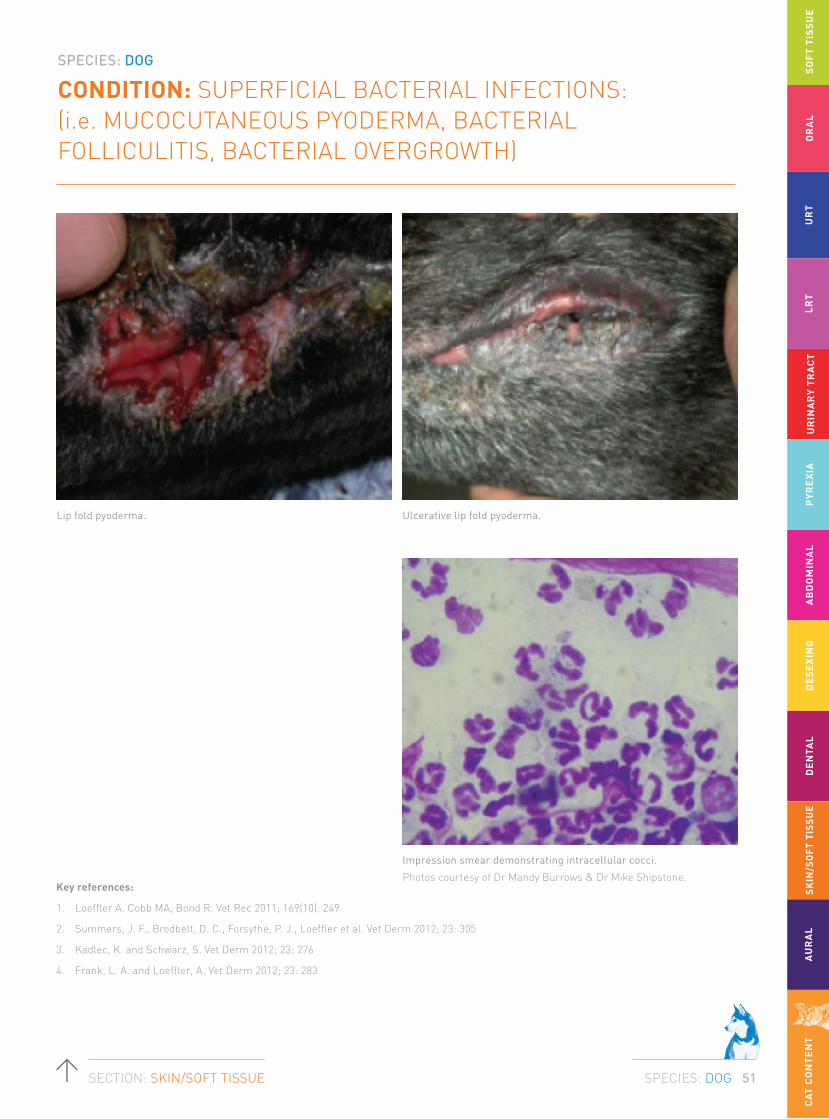
SPECIES: DOG
SPECIES: DOG 51SECTION: SKIN/SOFT TISSUE
CONDITION: SUPERFICIAL BACTERIAL INFECTIONS: (i.e. MUCOCUTANEOUS PYODERMA, BACTERIAL FOLLICULITIS, BACTERIAL OVERGROWTH)
SO
FT
TIS
SU
EO
RA
LU
RT
LR
TU
RIN
AR
Y T
RA
CT
PY
RE
XIA
AB
DO
MIN
AL
DE
SE
XIN
GD
EN
TAL
SKIN
/SO
FT T
ISSU
EA
UR
AL
CA
T C
ON
TE
NT
Key references:
1. Loeffler A. Cobb MA, Bond R. Vet Rec 2011; 169(10): 249
2. Summers, J. F., Brodbelt, D. C., Forsythe, P. J., Loeffler et al. Vet Derm 2012; 23: 305
3. Kadlec, K. and Schwarz, S. Vet Derm 2012; 23: 276
4. Frank, L. A. and Loeffler, A. Vet Derm 2012; 23: 283
Lip fold pyoderma. Ulcerative lip fold pyoderma.
Impression smear demonstrating intracellular cocci.
Photos courtesy of Dr Mandy Burrows & Dr Mike Shipstone.

SPECIES: DOG
SPECIES: DOG 52SECTION: SKIN/SOFT TISSUE
CONDITION: DEEP BACTERIAL INFECTIONS:(i.e. FURUNCULOSIS WITH DRAINING TRACTS)
SO
FT
TIS
SU
EO
RA
LU
RT
LR
TU
RIN
AR
Y T
RA
CT
PY
RE
XIA
AB
DO
MIN
AL
DE
SE
XIN
GD
EN
TAL
SKIN
/SO
FT T
ISSU
EA
UR
AL
CA
T C
ON
TE
NT
Deep pyoderma may be seen without evidence of prior superficial pyoderma or as a sequelae to superficial bacterial folliculitis. Deep follicular inflammation leads to follicular rupture releasing hair shaft keratin, bacteria and bacterial products into the dermis resulting in a furunculosis or infection of the dermis and subcutis. Bacteria present are usually S. pseudintermedius, but Proteus, Pseudomonas and E. coli are seen more frequently in deeper infections. Pseudomonas aeruginosa has also been isolated from dorsal furunculosis following shampoo therapy.
Generalised furunculosis: Underlying triggers commonly initiate deep pyoderma. Generalised demodicosis is the most common cause of deep pyoderma. Other predisposing causes include hyperadrenocorticism, the injudicious use of glucocorticoids (iatrogenic hyperglucocorticoidism), actinic disease and immunologic defects. Comedonal diseases such as calluses, actinic comedones and Schnauzer comedone syndrome also predispose to deep pyoderma via rupture of the abnormal follicle.
Triggers are not always identified in deep pyoderma.
Distribution: Glabrous skin of axillae, inguinal region.
Skin lesions: Papules, pustules that rupture forming fistulous tracts discharging seropurulent or haemorrhagic exudate with necrotic, friable tissue and haemorrhagic crusts; erosions and ulcers develop secondary to inflammation, necrosis and self-trauma. Haemorrhagic bullae are a distinctive clinical feature of canine deep pyoderma. Well circumscribed, firm nodules exhibit a deep dark red to blue hue. Lethargy, depression and fever; pain and pruritus are common with accompanied regional and generalised lymphadenopathy.
Localised syndromes: Deep bacterial folliculitis and furunculosis may be manifest in skin diseases with a traumatic component, and may be termed ‘traumatic furunculosis’ in this context. Diseases featuring traumatic furunculosis include post grooming furunculosis, canine acne, callus pyoderma and interdigital furunculosis.
Deep bacterial folliculitis and furunculosis is a common, somewhat heterogeneous group of bacterial skin diseases.
BACKGROUND/NATURE OF INFECTION/
ORGANISMS INVOLVED

SPECIES: DOG
SPECIES: DOG 53SECTION: SKIN/SOFT TISSUE
CONDITION: DEEP BACTERIAL INFECTIONS:(i.e. FURUNCULOSIS WITH DRAINING TRACTS)
SO
FT
TIS
SU
EO
RA
LU
RT
LR
TU
RIN
AR
Y T
RA
CT
PY
RE
XIA
AB
DO
MIN
AL
DE
SE
XIN
GD
EN
TAL
SKIN
/SO
FT T
ISSU
EA
UR
AL
CA
T C
ON
TE
NT
Interdigital furunculosis: is a common presentation. Lesions occur predominantly in the interdigital webs but may affect the digits as well. Although interdigital furunculosis is multifactorial, trauma to hair follicles is probably a common precipitating cause.
Licking/trauma of the interdigital skin leads to penetration of hair shaft into dermis or follicular rupture resulting in a foreign body reaction to the hair shaft keratin with pyogranulomatous inflammation and secondary bacterial infection. Thus allergic skin disease such as canine atopic dermatitis and adverse food reactions may initiate this syndrome.
Interdigital pyoderma may also present as a sequelae to rupture of obstructed, keratin-filled follicles that result from chronic friction or other trauma.
Large/giant breeds and dogs with abnormal foot conformation are predisposed and most commonly the front feet are involved due to increased weight bearing. Follicular dilation and comedone formation result forming follicular cysts. Follicular cysts rupture and the release of the keratin protein results in a foreign body reaction.
Skin lesions: Nodules, haemorrhagic bullae, fistulae discharging serosanguinous to seropurulent exudate; alopecia from constant licking leading to maceration, chronic moistness and surface secondary bacterial and Malassezia overgrowth; varying degrees of pain and lameness, pruritus and paronychia may be present.
Distribution: Dorsal interdigital webs; the front feet are most commonly and usually most severely affected.
SPECIES: DOG
SPECIES: DOG 54SECTION: SKIN/SOFT TISSUE
CONDITION: DEEP BACTERIAL INFECTIONS:(i.e. FURUNCULOSIS WITH DRAINING TRACTS)
SO
FT
TIS
SU
EO
RA
LU
RT
LR
TU
RIN
AR
Y T
RA
CT
PY
RE
XIA
AB
DO
MIN
AL
DE
SE
XIN
GD
EN
TAL
SKIN
/SO
FT T
ISSU
EA
UR
AL
CA
T C
ON
TE
NT
Perform a direct smear of the contents of the pustule, nodule or bulla by puncturing or squeezing the lesion; transfer and spread the contents directly onto a microscope slide for cytological evaluation and stain the sample with DiffQuik. If there are no intact lesions then sample the exudate draining from a fistulous tract and make an impression smear of the fluid contents although microorganisms are usually less evident than in surface and superficial bacterial infections. If there are abundant bacilli, then tissue should be submitted for culture and sensitivity. In all deep pyodermas, it is important to rule out underlying demodicosis by collecting multiple deep skin scrapings and or trichograms.
Histological evaluation is often useful in the diagnostic work-up of deep bacterial pyoderma because the biopsy sample can be divided, with half submitted fresh, for bacterial and fungal culture and the rest submitted in formalin for histological examination to rule out pododemodicosis. It is routine for us to skin biopsy all other cases of localised and generalised deep bacterial folliculitis and furunculosis.
Further diagnostic workup involves evaluation of spontaneous or iatrogenic hyperglucocorticoidism (CBC, biochemistry, urinalysis, ACTH stimulation testing), hypothyroidism (free T4 with TSH) and for interdigital furunculosis, evaluation of underlying allergic skin disease (elimination diet trials and intradermal/serological allergy testing) as well as mechanical and traumatic factors.
KEY STEPS
01 Collect surface/exudative cytology from draining or intact lesions.
02 Multiple deep skin scrapings or trichograms to evaluate for demodicosis.
03 Biopsy for deep tissue bacterial and fungal culture for recurrent/non-responsive cases.
04 Treat for adequate length of time (4-6 weeks minimum) with systemic antimicrobial therapy.
05 Evaluate for underlying cause/s.
TESTS FOR DIAGNOSIS
SPECIES: DOG
SPECIES: DOG 55SECTION: SKIN/SOFT TISSUE
CONDITION: DEEP BACTERIAL INFECTIONS:(i.e. FURUNCULOSIS WITH DRAINING TRACTS)
SO
FT
TIS
SU
EO
RA
LU
RT
LR
TU
RIN
AR
Y T
RA
CT
PY
RE
XIA
AB
DO
MIN
AL
DE
SE
XIN
GD
EN
TAL
SKIN
/SO
FT T
ISSU
EA
UR
AL
CA
T C
ON
TE
NT
TREATMENT
Systemic antibiotics, namely cephalosporin, β-lactam and fluoroquinolone antimicrobial agents should be used for the treatment of deep bacterial infections.
Interdigital furunculosis is usually managed with systemic antimicrobial agents using either cephalosporin or beta-lactams. If Gram-negative organisms are implicated then either trimethoprim-sulphonamide or enrofloxacin is used for an extended treatment period of 8-12 weeks. Metronidazole may be used as an adjunct treatment. Most affected animals are allergic and so diagnostic investigation of the underlying allergic skin disease and the use of cyclosporin and other drug modalities to control the underlying allergic symptoms may be indicated once the secondary infection is resolved. Laser ablation of the interdigital follicular cysts and surgical fusion podoplasty have also been described for these cases, particularly the large or giant breeds that present with comedone formation.

SPECIES: DOG
SPECIES: DOG 56SECTION: SKIN/SOFT TISSUE
CONDITION: DEEP BACTERIAL INFECTIONS:(i.e. FURUNCULOSIS WITH DRAINING TRACTS)
SO
FT
TIS
SU
EO
RA
LU
RT
LR
TU
RIN
AR
Y T
RA
CT
PY
RE
XIA
AB
DO
MIN
AL
DE
SE
XIN
GD
EN
TAL
SKIN
/SO
FT T
ISSU
EA
UR
AL
CA
T C
ON
TE
NT
AIDAP TOP TIPS ANTIBIOTICS USED
USAGE RECOMMENDATION
First line:
Cephalexin 22 mg/kg q 12h; Cefovecin 8 mg/kg q 14d SC for ≥ 3 administrations (off-label).
Second line:
Higher doses of fluoroquinolones e.g. enrofloxacin 10-15 mg/kg q 24h (off-label) or marbofloxacin 5.5 mg/kg q 24h.
Topical therapy:
3% benzoyl peroxide or 3% chlorhexidine shampoo/lotion/gel.
Full therapeutic dose for 6 weeks, or 10 days beyond complete clinical resolution.
Always look for Demodex canis as an underlying cause of recurrent deep bacterial pyoderma in dogs.

SPECIES: DOG
SPECIES: DOG 57SECTION: SKIN/SOFT TISSUE
CONDITION: DEEP BACTERIAL INFECTIONS:(i.e. FURUNCULOSIS WITH DRAINING TRACTS)
SO
FT
TIS
SU
EO
RA
LU
RT
LR
TU
RIN
AR
Y T
RA
CT
PY
RE
XIA
AB
DO
MIN
AL
DE
SE
XIN
GD
EN
TAL
SKIN
/SO
FT T
ISSU
EA
UR
AL
CA
T C
ON
TE
NT
Key references:
1. Duclos DD, Hargis AM, Hanley PW. Vet Derm 2008; 19(3): 134-141
2. Summers, J. F., Brodbelt, D. C., Forsythe P.J. et al. Vet Derm 2012; 23: 30
3. Kovacs MS, McKiernan S, Potter DM et al. Contemp Top Lab Anim Sci 2005; 44(4): 17-21
Deep bacterial infection on lateral thigh with multiple discharging sinuses.
Deep bacterial infection of callus.
Interdigital cyst formation.
Photos courtesy of Dr Mandy Burrows & Dr Mike Shipstone.
Interdigital cyst formation.

SPECIES: DOG
SPECIES: DOG 58SECTION: SKIN/SOFT TISSUE
CONDITION: MYCOBACTERIA AND NOCARDIA AS CAUSES OF DEEP DRAINING SINUS TRACTS
SO
FT
TIS
SU
EO
RA
LU
RT
LR
TU
RIN
AR
Y T
RA
CT
PY
RE
XIA
AB
DO
MIN
AL
DE
SE
XIN
GD
EN
TAL
SKIN
/SO
FT T
ISSU
EA
UR
AL
CA
T C
ON
TE
NT
(i) Infections of the subcutaneous panniculus generally with rapidly growing mycobacteria and;
(ii) granulomatous or pyogranulomatous masses of the skin and subcutis (generally due to non-cultivable mycobacteria e.g. leproid granulomas etc. and sometimes mycobacterium avium complex infections).
The taxonomy of these organisms is evolving, and currently they are divided into complexes:
a. M. smegmatis complex (including M. goodii) – drugs of choice doxycycline, moxifloxacin, gentamicin.
b. M. fortuitum complex – drugs of choice clarithromycin, moxifloxacin, gentamicin.
c. M. chelonae/abscesses complex – drug of choice clarithromycin, rest depends on susceptibility testing.
Generally speaking they are all resistant to rifampicin, and all susceptible to clofazamine.
Mycobacteria cause two major types of disease affecting the skin and subcutis.
BACKGROUND/NATURE OF INFECTION/
ORGANISMS INVOLVED

SPECIES: DOG
SPECIES: DOG 59SECTION: SKIN/SOFT TISSUE
CONDITION: MYCOBACTERIA AND NOCARDIA AS CAUSES OF DEEP DRAINING SINUS TRACTS
SO
FT
TIS
SU
EO
RA
LU
RT
LR
TU
RIN
AR
Y T
RA
CT
PY
RE
XIA
AB
DO
MIN
AL
DE
SE
XIN
GD
EN
TAL
SKIN
/SO
FT T
ISSU
EA
UR
AL
CA
T C
ON
TE
NT
1. The cornerstone of therapy is obtaining a positive culture.
2. This is obtained by aspirating purulent exudate present in the subcutis through intact skin, after preparation of the skin with 70% ethanol (and allowing time for drying).
3. Primary isolation can be done in a veterinary laboratory, although it important to keep the plates for the 4-5 days it takes for the colonies to appear.
4. Positive cultures should be forwarded to a human mycobacteria reference laboratory for species identification and C+S testing.
Infections of the skin and subcutis with rapidly growing mycobacteria.
Reference laboratories managing culture and PCR of Mycobacteria and Nocardia:
VICTORIA
Dr Janet Fyfe Email: [email protected]
Victorian Infectious Diseases Reference Laboratory
10 Wreckyn Street, North Melbourne VIC 3051
Ph: (03) 9342 2600
WESTERN AUSTRALIA
Dr Ian Arthur Email: [email protected]
PathWest Laboratory Medicine WA
QEII Medical Centre, Nedlands WA 6009
KEY STEPS
KEY ISSUES
01 Draining sinus tracts should alert the practitioner to the presence of saprophytic pathogens such as mycobacteria, Nocardia spp. and fungi.
02 Involvement of the inguinal panniculus is suggestive of mycobacterial and nocardial disease.
03 Preliminary cytology stained with DiffQuik can be very helpful in cases where Nocardia and fungi are involved, whereas culture on routine media is far more expedient a way to diagnose mycobacterial infections caused by rapidly growing saprophytic mycobacteria.
a. Rapidly growing mycobacteria, Nocardia spp. and fungi can all give rise to deep draining tracts that discharge to the skin surface.
b. Rapidly growing mycobacteria (and to a lesser extent Nocardia nova) have a predilection for the fatty subcuatenous panniculuis, especially in the inguinal region.
TESTS FOR DIAGNOSIS
SPECIES: DOG
SPECIES: DOG 60SECTION: SKIN/SOFT TISSUE
CONDITION: MYCOBACTERIA AND NOCARDIA AS CAUSES OF DEEP DRAINING SINUS TRACTS
SO
FT
TIS
SU
EO
RA
LU
RT
LR
TU
RIN
AR
Y T
RA
CT
PY
RE
XIA
AB
DO
MIN
AL
DE
SE
XIN
GD
EN
TAL
SKIN
/SO
FT T
ISSU
EA
UR
AL
CA
T C
ON
TE
NT
TREATMENT
These infections require months to years of antimicrobial therapy, and in some cases surgery is required to debulk lesions to enable a clinical cure to be achieved.
Generally speaking, topical therapy is not useful in the management of these infections as the disease process is situated in the subcutis and involves the skin secondarily.
C+S is strongly advised in these cases.
Mycobacteria
First line:
Doxycycline (5 mg/kg q12h†) and moxifloxacin (5 mg/kg q12h‡ (compounded) for M. smegmatis complex infections; clarithromycin (5-15 mg/kg q12h‡) and moxifloxacin (5 mg/kg q12h‡) for other rapidly growing mycobacteria.
Second line:
Clofazimine (4-10 mg/kg q24h‡ compounded), amikacin (10-15 mg/kg q24h‡ IV/IM/SC).
Nocardia
First line:
Trimethoprim/sulphonamide combinations at a dose of 12.5 to 30 mg/kg q12h (do not split or otherwise divide the coated tablet) combined with a second drug depending on C+S testing; beware ketatoconjunctivitis sicca during therapy.
Second line:
Amoxicillin 20 mg/kg twice a day for N. nova (but not amoxicillin clavulanate). Clarithromycin (5-15 mg/kg q12h) or moxifloxacin (10 mg/kg once a day).
ANTIBIOTICS USED

SPECIES: DOG
SPECIES: DOG 61SECTION: SKIN/SOFT TISSUE
CONDITION: MYCOBACTERIA AND NOCARDIA AS CAUSES OF DEEP DRAINING SINUS TRACTS
SO
FT
TIS
SU
EO
RA
LU
RT
LR
TU
RIN
AR
Y T
RA
CT
PY
RE
XIA
AB
DO
MIN
AL
DE
SE
XIN
GD
EN
TAL
SKIN
/SO
FT T
ISSU
EA
UR
AL
CA
T C
ON
TE
NT
USAGE RECOMMENDATION
See current textbooks for detailed guidelines. Ensure doxycycline given with food or water bowl provided.
Key references:
1. Reppas G, Nosworthy P, Hansen T, et al. Aust Vet J 2010; 88(5): 197-200
2. Escalonilla P, Esteban J, Soriano ML, et al. Clin Exp Dermatol 1998; 23(5): 214-221
3. Malik R, Krockenberger MB, O’Brien CR, et al. Aust Vet J 2006; 84(7): 235-245
4. Malik R, Love DN, Wigney DI, et al. Aust Vet J 1998; 76(6): 403-407
5. Charles J, Martin P, Wigney DI, et al. Aust Vet J 1999; 77(12): 799-803
6. Fyfe JA, McCowan C, O’Brien CR, et al. J Clin Microbiol 2008; 46(2): 618-626
7. Malik R, Shaw SE, Griffin C, et al. J Small Anim Pract 2004; 45(10): 485-494
8. Malik R, Hughes MS, James G, et al. J Feline Med Surg 2002; 4(1): 43-59
9. Govendir M, Norris JM, Hansen T, et al. Vet Microbiol 2011; 153(3-4): 240-245
10. Malik R, Wigney DI, Dawson D, et al. J Feline Med Surg 2000; 2(1) 35-48
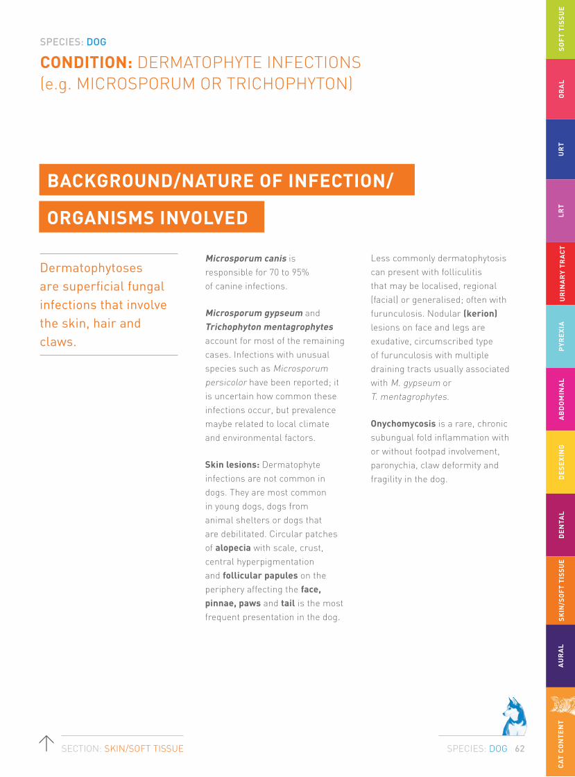
SPECIES: DOG
SPECIES: DOG 62SECTION: SKIN/SOFT TISSUE
CONDITION: DERMATOPHYTE INFECTIONS (e.g. MICROSPORUM OR TRICHOPHYTON)
SO
FT
TIS
SU
EO
RA
LU
RT
LR
TU
RIN
AR
Y T
RA
CT
PY
RE
XIA
AB
DO
MIN
AL
DE
SE
XIN
GD
EN
TAL
SKIN
/SO
FT T
ISSU
EA
UR
AL
CA
T C
ON
TE
NT
Microsporum canis is responsible for 70 to 95% of canine infections.
Microsporum gypseum and Trichophyton mentagrophytes account for most of the remaining cases. Infections with unusual species such as Microsporum persicolor have been reported; it is uncertain how common these infections occur, but prevalence maybe related to local climate and environmental factors.
Skin lesions: Dermatophyte infections are not common in dogs. They are most common in young dogs, dogs from animal shelters or dogs that are debilitated. Circular patches of alopecia with scale, crust, central hyperpigmentation and follicular papules on the periphery affecting the face, pinnae, paws and tail is the most frequent presentation in the dog.
Less commonly dermatophytosis can present with folliculitis that may be localised, regional (facial) or generalised; often with furunculosis. Nodular (kerion) lesions on face and legs are exudative, circumscribed type of furunculosis with multiple draining tracts usually associated with M. gypseum or T. mentagrophytes.
Onychomycosis is a rare, chronic subungual fold inflammation with or without footpad involvement, paronychia, claw deformity and fragility in the dog.
Dermatophytoses are superficial fungal infections that involve the skin, hair and claws.
BACKGROUND/NATURE OF INFECTION/
ORGANISMS INVOLVED
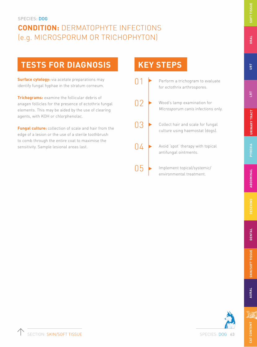
SPECIES: DOG
SPECIES: DOG 63SECTION: SKIN/SOFT TISSUE
CONDITION: DERMATOPHYTE INFECTIONS (e.g. MICROSPORUM OR TRICHOPHYTON)
SO
FT
TIS
SU
EO
RA
LU
RT
LR
TU
RIN
AR
Y T
RA
CT
PY
RE
XIA
AB
DO
MIN
AL
DE
SE
XIN
GD
EN
TAL
SKIN
/SO
FT T
ISSU
EA
UR
AL
CA
T C
ON
TE
NT
Surface cytology: via acetate preparations may identify fungal hyphae in the stratum corneum.
Trichograms: examine the follicular debris of anagen follicles for the presence of ectothrix fungal elements. This may be aided by the use of clearing agents, with KOH or chlorphenolac.
Fungal culture: collection of scale and hair from the edge of a lesion or the use of a sterile toothbrush to comb through the entire coat to maximise the sensitivity. Sample lesional areas last.
KEY STEPS
01 Perform a trichogram to evaluate for ectothrix arthrospores.
02 Wood’s lamp examination for Microsporum canis infections only.
03 Collect hair and scale for fungal culture using haemostat (dogs).
04 Avoid ‘spot’ therapy with topical antifungal ointments.
05 Implement topical/systemic/environmental treatment.
TESTS FOR DIAGNOSIS

SPECIES: DOG
SPECIES: DOG 64SECTION: SKIN/SOFT TISSUE
CONDITION: DERMATOPHYTE INFECTIONS (e.g. MICROSPORUM OR TRICHOPHYTON)
SO
FT
TIS
SU
EO
RA
LU
RT
LR
TU
RIN
AR
Y T
RA
CT
PY
RE
XIA
AB
DO
MIN
AL
DE
SE
XIN
GD
EN
TAL
SKIN
/SO
FT T
ISSU
EA
UR
AL
CA
T C
ON
TE
NT
TREATMENT
In most healthy animals, dermatophytosis is a self-curing disease, with full resolution of disease in 10-16 weeks without therapy. The best treatment protocol is a combination of three modalities:
1. Topical treatment: to kill infective material and prevent its dissemination into the environment.
2. Systemic treatment: to shorten the time of infection in the individual animal.
3. Environmental treatment: to help prevent recurrence of infection or spread to other animals or people in the household.
A. Clipping the hair coat: should you clip?
Clipping of the hair coat will mechanically remove fragile hairs that will fracture and release spores into the environment. Clipping of the entire hair coat is optimal but not always possible or practical. Clipping is time consuming and often requires sedation and is irritating to cats. Owners are often unwilling to commit to clipping their pets. Short-haired dogs with fewer than five focal lesions do not need to be clipped.
When dogs have more than five lesions, long hair and there are multiple pets in the environment and the affected pet cannot be segregated, clipping the entire pet is optimal. Clip the hair short and gently to avoid spreading the infection due to the microtrauma and mechanical spread of the spores.
The owner should be warned that a temporary exacerbation of lesions may occur after clipping.
Note: If the animal is to be clipped in the clinic all debris produced must be considered to be infectious with zoonotic potential and so rigorous infection control measures should be observed.
i.e. cover table surface with disposable drape, gown and glove, collect all material and double bag, thoroughly disinfect the room and all equipment used with an appropriate antifungal agent.
B. Topical therapy: which one is best?
i. Localised (treating only the spots) or whole body topical therapy
In animals, not all the lesions may be visible due to the long hair coat. It is almost certain that there are infective spores in non-lesional areas. Therefore ‘spot treatment’ with topical drugs is not recommended even for focal lesions because infection beyond the margin of visible lesions is likely. There is no clinical data to support that the use of spot treatment clear lesions any more rapidly than whole-body treatment alone. If the owner insists, the best products are probably 1% terbinafine solution, lotion, cream or spray (Lamisil®) and 2% clotrimazole cream (Canesten®).
ii. Total body treatment
Topical therapy inactivates fungal spores and mycelia on and within hair shafts reducing environmental contagion and results in a faster cure than systemic therapy alone. Shampoo therapy, dipping or rinsing with topical antifungal agents is preferred. The choice of topical antifungal agent is important because studies have shown that many topical antifungal agents are ineffective. In vitro and in vivo studies have shown that the most consistently effective topical treatments are lime sulphur, enilconazole, and miconazole; the latter with or without chlorhexidine. Miconazole and chlorhexidine (Malaseb®) shampoo has been studied in cats as an adjunct treatment to oral griseofulvin.

SPECIES: DOG
SPECIES: DOG 65SECTION: SKIN/SOFT TISSUE
CONDITION: DERMATOPHYTE INFECTIONS (e.g. MICROSPORUM OR TRICHOPHYTON)
SO
FT
TIS
SU
EO
RA
LU
RT
LR
TU
RIN
AR
Y T
RA
CT
PY
RE
XIA
AB
DO
MIN
AL
DE
SE
XIN
GD
EN
TAL
SKIN
/SO
FT T
ISSU
EA
UR
AL
CA
T C
ON
TE
NT
Systemic therapy
The role of systemic therapy in treating dermatophytosis is to accelerate the resolution of infection in the individual animal. Several effective drugs are available, and the appropriate choice should be made depending on cost, fungal species, patient species and potential for toxicity. Systemic therapy is the treatment of choice for dermatophytosis. It is important to remember that systemic antifungal therapy does not rapidly reduce contagion and should be used in conjunction with clipping and topical antifungal agents.
Griseofulvin
Griseofulvin is administered at 25 mg/kg q12h. The absorption is enhanced when administered as a divided dose and with a fatty meal. The most common side-effects – anorexia, vomiting and diarrhoea – can be avoided by dividing or lowering the dose. The drug is highly teratogenic and therefore contraindicated in pregnant animals.
Do not administer to dogs less than 6 weeks of age.
Griseofulvin is a good systemic drug for dermatophytosis in dogs, where myelotoxicity is not a concern. Nearly all patients with Microsporum infections, and many with Trichophyton infections will be cured with this drug,
Ketoconazole (KTZ)
Ketoconazole 10 mg/kg q12h‡ (Nizoral®) is an alternative but perhaps best reserved for resistant Trichophyton infections in dogs. Some strains of Microsporum canis appear resistant to ketoconazole. Therefore ketoconazole has no real advantage over other drugs for routine cases of dermatophytosis.
Itraconazole (ITZ)
Itraconazole (Sporanox®) is a fungicidal triazole drug that is extremely useful for dermatophytosis. The recommended dose is 5-10 mg/kg/day PO. Itraconazole persists in the skin and nails for weeks to months after dosing, and intermittent or pulse therapy is frequently prescribed for skin infections or onychomycosis.
Itraconazole is generally well tolerated; reported side-effects include vomiting and/or anorexia and an idiosyncratic cutaneous vasculitis has been reported in dogs. Itraconazole is reportedly not teratogenic when used at a dose of 5 mg/kg.
Fluconazole
Fluconazole (Diflucan®) is receiving some recent attention as an alternative drug because it has now become inexpensive through some compounding pharmacies. Several recent in vitro studies have shown that the MICs of fluconazole against dermatophytes are much higher that the MICs of itraconazole suggesting that itraconazole may be the superior drug. Current evidence and clinical anecdotes would suggest that there is no advantage of this drug over itraconazole and currently we do not recommend it for the treatment of dermatophytosis.

SPECIES: DOG
SPECIES: DOG 66SECTION: SKIN/SOFT TISSUE
CONDITION: DERMATOPHYTE INFECTIONS (e.g. MICROSPORUM OR TRICHOPHYTON)
SO
FT
TIS
SU
EO
RA
LU
RT
LR
TU
RIN
AR
Y T
RA
CT
PY
RE
XIA
AB
DO
MIN
AL
DE
SE
XIN
GD
EN
TAL
SKIN
/SO
FT T
ISSU
EA
UR
AL
CA
T C
ON
TE
NT
Environmental treatment
The critical role of environmental disinfection in eradication of M. canis from a household cannot be overemphasised. Environmental contamination with M. canis spores is widespread, difficult to eliminate and routinely transported by the fur of uninfected dogs. Such contamination is a major reservoir for recurrence of infection. M. canis spores remain viable in the environment for up to 18 months.
Studies using isolated infected hairs or spores or field studies using dermatophyte-contaminated environments have shown that the following disinfectant products are consistently effective: lime sulphur (1:33), enilconazole (0.2%), and 1:10 to 1:100 household bleach (10 mL/L). In addition, a study has also shown that strain variation of M. canis with respect to susceptibility to disinfectants is not present.
For treatment of routine infections with one or a few animals in the household, extensive environmental decontamination is generally impractical and unnecessary. Thorough vacuuming and mechanical cleaning will remove infective material. All hard surfaces should be mopped with 1:100 bleach solution.
During treatment these few animals should be confined to a small easily cleaned room without carpeting until they have received systemic antifungal therapy for at least two weeks and have been dipped at least four times with topical preparations. All bedding, brushes, combs, rugs, cages, carriers can be washed daily in hot water, detergent and a 1:10 dilution of household bleach.
Carpeted areas are problematic because of the lack of effective disinfectant that preserves carpeting. Frequent vacuuming on a daily basis or steam cleaning mechanically removes many but not all spores. Steam cleaning may not be a reliable method of killing M. canis unless an antifungal disinfectant such as chlorhexidine or sodium hypochlorite is added to the water. Draperies should be dry cleaned and not replaced until the infection is eradicated.
Length of treatment
Dogs with dermatophytosis should be treated until complete resolution of clinical signs (clinical cure) and then continued until the fungus cannot be cultured from the hair coat on at least two sequential cultures a week or more apart (mycologic cure)
Weekly fungal cultures should be started after the dog has received 4-6 weeks of therapy and thereafter on a 2 week schedule. Once the culture results are negative, monitoring can be done on a once weekly basis. Dogs appear healthy before their skin and hair are cleared of fungal organisms.
It is not always possible or practical however to re-culture every patient. In otherwise healthy dogs, systemic and topical treatments should generally be continued for 6-10 weeks, preferably until 2 weeks after clinical resolution.

SPECIES: DOG
SPECIES: DOG 67SECTION: SKIN/SOFT TISSUE
CONDITION: DERMATOPHYTE INFECTIONS (e.g. MICROSPORUM OR TRICHOPHYTON)
SO
FT
TIS
SU
EO
RA
LU
RT
LR
TU
RIN
AR
Y T
RA
CT
PY
RE
XIA
AB
DO
MIN
AL
DE
SE
XIN
GD
EN
TAL
SKIN
/SO
FT T
ISSU
EA
UR
AL
CA
T C
ON
TE
NT
USAGE RECOMMENDATION
Significant duration: 6-10 weeks; treat until two successive negative fungal cultures obtained one week apart or 14 days beyond a clinical cure.
AIDAP TOP TIPS
Our treatment recommendations for dermatophytosis for dogs:
• 2% miconazole, 2% chlorhexidine shampoo baths twice a week
• 0.2% enilconazole (Imaverol®) rinse twice a week (not registered for use in cats)
• Itraconazole 5 mg/kg q24h 7d, 7d break and repeat pulse for 3 treatment cycles OR
• Griseofulvin 25 mg/kg q12h
• Environmental decontamination.
ANTIFUNGAL AGENTS USED
First line:
Itraconazole 5 mg/kg q24h‡ 7d, 7d break and repeat pulse for 3 treatment cycles.
Second line:
Griseofulvin 25 mg/kg q12h.

SPECIES: DOG
SPECIES: DOG 68SECTION: SKIN/SOFT TISSUE
CONDITION: DERMATOPHYTE INFECTIONS (e.g. MICROSPORUM OR TRICHOPHYTON)
SO
FT
TIS
SU
EO
RA
LU
RT
LR
TU
RIN
AR
Y T
RA
CT
PY
RE
XIA
AB
DO
MIN
AL
DE
SE
XIN
GD
EN
TAL
SKIN
/SO
FT T
ISSU
EA
UR
AL
CA
T C
ON
TE
NT
Characteristic presentation of T. mentagrophytes (before)
Same dog after treatment for T. mentagrophytes
Characteristic presentation of T. mentagrophytes
Photos courtesy of Dr Mike Shipstone.
Severe dermatophytosis causing soft tissue swelling.
Photos courtesy of Dr Mandy Burrows & Dr Mike Shipstone.
Multifocal alopecia dt Trichophyton infection.
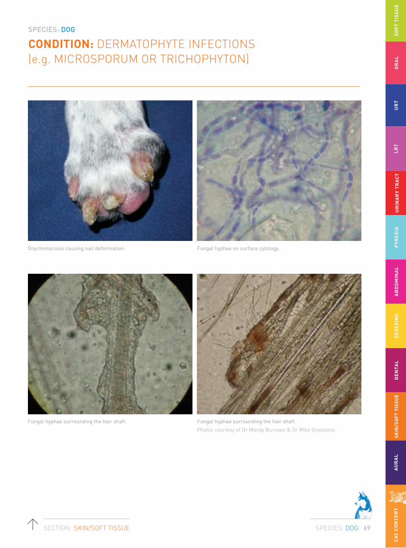
SPECIES: DOG
SPECIES: DOG 69SECTION: SKIN/SOFT TISSUE
CONDITION: DERMATOPHYTE INFECTIONS (e.g. MICROSPORUM OR TRICHOPHYTON)
SO
FT
TIS
SU
EO
RA
LU
RT
LR
TU
RIN
AR
Y T
RA
CT
PY
RE
XIA
AB
DO
MIN
AL
DE
SE
XIN
GD
EN
TAL
SKIN
/SO
FT T
ISSU
EA
UR
AL
CA
T C
ON
TE
NT
Onychomycosis causing nail deformation. Fungal hyphae on surface cytology.
Fungal hyphae surrounding the hair shaft.
Photos courtesy of Dr Mandy Burrows & Dr Mike Shipstone.
Fungal hyphae surrounding the hair shaft.
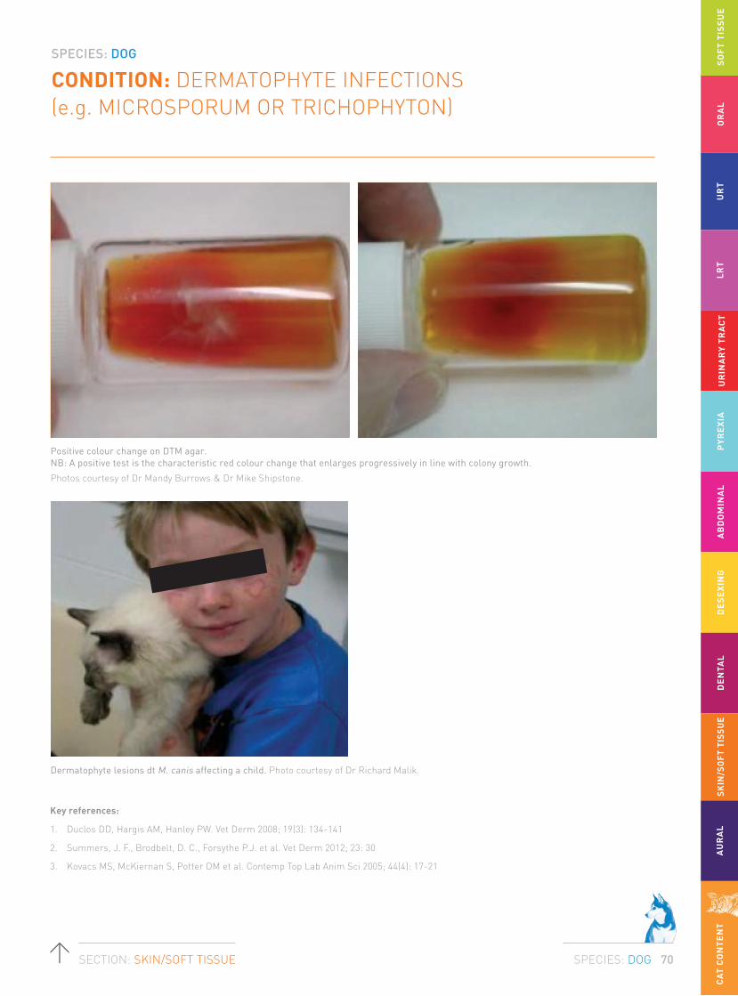
SPECIES: DOG
SPECIES: DOG 70SECTION: SKIN/SOFT TISSUE
CONDITION: DERMATOPHYTE INFECTIONS (e.g. MICROSPORUM OR TRICHOPHYTON)
SO
FT
TIS
SU
EO
RA
LU
RT
LR
TU
RIN
AR
Y T
RA
CT
PY
RE
XIA
AB
DO
MIN
AL
DE
SE
XIN
GD
EN
TAL
SKIN
/SO
FT T
ISSU
EA
UR
AL
CA
T C
ON
TE
NT
Key references:
1. Duclos DD, Hargis AM, Hanley PW. Vet Derm 2008; 19(3): 134-141
2. Summers, J. F., Brodbelt, D. C., Forsythe P.J. et al. Vet Derm 2012; 23: 30
3. Kovacs MS, McKiernan S, Potter DM et al. Contemp Top Lab Anim Sci 2005; 44(4): 17-21
Positive colour change on DTM agar. NB: A positive test is the characteristic red colour change that enlarges progressively in line with colony growth.
Photos courtesy of Dr Mandy Burrows & Dr Mike Shipstone.
Dermatophyte lesions dt M. canis affecting a child. Photo courtesy of Dr Richard Malik.

SPECIES: DOG
SPECIES: DOG 71SECTION: SKIN/SOFT TISSUE
CONDITION: SUPERFICIAL YEAST (MALASSEZIA) INFECTIONS OF THE SKIN (NOT INCLUDING EARS)
SO
FT
TIS
SU
EO
RA
LU
RT
LR
TU
RIN
AR
Y T
RA
CT
PY
RE
XIA
AB
DO
MIN
AL
DE
SE
XIN
GD
EN
TAL
SKIN
/SO
FT T
ISSU
EA
UR
AL
CA
T C
ON
TE
NT
In healthy animals it exists at higher population densities at the lips and interdigital skin than in the ears. The anus seems to be the most frequently colonised mucosal site. M. pachydermatis acts as an opportunistic pathogen and factors promoting its pathogenicity may include increased temperature and humidity, excessive lipid secretion and pruritic inflammatory diseases (allergic and parasitic), endocrinopathies and some metabolic (hepatocutaneous syndrome, zinc-responsive dermatoses) and occasionally cutaneous or internal neoplasia.
(Malassezia dermatitis is typically secondary to allergy, endocrinopathy, or neoplasia (reported with thymoma-induced exfoliative dermatitis and pancreatic and/or hepatobiliary paraneoplastic alopecia) in cats and may play a primary role in feline acne. Malassezia dermatitis is rare in cats when compared to dogs).
Breed-related factors are important in Malassezia dermatitis; Basset hounds
and West Highland white terriers are particularly predisposed. Since S. pseudintermedius and M. pachydermatisare inhabitants of the mucosae, including the oral cavity, they will continually be transferred to the skin, particularly in areas which require cleaning or grooming, and which are pruritic. Thus there is potential for the establishment of microbial overgrowth whenever the skin is damaged or there is underlying disease impairing cutaneous function.
Distribution: Lips, between the pads and digits, groin, perivulvar and perianal areas, ventral abdomen, axillae, pinnae, and in skin folds; clawbeds.
Lesion: Erythema, greasiness or exudation, scaling, pruritus and saliva staining; reddish-brown staining of the proximal claw or a waxy exudate in the claw fold, with inflammation of the surrounding soft tissue. Pruritus may be quite marked. In chronic or severe lesions there may be excoriation and lichenification. There is commonly odour.
Malassezia pachydermatis is present as a commensal of the skin and mucosae of most dogs.
BACKGROUND/NATURE OF INFECTION/
ORGANISMS INVOLVED

SPECIES: DOG
SPECIES: DOG 72SECTION: SKIN/SOFT TISSUE
CONDITION: SUPERFICIAL YEAST (MALASSEZIA) INFECTIONS OF THE SKIN (NOT INCLUDING EARS)
SO
FT
TIS
SU
EO
RA
LU
RT
LR
TU
RIN
AR
Y T
RA
CT
PY
RE
XIA
AB
DO
MIN
AL
DE
SE
XIN
GD
EN
TAL
SKIN
/SO
FT T
ISSU
EA
UR
AL
CA
T C
ON
TE
NT
Diagnosis is confirmed by cytology using tape strip samples, slide impressions or swab smears stained with DiffQuik showing elevated populations of Malassezia.
Tape strips are preferred because the organisms are sometimes not located at the surface of the lesions and repeated application of the tape to the same site will reveal deeper populations. The technique is quick and easy to perform and, with experience, tapes can be examined quickly under the microscope permitting a diagnosis to be made rapidly.
The presence of numbers of Malassezia above 2 per high power x1000 oil immersion field is suggestive of microbial overgrowth. Common populations are very much higher but the organisms may be found in clusters so at least 20 high power fields should be examined. For diagnosis of Malassezia paronychia, the broken end of a wooden cotton-tip swab is used to scrape the claw fold, and exudate is pressed and rolled onto a glass slide. For examination of ear exudate in dogs with ceruminous or exudative otitis externa, rolling of exudate in a thin layer on glass slides with a cotton-tip swab is the preferred method.
KEY ISSUES
01 Identify yeast overgrowth or infection via surface cytology.
02 Response to trial treatment with appropriate topical and systemic antimicrobial therapy.
TESTS FOR DIAGNOSIS

SPECIES: DOG
SPECIES: DOG 73SECTION: SKIN/SOFT TISSUE
CONDITION: SUPERFICIAL YEAST (MALASSEZIA) INFECTIONS OF THE SKIN (NOT INCLUDING EARS)
SO
FT
TIS
SU
EO
RA
LU
RT
LR
TU
RIN
AR
Y T
RA
CT
PY
RE
XIA
AB
DO
MIN
AL
DE
SE
XIN
GD
EN
TAL
SKIN
/SO
FT T
ISSU
EA
UR
AL
CA
T C
ON
TE
NT
TREATMENT
The condition normally responds to topical therapy with lotions, cream and ointments or antimicrobial shampoos that are active against staphylococci and Malassezia.
Benzoyl peroxide shampoo can also be used. In severe or extensive cases of microbial overgrowth or when washing/spraying of the affected areas is not practical, systemic therapy with imidazoles (e.g. ketoconazole or itraconazole) or terbinafine can be utilised. Ketoconazole is the drug of choice for the treatment of Malassezia dermatitis in the dog.
Itraconazole is the drug of choice in the cat. Itraconazole persists in the stratum corneum and therefore pulse therapy can be employed. Anecdotal evidence suggests that fluconazole is not as clinically effective as the other azoles.
Topical agents containing miconazole (Daktarin®) or clotrimazole (Canesten®) spray, lotion, cream for ‘spot’ therapy. Shampoos containing chlorhexidine, or chlorhexidine and miconazole (active against staphylococci and Malassezia). Systemic therapy with ketoconazole (5-10 mg/kg q12h‡; dog only) or itraconazole (5 mg/kg q24h‡; dog [and cat]) or terbinafine (30 mg/kg q12h‡; dog [and cat]).
First line:
Topical chlorhexidine/miconazole shampoo with or without ketoconazole (5-10 mg/kg q12h‡; dog only).
Second line:
Oral itraconazole.
ANTIMICROBIALS USED

SPECIES: DOG
SPECIES: DOG 74SECTION: SKIN/SOFT TISSUE
CONDITION: SUPERFICIAL YEAST (MALASSEZIA) INFECTIONS OF THE SKIN (NOT INCLUDING EARS)
SO
FT
TIS
SU
EO
RA
LU
RT
LR
TU
RIN
AR
Y T
RA
CT
PY
RE
XIA
AB
DO
MIN
AL
DE
SE
XIN
GD
EN
TAL
SKIN
/SO
FT T
ISSU
EA
UR
AL
CA
T C
ON
TE
NT
AIDAP TOP TIPS
Ketoconazole: Nizoral® 5-10 mg/kg q12h PO for 21-28 days.
A low-dose regimen of ketoconazole 5 mg/kg every 24 hours PO for 10 days, followed by 5 mg/kg every 48 hours PO for 10 doses has been reported to be successful in the majority of cases, and lessens the expense of therapy.
Itraconazole: Sporanox® 5 mg/kg q12h PO or 10 mg/kg q24h PO for 21–28 days.
Itraconazole persists in the stratum corneum and therefore pulse therapy can be employed. One study found that dogs treated with 5 mg/kg q24h for 2 days followed by 5 days without treatment for 3 cycles (3 weeks) responded as well as dogs who had received the medication at 5 mg/kg/day for 21 days.
Prophylaxis for chronic/relapsing Malassezia dermatitis
Regular shampoo therapy (weekly or biweekly).
Pulse oral ketoconazole or itraconazole (5-10 mg/kg given 2 consecutive days per week).
Monitor for hepatotoxicity with CBC and biochemistry every 6 months.
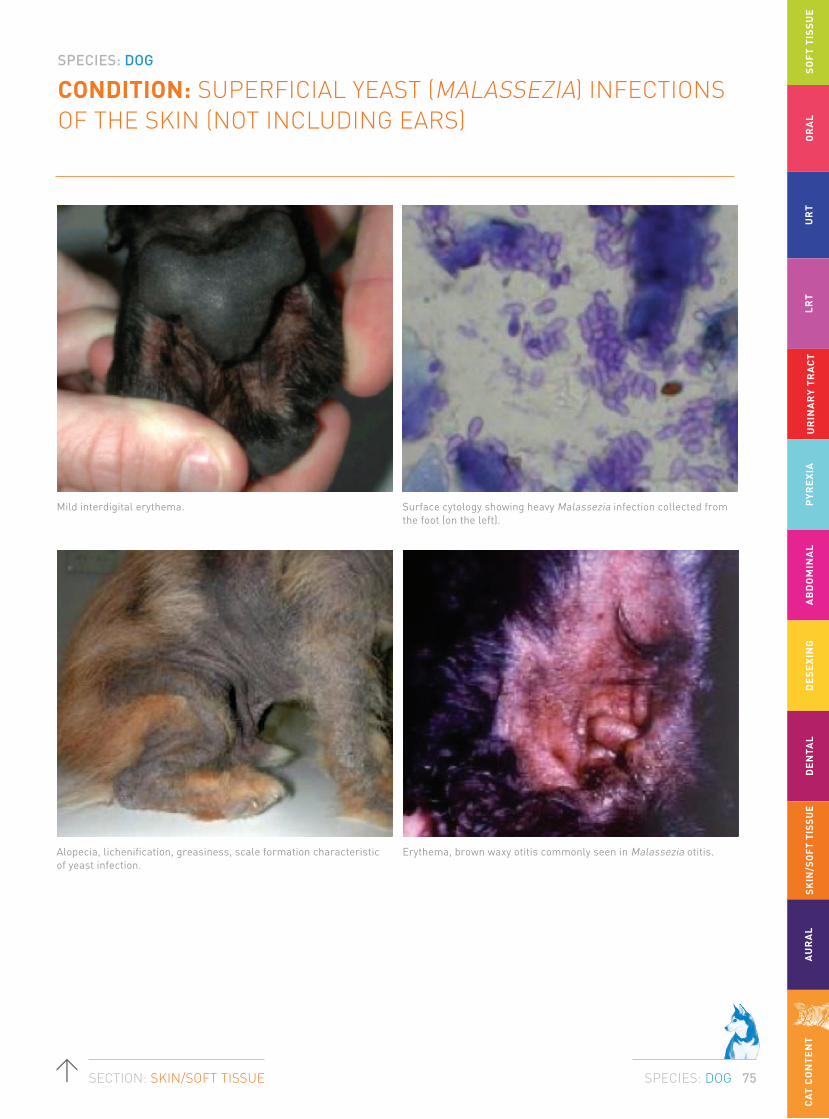
SPECIES: DOG
SPECIES: DOG 75SECTION: SKIN/SOFT TISSUE
CONDITION: SUPERFICIAL YEAST (MALASSEZIA) INFECTIONS OF THE SKIN (NOT INCLUDING EARS)
SO
FT
TIS
SU
EO
RA
LU
RT
LR
TU
RIN
AR
Y T
RA
CT
PY
RE
XIA
AB
DO
MIN
AL
DE
SE
XIN
GD
EN
TAL
SKIN
/SO
FT T
ISSU
EA
UR
AL
CA
T C
ON
TE
NT
Mild interdigital erythema. Surface cytology showing heavy Malassezia infection collected from the foot (on the left).
Erythema, brown waxy otitis commonly seen in Malassezia otitis.Alopecia, lichenification, greasiness, scale formation characteristic of yeast infection.
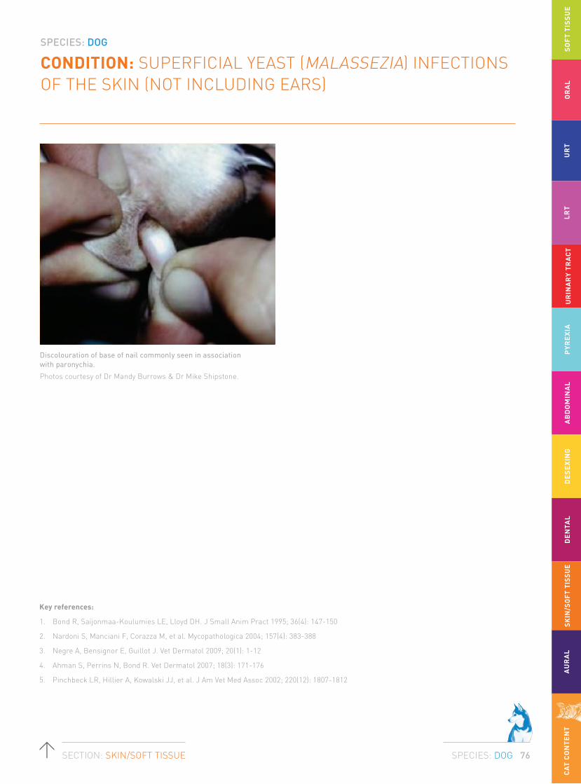
SPECIES: DOG
SPECIES: DOG 76SECTION: SKIN/SOFT TISSUE
CONDITION: SUPERFICIAL YEAST (MALASSEZIA) INFECTIONS OF THE SKIN (NOT INCLUDING EARS)
SO
FT
TIS
SU
EO
RA
LU
RT
LR
TU
RIN
AR
Y T
RA
CT
PY
RE
XIA
AB
DO
MIN
AL
DE
SE
XIN
GD
EN
TAL
SKIN
/SO
FT T
ISSU
EA
UR
AL
CA
T C
ON
TE
NT
Key references:
1. Bond R, Saijonmaa-Koulumies LE, Lloyd DH. J Small Anim Pract 1995; 36(4): 147-150
2. Nardoni S, Manciani F, Corazza M, et al. Mycopathologica 2004; 157(4): 383-388
3. Negre A, Bensignor E, Guillot J. Vet Dermatol 2009; 20(1): 1-12
4. Ahman S, Perrins N, Bond R. Vet Dermatol 2007; 18(3): 171-176
5. Pinchbeck LR, Hillier A, Kowalski JJ, et al. J Am Vet Med Assoc 2002; 220(12): 1807-1812
Discolouration of base of nail commonly seen in association with paronychia.
Photos courtesy of Dr Mandy Burrows & Dr Mike Shipstone.
SPECIES: DOG
SPECIES: DOG 77SECTION: AURAL
CONDITION: OTITIS EXTERNA (UNCOMPLICATED, FIRST EPISODE AND COMPLICATED, RECURRENT)
SO
FT
TIS
SU
EO
RA
LU
RT
LR
TU
RIN
AR
Y T
RA
CT
PY
RE
XIA
AB
DO
MIN
AL
DE
SE
XIN
GD
EN
TAL
SKIN
/SO
FT T
ISSU
EA
UR
AL
CA
T C
ON
TE
NT
The normal flora is generally Gram-positive, with higher bacterial counts retrieved from the vertical external ear canal than the horizontal ear canal. Commensal and pathogenic bacteria rapidly colonise the external ear canal where changes in the microclimate subsequent to inflammation modify the environment. The microbial proliferation exacerbates and perpetuates the inflammatory response within the ear canal. Once inflamed, there is a shift towards increased bacterial numbers, initially coagulase positive staphylococci and with more chronic inflammation, Gram-negative bacteria.
Because potential pathogens can be recovered in the absence of disease (as they can from the skin surface), it is assumed that they are unable to initiate disease in the ear.
However, once the ear becomes inflamed or macerated, proliferation may occur and it is for this reason that bacteria are considered perpetuating rather than primary or predisposing factors in otitis externa.
In dogs, coagulase positive Staphylococcus spp. are most commonly isolated in acute otitis and as a single organism. Streptococcus spp., Pseudomonas aeruginosa, Proteus mirabilis, E. coli, Corynebacterium spp., Klebsiella spp. are also frequently identified. Pseudomonas organisms are frequently identified in chronic recurrent cases or those cases that have had long-term antimicrobial treatment.
The external ear canal of most normal dogs harbours low numbers of a variety of commensal and potentially pathogenic bacteria with between 2 and 52% of healthy ears having bacteria identified on culture.
BACKGROUND/NATURE OF INFECTION/
ORGANISMS INVOLVED

SPECIES: DOG
SPECIES: DOG 78SECTION: AURAL
CONDITION: OTITIS EXTERNA (UNCOMPLICATED, FIRST EPISODE AND COMPLICATED, RECURRENT)
SO
FT
TIS
SU
EO
RA
LU
RT
LR
TU
RIN
AR
Y T
RA
CT
PY
RE
XIA
AB
DO
MIN
AL
DE
SE
XIN
GD
EN
TAL
SKIN
/SO
FT T
ISSU
EA
UR
AL
CA
T C
ON
TE
NT
In many cases of otitis, a single organism can be isolated on bacterial culture of exudate, but in others, multiple potentially pathogenic organisms are identified. Thus it is of critical importance to combine cytological examination of the otic discharge when a C+S test is performed. This allows determination of the dominant population of bacteria evident, the presence of leukocytes, and the presence of phagocytosed bacteria.
Cytology is the first step. It is mandatory in ALL cases of otitis externa and should be repeated at each visit. A number of studies have shown that cytology is more sensitive than C+S testing in identifying the presence of bacteria or yeast (e.g. sensitivity of cytology for detection of Gram-positive cocci, Gram-negative rods, and yeasts was 84%, 100% and 100% respectively). The sensitivity of culture for detection of these organisms was 59%, 69% and 50% respectively.
Normal cerumen does not have high stain uptake because of the high lipid content. Outlines of occasional squames may be seen. Inflammation leads to increased numbers of squames (some of which may be nucleated indicting faster epithelial turnover with incomplete keratinisation before desquamation). As the severity of inflammation increases inflammatory cells may be seen along with increasing numbers of organisms. Higher cellular content of cerumen may also be appreciated by increasing stain uptake on the stained slide (before microscopic examination is even started).
The number of organisms and inflammation should be assessed on a 1-4+ scale. Normal ears may have a few yeast and Gram-positive cocci per oil field but not rods. The finding of yeast or cocci should be correlated with the findings of the otoscopic examination. Some animals may have few organisms yet show marked inflammation and exudation, whilst others seem to be able to tolerate quite large numbers without any pathologic changes. Repeating the cytology at each revisit allows accurate assessment of response to therapy. Medical treatment should continue until otoscopic and cytologic examinations demonstrate no pathologic change.
KEY STEPS
Otic examination alone is not sufficient and the following minimum database is necessary in order to identify both the nature and type of the otitis as well as any underlying primary or predisposing factors.
01 Thorough dermatological history.
02 Complete physical examination of all areas of integument.
03 Thorough otic examination (may require sedation/anaesthesia).
04 Collect otic cytology.
05 Implement topical antimicrobial therapy on the basis of cytological findings.
06 Systemic antibiotic therapy is not indicated for otitis externa.
TESTS FOR DIAGNOSIS

SPECIES: DOG
SPECIES: DOG 79SECTION: AURAL
CONDITION: OTITIS EXTERNA (UNCOMPLICATED, FIRST EPISODE AND COMPLICATED, RECURRENT)
SO
FT
TIS
SU
EO
RA
LU
RT
LR
TU
RIN
AR
Y T
RA
CT
PY
RE
XIA
AB
DO
MIN
AL
DE
SE
XIN
GD
EN
TAL
SKIN
/SO
FT T
ISSU
EA
UR
AL
CA
T C
ON
TE
NT
TREATMENT
Topical therapy is the key to successful resolution of the majority of cases of otitis externa which is essentially a surface infection. Essential to this therapy though is the successful removal of exudate. If the medication cannot penetrate the full length of the ear canal, then treatment is likely to fail. The choice of appropriate active ingredients and vehicles for treatment of otitis externa is usually made empirically based on cytological examination of ear canal exudates and otoscopic examination of the inflamed ear canals.
Most commercially produced topical products contain one or more active antibacterial, antifungal and anti-inflammatory agents in various combinations as well as vehicle and various solubilisers, stabilisers and surfactants.
Clients may need to be shown how to administer medications correctly. Failure to do this is a significant cause for treatment failure. An adequate volume of medication must be delivered to line the entire canal. Getting clients to count drops increases the time for administration and fundamentally means that the nozzle of the bottle is not in the canal, reducing penetration of the medication. Putting the nozzle of the bottle in the canal and telling clients to use a “squeeze” means that both under and overdosing are risked because the amount to medication is not measured out. We use the Terumo brand of syringe to most accurately measure ear medications and dispense them into the ear canal.
A broad guideline depending on the length and diameter of the ear canal would be:
• 0.3 mL for a Shih tzu.
• 0.6 mL for a Labrador.
• 1 mL for a German Shepherd or very large dog.
• Twice daily dosing may require slightly smaller volumes to avoid overdosing.
Duration of therapy
For acute disease a minimum of 5 to 14 days therapy depending on the degree of inflammatory change (oedema, hyperplasia, and erosion, ulceration) is to be expected. Rechecks every one to two weeks are necessary to ensure that ears are cytologically and otoscopically resolved prior to cessation of therapy. It is not uncommon to have a dog clinically resolved with otoscopically normal ears because of anti-inflammatory medications, where cytology is still not normal.

SPECIES: DOG
SPECIES: DOG 80SECTION: AURAL
CONDITION: OTITIS EXTERNA (UNCOMPLICATED, FIRST EPISODE AND COMPLICATED, RECURRENT)
SO
FT
TIS
SU
EO
RA
LU
RT
LR
TU
RIN
AR
Y T
RA
CT
PY
RE
XIA
AB
DO
MIN
AL
DE
SE
XIN
GD
EN
TAL
SKIN
/SO
FT T
ISSU
EA
UR
AL
CA
T C
ON
TE
NT
Antimicrobial therapy for ears with mainly cocci on cytology
Coccoid organisms will be Staphylococcus spp. or Streptococcus spp. The challenge for empirical therapy for cocci is the relative resistance of streptococci to some of the routine antibiotics, which otherwise tend to have reasonable activity for most Staphylococci spp. For this reason products containing antibiotics with good efficacy against both bacteria are desirable. For this reason, Canaural® is useful if the TM is intact because of the framycetin and fusidic acid. Other reasonable choices would be Otomax® or Mometamax® where both the gentamicin and clotrimazole have anti-coccal activity. Remember that gentamicin is degraded by organic debris and purulent exudate so the ear must be clean and inflammation well controlled for best effect and that gentamicin is not middle ear safe, at least not in commercial preparations.
When the TM is ruptured, enrofloxacin is the only real choice although its activity against streptococcal infections is not always reliable. If this inadequate, then options include the use of systemic antibiotics based on culture and sensitivity and also ear wicks impregnated with more effective (but not middle ear safe) ointments.
Systemic antibiotics are used if there is significant involvement of the pinna, if a methicillin resistant Staphylococcal infection is identified on culture and sensitivity or if otitis media is evident. They are unreliable in our experience used as a sole therapy of otitis externa.
Antimicrobial therapy for ears with mainly rods on cytology
Rods are rarely found in healthy ears. In Australia, the majority of rods identified on culture are Pseudomonas aeruginosa with Proteus and E. coli both identified at about 11% to 20% of the otitis ears. Other less common rods include Corynebacterium spp. and Klebsiella. While Corynebacterium is not uncommonly found on culture from ears with otitis it is usually found as part of a mixed culture and is probably of minimal significance unless isolated in pure growth.
Tris-EDTA acts as a chelating agent and enhances activity of topical antibiotics against otic pathogens by decreasing stability and increasing permeability of the cell wall. The ear canal should be filled with the solution 15 to 30 min before the topical antibiotic is applied every 12 hours. First line antibiotic therapy includes enrofloxacin (compounded enrofloxacin 1.5% with dexamethasone and once the tympanic membrane is intact and the inflammation controlled then products containing gentamicin Otomax® q 12hrs and Mometamax® q 24hrs as long as the ear is clean. Culture and sensitivity testing is indicated if the infection fails to respond. Timentin® 6% q 12hrs or ciprofloxacin can be used topically as a second-line antibiotic. Systemic antibiotics are only used if there is significant involvement of the pinna or if otitis media is evident. They are unreliable in our experience used as a sole therapy of otitis externa.

SPECIES: DOG
SPECIES: DOG 81SECTION: AURAL
CONDITION: OTITIS EXTERNA (UNCOMPLICATED, FIRST EPISODE AND COMPLICATED, RECURRENT)
SO
FT
TIS
SU
EO
RA
LU
RT
LR
TU
RIN
AR
Y T
RA
CT
PY
RE
XIA
AB
DO
MIN
AL
DE
SE
XIN
GD
EN
TAL
SKIN
/SO
FT T
ISSU
EA
UR
AL
CA
T C
ON
TE
NT
Antimicrobial therapy for ears with mainly yeast on cytology
Malassezia can be retrieved from up to 80% of otitis ears. Dogs with atopy can produce IgE to Malassezia which means that the degree of inflammation depends on the host response rather than the number of yeast.
Disinfectants are useful as sole therapy where there are low numbers of yeast and minimal inflammation or occasionally in cases apparently resistant to other antifungal ear medications. The only two with any proven efficacy against Malassezia are Epiotic® and Malacetic Otic®. Neither are particularly good ceruminolytics so penetration is an issue where there is significant exudate. Alpha Ear Cleaner® (Troy) has good activity against yeast and is a good ceruminolytic. None of these products is middle ear safe.
Most of the major commercial combination ear products (except Baytril Otic®) are reliable in the therapy of an uncomplicated yeast otitis. Surolan® q 12hrs, Otomax® q 12hrs, Mometamax® q 24hrs containing miconazole and clotrimazole are both useful first line treatment. In cases of product failure, both clotrimazole and miconazole resistance has been reported and in these instances nystatin (Canaural®) has proven useful. None of these products are middle ear safe.
Systemic use of antifungal medication is a consideration where there is a fungal otitis media and for sole or adjunctive therapy where topical medications are not possible or there are severe proliferative changes in the ear canal.
Secondary changes are sequelae that occur due to acute and chronic inflammation of the external ear canal that when present will increase the likelihood of relapse of otitis externa irrespective of whether the trigger factor has been controlled. Sequelae secondary to otitis externa include epidermal or glandular hyperplasia, inflammatory polyps, fibrosis, stenosis, calcification, ceruminoliths, otitis media and complete occlusion of the external ear canals.

SPECIES: DOG
SPECIES: DOG 82SECTION: AURAL
CONDITION: OTITIS EXTERNA (UNCOMPLICATED, FIRST EPISODE AND COMPLICATED, RECURRENT)
SO
FT
TIS
SU
EO
RA
LU
RT
LR
TU
RIN
AR
Y T
RA
CT
PY
RE
XIA
AB
DO
MIN
AL
DE
SE
XIN
GD
EN
TAL
SKIN
/SO
FT T
ISSU
EA
UR
AL
CA
T C
ON
TE
NT
AIDAP TOP TIPS
Bacterial C+S testing
The commonly accepted practice is that a bacterial C+S testing should be performed if:
• rods are seen on cytology
• ulceration of the epithelium is present
• the condition is recurrent
• there is no response to appropriate treatment
• otitis media is present.
However there have been several recent studies raising doubts as to the usefulness and accuracy of culture results (Graham-Minze and Rosser 2004). It has been suggested that the culture may identify organisms from the external ear canal that are low in number and possibly irrelevant in the pathogenesis of the disease state. As such the initial cytology may be a better indicator of the relative importance of the different organisms present.
Robson (2008) has proposed the following: “That bacterial C+S testing should be performed when cytology shows a uniform or near uniform pattern of bacteria AND when appropriate empirical therapy has failed AND all other causes of failure of therapy have been ruled out as well as causes of otitis media”.
Sample on right showing marked stain uptake due to presence of neutrophils.
Photo courtesy of Dr Mandy Burrows & Dr Mike Shipstone.
Key references:
1. Sharma VD and Rhodes HE. J. Sm An Pract 1975; 156: 241-247
2. McCarthy G and Kelly WR. Irish V J 1983; 36: 53-56
3. Colombini S, Merchant SR, Hosgood G. Vet Dermatol 2000; 196: 84-90
4. Graham-Minze CA and Rosser EJ J. Am An Hosp Assoc 2004; 40(2): 102–108

SO
FT
TIS
SU
EO
RA
LU
RT
LR
TU
RIN
AR
Y T
RA
CT
PY
RE
XIA
AB
DO
MIN
AL
DE
SE
XIN
GD
EN
TAL
SKIN
/SO
FT T
ISSU
EA
UR
AL
DO
G C
ON
TE
NT
SPECIES: CAT
SPECIES: CAT 83SECTION: SOFT TISSUE
CONDITION: SUBCUTANEOUS ABSCESS/CELLULITIS
The subcutaneous tissues and underlying muscle are damaged by the crushing action of the long canine teeth of the feline perpetrator. The teeth of the perpetrator cut deep and crush muscles, releasing myoglobin and hence iron, but leave no connection with the atmosphere (because there is no ripping action). The reduced oxygen environment sets the scene for the evolution of an anaerobic infection, initially a cellulitis. This starts as a cellulitis, and will usually evolve into a full blown abscess that eventually will 'point' and burst, discharging pus to the skin surface. Once this occurs, the infection will often heal spontaneously due to oxygenation of the wound and drainage of pus and necrotic debris, especially if the location of the abscess is amenable to licking and the debriding action of the victim's tongue.
The anaerobic nature of the infection results in a characteristic foetid odour (due to volatile fatty acids and other fermentation products of the anaerobic bacteria).
Infections are typically polymicrobial, involving variable combinations of a variety of obligate anaerobic organisms (Bacteriodes spp., Porphyromonas spp., Fusobacterium spp., Prevotella spp.). Some facultative anaerobic organisms, such as Pasteurella multocida and streptococci, are also often present.
Rare: Nocardia spp. (especially N. nova), Corynebacterium spp., Rhodococcus equi.
Cat fight abscesses are polymicrobial anaerobic infections that result from inoculation of the microbial biofilm from the gingival cleft deep into a bite wound.
BACKGROUND/NATURE OF INFECTION/
ORGANISMS INVOLVED
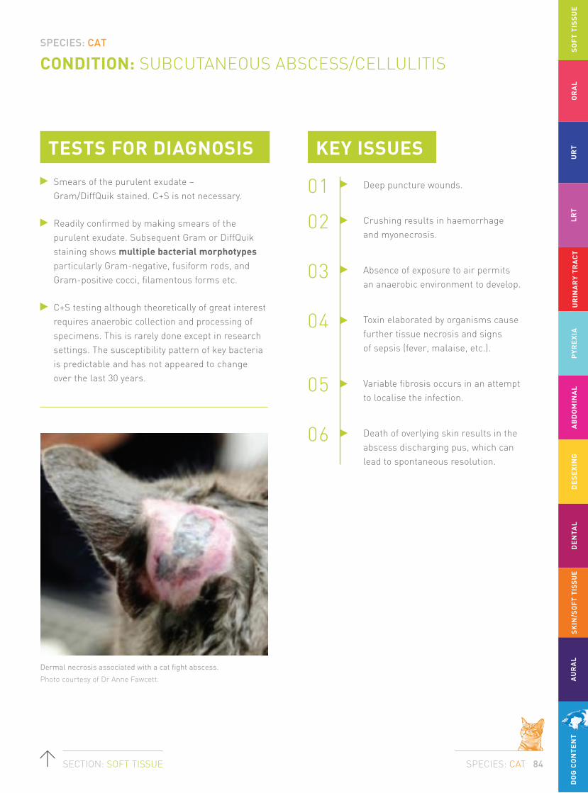
SO
FT
TIS
SU
EO
RA
LU
RT
LR
TU
RIN
AR
Y T
RA
CT
PY
RE
XIA
AB
DO
MIN
AL
DE
SE
XIN
GD
EN
TAL
SKIN
/SO
FT T
ISSU
EA
UR
AL
DO
G C
ON
TE
NT
SPECIES: CAT
SPECIES: CAT 84SECTION: SOFT TISSUE
CONDITION: SUBCUTANEOUS ABSCESS/CELLULITIS
Smears of the purulent exudate – Gram/DiffQuik stained. C+S is not necessary.
Readily confirmed by making smears of the purulent exudate. Subsequent Gram or DiffQuik staining shows multiple bacterial morphotypes particularly Gram-negative, fusiform rods, and Gram-positive cocci, filamentous forms etc.
C+S testing although theoretically of great interest requires anaerobic collection and processing of specimens. This is rarely done except in research settings. The susceptibility pattern of key bacteria is predictable and has not appeared to change over the last 30 years.
KEY ISSUES
01 Deep puncture wounds.
02 Crushing results in haemorrhage and myonecrosis.
03 Absence of exposure to air permits an anaerobic environment to develop.
04 Toxin elaborated by organisms cause further tissue necrosis and signs of sepsis (fever, malaise, etc.).
05 Variable fibrosis occurs in an attempt to localise the infection.
06 Death of overlying skin results in the abscess discharging pus, which can lead to spontaneous resolution.
Dermal necrosis associated with a cat fight abscess.
Photo courtesy of Dr Anne Fawcett.
TESTS FOR DIAGNOSIS

SO
FT
TIS
SU
EO
RA
LU
RT
LR
TU
RIN
AR
Y T
RA
CT
PY
RE
XIA
AB
DO
MIN
AL
DE
SE
XIN
GD
EN
TAL
SKIN
/SO
FT T
ISSU
EA
UR
AL
DO
G C
ON
TE
NT
SPECIES: CAT
SPECIES: CAT 85SECTION: SOFT TISSUE
CONDITION: SUBCUTANEOUS ABSCESS/CELLULITIS
TREATMENT
For an uncomplicated cat fight abscess, aeration of the wound, surgical debridement and drainage (sometimes using a latex drain) is recommended in combination with antimicrobial therapy.
IV (during anaesthesia) and/or subcutaneous fluid therapy to re-establish and maintain hydration.
On day 1, opioid analgesia may be indicated in selected cases.
The practitioner should be wary of using any NSAID in a potentially dehydrated cat, subjected to sepsis and general anaesthesia. These drugs are much more safely given from day 2 onwards, once the cat has resumed eating and drinking normally.
Based on the human literature where there is more of an evidence-base to draw upon, an injection of amoxicillin-clavulanate (or amoxicillin) will produce high and predictable blood levels, and is easily given under anaesthesia, and can be followed up by the same drug given orally.
Doxycycline monohydrate is often preferred because it has excellent anaerobic activity, is good at diffusing into marginally perfused tissues, available in tablets and paste which are both easy to administer (which greatly facilitates compliance); the anti-inflammatory action of the drug is helpful also.
Cases that fail to respond to standard therapy should be sampled appropriately (from deep within the lesion) to permit cytology (DiffQuik), Gram staining, and C+S.
First line:
Amoxicillin-clavulanate (12.5 mg/kg q12h) or doxycycline monohydrate (5 mg/kg q12h†); clindamycin (5-11 mg/kg q12h) and metronidazole (10 mg/kg q12h) are also suitable, but perhaps less so than the recommended agents; some clinicians may combine amoxicillin-clavulanate and metronidazole or clindamycin for an especially severe or extensive anaerobic infection.
Second line:
Based on C+S.
Cefovecin is suitable for any case where there are concerns of compliance, or there are difficulties with oral dosing.
Note: Currently registered veterinary fluoroquinolones are completely INAPPROPRIATE in this setting as they have virtually no activity against anaerobes.
RECOMMENDED ANTIBIOTICS
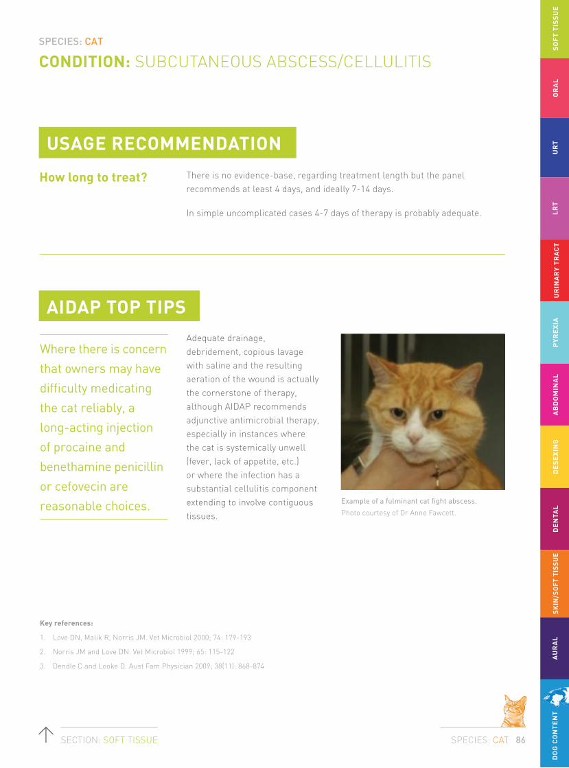
SO
FT
TIS
SU
EO
RA
LU
RT
LR
TU
RIN
AR
Y T
RA
CT
PY
RE
XIA
AB
DO
MIN
AL
DE
SE
XIN
GD
EN
TAL
SKIN
/SO
FT T
ISSU
EA
UR
AL
DO
G C
ON
TE
NT
SPECIES: CAT
SPECIES: CAT 86SECTION: SOFT TISSUE
CONDITION: SUBCUTANEOUS ABSCESS/CELLULITIS
How long to treat?
AIDAP TOP TIPSAdequate drainage, debridement, copious lavage with saline and the resulting aeration of the wound is actually the cornerstone of therapy, although AIDAP recommends adjunctive antimicrobial therapy, especially in instances where the cat is systemically unwell (fever, lack of appetite, etc.) or where the infection has a substantial cellulitis component extending to involve contiguous tissues.
Example of a fulminant cat fight abscess.
Photo courtesy of Dr Anne Fawcett.
USAGE RECOMMENDATION
There is no evidence-base, regarding treatment length but the panel recommends at least 4 days, and ideally 7-14 days.
In simple uncomplicated cases 4-7 days of therapy is probably adequate.
Where there is concern
that owners may have
difficulty medicating
the cat reliably, a
long-acting injection
of procaine and
benethamine penicillin
or cefovecin are
reasonable choices.
Key references:
1. Love DN, Malik R, Norris JM. Vet Microbiol 2000; 74: 179-193
2. Norris JM and Love DN. Vet Microbiol 1999; 65: 115-122
3. Dendle C and Looke D. Aust Fam Physician 2009; 38(11): 868-874
SPECIES: CAT
SPECIES: CAT 87SECTION: ORAL
CONDITION: CHRONIC GINGIVOSTOMATITIS/FAUCITIS
SO
FT
TIS
SU
EO
RA
LU
RT
LR
TU
RIN
AR
Y T
RA
CT
PY
RE
XIA
AB
DO
MIN
AL
DE
SE
XIN
GD
EN
TAL
SKIN
/SO
FT T
ISSU
EA
UR
AL
DO
G C
ON
TE
NT
It is thought that this occurs as a result of feeding soft food or highly refined carbohydrate (readily fermentable by gingival anaerobes), without the natural flossing action of stripping meat and sinew from prey tissues. This leads to a shift in the host: bacteria relationship with overgrowth of pathogenic organisms specifically Porphyromonas spp. closely related to the human periodontal disease pathogen Porphyromonas gingivalis. At some level, this is a ‘physiological’ disease condition, in which dietary factors, host factors and microbial factors interplay in a complex manner. The bacteria involved are normal constituents of the microbial flora of the gingival cleft, although they can behave as true pathogens in this scenario. The amount of inflammation in the gum is usually commensurate with the extent of the build-up of plaque and tartar, although irreversible changes in local anatomy (gingival recession with exposure of dentine, odontoclastic resorptive lesions) contribute. Abnormal dentition, e.g. crowding and misalignment of teeth in brachycephalic cats,
can also contribute to disease occurrence and progression. Critically, there is no concurrent inflammation of the palatoglossal arches (‘fauces’) or contiguous pharyngeal or palatine mucosa.
Chronic gingivostomatitis/faucitis signifies a different syndrome in which the amount of inflammation in the gums and contiguous tissues is vastly disproportionate to the extent of plaque and tartar build up. Indeed, the disease can occur in young cats with hardly any ‘traditional’ periodontal disease, and this condition is often termed juvenile gingivitis. This syndrome is a manifestation of chronic low grade calicivirus infection. Because of (perhaps) the specific strain of FCV, the immune status of the cat, or (most likely) a combination of the two – some cats can develop a latent infection associated with chronic shedding of virus, in the presence of a florid antibody-mediated immune response that causes pain, erythema and dysfunction, but fails to clear virus from the gums possibly due to insufficient cell mediated immunity.
Periodontal disease can occur as a discrete entity, in the older cat, in association with a build-up of plaque (a biofilm of obligate anaerobes and salivary mucoproteins), tartar (mineralised plaque) and gingival recession.
BACKGROUND/NATURE OF INFECTION/
ORGANISMS INVOLVED

SPECIES: CAT
SPECIES: CAT 88SECTION: ORAL
CONDITION: CHRONIC GINGIVOSTOMATITIS/FAUCITIS
SO
FT
TIS
SU
EO
RA
LU
RT
LR
TU
RIN
AR
Y T
RA
CT
PY
RE
XIA
AB
DO
MIN
AL
DE
SE
XIN
GD
EN
TAL
SKIN
/SO
FT T
ISSU
EA
UR
AL
DO
G C
ON
TE
NT
1. Examination of the oral cavity under general anaesthesia is very helpful, with probing of the periodontal pockets.
2. Dental radiography can be very helpful.
3. Biopsy of gums is of limited value, although immunohistology (available through Assoc Prof. Jacqui Norris, The University of Sydney) can confirm the presence of FCV antigen.
KEY ISSUES
01 Chronic gingivostomatitis can be due to severe periodontal disease.
02 More often it is due to chronic, persistence of FCV in the gums, palatoglossal arches and adjacent mucosa.
03 There is abundant antibody mediated inflammation directed at FCV deep within the host tissues, but insufficient or ineffective cellular immunity to eliminate the chronic persistent carrier state.
Severe gingivostomatitis and faucitis.
Photos courtesy of Dr Richard Malik.
TESTS FOR DIAGNOSIS
SPECIES: CAT
SPECIES: CAT 89SECTION: ORAL
CONDITION: CHRONIC GINGIVOSTOMATITIS/FAUCITIS
SO
FT
TIS
SU
EO
RA
LU
RT
LR
TU
RIN
AR
Y T
RA
CT
PY
RE
XIA
AB
DO
MIN
AL
DE
SE
XIN
GD
EN
TAL
SKIN
/SO
FT T
ISSU
EA
UR
AL
DO
G C
ON
TE
NT
TREATMENT1. Remove tartar and plaque, scale and polish
enamel and exposed dentine, remove unsalvageable teeth and administer (perhaps) a 2-week course of antimicrobials highly effective against obligate anaerobes involved in periodontal disease.
2. Further treatment options for affected cats are controversial. The following comments apply to cats with chronic FCV-associated disease:
i. The best evidence based medicine suggests ‘radical dentistry’ has the highest chance to affect a cure or substantial benefit. This involves removal of all the cat’s teeth or at least all of the cheek teeth. This somehow alters the host: virus relationship leading to eradication of the chronic calicivirus carrier state. Unfortunately, this is only successful in perhaps 50-70%
of cases (some are cured, some are greatly improved, some are somewhat improved).
ii. Other antiviral strategies are the use of omega interferon topically, intra-lesionally or subcutaneously; and topical antiviral agents such as lactoferrin.
iii. An alternate approach is to ‘give up’ trying to clear the virus, but instead dampen down the inflammatory response using immunomodulatory drugs including corticosteroids, cyclosporine, megestrol, thalidomide, chlorambucil and NSAIDs (meloxicam, Bonjella). In the USA, laser therapies of varying intensities are also commonly used, although the evidence-base for the success rate of these therapies is impossible to find.
Clindamycin, metronidazole (alone or with spiramycin) and doxycycline monohydrate (5 mg/kg q12h†) are preferred over amoxicillin-clavulanate because they have been shown to be clinically superior. This may be because these agents attain more effective levels within the biofilm in the vicinity of the periodontal ligament.
First line:
Doxycycline monohydrate (5 mg/kg q12h†), Clindamycin (5-11 mg/kg q12h) or Metronidazole (10 mg/kg q12h).
Second line:
Amoxicillin-clavulanate (12.5 mg/kg q12h).
ANTIBIOTICS USED

SPECIES: CAT
SPECIES: CAT 90SECTION: ORAL
CONDITION: CHRONIC GINGIVOSTOMATITIS/FAUCITIS
SO
FT
TIS
SU
EO
RA
LU
RT
LR
TU
RIN
AR
Y T
RA
CT
PY
RE
XIA
AB
DO
MIN
AL
DE
SE
XIN
GD
EN
TAL
SKIN
/SO
FT T
ISSU
EA
UR
AL
DO
G C
ON
TE
NT
AIDAP TOP TIPS
Chewing encourages a scissors action of the carnassials on fresh meat and ‘flossing’ by stripping muscle from periosteum and bone, reducing fermentable carbohydrates in the diet and using adjunctive products such as chews, brushing, chlorhexidine, T/D diets etc.
C+S testing is worthless in this disease. The bacteria involved are fastidious obligate anaerobes and it is very difficult to collect a meaningful specimen and transport it to the laboratory in a timely manner that would permit the causal organisms to be cultured. Susceptibility testing for anaerobes is not available routinely in any private or institutional vet laboratories in Australia currently. Other techniques e.g. immunohistology for FCV, in situ PCR and fluorescent in situ hybridisation (FISH) have not become routinely available to be useful in a general practice setting.
USAGE RECOMMENDATION
Typically for 1-2 weeks, but longer in certain circumstances. Ensure doxycycline given with a small bolus of water, or a small dab of margarine. Consider topical treatment with lactoferrin from a compounding pharmacist. Consider topical or intra-lesional interferon. Consider ‘radical dentistry’ with extraction of the cheek teeth in refractory cases.
Further therapy is
directed at changing
the diet to include
more chewing.
Key references:
1. Norris JM and Love DN. Aust Vet J 2000; 78(8): 533-537
2. Harley R, Gruffydd-Jones TJ, Day MJ. J Comp Pathol 2011; 144(4): 239-250
3. Hennet PR, Camy GA, McGahie DM, et al. J Feline Med Surg 2011; 13: 577-587
4. Southerden P and Gorrel C. J Small Anim Pract 2007 48(2): 104-6

SPECIES: CAT
SPECIES: CAT 91SECTION: URT
CONDITION: ACUTE URT DISEASE
SO
FT
TIS
SU
EO
RA
LU
RT
LR
TU
RIN
AR
Y T
RA
CT
PY
RE
XIA
AB
DO
MIN
AL
DE
SE
XIN
GD
EN
TAL
SKIN
/SO
FT T
ISSU
EA
UR
AL
DO
G C
ON
TE
NT
Dual infections also occur. In some instances Chlamydophila felis, Mycoplasma felis and possibly Bordetella bronchiseptica can also be primary pathogens, but all except the first three organisms tend to be ocular pathogens primarily, rather than naso-ocular pathogens. Although cryptococcal rhinitis can occur as a primary disease entity, it is very unusual for this to be detected until the disease has entered a more chronic stage. Primary viral disease is frequently complicated by secondary bacterial involvement, and typically it is the normal anaerobic flora of the sino-nasal cavity that is involved although mycoplasmas, Pasteurella and Bordetella can also be significant pathogens.
Acute URT disease in cats is in the great majority of instances a primary viral disease, with FHV-1 the most important primary pathogen, and FCV implicated in a smaller proportion of cases.
BACKGROUND/NATURE OF INFECTION/
ORGANISMS INVOLVED
SPECIES: CAT
SPECIES: CAT 92SECTION: URT
CONDITION: ACUTE URT DISEASE
SO
FT
TIS
SU
EO
RA
LU
RT
LR
TU
RIN
AR
Y T
RA
CT
PY
RE
XIA
AB
DO
MIN
AL
DE
SE
XIN
GD
EN
TAL
SKIN
/SO
FT T
ISSU
EA
UR
AL
DO
G C
ON
TE
NT
1. Multiplex PCR panels for feline respiratory pathogens can be informative if taken carefully and early in the course of the disease, although they are expensive.
2. C+S of nasal discharge is completely unhelpful in most instances, as the material that appears in the nares is not representative of the deeper underlying infective process and is contaminated by commensal microbiota. Deep nasal flushes are not commonly performed in cases with acute disease, and most labs do not handle specimens in such a way that fastidious organisms such as obligate anaerobes and mycoplasmas can be detected. Isolating Bordetella bronchiseptica in pure culture may implicate its role as a primary pathogen or secondary invader, and permits susceptibility testing.
3. In the exceedingly rare case of acute cryptococcosis, budding capsulated yeasts are easy to see on cytology and can be cultured on appropriate media e.g. bird seed agar (plus antibiotics). However, Cryptococcus is rarely cultured from the nasal cavities of normal cats (but you do not see them cytologically – it is likely that it is inhaled spores that germinate).
4. Radiography, endoscopy or advanced cross sectional imaging (CT or MRI) has usually little to offer in acute disease unless a foreign body is suspected (peracute onset of sneezing and nasal discomfort, possibly with stertor).
KEY ISSUES
01 Most URT disease in cats is viral.
02 FHV-1 (particularly) and FCV are the most common causes.
03 Secondary bacterial involvement is common, with mixed anaerobes, Mycoplasma felis and Bordetella bronchiseptica all being potential pathogens.
04 Acute disease may represent recurrence of latent FHV-1 disease, rather than primary infection. This is common in cats that are stressed (e.g. boarding) or given corticosteroids.
TESTS FOR DIAGNOSIS
SPECIES: CAT
SPECIES: CAT 93SECTION: URT
CONDITION: ACUTE URT DISEASE
SO
FT
TIS
SU
EO
RA
LU
RT
LR
TU
RIN
AR
Y T
RA
CT
PY
RE
XIA
AB
DO
MIN
AL
DE
SE
XIN
GD
EN
TAL
SKIN
/SO
FT T
ISSU
EA
UR
AL
DO
G C
ON
TE
NT
TREATMENT 1. The treatment of acute viral URT disease should
be based on the fact that most cases are due to FHV-1/FCV, with variable secondary invaders.
2. It should also be remembered that the cost of a diagnostic PCR panel may exceed the cost of definitive therapy. So the cornerstone of therapy should be appropriate doses of famciclovir, the only safe and effective anti-herpesvirus agent available in feline practice.
3. To treat the other potential naso-ocular pathogens (primary and secondary), such as Bordetella, Pasteurella, Chlamydia and obligate anaerobes, an antibiotic should be given concurrently in most cases. Doxycycline monohydrate and clindamycin probably are the most cogent choices. Although many papers have been written on this subject, most are low down on the ‘evidence based pyramid’, for a variety of reasons.
4. Additional measures can be very helpful, such as subcutaneous fluid therapy, nutritional support, nebulisation with saline etc. Omega-interferon may be helpful for acute FCV infections.
5. In the shelter or pet shop setting, timely administration of famciclovir early in the disease course can have dramatic effects. Anecdotal reports from CPS/RSPCA and shelter veterinarians support the successful use of famciclovir in acute feline URT disease. Recent work, utilising experimental infections provides a solid evidence base for these recommendations, although more studies under ‘field conditions’ would be helpful.2
First line:
Famciclovir (30-40 mg/kg q8-12h‡) plus Doxycycline monohydrate (5 mg/kg q12h†).
Second line:
Clindamycin (5-11 mg/kg q12h).
ANTIMICROBIALS USED
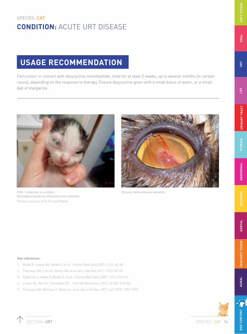
SPECIES: CAT
SPECIES: CAT 94SECTION: URT
CONDITION: ACUTE URT DISEASE
SO
FT
TIS
SU
EO
RA
LU
RT
LR
TU
RIN
AR
Y T
RA
CT
PY
RE
XIA
AB
DO
MIN
AL
DE
SE
XIN
GD
EN
TAL
SKIN
/SO
FT T
ISSU
EA
UR
AL
DO
G C
ON
TE
NT
FHV-1 infection in a kitten. Secondary bacterial infections are common.
Photos courtesy of Dr Richard Malik.
Chronic feline herpes keratitis.
USAGE RECOMMENDATION
Famciclovir in concert with doxycycline monohydrate; treat for at least 2 weeks, up to several months [in certain cases], depending on the response to therapy. Ensure doxycycline given with a small bolus of water, or a small dab of margarine.
Key references:
1. Malik R, Lessel NS, Webb S, et al. J Feline Med Surg 2009; 11(1): 40-48
2. Thomasy SM, Lim CC, Reilly CM, et al. Am J Vet Res 2011; 72(1): 85-95
3. Egberink H, Addie D, Belák S, et al. J Feline Med Surg 2009; 11(7): 610-614
4. Litster AL, Wu CC, Constable PD. J Am Vet Med Assoc 2012; 241(2): 218-226
5. Thomasy SM, Whittem T, Bales JL, et al. Am J Vet Res. 2012 Jul; 73(7): 1092-1099

SPECIES: CAT
SPECIES: CAT 95SECTION: URT
CONDITION: CHRONIC RHINOSINUSITIS
SO
FT
TIS
SU
EO
RA
LU
RT
LR
TU
RIN
AR
Y T
RA
CT
PY
RE
XIA
AB
DO
MIN
AL
DE
SE
XIN
GD
EN
TAL
SKIN
/SO
FT T
ISSU
EA
UR
AL
DO
G C
ON
TE
NT
It is seen commonly in general practice, although perhaps less commonly than in the 1970s and 1980s. Although there is no universal agreement, many authorities consider this to be sequel of viral rhinitis, and in the vast majority of cases it is thought to be caused by FHV-1. Most cases occur in younger cats, however occasionally it is seen as a first presentation in older cats.
FHV-1 is an epitheliotropic virus, causing necrosis, neutrophilic and sometimes eosinophilic inflammation. This results in destruction of normal turbinate architecture, with loss of bone and cartilage. Subsequent failure of normal drainage pathways can lead to abnormal accumulation of mucus and inflammatory exudate. Anatomic pockets not cleared by normal mucociliary clearance mechanisms develop and there can be scarring and web formation within the nasal cavity and also in the nasophaynyx (nasopharyngeal
stenosis). Distortion of normal anatomy interferes with defence against commensal bacteria, including mycoplasmas, obligate anaerobes, Pasteurella spp., Bordetella and sometimes other infectious agents including fungi (Cryptococcus spp. complex, Aspergillus spp., Neosartorya spp.) and also filamentous bacteria (Actinomyces, Streptomyces).
This syndrome is hard to diagnose definitively. Indeed, it is essentially diagnosed by exclusion, in the presence of supportive historical, physical and imaging findings. Perhaps in the future immunohistology or fluorescent in situ hybridisation (FISH) will provide more definitive evidence of the underlying patho-mechanisms. Despite its commonness, there is no universally acceptable approach to the management of these cases, and various different authorities recommend different approaches.
Chronic rhinosinusitis syndrome is known by a variety of different colloquial terms – ‘chronic snuffler syndrome’, ‘chronic snorter syndrome’, ‘chronic post-viral rhinitis, ‘lymphocytic plasmacytic rhinosinusitis’ etc.
BACKGROUND/NATURE OF INFECTION/
ORGANISMS INVOLVED

SPECIES: CAT
SPECIES: CAT 96SECTION: URT
CONDITION: CHRONIC RHINOSINUSITIS
SO
FT
TIS
SU
EO
RA
LU
RT
LR
TU
RIN
AR
Y T
RA
CT
PY
RE
XIA
AB
DO
MIN
AL
DE
SE
XIN
GD
EN
TAL
SKIN
/SO
FT T
ISSU
EA
UR
AL
DO
G C
ON
TE
NT
Full investigation of these cases requires cross section imaging, anterior and posterior rhinoscopy, pathological studies from nasal washing and biopsy material (for cytology, routine culture, fungal culture, FISH, PCR and immunohistochemical studies). Such investigations are beyond the financial limitation of many owners, and so empiric therapy after limited investigations is commonly done in practice. It is prudent to rule out cryptococcosis and invasive diseases that cause bone lysis, but even this is not always possible. In the older cat, the possibility of neoplasia should be explored as the most common nasal malignancy is lymphoma, and this often responds to sequential multi-agent chemotherapy.
KEY ISSUES
01 Most cats with chronic rhinosinusitis develop it as a consequence of long standing FHV-1 infection with distortion of local anatomy and secondary bacterial invasion.
02 Most chronic snuffler cats have negative or non-specific findings on radiography, CT, biopsy and culture.
03 It is important to rule out some specific aetiologies such as cryptococcosis, filamentous fungal infections and neoplasia. This is best achieved by biopsy of representative material obtained using cupped alligator forceps, ideally guided by imaging findings.
TESTS FOR DIAGNOSIS
Bilateral nasal discharge in a cat with a nasopharyngeal polyp – a differential diagnosis for chronic rhinonsinusitis in cats.
Photo courtesy of Dr Vanessa Barrs.

SPECIES: CAT
SPECIES: CAT 97SECTION: URT
CONDITION: CHRONIC RHINOSINUSITIS
SO
FT
TIS
SU
EO
RA
LU
RT
LR
TU
RIN
AR
Y T
RA
CT
PY
RE
XIA
AB
DO
MIN
AL
DE
SE
XIN
GD
EN
TAL
SKIN
/SO
FT T
ISSU
EA
UR
AL
DO
G C
ON
TE
NT
TREATMENT
Antimicrobial therapy is considered a critical component of therapy to control secondary opportunistic infection. Some clinicians recommend surgical interventions. Some clinicians utilise judicious use of NSAIDs such as meloxicam, while others sometimes utilise other strategies such as nebulisation, instillation of saline nose drops, and even low dose corticosteroid therapy.
There is anecdotal evidence that use of famciclovir is helpful as an adjunct to antibacterial therapy in a subset of cases, especially where bony erosion within or adjacent to the nasal cavity is present, or where FHV-1 associated ocular disease or dermatitis is present concurrently.
Most commonly recommended agents are doxycycline, and clindamycin. Anti-Pseudomonas drugs (such as enrofloxacin, marbofloxacin and pradofloxacin) are usually reserved for cases where there is a pure heavy growth of P. aeruginosa from deep nasal washing or nasal biopsy specimens.
First line:
Doxycycline monohydrate (5 mg/kg q12h†).
Second line:
Clindamycin (11 mg/kg q12h).
Consider using concurrent anti-herpes therapy with Famciclovir (30-40 mg/kg q8-12h‡) if cost permits, or if there is definitive evidence of the involvement of FHV-1.
ANTIBIOTICS USED

SPECIES: CAT
SPECIES: CAT 98SECTION: URT
CONDITION: CHRONIC RHINOSINUSITIS
SO
FT
TIS
SU
EO
RA
LU
RT
LR
TU
RIN
AR
Y T
RA
CT
PY
RE
XIA
AB
DO
MIN
AL
DE
SE
XIN
GD
EN
TAL
SKIN
/SO
FT T
ISSU
EA
UR
AL
DO
G C
ON
TE
NT
AIDAP TOP TIPS
USAGE RECOMMENDATION
The nature of the underlying pathological process is deep-seated neutrophilic and lymphoplasmacytic inflammation, with bone and cartilage atrophy and necrosis – i.e. bacterial chondritis and osteomyelitis. In cases where there is initially a favourable response to therapy, it is imperative that treatment be run out for many months, typically 2-3 months past clinical remission. Ensure doxycycline given with a small bolus of water, or a small dab of margarine.
Key references:
1. Quimby J and Lappin M. Compend Contin Educ Vet 2010; 32(1): E1-E10
2. Quimby J and Lappin M. Compend Contin Educ Vet 2009; 31(12): 554-564
3. Reed N and Gunn-Moore D. J Feline Med Surg 2012; 14(5): 317-326
4. Malik R, Lessels NS, Webb S, et al. J Feline Med Surg 2009; 11(1): 40-48
5. Kuehn NF. Clin Tech Small Anim Pract 2006; 21(2): 69-75
6. Johnson LR, Foley JE, De Cock HE, et al. J Am Vet Med Assoc 2005; 227(4): 579-585
Deep nasal washings or biopsy specimens procured with small cutting alligator forceps (or endoscopy biopsy forceps) are more useful for cytology and culture.
Nasal discharge is
an inferior sample,
although it can be
useful for diagnosing
cryptococcosis.

SPECIES: CAT
SPECIES: CAT 99SECTION: LRT
CONDITION: ACUTE LRT INFECTION
SO
FT
TIS
SU
EO
RA
LU
RT
LR
TU
RIN
AR
Y T
RA
CT
PY
RE
XIA
AB
DO
MIN
AL
DE
SE
XIN
GD
EN
TAL
SKIN
/SO
FT T
ISSU
EA
UR
AL
DO
G C
ON
TE
NT
Pasteurella spp. and obligate anaerobes are commensals of the oropharynx and aspiration of these bacteria is the most likely route of infection, for example after viral URT infection or other disease resulting in impairment of mucociliary clearance. Infection with these bacteria often results in pleuropneumonia and pyothorax (see pyothorax). Bordetella bronchiseptica is a primary respiratory pathogen that can cause acute or chronic LRT disease. Risk factors for infection include high population density (eg catteries or shelters), poor hygiene, young age/immunosuppression and exposure to dogs with respiratory disease. B. bronchiseptica is a rare zoonosis of humans, particularly if immunosuppressed. Clinical disease is most likely in kittens less than 10 weeks of age and signs range from sneezing, ocular discharge and mild cough to severe dyspnoea and respiratory distress, which can be fatal.
Mycoplasmas are also an important cause of LRT infection in cats. Whether they act as primary or secondary pathogens in this location is less clear.
Some investigators consider that mycoplasmas can only colonise inflamed/infected lower airways (e.g. secondary to chronic bronchitis/asthma) while others consider they can act as primary LRT pathogens. Mycoplasmas (M. felis, M. gateae, M. feliminutum) have been detected by culture or PCR in 15 to 22% of cats with LRT disease in the USA, UK and Australia. (Foster et al 2004, Randolph et al 1993, Reed et al 2012). They have been reported to cause bronchopneumonia, focal pulmonary abscessation and pyothorax spontaneously in owned cats and pneumonia after experimental inoculation in kittens. Evidence is mounting for a pathogenic role of mycoplasmas (M. pneumoniae) in triggering acute exacerbations in humans with chronic asthma.
Non-bacterial LRT pathogens of cats include lungworm, heartworm, toxoplasmosis, viral infection and fungi. These aetiologies should be considered in the diagnostic investigation of cats with acute LRT signs.
The most common bacteria to cause lower respiratory tract (LRT) infections in cats are B. bronchiseptica, Pasteurella spp., Mycoplasma spp., Streptococcus spp. and E. coli. Other less common bacterial pathogens include mycobacteria, Salmonella typhimurium, Pseudomonas spp. and obligate anaerobes.
BACKGROUND/NATURE OF INFECTION/
ORGANISMS INVOLVED

SPECIES: CAT
SPECIES: CAT 100SECTION: LRT
CONDITION: ACUTE LRT INFECTION
SO
FT
TIS
SU
EO
RA
LU
RT
LR
TU
RIN
AR
Y T
RA
CT
PY
RE
XIA
AB
DO
MIN
AL
DE
SE
XIN
GD
EN
TAL
SKIN
/SO
FT T
ISSU
EA
UR
AL
DO
G C
ON
TE
NT
1. Careful clinical examination to rule out non-infectious respiratory disease.
2. Thoracic imaging (radiography first, then possibly CT where available).
3. BAL samples for cytology and C+S.
4. Bronchoscopy, where indicated.
5. PCR of BAL fluid for specific pathogens such as Mycoplasma spp. and Bordetella bronchiseptica in some settings. Bordetella bronchiseptica can also be detected by PCR of oropharyngeal swabs.
KEY ISSUES
01 Although infectious LRTI occurs in cats, allergic lower airway disease is more common, and at some level is a diagnosis of exclusion.
02 Mycoplasma species may cause primary LRTI, or occur as a complication of “feline asthma”.
03 Bordetella bronchiseptica is a primary respiratory pathogen and clinical disease is most common in kittens.
04 Because of the difficulty of determining whether bacterial involvement is primary or secondary in cases with feline bronchial disease, antimicrobials are often used as a component of therapy, even when the primary aetiology is thought to be allergic.
TESTS FOR DIAGNOSIS
DiffQuik-stained smear of bronchial lavage fluid from a cat with a Bordetella bronchiseptica lower respiratory tract infection. Clusters of rods are attached to ciliated respiratory epithelial cells.
Photo courtesy of Dr Patricia Martin.

SPECIES: CAT
SPECIES: CAT 101SECTION: LRT
CONDITION: ACUTE LRT INFECTION
SO
FT
TIS
SU
EO
RA
LU
RT
LR
TU
RIN
AR
Y T
RA
CT
PY
RE
XIA
AB
DO
MIN
AL
DE
SE
XIN
GD
EN
TAL
SKIN
/SO
FT T
ISSU
EA
UR
AL
DO
G C
ON
TE
NT
TREATMENT
Therapy should be based on antimicrobial susceptibility testing of bacteria cultured from BAL fluid or lung FNAs where available. Mycoplasma spp. are generally susceptible to doxycycline, macrolides and fluoroquinolones. B. bronchiseptica is usually susceptible to doxycycline and fluoroquinolones. Resistance has often been detected to ampicillin and trimethoprim. Amoxicillin-clavulanate is not recommended, even if susceptibility is documented, due to poor distribution into respiratory secretions. Early treatment of URT infections caused by B. bronchiseptica is recommended to prevent LRT involvement.
For empirical therapy of feline LRTI where the aetiological agent has not been cultured but is suspected to be bacterial, doxycycline is the recommended first-choice antimicrobial and has good efficacy for treatment of infections caused by B. bronchiseptica, mycoplasmas, Pasteurella spp. and anaerobes.
Acute life-threatening bronchopneumonia: Empiric therapy with ampicillin (20 mg/kg IV every 8 hours‡) and gentamicin (5-6 mg/kg IV once daily) while receiving IV fluid therapy, plus nebulisation with saline and thoracic percussion and coupage. Change agents on the basis of C+S from BAL specimens.
Chronic LRTI: Empiric treatment with doxycycline monohydrate (5 mg/kg q12h†). Change therapy on the basis of C+S data if available.
Re-assess drug choices in the light of the response to therapy.
Note: As mycoplasmas lack a peptidoglycan cell they are resistant to all β-lactam drugs.
ANTIBIOTICS USED

SPECIES: CAT
SPECIES: CAT 102SECTION: LRT
CONDITION: ACUTE LRT INFECTION
SO
FT
TIS
SU
EO
RA
LU
RT
LR
TU
RIN
AR
Y T
RA
CT
PY
RE
XIA
AB
DO
MIN
AL
DE
SE
XIN
GD
EN
TAL
SKIN
/SO
FT T
ISSU
EA
UR
AL
DO
G C
ON
TE
NT
AIDAP TOP TIPS
1. As well as systemic antimicrobials, nebulisation with oxygen and saline, combined with thoracic physiotherapy can be helpful in cats with severe LRTI.
2. Maintain a high index of suspicion for pulmonary toxoplasmosis in cats receiving combination therapy using prednisolone and either cyclosporine or cytotoxic drug therapy.
USAGE RECOMMENDATION
Minimum treatment durations for effective treatment of LRTI caused by mycoplasmas or B. bronchiseptica have not been established, but extrapolating from URT infections and because of virulence factors of B. bronchiseptica which favour persistence six weeks therapy is recommended. For other bacterial LRT infections three weeks may be adequate. Ensure doxycycline given with a small bolus of water, or a small dab of margarine.
Key references:
1. Foster SF and Martin P. J Feline Med Surg 2011; 13(5): 313-332
2. Bart M, Guscetti F, Zurbriggen A, et al. Vet J 2000; 159(3): 220-230
3. Macdonald ES and Norris CR. J Am Vet Med Assoc 2003; 223(8): 1142-1150
4. Foster SF, Martin P, Allan GS, et al. J Feline Med Surg 2004; 6(3): 167-180
5. Barrs VR, Martin PM and Beatty JA. Aust Vet J 2006; 84: 30-35

SPECIES: CAT
SPECIES: CAT 103SECTION: LRT
CONDITION: PYOTHORAX
SO
FT
TIS
SU
EO
RA
LU
RT
LR
TU
RIN
AR
Y T
RA
CT
PY
RE
XIA
AB
DO
MIN
AL
DE
SE
XIN
GD
EN
TAL
SKIN
/SO
FT T
ISSU
EA
UR
AL
DO
G C
ON
TE
NT
Isolates include Bacteroidaceae (Bacteroides spp., Porphyromonas spp., Prevotella spp.), Fusobacterium spp., Peptostreptococcus spp., Actinomyces spp., Eubacterium spp., Propionibacterium spp., Filifactor villosus, Pasteurella multocida, Streptococcus spp. and Mycoplasma spp.
Less than 20% of cases of feline pyothorax are caused by infectious agents other than oropharyngeal microbiota such as Staphylococcus spp., Rhodococcus equi, Nocardia spp., enteric Gram-negative organisms (E. coli, Salmonella spp., Klebsiella spp., Proteus spp.) non-enteric Gram-negative organisms (Pseudomonas spp.) and protozoa (Toxoplasma gondii).
Fungal causes of feline pyothorax are rare and include Cryptococcus spp. and Candida albicans. In contrast with dogs, infection with Enterobacteriaceae is uncommon, with E. coli being isolated in 0-7% of cases.
Pyothorax is mostly a disease of young cats (4-6 years), although cats of any age can be affected. There is no breed or gender predisposition.
Possible routes of infection include extension from an adjacent structure (bronchopneumonia, para-pneumonic spread, oesophageal rupture, mediastinitis or sub-phrenic infection), direct inoculation (penetrating trauma, migrating foreign body, thoracocentesis or thoracic surgery) or haematogenous or lymphatic spread from a distant site (systemic sepsis). Oropharyngeal microbiota could gain access to the pleural space by aspiration, direct penetration from a bite wound or by haematogenous spread from a distant wound. The evidence suggests that aspiration of oropharyngeal microbiota, subsequent colonisation of the LRT and direct extension of infection from the bronchi and lungs is the most common cause of feline pyothorax, as it is also in cases of human pyothorax and equine pleuropneumonia that also often involve obligate anaerobes. Viral URT infection can impair mucociliary clearance of respiratory secretions and predispose to accumulation of aspirated oropharyngeal secretions, resulting in colonisation of the LRT then pleuropneumonia.
Similar to cat bite abscesses, the majority of cases of pyothorax in cats are polymicrobial infections caused by obligate and facultative anaerobic bacteria derived from the feline oral cavity.
BACKGROUND/NATURE OF INFECTION/
ORGANISMS INVOLVED
SPECIES: CAT
SPECIES: CAT 104SECTION: LRT
CONDITION: PYOTHORAX
SO
FT
TIS
SU
EO
RA
LU
RT
LR
TU
RIN
AR
Y T
RA
CT
PY
RE
XIA
AB
DO
MIN
AL
DE
SE
XIN
GD
EN
TAL
SKIN
/SO
FT T
ISSU
EA
UR
AL
DO
G C
ON
TE
NT
1. Ultrasonography (U/S) of the chest, followed by thoracocentesis using a 23 gauge butterfly needle, ideally using U/S guidance. Note the odour of the discharge, which is usually foetid in anaerobic or mixed infections with an anaerobic component.
2. Where U/S guidance is not available, thoracocentesis can be performed safely at the ventral third of the 6th, 7th or 8th intercostal space with the cat standing or in ventral recumbency. Avoid the intercostal vessels and nerves located near the caudal rib margin.
3. Inoculate aerobic and anaerobic culture bottles at the cage side if anaerobic culture is contemplated. Do NOT permit air into the syringe after aspiration of purulent fluid from the pleural space.
4. If U/S is not available, judiciously take thoracic radiographs to confirm fluid is present in the pleural space and to identify a prudent site for attempted thoracocentesis. Examine pleural fluid specimen by making smears, stained with DiffQuik and Burke’s modification of the Gram stain (if available).
5. Submit purulent exudates collected whilst maintaining anaerobic conditions to the laboratory for C+S.
KEY ISSUES
01 Most cats with pyothorax have polymicrobial infections dominated by obligate anaerobes.
02 These infections typically begin in the lung after aspiration of orpharyngeal microbiota and spread secondarily to the pleura and pleural space. Thus, pyothorax is often a complication some weeks after acute viral URT infection in cats. Rarely, infection occurs after penetrating cat bite injuries to the thoracic wall.
03 Unlike dogs, grass seeds are rarely the cause of pyothorax in cats, although they can cause signs as tracheal foreign bodies.
TESTS FOR DIAGNOSIS
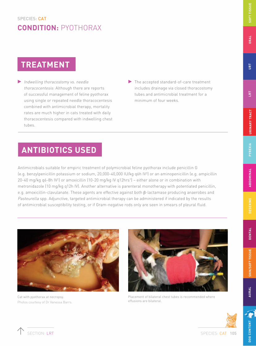
SPECIES: CAT
SPECIES: CAT 105SECTION: LRT
CONDITION: PYOTHORAX
SO
FT
TIS
SU
EO
RA
LU
RT
LR
TU
RIN
AR
Y T
RA
CT
PY
RE
XIA
AB
DO
MIN
AL
DE
SE
XIN
GD
EN
TAL
SKIN
/SO
FT T
ISSU
EA
UR
AL
DO
G C
ON
TE
NT
TREATMENT
Indwelling thoracostomy vs. needle thoracocentesis: Although there are reports of successful management of feline pyothorax using single or repeated needle thoracocentesis combined with antimicrobial therapy, mortality rates are much higher in cats treated with daily thoracocentesis compared with indwelling chest tubes.
The accepted standard-of-care treatment includes drainage via closed thoracostomy tubes and antimicrobial treatment for a minimum of four weeks.
Antimicrobials suitable for empiric treatment of polymicrobial feline pyothorax include penicillin G (e.g. benzylpenicillin potassium or sodium, 20,000-40,000 IU/kg q6h IV‡) or an aminopenicillin (e.g. ampicillin 20-40 mg/kg q6-8h IV‡) or amoxicillin (10-20 mg/kg IV q12hrs†) – either alone or in combination with metronidazole (10 mg/kg q12h IV). Another alternative is parenteral monotherapy with potentiated penicillin, e.g. amoxicillin-clavulanate. These agents are effective against both β-lactamase producing anaerobes and Pasteurella spp. Adjunctive, targeted antimicrobial therapy can be administered if indicated by the results of antimicrobial susceptibility testing, or if Gram-negative rods only are seen in smears of pleural fluid.
ANTIBIOTICS USED
Cat with pyothorax at necropsy.
Photos courtesy of Dr Vanessa Barrs.
Placement of bilateral chest tubes is recommended where effusions are bilateral.
SPECIES: CAT
SPECIES: CAT 106SECTION: LRT
CONDITION: PYOTHORAX
SO
FT
TIS
SU
EO
RA
LU
RT
LR
TU
RIN
AR
Y T
RA
CT
PY
RE
XIA
AB
DO
MIN
AL
DE
SE
XIN
GD
EN
TAL
SKIN
/SO
FT T
ISSU
EA
UR
AL
DO
G C
ON
TE
NT
AIDAP TOP TIPS
1. Pyothorax in cats occurs as a sequel to URT infection, and is due to spread of aspirated bacteria from the lung to the pleural space.
2. Although cats may present acutely, they have actually been sick for quite some time.
3. Ultrasound scans for pleural effusion is often the most cost effective test in cats with dyspnoea and cats with fever of unknown origin.
4. Of all the causes of pleural effusion in the cat, bacterial pyothorax has the best long-term prognosis.
5. The presence of volatile fatty acids and other fermentation products in the expired air of affected cats cause halitosis, which can be a tip off to the presence of pyothorax if the cats have good oral hygiene.
USAGE RECOMMENDATION
Successful treatment of feline pyothorax requires pleural drainage and lavage plus antimicrobial therapy.
Indwelling thoracostomy tubes (chest tubes) are the gold-standard for pleural space drainage, and bilateral tubes should be placed where effusions are bilateral.
Before anaesthesia for chest tube placement, the pleural effusion should be drained as completely as possible using needle thoracocentesis.
Treatment of anaerobic infections associated with devitalised tissue requires high doses of antimicrobials administered for extended periods. There may be risk of relapse if therapy is discontinued prematurely. For ongoing treatment once clinical improvement is seen and the patient is eating well, oral antibiotics may be substituted for IV agents. Antimicrobial therapy for treatment of feline pyothorax should be administered for 4-6 weeks.
Key references:
1. Love DN, Vekselstein R, Collings S. Vet Microbiol 1990; 22(2–3): 267-275
2. Love DN, Johnson JL, Moore LV. Vet Microbiol 1989; 19(3): 275-281
3. Love DN, Jones RF, Bailey M, et al. Vet Microbiol 1982; 7(5): 455-461
4. Barrs VR and Beatty JA. Vet J 2009; 179: 163-170
5. Barrs VR and Beatty JA. Vet J 2009; 179: 171-178
6. Walker AL, Jang SS, Hirsh DC. J Am Vet Med Assoc 2000; 216(3): 359-363
7. Waddell LS, Brady CA, Drobatz KJ. J Am Vet Med Assoc 2002; 221(6): 819-824
8. Barrs VR, Allan GS, Martin P, et al. J Feline Med Surg 2005; 7(4): 211-222
9. Demetriou JL, Foale RD, Ladlow J, et al. J Small Anim Pract 2002; 43(9): 388-394

SPECIES: CAT
SPECIES: CAT 107SECTION: URINARY TRACT
CONDITION: ACUTE LOWER UTI/CYSTITIS (FIRST OCCURRENCE)
SO
FT
TIS
SU
EO
RA
LU
RT
LR
TU
RIN
AR
Y T
RA
CT
PY
RE
XIA
AB
DO
MIN
AL
DE
SE
XIN
GD
EN
TAL
SKIN
/SO
FT T
ISSU
EA
UR
AL
DO
G C
ON
TE
NT
Although presentation for lower urinary tract (LUT) signs including dysuria, haematuria, stranguria, pollakiuria and periuria (inappropriate urination) is common in feline practice, the prevalence of bacterial UTI overall in cats with LUT signs is less than 3%, though the prevalence may be higher in older cats. The major differential diagnosis for cats presenting with LUT signs, in up to 55% of cats, is idiopathic cystitis (also known as interstitial cystitis or feline urological syndrome). Urethral obstruction in male cats (mostly due to struvite plugs or calcium oxalate uroliths) is the next most common cause of LUT signs while less common causes include urolithiasis, anxiety disorder (where inappropriate urination is the presenting sign) and neoplasia. Age (≥ 10 years) is a risk factor for UTI in cats which may be related to lower urine osmolarity in older cats with renal disease.
E. coli is the most common Gram-negative bacterium and Enterococcus spp. and S. felis are the most common Gram-positive bacteria to cause UTI in Australian cats. This was demonstrated in a study of LUT infections in Australian cats presenting with LUT signs, where infections were due to E. coli (37%), Enterococcus spp. (30%), Staphylococcus felis (20%), Proteus spp. (5%), Enterobacter/Klebsiella spp. (3%), Pseudomonas aeruginosa (<2%), S. aureus (<2%) and S. pseudintermedius (<2%). A total of 94% of E. coli isolates in that study were susceptible to amoxicillin-clavulanate, as were all Enterococcus and S. felis isolates. Amoxicillin has been suggested as an appropriate first-choice empiric antimicrobial for uncomplicated UTI in cats by ISCAID. While all Enterococcus spp. are likely to be susceptible to amoxicillin, 12% of S. felis and 32% of E. coli in an Australian study were resistant.
First occurrence UTI in an otherwise healthy cat with normal urinary tract anatomy and function is defined as an uncomplicated UTI.
BACKGROUND/NATURE OF INFECTION/
ORGANISMS INVOLVED

SPECIES: CAT
SPECIES: CAT 108SECTION: URINARY TRACT
CONDITION: ACUTE LOWER UTI/CYSTITIS (FIRST OCCURRENCE)
SO
FT
TIS
SU
EO
RA
LU
RT
LR
TU
RIN
AR
Y T
RA
CT
PY
RE
XIA
AB
DO
MIN
AL
DE
SE
XIN
GD
EN
TAL
SKIN
/SO
FT T
ISSU
EA
UR
AL
DO
G C
ON
TE
NT
Also, given that three times daily dosing is recommended for amoxicillin, compliance is likely to be a key issue for cat-owners. Trimethoprim-sulfonamide has also been recommended for first-line therapy of uncomplicated UTIs by the International Society for Companion Animal Infectious Diseases (ISCAID) – 90% of Australian feline isolates of E. coli and all Enterococcus spp. and S. felis were susceptible to this agent. However, some formulations of trimethoprim-sulfonamide such as coated tablets can induce profound salivation in cats if the exterior coating is broken so may not be appropriate oral therapy unless available in capsules.
For cats that cannot be orally medicated with amoxicillin-clavulanate, empirical treatment with doxycycline may be an alternative as 80% of E. coli isolates from Australia were susceptible to this agent and it reaches appropriate concentrations in urine. It should be noted, however that some less prevalent Gram-negative pathogens (i.e. Proteus spp.) are intrinsically resistant to doxycycline.

SPECIES: CAT
SPECIES: CAT 109SECTION: URINARY TRACT
CONDITION: ACUTE LOWER UTI/CYSTITIS (FIRST OCCURRENCE)
SO
FT
TIS
SU
EO
RA
LU
RT
LR
TU
RIN
AR
Y T
RA
CT
PY
RE
XIA
AB
DO
MIN
AL
DE
SE
XIN
GD
EN
TAL
SKIN
/SO
FT T
ISSU
EA
UR
AL
DO
G C
ON
TE
NT
1. A full urinalysis is recommended for all cats presenting with LUT signs, using a urine sample collected by cystocentesis.
2. Diagnosis of an uncomplicated UTI should be made on the basis of presence of LUT signs with supporting evidence of UTI (epithelial cells, RBC, WBC and bacteria) on a full urinalysis including examination of Gram or DiffQuik stained urine sediment. It is recommended that urine collected for urinalysis also be submitted for C+S testing since false positive diagnoses can be made on urine sediments, usually due to the presence of stain precipitates mimicking bacteria.
3. Free-catch urine samples are inferior and should generally be avoided.
4. Where subclinical bacteriuria is identified (positive urine culture in the absence of clinical and cytological evidence of UTI) treatment is generally not recommended in otherwise healthy patients with normal urinary tract anatomy and function.
KEY ISSUES
01 The majority of acute first occurrence cystitis cases in cats are sterile and due to idiopathic cystitis, also known as feline interstitial cystitis.
02 UTIs are uncommon in young cats.
03 E. coli is the most common Gram-negative bacterium while Enterococcus spp., and Staphylococcus felis are the most common Gram-positive bacteria to cause UTIs in Australian cats.
04 Considering both antimicrobial susceptibility and compliance issues, amoxicillin-clavulanate and doxycycline are appropriate empiric choices for treating uncomplicated UTIs in cats.
05 GIT side-effects, primarily vomiting, occur in a small minority of cats after administration of amoxicillin-clavulanate, in which case a lower-dose or changing to another antimicrobial, such as doxycycline is recommended.
06 Collection of urine by cystocentesis for full urinalysis including sediment examination can be difficult on an out-patient basis in some cases due to LUT signs resulting in an empty bladder at first presentation. Consider admitting the cat, if required, to obtain a urine sample by cystocentesis.
07 For cats with urethral obstruction and indwelling urinary tract catheters, prophylactic antimicrobials should never be given during the period of catheterisation and culture of urine from urine-collection bags is contraindicated. Culture of urine-catheter tips after removal is not necessary. Post-catheter UTI should be diagnosed on the basis of presence of clinical signs and cytological evidence of infection using a urine-sample collected by cystocentesis.
TESTS FOR DIAGNOSIS
SPECIES: CAT
SPECIES: CAT 110SECTION: URINARY TRACT
CONDITION: ACUTE LOWER UTI/CYSTITIS (FIRST OCCURRENCE)
SO
FT
TIS
SU
EO
RA
LU
RT
LR
TU
RIN
AR
Y T
RA
CT
PY
RE
XIA
AB
DO
MIN
AL
DE
SE
XIN
GD
EN
TAL
SKIN
/SO
FT T
ISSU
EA
UR
AL
DO
G C
ON
TE
NT
TREATMENT
1. Empiric antimicrobial therapy is appropriate while waiting for urine culture results since LUT signs cause pain and discomfort.
2. Recommended first-line choice for empiric therapy in cats include amoxicillin-clavulanate at 12.5 mg/kg q12h PO (using the higher dose will achieve a concentration in urine that is likely to exceed the MIC for most pathogens). Doxycycline 5 mg/kg q12h†, then 2.5 mg/kg q24h† can be considered as an alternative for non-compliant cats or cats that vomit on amoxicillin-clavulanate.
3. The recommended treatment duration is 7-14 days, although shorter treatment times may be effective.
First line:
Amoxicillin-clavulanate (12.5 mg/kg q12h) or doxycycline (5 mg/kg q12h†) are recommended for empiric treatment.
Cefovecin should only be considered where compliance is an issue, or there are difficulties with medicating orally.
Second line:
Consider a fluoroquinolone (marbofloxacin, 2.75-5.5 mg/kg q24h) on the basis of C+S if the bacteria are resistant to first line therapy or the infection is serious.
ANTIBIOTICS USED

SPECIES: CAT
SPECIES: CAT 111SECTION: URINARY TRACT
CONDITION: ACUTE LOWER UTI/CYSTITIS (FIRST OCCURRENCE)
SO
FT
TIS
SU
EO
RA
LU
RT
LR
TU
RIN
AR
Y T
RA
CT
PY
RE
XIA
AB
DO
MIN
AL
DE
SE
XIN
GD
EN
TAL
SKIN
/SO
FT T
ISSU
EA
UR
AL
DO
G C
ON
TE
NT
AIDAP TOP TIPS
Cystocentesis should be performed routinely to collect urine for urinalysis in cats presenting with LUT signs. It is easy to perform with the cat restrained in lateral recumbency, the bladder gently supported in the left hand while aspirating urine using a 23 gauge needle and syringe held in the right hand. Cystocentesis is generally safe in cats providing the bladder is not overly distended. It is also important not to squeeze the bladder during urine aspiration or transient uroabdomen can result.
Photo courtesy of Dr Vanessa Barrs.
USAGE RECOMMENDATION
Recommended duration of therapy is 7 to 14 days for uncomplicated UTIs. Ensure doxycycline given with a small bolus of water, or a small dab of margarine.
Consider bacterial UTI
in cats with underlying
risk factors (e.g. age,
renal impairment,
metabolic disease)
and perform a full
diagnostic workup
including C+S testing.
Key references:
1. Litster A, Thompson M, Moss S, et al. Vet J 2011; 187(1): 18-22
2. Litster AL, Moss SM, Honnery M, et al. Vet Microbiol 2007; 121: 182-188

SPECIES: CAT
SPECIES: CAT 112SECTION: URINARY TRACT
CONDITION: COMPLICATED UTIs: RECURRENT LOWER UTI /CYSTITIS AND CKD WITH PYURIA
SO
FT
TIS
SU
EO
RA
LU
RT
LR
TU
RIN
AR
Y T
RA
CT
PY
RE
XIA
AB
DO
MIN
AL
DE
SE
XIN
GD
EN
TAL
SKIN
/SO
FT T
ISSU
EA
UR
AL
DO
G C
ON
TE
NT
Complicated UTIs occur where there is an underlying anatomic or functional abnormality or where there is a concurrent disease that predisposes to UTI, for example CKD. Recurrent UTIs, occurring within six months after successful treatment of the first infection, can be re-infections caused by a different bacterial species to the original isolate or relapses caused by the same bacterial species as the original isolate.
The most common underlying anatomic abnormality associated with complicated UTIs in cats is perineal urethrostomy in previously obstructed male cats with approximately 1 in 5 cats developing recurrent UTIs.
The most common concurrent diseases associated with complicated UTIs in cats are CKD, hyperthyroidism and diabetes mellitus. In two retrospective studies of 224 and 614 cases, UTIs were identified in 17 and 22% of cats with CKD, in 12 and 22% of cats with hyperthyroidism and in 12 and 13% of cats with diabetes mellitus, respectively. The majority of cats with UTIs secondary to these diseases did not present with LUT signs. Risk factors for complicated UTIs in cats include female gender, increasing age and decreasing body weight. In some studies low urine specific gravity has also been identified as a risk factor for UTI caused by Gram-negative bacteria.
Recurrent UTIs are a subset of complicated UTIs.
BACKGROUND/NATURE OF INFECTION/
ORGANISMS INVOLVED

SPECIES: CAT
SPECIES: CAT 113SECTION: URINARY TRACT
CONDITION: COMPLICATED UTIs: RECURRENT LOWER UTI /CYSTITIS AND CKD WITH PYURIA
SO
FT
TIS
SU
EO
RA
LU
RT
LR
TU
RIN
AR
Y T
RA
CT
PY
RE
XIA
AB
DO
MIN
AL
DE
SE
XIN
GD
EN
TAL
SKIN
/SO
FT T
ISSU
EA
UR
AL
DO
G C
ON
TE
NT
1. To identify diseases that cause recurrent LUT signs in cats an appropriate minimum data base should include the following:
(i) a detailed history of previous antimicrobial therapy used including dose, duration of therapy and owner compliance.
(ii) a full urinalysis and C+S testing of a urine sample collected by cystocentesis.
(iii) haematology, serum biochemistry and a total T4.
(iv) abdominal imaging (sonography and radiography).
2. For recurrent UTI where concurrent disease or an underlying bladder abnormality cannot be detected after the investigations above consider referral for further investigation, e.g. cystoscopy.
3. Where subclinical bacteriuria is identified (positive urine culture in the absence of clinical and cytological evidence of UTI) treatment is based on the risk of ascending or systemic infection.
KEY ISSUES
01 UTIs are most common in older female cats. Females are predisposed to ascending infections from the gastrointestinal tract due to a relatively wide and short urethra compared to males.
02 Other risk factors for complicated UTIs in cats are concurrent diseases, most commonly CKD, hyperthyroidism and diabetes mellitus.
03 Causes of recurrent sterile cystitis including urolithiasis and bladder neoplasia need to be excluded in any diagnostic investigation.
04 Recurrent UTIs in young cats should arouse suspicion of an underlying abnormality or dysfunction of the LUT, e.g. ectopic ureter
05 In addition to identification and treatment of underlying predisposing causes of recurrent UTIs accurate identification of the underlying bacterial isolate(s), antimicrobial susceptibility testing and appropriate duration treatment are important for successful management of recurrent UTI.
06 Because MDR pathogens are becoming increasingly recognised globally, careful consideration must be given to selection of antimicrobials used for treatment of UTI and should be guided by C+S findings.
TESTS FOR DIAGNOSIS

SPECIES: CAT
SPECIES: CAT 114SECTION: URINARY TRACT
CONDITION: COMPLICATED UTIs: RECURRENT LOWER UTI /CYSTITIS AND CKD WITH PYURIA
SO
FT
TIS
SU
EO
RA
LU
RT
LR
TU
RIN
AR
Y T
RA
CT
PY
RE
XIA
AB
DO
MIN
AL
DE
SE
XIN
GD
EN
TAL
SKIN
/SO
FT T
ISSU
EA
UR
AL
DO
G C
ON
TE
NT
TREATMENT1. Empiric antimicrobial therapy is not
recommended unless clinical signs necessitate it. Recommended antimicrobials for empiric therapy are as for uncomplicated UTIs. Fluoroquinolones are also possible second line choices in cases of resistance to first line drugs. Organisms isolated from complicated UTI cases are more likely to be resistant to doxycycline. Where possible the drug class selected should be different from that used to treat the original UTI. If the bacterial isolate is resistant to the antimicrobial chosen for empiric therapy, treatment should be changed to an antimicrobial to which the isolate is susceptible and if possible is excreted in its active form primarily in urine. Consider complete blood counts during therapy to monitor for haematological side-effects.
2. In cats with asymptomatic bacteriuria and underlying disease (e.g. CKD) wait until C+S test results are available to initiate treatment using an appropriate antimicrobial.
3. For mixed infections consisting of Enterococcus spp. and another bacterial isolate infection by the former will often resolve when the other organism is successfully treated, though infections where Enterococcus is the sole pathogen may be refractory to treatments of short duration. Ideally a single antimicrobial or antimicrobial combination effective against both organisms should be selected, however if this is not possible due to resistance antimicrobial therapy should be based on efficacy against the organism perceived to be most clinically relevant.
4. Where multi-drug resistant (MDR) organisms are identified, consultation with colleagues with expertise in infectious diseases is recommended. Antimicrobials including carbapenems, vancomycin and linezolid should never be used for treatment of subclinical bacteriuria and are reserved for treatment of complicated UTI diagnosed by C+S testing of a urine sample obtained by cystocentesis in patients with treatable diseases in which all other possible antimicrobials have been considered.
First line:
Amoxicillin-clavulanate (12.5 mg/kg q12h PO; higher doses may be used off-label but vomiting may occur).
Second line:
Consider a fluoroquinolone* on the basis of C+S if the bacteria are resistant to first line therapy or for serious infections.
Cefovecin is not recommended as a treatment for cats with complicated or recurrent UTI as the strains involved could potentially acquire extended spectrum β-lactamases following repeated treatments.
*Enrofloxacin should not be used in a cat as it has been reported to cause retinal toxicity, so other fluoroquinolones are recommended.
ANTIBIOTICS USED
SPECIES: CAT
SPECIES: CAT 115SECTION: URINARY TRACT
CONDITION: COMPLICATED UTIs: RECURRENT LOWER UTI /CYSTITIS AND CKD WITH PYURIA
SO
FT
TIS
SU
EO
RA
LU
RT
LR
TU
RIN
AR
Y T
RA
CT
PY
RE
XIA
AB
DO
MIN
AL
DE
SE
XIN
GD
EN
TAL
SKIN
/SO
FT T
ISSU
EA
UR
AL
DO
G C
ON
TE
NT
AIDAP TOP TIPS
USAGE RECOMMENDATION
The recommended treatment duration is four weeks although shorter treatment times may be effective. For recurrent infections, consider urine culture 5 to 7 days after starting therapy and 7 days after stopping oral therapy.
In cases of complicated
cystitis in cats where a
fluoroquinolone is indicated
on the basis of C+S,
administer at the higher
end of the registered dose
rate. Enrofloxacin has been
reported to be associated
with retinal toxicity in cats.
Key references:
1. Weese JS, Blondeau JM, Boothe D, et al. Vet Met Int 2011; Article ID 263768: 1-9
2. Mayer-Roenne B, Goldstein RE, Erb HN. J Feline Med Surg 2007; 9: 124-132
3. Bailiff NL, Westropp JL, Nelson RW, et al. Vet Clin Pathol 2008; 37: 317-322
4. Litster A, Moss S, Platell J, et al. Vet Microbiol 2009; 136: 130-134
5. Griffin DW and Gregory CR. J Am Vet Med Assoc 1992; 200: 681-684
6. Plumb DC 2011. Plumb’s veterinary handbook (7th edition). Wiley-Blackwell
7. Dierikx CM, van Duijkeren E, Schoormans AH, et al. J Antimicrob Chemother 2012; 67(6): 1368-1374

SPECIES: CAT
SPECIES: CAT 116SECTION: PYREXIA
CONDITION: ACUTE FEBRILE ILLNESS
DO
G C
ON
TE
NT
SO
FT
TIS
SU
EO
RA
LU
RT
LR
TU
RIN
AR
Y T
RA
CT
PY
RE
XIA
AB
DO
MIN
AL
DE
SE
XIN
GD
EN
TAL
SKIN
/SO
FT T
ISSU
EA
UR
AL
DO
G C
ON
TE
NT
Common causes may be bacterial (e.g. cat fight cellulitis) or viral (feline infectious peritonitis, FHV-1, FCV). It is uncommon to have immune-mediated disease in cats when compared with dogs, although sterile polyarthropathies can mimic infection-related fever. Pasteurella, Staphylococcus pseudintermedius, obligate anaerobic spp. and Streptococcus spp. can be involved.
Many cats with acute febrile illness have an underlying infectious cause.
BACKGROUND/NATURE OF INFECTION/
ORGANISMS INVOLVED
SPECIES: CAT
SPECIES: CAT 117SECTION: PYREXIA
CONDITION: ACUTE FEBRILE ILLNESS
DO
G C
ON
TE
NT
SO
FT
TIS
SU
EO
RA
LU
RT
LR
TU
RIN
AR
Y T
RA
CT
PY
RE
XIA
AB
DO
MIN
AL
DE
SE
XIN
GD
EN
TAL
SKIN
/SO
FT T
ISSU
EA
UR
AL
DO
G C
ON
TE
NT
1. History and thorough clinical examination remain the corner stone. Was the cat boarded recently? Are mouth ulcers present? Is there oculonasal discharge. Was the cat recently in a fight?
2. Systematic palpation for regions of hyperaesthesia.
3. Consider thoracic radiographs and ultrasound examination of the thoracic and abdomen for fluid.
4. Consider echocardiography to rule in or rule out bacterial endocarditis.
5. Consider blood culture if there is an index of suspicion for sepsis.
6. Where joint effusions are detected, imaging and cytological analysis/culture of joint effusions are recommended.
KEY ISSUES
01 A key element of any acute febrile disease is making a diagnosis as early as possible. A thorough physical examination is required, in particular a search for cat bite cellulitis lesions.
02 A high creatine kinase activity in a serum biochemistry profile in a cat with acute fever can suggest myonecrosis secondary to undetected cat bite wounds.
03 Acute upper respiratory viral disease is a common cause of fever in the cat.
04 Early pyothorax can present for fever and little else, as there is insufficient fluid to impair ventilation. Thoracic radiographs or thoracic sonography may reveal a small amount of septic pleural exudate in this case.
05 Palpate joints carefully for thickening joint capsules, effusion and pain.
TESTS FOR DIAGNOSIS
SPECIES: CAT
SPECIES: CAT 118SECTION: PYREXIA
CONDITION: ACUTE FEBRILE ILLNESS
DO
G C
ON
TE
NT
SO
FT
TIS
SU
EO
RA
LU
RT
LR
TU
RIN
AR
Y T
RA
CT
PY
RE
XIA
AB
DO
MIN
AL
DE
SE
XIN
GD
EN
TAL
SKIN
/SO
FT T
ISSU
EA
UR
AL
DO
G C
ON
TE
NT
TREATMENT
Clearly investigating and reaching a diagnosis is very important, but in the initial situation antibiotics may be used if there is a strong suspicion of a bacterial infection.
Given the high likelihood of cat fight wounds or a non-bacterial infection then if an antibiotic is to be used, a broad-spectrum antibiotic with good activity against common bacterial found in CFA or on skin would be required.
Amoxicillin-clavulanate is a rational first choice (12.5 mg/kg q12h).
Doxycycline monohydrate (5 mg/kg q12h†) is a good second choice, or in situations where the use of paste or a small easy to use pill will improve compliance.
ANTIBIOTICS USED
SPECIES: CAT
SPECIES: CAT 119SECTION: PYREXIA
CONDITION: ACUTE FEBRILE ILLNESS
DO
G C
ON
TE
NT
SO
FT
TIS
SU
EO
RA
LU
RT
LR
TU
RIN
AR
Y T
RA
CT
PY
RE
XIA
AB
DO
MIN
AL
DE
SE
XIN
GD
EN
TAL
SKIN
/SO
FT T
ISSU
EA
UR
AL
DO
G C
ON
TE
NT
AIDAP TOP TIPS
1. Although there is a strong compassionate desire to use a NSAID to reduce the fever and make the patient feel more comfortable, this is only recommended when the diagnosis is known – for example detection of cat puncture wounds, but no abscess. In other situations – it is more prudent to use antibiotics alone – and let the response to therapy (e.g. resolution of fever, resumption of eating) confirm that there is likely an underlying bacterial aetiology.
2. If there is a favourable response to antimicrobial therapy – consider a longer course, rather than a shorter course, in case there is undetected significant disease such as pneumonia or purulent pleurisy.
USAGE RECOMMENDATION
An initial injection of amoxicillin-clavulanate subcutaneously followed by administration of oral therapy as required.
Modifications of this pending result of further diagnostic testing. Ensure doxycycline given with a small bolus of water, or a small dab of margarine.
Key references:
1. Hause WR. Mod Vet Pract 1984; 65(6): 461-465

SPECIES: CAT
SPECIES: CAT 120SECTION: ABDOMINAL
CONDITION: ACUTE ABDOMINAL PAIN AND PYREXIA /ABDOMINAL INFECTION AND LEUKOPENIA
SO
FT
TIS
SU
EO
RA
LU
RT
LR
TU
RIN
AR
Y T
RA
CT
PY
RE
XIA
AB
DO
MIN
AL
DE
SE
XIN
GD
EN
TAL
SKIN
/SO
FT T
ISSU
EA
UR
AL
DO
G C
ON
TE
NT
The source of the infection is often bowel leakage, but can be from pyometra, prostatitis, pyothorax, hepatic or kidney abscess rupture. Migrating grass awns and metallic foreign bodies (needles) may also instigate peritonitis. A ruptured gall bladder may occur in animals with infectious cholecystitis. Penetrating bite wounds may also cause peritonitis. In cats (and dogs) primary bacterial peritonitis is also reported where no predisposing cause is identified. Mortality rate is high and delayed treatment and diagnosis may result in a poor outcome.
For example a retrospective study of 12 client-owned animals reviewed clinical findings, laboratory and microbial culture results, radiographic findings, diagnosis, treatment and outcome. The overall mortality rate of the cats was 31%, consistent with previous reports of septic peritonitis in cats. All cats that were both bradycardic and hypothermic on presentation did not survive. Results suggest that clinico-pathological findings and outcomes in cats with primary septic peritonitis are similar to those in cats with septic peritonitis from a determined cause. A specific mechanism of inoculation has yet to be determined, but an oral source of bacteria is suggested for cats with primary bacterial septic peritonitis. There is little published information on the bacteria aetiologies and their sensitivity profiles.
This situation is clearly a severe life or death scenario with no time for C+S data prior to initiation of therapy.
BACKGROUND/NATURE OF INFECTION/
ORGANISMS INVOLVED
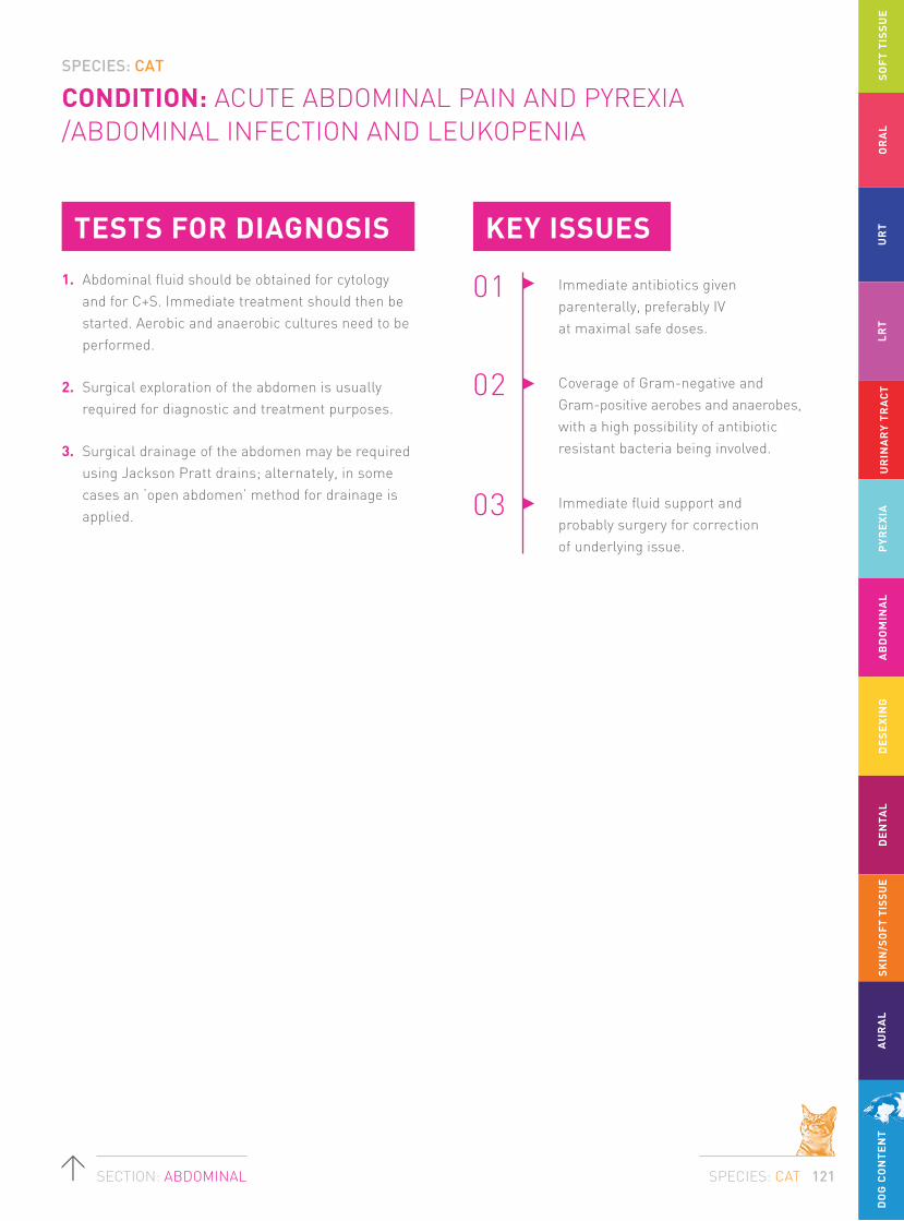
SPECIES: CAT
SPECIES: CAT 121SECTION: ABDOMINAL
CONDITION: ACUTE ABDOMINAL PAIN AND PYREXIA /ABDOMINAL INFECTION AND LEUKOPENIA
SO
FT
TIS
SU
EO
RA
LU
RT
LR
TU
RIN
AR
Y T
RA
CT
PY
RE
XIA
AB
DO
MIN
AL
DE
SE
XIN
GD
EN
TAL
SKIN
/SO
FT T
ISSU
EA
UR
AL
DO
G C
ON
TE
NT
1. Abdominal fluid should be obtained for cytology and for C+S. Immediate treatment should then be started. Aerobic and anaerobic cultures need to be performed.
2. Surgical exploration of the abdomen is usually required for diagnostic and treatment purposes.
3. Surgical drainage of the abdomen may be required using Jackson Pratt drains; alternately, in some cases an ‘open abdomen’ method for drainage is applied.
KEY ISSUES
01 Immediate antibiotics given parenterally, preferably IV at maximal safe doses.
02 Coverage of Gram-negative and Gram-positive aerobes and anaerobes, with a high possibility of antibiotic resistant bacteria being involved.
03 Immediate fluid support and probably surgery for correction of underlying issue.
TESTS FOR DIAGNOSIS

SPECIES: CAT
SPECIES: CAT 122SECTION: ABDOMINAL
CONDITION: ACUTE ABDOMINAL PAIN AND PYREXIA /ABDOMINAL INFECTION AND LEUKOPENIA
SO
FT
TIS
SU
EO
RA
LU
RT
LR
TU
RIN
AR
Y T
RA
CT
PY
RE
XIA
AB
DO
MIN
AL
DE
SE
XIN
GD
EN
TAL
SKIN
/SO
FT T
ISSU
EA
UR
AL
DO
G C
ON
TE
NT
TREATMENT
1. Four-quadrant IV antibiotic therapy.
2. Surgical drainage.
3. Management of sepsis syndrome with fluids, plasma or colloids, sometimes a whole blood transfusion and inotropic support, as needed.
1. Amoxicillin 20 mg/kg IV q6h† or ampicillin (20 mg/kg IV q6-8h‡) or cefazolin (22 mg/kg IV q8h‡) /cefoxitin (30 mg/kg IV q8h‡).
2. Use an aminoglycoside (e.g. gentamicin 6-8 mg/kg IV q24h).
3. Metronidazole 10 mg/kg IV q8h.
ANTIBIOTICS USED
Key references:
1. Ruthrauff CM, Smith J, Glerum L. J Am Anim Hosp Assoc 2009; 45(6): 268-276
2. Culp WT, Zeldis TE, Reese MS, Drobatz KJ. J Am Vet Med Assoc 2009; 234(7): 906-913
3. Mueller MG, Ludwig LL, Barton LJ. J Am Vet Med Assoc. 2001; 219(6):789-794
2 weeks post-recovery
USAGE RECOMMENDATION
The upper limit of the recommended dose range should be given.
Note that beta-lactams (penicillins and cephalosporins) and aminoglycosides (gentamicin, amikacin) need to be given separately as they precipitate in the fluid line if given simultaneously. These agents should be given by slow IV push over several minutes, separated by a flush with saline.
SPECIES: CAT
SPECIES: CAT 123SECTION: DESEXING
CONDITION: ANTIBIOTIC USE AFTER ROUTINE DESEXING
SO
FT
TIS
SU
EO
RA
LU
RT
LR
TU
RIN
AR
Y T
RA
CT
PY
RE
XIA
AB
DO
MIN
AL
DE
SE
XIN
GD
EN
TAL
SKIN
/SO
FT T
ISSU
EA
UR
AL
DO
G C
ON
TE
NT
As a general rule desexing operations conducted using sterile technique and not taking longer than average for an experienced veterinarian to complete would not be given antibiotics prophylactically.
In one study, peri operative antimicrobial prophylaxis decreased postoperative infection rate in dogs undergoing elective orthopaedic surgery, compared with the infection rate in control dogs. Cefazolin was not more efficacious than potassium penicillin G in these dogs.
This would suggest that prolonged surgery times for routine desexing might be considered an increased risk of infection, as might a breach in aseptic technique. Definitive studies of what time limits are associated with increased infection in desexing operations are lacking. However, antibiotics with higher efficacy for Staphylococcus spp. would be considered in the event of a prolonged desexing operation.
Several studies have shown that length of time of surgery and the more people in the room at the time of surgery, the greater the risk of infections.
BACKGROUND/NATURE OF INFECTION/
ORGANISMS INVOLVED

SPECIES: CAT
SPECIES: CAT 124SECTION: DESEXING
CONDITION: ANTIBIOTIC USE AFTER ROUTINE DESEXING
SO
FT
TIS
SU
EO
RA
LU
RT
LR
TU
RIN
AR
Y T
RA
CT
PY
RE
XIA
AB
DO
MIN
AL
DE
SE
XIN
GD
EN
TAL
SKIN
/SO
FT T
ISSU
EA
UR
AL
DO
G C
ON
TE
NT
KEY ISSUES
01 There is no need for prophylactic antimicrobials for routine desexing.
02 If the procedure is unduly prolonged, or there is a breach in asepsis, then a single injection of amoxicillin-clavulanate or a 1st generation cephalosporin might be appropriate.
TREATMENT
Antibiotics are considered unnecessary in routine short surgery conducted under sterile conditions. Given the use of gloves and sterile conditions, the routine use of prophylactic antibiotics for spays is not required. Also given that most potential contaminants arise from the skin of the dog or the veterinary staff, a single shot of procaine penicillin offers insufficient coverage for Staphylococcus pseudintermedius or Staphylococcus aureus infection.
Routine use of antibiotics not suggested.
ANTIBIOTICS USED
USAGE RECOMMENDATION
N/A.
SPECIES: CAT
SPECIES: CAT 125SECTION: DENTAL
CONDITION: USE OF ANTIBIOTICS IN DENTAL PROPHYLAXIS
SO
FT
TIS
SU
EO
RA
LU
RT
LR
TU
RIN
AR
Y T
RA
CT
PY
RE
XIA
AB
DO
MIN
AL
DE
SE
XIN
GD
EN
TAL
SKIN
/SO
FT T
ISSU
EA
UR
AL
DO
G C
ON
TE
NT
Pasteurella multocida, and anaerobic Gram-negative rods including Capnocytophaga are frequently involved and these are all sensitive to penicillins, including benzyl penicillin and amoxicillin-clavulanate.
There is a very small risk that bacteraemia associated with the use of ultrasonic scaling devices and extractions could produce infections elsewhere, such as bacterial endocarditis. This is most unlikely in normal patients, but the risk is increased with structural heart disease, especially subaortic stenosis which has been associated with increased risk for the development of bacterial endocarditis.
For this reason, it may be prudent to administer prophylactic bactericidal antibiotics so that high blood levels are obtained during and immediately after the dental procedures.
Most bacteria found in the mouths of cats (and dogs) are similar to what is recovered in bite wounds.
BACKGROUND/NATURE OF INFECTION/
ORGANISMS INVOLVED

SPECIES: CAT
SPECIES: CAT 126SECTION: DENTAL
CONDITION: USE OF ANTIBIOTICS IN DENTAL PROPHYLAXIS
SO
FT
TIS
SU
EO
RA
LU
RT
LR
TU
RIN
AR
Y T
RA
CT
PY
RE
XIA
AB
DO
MIN
AL
DE
SE
XIN
GD
EN
TAL
SKIN
/SO
FT T
ISSU
EA
UR
AL
DO
G C
ON
TE
NT
Histopathology and culture of infected tissue is suggested if initial prophylaxis fails to cure an ulcerated mouth lesion.
KEY ISSUES
01 Prophylactic antibiotics are best administered prior to the procedure e.g. procaine penicillin or amoxicillin-clavulanate administered SC or IM after premedication or immediately after anaesthetic induction.
TESTS FOR DIAGNOSIS
TREATMENT
N/A.
Amoxicillin or amoxicillin-clavulanate would cover the great majority of potential pathogens in this setting.
First line:
Amoxicillin 10 mg/kg q12h/amoxicillin-clavulanate (12.5 mg/kg q12h).
Second line:
Clindamycin (5-11 mg/kg q12h) or doxycycline monohydrate (5 mg/kg q12h†).
ANTIBIOTICS USED
SPECIES: CAT
SPECIES: CAT 127SECTION: DENTAL
CONDITION: USE OF ANTIBIOTICS IN DENTAL PROPHYLAXIS
SO
FT
TIS
SU
EO
RA
LU
RT
LR
TU
RIN
AR
Y T
RA
CT
PY
RE
XIA
AB
DO
MIN
AL
DE
SE
XIN
GD
EN
TAL
SKIN
/SO
FT T
ISSU
EA
UR
AL
DO
G C
ON
TE
NT
USAGE RECOMMENDATION
If there are extractions or bleeding, which occurs in most cases, then a 7-10 day course of antibiotics is required depending on the healing period. Ensure doxycycline given with a small bolus of water, or a small dab of margarine.
Key references:
1. Love D 1990 et al (isolated lots of anaerobes but possibly no data on their sensitivities)
2. Bowersock TL, Wu CC, Inskeep GA, Thoracicer ST. J Vet Dent 2000; 17(1): 11-16
3. Zetner K and Rothmueller G. Vet Ther 2002; 3(4): 441-452
4. Nielsen D, Walser C, Kodan G, et al. Vet Ther 2000; 1(3): 150-158
5. Warrick JM, Inskeep GA, Yonkers TD, et al. Vet Ther 2000; 1(1): 5-16
6. Sarkiala E and Harvey C. Semin Vet Med Surg (Small Anim) 1993; 8(3): 197-203
7. Zetner K and Thiemann G. J Vet Dent 1993; 10(2): 6-9
8. Wilcke JR. Probl Vet Med 1990; 2(2): 298-311

SPECIES: CAT
SPECIES: CAT 128SECTION: SKIN/SOFT TISSUE
CONDITION: MYCOBACTERIA AND NOCARDIA AS CAUSES OF DEEP DRAINING SINUS TRACTS
SO
FT
TIS
SU
EO
RA
LU
RT
LR
TU
RIN
AR
Y T
RA
CT
PY
RE
XIA
AB
DO
MIN
AL
DE
SE
XIN
GD
EN
TAL
SKIN
/SO
FT T
ISSU
EA
UR
AL
DO
G C
ON
TE
NT
(i) Infections of the subcutaneous panniculus generally with rapidly growing mycobacteria and;
(ii) granulomatous or pyogranulomatous masses of the skin and subcutis (generally due to non-cultivable mycobacteria e.g. feline leprosy-like syndromes, leproid granulomas etc. and sometimes mycobacterium avium complex infections).
The taxonomy of these organisms is evolving, and currently they are divided into complexes:
a. M. smegmatis complex (including M. goodii) – drugs of choice doxycycline, moxifloxacin, gentamicin.
b. M. fortuitum complex – drugs of choice clarithromycin, moxifloxacin, gentamicin.
c. M. chelonae/abscesses complex – drug of choice clarithromycin, rest depends on susceptibility testing.
Generally speaking they are all resistant to rifampicin, and all susceptible to clofazamine.
Mycobacteria cause two major types of disease affecting the skin and subcutis.
BACKGROUND/NATURE OF INFECTION/
ORGANISMS INVOLVED
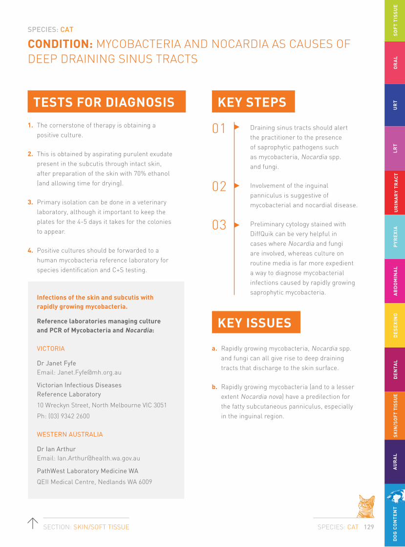
SPECIES: CAT
SPECIES: CAT 129SECTION: SKIN/SOFT TISSUE
CONDITION: MYCOBACTERIA AND NOCARDIA AS CAUSES OF DEEP DRAINING SINUS TRACTS
SO
FT
TIS
SU
EO
RA
LU
RT
LR
TU
RIN
AR
Y T
RA
CT
PY
RE
XIA
AB
DO
MIN
AL
DE
SE
XIN
GD
EN
TAL
SKIN
/SO
FT T
ISSU
EA
UR
AL
DO
G C
ON
TE
NT
1. The cornerstone of therapy is obtaining a positive culture.
2. This is obtained by aspirating purulent exudate present in the subcutis through intact skin, after preparation of the skin with 70% ethanol (and allowing time for drying).
3. Primary isolation can be done in a veterinary laboratory, although it important to keep the plates for the 4-5 days it takes for the colonies to appear.
4. Positive cultures should be forwarded to a human mycobacteria reference laboratory for species identification and C+S testing.
Infections of the skin and subcutis with rapidly growing mycobacteria.
Reference laboratories managing culture and PCR of Mycobacteria and Nocardia:
VICTORIA
Dr Janet Fyfe Email: [email protected]
Victorian Infectious Diseases Reference Laboratory
10 Wreckyn Street, North Melbourne VIC 3051
Ph: (03) 9342 2600
WESTERN AUSTRALIA
Dr Ian Arthur Email: [email protected]
PathWest Laboratory Medicine WA
QEII Medical Centre, Nedlands WA 6009
KEY STEPS
KEY ISSUES
01 Draining sinus tracts should alert the practitioner to the presence of saprophytic pathogens such as mycobacteria, Nocardia spp. and fungi.
02 Involvement of the inguinal panniculus is suggestive of mycobacterial and nocardial disease.
03 Preliminary cytology stained with DiffQuik can be very helpful in cases where Nocardia and fungi are involved, whereas culture on routine media is far more expedient a way to diagnose mycobacterial infections caused by rapidly growing saprophytic mycobacteria.
a. Rapidly growing mycobacteria, Nocardia spp. and fungi can all give rise to deep draining tracts that discharge to the skin surface.
b. Rapidly growing mycobacteria (and to a lesser extent Nocardia nova) have a predilection for the fatty subcutaneous panniculus, especially in the inguinal region.
TESTS FOR DIAGNOSIS
SPECIES: CAT
SPECIES: CAT 130SECTION: SKIN/SOFT TISSUE
CONDITION: MYCOBACTERIA AND NOCARDIA AS CAUSES OF DEEP DRAINING SINUS TRACTS
SO
FT
TIS
SU
EO
RA
LU
RT
LR
TU
RIN
AR
Y T
RA
CT
PY
RE
XIA
AB
DO
MIN
AL
DE
SE
XIN
GD
EN
TAL
SKIN
/SO
FT T
ISSU
EA
UR
AL
DO
G C
ON
TE
NT
TREATMENT
These infections require months to years of antimicrobial therapy, and in some cases surgery is required to debulk lesions to enable a clinical cure to be achieved.
Generally speaking, topical therapy is not useful in the management of these infections as the disease process is situated in the subcutis and involves the skin secondarily.
C+S is strongly advised in these cases.
Mycobacteria
First line:
Doxycycline (5 mg/kg q12h†) and moxifloxacin (5 mg/kg q12h‡ (compounded) for M. smegmatis complex infections; clarithromycin (5-15 mg/kg q12h‡) and moxifloxacin (5 mg/kg q12h‡) for other rapidly growing mycobacteria.
Second line:
Clofazimine (4-10 mg/kg q24h‡ compounded), amikacin (10-15 mg/kg q24h‡ IV/IM/SC).
Nocardia
First line:
Trimethoprim/sulphonamide combinations at a dose of 125 mg per cat once daily (do not split or otherwise divide the coated tablet) combined with a second drug depending on C+S testing; note that many cats can tolerate chronic dosing with trimethorpim/sulphonamide combinations.
Second line:
Amoxicillin 20 mg/kg twice a day for N. nova (not amoxicillin clavulanate).
Clarithromycin (5-15 mg/kg q 12h) or moxifloxacin (10 mg/kg once a day).
ANTIBIOTICS USED
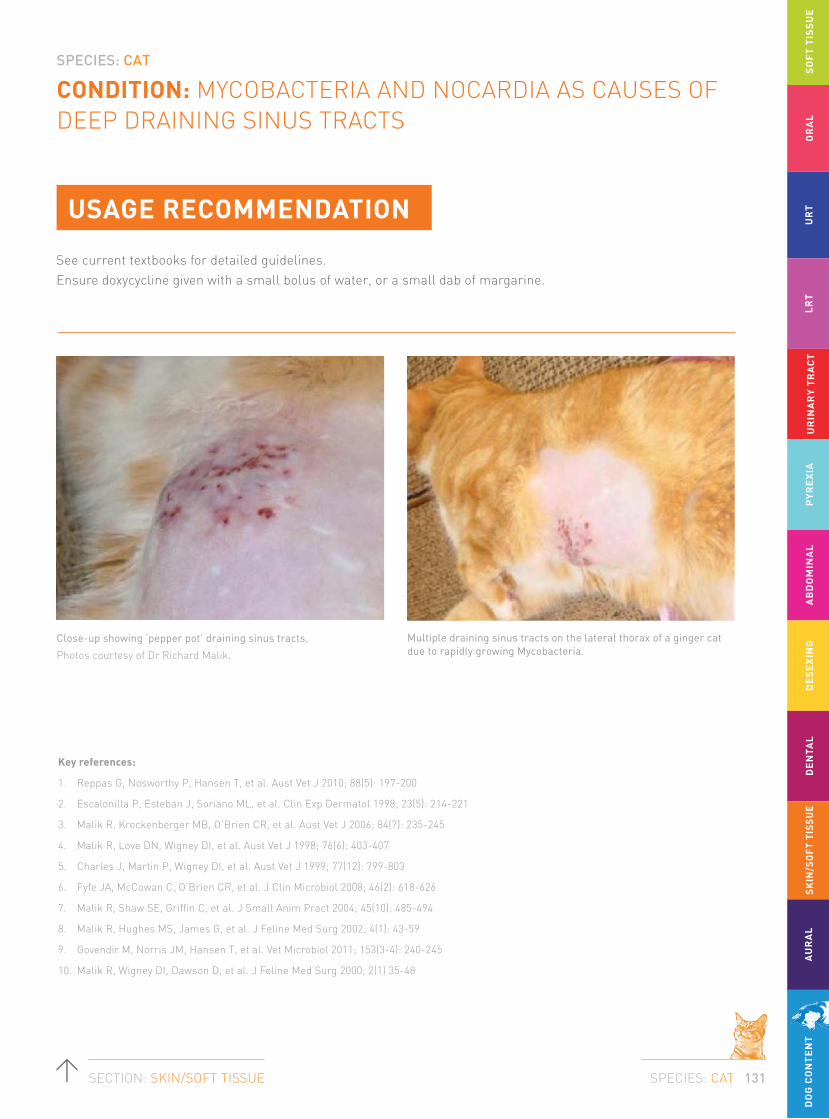
SPECIES: CAT
SPECIES: CAT 131SECTION: SKIN/SOFT TISSUE
CONDITION: MYCOBACTERIA AND NOCARDIA AS CAUSES OF DEEP DRAINING SINUS TRACTS
SO
FT
TIS
SU
EO
RA
LU
RT
LR
TU
RIN
AR
Y T
RA
CT
PY
RE
XIA
AB
DO
MIN
AL
DE
SE
XIN
GD
EN
TAL
SKIN
/SO
FT T
ISSU
EA
UR
AL
DO
G C
ON
TE
NT
USAGE RECOMMENDATION
See current textbooks for detailed guidelines. Ensure doxycycline given with a small bolus of water, or a small dab of margarine.
Key references:
1. Reppas G, Nosworthy P, Hansen T, et al. Aust Vet J 2010; 88(5): 197-200
2. Escalonilla P, Esteban J, Soriano ML, et al. Clin Exp Dermatol 1998; 23(5): 214-221
3. Malik R, Krockenberger MB, O’Brien CR, et al. Aust Vet J 2006; 84(7): 235-245
4. Malik R, Love DN, Wigney DI, et al. Aust Vet J 1998; 76(6): 403-407
5. Charles J, Martin P, Wigney DI, et al. Aust Vet J 1999; 77(12): 799-803
6. Fyfe JA, McCowan C, O’Brien CR, et al. J Clin Microbiol 2008; 46(2): 618-626
7. Malik R, Shaw SE, Griffin C, et al. J Small Anim Pract 2004; 45(10): 485-494
8. Malik R, Hughes MS, James G, et al. J Feline Med Surg 2002; 4(1): 43-59
9. Govendir M, Norris JM, Hansen T, et al. Vet Microbiol 2011; 153(3-4): 240-245
10. Malik R, Wigney DI, Dawson D, et al. J Feline Med Surg 2000; 2(1) 35-48
Close-up showing ‘pepper pot’ draining sinus tracts.
Photos courtesy of Dr Richard Malik.
Multiple draining sinus tracts on the lateral thorax of a ginger cat due to rapidly growing Mycobacteria.
SPECIES: CAT
SPECIES: CAT 132SECTION: SKIN/SOFT TISSUE
CONDITION: DERMATOPHYTE INFECTIONS (e.g. MICROSPORUM OR TRICHOPHYTON)
SO
FT
TIS
SU
EO
RA
LU
RT
LR
TU
RIN
AR
Y T
RA
CT
PY
RE
XIA
AB
DO
MIN
AL
DE
SE
XIN
GD
EN
TAL
SKIN
/SO
FT T
ISSU
EA
UR
AL
DO
G C
ON
TE
NT
Microsporum canis is responsible for 94 to 99% of feline infections.
Microsporum gypseum and Trichophyton mentagrophytes account for most of the remaining cases. Infections with unusual species such as Microsporum persicolor have been reported; it is uncertain how common these infections occur, but prevalence maybe related to local climate and environmental factors.
Skin lesions: There are many clinical presentations of feline dermatophytosis. Pruritus is variable and can range from nil to severe. In kittens, irregular, annular to circular patches of alopecia with scale, crust and erythema affecting face, ears and forelegs are common. In adult cats, focal, multifocal or generalised patchy alopecia with or without scale occurs frequently, especially in long haired cats.
Dermatophytoses are superficial fungal infections that involve the skin, hair and claws.
BACKGROUND/NATURE OF INFECTION/
ORGANISMS INVOLVED
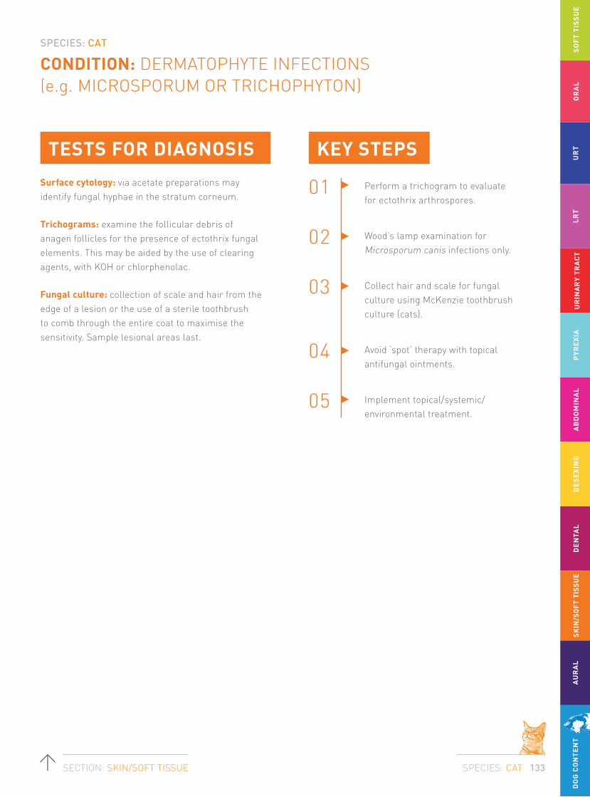
SPECIES: CAT
SPECIES: CAT 133SECTION: SKIN/SOFT TISSUE
CONDITION: DERMATOPHYTE INFECTIONS (e.g. MICROSPORUM OR TRICHOPHYTON)
SO
FT
TIS
SU
EO
RA
LU
RT
LR
TU
RIN
AR
Y T
RA
CT
PY
RE
XIA
AB
DO
MIN
AL
DE
SE
XIN
GD
EN
TAL
SKIN
/SO
FT T
ISSU
EA
UR
AL
DO
G C
ON
TE
NT
Surface cytology: via acetate preparations may identify fungal hyphae in the stratum corneum.
Trichograms: examine the follicular debris of anagen follicles for the presence of ectothrix fungal elements. This may be aided by the use of clearing agents, with KOH or chlorphenolac.
Fungal culture: collection of scale and hair from the edge of a lesion or the use of a sterile toothbrush to comb through the entire coat to maximise the sensitivity. Sample lesional areas last.
KEY STEPS
01 Perform a trichogram to evaluate for ectothrix arthrospores.
02 Wood’s lamp examination for Microsporum canis infections only.
03 Collect hair and scale for fungal culture using McKenzie toothbrush culture (cats).
04 Avoid ‘spot’ therapy with topical antifungal ointments.
05 Implement topical/systemic/environmental treatment.
TESTS FOR DIAGNOSIS

SPECIES: CAT
SPECIES: CAT 134SECTION: SKIN/SOFT TISSUE
CONDITION: DERMATOPHYTE INFECTIONS (e.g. MICROSPORUM OR TRICHOPHYTON)
SO
FT
TIS
SU
EO
RA
LU
RT
LR
TU
RIN
AR
Y T
RA
CT
PY
RE
XIA
AB
DO
MIN
AL
DE
SE
XIN
GD
EN
TAL
SKIN
/SO
FT T
ISSU
EA
UR
AL
DO
G C
ON
TE
NT
TREATMENT
In most healthy animals, dermatophytosis is a self-curing disease, with full resolution of disease in 10-16 weeks without therapy. The best treatment protocol is a combination of three modalities:
1. Topical treatment: To kill infective material and prevent its dissemination into the environment.
2. Systemic treatment: To shorten the time of infection in the individual animal.
3. Environmental treatment: To help prevent recurrence of infection or spread to other animals or people in the household.
A. Clipping the hair coat: should you clip?
Clipping of the hair coat will mechanically remove fragile hairs that will fracture and release spores into the environment. Clipping of the entire hair coat is optimal but not always possible or practical. Clipping is time consuming and often requires sedation and is irritating to cats. Owners are often unwilling to commit to clipping their pets. Short-haired cats with fewer than five focal lesions do not need to be clipped.
When cats have more than five lesions, long hair and there are multiple pets in the environment and the affected pet cannot be segregated, clipping the entire pet is optimal. Clip the hair short and gently to avoid spreading the infection due to the microtrauma and mechanical spread of the spores.
The owner should be warned that a temporary exacerbation of lesions may occur after clipping.
Note: If the animal is to be clipped in the clinic all debris produced must be considered to be infectious with zoonotic potential and so rigorous infection control measures should be observed.
i.e. cover table surface with disposable drape, gown and glove, collect all material and double bag, thoroughly disinfect the room and all equipment used with an appropriate antifungal agent.
B. Topical therapy: which one is best?
i. Localised (treating only the spots) or whole body topical therapy
In animals, not all the lesions may be visible due to the long hair coat. It is almost certain that there are infective spores in non-lesional areas. Therefore ‘spot treatment’ with topical drugs is not recommended even for focal lesions because infection beyond the margin of visible lesions is likely. There is no clinical data to support that the use of spot treatment clear lesions any more rapidly than whole-body treatment alone. If the owner insists, the best products are probably 1% terbinafine solution, lotion, cream or spray (Lamisil®) and 2% clotrimazole cream (Canesten®).
ii. Total body treatment
Topical therapy inactivates fungal spores and mycelia on and within hair shafts reducing environmental contagion and results in a faster cure than systemic therapy alone. Shampoo therapy, dipping or rinsing with topical antifungal agents is preferred. The choice of topical antifungal agent is important because studies have shown that many topical antifungal agents are ineffective. In vitro and in vivo studies have shown that the most consistently effective topical treatments are lime sulphur, enilconazole, and miconazole; the latter with or without chlorhexidine. Miconazole and chlorhexidine (Malaseb®) shampoo has been studied in cats as an adjunct treatment to oral griseofulvin.
SPECIES: CAT
SPECIES: CAT 135SECTION: SKIN/SOFT TISSUE
CONDITION: DERMATOPHYTE INFECTIONS (e.g. MICROSPORUM OR TRICHOPHYTON)
SO
FT
TIS
SU
EO
RA
LU
RT
LR
TU
RIN
AR
Y T
RA
CT
PY
RE
XIA
AB
DO
MIN
AL
DE
SE
XIN
GD
EN
TAL
SKIN
/SO
FT T
ISSU
EA
UR
AL
DO
G C
ON
TE
NT
Systemic therapy
The role of systemic therapy in treating dermatophytosis is to accelerate the resolution of infection in the individual animal. Several effective drugs are available, and the appropriate choice should be made depending on cost, fungal species, patient species and potential for toxicity. Systemic therapy is the treatment of choice for dermatophytosis. It is important to remember that systemic antifungal therapy does not rapidly reduce contagion and should be used in conjunction with clipping and topical antifungal agents.
Griseofulvin
Griseofulvin is administered at 25 mg/kg q12h. The absorption is enhanced when administered as a divided dose and with a fatty meal. The most common side-effects – anorexia, vomiting and diarrhoea – can be avoided by dividing or lowering the dose. The drug is highly teratogenic and therefore contraindicated in pregnant animals.
Bone marrow suppression producing anaemia and leukopenia is a relatively uncommon yet severe and unpredictable adverse effect of griseofulvin in cats. These effects can reverse when treatment is withdrawn but irreversible fatal idiosyncratic pancytopenia has been reported. Myelosuppression does not appear to depend on dose, breed or duration of treatment. Blood counts are recommended once a month when using this drug in cats. Severe neutropenic reactions have been reported in cats with dermatophytosis associated with FIV infection. Griseofulvin should not be used in cats with FIV. All cats should be tested for FIV before griseofulvin is administered. Do not administer cats less than 6 weeks of age.
Nearly all patients with Microsporum infections, and many with Trichophyton infections will be cured with this drug,
Ketoconazole (KTZ)
This drug should be avoided in cats due to hepatotoxicity. Some strains of Microsporum canis appear resistant to ketoconazole. Therefore ketoconazole has no real advantage over other drugs for routine cases of dermatophytosis and should not be used in cats due to the adverse effects in these species.
Itraconazole (ITZ)
Itraconazole (Sporanox®) is a fungicidal triazole drug that is extremely useful for dermatophytosis. The recommended dose is 5-10 mg/kg/day PO. Itraconazole persists in the skin and nails for weeks to months after dosing, and intermittent or pulse therapy is frequently prescribed for skin infections or onychomycosis. In cats a regime has been reported using 5 mg/kg/day every 24 hours for three one week-on and one week-off cycles and this is recommended by the authors.
Itraconazole is generally well tolerated; reported side- effects include vomiting and/or anorexia in cats. Signs of dose related hepatotoxicosis have been reported rarely in cats. Itraconazole is reportedly not teratogenic when used at a dose of 5 mg/kg.
Fluconazole
Fluconazole (Diflucan®) is receiving some recent attention as an alternative drug because it has now become inexpensive through some compounding pharmacies. Several recent in vitro studies have shown that the MICs of fluconazole against dermatophytes are much higher that the MICs of itraconazole suggesting that itraconazole may be the superior drug. Current evidence and clinical anecdotes would suggest that there is no advantage of this drug over itraconazole and currently we do not recommend it for the treatment of dermatophytosis.
SPECIES: CAT
SPECIES: CAT 136SECTION: SKIN/SOFT TISSUE
CONDITION: DERMATOPHYTE INFECTIONS (e.g. MICROSPORUM OR TRICHOPHYTON)
SO
FT
TIS
SU
EO
RA
LU
RT
LR
TU
RIN
AR
Y T
RA
CT
PY
RE
XIA
AB
DO
MIN
AL
DE
SE
XIN
GD
EN
TAL
SKIN
/SO
FT T
ISSU
EA
UR
AL
DO
G C
ON
TE
NT
Terbinafine
Terbinafine (Lamisil®) is a fungicidal allylamine useful in the treatment of superficial dermatophytosis and onychomycosis in humans. There is little data on the use of terbinafine in cats and this drug appears to offer no advantages over itraconazole. Preliminary studies indicate that a dose of 30-40 mg/kg/day is the most efficacious in the cat.
Terbinafine is generally well tolerated; reported adverse effects include vomiting and asymptomatic elevation in liver enzymes. Idiosyncratic acute hepatotoxicity has been reported occasionally. No teratogenicity has been reported. The drug reaches very high concentrations in sebum and stratum corneum and fungicidal concentrations persist in the skin for several weeks after administration in humans.
Environmental treatment
The critical role of environmental disinfection in eradication of M. canis from an endemic cattery or household cannot be overemphasised. Environmental contamination with M. canis spores is widespread, difficult to eliminate and routinely transported by the fur of uninfected cats. Such contamination is a major reservoir for recurrence of infection. M. canis spores remain viable in the environment for up to 18 months.
Studies using isolated infected hairs or spores or field studies using dermatophyte-contaminated environments have shown that the following disinfectant products are consistently effective: lime sulphur (1:33), enilconazole (0.2%), and 1:10 to 1:100 household bleach (10 mL/L). In addition, a study has also shown that strain variation of M. canis with respect to susceptibility to disinfectants is not present.
For treatment of routine infections with one or a few animals in the household, extensive environmental decontamination is generally impractical and unnecessary. Thorough vacuuming and mechanical cleaning will remove infective material. All hard surfaces should be mopped with 1:100 bleach solution.
During treatment these few animals should be confined to a small easily cleaned room without carpeting until they have received systemic antifungal therapy for at least two weeks and have been dipped at least four times with topical preparations. All bedding, brushes, combs, rugs, cages, carriers can be washed daily in hot water, detergent and a 1:10 dilution of household bleach.
Carpeted areas are problematic because of the lack of effective disinfectant that preserves carpeting. Frequent vacuuming on a daily basis or steam cleaning mechanically removes many but not all spores. Steam cleaning may not be a reliable method of killing M. canis unless an antifungal disinfectant such as chlorhexidine or sodium hypochlorite is added to the water. Draperies should be dry cleaned and not replaced until the infection is eradicated.
Length of treatment
Cats or dogs with dermatophytosis should be treated until complete resolution of clinical signs (clinical cure) and then continued until the fungus cannot be cultured from the hair coat on at least two sequential cultures a week or more apart (mycologic cure)
Weekly fungal cultures should be started after the cat has received 4-6 weeks of therapy and thereafter on a 2 week schedule. Once the culture results are negative, monitoring can be done on a once weekly basis. Cats appear healthy before their skin and hair are cleared of fungal organisms.
It is not always possible or practical however to re-culture every patient. In otherwise healthy cats, systemic and topical treatments should generally be continued for 6-10 weeks, preferably until 2 weeks after clinical resolution.
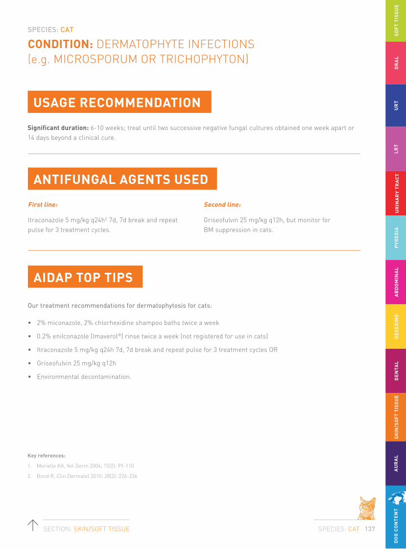
SPECIES: CAT
SPECIES: CAT 137SECTION: SKIN/SOFT TISSUE
CONDITION: DERMATOPHYTE INFECTIONS (e.g. MICROSPORUM OR TRICHOPHYTON)
SO
FT
TIS
SU
EO
RA
LU
RT
LR
TU
RIN
AR
Y T
RA
CT
PY
RE
XIA
AB
DO
MIN
AL
DE
SE
XIN
GD
EN
TAL
SKIN
/SO
FT T
ISSU
EA
UR
AL
DO
G C
ON
TE
NT
USAGE RECOMMENDATION
Significant duration: 6-10 weeks; treat until two successive negative fungal cultures obtained one week apart or 14 days beyond a clinical cure.
Key references:
1. Moriello KA, Vet Derm 2004; 15(2): 99-110
2. Bond R, Clin Dermatol 2010; 28(2): 226-236
AIDAP TOP TIPS
Our treatment recommendations for dermatophytosis for cats:
• 2% miconazole, 2% chlorhexidine shampoo baths twice a week
• 0.2% enilconazole (Imaverol®) rinse twice a week (not registered for use in cats)
• Itraconazole 5 mg/kg q24h 7d, 7d break and repeat pulse for 3 treatment cycles OR
• Griseofulvin 25 mg/kg q12h
• Environmental decontamination.
ANTIFUNGAL AGENTS USED
First line:
Itraconazole 5 mg/kg q24h‡ 7d, 7d break and repeat pulse for 3 treatment cycles.
Second line:
Griseofulvin 25 mg/kg q12h, but monitor for BM suppression in cats.
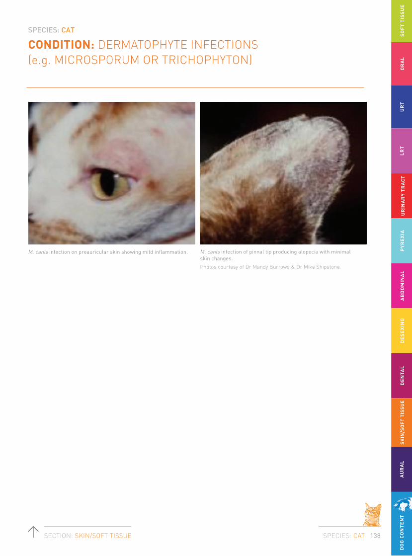
SPECIES: CAT
SPECIES: CAT 138SECTION: SKIN/SOFT TISSUE
CONDITION: DERMATOPHYTE INFECTIONS (e.g. MICROSPORUM OR TRICHOPHYTON)
SO
FT
TIS
SU
EO
RA
LU
RT
LR
TU
RIN
AR
Y T
RA
CT
PY
RE
XIA
AB
DO
MIN
AL
DE
SE
XIN
GD
EN
TAL
SKIN
/SO
FT T
ISSU
EA
UR
AL
DO
G C
ON
TE
NT
M. canis infection on preauricular skin showing mild inflammation. M. canis infection of pinnal tip producing alopecia with minimal skin changes.
Photos courtesy of Dr Mandy Burrows & Dr Mike Shipstone.

SPECIES: CAT
SPECIES: CAT 139SECTION: SKIN/SOFT TISSUE
CONDITION: DERMATOPHYTE INFECTIONS (e.g. MICROSPORUM OR TRICHOPHYTON)
SO
FT
TIS
SU
EO
RA
LU
RT
LR
TU
RIN
AR
Y T
RA
CT
PY
RE
XIA
AB
DO
MIN
AL
DE
SE
XIN
GD
EN
TAL
SKIN
/SO
FT T
ISSU
EA
UR
AL
DO
G C
ON
TE
NT
Fungal hyphae on surface cytology.
Fungal hyphae surrounding the hair shaft.
Photos courtesy of Dr Mandy Burrows & Dr Mike Shipstone.
Fungal hyphae surrounding the hair shaft.
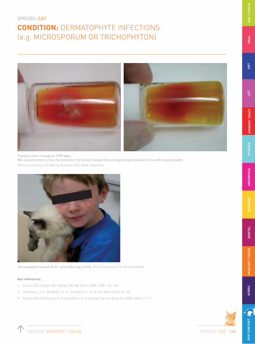
SPECIES: CAT
SPECIES: CAT 140SECTION: SKIN/SOFT TISSUE
CONDITION: DERMATOPHYTE INFECTIONS (e.g. MICROSPORUM OR TRICHOPHYTON)
SO
FT
TIS
SU
EO
RA
LU
RT
LR
TU
RIN
AR
Y T
RA
CT
PY
RE
XIA
AB
DO
MIN
AL
DE
SE
XIN
GD
EN
TAL
SKIN
/SO
FT T
ISSU
EA
UR
AL
DO
G C
ON
TE
NT
Key references:
1. Duclos DD, Hargis AM, Hanley PW. Vet Derm 2008; 19(3): 134-141
2. Summers, J. F., Brodbelt, D. C., Forsythe P.J. et al. Vet Derm 2012; 23: 30
3. Kovacs MS, McKiernan S, Potter DM et al. Contemp Top Lab Anim Sci 2005; 44(4): 17-21
Positive colour change on DTM agar. NB: a positive test is the characteristic red colour change that enlarges progressively in line with colony growth.
Photos courtesy of Dr Mandy Burrows & Dr Mike Shipstone.
Dermatophyte lesions dt M. canis affecting a child. Photo courtesy of Dr Richard Malik.
SPECIES: CAT
SPECIES: CAT 141SECTION: AURAL
CONDITION: OTITIS EXTERNA (UNCOMPLICATED, FIRST EPISODE AND COMPLICATED, RECURRENT)
SO
FT
TIS
SU
EO
RA
LU
RT
LR
TU
RIN
AR
Y T
RA
CT
PY
RE
XIA
AB
DO
MIN
AL
DE
SE
XIN
GD
EN
TAL
SKIN
/SO
FT T
ISSU
EA
UR
AL
DO
G C
ON
TE
NT
The normal flora is generally Gram-positive, with higher bacterial counts retrieved from the vertical external ear canal than the horizontal ear canal. Commensal and pathogenic bacteria rapidly colonise the external ear canal where changes in the microclimate subsequent to inflammation modify the environment. The microbial proliferation exacerbates and perpetuates the inflammatory response within the ear canal. Once inflamed, there is a shift towards increased bacterial numbers, initially coagulase positive staphylococci and with more chronic inflammation, Gram-negative bacteria.
Because potential pathogens can be recovered in the absence of disease (as they can from the skin surface), it is assumed that they are unable to initiate disease in the ear.
However, once the ear becomes inflamed or macerated, proliferation may occur and it is for this reason that bacteria are considered secondary rather than primary or predisposing factors in otitis externa.
In cats, Staphylococcus pseudintermedius and Pasteurella multocida are commonly isolated from otitis externa cases.
Malassezia are relatively more important and have been found in more than 95% of cases of otitis externa.
BACKGROUND/NATURE OF INFECTION/
ORGANISMS INVOLVED
Otitis externa is a relatively common disease with 2-6.5% of cats affected.

SPECIES: CAT
SPECIES: CAT 142SECTION: AURAL
CONDITION: OTITIS EXTERNA (UNCOMPLICATED, FIRST EPISODE AND COMPLICATED, RECURRENT)
SO
FT
TIS
SU
EO
RA
LU
RT
LR
TU
RIN
AR
Y T
RA
CT
PY
RE
XIA
AB
DO
MIN
AL
DE
SE
XIN
GD
EN
TAL
SKIN
/SO
FT T
ISSU
EA
UR
AL
DO
G C
ON
TE
NT
In many cases of otitis, a single organism can be isolated on bacterial culture of exudate, but in others, multiple potentially pathogenic organisms are identified. Thus it is of critical importance to combine cytological examination of the otic discharge when a C+S test is performed. This allows determination of the dominant population of bacteria evident, the presence of leukocytes, and the presence of phagocytosed bacteria.
Cytology is the first step. It is mandatory in ALL cases of otitis externa and should be repeated at each visit.
Normal cerumen does not have high stain uptake because of the high lipid content. Outlines of occasional squames may be seen. Inflammation leads to increased numbers of squames (some of which may be nucleated indicting faster epithelial turnover with incomplete keratinisation before desquamation). As the severity of inflammation increases inflammatory cells may be seen along with increasing numbers of organisms. Higher cellular content of cerumen may also be appreciated by increasing stain uptake on the stained slide (before microscopic examination is even started).
The number of organisms and inflammation should be assessed on a 1-4+ scale. Normal ears may have a few yeast and Gram-positive cocci per oil field but not rods. The finding of yeast or cocci should be correlated with the findings of the otoscopic examination. Some animals may have few organisms yet show marked inflammation and exudation, whilst others seem to be able to tolerate quite large numbers without any pathologic changes. Repeating the cytology at each revisit allows accurate assessment of response to therapy. Medical treatment should continue until otoscopic and cytologic examinations demonstrate no pathologic change.
KEY STEPS
Otic examination alone is not sufficient and the following minimum database is necessary in order to identify both the nature and type of the otitis as well as any underlying primary or predisposing factors.
01 Thorough dermatological history.
02 Complete physical examination of all areas of integument.
03 Thorough otic examination (cats in particular may require sedation/anaesthesia).
04 Collect otic cytology.
05 Implement topical antimicrobial therapy on the basis of cytological findings.
06 Systemic antibiotic therapy is not indicated for otitis externa.
TESTS FOR DIAGNOSIS
SPECIES: CAT
SPECIES: CAT 143SECTION: AURAL
CONDITION: OTITIS EXTERNA (UNCOMPLICATED, FIRST EPISODE AND COMPLICATED, RECURRENT)
SO
FT
TIS
SU
EO
RA
LU
RT
LR
TU
RIN
AR
Y T
RA
CT
PY
RE
XIA
AB
DO
MIN
AL
DE
SE
XIN
GD
EN
TAL
SKIN
/SO
FT T
ISSU
EA
UR
AL
DO
G C
ON
TE
NT
TREATMENT
Topical therapy is the key to successful resolution of the majority of cases of otitis externa which is essentially a surface infection. Essential to this therapy though is the successful removal of exudate. If the medication cannot penetrate the full length of the ear canal, then treatment is likely to fail. The choice of appropriate active ingredients and vehicles for treatment of otitis externa is usually made empirically based on cytological examination of ear canal exudates and otoscopic examination of the inflamed ear canals.
Most commercially produced topical products contain one or more active antibacterial, antifungal and anti-inflammatory agents in various combinations as well as vehicle and various solubilisers, stabilisers and surfactants.
Clients may need to be shown how to administer medications correctly. Failure to do this is a significant cause for treatment failure. An adequate volume of medication must be delivered to line the entire canal. Getting clients to count drops increases the time for administration and fundamentally means that the nozzle of the bottle is not in the canal, reducing penetration of the medication. Putting the nozzle of the bottle in the canal and telling clients to use a “squeeze” means that both under and overdosing are risked because the amount to medication is not measured out. We use the Terumo brand of syringe to most accurately measure ear medications and dispense them into the ear canal.
A broad guideline depending on the length and diameter of the ear canal would be:
• 0.15-0.2 mL for a cat.
• Twice daily dosing may require slightly smaller volumes to avoid overdosing.
• It is important to remember that the bulla of cats it divided by an incomplete bony septum. This septum is rooved by a sympathetic nerve plexus that can be easily damaged causing Horner’s syndrome. Therefore, products should be used with caution if the tympanum is ruptured.
Duration of therapy
For acute disease a minimum of 5 to 14 days therapy depending on the degree of inflammatory change (oedema, hyperplasia, and erosion, ulceration) is to be expected. Rechecks every one to two weeks are necessary to ensure that ears are cytologically and otoscopically resolved prior to cessation of therapy. It is not uncommon to have a cat clinically resolved with otoscopically normal ears because of anti-inflammatory medications, where cytology is still not normal.

SPECIES: CAT
SPECIES: CAT 144SECTION: AURAL
CONDITION: OTITIS EXTERNA (UNCOMPLICATED, FIRST EPISODE AND COMPLICATED, RECURRENT)
SO
FT
TIS
SU
EO
RA
LU
RT
LR
TU
RIN
AR
Y T
RA
CT
PY
RE
XIA
AB
DO
MIN
AL
DE
SE
XIN
GD
EN
TAL
SKIN
/SO
FT T
ISSU
EA
UR
AL
DO
G C
ON
TE
NT
Antimicrobial therapy for ears with mainly cocci on cytology
Coccoid organisms will be Staphylococcus spp. or Streptococcus spp. The challenge for empirical therapy for cocci is the relative resistance of streptococci to some of the routine antibiotics, which otherwise tend to have reasonable activity for most Staphylococci spp. For this reason products containing antibiotics with good efficacy against both bacteria are desirable. For this reason, Canaural® is useful if the TM is intact because of the framycetin and fusidic acid. Other reasonable choices would be Otomax® or Mometamax® where both the gentamicin and clotrimazole have anti-coccal activity. Remember that gentamicin is degraded by organic debris and purulent exudate so the ear must be clean and inflammation well controlled for best effect and that gentamicin is not middle ear safe, at least not in commercial preparations.
When the TM is ruptured, enrofloxacin is the only real choice although its activity against streptococcal infections is not always reliable. If this is inadequate, then consider the use of systemic antibiotics based on culture and sensitivity.
Systemic antibiotics are used if there is significant involvement of the pinna, if a methicillin resistant Staphylococcal infection is identified on culture and sensitivity or if otitis media is evident. They are unreliable in our experience used as a sole therapy of otitis externa.
Antimicrobial therapy for ears with mainly rods on cytology
Rods are rarely found in healthy ears. In Australia, the majority of rods identified on culture are Pseudomonas aeruginosa with Proteus and E. coli both identified at about 11% to 20% of the otitis ears. Other less common rods include Corynebacterium spp. and Klebsiella. While Corynebacterium is not uncommonly found on culture from ears with otitis it is usually found as part of a mixed culture and is probably of minimal significance unless isolated in pure growth.
Tris-EDTA acts as a chelating agent and enhances activity of topical antibiotics against otic pathogens by decreasing stability and increasing permeability of the cell wall. The ear canal should be filled with the solution 15 to 30 min before the topical antibiotic is applied every 12 hours. First line antibiotic therapy includes enrofloxacin (compounded enrofloxacin 1.5% with dexamethasone and once the tympanic membrane is intact and the inflammation controlled then products containing gentamicin Otomax® q 12hrs and Mometamax® q 24hrs as long as the ear is clean. Culture and sensitivity testing is indicated if the infection fails to respond. Timentin® 6% q 12hrs or ciprofloxacin can be used topically as a second-line antibiotic. Systemic antibiotics are only used if there is significant involvement of the pinna or if otitis media is evident. They are unreliable in our experience used as a sole therapy of otitis externa.
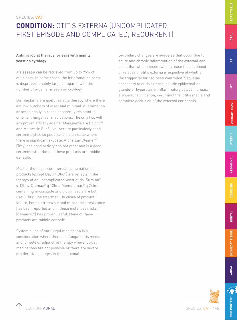
SPECIES: CAT
SPECIES: CAT 145SECTION: AURAL
CONDITION: OTITIS EXTERNA (UNCOMPLICATED, FIRST EPISODE AND COMPLICATED, RECURRENT)
SO
FT
TIS
SU
EO
RA
LU
RT
LR
TU
RIN
AR
Y T
RA
CT
PY
RE
XIA
AB
DO
MIN
AL
DE
SE
XIN
GD
EN
TAL
SKIN
/SO
FT T
ISSU
EA
UR
AL
DO
G C
ON
TE
NT
Antimicrobial therapy for ears with mainly yeast on cytology
Malassezia can be retrieved from up to 95% of otitis ears. In some cases, the inflammation seen is disproportionately large compared with the number of organisms seen on cytology.
Disinfectants are useful as sole therapy where there are low numbers of yeast and minimal inflammation or occasionally in cases apparently resistant to other antifungal ear medications. The only two with any proven efficacy against Malassezia are Epiotic® and Malacetic Otic®. Neither are particularly good ceruminolytics so penetration is an issue where there is significant exudate. Alpha Ear Cleaner® (Troy) has good activity against yeast and is a good ceruminolytic. None of these products are middle ear safe.
Most of the major commercial combination ear products (except Baytril Otic®) are reliable in the therapy of an uncomplicated yeast otitis. Surolan® q 12hrs, Otomax® q 12hrs, Mometamax® q 24hrs containing miconazole and clotrimazole are both useful first line treatment. In cases of product failure, both clotrimazole and miconazole resistance has been reported and in these instances nystatin (Canaural®) has proven useful. None of these products are middle ear safe.
Systemic use of antifungal medication is a consideration where there is a fungal otitis media and for sole or adjunctive therapy where topical medications are not possible or there are severe proliferative changes in the ear canal.
Secondary changes are sequelae that occur due to acute and chronic inflammation of the external ear canal that when present will increase the likelihood of relapse of otitis externa irrespective of whether the trigger factor has been controlled. Sequelae secondary to otitis externa include epidermal or glandular hyperplasia, inflammatory polyps, fibrosis, stenosis, calcification, ceruminoliths, otitis media and complete occlusion of the external ear canals.
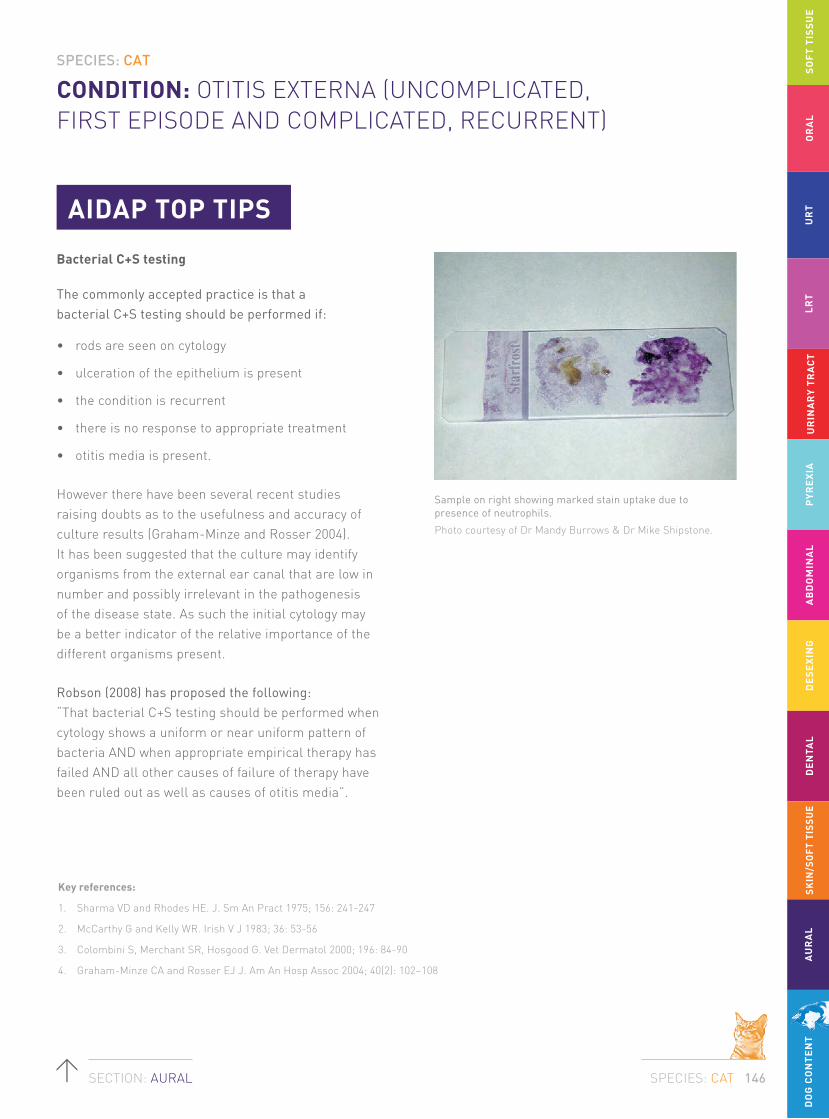
SPECIES: CAT
SPECIES: CAT 146SECTION: AURAL
CONDITION: OTITIS EXTERNA (UNCOMPLICATED, FIRST EPISODE AND COMPLICATED, RECURRENT)
SO
FT
TIS
SU
EO
RA
LU
RT
LR
TU
RIN
AR
Y T
RA
CT
PY
RE
XIA
AB
DO
MIN
AL
DE
SE
XIN
GD
EN
TAL
SKIN
/SO
FT T
ISSU
EA
UR
AL
DO
G C
ON
TE
NT
AIDAP TOP TIPS
Bacterial C+S testing
The commonly accepted practice is that a bacterial C+S testing should be performed if:
• rods are seen on cytology
• ulceration of the epithelium is present
• the condition is recurrent
• there is no response to appropriate treatment
• otitis media is present.
However there have been several recent studies raising doubts as to the usefulness and accuracy of culture results (Graham-Minze and Rosser 2004). It has been suggested that the culture may identify organisms from the external ear canal that are low in number and possibly irrelevant in the pathogenesis of the disease state. As such the initial cytology may be a better indicator of the relative importance of the different organisms present.
Robson (2008) has proposed the following: “That bacterial C+S testing should be performed when cytology shows a uniform or near uniform pattern of bacteria AND when appropriate empirical therapy has failed AND all other causes of failure of therapy have been ruled out as well as causes of otitis media”.
Sample on right showing marked stain uptake due to presence of neutrophils.
Photo courtesy of Dr Mandy Burrows & Dr Mike Shipstone.
Key references:
1. Sharma VD and Rhodes HE. J. Sm An Pract 1975; 156: 241-247
2. McCarthy G and Kelly WR. Irish V J 1983; 36: 53-56
3. Colombini S, Merchant SR, Hosgood G. Vet Dermatol 2000; 196: 84-90
4. Graham-Minze CA and Rosser EJ J. Am An Hosp Assoc 2004; 40(2): 102–108

Copyright © 2013 Zoetis Inc. All rights reserved. Zoetis Australia Pty Ltd. ABN 94 156 476 425. 38-42 Wharf Road, West Ryde NSW 2114. www.zoetis.com.au AM56255 JUNE 2013
AIAIDDAPAPAUSTRALASIAN INFECTIOUSDISEASES ADVISORY PANEL
Zoetis would like to thank the dedicated members of AIDAP for all their hard work and contribution towards these guidelines. AIDAP, the Australasian Infectious Diseases Advisory Panel, is a committee of Specialists with fields in Internal Medicine, Feline Medicine, Dermatology and Microbiology. The panel works together with Zoetis to assist with the ongoing understanding of the nature of infectious diseases; the understanding of how to treat infectious diseases; and also the current rationale for the appropriate use of antibiotics.
Please note, these recommendations are based entirely on the decisions made by the AIDAP committee, and some of these recommendations include the "off-label" use of certain medications. These off-label uses are not endorsed by Zoetis.
For more information please visit www.vetsaustralia.com.au.
ANTIBIOTICPRESCRIBINGGUIDELINES
AUSTRALASIAN INFECTIOUS DISEASES ADVISORY PANELAIDAP
ANTIBIOTICPRESCRIBINGDETAILED GUIDELINES
AUSTRALASIAN INFECTIOUS DISEASES ADVISORY PANELAIDAP