aesthetic surgery journal upper lid blepharoplasty ... · the eyebrow modifies the shape of the...
TRANSCRIPT

Oculoplastic Surgery
Aesthetic Surgery Journal33(1) 24 –30© 2013 The American Society for Aesthetic Plastic Surgery, Inc.Reprints and permission: http://www .sagepub.com/journalsPermissions.navDOI: 10.1177/1090820X12468751www.aestheticsurgeryjournal.com
The force of gravity and, more important, the traction con-nected to the contraction of the orbicular muscle eventu-ally cause relaxation of the integuments of the lateral part of the forehead. This results in lowering of the lateral third of the eyebrow, the appearance of excess skin in the upper eyelid, and the accumulation of an overabundance of skin in the area above the lateral corner.
McCord1 compared the mechanics of brow ptosis “to that of a curtain rod that has loosened and fallen, causing folding of the curtain.” This phenomenon, related to age as well as local anatomic structure, is often accompanied by functional inconveniences related to heaviness of the lid, which fre-quently are alleviated by frowning. As a result, the patient’s range of vision may be reduced, sometimes markedly.
Sagging of the eyebrows contributes to a sadder, older, and more fatigued appearance. Many patients seek remedies that will mask or correct this concern. Today, women of every
social class depilate the lower lateral part of the eyebrow to obtain the illusion of raising the eyebrow and/or create more
Upper Lid Blepharoplasty, Eyebrow Ptosis, and Lateral Hooding
Giacomo Bellinvia, MD; Francesco Klinger, MD; Luca Maione, MD; and Pietro Bellinvia, MD
AbstractBackground: Upper blepharoplasty is a common aesthetic surgical procedure. Different surgical techniques and markings have been described, some of which may incorporate browpexy or browlifting. Considering the upper periorbital area as an aesthetic unit, various techniques of browpexy and browlifting have been taken into consideration.Objectives: The authors describe their technique for upper blepharoplasty, which includes an extended cutaneous excision that permits good correction of lateral hooding, reducing the need for browpexy or browlifting. Indications and surgical outcomes are also presented.Methods: The authors retrospectively reviewed the records of 552 consecutive patients who underwent upper blepharoplasty according to the authors’ technique between January and December 2008. The patients received local anesthesia. Cutaneous excision extended beyond the eyelid to include the lower lateral portion of the eyebrow area.Results: Optimal functional correction was achieved in all cases, and the majority of patients were pleased with their aesthetic result. No significant complications occurred; 4 minor complications were noted and are described fully in the text.Conclusions: The authors’ modification of the usual technique of blepharoplasty results in excellent correction, both functional and aesthetic, of the periocular area, particularly the lateral portion. The technique is safe, and patient satisfaction is high.
Level of Evidence: 4
Keywordsupper blepharoplasty, lateral hooding, eyebrow ptosis, oculoplastics
Accepted for publication June 19, 2012.
Drs G. Bellinvia and P. Bellinvia are surgeons in private practice in Milan, Italy. Drs Klinger and Maione are staff surgeons in the Department of Plastic Reconstructive and Aesthetic Surgery, University of Milan, Italy.
Corresponding Author:Dr Giacomo Bellinvia, Via Monte Amiata, 3 20149, Milan, Italy. E-mail: [email protected]
INTE
RNAT
IONAL CONTRIBUTION
Scan this code with your smartphone to see the operative video. Need help? Visit www.aestheticsurgery.com.

Bellinvia et al 25
space between the eye and eyebrow. Some people depilate very extensively and end up completely or partly redrawing their brows in a higher position, using a pencil; others resort to “permanent makeup” (tattoos).
In the current study, 90% of women had modified the natural lines of their eyebrows before undergoing blepharoplasty. Therefore, independent of the presence or absence of hair, we defined the eyebrow as the strip of thick skin that initially follows the orbital arch and, with the passage of time, descends laterally.
The eyebrow and eyelid form an aesthetic unit. To obtain rejuvenation of this area as a whole, it is necessary to correct the excess skin of the eyelid and lateral third of the eyebrow, together with the cutaneous excess that derives from the latter.
The lowering that occurs with time is generally most pronounced in the lateral part of the eyebrow. Therefore, this should be the primary area of focus. Endoscopic lifting of the medial and central parts of the brow can be prob-lematic and may necessitate complex restorative operations later.2 According to Yaremchuk et al,2 a “young” eyebrow is low: “Young attractive women have low brows.” Moreover, Troilius3 believes that “surgeons are creating too many unnatural high brows.”
The shape of the eyebrow is more important than its height; the medial and lateral ends should lie at approxi-mately the same height.4 Resection of the sagging part of the eyebrow modifies the shape of the eyebrow itself, cor-recting it with an aspect nearly the same as surgical lifting of the lateral third. Therefore, additional procedures such as browpexy and browlifting (which increase time and cost and may not produce good results) may be avoided.
MEthOdsStudy PopulationThe study population consisted of consecutively treated patients (N = 552) who underwent upper blepharoplasty between January and December 2008 and whose records
were retrospectively reviewed. The study was conducted in the senior author’s private practice facility. No patient was excluded from the study. Most patients (387 women and 26 men) underwent upper blepharoplasty alone; some had undergone blepharoplasty previously. The others (129 women and 10 men) underwent lower blepharoplasty in addition to upper blepharoplasty.
Thorough evaluation of preoperative clinical photo-graphs revealed the following:
• In 58 patients (10.5% overall; 9.8% of women; 19.4% of men), the edge of the eyelid was com-pletely covered and hidden by excess skin. These patients required a distinctly functional outcome.
• In 170 patients (29.7% overall; 29% of women; 55.5% of men), there was a partially functional component: excess skin concealed the eyelid edge only laterally.
• In 217 patients (39.3% overall; 40.5% of women; 22.2% of men), there was no significant impact on the field of vision, but the excess skin repre-sented a functional inconvenience and was often accompanied by persistent frowning.
• In 107 patients (19.3% overall; 20.5% of women; 2.7% of men), the inconvenience was primarily aesthetic.
Preoperative Markings
With the patient seated and the eye closed, and without stretching the skin, a line was drawn at the top margin of the area to be excised, starting medially, 5 to 6 mm over the medial canthus. The marking was curvilinear and con-vex superiorly, leaving the thin lid skin to reach the thick skin over the super orbital rim, never going downward. The line ended laterally, over and beyond the area of lat-eral hooding, at about the height of the medial margin of the eyebrow (Figure 1). Sometimes the mark was placed just under the brow, particularly in men. In cases of severe
Figure 1. Preoperative markings are shown with (A) the eye open and (B) the eye closed, with the eyelid skin stretched upward.

26 Aesthetic Surgery Journal 33(1)
redundancy (and a hairless brow for cosmetic reasons), the mark often crossed the skin of the lateral brow region.
A horizontal line was then drawn at the lower margin of the incision lines, starting over the medial canthus and running horizontally for only half the lid, approximately 10 to 12 mm above the eyelid margin—without reference to the lid crease and generally above it. The line continued upward, gently slanting to meet the end of the upper mar-gin of the drawing. The vertical distance between the lat-eral canthus and the lower line generally varied from 15 to 25 mm. This allowed for preservation of a large amount of the lower, thin elastic skin. Precision of the markings was paramount because they could not be modified after the administration of local anesthesia.
Surgical Technique
All blepharoplasty procedures were performed in a licensed outpatient surgical facility, with the patient under local anesthesia after having been given anxiolytic drops (brom-azepam). The anesthetic (4-5 mL per side) consisted of 0.2% lidocaine and 1:280 000 epinephrine.
To obtain optimal correction of the cutaneous excess and the lateral sagging of the eyebrow, our technique evolved over the years to include a wider incision in the lateral area of the eyebrow (Figures 1 and 2). Skin excision was superfi-cial and therefore did not interfere with the vascular or lym-phatic supply to the lid. (This minimized edema and bruising and expedited recovery.) If indicated, the medial fat pads were removed. The strip of orbicular muscle was not excised.
We did not attempt to create variations in eyelid type, and we avoided correcting the tarsal sulcus or evacuating fat from “plump” eyelids. A certain “fullness” in the upper eyelid is typical of a youthful eye and therefore it was not modified. Fatty medial hernias were removed in some patients. In 3 cases, correction of monolateral ptosis was performed in conjunction with the blepharoplasty, as described by Carraway and Tran.5
The entire operation took less than 30 minutes. Two-thirds of that time was spent securing subcuticular run-ning sutures (6-0 nylon). Cold packs were applied before discharge. Sutures were removed 5 to 6 days postopera-tively, and a topical antibiotic ointment was applied to the wounds.
Assessments
To evaluate long-term patient satisfaction, a telephone inter-view was conducted with 100 patients, chosen randomly, whose surgery had taken place at least 3 years earlier. The survey was carried out by office staff, and the participants were selected alphabetically from the list of patients who underwent upper blepharoplasty in 2008. Patients were asked about their overall satisfaction with the operation (very satisfied, satisfied, dissatisfied) and the degree of vis-ibility of their scars (insignificant, modest, marked).
A video of the authors’ technique is available at www .aestheticsurgeryjournal.com. You may also use any
smartphone to scan the code on the first page of this article to be taken directly to the video on www.YouTube.com.
REsuLts
Although the authors have performed their technique of upper blepharoplasty on 5530 patients in the past 10 years (January 2002 to December 2011), the present study focuses on the operations that occurred from January 2008 through December 2008. During this 1-year period, 552 patients (516 women, 36 men) underwent upper blepharo-plasty. The year 2008 was chosen for this retrospective analysis because of the substantial number of patients treated and because it was the most recent year that offered sufficient time for follow-up.
Upper blepharoplasty alone was performed in 413 cases (387 women, 26 men). The other patients underwent lower blepharoplasty as well (129 women, 10 men). The overall age range was 30 to 87 years, and the average age was 54 years (Table 1). In 19 cases (17 women, 2 men), the upper blepharoplasty was secondary, sometimes to blepharoplasty performed elsewhere.
After the sutures were removed (5-6 days postopera-tively), patients were asked to return for a follow-up appointment 6 months later. Approximately 50% of the patients returned for this checkup. Observation beyond 6 months is common in our practice.
The surgery resolved all functional inconveniences in all patients who had experienced them preoperatively (80%), according to observation of preoperative photo-graphs. Although the correction of the shape of the eye-brow obtained by removing the lateral lower area is not the same as that of browpexy or browlifting, it does modify the area in a similar fashion.
Recovery time was brief, with bruising being absent or minimal. Only 1 patient (0.18%) experienced a complication: an allergic cutaneous reaction to the antibiotic ointment that included edema, itching, and marked reddening. In 3 cases (0.54%), secondary correction of the medial adipose hernias was necessary because of inadequate removal.
Six to 7 months after the operation, the surgical scar was undetectable in 92% of patients. In others, there was
Table 1. Type of Operation According to Age and Sex
Age Range, yUpper Blepharo-
plasty OnlyUpper and Lower Blepharoplasty
Secondary Upper Blepharoplasty
Total No. of Patients
30-39 1 M, 29 F 0 M, 7 F None 37
40-49 5 M, 79 F 2 M, 26 F None 112
50-59 8 M, 120 F 4 M, 49 F 0 M, 6 F 181
60-69 7 M, 91 F 3 M, 31 F 1 M, 5 F 132
70-79 3 M, 43 F 1 M, 10 F 1 M, 5 F 57
80-89 2 M, 25 F 0 M, 6 F 0 M, 1 F 33
Abbreviations: M, male; F, female.
Bellinvia et al 27
only minimal evidence of the scar (noted by approxi-mately 8% of patients during telephone follow-up). In a few of these patients, the visibility of the scar related to a difference in pigmentation between the 2 cutaneous mar-gins that were sutured. In 3 patients (0.54%) who had excessive sun exposure in the weeks immediately after surgery, pigmentation of the scar was observed laterally.
Our patients had been informed that it could take months for a lateral scar to become negligible. However, the outcomes were so successful that no patient com-plained of this. Hypertrophic scars were not observed. Even in patients prone to keloids whose excision extended beyond the orbital rim, scars became negligible within a few weeks or months, owing to the precise technique used for the subcuticular running suture.
Reddening of the scar typically lasted longer (some-times 7-8 months) in individuals with fair, thick skin, which is not unusual. Patients with dark lid skin had been informed that a permanent different shade might result from the scar. All final scars had a continuous gentle cur-vature, slanting upward; even without makeup, they
appeared to be a natural cosmetic line. Secondary correction of scars was not required for any patient. Intradermic suturing resulted in a low frequency of microcysts along the scar.
No patient requested further correction of the eyebrow.Results from the telephone survey of random patients
from this series are shown in Table 2. Satisfaction was very high for patients who underwent the procedure purely for cosmetic reasons and for those who required functional correction. Only 1 patient was not satisfied; this patient would have liked a more marked cutaneous removal. A high degree of satisfaction also was evidenced by what we subjectively observed as a great demand for the operation from people who had witnessed the results for these patients.
Clinical results and illustrations are shown in Figures 3 through 7.
disCussiOn
The traditional blepharoplasty excision6 allows for removal of excess skin by designing an ellipse with a dart that extends laterally and is slanted slightly upward. The lower “limb” of the elliptical excision is located approximately 5 to 7 mm above the ciliary margin. Even when the lateral cutaneous excess is removed, it is generally believed that for the suture line to be virtually undetectable, it must pass a few millimeters above the external canthus.
Various surgeons have modified the usual excision in an attempt to minimize the appearance of the scar. Eremia and Willoughby7 moved the lateral excision slightly upward so that the suture terminates in a lower position, halfway between the end of the eyebrow and the lateral projection of the external canthus. In this manner, the scar is hidden by the remaining lateral cutaneous excess. Har-Shai and Hirshowitz,8 using a cutaneous excision in the form of a scalpel blade, modified Rees’s design6 by widen-ing the lateral cutaneous excision upward. Har-Shai and Hirshowitz8 maintained the distance between the lower
Table 2. Survey Results for a Random Sample of Patients (Conducted 3+ Years Postoperatively)
Result No. of Patients
Overall satisfaction
Very satisfied 83
Satisfied 16
Dissatisfied 1
Visibility of scar
Insignificant 92
Modest 8
Marked 0
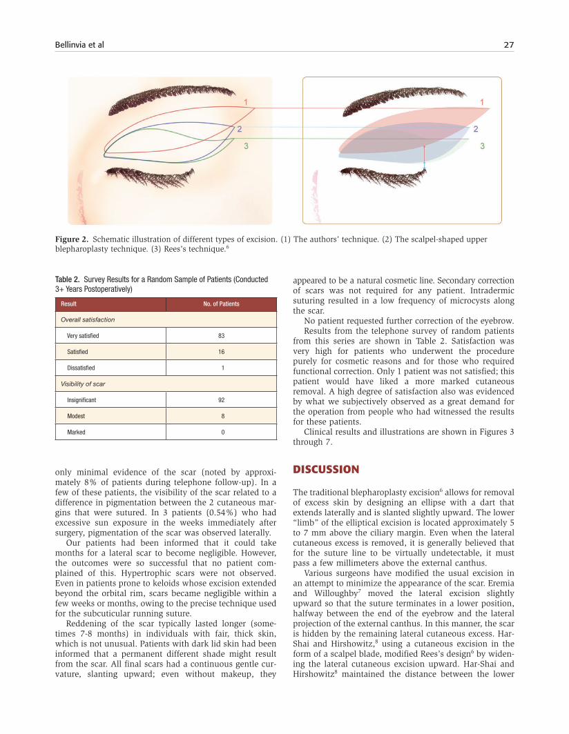
Figure 2. Schematic illustration of different types of excision. (1) The authors’ technique. (2) The scalpel-shaped upper blepharoplasty technique. (3) Rees’s technique.6

28 Aesthetic Surgery Journal 33(1)
incision line and the lateral canthus unchanged. To pre-vent the eyebrow from being stretched downward, they applied this technique only in patients who are not young, whose eyebrows are positioned normally, and “in whom there is little if any ptosis of the lateral eyebrow.” Lee and Law9 sometimes performed blepharoplasty under the eye-brow to correct the lateral cutaneous excess in Asian patients with particularly high eyebrows. This is an unu-sual excision, limited to selected cases and not appropriate for white patients. Har-Shai et al10 modified the technique of Castañares11 (the removal of skin above the eyebrow) to correct both the ptosis of the eyebrow and the lateral cuta-neous excess. In the case of lateral hooding, this technique raises the eyebrow too much. Moreover, it cannot be used if the eyebrow has been depilated or redrawn.
Over the years, we too have moved the cutaneous exci-sion of blepharoplasty upward, with markings that some-what resemble those of Har-Shai and Hirshowitz.8 However, our procedure differs because of the greater distance from the lid margin and, more important, from the lateral corner (Figure 2). The surplus skin removed laterally involves the eyebrow area, and the falling line is
modified as if lifting of the eyebrow had been carried out. A substantial amount of skin is left below the skin exci-sion, considering that the lower marking of the excision is above the tarsal sulcus, 10 to 12 mm from the eyelash. The vertical distance above the external canthus is even greater (15-25 mm). The lateral cutaneous excess is extended and redistributed upward. Thus, the “fallen” line is eliminated, and the result is an apparent lifting of the eyebrow (Figure 3). In essence, we have raised the rod of the curtain of McCord1: the thin elastic eyelid skin above the canthus is preserved and extended upward, which eliminates lateral hooding. In our experience, better and longer-lasting corrections have been achieved with this technique than with browlifting or temporal lifting. (If significant lateral hooding is present, raising the eyebrow to the height necessary for successful correction would bring it to an unnatural position.) Because the lateral cuta-neous excision is higher, the sutured cutaneous margins are of equal thickness, which minimizes the scar (Figure 4). The abundance and elasticity of residual skin allows for intradermic suturing without any tension on the mar-gins. The eventual tension on the margins of the suture is substantiated by the shape of the scar, which takes on the form of a lazy “S” when the tissues are stretched toward the lateral canthus (Figure 8). Residual scar visibility is lessened by a scar that, in our hands, never takes on the form of an “S”; rather, it remains uniformly curved, with a direction similar to that of a young person’s eyebrow: upward or horizontal (never downward), with perfect adherence to the aesthetic unity of the area (Figure 9).
Accurate intradermic suturing and the absence of trac-tion are necessary to obtain, even in an area of thick skin, optimal scars destined to become negligible in most cases. The cutaneous removal often is relevant in areas that depilation has made “available.” In patients who have designed the eyebrow above its original position, the lat-eral eyebrow skin may be removed partially or completely. This permits corrections that otherwise would not be pos-sible, particularly in older adults.
In men and women who have not depilated their eye-brows, the upper margin of excision coincides with the lower edge of the natural eyebrow. Sometimes a strip of eyebrow skin is removed. The excision markings of the eyelid do not take into account the tarsal sulcus, which generally remains lower. Since the lower marking of the excision is higher than
Figure 3. (A) This 43-year-old woman presented for rejuvenation of the eyelid area and correction of lateral hooding. (B) Preoperative markings. (C) Six months after upper blepharoplasty performed according to the authors’ technique.
Figure 4. This 61-year-old woman had undergone “traditional” blepharoplasty 13 years earlier and the authors’ blepharoplasty technique 3 years earlier. Although the new scar (arrow) is in a more visible area, it is much less noticeable than the original scar (which is lower and joins different types of skin).

Bellinvia et al 29
the tarsal sulcus, it is not necessary to remove a strip of orbicularis muscle, which is carried out to avoid thickening of the upper edge of the scar.
Superficial cutaneous removal respects the vascular and lymphatic network. Therefore, edema and ecchymosis are often absent, and recovery time is reduced. With this type of excision, it is possible to perform secondary blepharoplasty with good results, even if the previous operation had been performed traditionally. This type of correction is used by the authors for numerous orbital-palpebral morphologies. However, the specific excision lines should be adapted to each patient’s clinical scenario. Important considerations include patient age, the extent of lateral hooding, and the quality and pigmentation of the skin. The patient’s expectations also must also be taken into account. (Sometimes it is better to obtain a minor
correction and have negligible scarring than risk greater scarring from a major correction.)
Although visible scarring is always an aesthetic con-cern, particularly after considerable cutaneous excision in areas where the skin is thick, this has not been an issue with our surgical technique. There have been no com-plaints about scarring. We have performed upper blepha-roplasty, without problems, in a large number of patients over the past decade. This includes relatively young peo-ple as well as public figures. Even the patients whose cutaneous pigmentation put them at greater risk for scar-ring (of which they had been warned) obtained excellent correction with minimal scarring. The minimal scar has been well accepted by our patients, even when it is recent and visible. Because the upward direction resembles a makeup line, the scar is easily masked. The demand for
Figure 5. (A) This 50-year-old woman presented for rejuvenation of the eyelid area and correction of lateral hooding. (B) Preoperative markings. (C) Six months after upper blepharoplasty performed according to the authors’ technique.
Figure 6. (A) This 48-year-old woman presented for rejuvenation of the eyelid area and correction of lateral hooding. (B) Preoperative markings. (C) Six months after upper blepharoplasty performed according to the authors’ technique.
Figure 7. (A) This 70-year-old woman presented for rejuvenation of the eyelid area and correction of lateral hooding. (B) Preoperative markings. (C) Six months after upper blepharoplasty performed according to the authors’ technique.

30 Aesthetic Surgery Journal 33(1)
our procedure continues to grow, and many patients who underwent traditional blepharoplasty elsewhere have requested this technique.
COnCLusiOns
Our modification to traditional blepharoplasty involves treating the eyelid and eyebrow as an aesthetic unit.
Although the technique requires greater suturing preci-sion, the results have been superior and more natural than those of traditional blepharoplasty. Both functional and aesthetic results have been excellent, and no significant complications have occurred.
disclosures
The authors declared no potential conflicts of interest with respect to the research, authorship, and publication of this article.
Funding
The authors received no financial support for the research, authorship, or publication of this article.
REFEREnCEs
1. McCord CD. Upper blepharoplasty and eyebrow surgery. In: Chen WP, ed. Oculoplastic Surgery. New York, NY: Thieme; 2001:125-145.
2. Yaremchuk MJ, O’Sullivan N, Benslimane F. Reversing brow lifts. Aesthetic Surg J. 2007;27:367-379.
3. Troilius C. Subperiostal browlifts without fixation. Plas Reconstr Surg. 2004;114:1595-1603.
4. Pham S, Wilhelmi B, Mowlavi A. Eyebrow peak position redefined. Aesthetic Surg J. 2010;30:297-300.
5. Carraway JH, Tran P. Blepharoplasty with ptosis repair. Aesthetic Surg J. 2009;29:54-61.
6. Rees TD. Blepharoplasty. In: Rees TD, Woodsmith D, eds. Cosmetic Facial Surgery. Philadelphia, PA: Saunders; 1973:44-133.
7. Eremia S, Willoughby LA. Upper blepharoplasty with cor-rection of the lateral hooding. In: Moy RL, Fincher EF, eds. Blepharoplasty. Philadelphia, PA: Elsevier; 2006:53-76.
8. Har-Shai Y, Hirshowitz B. Extended upper blepharoplasty for lateral hooding of the upper eyelid using a scalpel blade excision: a 13-year experience. Plast Reconstr Surg. 2004;113:1028-1035.
9. Lee D, Law V. Subbrow blepharoplasty for upper eyelid rejuvenation in Asians. Aesthetic Surg J. 2009;29:284-289.
10. Har-Shai Y, Gil T, Metanes I, Scheflan M. Brow lift for the correction of visual field impairment. Aesthetic Surg J. 2008;28:512-517.
11. Castañares S. Forehead wrinkles, glabellar frown and ptosis of the eyebrows. Plast Reconstr Surg. 1964;34:406-413.
Figure 8. With the scalpel-shaped technique, the scar resembles a lazy “S” when the tissues are stretched downward toward the lateral canthus.
Figure 9. Lateral hooding is corrected by moving the thin elastic skin above the canthus in an upward direction.