advances in fertility options of azoospermic men
TRANSCRIPT

8
Advances in Fertility Options of Azoospermic Men
Bin Wu1, Timothy J. Gelety1 and Juanzi Shi2
1Arizona Center for Reproductive Endocrinology and Infertility Tucson, Arizona
2Shaanxi Province Hospital for Women and Children Health Care Xi’an, Shaanxi
1USA 2People’s Republic of China
1. Introduction
Infertility is defined as the failure of a couple to become pregnant after one year of regular,
unprotected, sexual intercourse. Misconceptions about infertility are very common.
Infertility affects roughly 15% of all couples during their reproductive lives. Infertility is not
"just a female problem" as there is a male infertility component in approximately 50% of
couples. A couple experiencing infertility should not underestimate the significance of the
problems that can exist in the male. Male infertility may be the sole contributing reason for
the couple's failure to conceive and should be best identified by a male infertility specialist.
According to the 2009 national summary report of the American Society for Assisted
Reproductive Technology, nearly 35% of infertility is attributed to the male factor. In the late
last century, treatment for severe male factor infertility was limited to inseminations or in vitro fertilization using donor sperm. However, most infertile couples, and particularly men,
are reluctant to use donor sperm because of bias maintained across cultural and ethnic
boundaries. Today, exciting advances in male infertility have introduced innovative
therapeutic options, in particular, intracytoplasmic sperm injection (ICSI), which offers men,
including those with no sperm (azoospermia) in their ejaculate due to genetic conditions, a
greatly improved chance to conceive their own biological offspring (Palermo, et al.. 1992).
Currently, despite severe male factor infertility, pregnancy may still be achieved. This is
mainly attributed to the success of the ICSI technique and advanced surgical testicular
sperm retrieval techniques. In recent years, the ICSI technique has evolved into
intracytoplasmic morphologically selected sperm injection (IMSI) and a method for selection
of hyaluronan bound sperm for use in ICSI (PICSI) (Parmegiani et al., 2010a,b; Said & Land
2011; Berger et al., 2011), resulting in significantly increased fertilization and pregnancy
rates over the world IVF clinics. Because the advanced ICSI technique in human IVF is
covered in depth by other chapters of this book, the main emphasis here will be on the
application of testicular biopsy sperm, round or elongated spermatids from azoospermic
men to human IVF as well as explore new technologies to produce artificial sperm from
stem cells and somatic cells.
www.intechopen.com

Advances in Embryo Transfer
116
2. Evaluation of male infertility-definition of azoospermia
There are a variety of conditions that may lead to male infertility. An important component in the treatment of men with infertility is establishing the correct diagnosis through a thorough clinical evaluation of each couple. The cornerstone of the male fertility evaluation is the semen analysis, which is also the simplest evaluation for a male fertility. Complete evaluation of the infertile male involves a comprehensive medical history, physical examination and two and more semen analyses.
In addition to semen analysis, specialized sperm function testing are available, including measurement of sperm capacitation and the acrosome reaction, computer assisted sperm motion analysis (CASA), sperm antibody testing, and leukocyte quantitation. According to the World Health Organization (WHO, 2010), the lower reference limit for semen characteristics should include a semen volume (1.4-1.7ml), sperm concentration (12-16 million/ml), motility (38-42%), and morphology (3.0-4.0), as an integral screen of sperm fertilization potential. A persistently abnormal finding outside of the normal range often indicates male infertility. An abnormal semen analysis may be the first sign of significant pathology that may be life threatening in up to 1-2% of cases. The diagnosis of infertility may not only indicate a problem with the husband but also may put the health of his offspring at risk. With a growing understanding about the genetics of male infertility, a genetic cause may exist in up to 20% of patients. The primary goal of the evaluation is to determine the cause of the problem and to exclude life threatening pathology. Thus, genetic diagnosis of male infertility plays an important role in the application of assisted reproductive technology. Today, genetic testing for cystic fibrosis, Karyotyping and Y microdeletion analysis are important male infertility diagnostic tests for couples who are at risk of inherited genetic diseases.
After detailed semen analysis, potential causes of male infertility may present (Comhaire & Vermenlen, 1995) as (1) complete absence of sperm (azoospermia), (2) low sperm count (oligozoospermia), (3) abnormal sperm shape (teratozoospermia), (4) problems with sperm movement (asthenozoospermia), (5) completely immobile sperm (necrozoospermia), where the sperm may be alive and not moving, or they may be dead (6) problems with sperm delivery, due to sexual dysfunction, an obstruction, previous vasectomy, or retrograde ejaculation. In an earlier generation of investigations, the most common cause of male infertility was reported to be the presence of a varicocele, which is the presence of a varicose vein found in the scrotum. The extra heat caused by the vein was suggested to lead to low sperm counts and impaired sperm movement. However, some clinical studies have failed to demonstrate a significant increase in pregnancy following varicocele repair. It is likely that genetic abnormalities noted in the modern studies of Y chromosomal micro-deletions account for abnormal parameters. With the development of intracytoplasmic sperm injection (ICSI) technology, a laboratory specialist uses microscopic tools to isolate a single sperm and then injects it directly into an egg. This technique, used in conjunction with in vitro fertilization (IVF), has been especially successful as a treatment for men who have a low sperm count (oligozoospermia), asthenozoospermia and women who have a small number of mature eggs. Also, oligozoospermic men often carry seminal populations demonstrating increased chromosomal aberrations and compromised DNA integrity. Therefore, the in vitro selection of sperm for ICSI is critical and directly influences the paternal contribution to preimplantation embryogenesis. Hyaluronan (H), a major constituent of the cumulus matrix, may play a critical
www.intechopen.com

Advances in Fertility Options of Azoospermic Men
117
role in the selection of functionally competent sperm during in vivo fertilization (Parmegiani et al., 2010a). Hyaluronan bound sperm (HBS) exhibit decreased levels of cytoplasmic inclusions and residual histones, an increased expression of the HspA2 chaperone protein and a marked reduction in the incidence of chromosomal aneuploidy. The relationship between HBS and enhanced levels of developmental competence led to the current clinical trial (Worrilow et al., 2010). Thus, as a HBS test, a PCISI technique, a method for selection of hyaluronan bound sperm for use in ICSI, has been developed to treat oligozoospermic and asthenozoospermic man infertility problem. Additionally, recently advanced IMSI technique has been used to treat sperm morphology problems (teratozoospermia). In cases of necrozoospermia, motile spermatozoa may be found by adding pentoxifylline or other chemicals such as 2-deoxyadenosine to treat semen and sperm (Angelopoulos et al., 1999), or live sperm will be determined by hypo-osmotic swelling test (Takahashi et al., 1990).
The most sever cases of male infertility are those presenting with no sperm in the ejaculate
(azoospermia). Some men have a condition where their reproductive ducts may be absent or
blocked (obstructive azoospermia or OA), where others may have no sperm production
with normal reproductive anatomy (non-obstructive azoospermia or NOA). Azoospermia is
found in 10% of male infertility cases. Patients with OA due to congenital bilateral absence
of the vas deferens or those in whom reconstructive surgery fails have historically been
considered infertile. Men who can not produce sperm in their testes with apparent absence
of spermatogenesis diagnosed by testicular biopsy are classified as NOA. Once testicular
and epidiymal function can be verified, surgery may be justified to correct or remove the
blockage. Alternatively, the current optimal method for treatment of azoospermic men is to
acquire sperm from the testis or epididymis by either non-surgical or surgical means.
3. Acquiring sperm from azoospermic men
For men with no sperm at all in their semen, sperm may be obtained by non-surgical or surgical techniques based on the complete evaluation and diagnosis of male factor infertility.
3.1 Non-surgical sperm retrieval techniques
3.1.1 Electro-ejaculation (EEJ)
Many factors may predispose spinal cord injured men to infertility. Ejaculatory dysfunction, abnormalities of sperm production, chronic infections and blockage of sperm within the male reproductive tract are all potential factors. Currently a number of different methods have been used to obtain sperm from azoospermic men. For example, sperm can often be obtained through vibratory stimulation to the head and shaft of the penis. Men with ejaculatory failure due to nerve damage caused by spinal injury, and occasionally by other conditions, can produce sperm by electrical stimulation of the ejaculatory ducts internally. Electro-ejaculation or sperm harvesting along the ejaculatory path from the vas deferens, epididymis, and directly from the testis is a very successful form of therapy for men who have normal sperm production but can not ejaculate because of a short circuit in the nervous system. Though sperm quality is often poor due to remaining too long in the body, the sperm are usually suitable for ICSI treatment. EEJ has also proven effective for loss of ejaculation in patients with other conditions such as diabetes, retroperitoneal lymph node dissection, pelvic surgery, multiple sclerosis, or unexplained loss of orgasm. EEJ is non-invasive and patients routinely
www.intechopen.com
Advances in Embryo Transfer
118
return back to desk type work that day after a pain-free procedure of 30 minutes with local anesthesia at an outpatient surgery center. Electro-ejaculation allows the retrieval of sperm in more than 90% of patients, but in most of cases, sperm concentration and sperm motility is relatively low. Combining EEJ with current ICSI techniques, this may yield up to a 50% pregnancy rate. Overall, the chances for pregnancy in the informed and motivated patient are similar to those of a healthy male. However, electro-ejaculation must be performed under satisfactory anesthesia in men with spinal cord injuries with sensation in or below the abdomen. To avoid the complication of autonomic dysreflexia, a complete urologic examination must be performed prior to the procedure to detect and treat any urinary tract infections. Often during this procedure retrograde ejaculation occurs, which is a backwards ejaculation into the bladder, and sperm must be collected from the urine.
3.1.2 Testicular sperm aspiration (TESA) and percutaneous epididymal sperm aspiration (PESA)
TESA and PESA have been used as minimally invasive or non-surgical sperm aspiration
techniques for use in conjuncture with assisted reproductive technology. This technique is a
quick and painless procedure performed under sedation. A tiny percutaneous needle is
used to extract sperm directly from the testis or epididymis. Using these minimally invasive
techniques, sperm can be obtained from men with vasectomy, failed vasectomy reversal,
absence of the vas deferens, or uncorrectable blockages anywhere along the seminal tract
(obstructive azoospermia). In addition, individual sperm occasionally may be obtained in
some cases of non-obstructive azoospermia (NOA). Sperm retrieval procedures are typically
done at an outpatient surgery and last about one hour. The advantages of percutaneous
aspiration techniques are that they can be performed with local anesthesia, without open
scrotal exploration and its attendant postoperative discomfort, and without microsurgical
expertise. Also it does not require hospitalization, and recovery is virtually immediate.
Patients return back to desk type work in one or two days. It should be noted that for some
men, a single percutaneous aspiration procedure may yield enough sperm to permit sperm
freezing for several subsequent ICSI attempts. While the ejaculate normally contains 100 to
300 million sperm, aspiration of as few as 100-200 sperm by this technique have been
enough to achieve pregnancy when it is combined with ICSI.
3.2 Surgical sperm retrieval techniques
In cases where sperm production is low or there is some atrophy of testes or epididymis, microsurgical techniques known as Microscopic Epididymal Sperm Aspiration (MESA) and Testicular Sperm Extraction (TESE) can be used to obtain sperm from the epididymis, the sperm rich tube at the back of the testis, or removing small samples of testis tissue for processing and eventual extraction of sperm. These two techniques are more aggressive ways of recovering sperm. Microscopic TESE (MicroTESE) is a very exacting search for sperm under high magnification in cases of extremely low sperm production. MESA and TESE procedures are the most popular because the goal is retrieval of sufficient sperm for freezing and use in future ICSI cycles. MESA involves direct retrieval of spermatozoa from epididymal tubuli, thus it minimizes contamination of the epididymal fluid by blood cells, which may affect spermatozoa fertilizing capacity during IVF (Cornell Physicians, 2010). However, in the most clinical situations, the results of MESA are often poor and
www.intechopen.com

Advances in Fertility Options of Azoospermic Men
119
unpredictable. Thus, testicular sperm extraction (TESE) is an effective method and a well accepted technique for sperm retrieval from men with non-obstructive azoospermia or obstructive azoospermia for ICSI. However, the conventional TESE technique requires multiple blind testis biopsies, with excision of large volumes (>700 mg) of testicular tissue and risks permanent damage to the testis. Recent research ((Pühse et al., 2011) has suggested that testicular biopsy is not necessary for diagnosis of OA because they have normal FSH and glucoside levels and normal testicular volumes. However, normal spermatogenesis often appears in the larger testis. Thus, the use of unilateral therapeutic biopsy in men with OA is feasible and the large side testicle should be operated on when there is a significant size difference between two testicles. Using optical magnification, a relatively avascular region of the testis is located for an incision. Currently, because many urology clinics do not have advanced micro-surgical instruments, testicular tissue is often removed from testis or epididymis by routine surgical biopsy. The testis is exposed and a long incision is made in the tunica to expose the testicular parenchyma. The seminiferous tubules are gently separated and examined under an operating microscope. Testicular sperm may be found within testicular tissue of many men with non-obstructive azoospermia. The optimal technique of sperm extraction would be minimally invasive and avoid destruction of testicular function without compromising the chance of retrieval adequate numbers of spermatozoa to perform ICSI. Since testicular biopsy is an invasive procedure, the efficient use of TESE would reduce surgical aspiration to single sperm retrieval by including cryopreservation.
4. Optimal use of testicular biopsy sperm
Surgical retrieval of testicular sperm for ICSI is now a widely practiced technique in the
treatment of azoospermic men, but testicular biopsy is an invasive procedure. Thus, the
optimal use of testicular biopsy tissue for patient treatment would reduce surgical extractions
to retrieval of a single sample. In the last decade, many groups have suggested methods to
optimize the use of testicular biopsy specimens. Several strategies have been developed to
optimally manage a testicular biopsy, including improvement of sperm motility and
increasing the motile sperm recovery rate after freezing (Wu et al., 2005). In most cases, only
few spermatozoa can be obtained from minced testicular tissue and many of these free
spermatozoa display complete immotility and occasionally only an individual sperm shows a
slowly “twitching” movement. The goal of therapy is to obtain sufficient motile sperm from
testicular biopsy tissue for use with ICSI to treat male infertility. In our clinical practice, we use
the following three methods to optimize use of a single testicular biopsy specimen.
4.1 Motile or non-motile sperm recovery from testicular biopsy tissue
The surgical biopsy of testicular tissue is often carried out by an urologist under local anesthesia. After obtaining the testicular biopsy tissue (TESE), an examination for the presence of sperm and their motility should be immediately performed, but the TESE samples often contain a lot of tissue, red blood cells and debris. In our laboratory, after mincing and cutting the specimen into small pieces, all tissue and medium are transferred into a centrifuge tube and washed with modified modified human tubal fluid (mHTF) medium (Irvine scientific, CA) by centrifuge. After removing supernatant, the pellet is suspended again with 1-2 ml mHTF medium and mixed. After that, a slide with a small piece tissue is made to check for the presence of sperm under the microscope.
www.intechopen.com

Advances in Embryo Transfer
120
4.2 In vitro maturation of testicular biopsy tissue
The only requirement for ICSI is one living spermatozoon per oocyte (Nagy et al., 1995). Sperm movement itself is not necessary for successful fertilization but it is the most reliable marker of viability. Although testicular sperm extraction is used frequently to obtain sufficient sperm for ICSI, very few spermatozoa demonstrate twitching motility or are completely immotile in the initial testicular biopsy samples. Our research observed less than 3% motile sperm (slight twitching) during the initial collection of testicular biopsy samples (Wu et al., 2005). In most cases, it is very difficult to find sufficient motile sperm in the initial or frozen-thawed TESE samples. Thus, our study indicated that culturing testicular biopsy tissue for a period has been a reliable method to obtain motile sperm with ICSI. In order to get more motile sperm from fresh or frozen-thawing testicular biopsy tissue, we conducted a comparative experiment with two culture medium, mHTF and HTF (Figure 1).
Fresh TBX Frozen TBX
mHTFmHTFHTFHTF HTFHTF
Different Time Culture
1, 2, 3, 4, 5 days1, 2, 3, 4, 5 days
5%CO237oC
5%CO2 37oC
37oC
TBX PVPPVP
ICSI dish ICSI dish
oocyte
Fig. 1. Procedure for culture of testicular biopsy tissue in two media (mHTF and HTF). Minced tissues were divided into two parts and were cultured in HTF with CO2 incubation or mHTF without CO2 incubation. After one to 5 days of culture, motile sperm numbers were compared.
Our result indicates that either fresh or frozen-thawing testicular biopsy tissue is cultured
slightly better in HTF medium under 5% CO2 at 37oC than in mHTF medium at 37oC, but
there was no significant difference (Table 1). Our further experiment showed that after 24
hour in vitro culture, the number of motile sperm increases remarkably, with a maximum
motility rate seen between 48-72 hours of culture (Figure 2). Motile spermatozoa still are
observed up to 120 hours in culture (Wu et al., 2005). Based on our research and clinical
practice, it appears that the optimal time for ICSI using testicular sperm is after 24-48
hours of culture. This may allow the testicular biopsy procedure to be performed 1 or 2
days before oocyte retrieval and provides flexibility in scheduling these procedures in
clinical practice.
www.intechopen.com

Advances in Fertility Options of Azoospermic Men
121
Culture Time 0 hr 24 hrs 48 hrs 72 hrs 96 hrs 120 hrs
HTF medium 2.1 ±2 9.0±7.2 15.0±6.5 35.0±15.3 25.4 ±11 15.0±9.3
mHTF medium 2.0±2 7.5±6.5 12.0±7.6 31.0 ±17.2 23.0±12 13.0 ±9.9
Note: At 0 hour, the initial motile sperm from collecting biopsy tissue just display twitching tail. After culture, some movement sperm were observed at different time. Motile sperm numbers in HTF medium are slightly higher than in mHTF medium, but there is no significant statistic difference (P>0.05).
Table 1. Effect of in vitro culture of testicular tissue in two media on sperm motility
0
5
10
15
20
25
0 24 48 72 96 120
In vitro culture time (hours)
Mo
tili
ty r
ate%
Fresh TESE Frozen TESE
Fig. 2. Effect of in vitro culture time of TESE on sperm motility.
4.3 Cryopreservation of testicular biopsy tissue
Open surgical testicular biopsy is an invasive intervention. Cryopreservation of the testicular biopsy specimen can avoid repeated testicular biopsies in azoospermic patients in whom the only source of spermatozoa is the biopsy. Cryopreservation of testicular sperm using a simple freezing protocol is promising in patients with obstructive and non-obstructive azoospermia augmenting the overall success achieved after surgical sperm retrieval (Friedle et al., 1997). Also, when the frozen-thawed testicular spermatozoa were cultured for 48 hours, sperm motility showed a significant improvement (Figure 2). Our studies indicate that frozen-thawed TESE specimen displayed similar motility to fresh TESE samples during in vitro culture. After 24 hours of culture, frozen-thawed TESE samples also showed an increased number of motile sperm. We did not observed a significant detrimental effect of freezing on testicular sperm survival in most specimens. However, we did observe freezing damage to other cells including spermatids and germ cells. The freezing protocol using test yolk buffer containing glycerol (Irvine Scientific, Santa Ana, CA) is simple, practical and provides reliable sperm survival. We therefore would recommend cryopreservation of all testicular biopsy specimens routinely in IVF clinical practice.
In summary, freezing and in vitro maturation culture of testicular biopsy specimens are useful approaches to the management of testicular biopsy samples from both obstructive
www.intechopen.com

Advances in Embryo Transfer
122
and non-obstructive azoospermic patients. These techniques offer the possibility of several attempts at IVF/ICSI from single testicular biopsy sample. One to three days of in vitro culture for fresh and frozen samples before an oocyte retrieval may increase the availability of motile spermatozoa for ICSI. Testicular biopsy freezing and subsequent culture may be a reliable alternative for patients to undergo testicular biopsy surgery prior to oocyte retrieval, allowing for flexibility in scheduling patients for clinical procedures.
5. Treatment for globozoospermia
Globozoospermia, found in <0.1% of infertile male patients, results in the inability of the sperm to fertilize an oocyte. Globozoospermia is an uncommon sperm disorder associated with infertility and is also a severe form of teratozoospermia, characterized by round-headed sperm with an absence of acrosome vesicles and a disorganization of the midpiece and tail (Holstein et al. 1973). Sigh et al., (1992) described different types of globozoospermia with 100% affected sperm or only partial teratozoospermia and complete lack of acrosome and spherical nucleus or with some remains of acrosome vesicles detached from the nucleus. Transmission electron micrographs of spermatozoon from a globozoospermic man demonstrate the typical round head, large vacuolated regions within the nucleus, absence of acrosome and an abnormal mid-piece and tail (Stone et al., 2000, Dam et al., 2007). Nuclear damage was also described with abnormal chromatin packaging (Larson et al., 2001) and mitochondria abnormalities (Battaglia et al., 1997). Morphological defects in human sperm cells raise suspicion for further anomalies within the sperm cell and could have implications in clinical practice. Recently, Dam et al., (2007) reviewed and summarized the phenotypic manifestations, and possible causes of the condition as well as implications for clinical practice of globozoospermia, as a prerequisite for genetic analysis. In globozoospermia, there is an absence of acrosomal structures leading to absence of spermatozoa binding to the zone pellucida. Thus, ICSI is an only treatment option for these patients, but poor fertilization and only few baby births have been reported since the first live-birth from globozoospermia was reported in 1995 (Trokoudes et al., 1995). This poor fertilization rate can probably be explained by abnormal oocyte activation (Battaglia et al., 1997; Nardo et al., 2002). Low fertilization rates after ICSI indicate that these sperm do not have an ability to activate the oocyte. Thus, various oocyte activation methods including conventional ICSI sucking ooplasm or calcium ionophore have been used with some pregnancies and live births have been reported (Rybouchkin et al., 1996; Eldar-Geva, et al., 2003). However, some cases have reported normal live-births after intracytoplasmic sperm injection for globozoospermia without assisted oocyte activation (Stone et al., 2000; Banker et al., 2009). Generally speaking, the use of the ICSI technique in conjuncture oocyte activation may allow the globozoospermic patient to realize their dream to have a baby, however low fertilization and pregnancy rates seriously influence the overall success. Also, the use of these sperm does not appear to increase abortion, miscarriage rates or aneuploid / birth defects in the offspring of such men (Dam et al. 2007). Currently in clinical IVF practice, the only treatment option in these cases is ICSI, although repeated treatment cycles can be performed and no apparent predictors of success have been identified.
6. Selection of spermatids
In some azoospermic men, even though extensive surgical techniques such as microepididymal sperm aspiration (MESA), fine needle aspiration (FNA), testicular sperm
www.intechopen.com

Advances in Fertility Options of Azoospermic Men
123
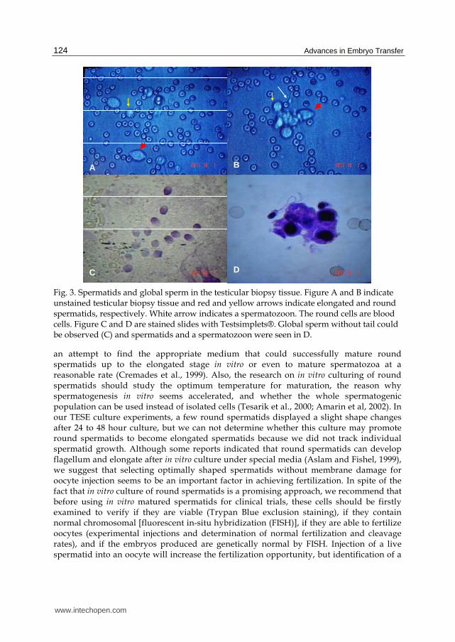
extraction (TESE) and testicular sperm aspiration (TESA) have been used, no spermatozoa can be found. Thus, many attempts have been made to develop new techniques of assisted fertilization using immature male germ cells. Some studies have showed that spermatids can be efficiently used as substitute gametes because of their high fertilizing ability comparable with that of mature sperm (Al-Hasani et al, 1999; Mansour et al., 2003). This technique is an exciting breakthrough in treatment of the infertile man with azoospermia. Spermatids are immature haploid germ cells that have not yet undergone the biochemical and morphological changes that accompany spermiogenesis through to the formation of spermatozoa. Morphologically, the shaping of the mammalian sperm involves the elongation and condensation of the spermatid nucleus, the development of the acrosome, and the transient appearance of the microtubular manchette, thus spermatids may display round or elongate shapes (Kierszenbaum et al., 2007). The elongating and elongated spermatids seem to have an advanced development compared with round spermatids. At this developmental stage, the elongating spermatids are easy to recognize among the other cell types in a wet preparation (Figure 3). Men with non-obstructive azoospermia can be treated by using intra-oocyte round spermatid injection (ROSI) or elongated spermatid injection (ELSI). It has been suggested that spermatids should be injected intact and that elongated spermatids are preferred to round spermatids where possible (Fishel, 1995). Spermatids can be retrieved from semen or from testis biopsy tissue before the final stage of spermatogenesis and the formation of sperm (Figure 3). The nucleus of these cells contains half the number of the chromosomes, thus may be injected directly into oocytes. The fertilization rate was not different between round and elongated spermatids, although the fertilization rates for round and elongated spermatids in the ejaculate were 33 and 18% respectively, compared with 22 and 38% respectively when testicular spermatids were utilized (Fishel et al., 1997). However, multiple comparisons have shown that spermatozoa and elongated spermatids gave better implantation and birth rates than did round spermatids, while spermatozoa and elongated spermatozoa were indistinguishable in their ability to support embryonic development (Ogonuki et al., 2010). Since the first baby was born as a result of spermatid injection (Tesarik et al., 1995), many healthy babies have been born using spermatid injection. In the past few years, our IVF center has injected round spermatids to five patients’ oocytes and a health boy was born in 2007. A typical case was that 6 oocytes were retrieved from a 23 age woman and only 4 matured oocytes were injected with her husband’s round spermatids from fresh testicular biopsy tissue in April 2006. One egg displayed normal two pronuclei and another egg was abnormal three pronuclei. Other two were unfertilized oocytes. On day 3, only normal fertilized embryo at 6-cell stage was transferred and finally a boy was born in February of 2007. Our clinical practice provides experienced diagnosis that when normal living spermatozoa or elongated spermatids cannot be found in the testicular biopsy specimen, round spermatids can be used successfully and result in the delivery of healthy offspring. However, the fertilization rate with round spermatid injection is relative low and only young couples may be good candidates successful for pregnancy.
Although spermatids may be used in azoospermic patients as a treatment option for their infertility, its efficiency is questionable because low fertilization and pregnancy rates influence seriously its success (Sousa et al., 1998; Akhondi et al., 2003). One major question facing spermatid use may be associated with problems related to incomplete nuclear protein maturation. Accordingly, many investigators are interested in spermatid in vitro culture and
www.intechopen.com
Advances in Embryo Transfer
124
A B
C D
Fig. 3. Spermatids and global sperm in the testicular biopsy tissue. Figure A and B indicate unstained testicular biopsy tissue and red and yellow arrows indicate elongated and round spermatids, respectively. White arrow indicates a spermatozoon. The round cells are blood cells. Figure C and D are stained slides with Testsimplets®. Global sperm without tail could be observed (C) and spermatids and a spermatozoon were seen in D.
an attempt to find the appropriate medium that could successfully mature round spermatids up to the elongated stage in vitro or even to mature spermatozoa at a reasonable rate (Cremades et al., 1999). Also, the research on in vitro culturing of round spermatids should study the optimum temperature for maturation, the reason why spermatogenesis in vitro seems accelerated, and whether the whole spermatogenic population can be used instead of isolated cells (Tesarik et al., 2000; Amarin et al, 2002). In our TESE culture experiments, a few round spermatids displayed a slight shape changes after 24 to 48 hour culture, but we can not determine whether this culture may promote round spermatids to become elongated spermatids because we did not track individual spermatid growth. Although some reports indicated that round spermatids can develop flagellum and elongate after in vitro culture under special media (Aslam and Fishel, 1999), we suggest that selecting optimally shaped spermatids without membrane damage for oocyte injection seems to be an important factor in achieving fertilization. In spite of the fact that in vitro culture of round spermatids is a promising approach, we recommend that before using in vitro matured spermatids for clinical trials, these cells should be firstly examined to verify if they are viable (Trypan Blue exclusion staining), if they contain normal chromosomal [fluorescent in-situ hybridization (FISH)], if they are able to fertilize oocytes (experimental injections and determination of normal fertilization and cleavage rates), and if the embryos produced are genetically normal by FISH. Injection of a live spermatid into an oocyte will increase the fertilization opportunity, but identification of a
www.intechopen.com

Advances in Fertility Options of Azoospermic Men
125
live spermatid is difficult. Thus, choosing optimally shaped cells may be helpful in distinguishing live spermatids from most cells in testicular biopsy specimens by simply respecting the criteria of cell size and detecting the presence of a round nucleus surrounded by a rim of cytoplasm in wet preparations with Hoffman optics (Figure 3; Silber et al., 2000).
As described above, spermatids have a nuclear protein immature statue, but activated oocyte cytoplasm may induce nuclear protein maturation after injecting spermatid to oocyte intracytoplasm. Thus, oocyte activation is very important for spermatid injection. Oocytes can be activated before or during injection in order to destroy maturation promoting factor (MPF) by various artificial treatments such as physically vigorous ooplasmic aspiration during injection, electrical pulses and injection of oscillin, and chemically calcium ionophores or ethanol stimulation. In fact, treatment of spermatid injected oocytes with inophore A 23187 has been shown the increase of the fertilization rate and has resulted in the birth of a healthy child with ROSI (Ahmady & Michael 2007). Based on our clinic practice, we suggest that the spermatid membrane should be broken by injection pipette when it is placed in oocyte cytoplasm without other oocyte activation. A round spermatid is 7-8µm in diameter and has a round shape with smooth outlines. When these spermatids are selected for injection into an oocyte, a same size (7-8µm) pipette in inner diameter may be used. Using this technique, the spermatid is aspirated into the pipette and the spermatid membrane can be broken by pipette squeezing when it is pushed out of the pipette. This simple technique may greatly increase spermatid fertilization.
7. Use of secondary spermatocytes
Round or elongated cytoplasmic spermatid injection can result in the delivery of normal
offspring in human IVF, but it is sometimes difficult to distinguish Hoffman optics whether
they are haploid round spermatids, diploid spermatocytes, spermatogonia, or even somatic
cells like Sertoli cell nuclei or Leydig cells. Can we consider the use of immature
spermatogenic germ cells for fertilization? Based on our understanding of the oocyte
maturation mechanism, oocyte maturation arrests at the meiosis II stage because the
mammalian oocyte contains two key factors: metaphase-promoting factor (MPF) and
cytostatic factor (CSF), in which the c-Mos proto-oncoprotein is a major component (Wu et
al, 1997a,b). These two key factors are active in the oocyte cytoplasm and maintain the
chromosomes of mature oocytes in metaphase of the second meiotic division. When the
oocyte is activated by the above-mentioned methods, the degradation of c-Mos onco-protein
causes CSF and MPF inactivity in the cytoplasm and the chromosomes of injected spermatid
or spermatozoon as well as the oocyte begin condensation, which leads to halploidization of
both chromosome sets and forms formal fertilized zygotes. However, when immature
diploid spermatocytes are injected into oocytes, oocyte activation should be absent. Thus,
the injected cell nucleus is exposed to MPF and CSF environment in which MPF and CSF
may induce injected cell nucleus to enter metaphase stage as the same way as does the
oocyte nucleus. (Tesarik, 1996). If the oocyte is activated, the disappearance of MPF and CSF
activity will lead the injected cell nucleus to undergo condensation and to complete meiosis
and form a haploid chromosome set. Thus, the premature chromosome condensation of
injected cells occurring in the absence of oocyte activation is essential for achieving
fertilization with secondary spermataocytes (Kimura and Yanagimachi 1995).
www.intechopen.com

Advances in Embryo Transfer
126
8. Adult somatic cell induction
Using the same theory and technique, somatic cells also may be induced into a haploid
chromosome set by immature or mature oocytes (Tesarik 2002; Keefer 2007; Neri et al., 2009)
because some important materials of germinal vesicle oocyte is essential for cell nucleus
remodeling (Gao et al., 2002). In our preliminary animal experiments, a bovine cumulus cell
from ovary follicular fluid was injected into an enucleated oocyte. After 24 hour in vitro
culture, injected cell nuclei were stained by Hoechst 33258 and two separated chromosome
sets were observed using fluorescence microscopy (Figure 4A). After two sets of
chromosome were taken out by manipulation, their chromosome numbers were counted.
Adult cattle have 60 diploid chromosomes. After induction, each set contains 30
chromosomes (Figure 4B and 4C). Thus, the immature oocyte can induce successfully
somatic cells to become haploid. When the haploid nucleus is moved out of the induced
matured oocyte and transferred into a normal female oocyte, using electro-fusion activation,
both the oocyte nucleus and the injected male nucleus begin condensation to form a similar
zygote which develops into an embryo for transfer (Figure 5).
Fig. 4. Preliminary experiments of using an oocyte to induce somatic cell chromosomes into a haploid chromosome set. A single bovine cumulus cell was injected into an enucleated immature oocyte. After 24 hour in vitro culture, injected cell nuclei were stained by Hoechst 33258 and two chromosome sets were observed using fluorescence microscopy (blue color, A). After each chromosome set was separated by manipulation, 30 chromosomes were counted in each set (green color, B and C). Authors greatly appreciate Dr. Bin Wang’s courtesy unpublished photos.
A
B
C
www.intechopen.com

Advances in Fertility Options of Azoospermic Men
127
GV oocyte
Inject malediploid cell GVBD MII
Normal MII
Biparental embryo
Meiosis
Male haploid
Female egg
Remove Nucleus
Fig. 5. Scenario for using immature oocytes to induce male somatic cell complete meiosis. The nucleus of the immature oocyte is removed and a male diploid cell was injected into the enucleated oocyte. After completing meiotic division, the induced haploid nucleus was transferred into a normal female mature oocyte to form a biparental embryo for transfer.
9. Sperm cell cloning
In some men with oligozoospermia and azoospermia, occasionally a single viable sperm may be found in their semen and/or testicular biopsy tissue. Therefore, such a sperm is precious to couples wishing to conceive. How can this single sperm be utilized to help patients realize their dream of having a baby? If only one sperm is found, we must be reluctant to use it for anything but fertilization. As described above, because the cytoplasm of oocyte may induce heterogonous cell nucleus condensation, we may try to inject a sperm into an enucleated oocyte to observe sperm nucleus kinetics (Jones et al., 2010; Ogonuki et al., 2010). Recent studies demonstrated possibility of artificially replicating a single sperm genome, which could help men with very low sperm counts become fathers. This is called “cloning of sperm cells or male genome cloning” (Figure 6). With this technique it should be possible to create enough sperm nuclei to ensure that produced and implanted embryo is healthy. The cloning of sperm cells has been practiced on mice by injecting a mouse sperm into a mouse oocyte which had its nucleus removed (Hopkin, 2007). The sperm may form a single male pronucleus in this enucleated oocyte by egg activation and electrofused. The haploid zygote may become cleaved embryos. When the resulting embryo develops to 4 to 8 cells, each blastomere may be separated by a specific solution (Figure 6). The process worked well in almost all cases and the produced blastomere from original sperm was found to be chromosomally identical to its originator in over 80% of the clones analyzed. Then, the resulting cells were fused with a normal egg that had been previously chemically activated. Based on this technique, Dr. Palermo’s research group created 64 blastocyst embryos and implanted them into 6 host mother mice resulting in only 4 developed into normal baby mice (Takeuchi et al., 2008). This offspring born as a result of such replication had shown a level of abnormalities consistent with that shown in cloned animals. This technique provides a new hope for azoospermic men to have a child, but we are a long way from the time when this will be able to be used in humans (Takeuchi et al., 2008). There is much work still needed to be done to understand why impaired development and abnormalities in the embryo occur, and to take steps to avoid that occurrence (Neri et al., 2009).
www.intechopen.com

Advances in Embryo Transfer
128
Fig. 6. Scenario for sperm genome cloning. A single sperm is injected into enucleated oocyte and this oocyte goes through a parthenogenesis process to become a 4-8 cell haploid embryo. A single blastomere is transferred into a normal mature oocyte to form a zygote. The developed embryos are transferred to recipient mice to deliver offspring.
10. Artificial sperm production
Development of embryo stem cell technology has resulted in a breakthrough for artificial sperm production (Ogawa, 2008). In the last decade, many research groups have attempted to re-create the process of sperm production in the laboratory using stem cells as the starting material, especially using mice as an animal model. In 2003, American scientists successfully coaxed embryonic stem cells from mice to mature into primitive sperm, which were then injected into eggs to form normal embryos. This “artificial sperm” capable of fertilizing an egg has been created in the laboratory for the first time in an experiment that could bring an end to male infertility. In 2005, British scientists took a step towards showing that the human egg and sperm could be created from stem cells and in 2009, this human sperm have been created from stem cells in world first from British university (Alleyne, 2009). This is the first time human sperm has been created anywhere in the world in a laboratory. However, the experiment has proved controversial, threatening to reopen the fierce debate over embryo research. Recently, Japanese researchers were able to turn mouse embryonic stem cells into early sperm cells called primordial germ cells (PGCs) and successfully implanted early sperm cells which were made from the stem cells into infertile mice, which after the treatment were able to produce healthy offspring (Hayashi et al., 2011).
The procedure of artificial sperm production was successfully repeated using another type of embryonic cell that was manipulated into a stem cell state. These stem cells are known as induced pluripotent stem (iPS) cells and have previously been created from a variety of different starting cells, including skin cells. Male embryonic stem cells with green protein makers may be selected and extracted from mouse embryos. These embryonic stem cells are a type of cell that has the ability to differentiate into any of the specialized cell types that make up the body. Under a given condition, the embryonic stem cells are induced to develop
SrCl2
MII
Inject a sperm
Haploid blastomere
Normal MII
Biparental embryo
Zygote
www.intechopen.com

Advances in Fertility Options of Azoospermic Men
129
into a particular type of cell, called “primordial germ cell-like cells or PGCs”. Primordial germ cells go on to form germ cells, which then produce sperm. Then the primordial germ cell-like cells were transplanted into the testes of mice that lacked their own germ cells. When these cells develop into sperm, they may be extracted from testes and injected into normal mouse oocytes. The embryos produced were transferred into female mice so that healthy offspring were born. This study has huge implications for furthering our understanding of how sperm are made, but may also one day lead to a clinical application whereby we could make sperm for infertile men. In next ten years, we believe that this technique could also be used to allow infertile couples without any sperm and spermatids to have children that are genetically their own. It could even be possible to create sperm from female stem cells, which would ultimately mean a woman having a baby without a man.
11. Conclusions
Exciting advances in male infertility have introduced innovative therapeutic options. This is mainly attributed to the success of the ICSI technique rapidly gaining popularity among infertile couples and advanced surgical testicular sperm retrieval techniques in azoospermic men, which offer infertile couples, including those with no sperm (azoospermia) in their ejaculate due to genetic conditions, a greatly improved chance to conceive their own biological offspring. Currently, despite severe male factor infertility, pregnancy may still be achieved by current developed technology. As the ICSI technique further improves, a single good morphologic and functional spermatozoon may be selected by IMSI or PICSI methods and an improved embryo implantation and pregnancy rate will be obtained. Testicular biopsy seems to be one of the most feasible techniques obtaining spermatozoa for azoospermic men because a single biopsy of testis may provide either spermatozoa if the specimen contains sperm or spermatids if biopsy tissues do not contain any sperm for ICSI. Using testis biopsy sperm for azoospermia may gain the same high pregnancy rate as the normal ejaculation sperm, but efficiency of spermatid application is doubted because low fertilization and pregnancy rates influence seriously its success. As cell cloning technology develops, other options for treatment of azoospermic men include use of secondary spermataocytes, sperm cell cloning, and artificial sperm production by inducing stem cells and adult somatic cells into sperm cells. Although these technologies still remain at animal research stage and demonstrate a low efficiency, we believe that application of these novel techniques will greatly improve chance for infertile couples (particular azoospermic men) to conceive their own biological offspring in the next decade.
12. References
Ahmady A, Michael E (2007) Successful pregnancy and delivery following intracytoplasmic injection of frozen-thawed non-viable testicular sperm and oocyte activation with calcium ionosphere. J Androl 28:13-14.
Akhondi MA, Sedighi MA, Amirjannati N, Sadri-Ardekani H (2003) Use of spermatide for treatment of non-obstructive azoospermic patients. J Reprod Infertil 4(3):177-183.
Al-Hasani S, Ludwig M, Palermo I, Küpker W, Sandmann J, Johannisson R, Fornara P, Sturm R, Bals-Pratsch M, Bauer O, Diedrich K (1999) Intracytoplasmic injection of round and elongated spermatids from azoospermic patients: results and review. Hum Reprod Suppl 1:97-107
www.intechopen.com

Advances in Embryo Transfer
130
Alleyne R, Science Correspondent 7:01AM BST 08 Jul 2009 Human sperm created from stem cells in world first, claims British university The Telgraph 08 Jul 2009.
Amarin ZO, Jamal HS, Rouzi AA (2002) Successful pregnancy after round spermatid microinjection. Saudi Medical Journal 23 (1): 113-114.
Angelopoulos T, Adler A, Krey L, Licciardi f, Noyes, McCullough A (1999) Enhancement or initiation of testicular sperm motility by in vitro culture of testicular tissue. Fertil Steril 71(2): 240-243.
Arakil Y, Ogawa S, Ohnol M, Moshizawa m, Araki S, Aslam I, Fishel S. (1999) Successful metaphase chromosome analysis of human elongated spermatids using mouse oocytes. Mol Hum Reprod 5 (8): 784-787.
Aslam I, Fishel S (1999) Evaluation of the fertilization potential of freshly isolated, in-vitro cultured and cryopreserved human spermatids by injection into hamster oocytes. Hum Reprod 14 (6): 1528-1533.
Banker MR, Patel PM, Joshi BV, Shah PB, Goyal B. Successful pregnancies and a live birth after intracytoplasmic sperm injection in globozoospermia J Hum Reprod Sci 2009, 2: 81-82.
Battaglia DE, Koehler JK, Klein NA and Tucker MJ (1997): Failure of oocyte activation after intracytoplasmic sperm injection using round-headed sperm. Fertil Steril 68,118–122.
Berger DS, AbdelHafez F, Russell H, Goldfarb J, Desai N (2011) Severe teratozoospermia and its influence on pronuclear morphology, embryonic cleavage and compaction. Reprod Biol Endocrinol 9: 37
Comhaire F, Vermenlen L (1995) Human semen analysis. Hum Reprod Update 1:343-362. Cornell Physicians (2011) Male infertility/sperm retrieval techniques. www.cornellurology.com/infertility/srt/icsi.shtml Cremades N, Bernabeu R, Barros A, Sousa (1999) In-vitro maturation of round spermatids
using co-culture on Vero cells. Hum Reprod 14 (5): 1287-1293. Dam AH, Feenstra I, Westphal JR, Ramos L, van Golde RJ, Kremer JA (2007)
Globozoospermia revisited. Hum Reprod Update 13:63-75. Eldar-Geva, T, Brooks B, Margalioth EJ, Zylber-Haran E, Gal M, Silber, SJ (2003). Successful
pregnancy and delivery after calcium ionophore oocyte activation in a normozoospermic patient with previous repeated failed fertilization after intracytoplasmic sperm injection Fertil Steril 79(suppl. 3):1656-1658
Fishel S, Green S, Hunter A, Lisi F, Rinaldi L, Lisi R, McDermott H (1997) Human fertilization with round and elongated spermatids. Hum Reprod 12 (2): 336-340.
Friedler S, Raziel A, Soffer Y, Strassburger D, Komarovsky D, Ron-EI R (1997) Introcytoplamic injection of fresh and cryopreserved testicular spermatozoa in patients with non-obstructive azoospermia: A comparative study. Fertil Steril 68:892-897.
Gao S, Gasperrini B, Mcgarry M, Ferrier T, Fletcher J, Harkness L, De Sousa P, Wilmut I (2002) Germinal Vesicle Material Is Essential for Nucleus Remodeling after Nuclear Transfer. Biol Reprod 67(3):928-934.
Girardi SK, Schlegel PN (1996) Microsurgical epididymal sperm aspiration: Review of techniques, preoperative considerations, and results. J Androl 1:5-9.
Hayashi K, Ohta H, Kurimoto K, Aramaki S, Saitoou M (2011) Reconstitution of the mouse germ cell specification pathway in culture by pluripotent stem cells. Cell 146:519-532.
Holstein AF, Schirren CG, Schirren C, Mauss J (1973) Round headed spermatozoa: A cause of male infertility. Dtsch Med Wochenschr 98:61-2.
Hopkin Mう2007えMice born from cloned sperm Technique raises hopes for infertile men. Published online 3 July 2007 | Nature | doi:10.1038/news070702-8
www.intechopen.com

Advances in Fertility Options of Azoospermic Men
131
http://www.nature.com/news/2007/070702/full/news070702-8.html Jones EL, Mudrak O, Zalensky AO (2010) Kinetics of human male pronuclear development
in a heterologous ICSI model. J Assist Reprod Genet 27(6): 277–283. Jow WW, Steckel J, Schlegel PN, Magid MS, Goldstein M (1993) Motile sperm in human
testis biopsy specimens. J Androl 14:194-198. Keefer Cl, (2008) Lessons learned from nuclear transfer (cloning). Theriogenology 69(1):48-54. Kierszenbaum AL, Rivkin E, Tres LL (2007) Molecular biology of sperm head shaping. Soc
Reprod Fertil Suppl. 65:33-43. Kimura Y, Yanagimachi R (1995) Development of normal mice from injected with secondary
spermatocytes nuclei. Biol Reprod 53:855-852. Larson KL, Brannian JD, Singh NP, Burbach JA, Jost LK, Hansen KP, Kreger DO, Evenson DP
(2001) Chromatin structure in globozoospermia: a case report. J Androl 22,424–431 Maggiulli R, Neri QV, Monahan D, Hu J, Takeuchi T, Rosenwaks Z, Palermo GD (2010)
What to do when ICSI fails. Syst Biol Reprod Med 6(5):376-87. Mansour RT, Fahmy IM, Taha AK, Tawab NA, Serour HI, Aboulghar MA (2003)
Intracytoplasmic spermatid injection can result in the delivery of normal offspring. J Andrology 24(5):
Matthews GJ, Goldstein M (1996) A simplified method of epididymal sperm aspiration. Urology 47:123-125.
Nagy Z, Liu J, Cecile J, Silber s, Devroey P, Van Steirteghem A (1995) Using ejaculated, fresh and frozen-thawed epididymal and testicular spermatozoa gives rise to comparable results after intracytoplasmic sperm injection. Fertil Steril 63:808-815.
Nardo LG, Sinatra F, Bartoloni G, Zafarana S and Nardo F (2002) Ultrastructural features and ICSI treatment of severe teratozoospermia: report of two human cases of globozoospermia. Eur J Obstet Gynecol Reprod Biol 104:40–42.
Neri QV, Takeuchi T, Rosenwaks Z, Palermo GD (2009): Treatment options for impaired spermatogenesis: germ cell transplantation and stem-cell based therapy. Minerva Ginecol 61(4):253-9
Ogawa T (2008) Reproductive stem cell research and its application to urology. International Journal of Urology 15(2):121–127.
Ogonuki N, Mori M, Shinmen A, Inoue K, Mochida K (2010) The effect on intracytoplasmic sperm injection outcome of genotype, male germ cell stage and freeze-thawing in mice. PLoS ONE 5(6): e11062. doi:10.1371/journal.pone.0011062
Palermo G, Joris H, Devroey P, Van Steirteghem AC (1992) Pregnancies after intracytoplasmic injection of a single spermatozoon into an oocyte. Lancet 340:17-18.
Parmegiani L, Cognigni GE, Bernardi S, Troilo E, Ciampaglia W, Filicori M (2010a) “Physiologic ICSI”: Hyaluronic acid (HA) favors selection of spermatozoa without DNA fragmentation and with normal nucleus, resulting in improvement of embryo quality. Fertil Steril 93: 598-604
Parmegiani L, Cognigni GE, Ciampaglia W, Pocognoli P, Marchi F, Filicori M (2010b) Efficiency of hyaluronic acid (HA) sperm selection. J Assist Reprod Genet 27(1):13-6
Pühse G, Hense J, Bergmann M, Kliesch S (2011): Bilateral histological evaluation of exocrine testicular function in men with obstructive azoospermia: condition of spermatogenesis and andrological implications? Human Reprod 26(10):2606-2612
Rybouchkin A, Dozortsev D, Pelinck MJ, De Sutter P, Dhont M (1996) Analysis of the oocyte activating capacity and chromosomal complement of round-headed human spermatozoa by their injection into mouse oocytes. Hum Reprod 11:2170-5.
www.intechopen.com

Advances in Embryo Transfer
132
Said TM, Land JA (2011) Effects of advanced selection methods on sperm quality and ART outcome: a systematic review. Hum Reprod Update doi: 10.1093/humupd/dmr032 First published online: August 25, 2011
Schlegel PN, Palermo GD, Goldstein M (1997) Testicular sperm extraction with ICSI for non-obstructive azoospermia. Urology 49:435-440.
Sheynkin YR, Schlegel PN (1997) Sperm retrieval for assisted reproductive technologies. Contemporary OB/GYN 15:113-129.
Silber S, Johnson L, Verheyen G, Van Steirteghem A (2000) Round spermatid injection. Fert Sterit 73:897-900.
Silber SJ, Nagy ZP, Liu J et al. (1994) Conventional in-vitro fertilization versus intracytoplamic sperm injection for patients requiring microsurgical sperm aspiration. Hum Reprod 9:1705-1709
Singh G (1992) Ultrastructural features of round-headed human spermatozoa. Int J Fertil 37:99–102.
Sousa M, Barros A, Tesarik J (1998) Current problems with spermatid conception. Human Reprod 13(2):255-258.
Stone S, O’Mahony F, Khalaf Y, Taylor A and Braude P (2000) A normal livebirth after intracytoplasmic sperm injection for globozoospermia without assisted oocyte activation: case report. Hum Reprod 15,139–141.
Takahashi K, Uchida A, Kitao M (1990) Hypoosmotic swelling test of sperm. Arch Androl 25(3):225-42.
Takeuchi T, Neri QV, Palermo GD (2008) Male gamete empowerment. Ann NY Acad Sci 1127:64-6
Tesarik J (1996) Fertilization of oocytes by injecting spermatozoa, spermatids and spermatocytes. Review of Reproduction 1:149-152.
Tesarik J (2002) Reproductive semi-cloning respecting biparental embryo origin: Embryos from syngamy between a gamete and a haploidized somatic cell. Hum Reprod 17 (8): 1933-1937.
Tesarik J, Mendoza C, GrecoE (2000) Immature germ cell conception ± in vitro germ cell manipulation. BaillieÁre's Clinical Endocrinology and Metabolism 14(3):437-452.
Tesarik J, Mendoza C, Testart J (1995) Viable embryos from injection of round spermatids into oocytes. New England Journal of Medicine 333:525.
Trokoudes KM, Danos N, Kalogirou L, Vlachou R, Lysiotis T, Georghiades N, Lerios S and Kyriacou K (1995) Pregnancy with spermatozoa from a globozoospermic man after intracytoplasmic sperm injection treatment. Hum Reprod 10,880–882.
WHO (2010) WHO laboratory manual for the examination and processing of human semen. Fifth Edition, World Health Organization, Switzerland, Page 223.
Worrilow KC, Eid S, Matthews J, Pelts E, Khoury C, Liebermann J (2010) Multi-site clinical trial evaluating PICSI®, a method for selection of hyaluronan bound sperm (HBS) for use in ICSI: improved clinical outcomes. Human Reprod 25(suppl 1): 6-9.
Wu B, Ignotz G, Currie WB, Yang X(1997a) Dynamics of maturation-promoting factor and its constituent protein during in vitro maturation of bovine oocytes. Boil Reprod 56:253-259.
Wu B, Ignotz G, Currie WB, Yang X(1997b) Expression of Mos proto-oncoprotein in bovine oocytes during maturation in vitro. Biol Reprod 56: 260-265.
Wu B, Wong D, Lu S, Dickstein S, Silver M, Gelety T (2005) Optimal use of fresh and frozen-thawed testicular sperm for intracytoplasmic sperm injection in azoospermic patients. J Assist Reprod Genet 22: 389-394.
www.intechopen.com
Advances in Embryo TransferEdited by Dr. Bin Wu
ISBN 978-953-51-0318-9Hard cover, 248 pagesPublisher InTechPublished online 14, March, 2012Published in print edition March, 2012
InTech EuropeUniversity Campus STeP Ri Slavka Krautzeka 83/A 51000 Rijeka, Croatia Phone: +385 (51) 770 447 Fax: +385 (51) 686 166www.intechopen.com
InTech ChinaUnit 405, Office Block, Hotel Equatorial Shanghai No.65, Yan An Road (West), Shanghai, 200040, China
Phone: +86-21-62489820 Fax: +86-21-62489821
Embryo transfer has become one of the prominent high businesses worldwide. This book updates and reviewssome new developed theories and technologies in the human embryo transfer and mainly focus on discussingsome encountered problems during embryo transfer, which gives some examples how to improve pregnancyrate by innovated techniques so that readers, especially embryologists and physicians for human IVFprograms, may acquire some new and usable information as well as some key practice techniques. Majorcontents include the optimal stimulation scheme for ovaries, advance in insemination technology, improvedembryo transfer technology and endometrial receptivity and embryo implantation mechanism. Thus, this bookwill greatly add new information for readers to improve human embryo transfer pregnancy rate.
How to referenceIn order to correctly reference this scholarly work, feel free to copy and paste the following:
Bin Wu, Timothy J. Gelety and Juanzi Shi (2012). Advances in Fertility Options of Azoospermic Men, Advancesin Embryo Transfer, Dr. Bin Wu (Ed.), ISBN: 978-953-51-0318-9, InTech, Available from:http://www.intechopen.com/books/advances-in-embryo-transfer/advances-in-fertility-options-of-azoospermic-men

© 2012 The Author(s). Licensee IntechOpen. This is an open access articledistributed under the terms of the Creative Commons Attribution 3.0License, which permits unrestricted use, distribution, and reproduction inany medium, provided the original work is properly cited.